pulmonary disorders ards postoperative respiratory failure obstructive pulmonary disease respiratory...
TRANSCRIPT
Pulmonary Disorders
• ARDS
• Postoperative Respiratory Failure
• Obstructive Pulmonary Disease
• Respiratory Tract Infections
• Pulmonary Vascular Disease
• Respiratory Neoplasms
ARDS (Acute Respiratory Distress Syndrome)
• Fulminant respiratory failure– Acute lung inflammation– Diffuse alveocapillary injury– 30% of all ICU admissions– Current mortality < 40%
• Etiology: – Sepsis & Multiple trauma (esp w/transfusions)– Pneumonia, burns, aspiration, CABG,
pancreatitis, drug overdose, smoke, O2, DIC
ARDS Pathophysiology
• Starts with alveolocapillary membrane damage and pulmonary edema– Direct damage– Indirectly (immune mediators)
• Final: Massive inflammatory response– Neutrophils, Macrophages, complement,
endotoxin, interleukin-1, TNF-α
Sequence
• Alveolocapillary membrane damage
• Platelet aggregation & thrombus– Attracts Neutrophils– Neutrophils release inflammatory mediators– Causes further damage, and increases
capillary membrane permeability– Pulmonary edema & hemorrhage– Vasoconstriction Pulmonary hypertension
• Uneven V/Q mismatching
Meanwhile, back at the ranch…
• Surfactant production is interrupted
• Compliance is impaired
• Ventilation is impaired
• Results in– Right to left shunting– Increased work of breathing– 24 – 48 hours: hyaline membrane forms– 7 days: progressive fibrosis destroys lung
Associated Problems
• SIRS– Systemic Inflammatory Response Syndrome
• MODS– Multi-organ Dysfunction Syndrome
• Death results from combination of Resp Failure and MODS
ARDS Manifestations
• Classic– Rapid, shallow, breathing– Resp alkalosis– Marked dyspnea– Hypoxemia– Diffuse alveolar infiltrates (x-ray)
• As progresses– Diffuse crackles, metabolic acidosis,
hypotension, decreased CO, death
ARDS Eval & Tx
• DX: exam, blood gas, x-ray– Criteria
• Hypoxemia, bilat x-ray infiltrates, exclusion of cardiogenic pulmonary edema
• TX: must catch early– Supportive therapy– Prevention of complications– You’ll learn a lot more about this is Critical
Care
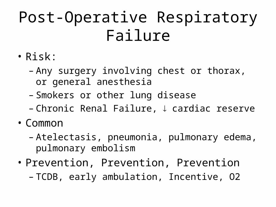
Post-Operative Respiratory Failure
• Risk: – Any surgery involving chest or thorax, or general
anesthesia
– Smokers or other lung disease
– Chronic Renal Failure, cardiac reserve
• Common– Atelectasis, pneumonia, pulmonary edema,
pulmonary embolism
• Prevention, Prevention, Prevention– TCDB, early ambulation, Incentive, O2

Obstructive Pulmonary Diseases
• Diseases that impair airflow– Upper or lower tract– Increase the work of breathing– Typically expiration is harder than inspiration
• Results in hyperinflated lungs• Symptom: dyspnea• Sign: wheezing
• Asthma• Emphysema• Chronic Bronchitis
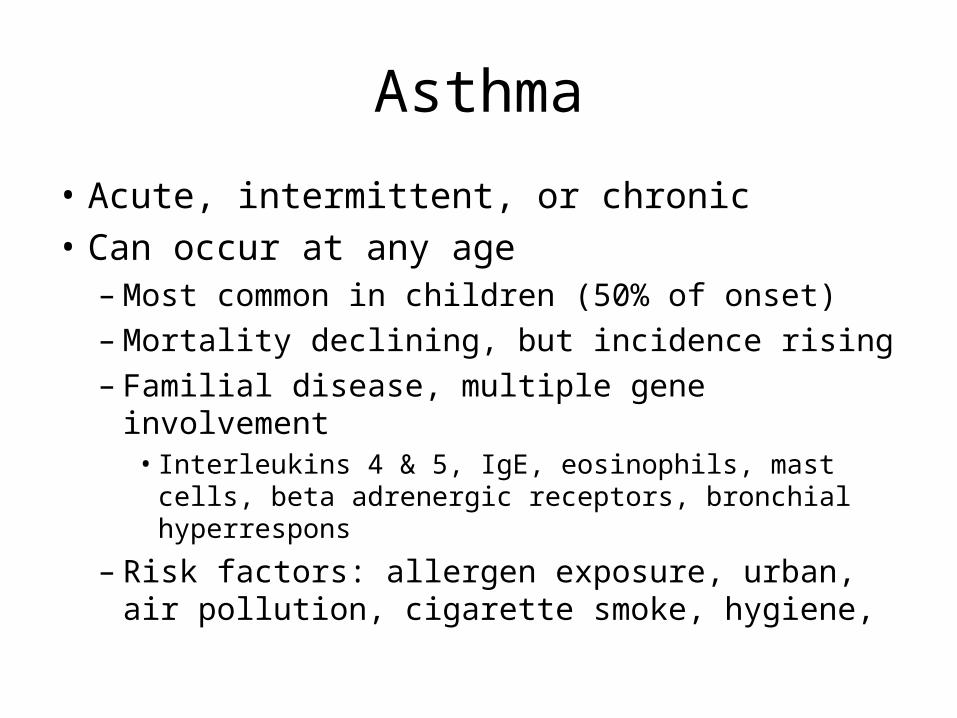
Asthma
• Acute, intermittent, or chronic
• Can occur at any age– Most common in children (50% of onset)– Mortality declining, but incidence rising– Familial disease, multiple gene involvement
• Interleukins 4 & 5, IgE, eosinophils, mast cells, beta adrenergic receptors, bronchial hyperrespons
– Risk factors: allergen exposure, urban, air pollution, cigarette smoke, hygiene,
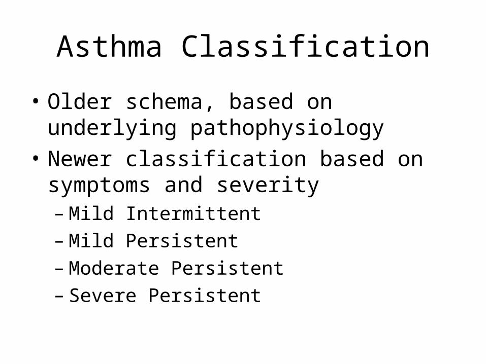
Asthma Classification
• Older schema, based on underlying pathophysiology
• Newer classification based on symptoms and severity– Mild Intermittent– Mild Persistent– Moderate Persistent– Severe Persistent
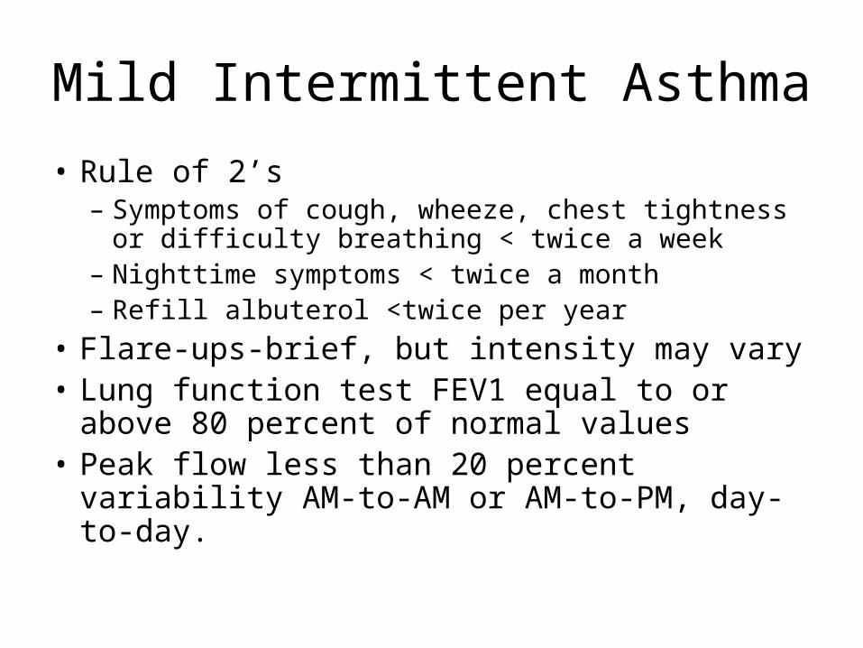
Mild Intermittent Asthma
• Rule of 2’s– Symptoms of cough, wheeze, chest tightness or
difficulty breathing < twice a week– Nighttime symptoms < twice a month– Refill albuterol <twice per year
• Flare-ups-brief, but intensity may vary• Lung function test FEV1 equal to or above 80
percent of normal values • Peak flow less than 20 percent variability AM-
to-AM or AM-to-PM, day-to-day.
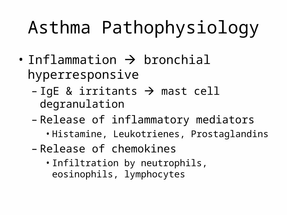
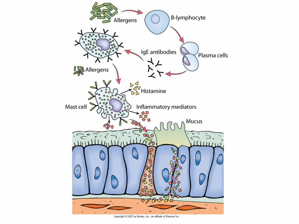
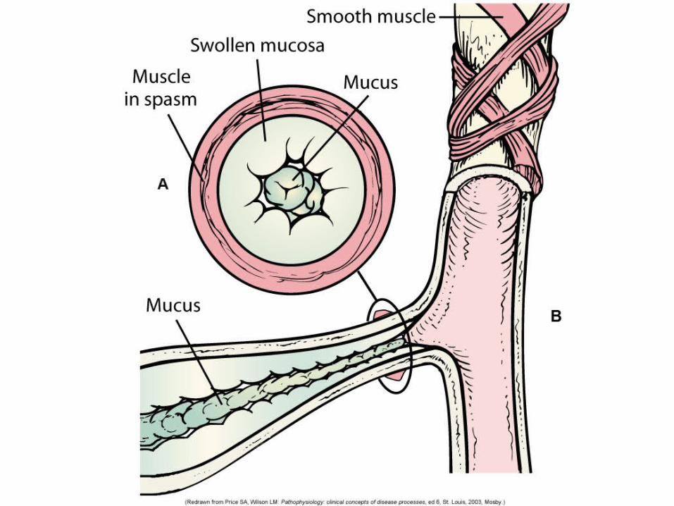
Asthma Pathophysiology
• Inflammation bronchial hyperresponsive– IgE & irritants mast cell degranulation– Release of inflammatory mediators
• Histamine, Leukotrienes, Prostaglandins
– Release of chemokines• Infiltration by neutrophils, eosinophils, lymphocytes

Asthma Pathophysiology
• Inflammatory response– Bronchospasm– ↑vascular permeability airway edema– Increased mucous production (thick)– Impaired mucociliary function– Thickening of airway walls– Muscarinic receptor stim increased
acteylcholine activity increased contraction– Epithelial destruction by eosinophils
(collateral damage)
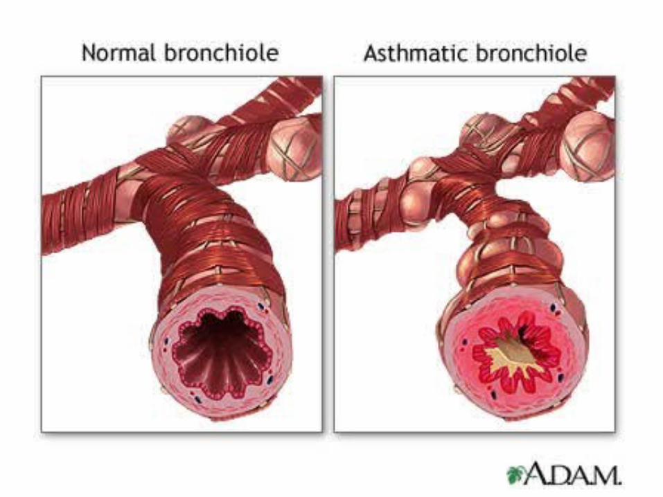
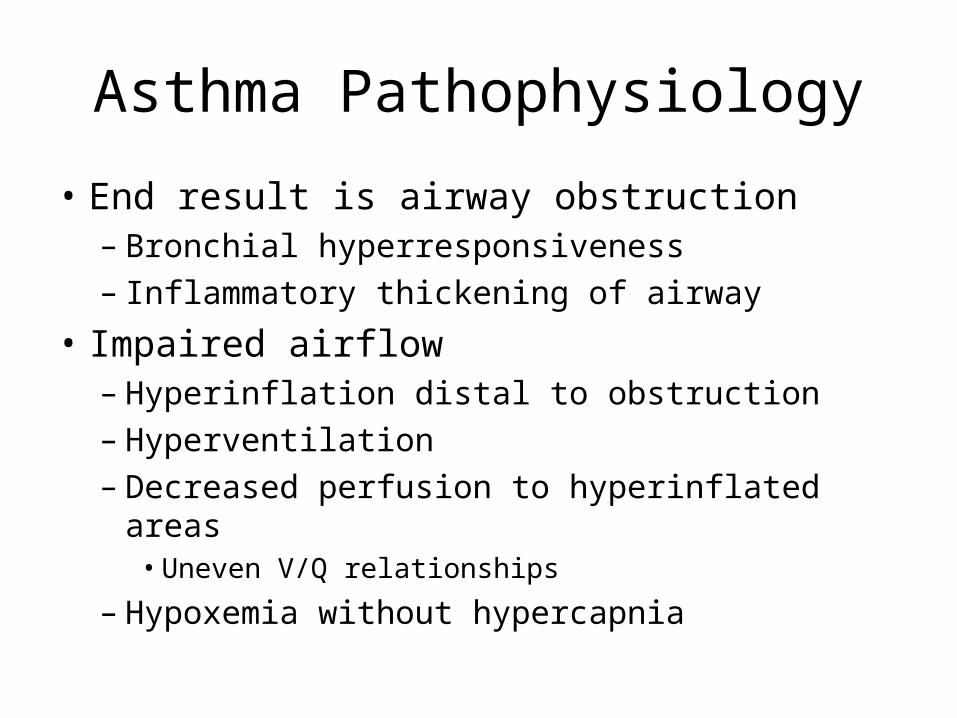
Asthma Pathophysiology
• End result is airway obstruction– Bronchial hyperresponsiveness– Inflammatory thickening of airway
• Impaired airflow– Hyperinflation distal to obstruction– Hyperventilation– Decreased perfusion to hyperinflated areas
• Uneven V/Q relationships
– Hypoxemia without hypercapnia
Asthma Pathophysiology
• If uncorrected– Hyperinflation of resp units results in
hyperexpansion of lungs– Resp muscles disadvantaged– Hypercapnia, resp acidosis
• Sign of resp failure

Asthma Clinical Manifestations
• Full remission: asymptomatic and PFTs normal
• Partial remission: asymptomatic but PFTs abnormal sign of impending flare?
• Asthma Attack– Slow onset acute asthma: days
• Often after URI
– Hyperacute asthma: minutes to hours• Often triggered by stress or exercise or allergens
Asthma Attack S/S
• Dyspnea & Wheezing
• Breath sounds decreased
• Peak flow early in attack
• If O2 sat < 90 ABGs
• Early: nonproductive cough, tachycardia, tachypnea, accessory muscle use
• Resolving: thick stringy mucus
Asthma: Eval & Tx
• Spirometry– Decreased FEV1 and FVC– Increased FRC & TLC
• Daily Peak flow (RECORD & GRAPH)
• Treatment– Avoid triggers (foods, airborne particles, etc.)– Get rid of carpets, vacuum regularly– Pharmacological Treatment

Asthma Treatment
• Acute treatment: – O2, bronchodilation, steroids, hospitalization?
• Chronic treatment: – Inflammatory reduction– Bronchodilation– Mucus reduction
• Status asthmaticus– Failure of conventional therapy to relieve attack– Life threatening

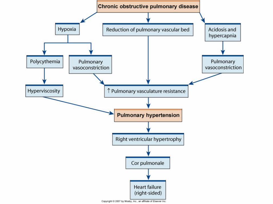
Chronic Obstructive Pulmonary Disease
• “Disease state characterized by airflow limitation that is not fully reversible.– Progressive– Abnormal inflammatory response
• Mixture of– Chronic Bronchitis– Emphysema
• Etiology– Smoking– Occupational exposure, air pollution, genetics
Chronic Bronchitis
• Hypersecretion of mucus and chronic productive cough > 3 month/year for at least 2 consecutive years
• More prevalent during winter
• 20x more incidence in smokers
• More common in elderly
• Associated with repeat infections
Chronic Bronchitis Patho
• Irritants normally cause ↑mucus secretion• In CB, irritants also cause
– Hyperplasia and hypertrophy of goblet cells– Thicker, stickier mucus
• Bacteria love this stuff and colonize it• Cilia function impaired, reducing clearance
– End result increased likelihood of infection– Bronchial walls become inflamed leading to
bronchospasm– Narrowed airway, difficulty expiring
CB Clinical Manifestations
• Decreased exercise tolerance
• Wheezing
• Dyspnea
• Productive cough: Mucus plugs
• Progression– Hypercapnia, Hypoxemia
• Polycythemia and Cyanosis• Later, pulmonary hypertension cor pulmonale• Disability and Death
Eval & Tx
• H&P, X-ray, PFT, ABG
• Best treatment? Prevention!!!!– Not reversible– Stopping smoking can prevent progression
• Tx– Bronchodilators, expectorants, anticholinergic– Chest PT– Antibiotics– Low O2– Steroids
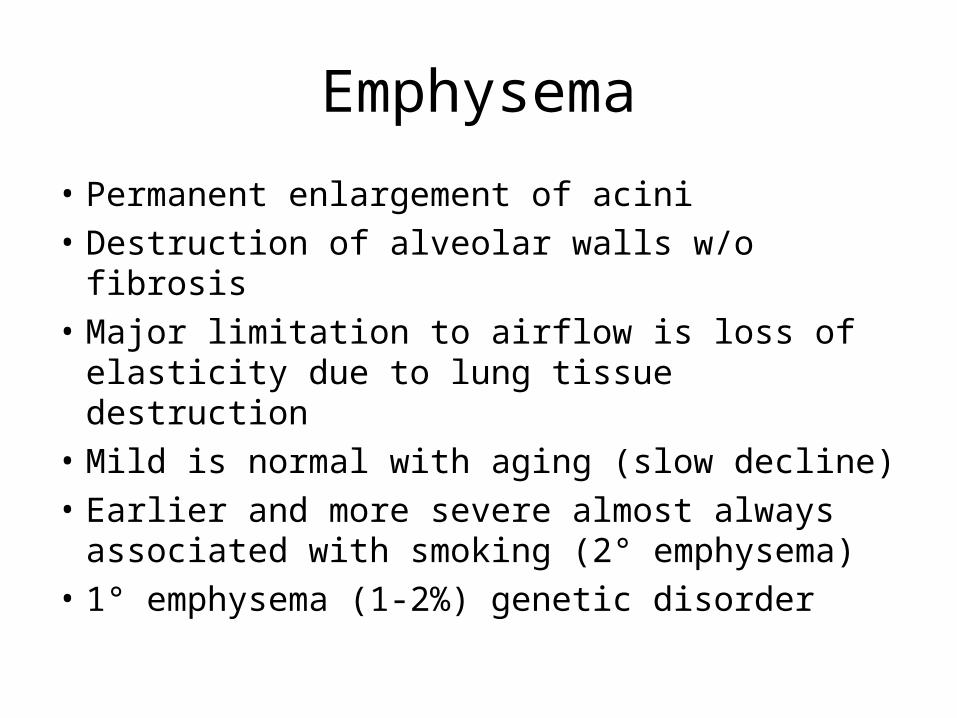
Emphysema
• Permanent enlargement of acini
• Destruction of alveolar walls w/o fibrosis
• Major limitation to airflow is loss of elasticity due to lung tissue destruction
• Mild is normal with aging (slow decline)
• Earlier and more severe almost always associated with smoking (2° emphysema)
• 1° emphysema (1-2%) genetic disorder
Emphysema Etiology
• Inability to inhibit lung proteolytic enzymes– Structural proteins are destroyed
• Primary Emphysema– α1-antitrypsin deficiency (plasma protein
responsible for inhibiting proteolytic enzymes)
• Secondary– Inhaled toxins inhibit antiproteases – Smoking, air pollution, etc.
Emphysema Patho
• Inhaled toxins – Epithelial inflammation and infiltration by leukocytes
– Inflammatory cytokines inhibit endogenous antiproteases (including α1-antitrypsin)
• Destruction of alveoli - Elastin proteolysis in alveoli septa– Decrease surface area lowered perfusion
– Capillary destruction pulmonary HTN
– Decreased elasticity difficulty expiring
– Increased air in acinus hyperinflation
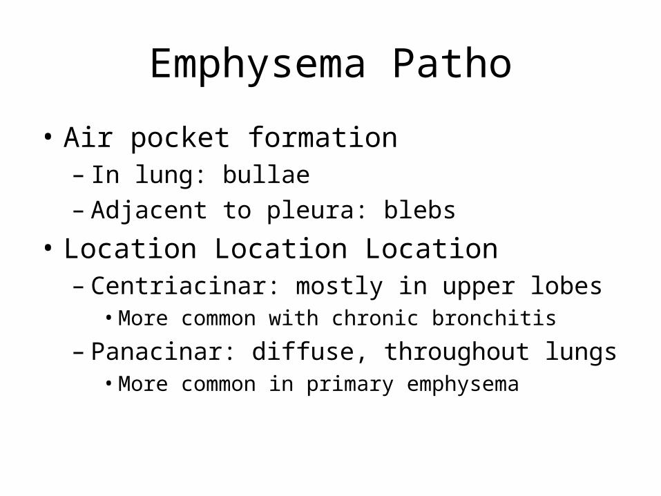
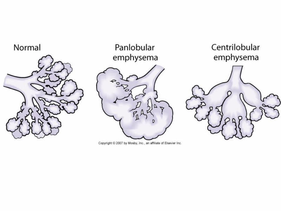
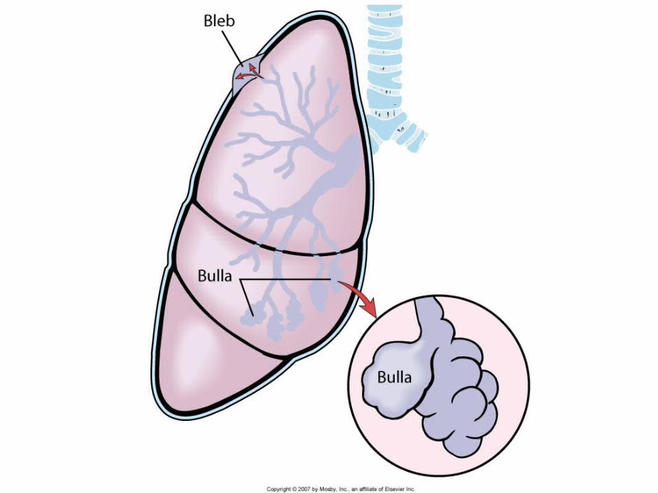
Emphysema Patho
• Air pocket formation– In lung: bullae– Adjacent to pleura: blebs
• Location Location Location– Centriacinar: mostly in upper lobes
• More common with chronic bronchitis
– Panacinar: diffuse, throughout lungs• More common in primary emphysema
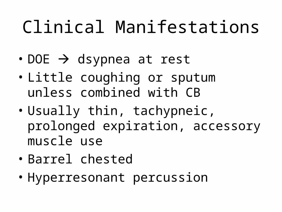
Clinical Manifestations
• DOE dsypnea at rest
• Little coughing or sputum unless combined with CB
• Usually thin, tachypneic, prolonged expiration, accessory muscle use
• Barrel chested
• Hyperresonant percussion
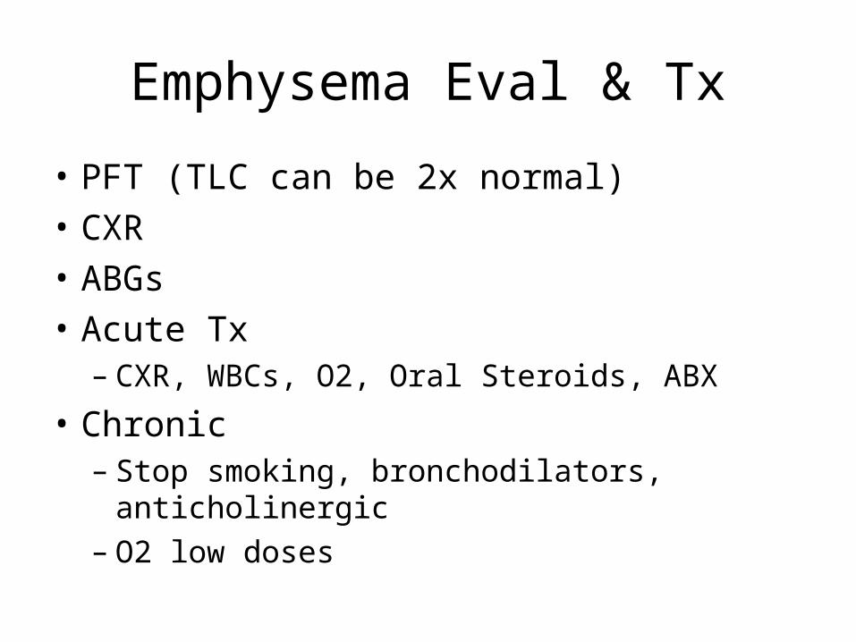
Emphysema Eval & Tx
• PFT (TLC can be 2x normal)
• CXR
• ABGs
• Acute Tx– CXR, WBCs, O2, Oral Steroids, ABX
• Chronic– Stop smoking, bronchodilators, anticholinergic– O2 low doses
Respiratory Tract Infections
• Rhinitis
• Sinusitis
• Pharyngitis
• Laryngitis
• Bronchitis
• Pneumonia

Pneumonia
• 6th leading cause of death in U.S.• Risk factors: age, immunocompromised,
lung disease, alcoholism, smoking, intubation, malnutrition, immobilization
• Causative organism: bacteria, fungus, protozoa, parasites
• Source– CAP (community acquired pneumnia)– Nosocomial
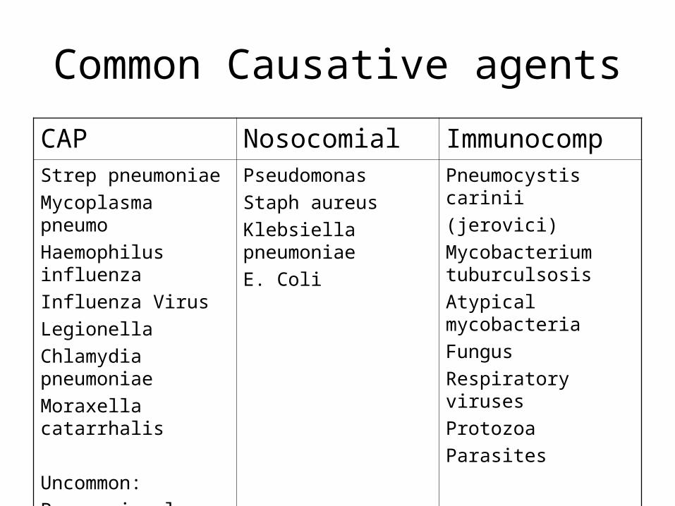
Common Causative agents
CAP Nosocomial ImmunocompStrep pneumoniae
Mycoplasma pneumo
Haemophilus influenza
Influenza Virus
Legionella
Chlamydia pneumoniae
Moraxella catarrhalis
Uncommon:
Pneumonic plague
Pseudomonas
Staph aureus
Klebsiella pneumoniae
E. Coli
Pneumocystis carinii
(jerovici)
Mycobacterium tuburculsosis
Atypical mycobacteria
Fungus
Respiratory viruses
Protozoa
Parasites
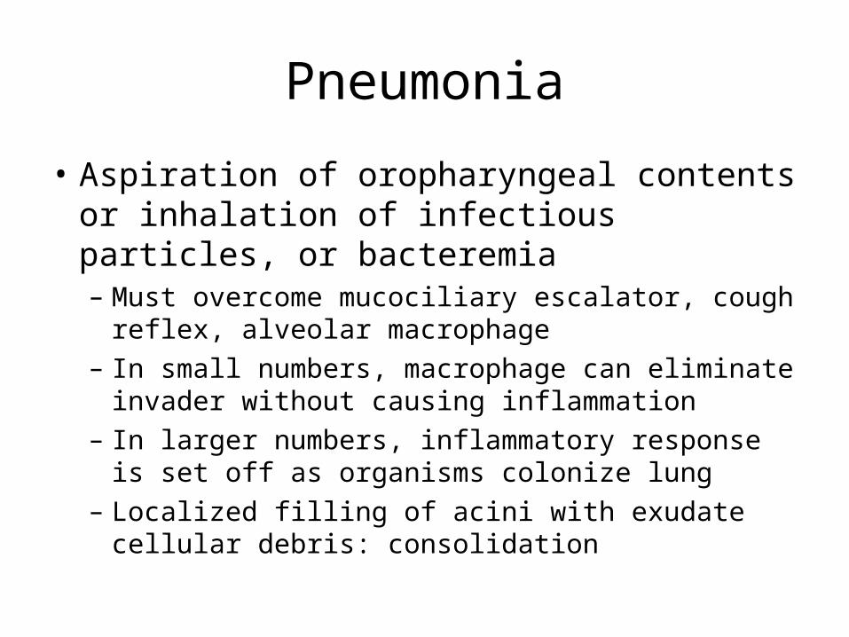
Pneumonia
• Aspiration of oropharyngeal contents or inhalation of infectious particles, or bacteremia– Must overcome mucociliary escalator, cough reflex,
alveolar macrophage– In small numbers, macrophage can eliminate invader
without causing inflammation– In larger numbers, inflammatory response is set off as
organisms colonize lung– Localized filling of acini with exudate cellular debris:
consolidation
Pneumonia Manifestations
• Usually preceded by URI or flu
• Cough (productive or unproductive)
• Dyspnea, fever
• Other: malaise, fatigue, chills, pleuritic pain
• Inspiratory crackles, localized decreased breath sounds, increased tactile fremitus

Eval & Treatment
• CXR (infiltrates: patchy, lobar, diffuse)• WBC, shift to right or left• Sputum gram stain and c/s• Tx
– Oxygenation & bronchodilation prn– Hydration and hygiene– Chest therapy– Antibiotics as appropriate
• Gatifloxacin or levofloxacin, ciprofloxacin
• Ceftriaxone + Azithro or clarithromycin
Pulmonary Vascular Disease
• Pulmonary Embolism– DVT, sudden dyspnea, hypotension, shock– Risk factor recognition and prevention– O2, rapid anti-coagulation, thrombolytic
• Pulmonary hypertension
• Cor pulmonale– Right ventricle enlargement
Respiratory Neoplasms
• Oral Cancer
• Lung cancer (13% of all U.S. cancer but 25 – 31% of cancer mortality)– Heavy smokers 20x risk
– Second hand smoke 1.3x risk
• Types of Lung Cancer– Non-Small Cell Lung Cancer
• Squamous Cell (30%), Adenocarcinoma (35-40%)
– Large Cell Carcinoma (10 – 15%)
– Small Cell Carcinoma (14%)