pulmonary defense
TRANSCRIPT

Pulmonary Defense
Dr. Manu Mohan K

Introduction
•Lungs are daily exposed to 10,000 liters of air which contain noxious particles, infectious agents, etc •Respiratory system must recognise and eliminate unwanted elements in inspired air to keep pulmonary structures free of infection.

•The fact that the normal respiratory tract is free of infection is a testimony to the efficiency of a defense system•Elements of defense system spread through out the respiratory tract

Nose and oropharynx
•Formidable barrier•Nasal hair exclude large particles•Sneezing and blowing •Rhinorrhoea•Mucociliary clearance•Air conditioning

Nose and oropharynx (contd.)
•Nasal secretion
•Oral cavity – tongue movements
•Secretory IgA in saliva

Cough
•Most important protective reflex as well as most common symptom of respiratory diseases.•Provoked by number of stimuli •Afferent – myelinated irritant nerve endings, intravascular nonmyelinated J receptors, via C fibers and myelinated fibers

Cough (contd.)
•Center - medulla 5 HT receptors
•Efferent – nerve supply to larynx, ribcage and diaphragm

Cough (contd.)
•Phases•Inspiration•Compression of intrathoracic gas against closed glottis•Explosive expulsion as glottis opens•Relaxation of airways

Cough (contd)
•Result•Expectoration of foreign debris and mucus from the larger airways due to extremely high local turbulence by the reflex•Chronic bronchitis – 50% clearance is contributed by cough

Cough (contd)
Cough reflex is inhibited by
•physiological – swallowing
•central action – codeine
•blocking the afferent signal – local anesthetics

Mucociliary clearance
• In healthy subjects cough ineffective in removing inhaled small particles
• Mucociliary clearance almost entirely responsible for tracheobronchial cleanliness
• Complex interaction between cilia and mucus

Mucociliary clearance• 200 cilia per one bronchial epithelial cell.• Cilia composed of contractile protein tubulin
arranged as nine outer and one central micro tubular pairs
• Outer microtubule has a pair of dyneine arms• Mucus forms a raft on top of cilia which
sweeps in the cephalad direction.• Cilia carry about 10 grams with out slowing• Cilia beats @ 12-14 times per second

Ultra structure of cilia

Mucociliary clearance
Test for ciliary motility
•Saccharin test
•Cine bronchography
•Imaging technique

Mucociliary clearance
• Mucus secreted by goblet cells and submucosal glands of the first several bronchial generations
• Secretions controlled by neuropeptides like substance P, Vasoactive intestinal peptide, bombesin, vagal stimulation - acetylcholine.
Mucus
Physical function
•To trap and clear particles
•Dilute noxious influences
•Lubricate airway
•Humidify inspired air

Mucociliary clearance• The viscoelastic or rheological properties of
mucus determined probably by concentration of different type of mucins
• External factors influencing mucociliary clearance– direct ciliary damage
• cigarette smoke, pollutants, local and general anaesthetics, bacterial and viral products, eosinophil products in asthma, etc
Primary Ciliary Dyskinesia
• Autosomal recessive• Ciliary dynein may be defective• Male infertility• Situs inversus• Result in repeated sinusitis and
respiratory infection and severe bronchiectasis

Surfactants•Complex surface active material lining the alveolar surface•Reduces surface tension and prevent lungs from collapsing•Simple and elegant way of alveolar clearance•At end expiration surface tension decrease and the surface film moves from the alveoli towards bronchioles

Surfactants•4 types – sp-A, sp-B, sp-C, sp-D
•Functions
•On alveolar macrophages
–Chemotaxis, enhancement of phagocytosis and killing of microorganisms
Surfactant proteins•SP- A most abundant •Closely resemble complement c1q•Enhances alveolar macrophage phagocytosis of microorganisms like staph aureus, pneumocystis carinii•SP- D share same effect•Inhibit endotoxin stimulated release of interleukin-1, IL-6, TNF

Surfactant•Can be damaged by noxious stimuli
•Alteration of surfactant quantitatively and qualitatively in ARDS
•Loss of lung function and gas exchange
•Susceptibility of injured lung to bacterial colonization and infection
•Surfactant replacement therapy

Protective Proteins of Lung
Antibacterialsurfactant proteins ( A & D), immunoglobulin ( IgA), defensin, lactoferrin, lysozyme, complementAntiproteinasesalpha 1 proteinase inhibitor, alpha 1 antichymotrypsin, alpha 2 macroglobulin

Immunoglobulins•IgA abundant•IgG and IgM in small quantities •Produced by B lymphocytes and plasma cells often associated with bronchial epithelial cells•IgA deficiency associated with local defects in immunity to bacterial infections

Complement proteins•Alveolar macrophage secrete c3a, c3b and c5a•C3 deficiency – recurrent infections•Enhances removal of bacteria by macrophages and other phagocytes
Antiproteinases•High molecular ( alpha 1 antiproteinase, alpha 2 macroglobulin)•Low molecular (secretory leukoproteinase inhibitor and elafin)•Protect local tissue against damage that would occur due to release of proteinases by inflammatory cells

Alveolar macrophages
•Derived from blood borne monocytes
•Patrol the alveolar lining
•Live for several weeks

Functions• Primary host defense - phagocytosis • Inflammatory response:
– Initiation• Generation of neutrophil chemokines ( IL-8)• Generation of monocyte chemokines (MIP-12)• Generation of agents that activate endothelial
cells (IL-1, TNF alpha)– Amplification
• stimulate bone marrow generation of leucocytes ( IL-1, TNF alpha, IL-3)
– Resolution• scavenging of necrotic and apoptotic cell
debris
Functions• Repair and fibrosis• Remodeling: elastase, collagenase• Scar formation: IL-1, PDGF, FGF• Immune response• Antigen presentation• Anti tumour effect
– Lysis of tumor cells by TNF alpha and nitric oxide dependent mechanisms

Pulmonary marginated pool of neutrophils
• Circulating pool • Marginated pool• Dynamic equilibrium• Exercise and epinephrine• Vascular bed of the lung and spleen • Neutrophils loitering in pulmonary
microvasculature- local immunity

Defense mechanism in the alveolar spaces

Effects of Smoking

`

Pulmonary function test

PFT
•Objectively measure the ability of lungs to ventilate and carry out gas exchange

Components
• Tests of airway function• Tests of pulmonary volume• Tests of gas exchange• Tests of respiratory muscle function• Tests of ventilatory control
mechanism• Tests of exercise performance

Indications • Establishing or excluding a respiratory cause of
dyspnea• Diagnosis of obstructive airway disease• Localize site of obstruction• Prognostic purpose • Evaluation of pulmonary involvement in systemic
diseases or side effect of drugs like amiodarone• Preoperative evaluation• Epidemiological observations• Evaluation of exercise performance

Principle
•Measure either displaced volume or air velocity
•All volumes are expressed at BTPS (body temperature ambient barometric pressure and saturation)

Tests of Airway Function• Peak expiratory flow• Most widely used • Measure maximum expiratory flow rate
over the first 10 milliseconds of an expiration
• Effort dependent• Reduced in airway obstruction and
expiratory muscle weakness

Forced expiratory volume
• Integrated flow over first second of expiration • Normal 70 – 80%• Airway obstruction• FeV1 reduced more than forced vital capacity
so that FeV1/FVC is reduced below 70%• Restrictive• Both FeV1 and FVC are reduced
proportionately, so that the FeV1/FVC is normal.

Normal flow volume loop

Flow volume loop in severe airway obstruction

Flow loop in restrictive lung disease

Maximum midexpiratory flow rate (MMEFR)
•FeF(25-75%) is the average expiratory flow during middle half of the forced vital capacity
•More variable than FeV1
•Sensitive index of small airway function

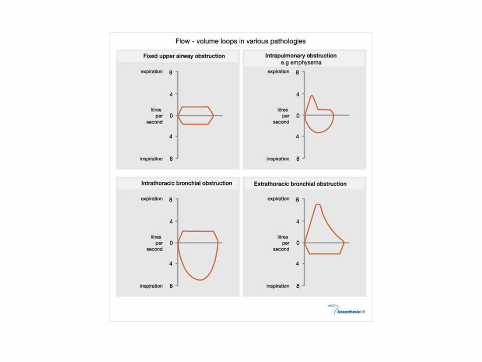
Flow volume loop•Is also useful for localizing the site of obstruction
•Whether intrathoracic or extrathoracic
•Whether fixed or variable

Flow volume loop in extrathoracic obstruction

Flow volume loop in variable intrathoracic
obstruction
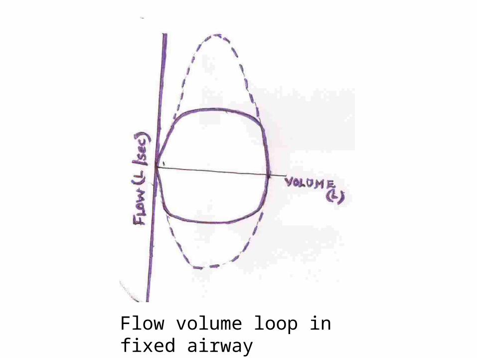
Flow volume loop in fixed airway obstruction

Test of Lung Volume• Measurement of lung volumes
provides valuable and functional information.
• Total lung capacity and residual volume measurement require indirect methods such as gas dilution technique or plethysmography


• Airway disease increase residual volume, the RV/TLC % is increased.
• Parenchymal diseases produce a restrictive ventilatory defect reducing total lung capacity and vital capacity with RV unaffected.

Respiratory muscle function
•Peak inspiratory and Peak expiratory maximal mouth pressure by sucking at and breathing out respectively against the shutter of pressure transducer device

Diffusing capacity•Carbon monoxide gas transfer •Mixture of helium and carbon monoxide 0.03%•The change in the concentration of gases are measured by automated equipment.• DLCo reduced in ILD,Pneumonia

Exercise test• Measure integrated
cardiorespiratory functions
• 6 or 12 minute walk test
• Plotting heart rate and minute ventilation against o2 uptake with estimation of respiratory quotient– cycle ergometer or tread mill.

•Arterial blood gas measurement
•Noninvasive oxymetry
