pulmonary atelectasis in anaesthesia
TRANSCRIPT

Pulmonary Atelectasis in Anaesthesia
And Critical CareBY
Dr. CHAMIKA HURUGGAMUWA
(Registrar in Anaesthesia)
Key points
Atelectasis during general anaesthesia (GA) is common, but usually does not cause clinically significant problems. Persistent prolonged atelectasis after GA increases perioperative respiratory complications.
Risk factors contributing to atelectasis include: obesity, chronic lung disease, thoracic or upper gastrointestinal surgery, and prolonged use of high-inspired oxygen concentration.
The aetiology and significance of atelectasis in critically ill patients is different from that seen in patients undergoing GA.
The treatment and prevention of atelectasis varies according to the aetiology, severity, and clinical context.

Atelectasis - Ateles and Ektasis, Meaning incomplete expansionIt is also referred to as collapse of the lung. Atelectasis is the loss of lung volume, either apart or all of a lung with or without mediastina shift. This is in contrast to consolidation where the lung volume is normal. In clinical practice, there is often a combination of both.
Atelectasis is common in the setting of anaesthesia and criticalcare.
Atelectasis can be broadly classified into
Obstructive and Non-obstructive, each having a particular radiological pattern.
Obstructive atelectasis is by far the most common.

Obstructive
Bronchial obstruction is a frequent cause of atelectasis.
This is usually due to a
neoplasm,
mucus plug, or
foreign body.
Progressive airway collapse develops distal to the obstruction.
Non-obstructive
Compressive:
Large Peripheral Tumour, Bullae, Or Extensive Air Trapping (emphysema) compresses adjacent normal lung tissue
Passive:
This is also referred to as relaxation atelectasis, and it is caused by the loss of contact between parietal and visceral pleura,
for example, due to pleural effusion, pneumothorax, or pleural malignancy

Adhesive:
this is caused by a lack of or inactivation of surfactant and can be seen in severe acute lung injury (ALI), radiation pneumonitis, or neonatal respiratory distress syndrome.
Cicatrizing:
This occurs due to scar tissue formation as a result of granulomatous disease or necrotizing pneumonia.
The above types of atelectasis are most often described in the chronic setting.

Atelectasis Related to GA
Uneventful anaesthesia can lead to the development of collapse in 10–15% of lung tissue.
The following mechanisms have been proposed
#Atelectasis during GA.
Compression atelectasis
Absorption atelectasis
# Atelectasis in the critical care setting
Acute lung injury( ALI)
Surfactant depletion

Compression atelectasis
In a normal healthy adult in the upright position, the functional residual capacity (FRC) is 3 litres.
. This decreases by 0.7–0.8 litres in the supine position as abdominal contents push the diaphragm cephalad.
This is further accentuated with anaesthesia and paralysis, as abdominal pressure is easily transmitted to the thoracic cavity.
The loss of intercostal muscle function also contributes to FRC reduction, particularly in children.
Surgical manipulation during thoraco-abdominal procedures may worsen atelectasis caused by GA

Other factors accentuating compression atelectasis include
#Morbid obesity,
#Laparoscopic procedures,
#Head-down and lateral positioning.
During one-lung anaesthesia, the lung on the side of surgery is typically deliberately collapsed to allow surgical access on that side.

Absorption atelectasis Two different mechanisms:
(i) Complete airway occlusion can be seen in accidental bronchial intubation, one-lung anaesthesia, and with mucus plugging of small or large airways. Its pathophysiology is similar to that described in obstructive mechanisms.
(ii) Atelectasis can occur in the absence of obstruction. Lung zones with lower ventilation relative to perfusion are susceptible to collapse, this can occur when inspired oxygen concentration is increased leading to a higher flux of oxygen from the alveoli to capillary, and alveoli progressively become smaller.

Atelectasis in the critical care setting
Common cause of impaired gas exchange and X-ray opacification of lung regions in critically ill patients.
The incidence is likely to be high if the patient is immobile, has had a general anaesthetic, or if they have pre-existing lung disease, a smoking history, obesity, or advanced age.
pathophysiology is multifactorial:
obstructive, non-obstructive, or both.
Prolonged immobility and infection probably being the most common contributors.
Acute lung injury
Atelectasis in patients with ALI differs from atelectasis related to GA.
During lung injury, atelectasis is accompanied by inflammatory fluid filling up the alveoli and a phenomenon of cyclical collapse is seen.
Inflammatory response causing localized lung injury alongside systemic and distant organ dysfunction due to systemic release of inflammatory mediators (cytokines, proteases, and reactive oxygen
species).
#Take time to resolve
Surfactant depletion
Lipoprotein complex secreted by Type II alveolar cells.
It reduces surface tension within a theoretically spherical structure like an alveolus.
Laplace’s law ( pressure=2* tension/radius)
Surfactant reduces surface tension in all alveoli, and to a greater extent in smaller ones, thereby maintaining alveolar stability. The lack of surfactant function therefore leads to atelectasis.

Lung tissue deficient in surfactant is difficult to inflate leading to increased work of breathing and so likelihood of respiratory failure. This situation can be seen in premature neonates who may require artificial surfactant.
In adults, surfactant function may be impaired by lung infection or inflammation.

Pathophysiological effects of atelectasis
Decreased compliance - increased work of breathing
Impaired oxygenation
Increased pulmonary vascular resistance
Clinical presentation
Depends on the extent of atelectasis and rapidity with which it develops.
Small and slowly developing areas of collapse may be asymptomatic or present as a non-productive cough.
Rapidly developing large-scale atelectasis can present with features of hypoxia and respiratory failure.
Physical examination will reveal decreased movement in the affected lung area, dullness on percussion, absent breath sounds, and deviation of the trachea to the affected site.
Investigations
Chest X-ray The radiological signs of collapse will depend upon the aetiology,
degree of collapse, and associated consolidation or pleural pathology.
Signs may be
#direct (related to loss of lung volume and collapsed
lobes) or
#Indirect (occurring as result of compensatory changes due to volume loss),
leading to shift of the mediastinal structures
Direct signsThese include:
† Increased opacification in the area of atelectasis. Air bronchograms are normally a feature of consolidation but may also b present in lobar collapse.
† Displacement of fissures. This occurs with large degree of collapse.
† Loss of aeration. If the collapsed lung is adjacent to the mediastinum or diaphragm, then loss of definition of these structures indicates loss of aeration (the silhouette sign).
† Vascular signs. In partial collapse, crowding of vessels may beseen.

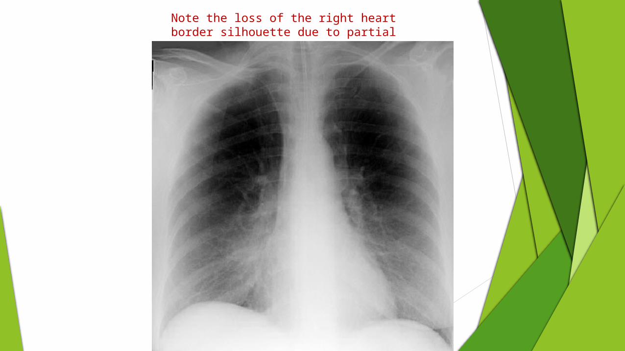
Note the loss of the right heart border silhouette due to partial atelectasis of the RML
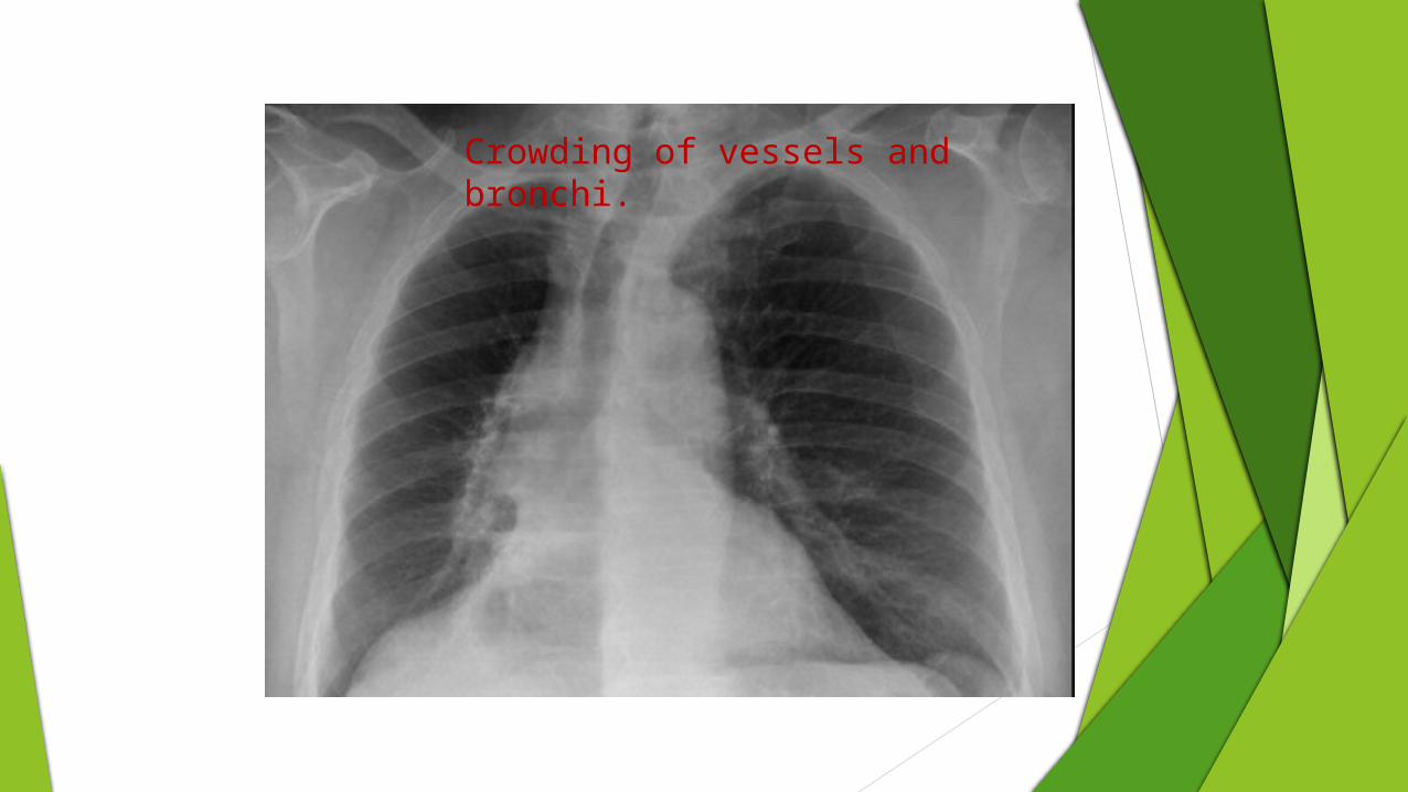
Crowding of vessels and bronchi.

Indirect signsThese include:
† Elevation of a hemi-diaphragm. This sign is of limited value as the normal position of the diaphragm is variable.
† Mediastinal displacement to the side of collapse. Some contents of the mediastinum which are easily seen on plain chest X-rays include the trachea, tracheal tube, central venous catheters in the superior vena cava, and nasogastric tubes in the oesophagus.
† Hilar displacement. The hilum may be elevated in the upper lobe collapse, and depressed in the lower lobe collapse.

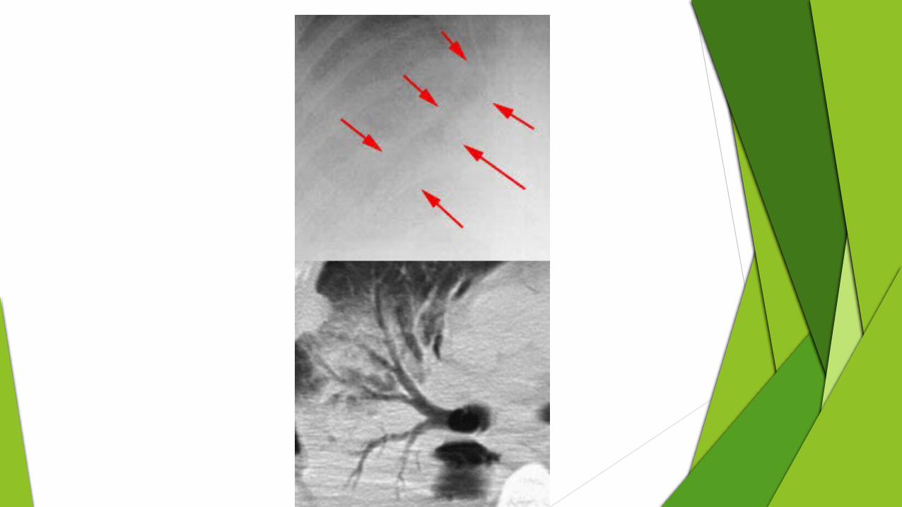
LLL atelectasis (arrows). Note the elevation of the left hemidiaphragm.

Total lung collapse due to obstructive pathology causing a white out of the right hemithorax. Note the trachea is pulled towards the collapsed lung

Total lung collapse of the left lung due to pressure from a pleural effusion leading to tracheal shift away from the left lung.

Lobar collapse
Right upper lobe (RUL) collapse
#Elevation of the right hilum and the minor fissure


CT scan
Features of lobar collapse and effusions are more obvious on CT than plain radiographs (Fig. 8) and it is useful for more atypical forms of collapse.
CT aids identification and localization of endobronchial lesions and any tumour spread, and differentiation of obstructive lesions from other forms of atelectasis.
Ultrasound
Obstructive atelectasis on ultrasound shows as an area of homogenous low echogenicity.

Management
Prevention of atelectasis is preferable to later treatment to re-open collapsed areas of the lung.
Evidence-based studies on the management of atelectasis are lacking.

Prevention of atelectasis begins in the preoperative period by identifying high-risk patients and introducing intensive respiratory therapy of physiotherapy, bronchodilators, cessation of smoking, and antibiotics when indicated, at least 5–7 days before operation for elective surgery.
Common risk factors include patients with preexisting lung problems (chronic obstructive pulmonary disease, asthma, bronchiectasis), smoking, obesity, advanced age, and sleep apnoea.
Baseline X-rays, blood gases, and lung function tests are useful for patients with moderate to severe respiratory and cardiovascular disease who are undergoing more major procedures.
Lung function test are not recommended as part of routine preoperative testing for adults (.16 yr) of any age classified as ASA I with no comorbidities undergoing surgery of any severity.

On induction
Using 100% oxygen at induction is common practice to improve margins of safety in relation to hypoxaemia, but there is good evidence that the use of 100% oxygen is associated with atelectasis.
A compromise is to reduce FIO2 to 80% to reduce atelectasis, or perform a recruitment manoeuvre after induction.
During induction of anaesthesia, application of continuous positive airway pressure (CPAP) can prevent the formation of atelectasis and can increase the margin of safety for oxygenation before intubation.
For example, application
of CPAP 10 cm H2O in morbidly obese patients is effective for the prevention of atelectasis during induction.

During anaesthesia
positive pressure ventilation with PEEP, rather than spontaneous ventilation is preferable in longer procedures in at-risk patients. Recruitment of atelectasis should be attempted if it is suspected clinically or in high-risk patients.
Suggested recruitment manoeuvres include:
† Vital capacity manoeuvre using an inflation pressure of 40 cm H2O sustained for 10–15 s.
† Increasing PEEP to 15 cm H2O and then increasing tidal
volumes to achieve peak inspiratory pressure of 40 cm H2O for 10 breaths before then returning to standard ventilator settings.
Higher levels of PEEP are generally not beneficial as the shunt does not improve due to redistribution of blood flow in the lung, and the high intra-thoracic pressure leads to decreased venous return and haemodynamic compromise.

Postoperative period
Atelectasis is one of the most common pulmonary complications in the postoperative period, often clinically insignificant.
The altered compliance of lung tissue, impaired regional ventilation, and retained airway secretions contribute to the development of atelectasis.
Postoperative pain interferes with spontaneous deep breathing and coughing resulting in decreases in FRC, leading to atelectasis.

variety of lung expansion exercises may reduce postoperative pulmonary complications
chest physical therapy, deep breathing exercises, intermittent positive pressure breathing, and CPAP.
Good postoperative pain control may help to minimize postoperative pulmonary complications by enabling earlier ambulation and improving the patient’s ability to take deep Breaths.

Management in critical care
differs from anaesthesia in that there is commonly a presence of background ALI or infection.
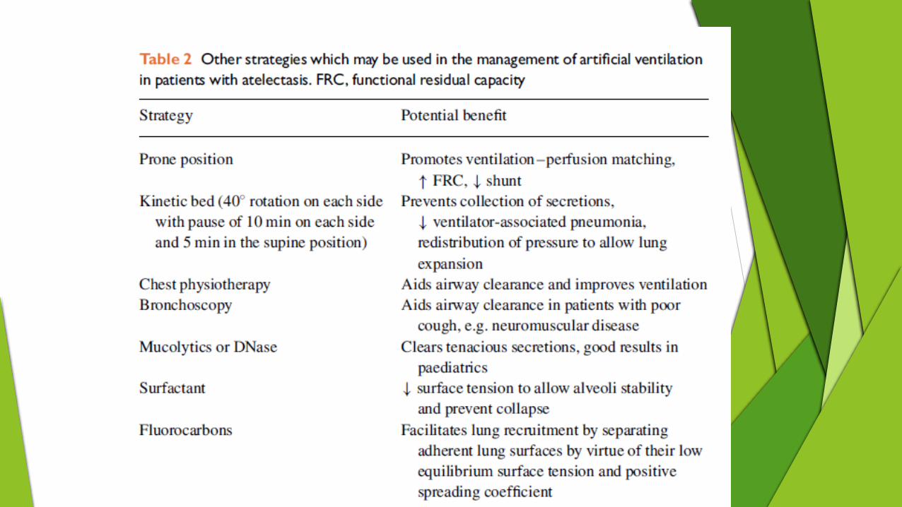
To minimize atelectasis during artificial ventilation in critical care patients,
CPAP is useful for the management of spontaneously breathing patients with non-obstructive atelectasis, who are unable to breathe deeply.
The aim is to open up collapsed alveoli to reduce shunt and improve ventilation–perfusion homogeneity, hence reversing hypoxaemia.

PEEP is a core component of artificial ventilation in ALI patient.
PEEP has a protective role in ALI by attenuating surfactant depletion, and reducing shearing stresses, parenchymal injury, and cytokine release.
Typical recruitment measures
Typical recruitment measures used in intensive care include
† Three consecutive volume-limited breaths per minute with a plateau pressure of 45 cm H2O (also called sigh).
† PEEP increased by 5 cm H2O every 30 s with a 2 ml kg21 decrease in tidal volume. When PEEP reaches 25 cm H2O, CPAP at 30 cm H2O is used for 30 s.
† CPAP: 35–40 cm H2O for 30 s.
Systemic hypotension can occur due to reduced cardiac output when a sustained inflation pressure is used in critically ill patients,
This can be reduced by adequate fluid filling pre-recruitment.
High-frequency oscillation ventilation (HFOV) may be considered and facilitates lung inflation and recruitment by maintaining mean airway pressure at a constant elevated level while using a piston to cycle the ventilation rate at several hundred times per minute.