pulling the trigger john a. mcgreal jr.,...
TRANSCRIPT

Excellence in Optometric Education
John A. McGreal Jr., O.D. Missouri Eye Associates McGreal Educational Institute
Pulling the Trigger
Cases from the Heartland
JAM
John A. McGreal Jr., O.D.
Missouri Eye Associates
11710 Old Ballas Rd.
St. Louis, MO. 63141
314.569.2020
314.569.1596 FAX
JAM
Summary
OCT allows unprecedented visualization of posterior
structures
OCT ushered in a far better understanding of the
relationship between the retina surface and the vitreous
OCT is now allowing better understanding of the role of
the choroid in retinal disease
OCT facilitates accurate clinical decision making, often
without referral, invasive testing, keeping patients in
primary care OD’s office
Elevates level of care provided, increases revenue, makes
clinical throughput easy and efficient
JAM
Case 1: The “Routine” Flashes
CC: “Flashes” HPI: OD/1d/mod/no floater
33 F, allergy – HC Meds: OCs, valacyclovir, flonase
VA = 20/20 OU w SCL IOP: 18OU AC: D&Q
Retina: multifocal choroidal lesions, myopic macular degeneration
OCT: Inner choroidal thickening and macular edema
IVFA: no CNV
JAM
Case 1: The “Routine” Flashes
CC: “Flashes” HPI: OD/1d/mod/no floater
IMP – Punctate Inner Choroidopathy OD, myopic
macular degeneration OU
PLAN – Medrol 4mg DOSPAK, Durezol tid OD,
Bromday qd OD
Prognosis - excellent
JAM
Case 1: The “PIC”
OCT Pearls
– OCT confirms (if not MAKES) the diagnosis
– Provides an image of the choroid that we cannot
evaluate clinically with any other technique

JAM
Case 2: The “Bonsai Floaties”
CC: “floaties on my trees when I work” HPI:
OD/1m/mod/ CABG 11 yrs
67 M, Meds: atenolol, maxide, plavix
MR OD: -3.50DS = 20/100
MR OS: -2.75DS = 20/25
IOP: 14OU AC: D&Q NS/CX+2OU
Retina: tear at equator with PVD OD, small micro-
aneurisms temporal to FAZ OS
OCT: OD 233u, OS 271u
JAM
Case 2: The “Bonsai Floaters”
IMP – Retinal tear OD, Juxta-foveal
Telangiectasia OS
PLAN – Laser photocoagulation for tear OD
today, return for IVFA OS 2wks
IVFA: NO leakage from JFT
PLAN: Observe JFT, order glucose screening,
schedule cataract consult
JAM
Case 2: The “JFT”
OCT Pearls
– OCT demonstrates abnormal thickness
– IVFA shows no leakage (SURPISE!)
– OCT & IVFA do not agree
– OCT is more sensitive test?
– Allows us to identify pathology at a subclinical level
follow closer for progression
avoiding vision loss
JAM
Case 3: The “CME” Solution
CC: “Not as good as 1st week” HPI: OU/1m/Cat-
IOL/ Lattice Deg OU / Retinal tears OU treated
with laser
66 F, VA OD = 20/15, VA OS = 20/40
PCIOL OU
IMP – Pseudophakic CME OS?
OCT: OD 306u, OS 383u
Plan: Predforte tid & Bromday qd OD
RTO: 2 wks
JAM
Case 3: The “CME” Solved
CC: “Getting better” HPI: OS/2 wks/ CME
/PF & Bromfenac
66 F, VA OD = 20/15, VA OS = 20/25+
PCIOL OU
IMP – Pseudophakic CME OS Resolved
OCT: OD 306u, OS 310u
Plan: Predforte tid & Bromday qd OD until gone,
new spec Rx
JAM
Case 3: The “CME Solved”
OCT Pearls
– OCT demonstrates abnormal thickness
– OCT allows for conservative treatment trial prior to
more invasive procedures
– OCT demonstrates rapid improvement in condition
– OCT keeps patient in primary care OD doctor’s office,
specialist not needed
– IVFA not needed

JAM
Case 4: The “glasses dilemma”
CC: “Had cataract surgery and cant see” HPI:
OS/1m/Cat-IOL/OD gave 2 new glasses, likes one
better than other
80 F, VA OD = 20/30, VA OS = 20/40+ at one
week post ops, admits to one drop per day?
PCIOL OU
IMP – Pseudophakic OU
PLAN – Predforte tid OU, release to optometrist
for continued care, emphasis on correct drug
dosing
JAM
Case 4: The “glasses dilemma”
CC: “Had cataract surgery and cant see” HPI:
OS/1m/Cat-IOL/OD gave 2 new glasses, likes one
better than other
Now after one month VA OD = 20/200 OS
Hands me the steroid prescription (never filled)
PCIOL OU
IMP – Pseudophakic CME OS?
OCT: OD 311u, OS 510u
Plan: Retina for IVK (failure with drop
compliance)
JAM
Case 4: The “Compliance dilemma”
OCT Pearls
– OCT demonstrates abnormal thickness OS
– IVFA not needed to confirm diagnosis
– Pseudophakic CME in this case related to
inflammation from non-compliance with steroidal
eyedrops
– Treatment with topicals may be beneficial but with
questionable ability to comply, intraocular depot drug
is the best choice
JAM
Case 5: The “Big Black Spot”
CC: “Black spot” HPI: OS/2
days/constant/decreased vision Cat-IOL/OU ROS:
recent diagnosis Hairy cell leukemia, chemotherapy
and spleenectomy, now anemic
76 F, VA OD = 20/40, VA OS = 20/400, was 20/25
two months prior
PCIOL OU IOP: 10 OU
Fundus: peripheral small retinal hemorrhages OU,
thick macular hemorrhage OS, schisis cavity inf OS
OCT: 286u/333u
JAM
Case 5: The “Big Black Spot”
IMP: Leukemic Retinopathy
PLAN: Retina consult
– TPA & gas to displace macular hemorrhage
– Continue oncologic care
Follow-up visit one month
– RE 20/25
– LE 20/30
– No retinopathy noted!
JAM
Case 5: The “Leukemic Retina”
Pearls
– OCT demonstrates abnormal thickness OS
– OCT clearly shows pre-retinal and intra-retinal nature
of hemorrhages
– Prognosis often very good

JAM
Case 6: The “Black Dot”
CC: “Black dot” HPI: OS/1 yr/constant/no worse
Referring doctor: Retinal detachment, partial, old
8 M, VA OD = 20/20, VA OS = CF CVF: central
scotoma OS, SLE: NL
Fundus: massive hemorrhage and exudative
retinopathy OS
OCT: massive elevation of macula
JAM
Case 6: The “Black Dot”
IMP: Coat’s Disease
PLAN: Retina consult
– R/O toxoplasmosis with serology
– Consideration of retinoblastoma
JAM
Case 6: The “Coats Disease”
Pearls
– OCT demonstrates markedly abnormal thickness OS
– Serologic testing to R/O toxoplasmosis, toxocariasis
– Always obtain retina consult (poor prognosis)
JAM
Case 7: The “Different Size Images”
CC: “Different sizes” HPI: OS/2 yr/constant/
worse/failed drivers test
PMH: colon CA, HTN
Referring doctor: Cataract surgery requested
69 M, VA OD = 20/30, VA OS =20/80 SLE:
symmetric NS
Fundus: Vitreous traction OS
OCT: 266u/338u
AL: 24.36/24.45
Ocriplasmin / ThromboGenetics, Inc
Non surgical treatment for vitreomacular adhesions
– Increased macular thickness
– CME Diagnosed 8% at slit lamp 30% with OCT
Vitrectomy vs Vitreolysis?
– Invasive
– Anesthesia
– Face down
– Retinal breaks
– Cataract
Ocriplasmin / ThromboGenetics, Inc
Truncated form of human plasmin produced by bacteria
Indications: developed for dissolving blood clots in
vascular disease
Single Intravitreal injection
Results – resolution 30% at 28 days, closure of hole 40%
at 28 days
– better than all other agents tried
Spin offs – DME, AMD, adjunct to vitrectomy
New England JourMed 2013

JAM
Case 7 : The “VMT”
Pearls
– OCT demonstrates abnormal thickness OS
– OCT demonstrates vitreo-macular traction clearly
– IVFA finds no leakage
– Previous solution limited to vitrectomy surgery
– Option now includes medical treatment first
– Ocriplasmin is perfect for this exact clinical
presentation
– If not successful, PPV remains option and will be
technically easier
Case 8: The “Lost My Monovision!”
77yowf CC: “Can’t read!”
HPI: 1 D duration / intermittent loss, altitudinal, preceded
episode / painless / OD
Meds: Amiodarone, ASA, Coumadin, Cartia, Zoloft, Advil,
Singulair, Cozaar, Norvasc
ROS: 190 lbs, recent Spinal surgery (L3-5), planned shoulder
(rotator cuff) surgery, Monovision
BVA: 20/60 OD 20/20 OS PERRL + APD
EOM: Full EXT: NL
SLE: ACIOL OD, PCIOL OS Blurred optic disc margin OD,
otherwise NL
What is the likely diagnosis?
1. Idiopathic optic neuritis
2. Ischemic optic neuropathy
3. Buried drusen
4. Papilledema
5. Cerebral vascular accident
What eye test would you order now?
1. Pachymetry
2. Visual fields
3. SCODI
4. ERG
5. IVFA / Photo
What other testing is indicated?
1. CBC with differential
2. Brain MRI
3. C-reactive protein
4. ESR
5. ESR & CRP
Tests results
1. Visual field = Mild central defect OD, normal OS
2. ESR = 17mm/Hr
– Reference 0-20mm/Hr
3. C-reactive protein = 0.899mg/L
– Reference 0.000-3.0mg/L

What should you do now?
1. Start Prednisone
2. Order biopsy of superficial temporal artery
3. Retina consult
4. Follow conservatively for NAION
Case 8: “NAION”
Clinical pearls
– R/O GCA most important
– Follow conservatively
– ASA debatable benefit
Case 9: The “Graduation”
83yowf from Memphis, TN CC: “Skim on my eye, then it
went black!”
HPI: 1 D duration / intermittent loss, altitudinal, preceded
episode / painless / OD
Meds: HCTZ, meclizine, centrum, naproxen
ROS: 115lbs, HA, stiffness
BVA: NLP OD 20/30 OS PERRL + APD
EOM: Full EXT: NL
SLE: PCIOL OD NS 2 OS Fundi: OD Blurred optic
disc margin, otherwise NL
What is the likely diagnosis?
1. Idiopathic optic neuritis
2. Ischemic optic neuropathy
3. Buried drusen
4. Papilledema
5. Cerebral vascular accident
What eye test would you order now?
1. Pachymetry
2. Visual fields
3. SCODI
4. ERG
What other testing is indicated?
1. CBC with differential
2. Brain MRI
3. C-reactive protein
4. ESR
5. ESR & CRP

Tests results
1. Visual field = absolute defect OD, normal OS
2. ESR = 44mm/Hr
– Reference 0-20mm/Hr
3. C-reactive protein = 0.158mg/L
– Reference 0.000-3.0mg/L
What should you do now?
1. Start Prednisone PO stat
2. Order biopsy of superficial temporal artery
3. Retina consult
Tests results
1. C-reactive protein = 27.5mg/L
– Reference 0.000-3.0mg/L
– Corrected C-reactive protein test delivered by mail five days
later!
Case 9: “GCA / AION”
Differential Diagnosis – Anterior ischemic optic neuropathy,
Giant cell arteritis, CVA, NAION
Additional Testing – STAT ESR, CRP, STA Biopsy +/-
Diagnosis – AION, GCA
Treatment Plan – Prednisone 80mg PO qd, chronic care with
internist or neurologist
Clinical Pearls – CRP best test, Don’t miss it, Prednisone dose =
1mg/kg/D, tapered with ESR, need to co-manage
Case 10: “I Lost Vision Last Night!”
35yowm CC: “Lost vision last night”
Pupils: PERRLA+MG
Meds: Glucophage for 3 years
VA 20/20 OD, HM OS
IOP: 17/18
SLE: Nl OU Fundus : As shown
What is the diagnosis?
1. Macular twig venous occlusion
2. Birdshot retinochoroidopathy
3. Hypercholesterolemia (retinal lipidemia)
4. CRAO
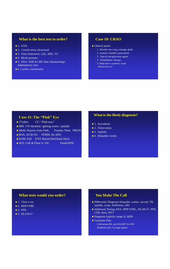
What is the best test to order?
1. IVFA
2. Carotid artery ultrasound
3. Total cholesterol, LDL, HDL, TG
4. Blood pressure
5. ANA / ESR & CRP other rheumatologic
inflammatory tests
6. Cardiac consult/echo
Case 10: CRAO
Clinical pearls
– 1. Breathe into a bag, massage globe
– 2. Anterior chamber paracentesis
– 3. Topical anti-glaucoma agents
– 4. Thrombolytic therapy
– 5. Must have a systemic cause
Find it & fix it!
Case 11: The “Pink” Eye
17yobm CC: “Pink-eyes”
HPI: 3 W duration / getting worse / painful
Meds: Ilotycin from Peds Trauma: None NKDA
BVA: 20/30 OU PERRL No APD
EOM: Full EXT: Raised Red Rash-Neck
SLE: Cell & Flare 3+ OU Fundi:WNL
What is the likely diagnosis?
1. Sarcoidosis
2. Tuberculosis
3. Syphilis
4. Idiopathic uveitis
What tests would you order?
1. Chest x-ray
2. RPR/VDRL
3. PPD
4. HLA B-27
You Make The Call
Differential Diagnosis-idiopathic uveitis, sarcoid, TB,
syphilis, Lyme, AS/Reiters, HIV
Additional Testing-ANA, RPR/VDRL, HLAB-27, PPD,
CXR, titers, HIV?
Diagnosis-Syphilis (stage 2), AIDS
Treatment Plan
– Ceftriaxone IM, start HAART for HIV,
– PredForte q2h, Cyclogel option

Case 11: “Uveitis/HIV”
Clinical Pearls
– R/O systemic causes in uveitis if bilateral, severe, young, or
high index of suspicion
– Granulomatous presentations more often underlying cause
– “Everyone lies”….Dr. House
Case 12: “Doc, I See Double”
57yobm CC: “Double vision”
HPI: OU / 3 D duration / Stable / not painful /
Horizontal
Past H: Colon cancer / surgery / radiation / Chemo
Meds: Multiple Trauma: None NKDA BVA:
20/30 OU PERRL No APD
EOM: R Adduction deficit, L Jerky nystagmus
SLE: NS OU Fundus : NL
What is the diagnosis?
1. Internuclear Ophthalmoplegia
2. Ocular Myasthenia Gravis
3. Duanes Retraction Syndrome
4. CN 3 Palsy
What is the best next step?
1. ESR
2. Neuro-ophthalmology consult
3. Neurology consult
4. MRI of head
Case 12: INO / Met CA
Differential Diagnosis - CN 3P, CN 6P, INO, Decompensating heterophoria
Additional Testing-old photos
Diagnosis- R INO, metastasis of colon CA
Lesion- R MLF
Treatment Plan- MRI, Neurology / Neurosurgery, Oncology, PCP, monocular occlusion
Pearls-INO often related to MS, stroke
– Needs imaging to differentially diagnose
Case 13: “Corneal Abrasion”
Age: 19yowm CC: Floaters
HPI: OD / 3wks / constant / worsening since corneal
abrasion with patching therapy
Meds: none
BVA: 20/20 OU Pupils: PERRL EOM:NL EXT: NL
SLE: small corneal defect / haze at limbus
IOP: 18/16
Fundi: As shown
PFSH & ROS: NL

What is the likely diagnosis?
1. Old CA with residual edema
2. Intraocular foreign body
3. Toxocara canis
4. Vitreous condensation
What tests would you order?
1. Ultrasound
2. Orbital CT
3. VF
Case 12 : IOFB
Differential Diagnosis – Old CA, retinal IOFB, primary retinal pathology
Additional Testing – US, Photography, VF
Diagnosis - IOFB
Treatment Plan – pars plana vitrectomy, FB removal, intravitreal antibiotics
Case 14: “Woke Up Blind!” Age: 19yobf CC: decrease VA
HPI: OU / rapid / severe / worsening
Meds: plaquenil 400mg, lopressor
BVA: CF OU Pupils:PERRL-APD EOM:NL
EXT: NL
SLE: NL
IOP:16/16
Fundi: as shown
PFSH & ROS: SLE x 3yrs, ischemic necrosis of hip secondary
to corticosteroids at initial flare
What is the likely diagnosis?
1. Diabetic retinopathy
2. Hypertensive retinopathy
3. Retinal vaculitis
4. Bilateral CRVO
What tests would you order?
1. BP
2. ESR
3. ANA
4. VF
5. Photo

Case 14: Lupus Retinal Vasculitis
Differential Diagnosis – SLE with retinal vasculitits,
HTN and retinopathy, DM and retinopathy,
hyperviscosity states
Additional Testing – IVFA, photos, ESR, ANA, C-
reactive protein, VF
Diagnosis – SLE and retinal vasculitis
Treatment Plan – IV corticosteroids, rheumatology
consult, retina consult
Pearl – ANA is elevated in acute Lupus
Case 15: The “Blue Freckle”
Age: 34yobm CC: blurred vision
HPI: OS / 1 yr / stable / constant
Meds: none
BVA: 20/20 OU Pupils: PERRL-APD EOM: full EXT:
pigmented lesions of face
SLE: pigmented lesions of the sclera
Gonio: pigment puddling
IOP: 19/29
Fundi: deeper retinal/choroidal pigmented, asymmetry of CDR
PFSH & ROS: NL
What is the likely diagnosis?
1. Nevus flammeus
2. Nevus of Ota
3. Sturge-Weber
4. POAG
What tests would you order?
1. Old photos
2. VF
3. Scanning lasers
4. Gonioscopy
You Make The Call Differential Diagnosis – Sturge-Weber, Nevus Ota,
Nevus flammeus, OAG
Additional Testing – VF, GDx/HRT, Gonio
Diagnosis – Nevus of Ota, OAG OD
Treatment Plan – Photodocument, blue tint spectacles,
latanoprost 0.005% qhs OS
Case 15: The “Nevus Ota”
Clinical Pearls
– Follow for malignant transformation of skin lesions
– Follow for pigment glaucoma
– Affects other systems
– Familial tendency
– More difficult in darker skin to diagnose

Case 16: “Headache” Lady
45yowf CC: “HA, Blurred vision”
HPI: Sudden / Explosive / Constant HA / photophobic
– Lower Extremity Amputee / Tracheotomy
Meds: None Trauma: None NKDA
BVA: 20/40 OD 20/20 OS PERRL No APD EOM: Full EXT: WNL
SLE: WNL Fundi: Globular Sub-Hyaloid Hemorrhage OD
What is the likely diagnosis?
1. Valsalva retinopathy
2. Terson’s syndrome
3. Diabetic retinopathy
4. Vitreous hemorrhage
What tests would you order?
1. MRI of the brain
2. Lumbar puncture
3. Fundus photography
4. Random blood glucose
You Make The Call
Differential Diagnosis-Drance hemorrhage, CNVM,
migraine, subarachnoid hemorrhage
Additional Testing-MRI/MRA, lumbar puncture+/-,
pupillary testing, physical examination (neurology)
Diagnosis
– ICA/SAH Terson’s Syndrome
You Make The Call
Treatment
– STAT admission/high mortality & morbidity
– Oxygenation
– Sedatives
– Control of blood pressure
– Monitor cerebral edema
– Surgery +/-
endovascular ballons, “clipping” of aneurisms
Case 16: ICA / SAH Clinical pearls
– Neurological/Neurosurgical emergency
– Prodromal sentinel signs common
– Rapid onset of pain/HA, nuchal rigidity, loss of
consciousness, loss of sight, obtundation, death
– Neurosurgery if stable
– Survivors-mild /severe cognitive impairment

Case 17: “LASIK Nevus”
34yowf CC: “Freckle in my eye”
HPI: OD / 2 wks duration / Lasik OU 1 wk
LASIK doctors request retinal evaluation
Meds: Allopurinol NKDA
BVA: 20/15 OU PERRL No APD
EOM: Full EXT: W&Q
SLE: Flaps OU IOP: soft OU
Fundus: as pictured
What is the likely diagnosis?
1. Epiretinal membrane
2. Congenital hypertrophy of RPE
3. Macular drusen
4. Choroidal osteoma
5. Benign choroidal nevus
6. Malignant melanoma
What eye test would you order now?
1. IVFA
2. Visual fields
3. SCODI
4. B scan ultrasound
What is the best course now?
1. Retina consult
2. Ocular Oncology
3. PCP
4. LASIK retreatment
5. Retire; I can’t take another day of this
Case 18: “Upper part missing”
65yowm referred for AION CC: “wavy things”
HPI: OD / 5 wks wavy / 2 wks upper part of vision missing / no pain / no flashes
Meds: MV, OM3, ASA NKDA
BVA: HM OD, 20/25 OS PERRL No APD
EOM: Full EXT: W&Q
SLE: NS 2 OU IOP: 15 OU
Fundus: as pictured
What is the likely diagnosis?
1. Retinal tear
2. Lattice degeneration
3. Vitreous hemorrhage
5. Benign choroidal nevus
6. Malignant melanoma
What is the best course now?
1. Retina consult
2. Ocular Oncology
3. PCP
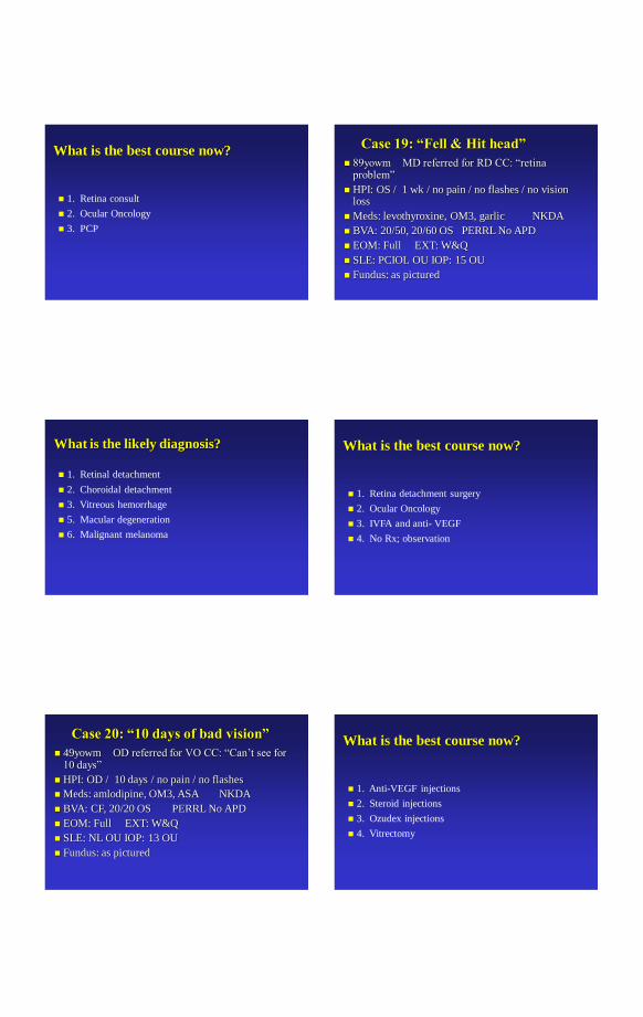
Case 19: “Fell & Hit head”
89yowm MD referred for RD CC: “retina problem”
HPI: OS / 1 wk / no pain / no flashes / no vision loss
Meds: levothyroxine, OM3, garlic NKDA
BVA: 20/50, 20/60 OS PERRL No APD
EOM: Full EXT: W&Q
SLE: PCIOL OU IOP: 15 OU
Fundus: as pictured
What is the likely diagnosis?
1. Retinal detachment
2. Choroidal detachment
3. Vitreous hemorrhage
5. Macular degeneration
6. Malignant melanoma
What is the best course now?
1. Retina detachment surgery
2. Ocular Oncology
3. IVFA and anti- VEGF
4. No Rx; observation
Case 20: “10 days of bad vision”
49yowm OD referred for VO CC: “Can’t see for 10 days”
HPI: OD / 10 days / no pain / no flashes
Meds: amlodipine, OM3, ASA NKDA
BVA: CF, 20/20 OS PERRL No APD
EOM: Full EXT: W&Q
SLE: NL OU IOP: 13 OU
Fundus: as pictured
What is the best course now?
1. Anti-VEGF injections
2. Steroid injections
3. Ozudex injections
4. Vitrectomy

Case 20: “Spot in vision”
63yowf MD referred for AMD CC: “Blur spot in center”
HPI: OD / 1 mos / no pain / constant Meds: synthroid, restasis, lotemax NKDA
BVA: 20/50 OD, 20/20 OS PERRL No APD
EOM: Full EXT: W&Q
SLE: PCIOL OU IOP: 8 OU
Fundus: as pictured
What is the best course now?
1. Anti-VEGF injections
2. Steroid injections
3. Ozudex injections
4. Vitrectomy
5 . Photodynamic therapy
Case 21: “Itchy retina”
35yowm OD referred for pink eye CC: “Itch, swollen”
HPI: OD / 1 d / no pain / constant NKDA
BVA: 20/30 OD, 20/20 OS PERRL No APD
EOM: Full EXT: conjunctival injection chemosis
SLE: same IOP: 14 OU
Fundus: as pictured
What is the best course now?
1. Anti-VEGF injections
2. PE, CBC, CXR, ANA, ESR, autoimmune,
coagulopathies
3. Ozudex injections
4. Photocoagulation
5 . Photodynamic therapy
Case 22: “Lost vision for 15 mins”
24yowm OD referred for retinal hemorrhage CC: “episode of lost vision”
HPI: OD / days / no pain / constant / had CL exam one month ago NKDA
BVA: 20/40 OD, 20/20 OS PERRL No APD
EOM: Full Ext: Nl
SLE: Nl IOP: 14 OU
Fundus: as pictured
What is the best course now?
1. Anti-VEGF injections
2. Pneumatic retinopexy
3. Vitrectomy +/- buckle
4. Photocoagulation to lattice
5 . Photodynamic therapy

Case 23: “I Want Chalazion
Removed” 55yowf CC: “OD dx chalazion”
Pupils: PERRLA-MG
Meds: Premarin, Zocor, HCTZ
VA 20/30 OU
IOP: 16/17
SLE: Lid lesion E2 OS Fundus : NL
What is the best option now?
1. Remove chalazion with I&D
2. Intralesional kenalog injection
3. Biopsy
4. Oral antiobiosis and hot packs
Case 24: “Exotopia” Age: 2yowm CC: R/O strabismus
HPI: XT OD / 4mos / constant / severe
Meds: none Ref: Peds
BVA: No Fix or follow Pupils: PERRL-APD
EOM: L XT 45 EXT: NL
SLE: NL
IOP: Soft
Fundi: ON abnormal OS
PFSH & ROS: NL
What is the likely diagnosis?
1. Coloboma optic nerve entrance
2. Morning glory syndrome
3. Retinal detachment
4. Cavernous hemangioma
What tests would you order?
1. PE
2. MRA brain and orbits
3. EUA
4. IVFA
You Make The Call
Differential Diagnosis – morning glory, retinal hemangioma, ON coloboma
Additional Testing – PE family members, eye examination family members
Diagnosis – cavernous hemangioma retina, strabismus, amblyopia
Treatment Plan – external plaque radiation, EOM surgery, patching treatment

Case25: “Light Sensitive” Age: 15 yowf CC: “Lights hurt”
HPI: OS / 2D / worsening / severe
Meds: none OcHx: Accuvue SCL denies sleeping in lens (Dr. House “everybody lies”) Renu
VAsRx: OD 20/100, OS LP Pupils: PERRL-APD EOM: full EXT: injected, ptosis
SLE: as pictured
IOP: not done
Fundi: not viewed
PFSH & ROS: NL
You Make The Call Differential Diagnosis – bacterial keratitis, fungal
keratitis, acanthameba keratitis, foreign body, hypopion uveitis
Additional Testing – culture/sensivity
Diagnosis – bacterial keratitis
Treatment Plan – moxifloxacin q1h, close watch
RTO 24h
Case26: “Glaucoma or Not?” Age: 68 yowm CC: “Need new doc for glaucoma”
HPI: OU / 6 yrs / Trav qhs OU / mild / No SE
Meds: Metformin, diovan, crestor, zetia, ranitidine
VAsRx: OD 20/25 OU Pupils: PERRL-APD EOM: full EXT: nl +Fam Hx Glauc mother
SLE: NS/CX 1 OU
IOP: 13 OU OCT normal VF normal
Fundi: Normal CDR .75 OU symmetric rims
PFSH & ROS: NL
You Make The Call Differential Diagnosis – POAG vs normal
Additional Testing – VEP
Diagnosis – Probable POAG OU based on VEP
Treatment Plan – continue medications, switch to generic latanoprost qhs
Thank you
Excellence in Optometric Education
Missouri Eye Associates
McGreal Educational Institute