public health and environmental laboratories …
TRANSCRIPT
1
November, 2014
New Jersey Laboratory Guidelines for Handling Specimens from Patients with
Suspected and Confirmed Ebola Virus Disease (EVD)
Purpose The following guidelines are provided for New Jersey clinical laboratories that may receive and test specimens from patients who are persons under investigation (PUI) or Laboratory Confirmed Cases of Ebola Virus Disease (EVD). Local and state public health authorities (in consultation with CDC) will assess each case and determine whether testing is warranted. Testing for the Ebola virus may be done locally, regionally or at CDC, at the discretion of the evaluation team.
1. Person Under Investigation (PUI):
A person who has both consistent signs or symptoms and risk factors as follows: • Elevated body temperature or subjective fever or symptoms, including severe
headache, fatigue, muscle pain, vomiting, diarrhea, abdominal pain, or unexplained hemorrhage; AND
• An epidemiologic risk (http://www.cdc.gov/vhf/ebola/exposure/risk-factors-when-evaluating-person-for-exposure.html) factor within the 21 days before the onset of symptoms. Exposure may be considered high, some, low, or no risk.
2. Persons Confirmed as having EVD with a laboratory test
Molecular EVD testing in New Jersey
Molecular diagnosis for EVD is available at the NJDOH Public Health and Environmental Laboratories (PHEL) using the DOD real time PCR Ebola Zaire assay that has been FDA-cleared under Emergency Use Authorization (EUA). Refer to the PHEL technical bulletin for information on implementation of the assay. The first step will be for hospital officials to contact the local health department within the patient’s jurisdiction. http://nj.gov/health/lh/documents/lhdirectory.pdf
PUBLIC HEALTH AND
ENVIRONMENTAL LABORATORIES TECHNICAL BULLETIN

2
EVD Transmission and Decontamination
A person infected with Ebola virus is not contagious until symptoms appear. •EVD is transmitted through direct contact (via broken skin or mucous membranes) with blood or body fluids from an EVD patient, or through contact with objects contaminated with blood or body fluids from an EVD patient. There is no evidence of airborne transmission. •Ebola virus is readily inactivated by standard chemical decontamination procedures used in laboratories.
Please note the following points with regard to EVD:
Ebola virus is present in numerous body fluids of patients with EVD1. Although detected much less frequently, it has also been shown to be present in some environmental samples contaminated with blood or body fluid from an EVD patient consistent with a risk of transmission from fomites1.
Biosafety Considerations
Laboratory Safety Requirements: For ALL blood and body fluids tested in clinical laboratories: All blood and body fluids in a clinical laboratory should be handled as if they contained a human pathogen. 1. Specimens should be processed and tested in accordance with standard OSHA bloodborne
pathogen requirement 42CFR1910.1030) https://www.osha.gov/pls/oshaweb/owadisp.show_document?p_table=STANDARDS&p_id=10051
2. Implement and enforce SOP-specific biosafety procedures based on procedural risk assessment (examples found in this document) http://www.cdc.gov/mmwr/pdf/other/su6101.pdf 3. Standard EPA disinfection guidance (10% v/v solution of household bleach to be
prepared fresh daily or equivalent) http://www.cdc.gov/vhf/ebola/hcp/environmental-infection-control-in-hospitals.html
4. Frequent handwashing
Laboratory Safety Requirements: For Specimens from EVD-Confirmed Laboratory Cases or PUI with a High/Some/Low Risk Exposure
Ebola virus is indisputably a highly pathogenic agent14. All laboratory directors should review their circumstances, facilities, resources and procedures, as well as the training and experience of their staff, in order to perform a thorough biohazard risk assessment and implement appropriate procedures for risk mitigation. However, any additional precautions or procedures should not interfere with the ability to provide appropriate medical care for suspected or confirmed EVD patient.

3
1. Specimens should be processed and tested in accordance with standard OSHA bloodborne pathogen requirements (42 CFR 1910.1030) http://www.osha.gov/pls/oshaweb/owadisp.show_document?p_table=STANDARDS&p_id+10051 2. Implement and enforce SOP-specific biosafety procedures based on procedural risk assessment (examples found in this document) http://www.cdc.gov/mmwr/pdf/other/su6101.pdf 3. Standard EPA disinfection guidance (10% v/v solution of household bleach to be prepared fresh daily or equivalent) http://www.cdc.gov/vhf/ebola/hcp/environmental-infection-control-in-hospitals.html 4. Frequent hand washing
Additional CDC Ebola biosafety guidance
Guidance from the CDC recommends that PUIs who report Low, Some or High Risk exposure, or laboratory confirmed Ebola cases, be managed in US hospitals with standard contact and droplet precautions. Laboratory personnel are advised to adhere strictly to safety procedures for the prevention of transmission of bloodborne pathogens when handling specimens from these patients (www.cdc.gov/vhf/ebola/hcp/interim-guidance-specimen-collection-submission-patients--suspected- infection-ebola.html) including the following:
• PPE for Specimen collection
Full face protection (mask and goggles or face shield), double gloves, impermeable gown
• PPE for Laboratory testing Full face protection (mask and goggles or face shield), double gloves, impermeable gown and use of certified class II Biosafety cabinet or splash shield
• Donning and Doffing PPE – PRACTICE-PRACTICE-PRACTICE Supervised donning and doffing PPE should be done in accordance with CDC recommended procedure. In order to provide appropriate barrier protection from exposure to blood and body fluids the process of donning PPE must assure adequate coverage of eyes, nose, mouth, hands and skin. In certain circumstances respiratory protection may also be required and the process for fit testing implemented and monitored. The link provides guidance on donning and doffing. http://www.cdc.gov/vhf/ebola/hcp/procedures-for-ppe.html
Note, the above guidance refers to all laboratory work including the routine hematology and clinical chemistry testing that is essential for the appropriate care and treatment of patients.

4
Risk Assessment of Laboratory Procedures Procedural risk assessment is an important part of all clinical laboratory safety programs. A safety manual can outline laboratory hazards and general guidance related to hazards, engineering controls and general safety practices. General use of PPE, decontamination, evacuation and chemical hygiene can be addressed as well as other general topics. Task-specific procedural safety guidance cannot be detailed in the safety manual, and must be incorporated into each SOP. Guidance on performance of risk assessments may be found in the 2012 MMWR publication, Guidance for Safe Work Practices in Human and Animal Diagnostic Laboratories http://www.cdc.gov/mmwr/pdf/other/su6101.pdf For example, if a procedure requires a vortex or homogenization step of biohazardous material, the procedure should indicate that that step is to be performed in the Biosafety Cabinet II (BSC II) or behind shielding. The common laboratory procedures which follow have been identified as being part of those which might occur during care of an EVD confirmed or PUI, and should be assessed in your laboratory as a potential source of exposure.
Important: General Laboratory Comments
Review biosafety practices with all staff to include donning and doffing procedures described above. Drill staff until proficient.
Review of all SOPs to assure that biosafety risk assessments have been completed, incorporated into the SOPs and implemented.
Limit testing to those tests essential to patient care. However, patient care and wellbeing should not be compromised.
Specimens should be labeled to indicate that they have originated from a suspected High, Some or Low risk PUI, or confirmed EVD patient.
Facilities should maintain a log of personnel handling specimens from these cases.
Laboratories should review their protocols for occupational exposure and consult with their hospital epidemiologist and the local health department immediately if a potential exposure occurs. Local contact information can be found at: localhealth.nj.gov. If the local health department cannot be reached, contact the state health department at: NJDOH Communicable Disease Service 609-826-5964 (business hours), 609-392-2020 (after hours)
If available, the use of Point-of-Care instruments and methods inside or nearby the patient’s isolation room may be a preferred option, to provide reduced specimen transport and limit the need for testing in routine laboratories.
For testing that requires transport of samples to the hospital laboratory, specimens should be double-bagged, placed in a biohazard transportation container, and hand-carried to the laboratory. DO NOT use a pneumatic tube system.

5
Risk Assessment on Specific Laboratory Procedures Procedure
Recommendation
Centrifugation Should be performed with sealed buckets or sealed rotor. Homogenization
Procedures requiring homogenization of any specimen type should be avoided or performed with extreme care due to the risk of spray or splash.
Clinical chemistry and hematology
Numerous issues pertaining to routine testing in these areas need to be considered and are highly variable depending on the type of equipment used, volume of testing performed, laboratory workflow and layout, and many other factors. A full risk assessment should be made at each site, including options for decontamination 15 . For automated instruments, decontamination procedures should be those advised by the manufacturer or vendor for enveloped viruses.
Malaria testing Rapid antigen tests or thin blood smears are preferred: recognizing that rapid tests are inherently less sensitive but positive results generally reliable. The effects of some inactivation/decontamination procedures on the performance of some rapid antigen tests for malaria have been investigated16. Thin blood smears should be fixed in methanol for 30 minutes and dried prior to staining. The use of additional heat inactivation is not considered necessary for Ebola decontamination and has been found by some parasitologists to cause disruption to the morphology of the parasites.
Blood Cultures Systems using plastic blood culture bottles are preferred. Blood culture in glass bottles should be avoided.
Other specimens for bacterial culture
“Pan-cultures” should not be performed. Procedures essential for patient management should be performed in a Class II BSC with PPE.
Wet preps Should be avoided. Viral cultures DO NOT perform viral culture, including any rapid culture systems, on any
specimen.
Pre-transfusion testing
Please refer to the American Association of Blood Banks’ Ebola information sheet http://www.aabb.org/press/Pages/Infection-Control-for-Handling-Blood- Specimens-from-Suspected-Ebola-Patients.aspx
Post-mortem examinations
Should not be performed.
Specimen storage With the exception of circumstances where retention is required by regulations, long-term storage of specimens is discouraged. It is recommended that specimens collected from suspected or confirmed EVD cases be isolated from other specimens in the laboratory and disposed of in an appropriate manner (see below) as soon as is practical after testing has been completed.
Specimen decontamination and disposal
Autoclave specimens if facilities are available. Alternatively, decontaminate specimens in 10% bleach for 24 hours, then place in standard biohazard infectious waste disposal.

6
Packaging and Shipping Specimens for Ebola Virus Testing Collection of blood specimens for EVD testing at PHEL is outlined in the PHEL Technical Bulletin. Follow the details outlined in the PHEL Technical Bulletin for safe collection, handling and transport of the blood tubes within your facility.
1. Please note that two filled 3 or 4 ml plastic EDTA tubes are required with CLIA approved labelling
2. At the bedside: Please be sure that the two blood tubes are labelled, decontaminated and each placed inside a separate plastic Ziploc bag. Each bag must contain sufficient absorbent material to absorb the entire contents of the tube if broken. Bags must be sealed (expel all air first) and decontaminated prior to leaving the patient’s bedside. Use 10%v/v bleach solution, prepared fresh daily, or other EPA approved disinfectant.
3. Receiving/Transport staff: DO NOT bring any packaging materials into the patient area. REMEMBER: that this is a contact isolation situation, so any surfaces coming in contact with blood or body fluids, OR any surfaces coming in contact with contaminated gloves, contaminated PPE or contaminated surfaces themselves become a source of contamination. Make sure that staff receiving the Ziploc bags from the patient area decontaminate the bags, change gloves and decontaminate the bags again, BEFORE beginning the packaging process. Transfer the decontaminated bagged specimens, containing sufficient absorbent material to the packaging area using a rigid specimen carrier. The carrier should be decontaminated before and after use.
4. Packaging/documenting staff (in the laboratory): Certified shippers packaging the specimen should do so in a two-step process involving “dirty” and “clean” steps. Dirty: Transfer of specimens into secondary container: It is recommended that the hard cooler be placed inside a Class II BSC along with the secondary container from the UN Certified Class 6.2 shipping system to be used for shipping the specimen. With gloved hands, the bags containing the specimen can be removed from the transport container (cooler), decontaminated again, individually wrapped in bubble wrap and placed inside the secondary container within the BSC. The secondary container can be sealed and deconned before removal from the BSC. Clean: Completion of packaging/paperwork (clean): The remainder of the packaging and documentation process should be completed in a clean area, with clean lab coat and without gloves.
5. Packaging/documenting staff (outside the laboratory): If packaging and preparation of documents is being conducted in a designated area outside of the laboratory, all attempts should be made to mimic the conditions in the laboratory to achieve the same level of safety and separation of clean and dirty work areas as described above.

7
6. Federal (USDOT) and international (IATA) transport regulations require correct classification of any hazardous materials (dangerous goods) offered for transport. Blood from known Ebola cases or PUI meets the USDOT and IATA definition for an Infectious Substance, which is “a substance known to contain or reasonably expected to contain a pathogen.” Any Infectious substance, whether it be a culture or a patient specimen, known to contain or reasonably expected to contain Ebola virus, must, according to regulation, be classified as a Category A Infectious Substance.
7. Category A Infectious substances with a potential for air transport should be packaged
utilizing the International Air Transport Association (IATA) packaging instruction 620. The USDOT Infectious Substance Category A equivalent instruction (with minor differences) is 49 CFR 173.196. Any Category A substances being transported by air, and using a commercial carrier such as FedEx or World Courier, must be in compliance with IATA.
The diagram below illustrates the requirements of the packaging instruction. All shippers of Category A substances must be certified by their employer to ship these substances. Employers are required to provide UN certified shipping materials, ensure staff certification and recertification and maintenance of training and shipment records. Federal Aviation Agency (FAA) is the enforcement arm of the USDOT for shipment of hazardous materials.
8
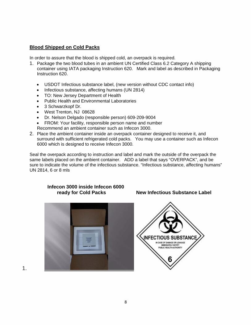
Blood Shipped on Cold Packs In order to assure that the blood is shipped cold, an overpack is required. 1. Package the two blood tubes in an ambient UN Certified Class 6.2 Category A shipping
container using IATA packaging Instruction 620. Mark and label as described in Packaging Instruction 620.
• USDOT Infectious substance label, (new version without CDC contact info) • Infectious substance, affecting humans (UN 2814) • TO: New Jersey Department of Health • Public Health and Environmental Laboratories • 3 Schwarzkopf Dr. • West Trenton, NJ 08628 • Dr. Nelson Delgado (responsible person) 609-209-9004 • FROM: Your facility, responsible person name and number
Recommend an ambient container such as Infecon 3000. 2. Place the ambient container inside an overpack container designed to receive it, and
surround with sufficient refrigerated cold packs. You may use a container such as Infecon 6000 which is designed to receive Infecon 3000.
Seal the overpack according to instruction and label and mark the outside of the overpack the same labels placed on the ambient container. ADD a label that says “OVERPACK”, and be sure to indicate the volume of the infectious substance. “Infectious substance, affecting humans” UN 2814, 6 or 8 mls Infecon 3000 inside Infecon 6000 ready for Cold Packs New Infectious Substance Label
1.

9
Shippers Declaration The most important section of the Shippers Declaration is the section which describes the Nature and Quantity of Dangerous Goods being transported. Please make sure the following information is included: UN2814, Infectious substance, affecting humans (Suspected Ebola virus), Class 6.2, 6 or 8 ml all packed in one fibreboard box, Overpack Used, Packaging Instruction 620 Prepare the shippers declaration in triplicate. This is the ONLY place the technical name, Suspected Ebola virus, should appear. Supporting information Information in support of these recommendations is provided below.
Recent experiments in Canada have demonstrated the absence of airborne Ebola transmission in non-human primate experiments2. An investigation of 173 contacts in 27 households demonstrated Ebola transmission only to those with direct physical contact or exposure to body fluids of the ill household member, and no transmission to the 78 household members who had no physical contact with the ill person3. An investigation of three generations of Ebola transmission during an outbreak in Uganda, demonstrated direct contact with patient body fluids as the strongest risk factor for transmission, with contaminated fomites as a possible lesser risk factor4. Several patients with viral hemorrhagic fever (VHF) have been cared for prior to being recognized as having VHFs in US and Western European medical facilities during the last several years. Although subsequently diagnosed as Lassa or Marburg fever, extensive follow-up of hundreds of potentially exposed healthcare workers including laboratory personnel, have found no instances of transmission of infection5,6,7,8. In 1996, a physician who had been working in West Africa and an anesthetics assistant previously involved in his care, became severely ill in Johannesburg, South Africa. Despite hospitalization for more than a week before being diagnosed with Ebola, and the performance of some potentially high risk medical procedures, none of the more than 300 exposed health -care workers, including laboratory personnel, contracted the virus9. Lassa fever was detected in March/April 2014 in a patient in Minnesota with renal failure. The possibility of a VHF was not initially recognized and numerous health care workers including laboratory personnel were potentially exposed. However, there were no cases of disease transmission10. Guidance documents from the UK note that one to two patients per year are diagnosed there with VHFs11. Some are not initially recognized as having VHF and are managed with standard precautions, yet there have been no reports of transmissions to healthcare workers. While VHF refers to a list of agents, not Ebola specifically; all are considered pathogens of “high

10
consequence”. Reports in the literature of laboratory-acquired Ebola infections refer to events prior to the implementation of universal precautions and the availability of relevant safety devices su ch as retractable needles12 or to infections acquired during the performance of animal necropsy and other animal experiments13. On average, routine laboratory testing is performed on a few patients per year collectively at healthcare facilities in the UK, US and Europe. In some cases dozens of samples per case are processed and tested before the patient is diagnosed with VHF. Therefore collectively in these countries since the implementation of universal precautions approximately 30 years ago, it would appear that hundreds of samples have been tested in laboratories using these procedures routinely, with no documented transmission to laboratory workers. To assist with the current outbreak in West Africa, laboratory personnel have been deployed to the European field laboratory in Guinea since mid-March, the Canadian field laboratory since June, and the two CDC laboratories since early August. Additionally, three other field laboratories set up by international partner groups are operational there. These laboratories process 200-300 specimens per day, yet there have been no documented cases of Ebola transmission to any of the laboratory scientists working at them. Earlier in the outbreak, some local West African laboratory personnel who were not wearing appropriate PPE and were performing procedures such as blood smear preparations without gloves, did acquire EVD. However, this has not occurred in any personnel wearing correct PPE and adhering to recommended procedures.
This document has been adapted from a NYSDOH/NYCDHMH Guidance document which was prepared in consultation with more than 40 microbiology, clinical chemistry, and hematology laboratory directors, infectious disease clinicians, epidemiologists, and scientific specialists in VHF at the CDC. The NJDOH would like to thank the NYSDOH and NYCDHMH for sharing most of the information contained in this document.
11
References:
1. Bausch DG, Towner JS Dowell SF, et (2007): Assessment of the risk of Ebola virus transmission from bodily fluids and fomites. J Infect Dis 196 Suppl 2: S142-7. 2. Alimonti J, Leung A, Jones S et al (2014): Evaluation of transmission risks associated with in vivo replication of several high containment pathogens in a biosafety level 4 laboratory. Sci Rep :5824. doi:10.1038/srep05824. 3. Dowell SF, Mukunu R, Ksiazek TG, et al (1999): Transmission of ebola hemorrhagic fever: a study of risk factors in family members, Kikwit, Democratic Republic of Congo, 1995. J Infect Dis 179 (Suppl 1): S87-91. 4. Francesconi P, Yoti Z, Declich S, et al (2003): Ebola Hemorrhagic Fever Transmission and Risk
Factors of Contacts, Uganda. Emerg Infect Dis 9:1430-37 5. Centers for Disease Control and Prevention (CDC) et al (2004): Imported Lassa Fever – New
Jersey, 2004. MMWR Oct 1, 53(38): 894-97. 6. Centers for Disease Control and Prevention (CDC) et al (2009): Imported case of Marburg
Hemorrhagic Fever – Colorado, 2008. MMWR Dec 18, 58(49): 1377-81 7. Timen V, MacNeil A, McConnell R et al (2009): Response to imported case of Marburg
Hemorrhagic Fever, The Netherlands. Emerg Infect Dis 15: 1171-75. 8. Timen A, Coopmans MPG, Vossen ACTM et al (2009): Response to imported case of Marburg
Hemorrhagic Fever, the Netherlands. Emerg Infect Dis 15: 1171-75.
9. Amorosa V, MacNeil a, McConnell R et al (2010): Imported Lassa Fever, Pennsylvania, USA, 2010. Emerg Infect Dis 16: 1598-1600. 10. Richards G, Murphy S, Jobson R, et al (2000): Unexpected Ebola virus in a tertiary setting:
Clinical and epidemiologic aspects. Crit Care Med 28:240-44
11. Personal communication, Aaron Devies, Minnesota Department of Health. 12. UK Department of Health, Advisory Committee on dangerous pathogens, Management of Hazard Group 4 viral hemorrhagic fevers and similar human infectious diseases of high consequence. Appendix 7: Laboratory Procedures. 13. Emond RTD, Evans B, Bowen ETW and Lloyd G (1977): A case of Ebola virus infection. BMJ 2: 541-44. 14. Formenty P, Hatz C, Le Guenno B, et al (1999): Human Infection Due to Ebola Virus, Subtype Côte d’Ivoire: Clinical and Biologic Presentation. J Infect Dis 179(Suppl 1): S48-53. 15. Rollin et al. 2011. Arenaviruses and Filoviruses. In: Manual of Clinical Microbiology. (10thed). ASM Press.

12
16. Hersberger M, Nushbaumer C, Scholer A et al (2004): Influence of Practicable Virus Inactivation Procedures on tests for frequently Measuered Analytes in Plasma Clin Chem 50: 944-46 17. Loutfy MR, Assmar M. Burgess DCH, and Kain KC (1998): Effects of Viral Hemorrhagic Fever Inactivation Methods on the Performance of Rapid Diagnostic Tests for Plasmodium falciparum. J. Infect Dis 178: 1852-55
a Suspected cases who meet the CDC criteria for Persons Under Investigation include i) travel within 21 days before illness onset to an EVD outbreak affected area (See http://www.cdc.gov/vhf/ebola/resources/distribution-map-guinea- outbreak.html#areas for the current list of affected areas; ii) fever (> 38.6 oC or 101.5 oF); and iii) compatible symptoms for EVD (e.g., severe headache, myalgia, vomiting, diarrhea, abdominal pain or unexplained hemorrhage).
High risk exposure is defined as either i) percutaneous, mucous membrane or direct skin contact with blood or body fluid from a confirmed or suspected EVD patient without appropriate personal protective equipment (PPE); ii) laboratory handling of body fluids from a confirmed or suspected EVD patient without appropriate PPE or biosafety precautions, or iii) participation in funeral rites which include direct exposures to human remains in the geographic area where outbreak is occurring without appropriate PPE.
Low risk exposures are defined as i) healthcare workers in facilities that have treated confirmed or suspected EVD patients or ii) household members or others with direct contact with a confirmed or suspected EVD patient.
b No known exposures are defined as residence or travel to an EVD affected area without either High or Low risk exposures.