pseudotumors in association with well-functioning metal...
TRANSCRIPT
Pseudotumors in Association with Well-FunctioningMetal-on-Metal Hip Prostheses
A Case-Control Study Using Three-Dimensional Computed Tomographyand Magnetic Resonance Imaging
Alister J. Hart, MA, MD, FRCSG(Orth), Keshthra Satchithananda, BDS, FDSRCS, MBBS, FRCS, FRCR,Alexander D. Liddle, MBBS, MRCS, Shiraz A. Sabah, MBBS, BSc, Donald McRobbie, PhD,
Johann Henckel, MBBS, MRCS, Justin P. Cobb, BMBCh, FRCS, MCh,John A. Skinner, FRCS(Orth), and Adam W. Mitchell, MBBS, FRCS, FRCR
Investigation performed at Charing Cross Hospital and Imperial College, London, United Kingdom
Introduction: Many papers have been published recently on the subject of pseudotumors surrounding metal-on-metalhip resurfacing and replacement prostheses. These pseudotumors are sterile, inflammatory lesions within the peri-prosthetic tissues and have been variously termed masses, cysts, bursae, collections, or aseptic lymphocyte-dominatedvasculitis-associated lesions (ALVAL). The prevalence of pseudotumors in patients with a well-functioning metal-on-metalhip prosthesis is not well known. The purpose of this study was to quantify the prevalence of pseudotumors adjacent towell-functioning and painful metal-on-metal hip prostheses, to characterize these lesions with use of magnetic resonanceimaging, and to assess the relationship between their presence and acetabular cup position with use of three-dimensionalcomputed tomography.
Methods: We performed a case-control study to compare the magnetic resonance imaging findings of patients with awell-functioning unilateral metal-on-metal hip prosthesis and patients with a painful prosthesis (defined by either revisionarthroplasty performed because of unexplained pain or an Oxford hip score of <30 of 48 possible points). Thirty patientswith a painful hip prosthesis and twenty-eight controls with a well-functioning prosthesis were recruited consecutively. Allpatients also underwent computed tomography to assess the position of the acetabular component.
Results: Thirty-four patients were diagnosed with a pseudotumor. However, the prevalence of pseudotumors in patientswith a painful hip (seventeen of thirty, 57%) was not significantly different from the prevalence in the control group (seventeenof twenty-eight, 61%). No objective differences in pseudotumor characteristics between the groups were identified. No clearassociation between the presence of a pseudotumor and acetabular component position was identified. The Oxford hipscore in the group with a painful hip (mean, 20.2; 95% confidence interval [CI], 12.7 to 45.8) was poorer than that in thecontrol group (mean, 41.2; 95% CI, 18.5 to 45.8; p £ 0.0001).
Conclusions: A periprosthetic cystic pseudotumor was diagnosed commonly (in thirty-four [59%] of the entire study cohort)with use of metal artifact reduction sequence (MARS) magnetic resonance imaging in this series of patients with a metal-on-metal hip prosthesis. The prevalence of pseudotumors was similar in patients with a well-functioning hip prosthesis and
continued
Disclosure: None of the authors received payments or services, eitherdirectly or indirectly (i.e., via his or her institution), from a third party insupport of any aspect of this work. One or more of the authors, or his orher institution, has had a financial relationship, in the thirty-six monthsprior to submission of this work, with an entity in the biomedical arenathat could be perceived to influence or have the potential to influencewhat is written in this work. No author has had any other relationships,or has engaged in any other activities, that could be perceived to in-fluence or have the potential to influence what is written in this work.The complete Disclosures of Potential Conflicts of Interest submittedby authors are always provided with the online version of the article.
A commentary by Joshua J. Jacobs, MD, islinked to the online version of thisarticle at jbjs.org.
317
COPYRIGHT � 2012 BY THE JOURNAL OF BONE AND JOINT SURGERY, INCORPORATED
J Bone Joint Surg Am. 2012;94:317-25 d http://dx.doi.org/10.2106/JBJS.J.01508
patients with a painful hip. Pseudotumors were also diagnosed commonly in patients with a well-positioned acetabularcomponent. Although magnetic resonance imaging is useful for surgical planning, the presence of a cystic pseudotumor maynot necessarily indicate the need for revision arthroplasty. Further correlation of clinical and imaging data is needed todetermine the natural history of pseudotumors to guide clinical practice.
Level of Evidence: Therapeutic Level III. See Instructions for Authors for a complete description of levels of evidence.
Many papers have been published recently on the subjectof pseudotumors surrounding metal-on-metal hip re-surfacing and replacement prostheses1-7. These pseu-
dotumors have been variously termed masses, cysts, bursae,collections, or aseptic lymphocyte-dominated vasculitis-associatedlesions (ALVAL)1. The identification of these lesions has con-tributed to decision by the Medicines and Healthcare productsRegulatory Agency (MHRA) in the United Kingdom to publish asafety alert for all metal-on-metal hip replacements8. The rec-ommendations included the use of cross-sectional imaging withuse of metal artifact reduction sequence (MARS) magnetic res-onance imaging (MRI), computed tomography (CT), or ultra-sonography to detect pseudotumors, which are thought torepresent an adverse reaction to wear debris from metal-on-metal hip prostheses7.
MRI is an excellent imaging modality for assessing softtissues, the bone cortex, and the underlying bone marrow, butit is impaired by large magnetic-susceptibility artifacts frommetal-on-metal hip prostheses9. MRI sequences that reducethe artifacts resulting from metallic prostheses use a variety oftechniques including increasing the imaging bandwidth andemploying viewing-angle tilting10-25. MARS MRI is a recentlydeveloped technique that provides good metal-artifact sup-pression while minimizing image blurring and scanningtime12-14.
A recent study used ultrasonography to screen asymp-tomatic patients with a metal-on-metal hip prosthesis forpseudotumors and estimated the prevalence to be 6.5%26. Thesensitivity of ultrasonography for the detection of pseudotu-mors is not known, but it is likely that small, deep pseudotu-mors may be more reliably diagnosed with use of MARS MRI.
We are not aware of any studies that used MRI to quantify theprevalence of pseudotumors in patients with a well-functioningmetal-on-metal hip prosthesis or to compare the prevalencewith that in patients with a painful hip prosthesis. The pur-pose of this study was to perform a case-control study tocompare the prevalence and characteristics of pseudotumorsin patients with painful and well-functioning metal-on-metal hip prostheses. In addition, the relationship betweenthe presence of pseudotumors and acetabular cup positionwas characterized with use of three-dimensional computedtomography.
Materials and MethodsPower Analysis
Amedical statistician performed a two-proportion power analysis to deter-mine the sample size necessary to provide 90% power when a p value of
0.05 was considered significant. Assuming a pseudotumor prevalence of 60% inpatients with a painful metal-on-metal hip replacement
27and 4% in patients
with a well-functioning replacement26
, the minimum sample size of each groupwas calculated to be thirteen.
PatientsAll patients had a unilateral metal-on-metal hip prosthesis and were imagedprospectively. The case group was defined as patients with either unexplainedhip pain sufficient to result in revision or an Oxford hip score of <30 of apossible 48 points. Unexplained hip pain was defined as pain with an etiologythat remained unclear after assessment of the hip by means of the clinicalhistory, physical examination, laboratory blood tests for markers of infection,and serial radiographs. The control group was defined as patients who did notvolunteer pain as a symptom during the assessment and were satisfied with theresults of the arthroplasty.
Forty potential control patients were selected from among the pa-tients receiving routine follow-up at our arthroplasty clinic. After excludingpatients who did not meet the inclusion criteria, were lost to follow-up, or
TABLE I Method of Classification of Lesions with Use of MARS MRI*
Pseudotumor Type Wall Contents Shape
1 Thin-walled Fluid-like: hypointense on T1,hyperintense on T2
Flat, with walls mainlyin apposition
2a Thick-walled or irregular Fluid-like: hypointense on T1,hyperintense on T2
Not flat, with >50% ofthe walls not in apposition
2b Thick-walled or irregular Atypical fluid: hyperintense on T1,variable on T2
Any shape
3 Solid throughout Mixed signal Any shape
*MARS MRI = metal artifact reduction sequence magnetic resonance imaging.
318
TH E J O U R N A L O F B O N E & JO I N T SU R G E RY d J B J S . O R G
VO LU M E 94-A d NU M B E R 4 d F E B R UA RY 15, 2012PS E U D O T U M O R S I N AS S O C I AT I O N W I T H WE L L-FU N C T I O N I N G
ME TA L-O N -ME TA L HI P P R O S T H E S E S

were unwilling to take part in the study, twenty-eight suitable patients wereincluded in the control group. Potential patients in the case group wererecruited from either our follow-up clinic or tertiary referrals to our center.
The first thirty patients in this group to undergo CTand MRI formed the casegroup. We were blinded to the results of the clinical investigations prior topatient recruitment. No scans were repeated.
TABLE II Comparison of the Patient Groups
VariableWell-FunctioningGroup (N = 28)
Painful Group(N = 30) P Value
Type of implant (no.)
ADEPT 1 2ASR 0 5Biomet 0 1BHR 12 9Cormet 15 7Durom 0 6
Oxford hip score* 46 (41.5 to 48) 20 (10-30) <0.001
Cup inclination angle*† (deg) 45 (38-51) 44 (36-52) 0.869
Cup version angle*† (deg) 20 (12-26) 15 (8-21) 0.238
*The values are given as the median, with the interquartile range in parentheses. †Measured with use of three-dimensional computedtomography.
TABLE III Logistic Regression Analysis
Predictor Painful GroupWell-Functioning
GroupAdjusted Odds
Ratio95% Confidence
Interval P Value
Presence of pseudotumor 17 patients (57%) 17 patients (61%) 1.392 0.420 to 4.608 0.589
Age at primary arthroplasty 50 yr (42-59 yr)* 57 (51-64)* 1.054 per year 0.992 to 1.121 0.091
Time since primaryarthroplasty
31 mo (22-41 mo)* 47 (29-62)* 1.032 per month 1.000 to 1.065 0.051
*The values are given as the median, with the interquartile range in parentheses.
Fig. 1-A Fig. 1-B
Magnetic resonance images of a type-1 pseudotumor (arrows; see Table I for details) adjacent to a metal-on-metal hip prosthesis that was well functioning
(Fig. 1-A) and to one that was painful (Fig. 1-B).
319
TH E J O U R N A L O F B O N E & JO I N T SU R G E RY d J B J S . O R G
VO LU M E 94-A d NU M B E R 4 d F E B R UA RY 15, 2012PS E U D O T U M O R S I N AS S O C I AT I O N W I T H WE L L-FU N C T I O N I N G
ME TA L-O N -ME TA L HI P P R O S T H E S E S
Data CollectionHip function was assessed in each patient with use of the Oxford hip score,which is a validated, patient-completed questionnaire
28. After providing
informed consent, each patient underwent MARS MRI of the hip with useof a 1.5-T scanner (MAGNETOM 1.5T; Siemens Medical, Erlangen, Ger-many). MRI scans were interpreted by consensus agreement by two ex-perienced musculoskeletal radiologists who were blinded to the clinicaldetails. The presence or absence of periprosthetic soft tissue reactions ormasses was recorded; if a lesion was present, it was categorized as discussedbelow.
MRI Characterization of LesionsTable I summarizes the method of characterization of pseudotumors foundon MRI scans. We characterized the wall, shape, and contents. A pseudo-tumor with a wall thickness of £2 mm was classified as ‘‘thin-walled.’’ Acrude estimate of the volume of the lesion was made by approximation toa cuboid, using the maximal anterior-posterior, superior-inferior, andmedial-lateral diameters. The contents were classified according to the signal
intensity on T1-weighted and T2-weighted images, as in most MRI classifi-cation systems. The pseudotumor classification differentiates simple (type 1)from complex (type 2) fluid collections and those with a solid component(type 3).
CT Scanning of the HipCT images were reconstructed in three dimensions, and anatomical acetabularinclination and version were defined with reference to the anterior pelvicplane
29. These angles were converted to the radiographic equivalents with use of
accepted formulas30
.
HistologyHip neocapsule specimens were collected from patients in the case group whowere undergoing revision hip arthroplasty. The articular surface was markedwith a suture, and the nonarticular surface was marked circumferentially with athin line of permanent ink to permit orientation of the specimen under lightmicroscopy. Representative samples were selected and processed in paraffinwax. Four-micrometer-thick sections were cut and stained with hematoxylin
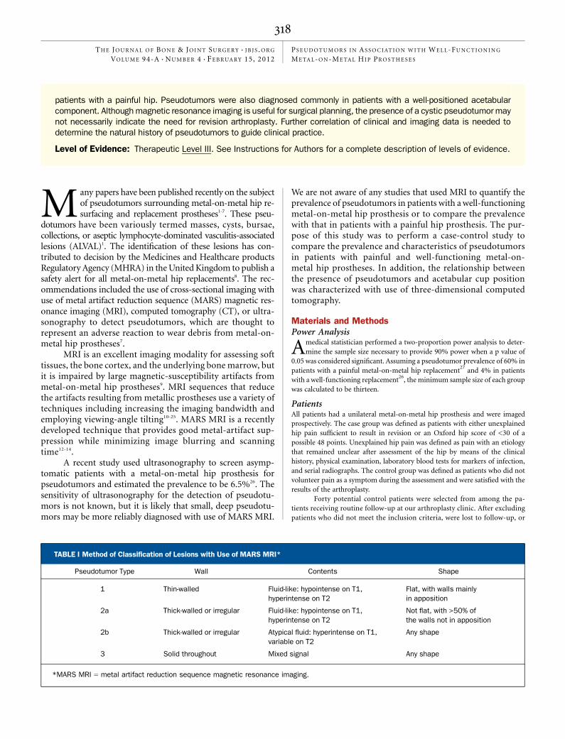
Fig. 2-A Fig. 2-B
Magnetic resonance images of a type-2a pseudotumor (arrow; see Table I for details) adjacent to a metal-on-metal hip prosthesis that was well functioning
(Fig. 2-A) and to one that was painful (Fig. 2-B).
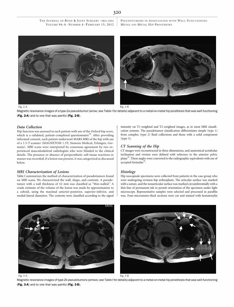
Fig. 3-A Fig. 3-B
Magnetic resonance images of type-2b pseudotumors (arrows; see Table I for details) adjacent to a metal-on-metal hip prosthesis that was well functioning
(Fig. 3-A) and to one that was painful (Fig. 3-B).
320
TH E J O U R N A L O F B O N E & JO I N T SU R G E RY d J B J S . O R G
VO LU M E 94-A d NU M B E R 4 d F E B R UA RY 15, 2012PS E U D O T U M O R S I N AS S O C I AT I O N W I T H WE L L-FU N C T I O N I N G
ME TA L-O N -ME TA L HI P P R O S T H E S E S
and eosin. Slides were analyzed according to the criteria established by Willertet al. for the diagnosis of ALVAL
31. The presence of all three findings of surface
necrosis, subsurface macrophage infiltrate, and perivascular lymphocyte infil-trate was defined as diagnostic for ALVAL. The presence of two of these three
findings was defined as ‘‘suggestive’’ for the presence of ALVAL. The presence ofone or none of the findings was defined as ‘‘nondiagnostic’’ for ALVAL. Allsamples were further analyzed by a consultant histopathologist for the presenceof neutrophils and other features suggestive of infection. Tissue samples weresent for microbiological analysis.
Statistical AnalysisThe difference in pseudotumor prevalence between the groups was analyzedwith use of logistic regression, adjusting for the known risk factors of sex, age
1,
and time since the arthroplasty. The difference in Oxford hip score between thegroups was tested with use of the Wilcoxon rank-sum test. A p value of 0.05(two-sided) was considered significant.
Source of FundingNo external funding was received for this study.
Results
The case group consisted of thirty patients with a painful hip(median age, fifty-five years; interquartile range, forty-six
to sixty-four years). The control group consisted of twenty-eight patients with a well-functioning hip (median age, sixty-four years; interquartile range, fifty-four to sixty-nine years).Thirty-four of the patients were men and twenty-four werewomen. The male:female ratio was 16:14 in the case group and18:10 in the control group; the difference between groups was
Fig. 4
Magnetic resonance images of a type-3 pseudotumor (arrows; see Table I
for details) adjacent to a painful metal-on-metal hip prosthesis.
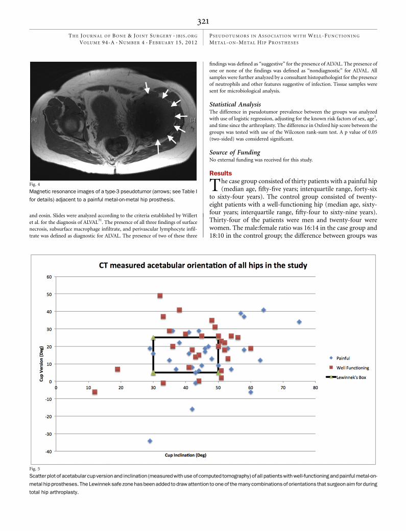
Fig. 5
Scatter plot of acetabular cup version and inclination (measured with use of computed tomography) of all patients with well-functioning and painful metal-on-
metal hip prostheses. The Lewinneksafe zone has been added to draw attention to one of the many combinations of orientations that surgeon aim for during
total hip arthroplasty.
321
TH E J O U R N A L O F B O N E & JO I N T SU R G E RY d J B J S . O R G
VO LU M E 94-A d NU M B E R 4 d F E B R UA RY 15, 2012PS E U D O T U M O R S I N AS S O C I AT I O N W I T H WE L L-FU N C T I O N I N G
ME TA L-O N -ME TA L HI P P R O S T H E S E S

not significant (chi-square = 0.791 with 1 degree of freedom,p = 0.397).
Additional characteristics of the patients in the twogroups are shown in Table II. The mean Oxford hip score was20.2 (95% confidence interval, 12.7 to 45.8) in the case groupand 41.2 (95% confidence interval, 18.5 to 45.8) in the controlgroup (p £ 0.0001). The functional score in the case group wassignificantly poorer than that in the control group (Mann-Whitney U = 773.5 with n1 = 30 and n2 = 28, p < 0.0001).
Logistic Regression ModelA logistic regression model to detect differences between thegroups with painful and well-functioning hip prostheses wasconstructed as described. Sex was removed from the finalmodel because of lack of significance (p > 0.1). The resultsfrom this model are shown in Table III. The groups did notdiffer significantly with regard to the prevalence of pseudotu-mors, age at the time of the primary arthroplasty, or time sincethe arthroplasty.
MRIPseudotumors were identified in thirty-four patients with useof MRI. A pseudotumor was diagnosed in seventeen (57%) ofthe thirty patients with a painful hip (the case group) and inseventeen (61%) of the twenty-eight asymptomatic patients
(the control group). The fluid-filled pseudotumors in the casegroup had similar characteristics to those in the control group(see Appendix for classification). Eight of the fourteen womenand nine of the sixteen men in the case group had a pseudo-tumor, compared with four of the ten women and thirteen ofthe eighteen men in the control group.
Examples of periprosthetic pseudotumors of types 1, 2a,and 2b in well-functioning and painful hips are shown in Figures1, 2, and 3, respectively. One solid pseudotumor was identified ina patient in the case group (Fig. 4). Pseudotumors were presentin patients with the following implants: ADEPT (Finsbury Or-thopaedics, Surrey, United Kingdom), ASR (Anatomic SurfaceReplacement; DePuy Johnson & Johnson, Warsaw, Indiana),BHR (BIRMINGHAM HIP Resurfacing System; Smith &Nephew, Memphis, Tennessee), Cormet (Corin, Cirencester,United Kingdom), and Durom (Zimmer, Warsaw, Indiana).The median pseudotumor volume was 25.1 cm3 (range, 0.9 to594.0 cm3). The pseudotumor volume was significantly largerin the patients with a painful hip (median, 79.2 cm3; range, 5.6to 594.0 cm3) than in the patients with a well-functioning hip(median, 15.7 cm3; range, 0.9 to 75.6 cm3; p = 0.016).
Cup Orientation Measured with Use of CTFigure 5 shows that the acetabular inclination and version anglesvaried widely among the patients in each group. Fourteen (47%)
Fig. 6
Scatter plot of acetabular cup versionand inclination (measured with use of computed tomography) of those patientswith well-functioning and painful metal-
on-metal hip prostheses who had a pseudotumor diagnosed on MRI. The Lewinnek safe zone has been added.
322
TH E J O U R N A L O F B O N E & JO I N T SU R G E RY d J B J S . O R G
VO LU M E 94-A d NU M B E R 4 d F E B R UA RY 15, 2012PS E U D O T U M O R S I N AS S O C I AT I O N W I T H WE L L-FU N C T I O N I N G
ME TA L-O N -ME TA L HI P P R O S T H E S E S

of the thirty hips in the painful group and six (21%) of thetwenty-eight hips in the well-functioning group were within theso-called ‘‘safe zone’’ described by Lewinnek et al.32. However, agreater number of hips in the painful group were furthest fromthe Lewinnek safe zone; for instance, all three acetabular cupsthat had an inclination angle of >60� and all three cups that weremore retroverted than 25� were in the painful group. Figure 6shows the cup orientation of only the patients with a pseudo-tumor; the acetabular cup was positioned within the Lewinneksafe zone in nine of the seventeen patients in the case group andin five of the seventeen patients in the control group.
HistologyEighteen patients in the painful group underwent revisionarthroplasty, and all had negative microbiological cultureresults. Samples from thirteen of these patients, includingsix patients with a visible pseudotumor on MARS MRI, wereavailable for histological analysis. Eight of the sampleswere ‘‘diagnostic’’ for ALVAL, one was ‘‘suggestive,’’ threewere ‘‘nondiagnostic,’’ and the remaining sample was entirelynecrotic and was not analyzed further. Five of the six histo-logical samples from patients with a pseudotumor were‘‘diagnostic’’ for ALVAL and one was ‘‘nondiagnostic.’’ Noneof the patients in the control group underwent revisionarthroplasty.
Discussion
To our knowledge, this is the first study to compare MRIfindings between patients with well-functioning and
painful metal-on-metal hip prostheses. The 61% pseudotumorprevalence in the well-functioning (control) group was unex-pectedly high, and both this prevalence and the characteristicsof the pseudotumors were similar to those in the painful (case)group. This high prevalence is probably due to use of the termpseudotumor for a spectrum of lesions—ranging from small,fluid-filled cysts (Fig. 1) to large, complex, and destructive le-sions with solid components (Fig. 4)—surrounding metal-on-metal hip prostheses7. Prior to this study, we had beenconcerned by the presence of any cystic mass that was visibleadjacent to a metal-on-metal hip prosthesis on MARS MRIbecause previous researchers had included cystic masses intheir definition of pseudotumors and had reported a poorclinical outcome associated with the presence of pseudotu-mors2. As a result of our study, we now place less clinical im-portance on the presence of a fluid-filled lesion visible onMARS MRI. However, we recognize the variability in proposeddefinitions of the term pseudotumor, and we recommend thatthe natural history and longitudinal imaging findings of theselesions be more fully analyzed.
We continue to use MRI in diagnosing the cause of apainful metal-on-metal hip arthroplasty and also find it usefulwhen planning revision arthroplasty (for instance, to deter-mine the extent of intrapelvic debridement required and toavoid neurological structures during the debridement). Weremain concerned about the solid pseudotumors (Fig. 4), butwe are seeking an alternative term, or a more restrictive use of
the term pseudotumor, to describe the spectrum of fluid-filledlesions that is observed. We suggest that surgeons and radiol-ogists consider the status of the tissues surrounding the fluidlesion—specifically, the destruction of adjacent muscle andother soft tissue.
The high prevalence of fluid-filled lesions in both groupsmay simply reflect the fact that the capsulotomy required duringimplantation of a hip prosthesis results in potential points ofweakness within the capsule27. A complete capsulotomy is rou-tinely performed to enable acetabular exposure during hip re-surfacing, in which the femoral head is retained, whereas a partialcapsulotomy is sufficient during a conventional hip arthroplasty,in which the femoral head is removed. The location of the fluidcollections may be related to pathways of low resistance createdby the capsulotomy, rather than to the surgical approach, whichwould explain the occurrence of collections in the iliopsoas.
A small number of fluid collections had atypical fluidsignal characteristics in the core of the lesion, with a highsignal on both T1 and T2-weighted images (Fig. 3). It is notclear whether this is due to the presence of proteinaceousmaterial or a high concentration of metal ions. These fluidcollections may represent a more clinically problematic groupof lesions, although there was no difference in their preva-lence between the two groups in this study. Therefore, al-though the classification of lesions into fluid-filled and solidtypes appears to be clinically relevant, we were unable todemonstrate the clinical importance of differences among thefluid-filled lesions.
This study also examined the histology of the lesions ina small number of patients who underwent revision arthro-plasty. Consistent with the findings reported by other groups5,7,we noted that five of six patients with a pseudotumor hadhistological results that were diagnostic for ALVAL. However,it was also evident that the remaining patient with a pseu-dotumor did not have features of ALVAL, and three patientswithout a pseudotumor had histological results that werediagnostic for ALVAL. We therefore caution that ALVAL-typehistology was not pathognomonic for the presence of apseudotumor, and we encourage future work to determinewhether an immunological mechanism is responsible forthese lesions.
We also investigated the relationship between acetabularcomponent position, hip function, and pseudotumor occur-rence. Unsurprisingly, we found that extremes of malposi-tioning were associated with poor hip function. However, wealso noted that 41% (fourteen) of the thirty-four pseudotumoroccurrences were associated with components positionedwithin the Lewinnek safe zone.
Other Studies Involving MARS MRI in Patients withMetal-on-Metal Hip ProsthesesA number of series have provided details of MARS MRI findingsin a smaller number of patients1,7,27,33,34. One previous study in-dicated that women were at greater risk of developing a pseu-dotumor than men were1. However, we did not find evidence tosupport this in our study. In our population, pseudotumors were
323
TH E J O U R N A L O F B O N E & JO I N T SU R G E RY d J B J S . O R G
VO LU M E 94-A d NU M B E R 4 d F E B R UA RY 15, 2012PS E U D O T U M O R S I N AS S O C I AT I O N W I T H WE L L-FU N C T I O N I N G
ME TA L-O N -ME TA L HI P P R O S T H E S E S

more common in male patients, but this difference was notsignificant. This finding is consistent with the similar pseudo-tumor prevalence in men and women in other reports27,33,34.
LimitationsOne limitation of our study is the inability to accuratelyclassify patients as having either a well-functioning or a painfulhip on the basis of the Oxford hip score, even though theOxford hip scores differed substantially and significantly be-tween the groups. For instance, one of the patients in the well-functioning group had a relatively low Oxford hip score (31 ofa possible 48 points) but was satisfied with the results of thehip arthroplasty and regarded the procedure as successful. Insuch cases, other areas of the body (commonly the lumbarspine) may reduce the overall Oxford hip score. In contrast,one of the patients in the painful group was dissatisfied withthe results of the hip arthroplasty despite a high Oxford hipscore (43 of 48 points) because he was unable to sail and playtennis.
Another limitation is that our MRI and clinical assess-ments took place at a single follow-up time point. It is thereforepossible that the pseudotumors observed in the well-functioninggroup will develop into symptomatic lesions. Investigation ofthis possibility will require longitudinal study of these lesions tounderstand their natural history, but to our knowledge such astudy has not yet been reported. For instance, variation in thesize of the lesion over time may be relevant; if small lesions aredetectable on MRI but not on ultrasonography, this may explainthe lower prevalence (six [10%] of sixty-one women) in a re-cently reported study involving ultrasonographic screening26.
SummaryA periprosthetic cystic pseudotumor was commonly diag-nosed (in 59% of the entire study cohort) with use of MARSMRI in patients with a metal-on-metal hip prosthesis, and the
prevalence was similar regardless of whether the hip wasfunctioning well or poorly. A pseudotumor was also com-monly found in patients with a well-positioned acetabularcomponent. Although MARS MRI is useful for surgicalplanning, the presence of a fluid-filled periprosthetic lesion(pseudotumor) may not necessarily indicate the need forrevision arthroplasty. Further correlation of clinical and im-aging data is needed to identify the natural history of pseu-dotumors to guide clinical practice.
AppendixA table summarizing the MARS MRI findings in the pa-tient groups is available with the online version of this
article as a data supplement at jbjs.org. n
NOTE: The authors acknowledge Gwynneth Lloyd, Angus Lewis, and the patients who contributed tothis study.
Alister J. Hart, MA, MD, FRCSG(Orth)Keshthra Satchithananda, BDS, FDSRCS, MBBS, FRCS, FRCRAlexander D. Liddle, MBBS, MRCSShiraz A. Sabah, MBBS, BScDonald McRobbie, PhDJohann Henckel, MBBS, MRCSJustin P. Cobb, BMBCh, FRCS, MChAdam W. Mitchell, MBBS, FRCS, FRCRDepartments of Orthopaedic Surgery (A.J.H, A.D.L., S.A.S., J.H.,and J.P.C.) and Radiology (K.S., D.M., and A.W.M.),Charing Cross Hospital (Imperial College Healthcare NHS Trust andImperial College London), Fulham Palace Road,London W6 8RF, United Kingdom.E-mail address for A.J. Hart: [email protected]
John A. Skinner, FRCS(Orth)Royal National Orthopaedic Hospital (University College London),Brockley Hill, Middlesex, London HA7 4LP, United Kingdom
References
1. Glyn-Jones S, Pandit H, Kwon YM, Doll H, Gill HS, Murray DW. Risk factors forinflammatory pseudotumour formation following hip resurfacing. J Bone Joint SurgBr. 2009;91:1566-74.2. Grammatopolous G, Pandit H, Kwon YM, Gundle R, McLardy-Smith P, Beard DJ,Murray DW, Gill HS. Hip resurfacings revised for inflammatory pseudotumour have apoor outcome. J Bone Joint Surg Br. 2009;91:1019-24.3. Harvie P, Giele H, Fang C, Ansorge O, Ostlere S, Gibbons M, Whitwell D. Thetreatment of femoral neuropathy due to pseudotumour caused by metal-on-metalresurfacing arthroplasty. Hip Int 2008;18:313-20.4. Kwon YM, Glyn-Jones S, Simpson DJ, Kamali A, McLardy-Smith P, Gill HS, MurrayDW. Analysis of wear of retrieved metal-on-metal hip resurfacing implants reviseddue to pseudotumours. J Bone Joint Surg Br. 2010;92:356-61.5. Pandit H, Glyn-Jones S, McLardy-Smith P, Gundle R, Whitwell D, Gibbons CL,Ostlere S, Athanasou N, Gill HS, Murray DW. Pseudotumours associated with metal-on-metal hip resurfacings. J Bone Joint Surg Br. 2008;90:847-51.6. Park SJ, Lee HK, Yi BH, Cha JG, Kim HC, Lee KW, Kim ME, Kwon GW. Pseudo-tumour in the bladder as a complication of total hip replacement: ultrasonography,CT and MR findings. Br J Radiol 2007;80:e119-21.7. Pandit H, Vlychou M, Whitwell D, Crook D, Luqmani R, Ostlere S, Murray DW,Athanasou NA. Necrotic granulomatous pseudotumours in bilateral resurfacing hiparthoplasties: evidence for a type IV immune response. Virchows Arch.2008;453:529-34.8. Medicines and Healthcare products Regulatory Agency. Medical device alert: allmetal-on-metal (MoM) hip replacements (MDA/2010/033). 2010. http://www.
mhra.gov.uk/Publications/Safetywarnings/MedicalDeviceAlerts/CON079157. Ac-cessed 12 Nov 2011.9. Laakman RW, Kaufman B, Han JS, Nelson AD, Clampitt M, O’Block AM, Haaga JR,Alfidi RJ. MR imaging in patients with metallic implants. Radiology 1985;157:711-4.10. Bellon EM, Haacke EM, Coleman PE, Sacco DC, Steiger DA, Gangarosa RE. MRartifacts: a review. AJR Am J Roentgenol 1986;147:1271-81.11. Eustace S, Goldberg R, Williamson D, Melhem ER, Oladipo O, Yucel EK, Jara H.MR imaging of soft tissues adjacent to orthopaedic hardware: techniques to mini-mize susceptibility artefact. Clin Radiol 1997;52:589-94.12. Eustace S, Jara H, Goldberg R, Fenlon H, Mason M, Melhem ER, Yucel EK. Acomparison of conventional spin-echo and turbo spin-echo imaging of soft tissuesadjacent to orthopedic hardware. AJR Am J Roentgenol 1998;170:455-8.13. Farahani K, Sinha U, Sinha S, Chiu LC, Lufkin RB. Effect of field strength onsusceptibility artifacts in magnetic resonance imaging. Comput Med Imaging Graph1990;14:409-13.14. Petersilge CA, Lewin JS, Duerk JL, Yoo JU, Ghaneyem AJ. Optimizing imagingparameters for MR evaluation of the spine with titanium pedicle screws. AJR Am JRoentgenol 1996;166:1213-8.15. Rupp R, Ebraheim NA, Savolaine ER, Jackson WT. Magnetic resonance imagingevaluation of the spine with metal implants. General safety and superior imagingwith titanium. Spine (Phila Pa 1976) 1993;18:379-85.16. Suh JS, Jeong EK, Shin KH, Cho JH, Na JB, Kim DH, Han CD. Minimizing artifactscaused by metallic implants at MR imaging: experimental and clinical studies. AJRAm J Roentgenol 1998;171:1207-13.
324
TH E J O U R N A L O F B O N E & JO I N T SU R G E RY d J B J S . O R G
VO LU M E 94-A d NU M B E R 4 d F E B R UA RY 15, 2012PS E U D O T U M O R S I N AS S O C I AT I O N W I T H WE L L-FU N C T I O N I N G
ME TA L-O N -ME TA L HI P P R O S T H E S E S

17. Tartaglino LM, Flanders AE, Vinitski S, Friedman DP. Metallic artifacts on MRimages of the postoperative spine: reduction with fast spin-echo techniques. Radi-ology 1994;190:565-9.18. Tormanen J, Tervonen O, Koivula A, Junila J, Suramo I. Image technique opti-mization in MR imaging of a titanium alloy joint prosthesis. J Magn Reson Imaging1996;6:805-11.19. Cho ZH, Kim DJ, Kim YK. Total inhomogeneity correction including chemicalshifts and susceptibility by view angle tilting. Med Phys. 1988;15:7-11.20. Ebraheim NA, Savolaine ER, Zeiss J, Jackson WT. Titanium hip implants forimproved magnetic resonance and computed tomography examinations. Clin OrthopRelat Res. 1992;275:194-8.21. Frazzini VI, Kagetsu NJ, Johnson CE, Destian S. Internally stabilized spine:optimal choice of frequency-encoding gradient direction during MR imaging mini-mizes susceptibility artifact from titanium vertebral body screws. Radiology1997;204:268-72.22. Olsen RV, Munk PL, Lee MJ, Janzen DL, MacKay AL, Xiang QS, Masri B. Metalartifact reduction sequence: early clinical applications. Radiographics 2000;20:699-712.23. Toms AP, Smith-Bateman C, Malcolm PN, Cahir J, Graves M. Optimization ofmetal artefact reduction (MAR) sequences for MRI of total hip prostheses. ClinRadiol 2010;65:447-52.24. Viano AM, Gronemeyer SA, Haliloglu M, Hoffer FA. Improved MR imaging forpatients with metallic implants. Magn Reson Imaging 2000;18:287-95.25. Pellicci PM, Potter HG, Foo LF, Boettner F. MRI shows biologic restoration ofposterior soft tissue repairs after THA. Clin Orthop Relat Res. 2009;467:940-5.
26. Kwon YM, Ostlere SJ, McLardy-Smith P, Athanasou NA, Gill HS, Murray DW.Asymptomatic" pseudotumors after metal-on-metal hip resurfacing arthroplasty:prevalence and metal ion study. J Arthroplasty 2011;26:511-8.27. Sabah SA, Mitchell AW, Henckel J, Sandison A, Skinner JA, Hart AJ. Magneticresonance imaging findings in painful metal-on-metal hips: a prospective study.J Arthroplasty 2011;26:71-6, 76.e1-2.28. Dawson J, Fitzpatrick R, Carr A, Murray D. Questionnaire on the perceptions ofpatients about total hip replacement. J Bone Joint Surg Br. 1996;78:185-90.29. Dandachli W, Nakhla A, Iranpour F, Kannan V, Cobb JP. Can the acetabularposition be derived from a pelvic frame of reference? Clin Orthop Relat Res.2009;467:886-93.30. Murray DW. The definition and measurement of acetabular orientation. J BoneJoint Surg Br. 1993;75:228-32.31. Willert HG, Buchhorn GH, Fayyazi A, Flury R, Windler M, Koster G, LohmannCH. Metal-on-metal bearings and hypersensitivity in patients with artificial hipjoints. A clinical and histomorphological study. J Bone Joint Surg Am. 2005;87:28-36.32. Lewinnek GE, Lewis JL, Tarr R, Compere CL, Zimmerman JR. Dislocations aftertotal hip-replacement arthroplasties. J Bone Joint Surg Am. 1978;60:217-20.33. Toms AP, Marshall TJ, Cahir J, Darrah C, Nolan J, Donell ST, Barker T, Tucker JK.MRI of early symptomatic metal-on-metal total hip arthroplasty: a retrospective re-view of radiological findings in 20 hips. Clin Radiol 2008;63:49-58.34. Hart AJ, Sabah S, Henckel J, Lewis A, Cobb J, Sampson B, Mitchell A, SkinnerJA. The painful metal-on-metal hip resurfacing. J Bone Joint Surg Br. 2009;91:738-44.
325
TH E J O U R N A L O F B O N E & JO I N T SU R G E RY d J B J S . O R G
VO LU M E 94-A d NU M B E R 4 d F E B R UA RY 15, 2012PS E U D O T U M O R S I N AS S O C I AT I O N W I T H WE L L-FU N C T I O N I N G
ME TA L-O N -ME TA L HI P P R O S T H E S E S