protocols - instituto estadual de hematologiahemorio.rj.gov.br/html/pdf/47043_1.pdf · protocols...
TRANSCRIPT


PROTOCOLS INSTITUTE OF HAEMATOLOGY
HEMORIO
This brochure is the Second Edition of the Book on Treatment Protocols adopted by the Technical Hematology Coordination and the First Edition of the Technical Hemotherapy Coordination of the Arthur de Siqueira Cavalcanti State Institute of Hematology – HEMORIO
MEDICAL DIRECTOR Revision - August 2009

COVER / ILLUSTRATION Marcos Monteiro
PICTURE
Marcos Araújo
PRINTING AND DIAGRAMMING Expresso Gráfica

COORDINATION AND REVISION Clarisse Lobo Claudia Máximo Maria Esther Duarte Vera Marra TECHNICAL TEAM Ana Claudia Celestino Leite Ana Maria Mach Queiroz Carla Maria Boquimpani de Moura Carolina Abrantes da Silva Cunha Claudia Julia Bruzzi Porto Coelho Domingos de Jesus Teixeira Lopes Eleonora D´Ávila Thomé Elias Hallack Ata Elizabeth Costa Ribeiro Cerqueira Erika Ribeiro Jardim Fabiane Dias Lopes Fernando Antonio Séllos Ribeiro Flavia Carolina Marques de Azevedo Heloisa Helena Pereira Miranda Silva Iracema da Costa Bragança Santos Luiz Carlos Rubim Luiz de Melo Amorim Filho Maria Margarida Nunes Pecego Marilia Sande Renni Maristela Gonçalves Olival Mauricio Drummond Alves Mônica Hermida Cerqueira Fernandes de Oliveira Patrícia Gomes Moura Regina Maria Guimarães Silva Renata de Souza Cravo Renato Prado Abelha Rita Cleide Amorim Coelho Roberto dos Santos Mitraud Shirley Lopes de Castilho Silvia Maia Farias de Carvalho Silvio Marcos de Oliveira Tania Silva Madeira Teresa Cristina de Oliveira Nobrega Teresa Cristina de Sá Miranda Viviani de Lourdes Rosa Pessôa Yung Bruno de Mello Gonzaga

PREFACE We are proud to present the HEMORIO Book on Treatment Protocols. It is a brochure that aims to standardize the hematological and hemotherapeutic assistance provided by HEMORIO.It may also be obtained at our website (www.hemorio.rj.gov.br). Its first part is composed by items related to hematological diseases, their clinical monitoring, their diagnostic guidelines and the therapeutic procedures in each stage of the disease. We begin with the hematological diseases of benign nature and then we move on to oncohematological diseases. To conclude this Second Edition, several meetings, with a massive participation of the teams involved with each infirmity, were held, having as main orientation the most recent scientific published works and the national and international guidelines. It must be stressed that the valuable experience accumulated by the professionals of the HEMORIO Technical Hematology Coordination, in treating almost 1,000 new cases a year, contributed decisively to the making of this book. The second part of this work corresponds to the Transfusion Protocols recommended by the HEMORIO Technical Hemotherapy Coordination, Rio de Janeiro State's Hemocenter, composed by professionals of great notoriety in the National scenario, due to their vast experience in the matter. In this First Edition we intend to address hemotherapeutic protocols for monitoring hemopathies. In a next Edition we will address the hemotherapeutic procedures recommended for the remaining infirmities. Finally, in the third section (supplements) we shortly describe topics we see as complimentary to the assistance provided for our patients, as for instance diagnostic sorting, pain addressing, chemotherapeutic patients care, and iron chelation, to cite a few.
Vera Marra HEMORIO Superintendency for Assistance

SUMMARY:
PART I - CLINICAL PROTOCOLS ON HEMATOLOGICAL DISEASES .................................................... 7
SICKLE CELL DISEASE .............................................................................................................................................................................................. 8 MAJOR AND INTERMIDIATE THALASSEMIA ......................................................................................................................................................... 40 HEREDIRATY SPHEROCYTOSIS ............................................................................................................................................................................ 42 G6PD DEFICIENCY ................................................................................................................................................................................................... 44 APLASTIC ANEMIA ................................................................................................................................................................................................... 46 SELFIMMUNE HEMOLYTIC ANEMIA ....................................................................................................................................................................... 48 IMMUNE THROMBOCYTOPENIC PURPURA ........................................................................................................................................................ 51 IMMUNE THROMBOCYTOPENIC PURPURA ........................................................................................................................................................ 52 THROMBOTIC THROMBOCYTOPENIC PURPURA ................................................................................................................................................ 54 HEMORRHAGIC SYNDROME .................................................................................................................................................................................. 56 PAROXYSMAL NOCTURNAL HEMOGLOBINURIA ................................................................................................................................................. 81 GAUCHER DISEASE ................................................................................................................................................................................................. 83 MYELODISPLASIC SYNDROME .............................................................................................................................................................................. 86 ACUTE MYELOID LEUKEMIA ................................................................................................................................................................................... 90 LMA IN CHILDREN AND TEENAGERS .................................................................................................................................................................... 98 CHRONIC MYELOID LEUKEMIA ............................................................................................................................................................................ 103 POLYCYTHEMIA VERA .......................................................................................................................................................................................... 106 ESSENTIAL THROMBOCYTHEMIA ....................................................................................................................................................................... 107 PRIMARY MYELOFIBROSIS .................................................................................................................................................................................. 109 ACUTE LYMPHOID LEUKEMIA .............................................................................................................................................................................. 110 NON-B ACUTE LYMPHOID LEUKEMIA AND LYMPHOBLAST LNH (CHILD) ...................................................................................................... 115 CHRONIC LYMPHOCYTIC LEUKEMIA .................................................................................................................................................................. 120 TRICHOLEUKEMIA ................................................................................................................................................................................................. 124 HODGKIN’S LYMPHOMA ........................................................................................................................................................................................ 125 NON-HODGKIN’S LYMPHOMA .............................................................................................................................................................................. 127 PROGNOSIS – FLIPI CRITERION .......................................................................................................................................................................... 128 LEUKEMIA / T-CELLS LYMPHOMA OF THE ADULT ............................................................................................................................................ 140 FUNGOID MYCOSIS AND SÉZARY SYNDROME ................................................................................................................................................. 141 MULTIPLE MYELOMA ............................................................................................................................................................................................. 144 WALDENSTRON MACROGLOBULINEMIA............................................................................................................................................................ 148 BONE MARROW TRANSPLANT ............................................................................................................................................................................ 149
PART II - TRANSFUSIONAL PROTOCOLS AT THE HEMATOLOGICAL DISEASES .......................... 158
MAIN BLOOD COMPONENTS ................................................................................................................................................................................ 159 TRANSFUSION OF RED BLOOD CELLS, PLATELETS, FROZEN FRESH PLASMA AND GENERAL INDICATIONS ...................................... 162 GENERAL INDICATIONS ........................................................................................................................................................................................ 162 TRANSFUSIONAL INDICATIONS AT THE HEMATOLOGICAL DISEASES ......................................................................................................... 169 SPECIAL PROCEDURES ........................................................................................................................................................................................ 177 TRANSFUSIONAL REACTIONS ............................................................................................................................................................................. 179 HEMOVIGILANCE AND TRANSFUSIONAL COMMITTEE .................................................................................................................................... 183 PRACTICAL ASPECTS OF THE TRANSFUSION .................................................................................................................................................. 183
ATTACHMENTS....................................................................................................................................... 185
ATTACHMENT I – HEMATOLOGICAL CLINICAL SCREENING ........................................................................................................................... 186 ATTACHMENT II – HEMORIO’S DISCHARGE CRITERIA .................................................................................................................................... 193 ATTACHMENT III – HEMATOLOGY SPECIAL TECHNIQUES .............................................................................................................................. 194 ATTACHMENT IV – PAIN APPROACH ROUTINE ................................................................................................................................................. 198 ATTACHMENT V – CHEMOTHERAPY SUBJECTS CARE ................................................................................................................................... 212 ATTACHMENT VI – RENAL AND HEPATIC ADJUSTMENT OF THE MOST USED DRUGS IN HEMATOLOGY ............................................... 213 ATTACHMENT VII – IRON CHELATION ................................................................................................................................................................ 214 ATTACHMENT VIII – SEDATION IN CHILDREN ................................................................................................................................................... 218 ATTACHMENT IX – TRANSFUSION OF PLATELETS CONCENTRATE .............................................................................................................. 219 AT THE HEMORRHAGIC DENGUE ....................................................................................................................................................................... 219

PART I - CLINICAL PROTOCOLS ON HEMATOLOGICAL DISEASES
International bibliographic references, Brazilian governmental guidelines, in addition to the expertise of Clinical Body from Technical Hematology Coordination, were used to elaborate the second edition of
hematological diseases treatment protocol.

8
SICKLE CELL DISEASE
1.INICIAL CLINICAL EVALUATION
CLINICAL EVALUATION - Anamneses and complete physical examination. - Guiding about the disease and alert signs. - To provide explanatory leaflet about the disease and sickle cell trait.
- Start Folic Acid and prescribe indicated vaccines - To provide multidisciplinary attendance – Nursing, Odontology, Nutrition, Social Worker, Psychology, Physiotherapy (to send to the specifics outpatien unit) .
ON CHILDREN: - Cephalic perimeter on child up to 12 months. - Start ATB prophylaxis, up to 5 years. - To stimulate breastfeeding - To teach the motherto palpate the spleen (see splenic sequestration)
INITIIAL LABORATORY EVALUATION - Complete blood test, reticulocyte and erythrocyte sedimentation speed. - Hb Study (Hb A2, fetal and G6PD dosage). - Arterial O2 by pulse oximeter - Immunohematological study and erythrocytic phenotype - Biochemistry - glycemia, hepatic and renal function tests, electrolyte, lipidogram and ferritin, serum folate, dosage of erythropoietin in patients older than 3 year.
- Serology to: hepatitis A, B and C / HIV / HTLV / Sifilis / Chagas Disease - TAP e PTT - SAE and Fecal Parasitology Survey - - Creatinine Clearance - 24 hour-Proteinuria on 2nd consultation - Microalbuminuria on 3rd consultation (for those who experienced normal proteinuria) - Abdominal USG FOR BABIES: Exams on the mother (Hb eletrophoresis + serology)
FAMILIAR STUDY: Hb eletrophoresis for the father, mother, children and siblings
2.PERIODIC CONTROLS MEDICAL CONSULTATION - Up to 2 years = consultation 2/2m - 2 years – 12 years = consultation 3/3m - 12 years = consultation 4/4m

9
-
LABORATORIAL TESTS
EVERY 4 MONTHS - Complete blood test + reticulocyte +SAE - Glycemia, hepatic, electrolyte and renal function tests
EVERY YEAR - Serology (hepatitis B,C, HIV, HTLV, Sifilis, Chagas Disease)
- Ferritin - Immunohaematological study
EVERY 6 MONTHS ON TRANSFUNDED PATIENTS
- Pulse oximeter - 24 hour-proteinuria and creatinine clearance - Lipidogram - Dosage (ferritine, erythropoietin (from 3 years), hemoglobin F (pts up to 15 years or HU usage) and serum Folate
- Evaluation to detect alloantibodies - TAP and PTT - Fecal Parasitology
EVERY 5 YEARS - Dosage of hemoglobin F (in patients older than 15 years)
SPECIALS EXAMS
EVERY 6 MONTHS - Odontological Evaluation - Nursing Evaluation – aiming at general guiding, with special focus on legs ulcers prevention.
EVERY YEAR
- 10 years – Cardiologic Evaluation (Echocardiogram and follow-up at the cardiologist discretion)
- 10 years – Ophthalmologic Evaluation – annual on SC patients and on those using Deferoxamine; every 2 years in other
- 02 years – Neurological evaluation – Transcranial Doppler, up to 20 years and follow-up at Neurology discretion
EVERY 2 YEARS - Physiatrist Evaluation - Abdominal ultrasound - Nutritional Evaluation – treatment as necessary (For ex: hyperuricemia)
EVERY 5 YEARS - Audiometry (1st evaluation at 7 years)
Note.: The intervals between consultation and exams are related to patients without intercurrence. So, these intervals may be shortened depending on the presented necessity by the patient.
3. SPECIAL CLINICAL SITUATIONS IMUNIZATION: In addition to follow the vaccine calendar, please introduce the following scheme:
2 MONTHS - Prevenar (pneumo 7) 3 MONTHS - Meningococcal Conjugate C 4 MONTHS - Prevenar (pneumo 7) 6 MONTHS - Prevenar (pneumo 7) 6 MONTHS - Influenza (flu) 12 MONTHS - Varicella and Hepatitis A 15 MONTHS - Prevenar (pneumo 7) 2 YEARS - Pneumo 23
4 – 6 YEARS - Reinforcement with Bacterial Triple (DTP or DTPa) - Triple viral
5 YEARS - Pneumo 23 14 – 16 YEARS - Double type adult IN THE BEGINNING OF THE
- Hepatitis A (HAV-G negative patients)

10
TREATMENTO AND IN ADULTS AT ANY AGE
- Hepatitis B (HbsAg, anti-Hbc and anti-Hbs (-) with a timeout < 3 m) - Pneumo 23 in non-vaccinated patients and/or that who will be submitted to splenectomy with 5-year reinforcement .
- Anti-Haemophyllus influenza in non-vaccinated. - Influenza (flu) – annually in the fall - Anti-tetanus every 10 years, specially in patients with leg ulcers
REINFORCEMENT ON EVERY FALL
- Influenza vaccine
INFECTION: In children less than 5 years old, the major cause of death from septicemia and meningitis is due to Streptococos pneumoniae and Haemophyllus influenza infection. Infections may precipitate vaso-occlusive crisis and anemia exacerbations, by erythropoietic suppression or hemolysis .
ANTIBIOTICS PROFILAXY: ORAL PENICILLIN V (PEN-VE-ORAL) - Initiated at the moment of the diagnostic - Maintained until 5 years, up to reinforcement of pneumococcal vaccine - Prophylaxis must be taken in consideration according to each case on elderly patients - Up to 10kg or 1 year = 1.5ml V.O. 12/12h - to 1year from 3 years = 2.5ml V.O. 12/12h - >3 years = 5ml V.O. 2x day
- PENICILLIN BENZATHINE - In case of gastric intolerance, noncompliance with the treatment with oral penicillin or impossibility of oral penicillin (approximately 50.000 U/Kg)
- 300,000 IU – patients weighting < 10 Kg, IM every 3 weeks - 600,000 IU – patients from 10 to 20 Kg, IM every 3 weeks - 1,200,000 IU – patients weighting > 20 Kg, IM every 3 weeks
- ERYTHROMYCINE - In case of penicillin allergy - Dose: 20 mg / kg / day divided twice a day
CONDUCTION IN THE INFECTIOUS PICTURE - Thoracic RX - Complete blood test + VHS + Ret + Ptn C-Reactive - Biochemistry (TGO, TGP, LDH, bilirubins, urea, creatinine) - SAE + Urine culture + Blood culture - Pulse oximeter - 12-hour observation
HOSPITALIZATION INDICATIONS (AT LEAST 1 FROM BELOW): - Major decrease of general state - Hypotension - Dehydrated or peripheral malperfusion - Oximetry Evaluation (STA) < 80% of saturation or experienced drop > 5% from baseline value - Pulmonary infiltrate - Leukocyte > 30000 or < 5000 / mm3 - Platelet Count < 100000 / mm3 - History or similar scene with S. pneumoniae infection

11
-
CONDUCTION IN THE HOSPITALIZATION:
Likely to ORAL therapy
Up to 12 years
Cefuroxime axetil 15 mg/kg VO 12/12 h + Azitromicine 10 to 12 mg/kg/day VO 1 X/day
> 12 years Cefuroxime axetil 500 mg VO 12/12 h + Azitromicine 500 mg VO 1 x day
Require ENDOVENOUS therapy
Up to 12 years
Cefuroxime axetil 15 mg/kg EV 12/12 h + Azitromicine 10 to 12 mg/kg/day EV 1 X/day
> 12 years Cefuroxime axetil 0.75 – 1.5mg/kg EV 8/8 h + Azitromicine 500 mg EV 1 X/day
- No improvement after 47 h using ORAL scheme
- History of hospitalization >24 h in the last 30 days
Moxifloxacine 400 mg IV 1X/day (may be performed sequential therapeutic with moxifloxacine 400 mg VO 1 X/day after clinical improvement)
4. SPLENIC SEQUESTRATION: CLINICAL PICTURE: - Suspect splenic sequestration in case of: acute anemia with large volume splenomegaly followed or not of reticulocytosis, as well as in the pictures of hypovolemic shock with splenomegaly.
- Subacute episodes may also occur characterized by moderate increase of the spleen, decrease of baseline Hb level to 2 and 3 g/dl and reticulocytosis.
- It is important to teach the family spleen palpation and to alert them to a possible occurrence of this complication and its severity. Tongue depressor is an easy access tool and that might be used by the assistant doctor during the consultation. Using as axis the gradil costal and directing the depressor to umbilical region, the tongue depressor is marked and given to the mother so she will have the initial size of the spleen.
CONDUCTION : - The first action to be made is patient hydration, once the initial shock is caused by hypovolemia and not by hypoxia.
- Use, preferably, plasmatic expandor at 10 and 15 ml/kg dose. In this absence, use 40ml/kg of saline solution to run within 2 hours at quick step. Bed rest using oxigenotherapy with mask .Elevate MMII
- Packed cells transfusion 10 to 15 ml/kg, to reach 6 to 7 g/dL Hb levels. (see transfusional protocol)
- Conduct after the 1st episode of splenic sequestration without infection:
- Children > 2 years: to confirm pneumococcal and Haemophilus vaccine; splenectomy –to perform hypertransfusion until splenectomy .
- Children < 2 years: must be conduct for follow-up by Hemotherapy Service to hypertransfusion program to delay a possible splenectomy.
NOTE: Theses measures do not apply to babies that developed sequestration due to an infectious picture, when splenomegaly is very often. Conduction in theses cases will depend on the frequency of the occurrence and the corresponding spleen increase. Splenectomy, in these cases, must be discussed at Multidisciplinary Group.

12
SPLENECTOMY - INDICATIONS: (1) CHILDREN WITH 2 YEARS OR OLDER – previously vaccinated, and had experienced, at least, one episode of spleen sequestration.
(2) CHILDREN WITH TWO YEARS OR YOUNGER – Recurrence, even with current transfusional program (after discussion with Interdisciplinary Group of Hemolytic Anemia).
5.TRANSITORY APLASTIC CRISIS CLINICAL PICTURE: Most of the aplastic crisis is due to Parvovirus B19 infection that causes infectious erythema or fifth disease. It is characterized for: fatigue increase, dyspnea, more severe anemia and marked reticulocytosis. It may occur signal of respiratory infection and fever.
CONDUCTION: - To ask for serology for Parvovirus B19 IgM for infection confirmation - Supportive Care: hydration and expandor when necessary. Oxigenotherapy and 10ml/kg of packed cell transfusion.
Note.: Strict follow-up with mother orientation, due to increase of AVE and STA prevalence for up to 3
months, after infectious picture.
6. VITAMINIC REPOSITION: FOLIC ACID: Daily necessity of folic acid (Obtained in a balance meal):
00 – 06 months 25 µg 01 – 03 years 50 µg 06 a 10 years 100 µg 06 – 12 months 35 µg 04 a 06 years 65 µg > 10 years 150 µg
RDA/OMS: 2003 Sources of Folate in food: milk (mother and cow), egg yolk, citrics fruits (orange, lemon, acerola, tangerine, and banana), beans, vegetables and leafy vegetables (potato, asparagus, Basella rubra, spinach, collard greens and broccoli), liver. Please, conduct the patients to Nutrition Service for orientation.
Drugs: - Up to 01 year or 10Kg = 02 drops VO 1X day – ½ tablet 2.5 mg, 3X/week - 10Kg or 01 year = 5mg VO 3 x for week
ZINC SULPHATE: Daily necessity of Zinc (RDA/OMS :2003) Children Men Women 00 – 06 months 2 mg 8 – 13 years 7 mg 08 – 13 years 7 mg 06 – 12 months 3 mg 14 – 16 years 11 mg 14 – 16 years 8 mg 01 – 03 years 3 mg 14 – 60 years 11 mg 17 – 60 years 7 mg 04 – 07 years 5 mg > 60 years 11 mg > 60 years 7 mg

13
Sources of Zinc in food: Milk ad derivates, eggs, oyster, beef, chicken and fish, cereal, rice, beans, lentil and nuts.
NOTE: Please conduct the patient to Nutritional and Diet Service for orientation regarding rich food in zinc.
Drugs: Please avoid using zinc sulphate, due to gastric intolerance.
IRON SULPHATE: The use of iron sulphate constitutes counter-indication, only being justified its using when severe iron deficiency has been comproved by lab tests.
Due to chronic anemia and the possibility to multiple transfusions over the lifetime, the person that experiences sickle cell disease tends to present increases stocks of iron in the body system.
Recommendation to avoid iron accumulation: - To ingest mate-tea, black-tea or coffee, at big meals time (lunch and dinner), because theses beverages reduce the absorption of these element.
- Avoid ingesting rich food in Vitamin C (orange, lemon, cashew, passion fruit) at big meals time (lunch and dinner). These foods must be ingested at small meals.
7. PAIN CRISIS GENERAL ACTIONS: - See also chapter appendix III (Pain approach) - Hydration IV with Glucose-Saline 5% (patients experiencing vomits or who are not ingesting liquids) - O2 determination by pulse oximetry (at least 1 X/d, at emergency room and compare with baseline saturation).
- - Prophylactic respiratory Physiotherapy - In case of thoracic pain or abdominal pain in children (referred pain), a chest X-ray must be performed daily to early diagnose Acute Thoracic Syndrome.
- Pulse oximetry daily - Chest x-ray in case of O2 decrease greater than 5%, by pulse oximetry - Sodium bicarbonate 3g/m2 must be used only in cases of proved metabolic acidosis and/or Nephropathy - Red cells packed transfusion, only in cases of Ht decrease > 20% regarding baseline value. - Due to multifactor pain, in severe cases may have an association of: DIAZEPAN - 5 - 10 mg, once a day and/or AMITRIPTYLINE - 25 mg once or twice a day.
- To conduct to PAIN Outpatient the cases with more than 2 episodes of pain in the last quarter.

14
OUTPATIENT TREATMENT: It is based on analog scale of pain that is provided to all patients:
GRADUATED PAIN 1 to 3:
1. Start DIPIRONE 4/4h 2. Withdrawn after 24h, WITHOUT
PAIN
GRADUATED PAIN 3 to 6:
1 Start DIPIRONE 4/4h + DICLOPHENAC 8/8 h (INTERCALATED) 2 After 24 hours WITHOUT PAIN, withdraw DICLOPHENAC, maintain DIPIRONE every 6/6h 3 IN CASE OF PAIN RECURRENCE- return to DICLOPHENAC + HEMORIO emergency
GRADUATED PAIN 6 to 10:
1. Start DIPIRONE 4/4h + DICLOPHENAC 8/8h + CODEÍNE 60 mg every 4/4 hours (INTERCALATED) 2. After 24 hours WITHOUT PAIN, withdraw DICLOPHENAC, maintain CODEINE every 6/6h, alternating
with DIPIRONE. 3. Following FURTHER 24h WITHOUT PAIN, give CODEINE every 8/8h, maintaining DIPIRONE, further
24h without pain, withdrawn CODEINE and maintaining DIPIRONE more 24h.
4. IF THE PAIN RETURNS – take again DICLOPHENAC + HEMORIO emergency
(1) Specially in patients above 10 years old, the use of DICLOPHENAC must be cautiously. In case of
necessity too often (more than 5 days a month), pain outpatient approach must be reviewed. In theses
cases, switch DICLOPHENAC for IBUPROFEN;
(2) To perform research about albuminuria (see 16.7)
EMERGENCY ROOM TREATMENT:
PAIN 1 to 6 Did you follow home treatment correctly?
PAIN 6 to 8 Did you follow home treatment correctly?
NO YES NO YES
DIPIRONE EV or DICLOPHENAC IM
CODEINE SC 0.5-1 mg/Kg/dose 4/4h intercalated with: DIPIRONE VO /EV 4/4h
MORFINA EV 0.1 mg/Kg/dose repeat if not improve after 30min
MORPHINE EV 4/4h
IF GET BETTER AFTER 1H – discharge with: DIPIRONE + DICLOPHENAC
IF GET BETTER APFTER 8H – DISCHARGE WITH: DIPIRONE + DICLOPHENAC + CODEINE
DIPIRONE EV 4/4h intercalate with DICLOPHENAC IM / VO 8/8 h
IF DOES NOT GET BETER AFTER 1H CHANGE TO CODEINE
IF GET WORSE AFTER 6H TAKE MORPHINE
IF GET WORSE AFTER 6H GO TO HOSPITALIZATION SCHEME
ATTENTION: Patients refractory to MORPHINE, start - METHADONE - 5 - 10 mg every 4/4 h. Withdrawn on 4 days, increasing the interval progressively.

15
HOSPITALIZATION TRETAMENT:
Note: To adjust daily dose and maintain SOS prescription, in addition to regular prescription.
OPIOID PAINKILLER:
MORPHINE:
1 amp = 1ml (1ml = 10 mg)
ADULTS: 0.1 mg/Kg/dose EV or IM or SC (IM administration is uncertain and depend on blood flow, which is not good to control the pain ) CHILDREN (OLDER THAN 6 MONTHS): 0.1 to 0.3 mg/Kg IV (infusion dose = 0.01 – 0.04 mg/Kg/hour = 10 a 40 mg/Kg/hour)
METHADONE
1amp = 1ml (1ml = 10 mg)
0.1 – 0.2 mg/Kg/dose SC or IM or IV. Administration interval must be increased every 4 days (Ex.: 6/6h then 8/8h, etc.) If the patient already take Methadone VO, dose may be started on VO 2:1 IV ratio (See conversion table for methadone administration under several ways of dosage forms and administration).
OPIOID ANTAGONIST
(NALOXONE)
ADULTS: 0.04 to 0.08 mg EV every 60 seconds until reversion of the picture. CHILDREN (OLDER THAN 6 MONTHS): 2 to 10 mg/Kg/iV in bolus. Repeat the dose until it becomes medically effective, reaching 100 mg/Kg. Then, repeat, as necessary,. It may be indicated an infusion at 1mg/Kg/h dose.
PAIN IN PREGNAT WOMEN:
INTRODUCTION Very few studies were performed about this issue. Pain treatment during pregnancy is complicated due to several changes that occur in the body system of a pregnant woman that influences pharmacodynamics and pharmakinetics processes, as gastric absorption delay, increase of distribution volume of the pregnant woman, etc. All this factors make difficult to have a prognostic about the amount of drug that will go effectively to the fetus.
Opioids are the most used drugs, but only on severe pain and for a little while.
RESOLVED ?
MORPHINE EV EVERY 4/4H + DICLOPHENAC EVERY 8/8H (ALTERNATING WITH DIPIRONE EVERY 4/4H) DURING 48H (RECEIVING AN PAIN KILLER EVERY 2 HOURS)
RESOLVED ?
MAINTAIN THE SAME SCHEME WITH REAVALUATION AFTER 48 H
MAINTAIN DICLOPHENAC + DIPIRONE + REDUCE OPIOID DOSE BY 25% EVERY 24 H YES
NO
YES
NO
TO EVALUATE THE CAUSE OF PAIN + CONSIDER DISCUSSION ABOUT TRANSFUSION WITH
INTERDISCIPLINARY GROUP
WHEN YOU ARE WITHOUT MORPHINE DISCHARGE WITH:
CODEÍNE + DICLOPHENAC PO (proceeding the drug withdrawn like in the outpatient model (pain 6 to 9).

16
ASPIRIN AND NSAID
Associated to 80% of abortion, it is not recommended to use some days after conception and 1 week later. In addition, at the first quarter, it may increase the risk of cause malformation, such as gastroschisis.
PARACETAMOL It seems to be the safer and it is not associated to the increased abortion index. However, measures must be taken regarding dose and treatment time.
OPIOID
In the uterus, continued opioid exposition seems to increase pain sensibility and increase tolerance, as well as some psychological and behavior changes. There are evidences of down-regulation in opioids recipients during second and third quarter, but it seems that these changes return to normal after a stopping administrating opioid, which reflects a quick change and development in the opioid in the immature brain, with a brief period of hypersensitivity Methadone: increases clearance, but it is the most used opioid in pregnant women to treat disintoxication, being the easiest to handling, despite of occurring Neonatal Abstinence Syndrome. Buprenorphine: has been used in some cases, however with fewer studies. In order to minimize the effects and risks, epidural via must be considered.
Dextrometorphane and Ketamine: Dextrometorphane is Codeine isomer and in Brazil is present as an antitussive, and once there is an antagonist effect of NMDA receptors, like ketamine, may be used during pregnancy.
ERGOTAMINE AND PROPANOLOL
Used for migraine and they are related in some papers about congenital defects, which require more researches.
ANTICONVULSIVE Phenotyoine, Valproate, carbamazepine, and phenobarbital are associated to congenital problems and malformations, so they should not be used.
ANTIDEPRESSANTS There are only studies about inhibitors of recaptation of serotonin, which are used on post partum depression. Diazepam, as sedative, is cited as safe during pregnancy, but during breastfeeding may cause lethargy and weight loss on the newborn.
8 – LEG ULCERS Patients may be conducted to Bandage Room to evaluation and conduction, whose protocol is:
PRESCRIPTION: Maintain hydrated skin, using socks and high top shoes.
CLEANING – lesion must be cleaned with saline solution warmed between 36 to 37ºC. At the perilesional skin, use digerm clorexedine 4%. Depending on the features of interface, there will have variation of techniques:
- Clean wounds and granulate: through jet; - Residual wounds: with dry sponge (without PVPI) – perform friction or carefully pressure; - Deep wounds, narrow or with dead space: irrigation through ureteral or retal catheter attached to a 20 ml syringe - Extremely dirty wounds, attaching in the bed or infected: dry sponge (without PVPI) – to perform friction with more mechanical strength.

17
TREATMENT – proposition from available cover in Institution: In order to better systematize the attendance, we establish as switching routine for special covers on a 6-day period, being secondary cover switch determined by its saturation.
Ointments must be switched in a shorter period (up to 48h),and it must follow the secondary cover saturation. According to ulcers wounds options are:
- Presence of necrosis: colagenase ointment, debridement with instrumental; - With phlogistic signals: activated charcoal (if there is exsudate), colagenase ointment, silver sulfadiazine cream; - Presence of corruption: colagenase ointment, Unna boot and/or mechanical debridement or with instrumental; - Predominance of granulated tissue: Unna boot, colagenase ointment. Notes:
(1) At perilesiona skin it will be indicated: dexamethasone cream and mineral oil (restoration of epidermal barrier). After scaring, use mineral oil.
(2) Anti-tetanus vaccination shall be updated.
(3) The difficult cases of outpatient conduction (complication) will be discussed between the room doctor and the assistant-doctor and/or the Clinical Leader
(4) In refractory cases, consider Hyperbaric Chamber with Switch Transfusion Program.
9. BONE ARTICULATIONS CHANGES: BONE NECROSIS: - Clinical Treatment: avoid carrying weight, use local warm and painkillers. In case of head and femur trauma, it is recommended orthosis to avoid underweight in the affected limb. The chronic use of anti-inflammatory is not indicated. - Surgical Treatment: to conduct the patient to be evaluated in Orthopedy - Rehabilitation Outpatient: to conduct all patients to evaluation and follow-up by a Physiatrist.
BONE INFARCT: Hydration, painkiller and anti-inflammatory (see pain crisis).
10. CARDIOPULMONARY COMPLICATIONS ACUTE THORACIC SYNDROME It corresponds to any acute episode associated to thoracic pain, fever, respiratory symptoms, hypoxemia and/or new infiltrate showed on chest x-ray.
18
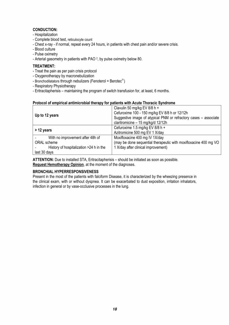
CONDUCTION: - Hospitalization - Complete blood test, reticulocyte count - Chest x-ray - if normal, repeat every 24 hours, in patients with chest pain and/or severe crisis. - Blood culture - Pulse oximetry - Arterial gasometry in patients with PAO 2, by pulse oximetry below 80.
TREATMENT: - Treat the pain as per pain crisis protocol - Oxygenotherapy by macronebulization - Bronchodilatators through nebulizers (Fenoterol = Berotec) - Respiratory Physiotherapy - Eritracitapherisis – maintaining the program of switch transfusion for, at least, 6 months.
Protocol of empirical antimicrobial therapy for patients with Acute Thoracic Syndrome
Up to 12 years
Clavulin 50 mg/kg EV 8/8 h + Cefuroxime 100 - 150 mg/kg EV 8/8 h or 12/12h Suggestive image of atypical PNM or refractory cases – associate claritromicine – 15 mg/kg/d 12/12h
> 12 years Cefuroxime 1.5 mg/kg EV 8/8 h + Azitromicine 500 mg EV 1 X/day
- With no improvement after 48h of ORAL scheme - History of hospitalization >24 h in the last 30 days
Moxifloxacine 400 mg IV 1X/day (may be done sequential therapeutic with moxifloxacine 400 mg VO 1 X/day after clinical improvement)
ATTENTION: Due to installed STA, Eritracitapherisis – should be initiated as soon as possible. Request Hemotherapy Opinion, at the moment of the diagnoses.
BRONCHIAL HYPERRESPONSIVENESS Present in the most of the patients with falciform Disease, it is characterized by the wheezing presence in the clinical exam, with or without dyspnea. It can be exacerbated to dust exposition, irritation inhalators, infection in general or by vase-occlusive processes in the lung.

19
TREATMENT: - Nebulization with 5-10 drops of phenoterol (Berotec) + Ipratropium Bromide (Atrovent) 20 drops + saline solution 3-5 ml and Oxygen at 6L/min (children 1 drop for each 3Kg – maximum 10 drops).
- Repeat up to 3 times with intervals 15-30 min - Reevaluate at 30 min: - If there is a partial improvement – discharge with regular NBZ and e SOS + Prednisone 40 mg VO prescription for 4 days - If the picture remained or aggravated – hospitalization, maintains NBZ every 30 minutes, corticoid EV (hydrocortisone 200 mg or methylprednisolone 40 – 60 mg EV) every 8h.
CHRONIC COMPLICATIONS:
COMPLICATION CHARACTERISTICS TREATMENT FOLLOW-UP
Pulmonary Arterial Hypertension
Exertional Dyspnea HU – maximum tolerated dose
Pulmonary vasodilator (Ca channel inhibitor)
Hypertransfusion Program
- Spirometry with bronchodilator proof – first routine exam at 14 years
- Repeat once a year:
Restrictive ventilatory disturb previously identified;
60 days after 2nd STA episode in less than 2 months ;
HAP recently diagnosed
Repeat twice a year (for those who experienced obstructive ventilatory disturb or as per medical evaluation)
Restrictive respiratory Syndrome
It may be related to multiple episodes of STA as consequence pulmonary fibrosis. Investigate HAP. Characterized by exertional dyspnea and spirometry with restrictive pattern.
1-Respiratoy Physiotherapy
2- Hypertransfusion Program, in case of H∆P
Obstructive respiratory Syndrome
Consequence of bronchial hyperresponsiveness. Release of chemical measurements from tissue lesion that occurs at vaso-occlusive episodes, resulting from bronchoespasms and obstructive pattern at spirometry.
1 – Regular B2-agonist associated to Ipratropium Bromide every 6/6 h.
2 - Inhaled corticosteroid (beclometasone)
ROUTINE PULSE OXIMETRY:
OUTPATIENT - Evaluation of saturation twice a year, at minimum every 6/6 months. - If Sat < 94%, conduct to physiotherapy to evaluate respiratory incentive - If no improvement after respiratory incentive use, conduct to Pneumology and Cardiology.
EMERGENCY ROOM AND HOSPITALIZATION
- Daily Evaluation - Respiratory incentive every hour

20
ONE, TWO-DIMENSIONAL DOPPLER ECHOCARDIOGRAM IN PULMONARY HYPERTENSION.
CONCEPTS
- Echocardiogram is a non-invasive method that allows an anatomical and functional evaluation of right cardiac cavities and the estimative of pulmonary arterial pressure.
- The most accurate method to estimate pulmonary arterial pressure is based on the measure of the speed from tricuspid valve regurgitation flow. Considering that speeds > 2.5cm/sec must guide the specific hematological approach (see 20.4.1 e 20.1.1).
- According to National Institute of Health-USA, it is considerate HAP, when systolic PAP is greater than 30mmHg. HAP grade is considerate as:
Mild --------- Moderate -- Severe ------
(> 30 to 38 mmHg) (de 40 to 54 mmHg) (> 55mmHg)
INDICATIONS - It is used on patients handling for the diagnoses and HAP prognostic evaluation. It is recommended to perform a Doppler Echocardiogram, from 14 years old, every 2 years and every year from 20 years old.
FOUNDS
- The mainly prognostic markers on echocardiograms founds were RA increase (right atrium) and the presence of pericardial effusion.
- The presence of pericardial effusion behaves as gravity independent marker.
- Approximately 36% of falcemic patients experience HP and this is the major cause of sudden death in this above 20-years population.
11. STROKE DIAGNOSES: OFTEN INDICATED EXAMS: - CCT (Cerebral Computadorized Tomography): with no contrast media (use contrast media in specific situations: unknown facility or neoplasm subacute or suspected or underlying infection). CCT may be normal at first 24 hours or indefinitely, if the lesion is at cerebral stem.
- Cardio-respiratory evaluation: ECG (detect IAM, arrhythmia); chest x-ray.
- DTC (Doppler Transcranial)
- Complete blood test, Platelet Count, Coagulonogram and arterial gasometry.
- Biochemistry: electrolytes , glucose, urea and creatinine. - Other blood exams: serology for Chagas Disease, serology for Sifilis, fibrinogen.
OCASIONALY INDICATED EXAMS: - MNR (Magnetic Nuclear Resonance): with spectroscopy, perfusion and diffusion. - Vertebral and Carotid Duplex–scans (elective measure to detect surgical indication stenosis). If it is the case, an angiography is performed. - One and two-dimensional Echocardiogram (if possible transesophageal when it is suspected of cardioembolic source). - If there is suspect of arrhythmia, it is recommended a Holter.

21
- Lumbar puncture (suspicion of HSA with normal CT) - Cervical column x-ray (In the suspicion of cervical or cranial trauma, height fall, pain or cervical rigidity). - Young subjects: rheumatologic screening and thyroidal function
- On special cases: C and S protein level, antithrombins III, Leiden factors VII, VIII and V, Tissue plasminogen activator (TPA) and plasminogen activator inhibitor PAI, homocistein, prothrombin mutationanticardiolipine (IgG and IgM) and lupus anticoagulant.
CLASSIFICATION OF STROKE: ORIGIN: - ISCHEMIC - HEMORRAGIC
EPISODE DURATION: - TIA –Transitory ischemic attack – neurologic deficit up to 24 h, without sequels.
- “in crescendo” TIA – two or more TIA episodes in 24 hours (it is one of the most important neurological emergency).
- RIND – Reversible Ischemic Neurological Deficit – lasts more than 24 h, with delayed deficit reversibility. More detailed exams show minimal sequels
- “ONGOING STROKE”: signals and symptoms aggravated
- COMPLETE CEREBRAL INFARCTION
STROKE – THERAPEUTIC APPROACH: 1 – Maintain free and functioning the respiratory airways 2 –ECG and oximetry monitoring: evaluate O2 suplementation
3 – Maintain elevated thorax and head at 30o 4 – Research cranial or cervical trauma and cardiovascular changes 5 –Neurological Exam: Conscience Level, convulsive episodes, Glasgow Coma Score, pupils (symmetry, reactivity) and four limbs movement. 6 - Hydration 6 - If PA is above 220 / 110 mmHg, it should not be acutely reduced 7 – Looking for reversible causes (bad positioning, pain, hypoxia) If there is arterial hypotension: volemic reposition, vasoactive amines. 8 – Cardiac monitoring
SUMMARY DESCRIPTION OF NEUROLOGIC EXAM
Dimidiate motor deficit Hemiparesis or hemiplegia or unilateral central facial paresia
Sensitivity Deficit Reduction or loss of painful or tactile sensitivity

22
Hemianopsia Homonymous deficit of visual field to confrontation
Superior Cerebral Dysfunction
Aphasia (word finding difficulty)
Deficit parietal (sensitivity or visual disregard, visual-spatial negligence and loss of notion of segmental position).
Cerebral stem deficit Ataxia, vertigo, Dysarthria (no aphasia) and oculo-motor paralysis (except conjugated deviation) with or without motor deficit or dimidiated sensitive, but without superior cerebral dysfunction.
TRANSCRANIAL DOPPLER (TCD)
- DTC is necessary to identify risk patients to first or new AVE and prevent them through transfusion.
- VFSC value to be considered is related to media and prior cerebral arteries.
- When the AVE predictive value is high (Cerebral Blood Flow Speed > 200cm/sec) is indicated to switch transfusion for 2-year period .
- After two years of transfusional therapy, beside DTC, encephalic MRI is performed.
- If NORMALS (DTC and MNR), patient is put hydroxyurea protocol.
STROKE ON TREATMENT
NEUROLOGICAL CHANGES
PATIENT WITH SCDS
NORMAL WITHDRAWN TT, after 5 years, if the patient is ok, and HU use for at least 6 months
NO-VASCULAR WITHOUT AVE RISK
RPM IMPROVEMENT LIKELY STROKE MODERATE RISK OF STROKE
RPE REFRACTORINESS STROKE HIGH RISK OF STROKE
RPM - Moderate Predictive Risk – Cerebral Blood Flow Speed (CBFS) between 160 and 200 cm/sec; RPE – Elevate Predictive Risk - CBFS above 200 cm/sec
CONDITIONAL VFSC 170-199 cm/s
NORMAL VFSC < 170 cm/s
Repeat after 1 year
Repeat after 3 months
TCD DF (2 – 16 years)
ANORMAL VFSC > 200 cm/s
Switch Transfusion for 2
VFSC > 200 cm/s Repeat in 1 year
RNM = N RNM = A
TCD in 6 months HU
TT + HU
INTERVAL ABSENCE
Perform another

23
BASIC ELEMENTS IN ROUTINE FOR CEREBRAL INFARCTION:
NEUROLOGICAL COMPLICATIONS: The principal neurological complications from cerebral infarction at acute phase are: - Cerebral edema and intracranial hypertension (HIC) - Hydrocephalia - Hemorrhagic transformation - Convulsive crisis - Early recurrence: (see picture below)
CLINICAL FACTORS AND PREDICTIVE LABS FOR DCV
CLINICAL FACTORS PREDICTIVE LABS
STROKE HF
Snoring or nocturne apnea
Amygdala Hypertrophy
Meningoencephalitis by HPP
Low fetal Hb
High Hemoglobin
Leucocytose Homocysteine deficiency
CEREBRAL EDEMA AND INTRACRANIAL HYPERTENSION TREATMENT - Do not use corticoid - Do not sedate the patient - Elevate headboard at 30o - Orotracheal intubation in case of hyperventilation and/or PCO2 of + 30 mmHg - PIC monitoring, in selected cases (Coma, major hemispherical lesions irresponsive to treatment). - Osmotic therapy on patients with HIC neurologic deterioration (10% of the cases)
Mannitol 20% 1-2 g/kg/24h in intervals of 4 h, infusion at 4 to 5 mL/min, for at least 3 days.
To take off gradually maintaining interval between doses.
Furosemide Use only on emergency cases (ex. Immediate transtentoyal hernia).
Dose: 70 mg or 7 ml IV slow. Don’t use to maintenance
COMORBIDITY - Diabetes - HAS - Epilepsy - Síndr de secr inapropr ADH - Heart failure
MONITORING - Neurologic - PA e TAx - Frequence cardiac - ECG (to admission)
PATIENT EVALUATION
TREATMENT - Hydrate 1.5 to 2 L (Do not use pure glucose) - Insulin if Gli ≥ 150-180 mg% - Hypertension: indication and drugs - Heparin/heparinoids-DVT prevention/ EP - Oxygen, if saturation ≤ 92-95% - Antipyretics, if rate > 37-37,5o Cl - AAS 160-300 mg/day - Culture and antibiotic therapy if there is infection) - UTI em complicações graves
DIAGNOSTIC - Hemorrhagic vs ischemic
- Topographic - Associated vascular factors
COMPLEMENTARY EXAMS - Glycemia - Blood count - Coagulogram - Electrolyte - Renal function - Lipidogram - Enzimas miocárdicas
CT
Cervical vases duplex scan cervicais
NOTE - Breathing - Peneumonia
- Hydration - Pulmonary edema - Swallowing - TVP
- Alimentation - ITU
- Sepse - Gangrene

24
TREATMENT OF CONVULSIVE EPISODES – GRAND MAL SEIZURES
0-5 Minutes
Diazepan EV – 0,1 a 0,3 mg/Kg (dose without dilution). It may be repeated every 10 minutes Oxygen through nasal cannula or masks; to put the head for optimal strength of airways; intubation (if necessary); oximetry. Labs exams: electrolytes, glucose, urea, creatinine, complete blood test, toxicological profile, DAE serum level, gasometry.
20-60 Minutes
Phenitoine via EV - 15 a 20 mg/Kg – loading dose, administrate at 30 minutes (dilute on Saline) Attention: EEG, ECG and BP monitoring
> 60 minutes
Phenitoine – 5 mg/Kg/day additional dose up to maximum of 30 mg/Kg (dilute on Saline) Pentobarbital - 15 mg/Kg IV (6-25 mg/Kg/h), initial dose, monitoring through EEG until the crises stop. Continue infusion at 1,5 mg/Kg/h, slowing down the speed every 4-6 hours. OR Midazolam - 0,2 mg/Kg with continuing infusion of 0.05 to 0.4 mg/Kg/h –EEG Monitoring OR Propopol - 1-3 mg/Kg followed by continuing infusion of 1-6mg/Kg/h.
12. OPHTHALMOLOGIC FOLLOW-UP MAIN OPHTHALMOLOGIC CHANGES Several ophthalmologic changes may be found according to falciform disease subgroup. The most common are “comma sign” in bulbar conjunctive in patients SS and facilform retinopathy, in the exam the fundus of the eye.
Non-Proliferative Retinopathy: more common in SS patients. It corresponds a group of fundoscopy manifestations that may be divided according to symptoms presentation. Symptomatic group corresponds to vascular occlusion of carotid and artery blockage and/or retinal vessels (or its branches). Asymptomatic group (the most common) included vascular tortuosity, spot like “salmon patch” (hemorrhagic areas), “black sunburst” (retinal atrophy due to resolution of hemorrhagic areas), iridescence spots (Hemosiderin depot), rupture of peripheral retinal and, rarely, angioids streaks.
Proliferative Retinopathy: more often in SC and Sthal patients, between 20 to 40 years old. Complication groups that occurred in a sequential manner, passing through 5 stages at retina peripheral: 1 – occlusion of peripheral arterioles; 2 – arteriovenous anastomosis; 3 – new vessels and/or new vessels tufts (“sea fan”); 4 – vitreous hemorrhage; 5 –detached retina. The clinical follow-up and treatment will be defined by the multiprofessional team at stages 1 and 2. In stage 3, laser photocoagulation is indicated in ischemic areas as fluorescein angiography founds and/or combined treatment with HU, according to personal evaluation. Patients that are on stages 5 and/or 5 wll be conducted to perform an ultrasound and follow-up or surgery (vitrectomy), as individual evaluation.

25
The patients who will be submitted to surgery must be conduct to hematologist for pre-surgical procedure. Hematologist will carry out pre-surgical exams (with surgical risk) and will conducted to Hemotherapy for transfusional schedule (switch transfusion or Eritracitapherisis –) as per each patient requirement .
ROUTINE:
The complete and basic ophthalmologic exam will be performed: directed anamnesis, inspection and appendices, ocular motility exam (iintrinsical and extrinsical), refraction, anterior segmental biomicroscopy, tonometry and fundoscopy. After, in a second consultation, retinal mapping will be conducted in all SS and SC and other, at ophthalmologic section discretion as follows::
From 0 to 10years: conduct only in case of specific ophthalmologic complain
From 10 to 20yeras: conduct all , even without complaining, once a year for SC and once every two years the other
From 20 to 40 years: conduct all even without complain and every 6 months the SC patients and once a year the other.
Above 40 year: patients will be conducted as the 10 to 20 years
All patients must be followed to look for lesions at initial stages. According to retinal mapping, the following flowchart will be followed: 13. GYNECOLOGICAL AND OBSTETRICAL FOLLOW-UP PREGANCY: PHYISIOPATHOLOGY: In Falciform Disease occurs placenta dysfunctional, in varies grades, leading to retarded intra-uterus growth and increase of peri-natal mortality.
Retinal mapping
First Consultation as routine
NORMAL Blur / hemor vitreous Non-proliferative Retinopathy erativa
New vessels
Asymptomatic symptomatic
Follow-up as age Individual conduction
Follow-up as age
Peripheric Non- PeriphericaPer
Laser Photocoagulatio

26
INITIAL ATTENDANCE TO PREGNANT WOMEN: 1. Clinical History: obtain information on renal disease, hypertension, smoking, drugs and allergies. 2. Obstetric History: number of deliveries and abortions, gestational age when it happened, newborn weight, types of labors and complications during gestational period or abortions.
3. Hemoglobin Electrophoresis of the father –to guide about fetus disease. 4. Labs exams: complete blood exam, reticulocyte, ferritine, bilrrubins, TGO, TGP, LDH, phosphatase alkaline, glucose, urea, creatinine, uric acid, serology for hepatitis A,B,C, HIV, HTLV1, CMV, measles, toxoplasmosis, VDRL; T. Coombs, fecal parasitology, PPD, SAE and urine culture. Repeat at the end of 1º, 2º and 3º quarters.
5. Ultrasound and Doppler. 6. Evaluation of fetal vitality
Note: To evaluate maternal nutrition and hydration and to monitor BP, pulse oximetry,, gain of weight, fundus of uterus, uterine colon exam.
TRANSFUSION: The efficacy of prophylactic transfusion is not supported by controlled papers. The indications are: - Toxemia - Gemellary Pregnancy - Previous peri-natal mortality - Septicemia / Bacteremy - Acute Renal Insufficiency - Acute Thoracic Syndrome - Pre-surgical procedure - Severe Anemia: 20% below hematocrits from baseline value or hemoglobin below 6g/dl.
RETARDED INTRA-UTERUS GROWTH (RIUG): - More frequent in SS, less in SC e Sβ + . - Implicated factors: (1) hypoxia: reduction of O2 from placental blood with inappropriate release to the fetus. (2) malnutrition - there is inappropriate transport of substances through placental membrane (maternal anemia, frequently episodes of vase-occulsion leading to hypotension). (3) early placental detached - area reduced of placenta switch
(4) previous placenta - with repeated bleeding it will become necrosis areas.
(5) toxemia

27
(6) multiple pregnancies
(7) smoking, alcohol and narcotics.
LABOR AND CHILDBIRTH: - To allow painkillers (see pain treatment on gestational period) - Peridural anesthesia - O2 and liquids reposition - Fetal monitoring - Cesarean by obstetric indication is common, being necessary pre-surgical transfusional preparation. - Avoid thromboembolism with early walking around, elastic socks, hydration and newborn cares.
PRETERM LABOR: The gestational age is smaller in the fetus whose mother has Falciform Disease. The media gestational age is 36 weeks.
Implicated factors: Anemia, DPP, previous placenta, toxemia, multiple gestations, urinary tract infection,
chorioamnionitis, smoking and narcotics.
PERINATAL MORTALITY: is increased in Falciform Disease, ranging from 20 to 50%.
RESULTS IMPROVEMENT: The principal factor to reduce mortality is neonatal care. The specialized attendance to the mother, which includes ultrasound, biophysical profile, Umbilical cord Doppler, cesarean with obstetrician indication. Transfusions in special cases must be discussed along with Hemotherapy. Great lesions in the placenta that occur earlier do not benefit form late transfusions..
PLACENTA CHANGE - DPP and Previous Placenta have increased incidence, being attributed to continuing vase-oclusion, decidute arterioles thrombosis, necrosis and subsequent venous hemorrhage by toxemia.
COMPLICATION DURING PREGNANCY - Episode of vase-occlusive crisis –may increase, especially, at the end of pregnancy period.
Prophylactic transfusions only for episodes of non-justified pain. - Pain Crisis _ also see “Pain Approach” – chapter III) - Anemia – there is an aggravation in the picture for increase of demand, hemodilution, medulla
suppression, infection or inflammation, vitamin or iron deficiency and aplastic crisis. - Infections – increased frequency at the 1st half of gestational period, mainly in the urinary tract and
respiratory system. - Bacteriurias must be treated. They cause CIUR and premature labor.

28
- It may occurs acute thoracic syndrome and congestive cardiac insufficiency secondary to anemia
aggravation. - Hypertension and toxemia may be related to preexisting renal disease. In some cases, patient may
need hemodialysis to support fetal development. Follow-up with nephrologists is always recommended in theses cases.
- CNS change – important to research the history of: thrombosis, hemorrhages, hypoxemias, continuing use of narcotics, headache, and toxemia.
ABORTION: - High prevalence (higher frequency on SS patients) - Probable causes – placental microvasculars accident, smoking, alcohol and frequent use of narcotics.
INFERTILITY – The retarded puberty may delay the 1st pregnancy
CONTRACEPTION: medroxyprogesterone acetate (Depo-Provera®) 150mg, intramuscular with intervals of 3/3 months or oral contraceptives with continuing progestogen (desogestrel). It is noted, in many cases, an improvement in the incidence of vaso-occlusive crisis.
DIU may be used, being well-controlled to avoid increase of blood loses. Patients may be notified as for
infection risks.
Request bone densitometry, before prescribing progestogen-based contraceptive. Repetition every two
years.
STERILIZATION All implication on new pregnancies, as well sterilization, must be discussed a lot with the patient. The orientation for this option can only be provided after the second child.
14. BILIARY LITHIASIS Noted from 1 year with the presence of biliary calculus, biliary mud, narrowing of vesicular wall and changes of common biliary ducts caliber of common biliary ducts.
DIAGNOSE: Abdominal ultrasound: Request in the 1st consultation and every 2 years, even asymptomatic as of 5 years old.
TREATMENT: CONSERVATIVE TREATMENT: In the presence of cholecystitis, carefully observation, venous hydration, and antibiotics are indicated. Support general actions, as antispasmodic and diet low-fat, some patients become symptoms-free for several years.
CHOLECYSTECTOMY: It is suggested to discuss with the patient the possibility of surgery. Urgent surgery must be avoid, unless there is an evidence of biliary obstruction.

29
- Currently there is a tendency to perform prophylactic cholescystectomy by laparoscopy in asymptomatic patients, with purpose of prevent possible pictures of cholecystitis, or colangitis and/or the necessity of urgency surgery. The occurrence of inflammatory/infectious pictures may also difficult the future use of videolaparoscopy due to viscosity formation.
- Pre-surgical preparation at Hemotherapy discretion. Due to possible complications during surgery, the patient, previously conducted to videolaparoscopy, may be submitted to laparotomy and respiratory physiotherapy with spirometer.
- Post-surgical cares – respiratory physiotherapy with spriometer and early walking around (prevention of STA and vase-occlusive).
15. HEPATITIS: After the diagnose by confirmatory test, conduct patients to a Reference Centre for treatment. Note.: RIBAVIRIN is not at MS protocol for DF, because its use in these patients are not established yet.
16. RENAL COMPLICATIONS HYPOSTHENURIA: Caused by the difficulty of the kidney to concentrate the urine. It is noted around 3 years old. This condition results a mandatory urinary output of more than 2.000ml/day, in adults. The increase of urinary loss becomes patient more likely to dehydration, which is a precipitate factor of vaso -occlusive crisis. Hyposthenuria also leads to nocturia in adult and persistent enuresis in children.
Conduct: to provide from 3 to 5 ml/day to adults and 150ml/Kg/24h to children.
NOCTURNAL ENURESIS (No need to treatment until 5 years old) - As of 5 years old: 1. Avoid ingestion of liquids near bedtime 2. To make conscious bladder control (training to hold urine for a longer period, urinate intermittently, urine before bedtime) 3. Drug treatment: Imipramine - 25mg/day – adult and 10mg/day- children
ATTENTION: Conduct to Nephrology ALL patients that experience any of the changes described below.
RENAL TUBULAR DYSFUNCTION: 1 Unable to acidificate urine. It may lead to metabolic acidosis (Start therapy with Sodium Bicarbonate) 2 Hyperkalemia (Higher risk of hyperkalemia in patients using beta-blockers, ACE blockers or Potassium-
sparing diuretic). The drug must be withdrawn.
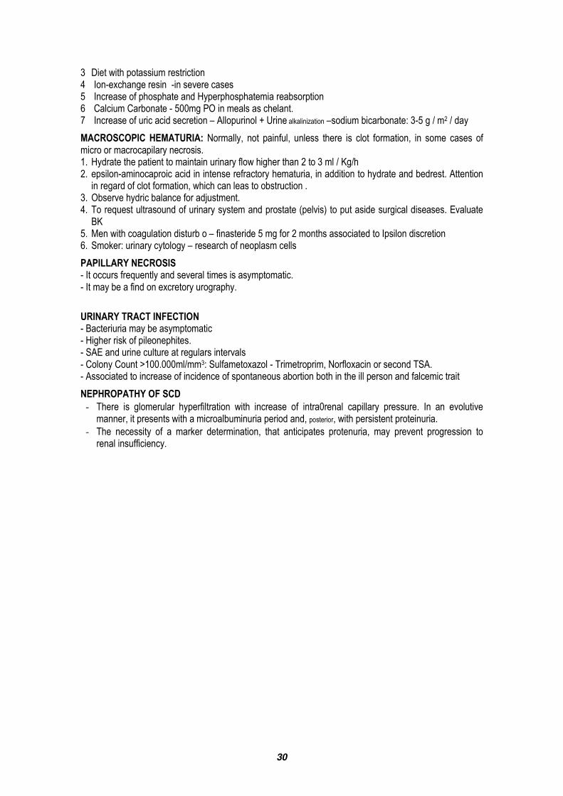
30
3 Diet with potassium restriction 4 Ion-exchange resin -in severe cases 5 Increase of phosphate and Hyperphosphatemia reabsorption 6 Calcium Carbonate - 500mg PO in meals as chelant. 7 Increase of uric acid secretion – Allopurinol + Urine alkalinization –sodium bicarbonate: 3-5 g / m2 / day
MACROSCOPIC HEMATURIA: Normally, not painful, unless there is clot formation, in some cases of micro or macrocapilary necrosis. 1. Hydrate the patient to maintain urinary flow higher than 2 to 3 ml / Kg/h 2. epsilon-aminocaproic acid in intense refractory hematuria, in addition to hydrate and bedrest. Attention
in regard of clot formation, which can leas to obstruction . 3. Observe hydric balance for adjustment. 4. To request ultrasound of urinary system and prostate (pelvis) to put aside surgical diseases. Evaluate
BK 5. Men with coagulation disturb o – finasteride 5 mg for 2 months associated to Ipsilon discretion 6. Smoker: urinary cytology – research of neoplasm cells
PAPILLARY NECROSIS - It occurs frequently and several times is asymptomatic. - It may be a find on excretory urography.
URINARY TRACT INFECTION - Bacteriuria may be asymptomatic - Higher risk of pileonephites. - SAE and urine culture at regulars intervals - Colony Count >100.000ml/mm3: Sulfametoxazol - Trimetroprim, Norfloxacin or second TSA. - Associated to increase of incidence of spontaneous abortion both in the ill person and falcemic trait
NEPHROPATHY OF SCD - There is glomerular hyperfiltration with increase of intra0renal capillary pressure. In an evolutive
manner, it presents with a microalbuminuria period and, posterior, with persistent proteinuria. - The necessity of a marker determination, that anticipates protenuria, may prevent progression to
renal insufficiency.

31
- Microalbuminuria is defined by increase of albumin excretion in the urine, in the absence of clinical
proteinuria anticipates proteinuria presence, which already indicates a more chronic way of nephropathy. - Macroalbuminuria is a sensitive marker of renal involvement before proteinuria, and some papers
have contributed to elect microalbuminuria as important marker of renal damage in DF.
Microalbuminuria 0 to 30mg/l first urine of the day, in the morning, 12-h or 24-h period.
Proteinuria Up to 150mg at 24-hour urine
Nephropathy monitoring
To request initially 24h-proteinuria for all patients, , clearance creatinine and nitrogen excretion annually and complementary with renal US.
Proteinuria above 150mg/24h
Conduct to nephrology where it will start with captopril or enalapril. HU must always be considered in these patients
Clinical Picture In addition to isolate proteinuria, patient may experience -Nephrotic Syndrome - proteinuria above or equal 3g, edema, and hypoalbuminemia.
Conduct
It is recommended salt restriction, water and diuretic use that might be venous or oral, depending on edemigenic picture. Renal biopsy may be indicated for histopathologic diagnose and evolutive prognostic of the picture and shall be evaluate in each case. Regular use of albumina is not established Hipoalbuminemia < 2 is frequently necessary the furosemide use during albumina infusion.
Protocol for microalbuminuria detection in patients withSCD disease :
Patients Screening All patients above 3 years old with falciform disease at outpatient at HEMORIO.
Exam Requirement During outpatient consultation, please request albuminuria dosage in the first sample in the morning urine.
Exclusion Criteria Patients with hypertension, previous proteinuria, with nitrogen excretion change, pregnancy women and diabetics .
Important Note to the Patient
If in the day of the exam, the patient experiences hematuria, pain episode or fever, the exam must be reschedule because these symptoms may interferer in the result.
Exam result Patients with microalbuminuria (albuminuria > 30 mg/l) shall be conducted to nephrology outpatient. Patients who experience albuminuria values below 30 mg/l should perform this exam annually.
Nephrology Follow-up
Determination of albuminuria on urine samples collected over the night or over 24h on three separated occasions at 30-day intervals for confirmation and monitoring.
Other labs exams: glucose, urea, creatinine, Na, K, Ca, P, Cl, uric acid, complete blood test, fetal hemoglobin. SAE, clearance creatinine. 24-hour proteinuria, viral markers, renal ultrasound.
Use of captopril – initial dose of 6,25 mg 2 x /day for one month; 12,5 mg 2x /day for two a three months and after increase to 3 x/day for six months, reaching 25mg, 2 to 3x a day (maximum 1mg/kg/day) or enalapril at 5mg/day dose. The blood pressure will be monitored one week after drug initiation and, after, in all consultations. .
After start MA treatment, in first month, in the third, and then every six months, also with execration monitoring and electrolytes, mainly, K level control, because it may occur hyperkalemia,.

32
MICROALBUMINURIA Screening
Patients > 3years
Dose albuminuria at 1st urine sample in the morning.
Patients with hematuria, pain or fever, reschedule the exam
CHRONIC RENAL INSUFFICIENCY: Clinical identification is frequently evidenced at third or fourth decade, so it is necessary regularly order nitrogen execration, proteinuria and renal ultrasound. - The initial treatment is conservative, with protein ingestion control and, as necessary, hydric restriction of potassium and salt (in case of edema and urinary volume decrease).
- Introduce HU - Supplementation with carbonate and oral calcium - Chronic Renal Insufficiency Terminal – dialytic treatment - It is noted after renal transplantation, increase of pain crisis by erythrocytosis. In some patients, change replacement may be indicated.
- ACE blockers seem to reduce this erythrocytosis.
ERYTHROPOIETIN (EPO):
Erythropoietin is responsible for red blood cell production. Its production is controlled by kidneys. Patients
with falciform disease have EPO high levels. When it aggravated with renal insufficiency, its levels will not
appropriately raise, aggravating anemia picture in these patients.
Microalbuminuria > 30 mg/l
NO
Repeat annually exam
Confirmation: Dose albuminuria urine sample overnight (at night 12h) or 24h
on 3 separate occasions at 1 month interval
YES
USE of captopril or enalapril
Conduct to nephrology

33
EPO serum dosage: in patients with chronic renal insufficiency of other etiology, anemia in general is present when glomerular filtration rate decreases to 40 ml/min. At DF, the measurement of renal function is jeopardize for muscle mass decrease and creatinina tubular secretion increased. In falciform disease, despite of chronic anemia, the serum EPO normal values in each age is unknown. Tubular tissue damage that occurs at AF may compromise EPO syntheses by renal O2 sensor destruction not always is compatible to renal insufficiency grade. Patients with DF have a significant reduction on renal function before clinically detected by clearance creatinine. Increased anemia, caused by ineffective erythropoietin, with EOP levels reduction, may occurs up to 54 months before azotemia, and it is a predictive of renal insufficiency in falciform anemia.
Initial dose: 50 to 150 IU/kg three times a week, preferable subcutaneously to increase the efficacy, it may be upgraded to 300 IU/kg, higher doses may indicate EPO resistance.
Adverse effects: hypertension, risk to thrombosis and hyperkalemia.
Indications: (1) severe or persistent anemia - HB < 6.0 in three dosage during 3 months, (2) symptomatic anemia, dependent transfusion or both, (3) fall of baseline hematocrit (fall of 20% of baseline HT or Hg on 3 dosage at 6-month period), (4) necessity to demonstrate EPO low level for EPO therapeutic follow-up
17. UROLOGIC COMPLICATION PRIAPISM ACUTE PHASE: - Hemotherapy Opinion in case of priapism lasting 6 hours or more. - Immediate analgesic treatment with NSAID (avoiding morphine and derivates) and general measures as in the pain crisis.
- Simple transfusion or early (depending on Ht levels) Eryitraciphere (better response before 12h of priapism). - Antibiotics to the cases that need surgical drainage (washing, multiple punctures or shunts).
Urologist handling:
Up to 6h Intravenous injection(IC)
6 – 12h IC + Lavage with Saline Solution
12 – 24h IC + Lavage + Transglandular Puncture (Winter) +/- Cavernous-sponge Shunt (CE)
> 24 h Winter +/- Shunt CE
Discharge patient with antibiotics administration (in case of drainage), Nsaid destilbenol 1 mg/day until outpatient return.

34
RECURRENCE OF PRIAPISM:
Objectives
- Pain Treatment - Reduce the chance of impotency – keep an erection - Reduce the chance of relapse – fibrosis - Reduce psychological disturbs - Non-expensive, simple and compliant treatment. - Information to target-population
Register - To ask priapism diary (write down frequency, duration).
General actions - Urinate always before bedtime, - Ingest less liquid at night, - Avoid alcohol and opioids
Home Treatment
- In case of priapism for more than 45 minutes, administrate 2 tablets Efortil DU. - In case of priapism persistence, administer 1 tablet of Diestilbestrol 1mg (DES) - In case of refractory for more than 3 hours, contact immediately an urologist
Urologist Conduction
- Start finasteride 5 mg 1x/day for 30 days and observe answer - Obtaining good response, reduce for 2,5mg/day during 30 days and then try maintenance with the lowest dose possible (1mg day or alternating days).
- Refractory cases to finasteride (5mg/day), associate DES 1 mg DU daily. - Achieving priapism control, start drug reduction (initiating with DES, 1/2mg day). Try keeping with the lowest dose of DES possible.
- Patients that experience severe side effects to DES (gynecomastism, delayed development) or not responsive to DES.
- Self-injection, intra-cavernous with etilephrine solution.
NOTE: Consider HU use, in case of severe or recurrent priapism
SEQUELS OF PRIAPISM (Erectile Dysfunction) - Study cavernous bodies with penile Doppler and IC drug - Intracavernous test - Encourage sildenafil, tadalafil, PO, use. - Encourage vacuum therapy (including preparation to prosthesis placement)
HEMOSPERMIA: - Request urinary system and prostate (pelvis) ultrasound to rule out surgical diseases. Evaluate BK - Men with coagulation disturb – finasteride 5 mg for 2 months associated to Ipsilon criteria - If smoker: urinary cytology – neoplasm cells research
MICROSCOPIC HEMATURIA: - Request urinary system and prostate (pelvis) ultrasound - Request dimorphism of erythrocyte (research of crenate red blood cells) – Hematuria study from glomerular origin x excretory tract, evaluate BK.
- If the exams are negative or show proteinuria presence, request nephrology opinion.

35
18. SURGERY PRESURGICAL CARES: Clinical Evaluation made by hematologist. - Complete Red Blood Cell Test - Coagulogram - Glucose, urea, creatinine, hepatogram -SAE - ECG – Cardiologic surgical risk - Chest x-ray - Hb A and S Dosage - O2 evaluation by pulse oxymeter - - Transfusion preparation (see item 19) - Immunohematology Study with erythrocytes of Phenotype for all systems - Keep plain hydration 12 hours before surgery
PRESURGICAL CARES: - Pleasant temperature in surgery room - O2 to 50% in combination with anesthetic agent. - Clinical monitoring - ECG, blood pressure, pulse, temperature urinary output. - lab monitoring - serum electrolytes dosage, O2 concentration inspirited pulse oxymeter or arterial
gasometry.
POSTSURGICAL CARES : - O2 at immediate postsurgical - pulse oxymeter - parenteral hydration - respiratory physiotherapy
SPECIAL SITUATIONS: Presurgical of falciform disease with recent history of priapism: Start 5 mg finasteride, 15 days before surgery and continue for more 20 days.
19. IRON CHELATION THERAPY: See also VI appendix – Iron Chelation
INCLUSION CRITERIA - All patients that are in a hypertransfusion program - All patients that experience ferritine levels > 2000, confirmed by 3 dosage performed
with 1-month interval with the patient at baseline.
DEFEROXAMINE DOSES
- 20-60 mg/kg/day SC every 8 h, by infusion pump (average of 40mg/kg/day), from Monday to Friday.
- Children below 3 years old, start 10 mg/kg/day to avoid hypodevelopment INTERRUPTION - Fever, abdominal pain and diarrhea (may be Yersinia infection)
FERRITINE CONTROL - Weekly drug dose may be reduced to 3 or 4 fold, when have therapeutic response .
SIDE EFFECTS
MORE FREQUENT - cyanosed (extremities, lips and skin) - Blur vision and other visual problems - seizures, - dyspnea or taquipnea, - tachycardia, - earring problems, - pain and/or administration site edema - rash or pruritus
LESS FREQUENT - diarrhea - dysuria - fever - cramps - abdominal
discomfort - bleeding

36
SPECIAL CARES
- The use in pregnant women is not recommended - Visual and earring problems are more common in younger patients at high doses and
prolonged use - Association with vitamin C must be used with caution in elderly patients, because they
are more likely to develop cardiac problems - Patients with renal problem have higher chance to experience side effects - Occurrence of orange urine may follow all treatment period
DEFERASIROX
- Discharge from MS, in 2006. - It must be used by PO, at 30mg/kg dose in patients continuing transfusion regime - Patients that are not at transfusion program, the dose can be adjusted to 20
mg/kg/day, and the iron balance must be monitored. - Presents only fecal elimination - The must be monitored on liver and renal function test monthly - The most frequent side effects are gastric intolerance and intestinal disturbs. This
manifestations are, generally, mild and the drug seems to be well-tolerated .
20. HYDROXYUREA ELIGIBILITY – INCLUSION CRITERIA TO PROTOCOL: 1. Hemoglobin electrophoresis - SS, SC, SD ou Sβ0 tal; 2. Older than 3 years old; 3. To show at periodic revisions; 4. Submit to lab exams every 2 weeks, at first month and, after, monthly; 5. Pregnancy test (β-HCG serum) negative for childbearing women; 6. At least one of the
complications, in the last 12 months:
- 3 or+ episode of vaso-occlusive crisis with medical consultation requirement - 2 episodes of STA (defined as acute thoracic pain with new pulmonary infiltrate fever
37,5o C or superior, taquipnea, pulmonary wheezing or cough); - 1 episode of severe priapism our recurrence of priapism; - bone ischemic necrosis; - renal insufficiency - 24h proteinuria higher or equal to 1 g - Severe and persistent anemia (Hgb < 6,0 on three dosage in 3-month period).
- Elevated LDH 2-fold normal in children and above 3-fold in adult - 2 DTC above 160 and up to 200 cm/s - patients with proliferative retinopathy - any other situations where there is an evidence of organ chronic lesion

37
EXCLUSION CRITERIA (should not be included in treatment protocol):
PERMANENTS Hypersensibility to HU
PROVISORY: Any one of the following:
Leucocytes count < 2.500/mm3;and/or neutrophil count < 1.500/mm3;
Hemoglobin < 4,5 g/dl;
Reticulocyte < 70.000/mm3 (when Hgb < 8 g/dl)
Pregnancy (there is evidence of animal teratogenicity, but not in human beings)
SPECIAL SITUATIONS: due to possible drug adverse effects, the risk/benefit ratio must be carefully evaluated in the following cases:
(a) BREASTFEEDING: It is known that HU is excreted in milk. Its use might be avoided during breastfeeding or be discontinued the breastfeeding;
(b) URICOSURIA: HU use may increase serum levels of uric acid. Patients with baseline levels above normal limits must be monitored every month.
(c) RENAL INSUFFICIENCY: evaluation together with nephrology.
(d) LIVER INSUFFICIENCY: There is no enough data to guide dose adjustment in this situation. Patients
(e) DRUG INTERACTION: The concomitant administration with other drugs that may produce medullar
depression must be close monitored.

38
TREATAMENT:
DRUG Hydroxiurea (hard gelatin capsules 500 mg)
INITIAL DOSE 15 mg/Kg/Day (only and daily administration).
DOSE ADJUSTMENT
Diary dose must be increased at 5 mg/Kg/day every 4 weeks until 30 mg/Kg/ day dose is reached or hematological toxicity or other serious adverse effects occur (see below)
HEPATOTOXICITY Defined as two-fold increase of transaminases baseline concentration. There is no data to adjust the dose,
MIELOTOXICITY
LEVELS ACCEPTABLES TOXIC
Neutrophil (cel/mm3) 2.500 < 1.500
Platelets (cel/mm3) > 85.000 < 70.000
Hemoglobin (g/dl) > 5,3 < 4,5
reticulocyte (cel/mm3) (when Hb < 8 g/dl)
85.000 < 70.000
If any value fulfills toxicity criteria, HU use must be interrupted, until it returns to superior and acceptable levels. Then treatment stars again with 2.5 mg/Kg/day dose, inferior to the last employed following the same scale of progressive increase, every 4 weeks. If there is toxicity twice in the same dosage, this dose will be considered the maximum tolerated dose and will not be use any longer.
RENAL INSUFFICIENCY
Dose adjustment has to be done according to creatinina clearance: 10 - 50 ml/min - administrate 50% of dose < 10 ml/min - administrate 20%of dose.
DURATION Treatment has to be maintained for, at least 2 years, and for undetermined period according to patient progression.
WARNINGS AND CAUTIONS:
FOLIC ACID HU lead to macrocytosis, difficulting folic acid deficiency recognization. In addition to AF 5mg 3X per week, it must be noted precaution with ingestion.
HIV+ PATIENTS HU increases the risk of peripheral neuropathy, especially when associated to antiretrovirals, a didanosine and stavudine. In HIV+ patients who experience pancreatitis or liver toxicity, HU use must be withdrawn and not indicated.
HEPATITIS B and C Patients with positive serology to Hepatitis B and C may use the drug, since they are monthly monitored with liver function test
ADVERSE EFFECTS
NEUROLOGIC: lethargy, headache, dizziness, disorientation, hallucination.
GASTRINTESTINALS: stomatitis, anorexia, nauseas, vomits, diarrhea and
constipation.
DERMATOLOGIC: macropapular eruptions, facial and periphery erythema, skin
ulcers or worsening of ulcers already existing and changes like dermatomiosity.
RENAL: creatinina and urea levels increased OTHER: fever, chills, malaise, asthenia.

39
EXPECTED BENEFITS
- Reduction of Pain Episode frequency, which can even, disappear. - Increase of hemoglobin F production and slight increase of Hb concentration - STA episodes reduced - Number of hospitalization reduced - Slowing down of organ chronic degeneration - Number of blood transfusion reduced
INFORMED CONSENT FORM
It is mandatory that patient or it legal representative is aware of potential risks and side effects related to drug use recommended in this protocol, or what has to be formalized by signing informed consent form.
TREATMENT MONITORING:
BEFORE TREATMENT INITIATION
- complete red blood cell test - reticulocyte - serology (HIV and hepatitis) - TGO, TGP, bilirubin - creatinine, urea, clearence - sodium - uric acid
- eletropheresis ptn - TAP - ferritine - hemoglobin F - cytogenetic - β-HCG - LDH
EVERY 2 WEEKS UNTIL REACH MAINTENANCE DOSE
AND AFTER EVERY 4 WEEKS - complete red blood cell test - reticulocyte
EVERY 4 WEEKS UNTIL REACH MAINTENANCE DOSE
AND AFTER EVERY 12 WEEKS - TAP, TGO, TGP, creatinine, gamma GT, alkaline phosphatase
EVERY 7 WEEKS UNTIL REACH MAINTENANCE DOSE
AND AFTER EVERY 24 WEEKS - hemoglobin F; - LDH

40
MAJOR AND INTERMIDIATE THALASSEMIA DIAGNOSTIC EXAMS - Peripheral blood study + complete red blood cell (VCM e CHM) + retyculocytes - Hb A2 dosage and fetal - Osmotic fragile curve - Family Study (parent and siblings)
MAJOR THALASSEMIAX INTERMEDIATE THALASSEMIA - MAJOR – Hb maintenance < 7g/dL - INTERMEDIATE - Hb maintenance > 8g/dL
PERIODIC CONTROL
DIAGNOSE
- Conduct to Interdisciplinary Group of Thalassemia treatment - Immunhematologic study (phenotype) - Serology (hepatitis A,B,C, HIV, HTLV, CMV) - Vaccination for hepatitis B (depend on serology) - PPD (for non reactors)
QUARTER
- Medical consultation - ferrokinetics (ferritine, TIBC) - Glucose, urea, creatinine, LDH - TGO, TGP, FAL, gamma-GT
SEMESTRAL
Cardiologic Evaluation - Echocardiogram - ECG (12-lead) - Chest x-ray
Serologic Evaluation - Hepatitis: A (anti HAV – IgG) / B (HBSAg, Anti HBS and anti HBC IgG / C (Anti HCV)
- Retrovirus (Anti HIV, Anti HTLV)
Evaluation of growth curve (compare to parent and siblings height) - Graphics Age X Weight X Height - Accurate measurement of height (stand and sit positions)
ANNUAL
Thyroid Evaluation (as of 12 years) - Free TRH, T4 and TSH Parathyroid Evaluation (as of 16 years) - Calcium - Phosphate serum - PTH Other hormonal evaluations - LH / FSH - Testosterone - Estradiol - cortisol on morning fasting Tanner staging of puberty
Ophthalmological Evaluation - Campimeter and Fundus of eyes Audiometric Evaluation
- Oral glucose tolerance test - Fist and hand x-rays - Densitometry or bone x-ray - Cholesterol total, HDL and triglyceride - RNM T2* (hepatic and cardiac evaluation)

41
- Abdominal US TREATAMENT HYPERTRANSFUSION: See HEMOTERAPIC PROTOCOLS INDICATIONS: - Major Thalassemia – all cases
- Intermediate Thalassemia – with facial changes, growth retard, pathologic fractures and/or extramedullar hematopoiesis
HYDROXYUREA INDICATIONS: Intermediate Thalassemia
Inclusion criteria, posology, monitoring, precautions and care are identical from HU protocol used in Falciform Disease.

42
HEREDIRATY SPHEROCYTOSIS CLINICAL EVALUATION Anamneses Complete clinical exam Orientation about disease Supply explicative manual about the disease Start Folic Acid
LABORATORIAL EVALUATION Complete Red Blood Cell Test Incubated osmotic fragility curve Biochemistry (glycemia, liver and renal function test, electrolytes) Serology (HIV, HTLV, hepatitis A, B, C, Chagas, VDRL) TAP and PTT SAE and Parasitology: feces
OUTPATIENT FOLLOW-UP Subsequential outpatient follow-up will be every four months, followed by labs exams (complete red blood cell, biochemistry). Serologic exams will be performed annually Only patients younger than 18 years old are included in this follow-up. Symptomatic patients with Hb <11g/dl at any age are also included.
DIAGNOSE: Presence of several espherocytes in peripheral blood Incubated osmotic fragility test positive CHCM >25g/dl Hb ranging 8-13g/dl Reticulocytose Indirect Bilirubinemia
TREATMENT: Use of folic acid Splenectomy See HEMOTERAPIC PROTOCOLS
SPLENECTOMY INIDICATIONS: 1-Major hemolytic anemia (Hb <8g/dl) with hypersplenism;
2-Development retard;
3-Moderate hemolytic anemia (Hb<11), but with symptomology and complications related to anemia/hemolyze with biliary litiase, legs ulcers or erythropoietic mass;
4-Children with serious hemolyze (Hb<6) must be splenectomized earlier, but not before 3-4 years old.
INFECTIONS: Splenectomized subjects have are more likely to sepsis by Pneumococcus, Neisseria meningitidis, Escherichia coli, Haemophilus influenzae, Estafilococccus and Estreptococcus. So, they need to receive vaccines anti-menigococica,and anti-Haemophilus, before splenectomy.

43
After splenectomy, antibiotic prophylaxis has to be made for 1-2 years with benzatine penicillin anos (600.000 IU) in children 10-20Kg and 1.200.000 IU above 20Kg) or 10mg/kg oral penicillin every 12/12h in children and 250-500mg every 12/12h in adult). In allergy patients, use erythromycin at usual dose.
THROMBOSIS
Splenectomized subjects have are more likely to thrombosis, so they have to continue using 100mg/day AAS.
PREGNANCY
Pregnant women with espherocytes must be kept with Hb≥10g/dl, it may need transfusions in case of anemia (Hb <10g/dl).
DISCHARGE CRITERIA FROM SPECIALIZED TREATMENT
Children must be followed every year until they complete 18 years, when it will be analyzed the necessity of specialized treatment. They have to perform previous labs exams and routine consultation.
Patients >18 years old, splenectomized who remained asymptomatic for 6 months after splenectomy;
Patients >18 years old non-splenectomized, asymptomatic with Hb>11g/dl;
The patients have to be discharged with orientation about screening reactivation criteria as: symptom of hemolytic anemia (pale, jaundice, tiredness), pregnant women during prenatal and major surgery.
SUMMARY OF DIAGNOSE OF SPHEROCYTE
Mild Moderate Slightly severe Severe
Hemoglobin 11 – 13% 8 – 11% 6 – 8% 6%
Reticulocyte 3 – 8% > 8% > 10% > 10%
Total Bilirubin 1 - 2 2 - 3 > 3
Fragility Curve N or slightly changed Clearly changed
Incubated Fragility Curve
Changed
Transfusion requirement
0 - 1 0 - 2 > 3 regular
Splenectmoy NI If vitality↓ Necessary < 5 year
Necessary < 3 years

44
G6PD DEFICIENCY INITIAL EVALUATION Anamnesis Complete clinical exam Orientation about disease Supply explicative manual about the disease LABORATORIAL EVALUATION Complete Red Blood Cell Test G6PD research Biochemistry (Glucose, urea, creatinine, sodium, potassium) Liver function test Annual Serology (HIV, HTLV, hepatitis A, B, C, Chagas Disease, VDRL) SAE and Parasitology: feces
DIAGNOSE G6PD research positive G6PD dosage
CLINICAL PICTURE Most of subjects is asymptomatic and diagnose is made by chance in familiar study.
It comes with hemolytic anemia variable as enzymatic deficiency levels, ranging since a mild way to severe pictures, triggered by infections (Staphylococcus aureus, Salmonella, Serratia marcescens, Escherichia coli, and fungus like Candida albicans, and Aspergillus).
Anemia can be triggered by infections, drugs. Osteomyelitis Oral and perioral lesions .
TREATMENT Avoid hemolyze by substance (see picture) Avoid infections; Transfusion when symptomatic and hemoglobin <10g/dl; Regular vaccination calendar and Hepatitis A (if marker is negative)
FOLLOW-UP Children have to be followed annually until reach 18 years, when the necessity of specialized treatment will be analyzed. They must perform previous labs tests and routine consultation.
Symptomatic patients related to basal disease will be followed every 6 months with previous labs exams..
Asymptomatic patients do not require specialized treatment.
The patients will be orientated about the disease as hemolyze risk by infection and substances use (oxidants drugs, food and other) and explanatory manual about disease as: appendix that contains substances that must be avoid or restricted.

45
AGENTS CAPABLE OF TRIGGERED HEMOLYZE ON ERITROCYTES WITH G6PD DEFICIENCY
CATEGORY MAY BE USED CAREFULLY MUST BE AVOIDED
ANALGESIC AND ANTIPYRETICS
Acetaminofen, acetophenetidin (phenacetin), acetylsalicylic acid, aminopyrine, antipyrine, phenacetin, paracetamol
Acetanilide, metamizol, fluotane, aminosalicylic acid
ANTIARRHYTHMIC Procainamide Quinidine ANTI-HELMÍNTIC Piperazine ANTIHYPERTENSIVES Captopril, enalapril (maleate), hydralazine
(cloridrate) ANTIMALARIC Cloroquine, pirimetamine, quinacrine,
quinine
Hydroxicloroquine, mefloquine, pamaquine, pentaquine, primaquine, quinocide
ANTIANGINOSOS
Isosorbide Mononitrate, Isosorbide dinitrate, nitroglycerine (trinitrine)
ANTIBACTERIANS P-aminobenzoic acid, Isoniazide, trimetropine Estreptomicine
Nalidixic acid, cloranfenicol, ciprofloxacine, dapsone, fenazopiridine, furaltodone, furmetonol, nitrofurantoine, nitrofurazone, norfloxacine, ofloxacine, Co-trimoxazol, furmetonol, neoarsfenamine
ANTIEPILETICS Fenitoin ANTIPARKINSONIAN L-Dopa ANTIPROTOZOAL Furazolidone ANTISEPTICS Metylene Blue ANTITOXICS Dimercaprol (BAL) ANTINFLAMATORY Probenicide ANTIHISTAMINICS Astemizol, azatadine,
bronpheniramine, cetirizine, clorfeniramine, ciproeptadine, difenidramine, dexclorfeniramine, difenidramine, elastine, hydroxyzine, loratadine, mequitazine, oxatomide, terfenadine
CYTOSTATICS Doxorrubicine (cloridrate) CONTRASTS Toluidine Blue ESTROGEN Mestranol SULFONAMIDES AND SULPHONES
Sulfisoxazol Sulfadiazine Sulfametoxipiridazine
Sulfacetamide, Sulfametoxazol, Sulfametoxipirimidine, Sulfapiridine, Sulfassalazine, Diaminodifenilsufone (DDS), Dapsone, Sulfanilamide, sulfamerazine, sulfatiazol, sulfoxone, tiazolsufone, N-acetilsufanilamide, sulfatiazol
VITAMINS Vitamin C (Ascorbic Acid) Vitamin K1, Vitamin K3 FOOD AND DOMESTIC USE Conservative, Naphthalene

46
APLASTIC ANEMIA LABS EXAMS TO DIAGNOSE
• Complete red blood cell with reticulocytes • Biochemistry with LDH, liver and renal function • Mielogram and Bone Narrow Biopsy • Hemoglobin electrophoresis (fetal Hb might be elevated at constitutional aplasia) • Serum iron, ferritine, transferrin saturation • Vitamin B12 dosage and folate • Immunophenotipage for HPN (if it is not available, HAM test and Sucrose) • Serology for viral infections • Direct Coombs • HLA Type if candidate to TMO • Cytogenetic study • Chest and bone x-ray RX, if suspected FAnconi Anemia • Total abdominal ultrasound • β -HCG • Rheumatic Function Test • Thyroid Hormones
CLASSIFICATION
Hereditary • Fanconi Anemia • Dyskeratosis congenital • Shwachman-Diamond Syndrome • Dysgenesis reticular Amegacariocitose • Familiar Medullar Dysfunction • Non-hematologic syndromes (Dubowitz, Seckel,Down)
Acquired – Secondary • Radiation • Drugs and chemical agents • Virose (EBV, Hepatitis, Parvovirus, HIV, CMV) • Immunological Diseases • Timema • Pregnancy • HPN • GVHD Transfusional

47
Idiopathic DIFERENTIAL DIAGNOSE
• SMD hypoplastic • HPN • Neoplasm invading MO • Osteopetrosis • Hypersplenism • Infectious Diseases (Calazar, sepsis, TB milliary, disseminated fungus disease, malaria,
AIDS ) • B12 Vitamin deficiency, pyridoxine and folic acid • Depot disease • LES
TREATMENT
Hemotherapy: see HEMOTHERAPEUTICAL PROTOCOLS
GCSF in case of current severe neutropenia of serious infections
Mild to moderate Aplastic Anemia • With no need of transfusion – observation • Depending of transfusion – ATG and Cyclosporine
Severe Aplastic Anemia • With donor – TMO • Without donor – ATG e Cyclosporine
Antitimocitc Globulin (ATG 25mg/ 5ml-Rabbit) – 2,5mg/Kg/day during 5 days
Antitimocitc Globuli (Linfoglobuline-ALG 100mg/ 5ml -Horse) – 15mg/Kg/d during 5 days
Cyclosporine – 10mg/Kg/day on 2 administrations, away from meals. Sodium, magnesium and potassium control, liver and renal function test. If Creatinine > 2 times baseline, reduce at 25% the dose and repeat the exam in 4 days, if persists, stop cyclosporine during 5 days.
BP control
Oral Hygienefo gum hypertrophy
Required for monitoring of Cyclosporine serum level
Schedule, in case of ATG use is not possible • CSA – 10mg/Kg/d up to 6 months and then gradually reduce • Prednisone – 1mg/Kg/d from D15 to D45. Then reduce 20% of the dose by week, until complete
withdrawal
• Response Criteria
• Complete – With no transfusion requirement Hb>11g/dl /Neutrophil >1.500/mm3 /Platelets >100..000/mm3
• Partial – With no transfusion requirement Hb>8g/dl / Neutrophil >500/mm3 /Platelets >20.000/mm3
• No response - Transfusion requirement persisted

48
SELFIMMUNE HEMOLYTIC ANEMIA CONCEPT: Selfimmune hemolytic anemia is defined as pathologies in which occurs premature destruction of red blood cells, mediated by fixes antibodies and antigens of erythrocyte membrane. This immune fixation triggers a number of reactions in cascade that finishes in these cells lysis (INTRAVASCULAR HEMOLYSE), in addition to phagocytose by macrophagolitic system (ETRAVASCULAR HEMOLYSE)
ETHIOLOGIC CLASSICATION
AHAI BY WARM ANTIBODY IgG, C3b
PRIMARY IDIOPATIC SECONDARY Lynphoproliferative
disease LLC, DH, LNH, MM
Self-immune disease
LES, self-immune hepatitis, rheumatoid arthritis, self-immune thyroiditis, Biermer anemia
Tumor Ovary Cyst, malignant neoplasm Infections Pneumonias and rhinopharyngitis Immunosuppressive Pregnancy and transplants Drugs Alpha-metildope, cimetidine, Procainamide
AHAI BY COLD ANTIBODY (COLD AGGLUTININ)
PRIMARY / IDIOPATIC
SECONDARY Lynphoproliferative disease Infections
DRUG-INDUCED AHAI
Cascade complement formation Intravascular hemolyze Drug absorption in cellular membrane Extra-vascular hemolyze Changes in cellular membrane
Adsorption non-immunologic
Self-antibodies formation Alpha-metildope
PAROXYSM COLD HEMOGLOBINURIA (biphasic hemolysins)
Fixes complement at low temperatures and promotes intravascular hemolyse > 37o C
Diagnose by Donath-Landsteiner test
IDIOPATIC
SECONDARY
More frequent in children associated to viroses (measles, chickenpox, rubeola, mononucleosis) More rare form of AHAI.
DIAGNOSE Clinical Diagnose: clinical signals of anemia, jaundice. In case of extravascular hemolyze, it may occurs splenomegaly and in case of cold antibodies, Raynaud phenomena and intolerance to cold, due to vase spasms, can be noted.

49
Laboratorial Diagnose:
Immunohematologic Study
Positive direct Coombs test (Negative on 4% of the cases) Positive indirect Coombs test Antibodies elution and fixation [for TCD (-) cases and to identify alo-antibodies (irregular agglutinin)] Antibody Identification Panel Erythrocyte phenotype
Peripheral Blood Study
Macrocytic Anemia Reticulocytose Spherocytte Policromasia RDW elevated
Biochemistry Study Extravascular hemolyze
Indirect Bilirubinemia Elevated LDH Haptoglobine reduced
Intravascular Hemolyze Hemoglobinuria Indirect Bilirubinemia
TREATMENT: Opinion to Hemotherapy for combined follow-up – see HEMOTHERAPIC PROTOCOLS
Red blood cells reserve for potential transfusion in more serious cases
Investigation for preexisting based-disease.
Mielogram to rule out the presence of lynphoprliferative disease.
Objectives: Reduce antibodies production, reduce viable antibodies quantity, reduce or stop hemolyze mediated by self-antibody. DRUGS-INDUCED AHAI Stop potential drugs-related event. AHAI – cold antibody: Maintain the patient warmed and in case of serious hemolyze, even with poor response to corticoid, we should continue with the immunosuppressive scheme, aiming a potential urgent hemotransfusion. AHAI – warm antibody: Minimal hemolyze: Folic acid and observation
Moderate to intense hemolyze: Corticosteroids (prednisone 1-2 mg/kg/day) during 2 to 4 weeks. If there is a response, gradually reduce to 30-90 days until there is no clinical evidence of hemolyze.
RESPONSE FAILURE TO CORTICOSTEROID OR RECIDIVE
1. Cytotoxics agents: ciclofosfamide: 60mg/m2/day PO or Azatioprine: 80mg/m2/day (or 1.5mg/kg/day) PO during 3-6 months.

50
2. Splenectomy: Vaccinate 14 days before surgery (pneumoccocus, meningoccocus and Influenza H),
wait 2-4 weeks). This is not indicate to cold antibody diagnose. This is indicate to patients dependents of high doses of corticosteroid for long period (above 40mg/day)
REFRACTORY TO CORTICOID OR UNTREATABLE SIDE EFFECTS (IATROGENIC CUSHING, DIFFICUL CONTROL HAS, DM AND GLAUCOMA)
1. Danazol (400/600/800 mg/day) 2. Ciclofosfamide at high doses – ciclofosfamide 500mg-700mg EV every 3-4 weeks 3. Ciclosporine A - 5-10mg/kg/dia, two administrations (monitor renal function, BP and electrolytes) 4. Rituximabe - 375mg/m2/dose evaluate response. Also for cold antibody cases.
CASES OF SEVERE HEMOLYZE WITHOUT RESPONSE TO INITIAL TREATMENT
1. Pulsotherapy with corticoid 2. Human Immunoglobulin doses at 1mg/kg/day, maximum 2 days or 400 mg/day for 5 days 3. Immunosuppressant agents 4. Rituximabe 5. Apheresis: limited efficacy - see HEMOTHERAPIC PROTOCOLS 6. Blood transfusion – see HEMOTHERAPIC PROTOCOLS 7. Support therapy (folic acid, erythropoietin )

51
ALGORITM FOR AHAI TREATMENT BY WARM ANTIBODY
MINIMAL
HEMOLYZE MODERATE
HEMOLYZE SEVERE
HEMOLYZE
OBSERVATION PRD – 1mg/Kg For 15 days
M-PRD or DEXA
+
TRANSFUSION
IF NECESSARY
RESPONSE
positive RESPONSE
negative or slow
GRADUAL
REDUCTION PRD – 2 mg/Kg
for 3 – 4 weeks
POSITIVE
RESPONSE
GRADUAL
REDUCTION
NEGATIVE
RESPONSE
SPLENECTOMY
NEGATIVE
RESPONSE
CFM + PRD 5 DAYS
MAINTENANCE PRD- 20 mg/day
DANAZOL –
200mg
NEGATIVE
RESPONSE POSITIVE
RESPONSE
PROLONGED MAINTENANCE PRD – 20 mg/Kg/day and / or
DANAZOL – 200 mg/day GAMMAGLOB
ULINE

52
IMMUNE THROMBOCYTOPENIC PURPURA LABS EXAMS TO DIAGNOSE - Anamnesis and physical exam - Complete red blood cell with hematoscopy - Serology for Hepatitis A, B, C, HIV, Measles, CMV, monocucleosis and e toxoplasmosis, FAN - Mielogram - Lupus anticoagulant research
TREATMENT INDICATION Clinical picture, platelets count, age, presence of risk factors for bleeding and life style. - Platelets < 20000/mm3 - Platelets between 20000 and 50000/mm3 with significant bleeding (hemorrhagic blisters in the mouth
mucose, conjunctiva hemorrhage) Severe bleeding with life threaten (hemorrhage CNS, HD).
TREATMENT Emergency Patients with severe or life threaten manifestations: CP<20.000/mm3, hemorrhagic blister un the mouth mucose, ocular conjunctiva hemorrhage on CNS
IgIV 1g/kg during 1 to 3 days + mehtylprednisolone IV 30mg/kg at 500ml of SG5% in infusion for 2 hours during 3 days. At subsequent days, reduce daily the dose to half until reach an equivalent dose at 1-2mg of prednisone. This dose must be kept for one month and should be withdrawn gradually with CP weekly control.
or Dexametasone 40mg/d during 4 days. CP may be transfused in case of life-threaten bleeding. See HEMOTHERAPIC PROTOCOLS
Initial
Patients with CP < 50.000/mm3 and bleeding, but with no life threaten
Prednisone 1 to 2mg/kg 3 to 4 weeks, followed by gradual reduction of 10mg by week until 0.5mg/kg dose and slowly withdrawn 5mg/week. Every 15 days clinical-laboratorial control.
Dexametasone 40mg/day for 4 days.
Patients with CP>50.000/mm3 Expecting conduct and laboratorial clinical control RESPONSE TO INITIAL TREATMENT Complete response: CP> 100.000/mm3 Partial response: CP>50.000/mm3 No response: CP< 50.000/mm3
TREATMENT AFTER CORTICOID Patients with complete remission = follow-up for 2 years and discharge from hospital. Patients with partial response = expecting conduction and clinical-laboratorial control Patients with CP between 20.000 and 50.000/mm3, the conduction depends on clinical picture, age and life style. Patients with com CP<20.000/mm3 decision made together with the patient. Discuss the risks of corticoid on low doses (prednisone 0,5mg/kg/day), other immunosuppressant and splenectomy.
Splenectomy: young patient, active life with bleeding and more than 6 months of PTI treatment
TREATMENT AFTER CORTICOID FAILURE Repeat mielogram, if corticoid refractory Rituximab 375mg/m2 for week to 4 weeks, if active bleeding Danazol 10 to 15 mg/kg/day + prednisone + azatioprine 1 to 2mg/kg.

53
Vincristine 1,5mg/m2 1x/week/4weeks + prednisone. Ciclofosfamide 1 to 2mg/kg/days + prednisone.

54
THROMBOTIC THROMBOCYTOPENIC PURPURA
PRIMARY CRITERIA: - Thrombocytopenia - Microangiopathic Hemolytic Anemia (defined as negative TCD, fragmented red blood cell and
laboratorial clinical evidence of hemolyze) - Absence of other causes for thrombocytopenia and anemia
OTHER ALTERATIONS: - Renal function alterations (proteinuria, hematuria, oliguria and acute renal insufficiency) - Neurologic alterations (agitation, disorientation, lethargy, coma and focal alterations) - Abdominal symptoms (pain, nauseas, diarrhea, vomit) - Asthenia - Fever
COMPLEMENTARY EXAMS: - Complete red blood cell with peripheral blood evaluation - Reticulocytes Count - Biochemistry - Hepatogram - LDH - SAE - Coagulogram, PDF and fibrinogen - Serology: HBV, HCV, HIV, HTLV - Immunohematologic Study
- Mielogram
TREATMENT: Plasmapheresis: see HEMOTERAPIC PROTOCOLS
Corticoid: - It may be used when plasmapheresis or plasma infusion failed. This is recommended when the patient
is submitted to plasmapheresis. - Metylprednisolone pulse: 20 mg/kg/day for 3 days, with gradual reduction posterior until reach the
equivalent to 1 mg/kg/day of prednisone. - Maintain PDN for 4 weeks, with gradual reduction
Splenectomy: indicated in PTT refractory cases
Immunosuppressant: - Vincristine: 2 mg/week for 4 weeks - Rituximab / Azatioprine / Ciclofosfamide / Ciclosporine

55
After platelets count recovering, it is recommended the use of low doses of aspirin (80 to 100 mg/day). Folic acid supplementation; Red blood cell pack transfusion, as necessary Relative counter indication of platelets transfusion
RESPONSE CRITERIA: Complete response: Partial Response: Absence of response: LDH normalization and platelets count + signals and symptoms vanished
Increase of 50% from platelets values Reduction of 50% of LDH concentration Some improvement of neurologic symptoms
No clinical or laboratorial improvement
Relapse: Return form symptoms and signals 30 days after complete remission without plasmapheresis
Exacerbation: Return from LDH increase and fall of platelets values, after initial improvement during treatment.

56
HEMORRHAGIC SYNDROME INTRODUCTION Hereditary Hemorrhagic syndromes are due to primary hemostasia alterations (vessel and platelets)-
hereditary purpura or alteration of coagulation cascade – hereditary coagulopathy.
FOLLOW-UP AOF PATIENT WITH HEMORRHAGE CLINICAL DIAGNOSE - Identify the age when the first hemorrhage occurs. - Evaluate the type of hemorrhage: petechia, schimose, mucous hemorrhage (epistaxes, gum
hemorrhage, digestive and gynecological, hematuria), hemartrhosis and hematomas. Spontaneous bleeding or post-trauma.
- Ask about drug uses, specially those, which interfere with hemostasia. - Ask about blood transfusion. - Investigate at family history about consanguinity, hemorrhagic pictures in the parents, siblings,
cousins, nephews, uncles and aunts, grandparents and children. If there is history in the family, try to know more about this relative and even try to submit him/her to labs exams.
INVESTIGATIONAL LABS EXAMS (also see appendix I – HEMATOLOGIC SCREENING) - Complete red blood cell - Biochemistry (hepatogram, ferrokinetics urea, creatinine and glucose) - Serology for hepatitis A, B and C; HTLVI/II; HIV and VDRL - Blood group and Rh Factor - According to screening laboratorial exams results, it is solicited, after awards, dosage of factor,
research of inhibitor, platelets aggregations and other - It is crucial that the patient do not use drugs that interfere at hemostasia, 15 days before exams.
DRUGS THAT GIVE SUPPORT TO PATIENTS WITH HEMORRHAGIC SYNDROME
Analgesics: paracetamol , paracetamol + codeine, Morphine and derivates Anti-inflammatory: ibuprofen Corticosteroids: prednisone Antifibrinolytic: The use cannot exceeded 14 days. Do not administrate in patients with hematuria macroscoscopic, with CNS hemorrhage and volume hematoma. Epsilon-Aminocaproic Acid (EACA)
- 25-50mg/kg of weight PO every 6/6 h (maximum de 12 g/d) - 1- 2g at 250ml of SF0, 9% or SG5% 6/6 h IV.

57
Tranexamic acid - 15-25mg/kg PO 8/8 h - 500-1000 mg IV 8/8 h. - Solution at 5% to rinse in the mouth: 10ml (6 times a day).
Desmopressin or DDAVP: analog synthetic of vasopressin (antidiuretic hormone). This is indicated for patients with mild hemophilia, von Willebrand disease, purpuras with alteration of platelets pool and Bernard Soulier syndrome, after therapeutic test. See using, administration and DDAVP doses , at von Willebrand disease treatment.
DRUGS THAT MIGHT BE AVOIDED IN PATIENT WITH HEMORRHAGIC SYNDROME - acetylsalicylic acid - Fenilbutazone, indometacine and other anti-inflammatory - Antihistaminic - Penicillin and derivates - Expectorant (most of has derivates of guaiacol that alters platelets function)
HOSPITALIZATION INDICATIONS - Volume hemarthrosis, relapses - Volume Hematomas at risk site (ileopsoas, calves, forearm, tongue and neck) - Neurologic signals and symptoms - Surgeries: pre and post surgery - Acute abdomen suspicion - Major hemorrhages
1- HEMOPHILIA CLINICAL FOLLOW-UP OF HEMOPHILIC PATIENT In hemophilic patients, all exams must be asked, every 6 months, except hemostasia study that will be replaced by research of periodic inhibitor. Patient who experiences positive inhibitor ins some phase of their evolution, must write down this fact in the medical chart so that any physician that, in the future, see him/her may know about it. As of this date, the control will be every three months with research and dosage of the inhibitor
PATIENTS WHO GO TO EMERGENCY ROOM AND DO NOT HAVE INHIBITOR RESEARCH, AT THE EXPECTED TERM, THIS MUST BE REQUIRED, IN THE EMERGENCY, BEFORE THE BEGINNING OF REPOSITION TREATMENT . Radiologic exam of joints has to be performed in patients who had submitted to hemarthrosis at periodical consultations intervals (at medical discretion).

58
MULTIDISCIPLINARY FOLLOW-UP OF HEMOPHILIC PATIENT The newly-diagnosed patient must have a consultation with hematologist, physiatrist, nurse, social worker, psychologist and physiotherapeutic from multidisciplinary group for general orientation about the illness. Deontological evaluation must be periodical. NOTE: All diagnosed cases, like hemophilia, must be notified to the hemostasia alterations group.
HEMOPHILIA CLASSIFICATION It is validated both A and B hemophilia and it is considerate that amount of coagulation activity of FVIII (A hemophilia) or FIX (B hemophilia).
CLASSIFICATION FACTOR SERIOUS <1% MODERATE 1-5% MILD 5-25%
TREATMENT In all hereditary coagulopahty, treatment is based on deficiency factor reposition and must respect individual biological characteristics of each factor (TABLE 1).
TABLE 1: PROPRIETY OF “IN VIVO” COAGULATION FACTORS: Factor
Plasmatic Concentration necessary to hemostasia
Half-Life of Transfunded Factor
Recovery in the blood (% total transfunded)
Stability on liquid plasma (stored at 4o C)
I 50 mg/dl 4 - 6 days 50% Stable II 40 UI / dL (40%) 3 days 40 - 80% Stable V 15 UI / dL (15%) 12 hours 80% Unstable VII 15 UI / dL (15%) 2 - 6 hours 70 - 80% Stable l VIII 30% 8 - 12 hours 60 - 80% Unstable IX 30% 18 - 24 hours 40 - 50% Stable l X 15% 2 days 50% Stable l X I 30% 3 days 90 - 100% Stable l XII - - - Stable l XIII 5% 6 - 10 days 5 - 100% Stable l

59
REPOSITION CALCULATION Reposition therapy depends on patients weight, clinical picture and amount of coagulation activity that patients presents. It follows the formulas below:
A Hemophilia: International Units (IU) of factor VIII = weight (Kg) x ∆/2 B Hemophilia: International Units (IU) of factor IX = weight (Kg) x ∆
∆= % to be elevated less % of factor that the patient has
Example for reposition calculation: Serious A Hemophilia (FVIII 0%) with hemarthrosis of the knee and 50Kg weight - (Elevate FVIII at 30%):
Notes: 1. Remember that half-life of FVIII is 50% every 8- 12 hours al normal conditions; 2. Half-life of FIX is 50% every12 hours 3. FVIII consume is greater when there is infection or active bleeding; 4. ∆ is depends on the severity of clinical picture. See Table 2. 5. Reposition therapy for each clinical situation is described at tables 2, 3,and 4; however, there is different ways to conduct it.
PRODUCTS THAT CONTAIN FVIII: FORZEN AND FRESH PLASMA (PFC): It contains all coagulation factors (approximately 1U/ml). It must avoid PFC, whenever possible, and specially on patients with hemophilia because there is safer products available in Brazil, with less risk of viral transmission. It may be administered at 10 a 20ml/Kg/day dose, divided in two and three administrations, on hemorrhagic episodes of minor importance, because it reaches plasmatic levels between 15 and 20%. Defrosted must be at 37°C and the infusion has to occur right after. Note: The Health Minister FORBIDS the use of frozen and fresh plasma and cryoprecipitated on patients with hemophilia (RDC n° 23, from January, 24, 2002).
CRYOPRECIPITATED: This is obtained from Hemotherapy Service through fast frozen method, and plasma centrifugation of a donor. Each bag contains FVIII 80IU, FvW, FXIII and fibrinogen 3 to 4,5g/dl. Mode of application: defrosted at maximum 10 minutes at 37°C. Mixture with the syringe the content of 5 bags in the same recipient. Transfunded with filter.
Note.: Use of CRYOPRECIPITATED is indicated on serious liver insufficiency, sepsis and XIII factor deficiency and fibrinogen.
The Health Minister FORBIDS the use of frozen and fresh plasma and cryoprecipitated on patients with hemophilia (RDC n° 23, from January, 24, 2002).
IU from FVIII = 50 X
(30 – 0) = 50 X 15 = 750 IU
2

60
LYOPHILIZED CONCENTRATES: They are products obtained by industrial methods of fractionating of plasma “pool” from more than 2000 donors, or more recently, from genetic engineering. There are several kinds of FVIII concentrates, ranging of grade of purity, according to fractionating technique and specific activity (SA):
FVIII CONCENTRATES OF INTERMIDIATE PURITY: They are obtained through serial protein precipitation method. Specific activity of these concentrates is between 1 and 50 IU/mg. They present in addition to FVIII, the following proteins: FvW, fibrinogen, immunoglobulin, immunocomplex and fibrinonectine. They are all indicated specially for patients with von Willebrand disease.
FVIII CONCENTRATES OF HIGH PUTIRTY - They are obtained through protein precipitation method plus separation by ionic exchange chromatography. SA of these concentrates ranges from 50 and 200 IU/mg. It contains non significant amount of FvW or other proteins. They are prescribed to patients with hemophilia.
FVIII CONCENTRATES OF TOO HIGH PURITY - They are obtained through chromatography by monoclonal antibodies or through genetic engineering (recombinants). It has SA above 2000IU/mg. In case of FVIII monoclonal concentrate, it is necessary to add stabilizers proteins like albumin or von Willebrand factor, because FVIII is an unstable protein. Third generation recombinants are being stabilizes with non-human plasma origin substances. They are indicated in patients with serious A hemophilia.
PRODUCTS CONTAINING FIX: PROTHROMBIN COMPLEX CONCENTRATES (PCC) – Are products of intermediate purity. In addition to FIX (2 IU/ml), they also present FII, FVII and FX. On B hemophilia is indicated when factor IX concentrate is not available to those with no evidence of hepatopathy. In these cases, the dose to be administered must be based on FIX calculation.
ACTIVATED PROTHROMBIN COMPLEX CONCENTRATES (APCC) – Are similar to those mentioned above. They have the same PCC factors activated. They are indicated to patients with hemophilia A and B with high titer inhibitors. Given the risk of thrombosis, they should not be used in hepatopathy patients. LYOPHILIZED CONCENTRATES OF FIX – May be of high purity or highest purity (plasma or recombinant origin). Indication: patients with hemophilia B, without inhibitor.
REPLACEMENT THERAPY TO PERFORM INVASIVE THERAPEUTIC PROCEDURES OR DIAGNOSIS: Should be preceded by replacement therapy, with proper levels according to TABLE 2.

61
TREATMENT OF HEMORRHAGIC EPISODES The replacement therapy to each clinical situation is described on TABLE 3; however, there are different ways to conduct it. As follows, we will describe the most used procedures for:
HEMATOMAS: Hematomas in the acute phase require replacement of deficient factor, immobilization, bed rest and ice bag for 20 minutes at most, every 2 hours, during the initial 48 hours (the exception is hemorrhage of iliopsoas muscle: bed rest during 4 days). After the acute phase the physiotherapy must be started with short waves and ultrasound. Do not use antifibrinolytics and do not deplete the hematomas. Hematomas located on the tongue, neck, forearm, calf, and iliopsoas muscles are considered risky. These hematomas deserve to be stressed by severity and due to the fact they leave disabling sequelae, if not treated properly. Hard to resolve hematomas may become pseudo-tumors, with required follow-up using ultrasound or MRI. It is important to stress that the clotting factor replaced does not help to reabsorb the hematoma. Therefore, the replacement suspension does not coincide with normalization of physical test.
Note: every patient with a hematoma must remain hospitalized, until the end of factor replacement. The patient needs to be forwarded to PHYSIATRY evaluation at all times.
HEMARTHROSIS: is the most common clinical manifestation found in the locomotive system. The most wounded articulations, by order of frequency are: knees, elbows, ankles, shoulders and hips. Replacement therapy, associated with bed rest sand ice on site, is enough to stop those bleedings. Measure the maximum diameter of articulation and movement amplitude. Start physiotherapy as soon as possible. Hemarthrosis may relapse, progressing to arthropathy. Patients presenting repeated hemarthrosis in a frequency of 3 episodes a month must be forwarded, mandatorily, to the Hemostasis Change Group.
Note: Every patient with hemarthrosis must be forwarded to PHYSIATRY, for follow-up.

62
TABLE 2: REPLACEMENT THERAPY TO PERFORM INVASIVE PROCEDURES
TYPE OF PROCEDURE F VIII FIX FREQUENCY (h)
DURATION GENERAL MEASURES % IU % IU FVIII FIX
IM VACCINES - - - - - - - Ice before and after ARTERIAL PUNCTURE 30 15 30 30 OD OD - Ice on site ELECTROMYOGRAPHY - - - - - - - - MYELOGRAM 30 15 30 30 OD OD - Ice on site
BIOPSY Skin and mucosa
30 15 30 30 OD OD - -
Muscle 50 25 50 50 OD OD Repeat I/N -
BRON CHOS COPY
WITHOUT biopsy
50 25 50 50 OD OD - Antifibrinolytic
WITH biopsy 50 30
25 15
50 30
50 30
1st dose 12/12
1st dose 12/12
1 day Antifibrinolytic
OPAQUE CLYSTER 30 15 30 30 OD OD - - EDA E EDB (endoscopy) 30 15 30 30 OD OD - -
EDA WITH BIOPSY 50 30
25 15
50 30
50 30
1st dose 12/12
1st dose 12/12
2 days Antifibrinolytic
EDB WITH BIOPSY 60 30
30 15
60 30
60 30
1st dose 12/12
1st dose 12/12
2-3 days Antifibrinolytic
LUMBAR PUNCTURE 100 50 100 100 OD OD - -
EXODONTIA 30 15 30 30 OD OD Repeat I/N Antifibrinolytic + liquid diet –
cold pasty
OD: only dose; I/N: if necessary.

63
HEMATURIA: - DO NOT start replacement therapy, at first. - General care: bed rest, vigorous hydration (oral). Avoid urinary infection - If in 72 hours macroscopic hematuria does not give in, increase FVIII or FIX according to Table 3, until macroscopic hematuria disappears.
- After hematuria is resolved, investigate etiology (ultrasound of urinary ways, EAS, excretion urography). - In refractory cases - Prednisone 1 to 2mg/kg weigh / day for 2 days. - NEVER administer Antifibrinolytics.
GI HEMORRHAGE: - Replacement of deficient factor (Table 3) - Antifibrinolytics - General clinical care: diet, antacid, ranitidine or omeprazol. - Hemostatic packed may be considered as a therapeutic option.
Note: Use in HDA cases, before and after cauterization: hemostatic packed: gelfoan 10 cm + adrenaline 1 ampoule + EACA 1-4g + SF0, 9%%, 250ml water or cold milk. Mix and drink 100ml 1/1h PO.
CRANIAL TRAUMA:
- MINOR TRAUMA: increase FVIII or FIX to 50%, every 24 hours, during 3 days; - MAJOR TRAUMA: increase F VIII or IX according to table 3. It is necessary to confirm the diagnosis of intracranial hemorrhage with: 1- Neurological evaluation 2- Fundus of eye evaluation 3- Cranial x-ray – fracture evaluation 4- Cranial CT always; in case there is no bleeding image, repeat it in 15 days for control.
- LIQUOR PUNCTURE REQUIRED: perform replacement of FVIII or IX according to Table 2.

64
CONDUCTION IN CONFIRMED INTRACRANIAL HEMORRHAGE: 1 WITHOUT NEUROLOGICAL SIGNS:
Increase FVIII or FIX to 100% in first infusion. Afterwards, 50% every 12 hours (for VIII factor) and every 24 h for IX factor, during 14 days.
2 WITH NEUROLOGICAL SIGNS:
Increase FVIII or FIX to 100% immediately and after, keep 50% every 8 hours (for VIII factor) and 12 h (for IX factor), during 7 days. If there is improvement of TCC image, maintain replacement at 50% every 12 h (for VIII factor) and 24 hours (for IX factor), until 14th day.
- General care: pain killers and anticonvulsants.
- Get to minimum level of clotting factor every 3 days. - Do not administer Antifibrinolytics.
CONDUCTION IN SURGICAL PROCEDURES Every surgical procedure must be preceded by inhibitor research or recovery curve of infused factor. Minimum level of clotting factor must be performed in post-operatory of medium and major surgeries.
Patients with mild hemophilia A, responsive to DDAVP (see treatment of Von Willebrand disease with DDAVP), may use this drug in pre-operatory of minor surgeries, such exodontia.
MINOR SURGERIES (1) PRE-OPERATORY: increase FVIII or FIX to 50% immediately before surgery. (2) POST-OPERATORY: if necessary (hematoma or external bleeding) increase FVIII or FIX to 30% every 24 hours, for over than 2 – 3 days.
- Use of Antifibrinolytic whenever possible. - On exodontias increase the factor to 30% only dose. - Attention to volume hematomas, they may lead to fibrinolysis.
MEDIUM SURGERIES (1) PRE-OPERATORY: Increase FVIII or FIX to 100% 30 to 60 minutes before surgery.
Example 1: Severe Hemophilia A (F VIII = 0%) - Weight 70 Kg
IU = 70 x (100 - 0) = 3.500 IU of F VIII UFVIII = weight (Kg) X ∆/2
2
Example 2: Severe Hemophilia B (FIX = 0%) - Weight 70 Kg UFIX = Weight (Kg) X ∆ IU = 70 x (100 - 0) = 7.000 IU of F IX
(2) POST-OPERATORY: D1 to D7 - increase FVIII or IX to 50% (every 12 hours) D8 to D14 - increase FVIII or IX to 50% (every 24 hours) until removal of stitches.

65
TABLE 3: TREATMENT OF HEMOPHILIAS
TYPE OF HEMORRHAGIA
FVIII FIX FREQUENCY (h) DURATION (days)
GENERAL MEASURES % IU % IU FVIII FIX
Superficial wound -
-
-
-
-
-
-
Compressive curative Gelfoan + Ice on site
Deep wound 40 20 40 40 24/24 24/24 2 a 3 Suture
Antifibrinolytic Antibiotics
Epistaxis
30
15
30
30
24/24
24/24
1 repeat I/N
bilateral nasal pressure Tamponage with glove finger Antifibrinolytic ORL Opinion
Ecchymosis - - - - - - - Ice on site
Superficial hematoma - - - - - - - Ice on site
Small muscular hematoma
30 15 30 30 24/24 24/24 2 to 3 Ice on site Immobilization
Large muscular hematoma
40-50 20-25 40-50 40-50 24/24 24/24 3 to 5 Ice on site Immobilization
Large muscular hematoma / risk sites / neurological impairment
80-100 40-50 80-100 80-100 1st dose 1st dose 3 to 7 Ice on site Immobilization Physiotherapy 50% 1st
dose 50% 1st
dose 50% 1st
dose 50% 1st
dose 12/12 12/12 Until
recovery
Hemarthrosis (early treatment or small volume)
40 20 40 40 24/24 24/24 1-3
Bed rest Ice on site
Hemarthrosis (late or volume treatment), hip, shoulder or target articulation hemarthrosis)
40 – 50 20 - 25 40 - 50 40 - 50 12/12 24/24 3-5
Hematuria 30 15 30 30 DU DU
Repeat I/N
Oral hydration Etiology investigation DO NOT USE ANTIFIBRINOLYTIC
Hemoptysis severe cases
50 25 50 50 1st dose 1st dose
50% 1st dose
50% 1st dose
50% 1st dose
50% 1st dose
24/24 24/24 3 -5
High or low digestive hemorrhage
80-100 40-50 80-100 80-100 1st dose 1st dose Until 3 Antifibrinolytic Hemostatic packed Endoscopy 50% 1st
dose 50% 1st dose
50% 1st dose
50% 1st dose
12/12 12/12 After resolved
Mild CET 50 25 50 50 24/24 24/24 3-5 Daily neurological evaluation + TCC
Severe CET without neurological change
100 50 100 100 1st dose 1st dose
14
Daily neurological evaluation Minimum level 2/2d Bed rest DO NOT USE ANTIFIBRINOLYTIC
50% 1st dose
50% 1st dose
50% 1st dose
50% 1st dose
12/12 24/24

66
Severe CET with neurological change
100 50 100 100 1st dose 1st dose Daily neurological evaluation Minimum level 2/2d Pain killers Anticonvulsants DO NOT USE ANTIFIBRINOLYTIC
50% 1st dose
50% 1st dose
50% 1st dose
50% 1st dose
8/8
12/12
1o- 7o
50% 1st dose
50% 1st dose
50% 1st dose
50% 1st dose
12/12 24/24 8o-14o
I.U. – international units, D.H.- digestive hemorrhage, I.N.- if necessary, C.E.T.- cranio-encephalic trauma.

67
Note: Surgical procedures must be preceded by inhibitor research or recovery curve of infused factor.
MAJOR SURGERIES (1) PRE-OPERATORY: - increase FVIII or FIX to 100%%, 30 to 60 minutes before surgery (2) POST-OPERATORY:
MAJOR SURGERIES PRE-OPERATORY D1 – D3 D4 – D7 D8 – D14 100%
50% 50% 50% FVIII - 8/8 H 12/12 h 24/24 h FIX – 12/12 H
WARNING: - Surgical procedures must be preceded by inhibitor research or recovery curve of infused factor. - Dose FVIII or FIX every 3 days (minimum level) - standardization of dose efficacy and identification of potential inhibitor. - Always re-evaluate replacement therapy doses according to clinic, especially due to volume hematomas and infection. - By guidance of Hemostasis Group, the factor’s continuous infusion may be performed.
CONTINUOUS INFUSION OF VIII FACTOR INDICATION - Hemorrhage of ileopsoas muscle - Compartment syndrome - Muscular or joint extended hemorrhage - Surgeries
MATERIAL - Conventional Infusion Pump - Saline - FVIII Concentrate - Heparin - Plastic Bag - Laminar Flow
MATERIAL PREPARATION: FVIII concentrates are diluted in the plastic bag with saline solution with total volume 200ml. Handling must be performed in sterile conditions, with laminar flow, gloves and masks. It is recommended the addition of 2-5 IU/ml heparin of total solution into the infusion pump to avoid thrombophlebitis at the puncture site. The bags are changed every 12 hours.

68
PHARMACOKINETICS OF CLOTTING FACTORS: the pharmacokinetics study of concentrate factor to be used must be performed within an interval of one week to 3 days before surgery. ISTH recommends the study to be performed with 30 IU/kg only dose infusion and serial sample draws for dosage of infused factor activity level. The first sample is pre-infusion, followed by 8 more samples drawn from the arm not infused, to determine the maximum peak of factor level, considering at least 3 samples post-infusion. Re-usage is given by the factor activity in these 3 samples and it is expressed in % by the formula (ml/kg/h) = infusion index (IU/kg/h) / plasma level (lU/ml).
METHOD: One FVIII dose in “bolus” is infused on pre-operatory. This dose is calculated to reach plasma levels between 60-80%. The continuous infusion starts right after the end of surgery or even during it. The dose administered is based on clearance obtained by the pharmacokinetics study according to the formula: FVIII level post- and pre-infusion X weight (Kg) / Factor dose given in IU.
NOTES: PTTa or FVIII dosage must be performed 4-6 hours after surgery to confirm if the dose is sufficient. Clearance is calculated every day, based on FVIII day activity to adjust the infusion and therefore, reach the desirable FVIII levels. For major surgery, the minimum desirable level is 50 and 30% during the 1st week and 2nd week, respectively.
In case of emergency surgery or major bleeding, the “bolus” dose is the same for elective surgeries and the continuous infusion must be installed immediately after the “bolus” dose.
When the pharmacokinetics study is not possible, it may be administered on the 1st infusion of 2 to 4IU/kg/h depending on the surgery or bleeding. In the next day, a sample is drawn to dose the factor plasma level and establish continuity.
Overall, it is noted a decreased amount of factor required to keep the same plasma level.
DOMICILE DOSE PROGRAM Intervention in the hemorrhage episode in hemophilia carriers, reaches its maximum efficiency with immediate access to the clotting factor, limiting the bleeding and the extension of resulting tissue damage. Early replacement therapy decreased the amount of clotting factor required to control the bleeding situation. The Domicile Dose Program aims to offer eligible hemophilia patients, 3 doses of clotting factor concentrate which increases the plasma level to 40%, for self-infusion at home, allowing early drug therapy. Thus, it reduces the “stress” arising from the need to go get the specific assistance to the drug and also allows the patient to participate in an active manner in his/her treatment.

69
ELIGIBILITY CRITERIA: - Have hemophilia diagnosis verified by laboratory, and do not present circulating anticoagulant (inhibitor); - Be willing to agree with the rules established by the program; - Favorable psychological evaluation; - Undergo proper training for self-infusion; - Have the knowledge about the pathology and treatment; - Be in proper domicile conditions to store, transport and return the material used in the assistance to be disposed in hospital waste; - Maintain clinical and laboratorial controls according to assistance routine; - Do not experience allergic reaction to drug.
NOTES FOR ASSISTANCE - Drug release registration is required; - It is required to return used bottles, allowing a complete control of drug use and also avoiding formation of « private storage »; - It is important that the patient records every application and brings the notes to the assistance, specifying the date, product, batch, bleeding site and infusion alternatives; - The participation of each patient must be re-evaluated periodically by the multidisciplinary group; - Special attention must be given to the storage, not allowing the lack of supply for emergency assistance. - Demand Treatment - the one performed due to bleeding episode. - Primary Prophylaxis – continuous treatment implemented before occurrence of any bleeding. - Secondary Prophylaxis - continuous treatment performed for 3 to 6 months, implemented after occurrence of more than one bleeding episode in one or more articulations. - DD – Domicile Dose – dose supplied to the patient for early domicile treatment, at the moment of bleeding episode.
TREATMENT OF HEMOPHILIA PATIENT WITH INHIBITOR About 5-15% of patients with Hemophilia A develop inhibitors, i.e., class IgG antibodies FVIII-oriented.
Among patients with Hemophilia B, the incidence of FIX inhibitors is of 3%. The most affected patients are, usually, the ones with severe hemophilia, but there is no direct correlation between the amount of FVIII or IX infusions and the appearance of inhibitors.
Clinically, the presence of inhibitors it is manifested by the bad response to usual treatment or by the increase of bleeding episodes in patients with hemophilia. In situations where there is inhibitor suspicion, request laboratorial research.
The presence of inhibitors is titrated through Bethesda method and by definition one unit of Bethesda (UBe) corresponds to the amount of circulating antibodies able to inactivate 50% of FVIII and IX existing in 01 ml of normal plasma.

70
INHIBITORS CLASSIFICATION The inhibitors may be classified according to the antigenic response and circulating antibody titer.
A) According to antigenic response: - Patients with high response: present increased antibody titers (higher than or equal to 5 UB/ml) and major increases of these titers after antigenic stimulation. - Patients with low response: present low antibody titers (lower than 5 UB/ml) and minor increases after antigenic stimulation.
This classification is important due to changes to the conduction in bleeding treatment, with the following exceptions:
a) Alone determination of the titer may be deceptive: low titers may be found in patients with high response not exposed to the antigen lately. Patient’s history is important to classify him/her as high or low response.
b) Patients with low response, after prolonged stimulation, may become patients with high response. Thus, if it is necessary to use FVIII in these patients, it is required to monitor the inhibitor titer.
B) According to titer may be classified as: - Low titer: levels inferior to 5 UBe - High titer: higher than or equal to 5 UBe
TREATMENT OF FVIII INHIBITORS In patients in whom the inhibitors were detected, administration of factor originally deficient, an anamnestic response may be generated, with fast increase of its titer. Therefore, the first recommendation is to avoid blood components and derivatives, seeking conservative measures whenever possible (curatives, antifibrinolytics, Ice on site).
For hemophilia patients with history of inhibitor whom arrive at the emergency room, the first measure to be taken is to request a research and inhibitor dosage, before administration of any medication. According to inhibitor titer, the antigenic response and the severity of clinical situation, one of the described treatments is administered:
A) High doses of FVIII: increase to the desired percentage + 20U FVIII/Kg/UB in initial dose, followed by infusion of same dose every twelve hours, in attempt to neutralize the existing antibody. It must be tried as first option in hemophilia patients with low titer and low anamnestic response inhibitors. Minimum level dosage must be performed daily, in order to verify if the ideal response is being obtained.
In case of patients with high anamnestic response, but with low inhibitor titer which present life threatening bleedings (CNS), we can indicate a 100 to 150 IU/Kg dose in adults, followed by 100 U/Kg/h in continuous infusion. Minimum level dosage is required.
If hemostatic levels are not obtained, plasmapheresis to partially deplete the patient from circulating antibodies is recommended.

71
B) Non-activated Prothrombin Complex Concentrates: the dosage to be used is arbitrary, but it is recommended that 50 to 75U/Kg doses every 12 or 24 hours must be tried in high titer inhibitors.
C) Activated Prothrombin Complex Concentrates (APCC): For patients with high titer inhibitor whom did not respond to the non-activated prothrombin complex or those who are in a life threatening situation (for example, intracranial bleeding). It is recommended a 50 to 100U/Kg dose per weight. Do not exceed 200U/Kg in 24h.
NOTE: Prothrombin Complex activated or not, should not be used for over 5 doses due to the risk of thromboembolic accidents. They should not be used in patients with hepatopathies or predisposed factors to consumption coagulopathy. The use of these substances has various results, and may or may not improve the hemorrhage. Follow with platelet count, TAP, PTT and fibrinogen dosage.
D) Recombinant Activated Factor VII Concentrate (rFVIIa): rFVIIa is produced through DNA recombinant technology. It is indicated to hemophilic with inhibitors, severe bleeding and those who do not respond to PCC, or the ones who undergo surgery.
- Doses: 90µg/kg/dose (90 to 120µg/kg), the first 24h in 2/2h, the second day, first 12h in 3/3h and then 4/4h. - IV bolus injection with administration time of 3-5 min. - The patient’s follow-up is performed through clinical improvement. - The simultaneous use of rFVIIa and PCC must be avoided . E) Immunossuppresion: Is more effective in acquired inhibitors in nonhemophilic patients.
- Prednisone 0.5 to 1.5mg/Kg/day for 5 days. - Cyclophosphamide 2 to 3mg/Kg/day (may be used alone or combined with Prednisone). - Azathioprine 0.2mg/Kg/day for 5 days - 6 - Mercaptopurine 25mg - 100mg/m2 /day - Immunoglobulins 400mg/Kg/day for 5 days IV or immunotolerance regimen.
F) Plasmapheresis: It may help to reduce fast the antibody titers, while waiting on immunossuppressive response in high titer inhibitors. Administration of FVIII must be performed right after plasmapheresis (while inhibitor level is lower).
SURGERY IN HEMOPHILIA PATIENTS WITH INHIBITORS - The hemophilia patient with inhibitor should not undergo elective surgeries com. The patient must be informed about the risks involved, and sign the specific informed consent form. - Minor surgeries, such as dental surgeries, may be performed only with local measures and Antifibrinolytics. - Extended dental surgeries or other medium surgeries must be performed with Prothrombin Complex reservations.

72
- Extended surgeries require increased doses of Prothrombin Complex, such as 75 to 100mg/Kg or rFVIIa. - All exceptional cases must be analyzed by the Hemostasis Group and evaluated by the Head of Assistance.
TABLE 4: TREATMENT OF BLEEDING EPISODES IN THE PRESENCE OF INHIBITOR
Ac BLEEDING FVIII pcc APCC Low
response Low titer
Mild high doses 12/12h - - Moderate high doses 12/12h - - Severe high doses 12/12h 50 to 75 U/kg/ dose 12/12h -
Low response High titer
Mild - 50 to 75 U/kg/ dose 12/12h - Moderate - 50 to 75 U/kg/ dose 12/12h - Severe - 75-100 U/kg / dose 12/12h
High response High titer
Mild - - 75-100 U/kg/dose 12/12hs Moderate - - 75-100 U/kg/dose 12/12hs Severe - - 75-100 U/kg/dose 12/12hs
High response Low titer
Mild - 50 to 75 U/kg/dose 12/12hs - Moderate - 50 to 75 U/kg/dose 12/12hs 50 -75 U/kg/dose 12/12hs Severe - 50 to 75 U/kg/dose 12/12hs 75-100 U/kg/dose 12/12hs
Note: The dose interval depends on the severity of the bleeding and the individual response. In cases of PCC and APCC it must not exceed 5 doses and the use of Antifibrinolytic should not be associated due to the risk of thrombosis.
2 - VON WILLEBRAND DISEASE (Ministry of Health – August, 2006) TYPES (Evan Sadler Classification):
TYPE DEFICIENCY
1 − 60-80% of VWD cases − Reduction of all multimeters, with preserved function − Dominant autosomal trait, with variable penetrance
2
− 10-30% of VWD cases − Reduction of functional activity of multimeters (types 2A, 2B, 2M and 2N) − Dominant autosomal or recessive trait − Subtypes: 2A, 2B, 2M e 2N
3
− 1-5% cases − Very reduced or undetectable levels − Recessive autossomal transmission − 10-15% patients develop antibodies against FVW, after several infusions
DIAGNOSIS: VWD diagnosis is based on the presence of three conditions:
a) Personal history of mucous or skin bleedings; b) Family history of bleeding manifestations; c) Laboratorial tests which demonstrate a quantitative and/or qualitative FvW defect.

73
DIAGNOSIS ALGORITHM:
Key:
FVIII:C ↓
FVW:Ag N or ↓
FVW:RCo ↓
FVW:RCo/FVW:Ag=0.7 – 1.2
VWD Type 1
VWD subtype 2B
VWD subtype 2A
VWD subtype 2M
VWD subtype 2N
VWD type 3
FVW:Ag ↓ ↓ ↓
FVW:RCo ↓ ↓ ↓
FVIII:C=0.5-1%
FVIII:C=10-40%
FVW:Ag N
FVW:RCo N
FVW:FVIII changed
FVW:CB N or ↓
Presence of MAPM
RIPA ↓ – RIPA ↑
FVW: Ag N or ↓
FVW:RCo ↓
FVW:RCo /FVW:Ag=0.7
FVIII:C N or ↓

74
CLINICAL DIAGNOSIS:
According to International Society of Thrombosis and Hemostasis (ISTH) the hemorrhagic events that may suggest the presence of VWD are: - Prolonged epistaxis without previous trauma history, which does not stops after 20 minutes with site
compression or which leads to anemia or require blood transfusion. Epistaxis which requires medical intervention or cauterization must be considered.
- Cutaneous or ecchymosis bleedings which arise from minimum trauma or even without apparent trauma or which require medical treatment;
- Prolonged bleeding in cutting wounds, with duration equal to or longer than 15 minutes, which require medical intervention to stop oral bleeding, such as gum hemorrhage, or after dental eruption or cutting wounds on the lips or tongue, which require medical treatment or which recurs in the 7 subsequent days;
- GI hemorrhage, which require medical evaluation or which causes anemia, acute or chronic, not explained by local lesion;
- Prolonged bleeding or recurring after exodontia or surgery, such as tonsillectomy and adenoidectomy, requiring medical evaluation;
- Menorrhage not associated to uterine problems; this symptom is more significant when the menorrhage started on menarche, or produces anemia, or require medical treatment;
- Prolonged bleeding of other mucous or skin surfaces, which require medical treatment.
LABORATORIAL DIAGNOSIS:
Test Type 1 Subtype 2A Subtype 2B Suptype 2M Subtype 2N Type 3 FVW:Ag ↓ ↓ ↓ ↓ N ↓↓↓ FVW:RCo ↓ ↓↓ ↓↓ ↓↓ N ↓↓↓ FVIII:C ↓ ↓ or N ↓ or N ↓ or N 5-30 IU/dl 0.05 -0.1
IU/dl FVW:RCo/ FVW:Ag
>0.7 <0.7 < 0.7 <0.7 >0.7 -
FVW:CB ↓ ↓ ↓ ↓ or N N ↓↓↓ RIPA N ↓ ↑ ↓ N ↓↓↓ Multimeters N Absence of
MAPM Absence of MAPM
N Absent
TREATMENT LOCAL MEASURES:
In VWD, as well as any other hemorrhagic disease, prolonged site compression (5-10 minutes) in minor lesions may be helpful and may have hemostatic power. Cauterization is not recommended. Fibrin seal may be used in surgical procedures, especially in the oral cavity. Mouth wash with antifibrinolytics agents may be also used in dental procedures (see Manual of Dental Assistance for Patients with Inherited Coagulopathies, Ministry of Health, 2005).
DDAVP: Desmopressin (1-deamino-8-D-arginine vasopressin or DDAVP) is a synthetic analogue of vasopressin (antidiuretic hormone), which produces increases of FVIII and FVW autologous plasma concentrations, usually without provoking important collateral effects when applied in normal individuals or patients with mild hemophilia A or VWD. It does not provoke vasoconstriction, arterial hypertension, uterine contractions or abdominal cramps. However, it has antidiuretic effect. It is acknowledged that Desmopressin increased the FVW plasma levels through induction of secretion of storage granule content of endothelial cell. Although the increase mechanism of FVIII is less clear, it is accepted to be related to the transport function performed by the FVW excreted.

75
DDAVP INDICATIONS Desmopressin is more effective in patients with VWD type 1. In other subtypes, the response varies. In subtype 2A there is increase of FVIII, without, however, any change to TS. In subtype 2B and Platelet Type VWD or pseudo-von Willebrand Disease, Desmopressin is counter-indicated due to risk of transient plateletopenia occurrence. In subtype 2M, the response pattern is variable and the decision to apply Desmopressin will depend on the type of response to the test infusion. Desmopressin in subtype 2N results in high concentrations of FVIII, although it has a short half-life. Type 3 patients, in general, do not respond to Desmopressin.
It is the chosen treatment for bleedings such as: epistaxis, hematuria, menorrhage, minor traumas and minor surgeries (dental extraction) in mild hemophilia patients and von Willebrand Disease Type 1 and 2A carries, the ones responding to DDAVP.
ADMINISTRATION Desmopressin may be administered through subcutaneous, intravenous or intranasal routes. SUS (Brazilian Public Health System) makes DDAVP available, in IV presentation. The recommended dose for intravenous use, in slow infusion of 30 minutes, is 0.3 µg/kg, diluted in 50-100 ml saline solution. Maximum dose of 20 µg. The concentration peak of FVIII occurs after 30 to 60 minutes of the end of infusion. It may be repeated in 12 to 24 h. Subsequent doses present less effective responses due to the tachyphylaxis, because the pre-existing stock would be empty. However, there are studies which show that the response to the second dose is approximately 30% smaller than the one obtained from the first dose and in which there are no subsequent reductions in the next doses. The amount of doses applied should not exceed three.
The recommended dose for subcutaneous use is the same (0.3 µg/kg), however applying to Desmopressin high concentration presentation (15-20 mcg/ampoule). For intranasal application the recommended dose is 300µg for adults and 150 µg for children. The use of subcutaneous and intranasal routes are convenient for the treatment of mild to moderate hemorrhages at home, although they are not yet made available by the Ministry of Health. After 30 to 60 minutes of Desmopressin administration intravenous, subcutaneous or intranasal) FVIII and FVW plasma concentrations increased from 3 to 5 times in relation to baseline values. Overall, the response pattern to Desmopressin test is similar in a family, which can be a guidance to the type of response other family members will present, with no need to submit them to the therapeutic test.
COLLATERAL EFFECTS Overall, the collateral effects have little relevance and are related to drugs vasomotor effects such as: facial redness, mild to moderate headache, hypotension/hypertension and tachycardia. Water retention and hyponatremia may also arise, due to antidiuretic effects of DDAVP.
NOTES: Special attention must be given to: 1. Elderly patients, due to cases of congestive cardiac failure; 2. Children younger than 3-years-old, especially if receiving endovenous hypotonic solutions, due to the possibility of developing hyponatremia and convulsions; 3. Patients experiencing unstable angina, due to reports of thromboembolitic phenomena; 4. Carriers of VWD subtype IIB, they may present plateletopenia; 5. Pregnant women, due to possibility of hypervolemia.
COUNTER-INDICATION 1. Patients with previous history of convulsion; 2. Patients with arterial hypertension and cardiopathy; 3. Patients Who developed plateletopenia after “test dose”; 4. Patients with polydypsia.

76
DDAVP TEST The “test dose” must be given to all volunteers to the use of the drug, since the responses are individual. The technique used for the “test dose” implies:
1. Draw a blood sample to perform TTPa, FVIII:C dosage, FvW dosage (not mandatory), platelet count, sodium and plasma chlorine dosage; 2. Perform bleeding test through Ivy method; 3. BP standard, radial pulse; 4. Infuse DDAVP in preconized doses; 5. BP standard and radial pulse during administration every 15 minutes and after end of infusion every 30 minutes until 2 hours; 6. After 60 minutes of infusion cycle, draw a new blood sample to determine TTPa, FVIII:C, FvW, platelet count, sodium and chlorine, in addition to perform a new TS Ivy; 7. It is considered as response: hemostasis test correction or increased FVIII: C three times in relation to baseline, in mild hemophilia patients or TS Ivy improvement for values within normality (other pathologies).
NOTE: Special attention must be given in case of water retention. The ingestion of liquids must be limited to the lowest quantity possible and the body weight must be monitored. Substances known for releasing antidiuretic hormone (tricyclic antidepressants, chlorpromazine, carbamezapine, etc.) may cause an additional antidiuretic effect, and, therefore, increasing the risk of water retention.
HEMODERIVATIVES Replacement therapy is indicated for patients who are NOT responding to Desmopressin or when the concentrations achieved after using the drug are inadequate for the situation in question.
In Brazil, the use of fresh frozen and cryoprecipitated plasma for replacement therapy in patients with hemophilia and VWD were forbidden by RDC nº 23, of January 24, 2002. Thus, these products should NOT be used for VWD treatment, with the exception of situations of absence or non-existence of factor concentrates.
INDICATIONS ON VWD - VWD type 2A, 2B and 3 and in patients with counter-indication for use of DDAVP. - VWD type1 rarely has indications for concentrate use containing FvW.
OBJECTIVES - To correct the FVW levels and subsequently, correct TS and FVIII deficiency.
PRODUCTS CONTAINING FVW - Cryoprecipitated (the use was forbidden by Ministry of Health) - FVIII Lyophilized Concentrates FVIII of intermediate purity: Hemate P (Aventis), Profilate (Alpha), FVIII-VHP-VWF (CRTS Lille), Khoate, 8Y (BPL).

77
NOTES: - In surgical cases, the pre-operatory tests include: FVIIIc dosage, FvW-inhibitor research (in cases of VWD type 3) and platelet count. - It is recommended to monitor FVIII:C every 12 h on surgery day and on a daily basis from the first day after post-operatory, aiming to keep the values under 100 IU / dl (100%), once the increase of plasma levels of FVIII:C may be associated to the occurrence of thromboembolic events. - During surgery, do it before FVIII:C replacement and 15 minutes after, FVIIIc dosage. - After surgery, it is recommended to dose FVIII:C levels daily before and after replacement. FvW dose in three first replacements. Recommended doses of FVIII/FVW concentrates in patients non-responsive to Desmopressin and/or in case of surgical procedure: Type of bleeding Dose (IU/kg) Frequency Objectives
Major surgery 40-50 Daily FVIII:C peak of 100% with minimum levels >50%, for 5-10 days according to the kind of severity of each case.
Minor surgery 30 Daily or alternate days FVIII:C peak of 60% with minimum levels >30% for 2-4 days.
Exodontia 20 Only dose FVIII:C peak of 40%
Spontaneous bleeding 25 Daily FVIII:C peak of 50% until bleeding stops (2-4 days)
Delivery and puerperium 40 Daily FVIII:C peak of 80% with minimum levels >30% for 3-4 days.
Adapted from Mannucci, 2001 * abdominal, chest, neurological or orthopedic surgeries in which need general anesthesia for over 30 minutes. ** Surgeries involving non-vital organs, with short-term limited dissection. THERAPEUTIC OPTIONS IN VWD TYPES: Von Willebrand disease Treatment of choice Alternative treatment Type 1 Desmopressin* Antifibrinolytics, estrogens Subtype 2A FVIII/FVW concentrate Antifibrinolytics, estrogens Subtype 2B FVIII/FVW concentrates Antifibrinolytics, estrogens Subtype 2M Desmopressin* FVIII/FVW concentrate,
Antifibrinolytics, estrogens Subtype 2N Desmopressin* FVIII/FVW concentrate,
Antifibrinolytics, estrogens Type 3 FVIII/FVW concentrate Desmopressin, platelet
concentrate, antifibrinolytics, estrogens
*With response evidences to Desmopressin in a patient or a family member.

78
TREATMENT OF BLEEDING SITUATIONS
SITUATIONS CONDUCTION FVIII FREQUENCY DURATION NASAL OR ORAL MUCOSA BLEEDING
- Locals (topical thrombin, cauterization, solution to rinse the
mouth with) - antifibrinolytic
- DDAVP
- - -
DENTAL EXTRACTION
- DDAVP only dose + antifibrinolytic (when indicated)
20 IU/Kg When indicated
Only dose -
MENOMETRORRHAGIA
- Contraceptive - Antifibrinolytic - NOR-etisteron
10mg 2x/day during10d, after -10mg/day for 10d
- DDAVP
20 IU/Kg
Only dose -
PREGNANCY VWD TYPE 1 (4-5d, after
childbirth, it might have some bleeding)
- DDAVP - antifibrinolytic is not indicated
- Normal birth
- - -
PREGNANCY VWD TYPE 2
(thrombocytopenia may occur)
Normal birth
30 - 50 IU/kg In cases of Severe
Thrombocytopenia
24/24h
Until Thrombocytopenia is corrected and until
scarring
PREGNANCY VWD TYPE 3
Normal birth or Cesarean section 40 - 60 IU/Kg 24/24h During 7 days
MINOR SURGERIES
Keep FVIII> 50U/dL until scarring 30 IU/Kg Once a Day in alternate days
Until scarring
MAJOR SURGERIES
Keep FVII > 50U/dl 50 IU/Kg Once a day 1st to 4th day
In alternate days 5th until scarring AUXILLIARY DRUGS Epsilon Amino Caproic Acid (EACA, 50 mg/kg/dose, 4 times a day, P.O.) and tranexamic acid (15-20 mg/kg/dose, 3 times a day, P.O.) are most frequently applied antifibrinolytics. The antifibrinolytics are very effective to control oral mucosa bleeding, epistaxis, menorrhages and after dental extraction. They may be used as only treatment, in minor severity bleeding in these sites, or associated to desmopressin, or factor concentrate, for more severe bleeding in pre- or post-operatory. Although they are more commonly used orally, antifibrinolytics can also be given through intravenous and topic routes. They are counter-indicated in cases of hematuria and present risk to anticipate vessel-occlusion events in post-thrombotic patients.
Estrogen-progesterone associations increase FVW plasma levels, but with variable and not-predictable response pattern, are not applied with therapy purposes, although, they are useful to reduce intensity of menorrhages in women with VWD. Even in low doses, the combined tablets of estrogen-progesterone decrease endometrial proliferation and may be enough to control mild bleeding. Combination with higher doses may be used where there is no control with lower doses. Tablets can be continuously administered during several months to reduce menstruation frequency, The use of intravenous estrogen, such Premarin® 25 mg every 4 hours for up to 6 doses, maybe administered to stop one severe menorrhage. Intravaginal rings or IUD with estrogen + progesterone release or progesterone release alone are well-tolerated in more mature women. Hysterectomy may be indicated for women with persistent menorrhage and to which completed family planning.
NOTES:
A – Pregnancy and Childbirth: As of 10th week of pregnancy, FVIII and FVW levels increase spontaneously in VWD types 1 and 2, being able to achieve normal levels. Pregnant patients with VWD

79
types 1 and 2 must, then, be monitored through FVIII:C dosage during the days prior childbirth and up to two weeks later, due to fast decrease of FVIII and FVW levels in this period with hemorrhage risks. The risk of bleeding is mild if the FVIII:C levels are superior to 40 IU/dl. When the levels are inferior to 20 IU/dl and the patient is responsive to desmopressin, is medication can be administered at the moment of childbirth and up to 2 days later, especially if the baby is delivered through a C-section . Desmopressin can be safely used in pregnant women with VWD and hemophilia carriers, at any time of pregnancy and as preparation for invasive procedures. A great surgical hemostasis is essential. In patients with VWD type 3 it is recommended the infusion of factor concentrate during and after childbirth at 40 IU/kg dose, with purpose to keep FVIII:C above 50% for 3-4 days. During pregnancy, in patients with VWD subtype 2B, the plateletopenia may worsen.
B – Pseudo-von Willebrand Disease Also known as platelet type VWD, pseudo-von Willebrand Disease is NOT a type of VWD, once there is no molecule defect of FVW. This condition is a platelet disease, arising from mutation with “gain of function” at GPIB, which increases its affinity by multimeters of high molecular weight of FVW. The heritage, of dominant autosomal character, has high penetrance and is very similar to subtype 2B of VWD. The patients present delayed TS, limit levels of FVIII: C and FVW: Ag, low levels of FVW:RCo, absence of multimeters of high molecular weight, RIPA hyper-aggregation with low doses of ristocetin and plateletopenia. Bleeding must be treated with platelet transfusion and the use of DDVAP or FVIII: C/FVW pellet is counter-indicated.
3- TREATMENT OF OTHER COAGULOPATHIES Replacement therapy in less frequent inherited coagulopathies can be found in Table 6.

80
TABLE 6 – REPLACEMENT THERAPY IN LESS FREQUENT INHERITED COAGULOPATHIES
Deficiency Product Initial Dose Maintenance Dose
Fibrinogen Cryoprecipitated 1,5 U Cryo/10 kg According to plasma levels
Prothrombin Plasma Prothrombin tablet
15 ml/kg 20 IU/Kg
10 ml/kg (daily) 10 IU/kg (daily)
Factor V P.F.C 20 ml/kg 10 ml/kg every 12 h
Factor VII Plasma Prothrombin tablet
10 ml/kg 10 IU./kg
5 ml/kg (daily) 10 IU/kg (daily)
Factor X Plasma Prothrombin tablet
15 ml/kg 15 IU/kg
10 ml/kg (daily) 10 IU/kg (daily)
Factor XI Plasma 10 ml/kg 5 ml/kg (daily)
Factor XIII Cryoprecipitated 1U Cryo/ 10kg every 2 or 3 week
CFXIII (not available by MS) 20 IU/Kg Every 28 days 4- PURPURA TREATMENTS DUE TO PLATELET DISORDERS - Glanzmann’s thrombasthenia - Bernard-Soulier syndrome - Purpura by Granule Deficiency - Purpura with changes to platelet release mechanism - SPD
GENERAL MEASURES - Ice on site - Compressive bandages - Antifibrinolytics
PLATELET CONCENTRATE Prepare 1U to each 10 kg of weight 1-2 times a day. Prepare the immunophenotyping of patient’s HLA platelets at the time of diagnosis.
DDAVP Patients with Glanzmann disease do not respond to DDAVP. Other patients with thrombopathy must undergo therapeutic test.

81
PAROXYSMAL NOCTURNAL HEMOGLOBINURIA WHEN TO SUSPECT
• Hemolytic anemia with or without hemoglobinuria, with red blood cells with normal appearance and without other defined etiologies;
• Hemolysis acquired with negative Coombs test; • Hemolysis acquired without splenomegaly; • Neutropenia, plateletopenia, hypoplasia or bone marrow aplasia associated to hemolysis; • Recurrent venous thrombosis, mainly in unusual sites (hepatic or mesenteric); • Recurrent abdominal pain or neurological symptoms associated to cytopenias.
TESTS TO DIAGNOSIS • More accurate and specific test: flow cytometry with CD55 and CD59 research (at least two
antigens in at least two cell lines); • HAM test and saccharose test (when unable to perform the first one) • Complete blood count, reticulocytes • Immunohematologic study • LDH • Haptoglobine • Leukocytary alkaline phosphatase • Iron kinetics • Renal and hepatic function • EAS • If other cytopenias and/or reticulocytopenia: myelogram and bone marrow biopsy (evaluation
of myelodysplasia and aplasia) • Other requirements according to the clinic
TREATMENT Anemia
• Prednisone: 0.3 to 0.5 mg/Kg in alternate days (in episodes of hemolysis intensification, increase to 1 mg/Kg/d). In case of response (HB increased), try and decrease to 20 mg in alternate days. If there is no response after 1 month, it must be suspended (unless there is thrombosis);
• Folic acid; • Ferrous sulfate (if not in transfusion scheme). Administration may cause hemolysis (evaluate
the use of prednisone or red blood cells transfusion); • Androgens: danazol 400 to 600 mg/d, oxymetholone (must be suspended if there is no effect
after 3-4 months) • Erythropoietin in higher doses: 24.000 a 40.000 U/ week • Transfusions, if clinically necessary: leukocyte-depleted and phenotyped red blood cells.
Thrombosis • Acute: To evaluate the use of thrombolytics (thrombosis in major veins, risk of death,
thrombus lower than 3 days, without counter-indications) followed by plain anticoagulant with heparin (continuous or low weight) for at least 7 to 10 days with oral anticoagulant for longer periods (superior to 6 months, maybe for the rest of life); Prednisone 0.5 to 1 mg/Kg/d (decreases complement activation);

82
• Primary prophylaxis: controversy, may be effective if there is HPN clone in granulocytes over 50%, platelets higher than 100.000 and there is no counter-indication. (cumarinic is used); it must be performed with heparin of low weight or low dose of heparin in Perioperatory periods, in prolonged immobilizations and pregnancy starting on the first trimester, up to 4 to 6 weeks after childbirth.
Marrow Aplasia • BMT if donor is available and good ER; • Treat as aplasia protocol; • Use of G-CSF may benefit

83
GAUCHER DISEASE CLINICAL PRESENTATION Clinical manifestations are associated to macrophagial hypertrophy and hyperplasia in several organs: spleen, liver, bone marrow, bone tissue, and rarely lungs, kidneys and heart. Therefore, we frequently find hepatosplenomegaly, cytopenias, bone infiltration and presence of Gaucher cells in the bone marrow.
DIFFERENTIAL DAGNOSIS - Nieman-Pick disease - Sea-blue Histiocyte - Histiocytosis - LMC - Mucopolysaccharidosis - Others: PTI, congenital ichthyosis...
DIAGNOSIS CONFIRMATION Is performed through peripheral blood drawn or in filter paper for beta-glycosidase enzyme dosage. DNA study, collected through oral mucosa Swab or peripheral blood identifies the mutation involved and often determines the type of disease (see CLASSIFICATION). CLASSIFICATION - GD type I: absence of involvement signs of central nervous system (associated to the presence of allele N370S) - GD type II: type acute neuropathic infantile - GD type III: type sub-acute neuropathic (L444P allele in homozygosis, for instance) CRITERIA FOR BEGINNING OF ENZYMATIC REPLACEMENT TREATMENT
It is required the presence of 2 major criteria and at least 1 minor criteria. - Major Criteria: Presence of enzymatic deficiency and have GD type I or III (type II does not
respond to TRE). - Minor Criteria: splenomegaly (5 times the normal size), previous splenectomy, bone, hepatic,
renal or cardiopulmonary impairment, signs of inadequate growth, impairing general symptoms, HB <10 g/dL other anemia causes were excluded, plateletopenia < 50 K/uL or < 100 K/uL with bleeding signs.
DISEASE SEVERITY CRITERIA - Presence of advanced bone disease (avascular necrosis, lytic lesions, pathological fracture...) or in activity. - Hepatomegaly higher than 2.5x and splenomegaly higher than 15 x normal values. - Plateletopenia lower than 60 K/uL or bleeding episodes - Hemoglobin < 8g/dL. - Hepatic, renal (portal hypertension, esophageal varices) and/or cardiopulmonary impairment. - Functional impairment

84
CLINICAL AND LABORATORIAL FOLLOW-UP ROUTINE - TO DIAGNOSIS: beta-glycosidase dosage, chitotriosidase (biomarkers) dosage, DNA study, skeleton
x-rays (lumbar, spine, chest, sacro-iliac, hip panorama, femoral and femurs), total abdomen ultra-sound, complete blood count and biochemistry* (*urea, creatinine, TGO, TGP, alkaline phosphatase, GGT,BTF, PTF, glucose, ferritin, calcium), clotting and serology studies. It is recommended to perform bone densitometry and MRI of lumbar spine, hips and femur in adults.
- First 2 years of treatment: monthly medical evaluation, complete blood count and biochemistry* every 3 months, total abdomen ultra-sound every 6 months, annual bone evaluation and serology. Annual repetition of chitotriosidase dosage.
- After 2 years of treatment: quarterly medical evaluation, complete blood count and biochemistry* every 3/6 months, total abdomen ultra-sound every 6m/1year, annual bone evaluation and serology. Repetition of chitotriosidase dosage every two years. Evaluate repetition of MRI and densitometry of lumbar spine, hip and femurs every 2 years for adults.
- Patients without treatment indication: medical evaluation, complete blood count and biochemistry*, every 3/6 months, total abdomen ultra-sound every 6 months, annual bone inventory. It is recommended to perform bone densitometry and, if possible, MRI of lumbar spine, hips and femurs.
- Other tests in specific situations: electrophoresis of proteins and immunoglobulin dosage, B12 vitamin dosage, bidimensional echocardiography with Doppler, anti-imiglucerase antibody dosage before starting enzyme replacement, specific IGE dosage for imiglucerase in cases of reactions to the drug, x-rays of other sites having pain crisis.
- Patients with GD type III: brain MRI, evoked potential, electroencephalography, follow-up with a neurologist.
ENZYMATIC REPLACEMENT TREATMENT (ERT) ADMINISTRATION: The enzyme is reconstituted in sterile water for injection, diluted in 100-200 ml of saline solution 0.9% and endovenous infusion in 1-2 hours, every 14/14 days (or 15/15 days). Doses are calculated individually, by U/Kg of patient’s weight. Administration during pregnancy must be evaluated according to risk x benefit.
INITIAL DOSE AND MAINTENANCE: Patients Initial Dose When does the dose should be
changed? Maintenance Dose
GD type I Adults and children without severe disease
30 U/kg After normalization of criteria
which lead to the beginning of the treatment
Children: 30U/Kg Stable Adults: 20 up to 15U/Kg
GD type I Adults and children with severe disease
60 U/Kg After 24 months, if there is any normalization of beginning of
treatment criteria 30 U/Kg
GD type III 60 – 120U/kg (bimonthly)
For treatment failure we suggest weekly infusion

85
CONCURRENT TREATMENTS - Sodium alendronate 10mg/day, for patients with osteoporosis. - The use of anticonvulsants is necessary for patients with type III. - Polyvitamin mixtures, especially for children and elderly. - Replacement with Vitamin B12, for the ones with anemia and low serum levels.
THERAPEUTIC PURPOSES - Hb levels increased, maintained and sustained, in the first 12/24 months to ≥ 11g/dL in women and
children, and to ≥12 g/dL, in men. - Platelet count increased in the first year of treatment, without spontaneous bleeding episodes and
without risk during surgical procedures. - Reduction of liver volume in 20/30% maintained in the first year and 30/40% in 3rd and 5th year of
treatment. - Reduction of spleen volume 30/50% in the first year and 50/60% in 2nd and 5th year of treatment,
resolving hypersplenism and discomfort associated to splenomegaly. - Decrease or eliminate and prevent crisis of bone pain within 12/24 months and improve bone quality. - Normalize the growth curve in children and delay normal signs of puberty in teenagers. - Monitoring of pulmonary hypertension signs, reversion of hepatopulmonary syndrome and oxygen
dependence.
MULTI-DISCIPLINARY FOLLOW-UP & GENETIC ADVICE Gaucher disease is multisystemic and therefore, it is recommended its evaluation with other specialists whenever necessary. The specialists are: orthopedist, ophthalmologist, endocrinologist and cardiologist are the ones most frequently requested. In HemoRio, there is a multi-disciplinary team following the patients with specialized pharmaceutical assistance, evaluation with social and psychological assistance. The children are followed along with pediatrics. And the geneticist appointment is also scheduled for performance of genetic advice.

86
MYELODISPLASIC SYNDROME LABORATORIAL TESTS TO DIAGNOSIS:
- Complete Blood Count with reticulocytes - Peripheral blood evaluation - Myelogram and BMO - Medullar iron/ ring sideroblasts - Cytogenetics - Erythropoietin dosage - Biochemistry with hepatogram - Serology: Hepatitis B, C and HIV - HLA if the patient is < 60-years-old - B12 vitamin dosage
DIAGNOSIS CRITERIA: - Atypical >10%, ratio G/E in 500 cells, ring sideroblasts (count in 300 cells) - Histology: evaluate cellularity, topography change, ALIP, fibrosis and reaction component - Crucial abnormalities:
- SP - Blasts, macrovalocytosis, hyposegmentation - Myelogram - Multinuclearity and megaloblastosis, blasts increased, hypogranulation and
hyposegmentation, monolobated, bilobated megakaryocytes and micromegakaryocytes - Histology - megakaryocytic clusters, micromega and mega monolobes, ALIP
CLASSIFICAÇÃO FAB
SUBTIPO % DE BLASTOS NO SP % DE BLASTOS NA MO
ANEMIA REFRATÁRIA (AR)
<1 <5
ANEMIA REFRATÁRIA COM SIDEROBLASTOS EM ANEL (ARSA)
<1 <5
ANEMIA REFRATÁRIA COM EXCESSO DE BLASTOS (AREB)
<5 5 – 20
ANEMIA REFRATÁRIA COM EXCESSO DE BLASTOS EM TRANSFORMAÇÃO (AREB-T)
> 5 21 – 30
CHRONIC MYELOMONOCYTIC LEUKEMIA (LMMC) - (>1000 MONOCYITES/ml)
<5 5 - 20

87
WHO CLASSIFICATION
SUBTYPE PERIPHERAL BLOOD BONE MARROW
REFRACTORY ANEMIA (AR)
Anemia; Without rare blasts
Erythroid dysplasia; < 5% blasts; <15 % ring sideroblasts
ANEMIA REFRATÁRIA COM DYSPLASIA MULTILINHAGEM
(ARDM)
Bi- or pancytopenia; Without or rare blasts; Absence of Auer rods; < 1000 Monocytes
Dysplasia in >10% cells in >2% myeloid line; < 5% blasts; <15 % ring sideroblasts; Absence of Auer rods.
ARSA Anemia; Absence of blasts
Erythroid dysplasia; Dysplasia; < 5% blasts; >15 %
ARDM and ring sideroblasts (ARDM-SA)
Bi- or pancytopenia; Without or rare blasts;
Absence of Auer rods; < 1000 Monocytes
Dysplasia in >10% cells in >2% myeloid line; >15 % ring sideroblasts; Absence of Auer rods.
AREB – 1 Cytopenias; < 5% blasts; Absence of Auer rods; < 1000 Monocytes
Dysplasia in one or more lines; 5 – 9% blasts; Absence of Auer rods.
AREB – 2 Cytopenias; 5 – 19% de blasts;
Presence or absence of Auer rods; < 1000 Monocytes
Dysplasia in one or more lines; 10 - 19% blasts; Presence or absence of Auer rods.
NON-CLASSIFIED MYELODISPLASTIC
SYNDROME
Cytopenias; With or without blasts; Absence of Auer rods
Granulocytic or megakaryocytic dysplasia; < 5% blasts; Absence of Auer rods;
SMD associated to Del 5q alone
Anemia; < 5% blasts;
Normal platelets or increased
< 5% blasts; Absence of Auer rods; Megacariocytes in normal amount or increased with hypolobulated nucleus; Del 5q alone.
IPSS
0 0.5 1.0 1.5 2.0 % BLASTS MO <5 5 – 10 11 - 20 21 - 30 CARIOTYPE Good Intermediate Bad CYTOPENIA 0/1 2/3
CATEGORY SCORE AVERAGE SURVIVAL WITHOUT TREATMENT (YEARS)
LOW RISK 0 9.4 INTERMEDIATE RISK 1
0.5 – 1.0 3.3
INTERMEDIATE RISK 2
1.5 – 2.0 1.1
HIGH RISK >= 2.5 0.2
TREATMENT: LOW RISK AND INTERMEDIATE RISK 1:

88
If EPO < 500 mU/ml : EPO (8000U 3x/week) associated or not to GCSF (300µg 1 to 3x/week). It is expected to obtain some response in up to 12 weeks.
If EPO > 500 mm/ml: Thymoglobulin (see protocol to ASA) – in patients younger than 60-year-old, MO hypo-cellular, HLA-Dr15, clone HPN + Cyclosporine – 3 to 5 mg/kg/day
INTERMEDIATE RISK 2 AND HIGH RISK: – With donor – TMO – No donor
� Chemotherapy: Ara-C 20 mg/m2/day SC 1x/day for 14 days � Evaluate the response after 3 cycles. If there is a response: maintenance with Ara-C 20
mg/m2/day , during 7 days, every 28 days, for 6 months. � Support
LMMC – Hyper-leukocitary and with splenomegaly: � Hydroxiurea – 40 mg/kg/day � Thioguanine – 2,5 mg/kg/day
SUPPORT TREATMENTS – See HEMOTHERAPEUTIC PROTOCOLS – GM CSF – it is not indicated as prophylaxis; indicated to recurring or resisting infections in neutropenic patients. – Infection treatment. – Antifibrinolytic agents in case of refractory bleeding to platelets concentrate and/or severe plateletopenia. – Evaluate iron chelation.

89
ALGORITHM FOR CLASSIFICATION
* Refractory cytopenia with Multiline Dysplasia with Ring Sideroblasts Keys: Dysplasia on MO/SP MO> 20% Blasts? Yes – no LMA – Monocytes > 1000 – Monocytes < 1000 LMMC – what is the blast percentage? >15% - <15% 2 or more lines > 10% dysplasia – 2 or more lines > 10% dysplasia Yes - no / yes – no CRDM-AS* - ARSA / CRDM -AR

90
ACUTE MYELOID LEUKEMIA LABORATORIAL TESTS TO DIAGNOSIS
• Complete blood count with hematoscopy of peripheral blood • Complete biochemistry including hepatogram and renal function • Coagulogram • SP cytochemistry and immunophenotyping, hyper-leukocytary • Myelogram (cytochemistry and immunophenotyping, if not possible by SP) • Cytogenetic (bone marrow) • BMO with Immunohistochemistry, in case inhaled dry • PCR for myeloid leukemia • Echocardiogram • Type HLA of patients and siblings • Enroll in REREME if there is a high risk and absence of donors • If there are neurological symptoms, perform cranial CT, if normal, lumbar puncture • Perform other tests according to clinical indication
IMMUNOPHENOTYPING
CELL LINE ANTIGENS
LYMPHOID B CD19, CD20, CD22c, CD23, CD79a
LYMPHOID T CD1, CD2, CD3c, CD4, CD5, CD7,CD8
MYELOMONOCYTIC Myeloperoxidase, CD11c, CD13, CD14, CD33, CD117
ERYTHROCYTIC Glycophorin A
MEGACARIOCYTIC CD41, CD61
WHO CLASSIFICATION
CATEGORY MORPHOLOGY
LMA with recurring cytogenetic translocations
• t(8;21) AML1/ETO / t(15;17) PML/RARa / t(16;16) or inv 16 CBFb/MYH11 / 11q23 abnormality (MLL)
LMA with SMD characteristics
• Multiline Dysplasia • Post SMD
LMA related to treatment
• Alkylant Agents • Epipodophilotoxins • Others
Non-specific LMA (FAB)
• Slightly different (M0) • No Maturation (M1) • Maturation (M2) • Myelomonocytic (M4) • Monocytic (M5) • Erythroleukemia (M6) • Megakaryocytic (M7) • Basophilic • Panmyelosis with Myelofibrosis
Biphenotypic leukemia

91
FAB CLASSIFICATION
M0 - Slightly different M1 - No maturation M2 - Maturation M3 - Promyelocytic M4 - Myelomonocytic M5a - Acute Monoblastic M5b - Acute Monocytic M6 - Erythroleukemia M7 - Megakaryoblastic
RISK STRATIFICATION
LOW RISK • t(15;17) • t(16;16) or inv 16 • t(8;21) without del 9q or complex karyotype
INTERMEDIATE RISK
• normal karyotype • +8,+6 • - Y • Del 12p
HIGH RISK
• -5 or del 5q • -7 or del 7q • 11q23 abnormalities; • Inv 3q; 20q, 21q; del 9q ;17p • t(6;9) ; t(8 ;21) with del 9q or complex karyotype • t(9;22) • three or more abnormalities
TREATMENT ALGORITHM

92
Keys: LMA LMA M3 – non-M3 LMA Protocol LMA M3 Volunteer to aggressive treatment No – Yes Palliative treatment – 7 + 3 induction Low risk – intermediate risk – high risk Chemotherapy – QT/auto/Allo TMO – Allo TMO
LMA - LOW RISK
LMA - HIGH RISK
7 + 3 induction
MO on D14
Blasts Aplasia
QT 5 + 2
Remission Absence of Remission
High Risk Protocol Relapse Protocol Palliative Treatment
Medullar recovery
Remission <5%blasts
Consolidation HDAC x 4

93
LMA – INTERMEDIATE RISK
LMA - RELAPSE
Induction 7 + 3
MO on D14
Blasts Aplasia
QT 5 + 2
Remission Absence of Remission
High Risk Protocol Relapse Protocol Palliative Treatment
Medullar Recovery
Remission <5%blasts
With donor Without donor
HDACx4 until TMO allo in 1st RC*
Consolidation HDAC +/- TMO auto
*Criteria for TMO in 1st RC: o FAB M0, M5, M6, M7 o CD34+ or 11b+ o Leukocytes > 30000
7 + 3 induction
MO on D14
Blasts Aplasia
QT 5 + 2
Absence of Remission
Remission
Relapse Protocol
Consolidation
Medullar Recovery
Remission <5%blasts
Not a donor -REREME
If there is a donor – Alo TMO
HDAC X 4 until TMO is possible
HDAC X 4 until TMO
Palliative Treatment

94
Key:
Volunteer to aggressive treatment
No – yes
Palliative treatment -2nd line treatment – re-induction with MEC
Remission – no remission
If donor – TMO allo / no donor – REREME / If donor – TMO allo / No donor
MEC x 3 until TMO - MEC until x 3 – Palliative Treatment – 3rd line Flag-Ida
TMO Allo not related – TMO auto if RC>8 months and DRM (-)

95
CHEMOTHERAPY SCHEMES
If hyperleukocitary, perform cytoreduction com Hydrea or Aracythin until leukometry < 30000 to start the protocol.
Induction - 7 + 3
• Ara-C 100mg/m2/d EV IC 24 h D1 – D7 • Idarubicin 12mg/m2/d EV D1 – D3
5 + 2
• Ara-C 100mg/m2/d EV IC 24h D1 - D5 • Daunoblastin 45mg/m2/d EV D1 and D2
Intensification - HDAC
• Ara-C 3g/m2 12/12h infusion 3h D1 – D3 Total 6 doses. • G-CSF 5mcg/kg/day, start 24 hours after end and kept until medullar recovery
Attention: • Eyewash Dexametasone 1gt AO 6/6h • Monitor cerebellar toxicity: in case of ataxy, nystagmus – ultimate counter-indication • Dose reduction to 1g/m2 if > 65 –years-old or Cl Cr < 50
Intervals between cycles
As soon as there is hematological recovery (Neutr>1500, increasing; Plat>100000)
RELAPSE SCHEMES
MEC
• Mitoxantrone 6mg/m2 EV bolus D1 – D6. Infuse after Ara-C. • Etoposide 80mg/m2/d EV 1h D1 – D6 • Ara-C 1g/m2/d EV Infuse in 6h. D1 – D6. Immediately after Etoposide. • G – CSF 5mg/Kg/day D7 until MO recovery
FLAG – IDA
• Fludarabine 30mg/m2 D1 – D5. Infuse in 30 minutes. • Ara-C 2g/m2 EV D1 – D5 for 2 h. Start 4 h after the beginning of Fludarabine • Idarubicin 10mg/m2 D1 – D3 • G – CSF 5mg/Kg/day D6 until MO recovery
PALLIATIVE TREATMENT
Transfusion-related support according to the clinic’s needs Antibiotic therapy for infectious cases
Palliative Chemotherapy
• Ara – C 40mg/m2 SC D1 – D4 – according to leukometry • 6TG 40 mg/m2 VO continuous – adjustment according to toxicity
PALLIATIVE TREATMENT MUST BE PERFORMED, ESPECIALLY AS OUTPATIENT REGIMEN

96
Acute Promyelocytic Leukemia
Transfusion-related support for coagulopathy • TX Platelets to keep count > 50000 • TX Plasma to keep INR and PTT < 1.5 • TX Cryoprecipitated to keep Fibrinogen > 100
Induction - AIDA
• ATRA 45mg/m2 VO /day until RC (hematological remission) • Idarubicin 12mg/m2/day EV, alternate days, (D2, D4, D6, D8)
Consolidation - 3 cycles of QT (outpatient treatment)
• 1st Cycle – Idarubicin 7mg/m2 EV D1 – D4 + ATRA • 2nd Cycle – Mitoxantrone 10mg/m2 EV D1 – D5 + ATRA • 3rd Cycle – Idarubicin 12mg/m2 EV D1 and D2 + ATRA
• ATRA 45mg/m2 D1-D15 in 3 cycles
Consider a cycle of HDAC in case PCR+ after 3 cycles of consolidation
Maintenance (in a way the total duration of the treatment is of 2 years)
• ATRA 45 mg/m2/days 15 days VO every 3 months • 6-MP 50mg/m2 VO/day • MTX 20mg/m2 VO/week
Monitoring
• PCR at the end of MO consolidation • PCR every 6 months for 4 years in MO • In case of PCR+, without hematological manifestation, repeat the test in 1 month and in case it
persists +, start treatment according RELAPSE protocol
Relapse
IDEALLY: Re-induction with Arsenic Trioxide
• As2O3 0.15mg/kg/day EV from Monday to Thursday until 2nd Complete Remission. Do not exceed 60 doses.
If arsenic is not available yet
• RC > 1 year – Treatment according to original protocol
• RC < 1 year - Treatment according to protocol LMA Low Risk + ATRA on Induction
At the end of treatment, evaluate: If PCR - - Auto TMO � If there is relapse after Auto TMO � Alo TMO If PCR + - If there is a donor � Alo TMO If there is no donor � Maintenance or palliative treatment
ATRA Syndrome
Diagnosis (without other causes that justify) • Fever • Weight increased • Respiratory discomfort • Pulmonary infiltrate • Pleural-pericardic effusion • Hypotension • Renal failure

97
Treatment
• Dexametasone 10mg EV 12/12h for at least 3 days • Consider ATRA suspension according to symptoms severity • Diuretic, if there are stable hemodynamic conditions • Always try and re-introduce ATRA in case it has already been suspended.
Prophylaxis - PDN 0.5 mg/kg/day

98
AML-BFM 2004(Special, modified version without randomisations for the Hospital Nino Jesus, Madrid, Spain)
A: ARA-C; cytarabineI: idarubicin E: etoposide-phosphate HA: high-dose cytarabine
M: mitoxantrone
HAE: high-dose cytarabine/ etoposide-phosphate R: randomisationSR: standard-risk groupHR: high-risk group MRD: minimal residual diseaseBMP: bone marrow punctureDocum.: obligatory documentation of the respective therapy course
1 Consider special rules for children with AML and Dow n’s syndrome/AM L FAB M3 ! a in patients with molecular marker2 1x /week for 4 weeks at the beginning of Maintenance Therapy3 only for children of the high-risk group
BMP
Day 1 15 21-28 42-56 88 ~112 ~140MRD MRD MRD MRD MRD MRDa MRDa
12 Gy vs.
18 Gy1
Induction 1 Induction 2
HAM haM
Maintenance Therapy 1 YearCytarabine IT 1,2
IntensificationConsolidierung
AIE1
AI
RSR
HR
Docum. Docum. Docum. Docum. Docum. Docum.
haM1
HAE
HAEAI1
SCT from HLA-identica l
siblings
AML-BFM 2004
LMA IN CHILDREN AND TEENAGERS
• Until 16-years-old � Children protocol � BFM 2004 • After 17-years-old � Adult protocol � 7+3
Protocol based on BFM 2004
The patients are divided in 2 risk groups:
STANDARD RISK HIGH RISK M3
LMA in Down Syndrome M0
M1/ M2 without Auer Rods M4 M5 M6 M7
M1 / M2 with Auer Rods M4 eosinophilic LMA with t(8; 21) /
Inv (16)
MO on D 15 with > 5% blasts
INDICATION OF STEM CELL TRANSPLANT:
1. Donors related to all high risk patients in 1st remission 2. Donors not related to all high risk patients with prolonged aplasia (over 4 weeks after HAM)
without signs of medullar regeneration.
Doses in infants:
In infants (< 12 months or < 10 kg), doses are calculated by weight (kg) and not by body surface (with the exception of HD-ARA-C; see its own table). Doses in m2 are divided by 30 to obtain doses in kg.

99
CYTOREDUCTIVE PHASE:
Patients with initial leukometry > 50.000/mm3 or major visceromegaly will receive a cytoreductive pre-treatment:
TG VO 40 mg/m2/d ARA-C IV/SC 40 mg/m2/d
If there is no leukometry appreciable reduction on D3, start induction.
Cytoreductive phase must not exceed 7 days.
1. INDUCTION
ARA-C IV 100mg/m2/Day D1-2 continuous infusion in 48 h ARA-C IV 100mg/m2/dose 12/12 h D 3-8 IDARUBICIN IV 12 mg/m2 in 4 hours D 3, 5,7 VP 16 IV 150 mg/m2 in 1 hour D 6, 7,8 ARA-C IT Dose by age D1 and 8
ARA-C IT
AGE DOSE < 1-year-old 20 mg Between 1-2-years-old 26 mg Between 2-3-years-old 34 mg > = 3-years-old 40 mg
Notes:
VP 6 hours before ara-c Idarubicin before ara-c. Bone marrow on D15 of induction.
Blast on D15 QT continuation <5% Approx. on D 28 if there is no sign of fever and in good general conditions > 5 % On D16 if there is no fever and no severe infection
2. SECOND INDUCTION:
HAM
HIGH RISK GROUP ONLY Patients with good D15 response must have leukocytes count > 1000/mm3 before the beginning of the pack and good general conditions. Patients with blasts on D15 receive the pack despite the leukocyte count, if the patient’s condition allows.
MO NO D1
HD ARA-C IV 3g/m2 in 3 h 12/12 h D 1-3 (total of 6 doses) MITOX. IV 10 mg/m2 in 30 minutes D 3,4 ARA-C IT DOSE BY AGE D1

100
Ara-c dose reduction by age:
Age in min Dose % > 24 100 20-24 90 17-19 80 14-16 70 11-13 60 10-8 50 6-7 40 4-5 30 <3 20
Support treatment: lubricant eyewash 2 drops/PO every 4-6 hours, starting 6 hours before ara-c and ending 12 h after the last dose.
Keep an interval of at least 2 hours between the 1st dose of ara-c and ara-c IT.
3. CONSOLIDATION (2 PARTS):
CONSOLIDATION Part I:
AI • Starts 4 weeks after HAM/ induction, respectively (by risk group). • Good general conditions • Absence of infection • PMN > 1000/mm3 and platelets > 80.000/mm3 • MO on D1
ARA-C IV 500 mg/m2 in continuous infusion D 1-4 IDA IV 7 mg/m2 in 60 minutes D 3, 5 ARA-C IT DOSE BY AGE D1, 6
CONSOLIDATION Part II:
HAM
• Starts 4 weeks after AI • Good general conditions • Absence of infection
• PMN > 1000/mm3 and platelets > 80.000/mm3
HD ARA-C IV 1g/m2 in 3 h 12/12 h D 1-3 (total of 6 doses) MITOX. IV 10 mg/m2 in 30 minutes. D 3,4 ARA-C IT DOSE BY AGE D1, 6
Note precautions pertaining to the use of ara-c.
4. INTENSIFICATION PHASE:
HAE (to all patients without expected SCT).
• Starts 2-4 weeks after HAM • Good general conditions • Absence of infection • PMN > 1000/mm3 and platelets> 80.000/mm3 • MO on D1
HD ARA-C IV 3g/m2 in 3 h 12/12 h D 1-3 (total of 6 doses) VP16 IV 125 mg/m2 in 60 minutes D 2-5 ARA-C IT DOSE BY AGE D1

101
VP 6 hours before do ara-c Note precautions pertaining to the use of ara-c.
5. MAINTENANCE: 1 year. Starts 4 weeks after the end of intensification, in parallel with radiotherapy, since general-hematological conditions allow.
ARA-C SC 40 mg/m2 D1-4 every 4 weeks TG VO 40 mg/m2 daily ARA-C IT By age D1, 8,15 and, 22
TG
Leukometry mm3 Dose % > 3000 150 > 2000 100 >1000-2000 50 < 1000 0
ARA-C: leukometry > 2000/mm3 and platelets> 80.000/mm3; in case the criteria are not fulfilled, postpone 1 week.
CNS TREATMENT:
Age Prophylactic dose Therapeutic dose 15 m -24 m 12 Gy 15 Gy 2-3-years-old 12 Gy 18 Gy > 3-years-old 12 Gy 18 Gy
LMA IN CHILDREN WITH DOWN SYNDROME:
Patients with Down Syndrome will be treated as Protocol patients, but with the following changes: 1. Ara-c dose reduction as in children younger than 2-years-old. 2. During induction (AIE), Idarubicin dose reduction: 8 mg/m2. 3. They will not receive pack . 4. IDA dose reduction on AI pack (5 mg/m2) and MITOX on HAM pack (7 mg/m2). 5. CNS treatment limited to 7 ara-c IT doses.
LMA M3
Patients with M3 are treated in standard risk, regardless the number of blasts on induction’s D15. Chemotherapy will remain following the patient’s clinical conditions. Patients with positive molecular marker after HAE pack, will receive ATRA until the marker is negative, and after HAM pack.

102
ATRA RECOMMENDATION:
ATRA is started right after diagnosis confirmation:
ATRA VO 25 mg/m2/day in 2 doses with meals. Maintain for 3 days before starting QT
The drug will be administered in a discontinuous manner for 14 days, with 7 days of therapeutic rest. Usually, RC is reached after ATRA’s third cycle. After that, ATRA will be administered again: 1. At HAE cycle start, for 14 days. 2. Three months after maintenance start, every 3 months for 14 days.
QT Start:
Initial Leukometry < 5000/mm3 After 3rd day of ATRA
Initial Leukometry > 5000/mm3 After o 1st day of ATRA
Initial Leukometry > 10.000/mm3 ATRA and QT simultaneously
ATRA SYNDROME:
Fever, pulmonary infiltrate, pleural-pericardic effusion, renal failure. Usually occurs after 2- 10 days of treatment. Reversible with interruption of ATRA and dexametasone. ATRA’s definitive suspension is not indicated. Dexametasone 0.5-2 mg/kg.
RELAPSE TREATMENT
FLAG scheme requested (fludarabin, ara-c and G-CSF) as induction, followed by second induction with FLAG and consolidation with the same cycle or allogeneic SCT.

103
CHRONIC MYELOID LEUKEMIA LABORATORIAL TESTS TO DIAGNOSIS Peripheral blood:
- Complete blood count (with specific leukometry + platelet count) - Uric acid, calcium, creatinine, TGO, TGP, alkaline phosphatase, LDH, triglycerides - Neutrophils alkaline phosphatase - patient and siblings’ HLA study - qualitative RT-PCR to bcr-abl
Bone marrow: - cytology - conventional cytogenetic ( 3- 5 ml of bone marrow in sodium heparin) - FISH in case of cytogenetic without mitosis - MO biopsy – histopatological test
Suspend hydroxiurea for at least 5 days before collection for cytogenetic
PROGNOSTIC CRITERION - Calculate Sokal index
DIAGNOSIS CONFIRMATION: - Presence of Philadelphia chromosome (Cr Ph) t(9;22)(q34;q11) or presence of BCR-ABL
PHASE DEFINITIONS: • Chronic Phase • Accelerated Phase
• Blasts SP or MO 10 to 29% • Basophils SP > 20% • Platelets < 100000 (not by treatment) • Platelets > 1000000 (no response to treatment) • Spleen ongoing increase and leukometry • Clonal progress
• Blastic Crisis • Blasts > 30 % SP or MO • Extra medullar proliferation • Blast nests on bone marrow biopsy
TREATMENT: After diagnosis confirmation, request imatinib’s release
Cytoreduction until leukometry gets to 15.000 /mm3: - hydroxiurea 15 to 40mg/kg/day in 2 to 3 PO administrations - alopurinol 300 -600 mg/day (children 10mg/kg/day) - oral hydration - hematological and biochemical control
Hyperleukocitary patients, evaluate hospitalization: - aracythin 100mg/m2 EV continuous infusion 24h

104
- venous hydration - leukapheresis in case of leukostasis signs
Imatinib: start after diagnosis verification by cytogenetic or PCR and leukometry < 15.000 /mm3 • chronic phase 400mg/day 1x/day PO • Accelerated phase or blastic crisis 600mg/day in 1 or 2 PO administrations • Children 400mg from SC>1 m2
TREATMENT PURPOSES: • Complete hematological response until 3 months • Minor cytogenetic response (<95% Ph) until 6 months • Major cytogenetic response (< 35% Ph) until 12 months • complete cytogenetic response (0% Ph) until 18 months • major molecular response (<0.1% ) until 18 months
RESPONSE CRITERIA: Hematological response
• Platelets < 450000; • Leuk < 10000; • Absence of Blasts in peripheral blood; • Basophils < 5%; • Impalpable spleen
Cytogenetic Response • Complete (< 1%) in 18 months • Major (<35%) in 12 months
Molecular Response (quantitative PCR) • Complete - PCR to bcr-abl with reduction of 4 log or < 0.01% • Major – Reduction of 3 log or < 0.1%
FOLLOW-UP: • Complete blood count with specific leukometry, every 15 days until hematological control and
after, every 2 months • TGO, TGP, GGT at least every 2 months • Cytogenetic study every 6 months until RCC and after, every 12 to 18 months, in order to monitor
clonal progress. • Molecular by quantitative PCR every 6 months
CRITERIA TO INCREASE THE DOSE (UP TO 600mg): • Fail or hematological response loss, cytogenetic and/or molecular, presenting follow-up criteria. • Research mutation existence (desirable).
MEDICINAL INTERACTION: • Increased Imatinib: Cetoconazol, Itraconazol, Erythromycin, Clarithromycin, Grapefruit

105
• Decreased Imatinib: Dexametasone, Phenitoin, Carbamazepin, Rifampicin, Phenobarbital, St. John’s wort
• Changing the concentration of other substances: Cyclosporin, calcium channel blockers, Metoprolol, contraceptives, statins, warfarin, and paracetamol
HANDLING ADVERSE EFFECTS: • Intolerance to medication
- Nausea - Symptomatic, take it with a full glass of water, take it before going to bed, or after a light meal
- Rash – If mild, evaluate anti-histaminic and corticoid. Do not suspend Imatinib. If severe, suspend Imatinib, treat it as allergic reaction.
- Diarrhea - Antispasmodic
• Liquid retention - Decrease the volume of liquids ingested - Use of Diuretics: Hydrochlorothiazide, Furosemide
• Progression to Neutropenia - Use of GCSF 1 to 3 times/week, if neutrophils < 1000/mm3 – avoid reduction and even
suspension of Imatinib
• Progression to Anemia - 8000U to 24.000U per week, if hemoglobin < 10g/dl - avoid reduction and even
suspension of Imatinib
• Progression to Thrombocytopenia - Try not to suspend. Below 50.000/mm3, suspend and re-evaluate in 15 days. Try and
keep the dose. In case it is not possible, reduce to 300mg.
PATIENTS INTOLERANT OR NOT RESPONDING TO IMATINIBE: • Evaluate possibility of allogeneic TMO, in case of donor, as soon as possible (discuss the
possibility with the patient) • Evaluate possibility to enroll Nilotinib or Dasatinib use in clinical trials • Maintain hematological control with hydroxiurea or Aracytin

106
POLYCYTHEMIA VERA
LABORATORIAL TESTS - Hemogram, reticulocytes, hematoscopy - Biochemistry with hepatogram and lipidogram - Dosage of erythropoietin - Chest X-Ray and abdominal ultrasound - Myelogram with conventional cytogenetics - Molecular biology of SP or MO to research BCR-ABL - Echocardiogram - Ferritin - Bone marrow biopsy - Other tests according to the clinical indication DIAGNOSE CRITERIA (See also appendix I - Screening) RISK STRATIFICATION > 60 years old or history of DVT Cardiovascular risk factors * LOW NO NO INTERMEDIATE NO YES HIGH YES Not applied Diabetes Mellitus, Hypertension, tabagism, dyslipidemia, obesity TREATMENT: All subjects PHLEBOTOMY -> target Ht < 45% in men and < 42% women + AAS 100 mg / day -> start with PLAT < 1500.000 + MODIFIABLE RISK FACTORS TREATMENT Phlebotomies – see HEMOTHERAPIC PROTOCOLS HIGH RISK - CURRENT THROMBOSIS OF PHLEBOTOMY + AAS - THROMBOCYTOSIS - SYMPTOMATIC / PROGRESSIVE SPLENOMEGALY - HYDROXYUREA 10 – 20 mg/kg/day VO if (+ Allopurinol 200 – 300mg/day) ALTERNATIVE - Refractivity to hydroxyurea, pregnancy, pruritus intractable (alternative: Paroxetine 20mg/d) - INF 3000000u SC 3X/week (UP TO 3,000,000 U/m2/d) SUBJECTS MUST NOT BE SUBMITTED TO ELECTIVE SURGICAL PROCEDURES BEFORE HAVING OBTAINED A SUITABLE HEMATOLOGICAL CONTROL. PAY ATTENTION TO THE PROPHYLAXIS OF DVT. SPECIAL CARE IN THE PREGNANCY: Before the delivery: AAS – 100 mg/day – interrupt 10 days before the delivery

107
Low molecular weight heparin – 40 mg/d SC - interrupt 12h before the delivery After the delivery: AAS – 100 mg/day + Low molecular weight heparin – 40 mg/d SC - for 6 weeks
ESSENTIAL THROMBOCYTHEMIA LABORATORY TESTS - Full Hemogram with reticulocytes and hematoscopy - Biochemistry with renal function, hepatogram and lipidogram - Ferritin - Bone marrow aspirated for myelogram and conventional cytogenetics (and ideally medullar iron) - Molecular biology of peripheral blood or bone marrow for BCR-ABL - Bone marrow biopsy - Other laboratorial or image tests, according to the clinical indication DIAGNOSE CRITERIA – WHO (all) 1 – PLATELETS > 450,000 for more than 2 months 2 – BMB [Bone marrow biopsy]: megakaryocytic hyperplasia and dysplasia, other strains with no changes 3 – With no evidence of CML [chronic myeloid leukemia]: cytogenetics MO with no cr Ph and absence of rearrangement BCR-ABL at the molecular biology 4 – With no evidence of MYELOFIBROSIS: Absence or minimal reticulin fibrosis at BMB 5 – With no evidence of MDS [myelodysplastic syndrome]: morphologic and cytogenetic: del(5q); t(3;3), inv(3) THROMBOTIC RISK STRATIFICATION
HIGH RISK (any factor)
Age > 60 years old Previous thrombosis Hemorrhage with platelets > 1,500,000/mm3
INTERMEDIATE RISK Age 40 – 60 years old With or without cardiovascular risk factors + Plat 1,000,000 – 1,500,000/mm3
LOW RISK Age < 40 years old With no cardiovascular risk factors
TREATMENT – 1st LINE – See HEMOTHERAPIC PROTOCOLS
HIGH RISK hydroxyurea + AAS low dose, if Plat < 1,500,000/mm3 INTERMEDIATE RISK AAS low dose +/- hydroxyurea
CARDIOVASCULAR RISK LOW RISK AAS low dose +/- Interferon - hydroxyurea 10 – 15mg/kg/3x/ week -> hematological control initially every 2 weeks and after reaching stable hematological levels, every 2 – 3 months - Purpose : PLAT between 450 – 550000 - Reduce the dosage if Leuko < 3000 or Hb < 12 - AAS 100mg/day after lunch, unless otherwise contraindicated. - Do not start if PLAT > 1,500,0000/mm3 - Cardiovascular risk factors control: Diabetes Mellitus, Hypertension, tabagism, dyslipidemia, obesity

108
TREATMENT – 2nd LINE - Indicated for subjects intolerant to hydroxyurea or those who can not reach a suitable control of platelets counting, even with an excessive hematological toxicity - INF 3000000 U – start 3x/week, with an adjustment in accordance to the tolerance and efficacy, and it can reach up to 3000000U/m2/day -> drug to choose (together with AAS), in the pregnancy - Option: Anagrelide, after discussion in the Service meeting TREATMENT – HEMORRHAGIC SETTINGS - Mild: interrupt AAS + local measures + antifibrinolytic (respecting the contraindications) + control of the platelets counting. - Severe (Digestive, intracranial hemorrhage): interrupt AAS + FVIII concentrated rich in vWillebrand multimers (alternative: DDAVP) + Plateletpheresis. SPECIAL CARE IN THE PREGNANCY: the same as Polycythemia vera

109
PRIMARY MYELOFIBROSIS DIAGNOSE CRITERIA: NECESSARY OPTIONAL 1 – Diffuse fibrosis of MO 2 – Absence of crPh and BCR/ABL
1 – Splenomegaly 2 – Anisopoikilocytosis + red blood cells in the tear 3 – Immature myeloid cells at the SP 4 – Erythroblasts at the SP 5 – Cluster of anomalous megacaryocytes / megakaryoblasts at the MO 6 – Myeloid Metaplasia
DIAGNOSIS: 2(N) + 2(O) if splenomegaly present 2(N) + 4(O) if splenomegaly absent LABORATORIAL TESTS - Hemogram, reticulocytes, hematoscopy - Full biochemistry with hepatogram - Abdominal ultrasound + Doppler of portal vein - Chest X-Ray - FAN / anti-DNA - Myelogram + conventional cytogenetics
- Bone marrow biopsy - Molecular biology of SP for BCR-ABL - Erythropoietin Dosage - HLA Typing subject and brothers if candidate to allogenic TMO - Other tests according to the clinical indication.
RISK STRATIFICATION RISK FACTORS RISK GROUPS
• Hb < 10 g/dL • LOW RISK: NO FACTOR • Leuko < 4000 or > 30,000/mm3 • INTERMEDIATE RISK: 1 FACTOR • Blasts SP > 1% • HIGH RISK: 2 FACTORS OR MORE • Constitutional symptoms
TREATMENT LOW RISK: quarterly control HIGH RISK: if < 50 years old and with a compatible-HLA donor - alo TMO, otherwise, similar to the intermediate risk INTERMEDIATE RISK: Anemic setting
Proliferative setting / Splenomegaly Extramedullar hematopoiesis Constitutional symptoms
ANEMIA ERYTHROPOIETIN 4000 – 10000 U SC 3x/week if low serum dosage DANAZOL 400 – 800 mg/day in 2 takes PREDNISONE – 0.5mg/kg/day + THALIDOMIDE 50-100mg/day
SPLENOMEGALY - According to the proliferative setting - Splenectomy: giant and painful splenomegaly, refractory cytopenias. Contraindicated if thrombocytosis or hepatic failure. - Splenic radiotherapy: giant and painful splenomegaly in candidates not indicated to the surgery. Contraindicated if important cytopenias FOLLOW-UP - Hemogram every 2 weeks after the introduction or change of any drug dosage and after being stable, every 2 – 3 months. - Biochemistry every 2 – 3 months. - Hepatogram every 2 weeks after the introduction or increase of the dosage of Danazol and, if stable, every 2 – 3 months

110
ACUTE LYMPHOID LEUKEMIA LABORATORIAL TESTS TO THE DIAGNOSIS - Full hemogram - Biochemistry with hepatic and renal functions, electrolytes, uric acid and LDH - Serology: HBV, HCV, HIV 1 and 2, HTLV I and II, syphilis - Myelogram - Cytochemistry and immunophenotype of peripheral blood or bone marrow - Karyotype (preferred bone marrow) - RT PCR for LLA of peripheral blood or bone marrow - BMB, if diagnosis doubt, with Immunohistochemistry - HLA classes I, II and high resolution of the subject and brothers if candidates to TMO (in the absence of brothers, register at REREME) - Chest X-Ray / Abdominal ultrasound - Echocardiogram - Other necessary, according to the history and physical exam IMMUNOPHENOTYPE RANK Strain B - Pre-pre B or pro B - HLA-DR+, TdT+,CD19+ - common LLA (CALLA) - HLA-DR+, TdT+,CD19+,CD10+ - Pre B - HLA-DR+, TdT+, CD10+/-, Ig cytoplasmic+ - B - HLA-DR+, CD19+, CD10+/-, Ig surface+ Strain T Pre-T - TdT+, CD3 cytoplasmic+,CD7+ T - TdT+, CD3 cytoplasmic+, CD1a/2/3+ CYTOGENETICS Good prognosis - hyperdiploid, Del 9p, t(12;21) Worst Prognosis - t(9;22); t(4;11); t(8;14) or variants t(2;8), t(8;22); t(1;19); complex karyotype; hypodiploid WORST PROGNOSIS FACTORS - Age> 30 years old - White blood cells LLA B above 30000/mm3; at LLA T leukometry above 100000/mm3 - Morphology L3 (B mature) - Time to full remission > 4 weeks (absence of full remission at D28) - Cytogenetics of bad prognosis, mainly Ph+ - Immunophenotype: pre T, B or B with myeloid markers, pre-pre-B ADULT TREATMENT General measures: anti-helminthic, allopurinol, vigorous hydration (3 l/m2) with alkalinization of urine (bicarbonate 4 meq / kg / day), oral hygiene. Prophylactic Bactrim 800/160 mg twice a day on Mondays, Wednesdays and Fridays during all the treatment up to two months after the conclusion of the maintenance. Evaluation by odontology. - LLA B mature, L3: protocol of Burkitt’s lymphoma

111
- LLA Ph+: Ideally use Imatinib associated to QT; however, up to the moment, it is not authorized by the Ministry of Health. The protocol in association to Imatinib usually used is HiperCVAD - Up to 21 years old: use the child’s protocol - Above 21 years old - protocol GMALL PROTOCOL GMALL 05/93 MODIFIED (total length of treatment, 2.5 years) INITIAL HYPERLEUKOCYTOSIS Cytoreduction with prednisone 30 mg/m2/day VO for 7 days and vincristine 0.75 mg/m2 at the first and seventh days of cytoreduction REMISSION EVALUATION: at D28 of induction IRRADIATION Brain and neuraxis radiotherapy (24 Gy in case of no initial attack and 30 Gy in case of initial attack) – is performed during phase II of induction after the full remission (RC). If RC is reached later, the irradiation will also be postponed. If there are cytopenias, chemotherapy must be discontinued, but the radiation must continue whenever possible. INDUCTION Phase I (weeks 1-4) - Prednisone 60mg/m2 VO D1-28 (after D28 decrease every 3 days) - Vincristine 1.5 mg / m2 (max 2mg) EV D 1, 8, 15, 22 - Daunorubicin 45 mg / m2 EV D 1, 8, 15, 22 - L-asparaginase 5,000U / m2 / dose every other day IM D15-28 - Methotrexate 15 mg IT D1 Phase II (weeks 5-8) - Cyclophosphamide 1g / m2 EV D29, 43, 57 - Cytarabine (1h) 75 mg / m2 EV D31-34, 38-41, 45-48, 52-55 - 6-mercaptopurine 60 mg / m2/d VO D29-57 - Methotrexate 15 mg IT D31, 38, 45, 52 CONSOLIDATION I HDAC/MITOX - Cytarabine (3h) 1g / m2 (every 12h) EV D1-4 (8 dosages) - Mitoxantrone 10 mg / m2 EV D3-5 After recovery: HDMTX/ASP - Methotrexate (24h) 1.5g / m2 EV D1 - Leucovorin (every 6 h) 30 mg / m2 EV 5 doses – 36h after the onset of MTX - L-asparaginase 10,000 U / m2 IM D2 - 6-mercaptopurine 25 mg / m2 VO D1-5

112
RE-INDUCTION Phase I (weeks 1-4) - Dexamethasone 8mg/m2 VO D1-28 (after D28 decrease every 3 days) - Vincristine 1.5mg / m2 (max 2mg) EV D1, 8, 15, 22 - Idarubicin 10 mg / m2 EV D1, 8, 15, 22 - MADIT D1 - Mtx 15 mg - AraC 40 mg - Dexa 4 mg Phase II (weeks 5-8) - Cyclophosphamide 1g / m2 EV D29 - Cytarabine (1h) 75 mg / m2 EV D31-34, 38-41 - 6-Thioguanine 60 mg / m2/d VO D29-42 - MADIT D29 - Mtx 15 mg - AraC 40 mg - Dexa 4 mg CONSOLIDATION II CYCLE / ARAC Cyclophosphamide 1g / m2 EV D 1 Cytarabine (24h) 500 mg / m2 EV D 1 After recovery: VM26 / ARAC Topside (1h) 100 mg/ m2 EV D1-5 Cytarabine (1h) 150 mg / m2 EV D1-5 CONSOLIDATION III (repeat consolidation II) MAINTENANCE - 6-mercaptopurine 60mg/m2/d (or until the limiting toxic dose, start with low doses) VO 18 months - Methotrexate - 20mg/m2 / weekly dose (or until limiting toxic dose, start with low doses) VO 18 months

113
LLA SUBJECT OVER 60 YEARS OLD: CALGB INDUCTION Cyclophosphamide EV 800 mg/m2 D 1 Daunorubicin EV 30 mg/m2 D 1-3 Vincristine EV 2 mg D 1, 8, 15, 22 Prednisone VO 60 mg/m2/d D 1-7 L-asparaginase IM 6000 IU/m2 Days 5, 8, 11, 15, 18, 22
G-CSF 5 ug/kg SC / d from D4 until neutrophils >1000/uL in 2 consecutive determinations COURSE IIA: EARLY INTENSIFICATION (4 weeks; repeat once the same cycle – Course IIB more 4 weeks) Methotrexate IT 15 mg D 1 Cyclophosphamide IV 1000 mg/m2 D 1 6-mercaptopurine VO 60 mg/m2/d D 1-14 Cytarabine SC 75 mg/m2/d D 1-4, 8-11 Vincristine IV 2 mg D 15, 22 L-asparaginase IM 6000 IU/m2 D 15, 18, 22, 25 COURSE III: CNS PROPHYLAXIS AND MAINTENANCE (12 WEEKS) Cranial Radiation 2400 cGy D 1-12 Methotrexate IT 15 mg D 1, 8, 15, 22, 29 6-mercaptopurine VO 60 mg/m2/d D 1-70 Methotrexate VO 20 mg/m2 D 36, 43, 50, 57, 64 COURSE IV: LATE INTENSIFICATION (8 WEEKS) Doxorubicin IV 30 mg/m2 D 1, 8, 15 Vincristine IV 2 mg D 1, 8, 15 Dexamethasone VO 10 mg/m2/d D 1-14 Cyclophosphamide IV 1000 mg/m2 D 29 6-Thioguanine VO 60 mg/m2/d D 29-42 Cytarabine SC 75 mg/m2/d D 29-32, 36-39 COURSE V: EXTENDED MAINTENANCE (UNTIL 24 MONTHS AFTER THE DIAGNOSIS) Vincristine IV 2 mg D 1 every 4 weeks Prednisone VO 60 mg/m2/d D 1-5 every 4 weeks 6-mercaptopurine VO 20 mg/m2/d D 1-28 Methotrexate VO 20 mg/m2 D 1, 8, 15, 22 RELAPSE LLA ( 2nd Line ) Relapse over 2 years after the diagnosis, the same treatment scheme can be used Early relapse: with the purpose to send to the allogenic TMO related or not (REREME) Protocol HiperCVAD Cycles 1, 3, 5, 7 - Cyclophosphamide 300mg/m2 in 2h every 12h D1 to D3. Total 6 doses Mesna 600mg/m2 IC D1 to D3. Start 1h before the 1st dose until 12h after the last dose of cyclophosphamide - Oncovin 2mg EV D4 and D11 - Doxorubicin 50mg/m2 EV D4 - Dexamethasone 40mg EV or VO D1 to 4 and D11 to 14

114
Cycles 2, 4, 6, 8 - Methotrexate 200mg/m2 in 2 h, followed by 800mg/m2 IC in 24h D1 - Leucovorin 25mg/m2 every 6h start 24h after the conclusion of MTX. Total 8 dosages - Cytarabine 3g/m2 in 2h every 12h 4 doses D2 and D3 (1g/m2 in subjects > 60 years old) - Methylprednisolone 50mg every 12h D1 to D3 - G-CSF 300mcg SC starting 24h after the conclusion of each cycle until medullar recovery, and the next cycle can be started CNS prophylaxis MTX IT 12 mg D 2 of each cycle ARA-C IT 100 mg D 8 of each cycle
FLAG/IDA Protocol (the same as LMA) ALLOGENIC TRANSPLANT Related allogenic TMO recommended in 1st RC for ALL adult subjects below 40 years old, irrespective the risk category. Not-related allogenic TMO recommended in 1st RC for high-risk subjects and < 40 years old with no donor related. IN SECOND REMISSION: All subjects Refractory LLA Alternate cycles scheme of QT - palliative St Jude Week 1 - Cyclophosphamide 300mg/m2 - Vincristine 1mg/m2 (maximum 2mg) Week 2 - Cytarabine 300mg/m2 - VM-26 150mg/m2
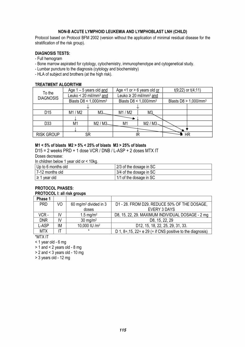
115
NON-B ACUTE LYMPHOID LEUKEMIA AND LYMPHOBLAST LNH (CHILD) Protocol based on Protocol BFM 2002 (version without the application of minimal residual disease for the stratification of the risk group). DIAGNOSIS TESTS: - Full hemogram - Bone marrow aspirated for cytology, cytochemistry, immunophenotype and cytogenetical study. - Lumbar puncture to the diagnosis (cytology and biochemistry) - HLA of subject and brothers (at the high risk). TREATMENT ALGORITHM
To the DIAGNOSIS
Age 1 – 5 years old and Age <1 or > 6 years old or t(9;22) or t(4;11) Leuko < 20 mil/mm3 and Leuko ≥ 20 mil/mm3 and Blasts D8 < 1,000/mm3 Blasts D8 < 1,000/mm3 Blasts D8 > 1,000/mm3
↓ ↓ D15 M1 / M2 M3 M1 / M2 M3 ↓ ↓
D33 M1 M2 / M3 M1 M2 / M3 ↓ ↓
RISK GROUP SR IR HR M1 < 5% of blasts M2 > 5% < 25% of blasts M3 > 25% of blasts D15 = 2 weeks PRD + 1 dose VCR / DNB / L-ASP + 2 doses MTX IT Doses decrease: In children below 1 year old or < 10kg. Up to 6 months old 2/3 of the dosage in SC 7-12 months old 3/4 of the dosage in SC ≥ 1 year old 1/1 of the dosage in SC PROTOCOL PHASES: PROTOCOL I: all risk groups Phase 1 PRD VO 60 mg/m2 divided in 3
doses D1 - 28. FROM D29, REDUCE 50% OF THE DOSAGE,
EVERY 3 DAYS VCR - IV 1.5 mg/m2 D8, 15, 22, 29. MAXIMUM INDIVIDUAL DOSAGE - 2 mg DNR IV 30 mg/m2 D8, 15, 22, 29 L-ASP IM 10,000 IU /m2 D12, 15, 18, 22, 25, 29, 31, 33. MTX IT * D 1, 8∗,15, 22∗ e 29 (∗ if CNS positive to the diagnosis)
*MTX IT < 1 year old - 6 mg > 1 and < 2 years old - 8 mg > 2 and < 3 years old - 10 mg > 3 years old - 12 mg

116
PHASE 2 Infusion CPM in 1 hour 1000mg/m2 D 36 and 64 ARA-C 75 mg/m2 D 38-41, 45-48, 52-55, 59-62 MP 60mg/m2 D 36-63 MTX Dose by age (see above) D 38 and 52 During the CPM: DIURESIS AND CYSTITIS PROPHYLAXIS: fluids (3,000 ml/m2), on the first 24 h + furosemide (0.5 mg/Kg IV), 6h and 12h after the infusion. It can be associated to MESNA (400 mg/m2), 4h and 6 h after the Infusion. PROTOCOL M ( Standard and mean risk groups): ¨MP VO 25 mg/m2 D 1- 56 HD-MTX IV 2g/m2 * in 24 hours D 8, 22, 36, 50 LCV IV 15 mg/m2 ** 42h, 48h and 54h hours after the onset of
MTX MTX (2 h after the onset of MTX)
IT Dose by age (see table)
* at LLA–T, use 5g/m2 if the determination of the serum level of MTX is possible. ** dose with a normal evolution of MTX level. Determination of the serum level of MTX: HOUR AFTER THE ONSET OF THE INFUSION EXPECTED SERUM LEVEL (ΜMOL/L)
Hour 24 ≤ 150.0 Hour 36 ≤ 3.0 Hour 42 ≤ 1.0 Hour 48 ≤ 0.4
Refer to the original Protocol (available at the Service) if MTX levels are above the expected values. BEFORE STARTING MTX: 1 - Hydration and alkalinization: 3000ml / m 2 SG 5% with sodium bicarbonate 40 mEq/L of serum 2 – URINE pH BEFORE, DURING AND AT LEAST 48H AFTER MTX INFUSION. 3 – If ph < 7.5:100 ml SG 5% + 20 mEq of NAHCO3 in 1 hour. HIGH-RISK GROUP: 3 treatment blocks that repeats sequentially in a total of 6 Blocks. Block HR 1’ DEXA VO 20mg/m2 D1-5 VCR IV 1.5mg/m2 D1 AND D6 HD-MTX IV 2g/m2 in 24 hours D1 LCV IV 15 mg/m2 Hour 42, 48 and 54 HDARA-C IV 2g/m2 every 12h D5 CPM IV 200 mg/m2 every 12h 5 doses D2-4 L-ASP IM 10000 IU /m2 D6 MTX / ARA-C / DEXA IT Dose by age D1

117
AGE MTX mg/m2 ARA-C mg/m2 DEXA mg/m2 < 1 year old 6 16 1 Between 1-2 years old 8 20 1 Between 2-3 years old 10 26 2 > 3 years old 12 30 2 MTX/LCV: see notes / support measures of Protocol M Block HR 2’ (HIGH RISK): DEXA VO 20 mg/m2 D1-5 VCR IV 1.5 mg/m2 D1 E D6 HD-MTX IV 2 g/m2 in 24 hours D1 LCV IV 15 mg/m2 Hour 42, 48 and 54 DNR IV 30 mg/m2 in 1 hour D5 IFOSFAMIDE IV 800 mg/m2 every 12h D2-4 for 5 doses Mesna IV 300 mg/m2 4h and 8 h after ifosfamide L-ASP IM 10000/m2 D6 MTX / ARA-C / DEXA IT IT Dose by age D1 MTX/LCV: see notes / support measures of Protocol M Note: vindesine (VDS) 3 mg/m2 (max. 5mg), announced at the original Protocol, was substituted by VCR because it was not, the first, available in Brazil. Block HR 3’(HIGH RISK): DEXA VO 20 mg/m2 D1-5 HD-ARA-C IV 2 g/m2 every 12h D1 -2 VP 16 IV 150 mg/m2 infusion 1 hour every 12 h. Total of 5 doses D3-5 L-ASP IM 10000 IU /m2 D6 MTX/ARA-C/DEXA IT Dose by age D5 Note: To LLA-T-bearing subjects, in the 1st remission, with an allergic reaction to the L-ASP, ERWINASE is indicated, at the same dose as L-ASP. PROTOCOL II (all risk groups): PHASE 1 DEXA VO 10 mg/m2 / day in 3
doses D1 – 22 (with a gradual decrease)
VCR IV 1.5 mg/m2 D8, D15, D22, D29 DOX IV 30 mg/m2 D8, D15, D22, D29 L-ASP IV/IM 10,000 IU / m2 D8, D11, D15, D18 MTX IT dose by age D1 AND D 8 (if CNS + to the diagnosis) PHASE 2 CPM IV infusion of 1 hour 1,000 mg/m2 D36 ARA-C IV 75 mg/m2 D38-41, D45 -48 TG 60 IV 60 mg/m2 D36 to D49 MTX IT dose by age D 38 AND 45 PROPHYLACTIC RADIOTHERAPY OF CNS: age dosage < 1 year old No irradiation ≥ 1 year old 12 Gy
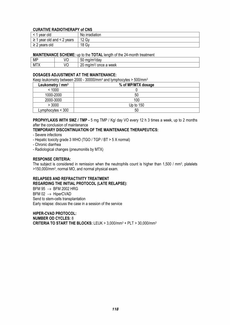
118
CURATIVE RADIOTHERAPY of CNS < 1 year old No irradiation ≥ 1 year old and < 2 years 12 Gy ≥ 2 years old 18 Gy MAINTENANCE SCHEME: up to the TOTAL length of the 24-month treatment MP VO 50 mg/m2/day MTX VO 20 mg/m2/ once a week DOSAGES ADJUSTMENT AT THE MAINTENANCE: Keep leukometry between 2000 - 30000/mm3 and lymphocytes > 500/mm3
Leukometry / mm3 % of MP/MTX dosage < 1000 0
1000-2000 50 2000-3000 100 > 3000 Up to 150
Lymphocytes < 300 50 PROPHYLAXIS WITH SMZ / TMP - 5 mg TMP / Kg/ day VO every 12 h 3 times a week, up to 2 months after the conclusion of maintenance TEMPORARY DISCONTINUATION OF THE MAINTENANCE THERAPEUTICS: - Severe infections - Hepatic toxicity grade 3 WHO (TGO / TGP / BT > 5 X normal) - Chronic diarrhea - Radiological changes (pneumonitis by MTX) RESPONSE CRITERIA: The subject is considered in remission when the neutrophils count is higher than 1,500 / mm3, platelets >150,000/mm3, normal MO, and normal physical exam. RELAPSES AND REFRACTIVITY TREATMENT REGARDING THE INITIAL PROTOCOL (LATE RELAPSE): BFM 95 → BFM 2002 HRG BFM 02 → HiperCVAD Send to stem-cells transplantation Early relapse: discuss the case in a session of the service HIPER-CVAD PROTOCOL: NUMBER OD CYCLES: 8 CRITERIA TO START THE BLOCKS: LEUK > 3,000/mm3 + PLT > 30,000/mm3

119
BLOCKS 1, 3, 5, 7: Hiper-CVAD CPM IV 300mg/m2/d every 12h total 6 doses D 1-3 MESNA IV 600 mg/m2 /d continuous infusion D 1-3 DEXA IV/VO 40 mg /day D 1-4, D11-14 VCR IV 2 mg /d D 4,11 DOXO IV 50 mg/m2/d D 4 G-CSF IV/SC 10 mcg /kg/day ( *) From D 5 (*) Up to leukometry > 3,000/mm3 and platelets > 60,000/mm3 BLOCKS 2, 4, 6 AND 8: HD-MTX- Ara-c
MTX IV 200 mg/m2 in 2 hours D 1 IV 800 mg/m2 continuous infusion in 24h D 1
LCV IV 15 mg every 6 h 8 doses Start 24 h. after the conclusion of MTX HD-ARA-C IV 3g/m2 in 2 hours every 12 h D 2-3 Methylprednisolone IV 50 mg every 12 hours D 1-3 G-CSF IV/SC 10 mcg /kg/day * From D 5 CNS Prophylaxis MTX IT 12 mg D 2 of each cycle ARA-C IT 100 mg D 6 of each cycle SUPPORT MEASURES: Antimicrobial prophylaxis Ciprofloxacin 500 mg every 12 h Fluconazole 200 mg once a day Acyclovir 200 mg every 12 h SMZ-TMP 2 tabl twice a day 3 times a week MAINTENANCE: FOR 2 YEARS - POMP 6-MP VO 50 mg 3x day continuous MTX VO 20mg/m2 weekly VCR IV 2 mg monthly PRED VO 200 mg For 5 days, together with Vcr. Note: protocol created for adults. To see the dosages in absolute values, consider SC ∆ 1.7-1.8 m2.

120
CHRONIC LYMPHOCYTIC LEUKEMIA LABORATORIAL TESTS TO THE DIAGNOSIS: Essential tests: - physical exam with special attention to the lymph nodes sites (including the Waldeyer ring) and liver and spleen size; - performance status; - presence of B symptoms; - full hemogram; - LDH, uric acid, β2-microglobulin; - renal and hepatic function; - direct Coombs test; - serology (HBV, HCV, HIV 1 and 2, HTLV I and II, syphilis and wounds); - electrophoresis of serum proteins and quantification of immunoglobulins; - simple PA and profile chest X-ray; - chest / abdomen / pelvis CT (if peripheral adenopathy); - peripheral blood or bone marrow immunophenotype using the following antibodies: CD3, CD5, CD20, CD23, CD38, FMC7 and cyclin D1; - bone marrow biopsy. Desirable tests: - determination of CD38 and ZAP-70 by flow cytometry or immunohistochemistry; - cytogenetics or FISH (preferred) to detect 17p- and 11q-. Diagnose criteria: - absolute lymphocytosis in the peripheral blood >5000/µL; - mature lymphocytes with less than 55% of pro-lymphocytes; - CD5+, CD19+,CD20+, CD23+, low density of surface immunoglobulins. SCORE FOR DIFFERENTIAL DIAGNOSIS BETWEEN LLC AND OTHER LNH B:
MARKER GRADE 1 GRADE 0 SmIg weak strong CD5 positive negative CD23 positive negative FMC7 negative positive
CD22 or CD79a weak strong The LLC score is usually > 3. At LNH B, the score is usually <3. BINET STAGING:
STAGE DESCRIPTION A Hb>= 10g/dL, platelets>= 100,000/mm3, less than 3 areas attacked B Hb>= 10g/dL, platelets>= 100,000/mm3, 3 or more areas attacked C Hb< 10g/dL or platelets< 100,000/mm3
Note 1: taking into account 5 areas: cervical, axillary, inguinal, liver and spleen. Note 2: other causes of anemia or plateletpenia must be excluded.

121
RAI STAGING: STAGE DESCRIPTION RISK
0 Isolated lymphocytosis Good I lymphadenopathy Intermediate II hepatomegaly and/or splenomegaly Intermediate III Hb< 11g/dL High IV platelets< 100,000/mm3 High
Note: other causes of anemia or plateletpenia must be excluded. PROGNOSTIC FACTORS: FAVORABLE UNFAVORABLE Sequencing DNA Vh >2% mutation ≤ 2% mutation ZAP70 (cytometry) >20% of the cells leukemic negative positive CD38> 30% negative positive 17p- (FISH) absent present 11q- (FISH) absent present TREATMENT INDICATIONS - progressive medullar failure by lymphomatous infiltration: development or worsening of anemia or thrombocytopenia (exclude other cause of anemia / thrombocytopenia); - progressive or massive lymphadenopathy (> 10 cm); - progressive or massive splenomegaly (6 cm below RCD); - progressive lymphocytosis: increase > 50% in 2 months or time of double lymphocytic counting < 6 months; - systemic symptoms: weight loss >10% last 6 months, fever >38o in the last 2 weeks, nocturnal sweating and extreme fatigue. Other causes of these symptoms must be excluded, such as, for example, infection; - self-immune cytopenias; - recurrent infections; - histological change.
RESPONSE CRITERIA (NCI): FULL RESPONSE PARTIAL RESPONSE PROGRESSION Physical exam normal decrease ≥ 50% increase ≥ 50% Symptoms absent Lymphocytes (x106/L)
≤4,000 decrease ≥50% from baseline increase >50%
Neutrophils (x106/L) ≥1,500 ≥1,500 or increase ≥50% from
baseline
Platelets (x106/L) >100,000 ≥100,000 or increase ≥ 50% from
baseline
Hemoglobin (g/dL) >11 (with no transfusion) >11 or increase ≥ 50% Myelogram <30% of lymphocytes BMB With no infiltration nodular infiltration Others length ≥ 2 months length ≥ 2 months Ritcher Syndrome Note: the response criteria tend to become more complex with the evolution of LLC treatment FIRST LINE TREATMENT Age <70 years old without 17p- (deletion of p53): 6 cycles of fludarabine associated to cyclophosphamide. Repeat every 28 days. Administer G-CSF prophylactically in the course of the treatment if the subject presents a severe neutropenia (<500 neutrophils x 106/L) after the first cycle.

122
VENOUS fludarabine 25 mg/m2 D1-D3 cyclophosphamide 250 mg/m2 D1-D3
ORAL fludarabine 24 mg/m2 D1-D5 cyclophosphamide 150 mg/m2 D1-D5
Note 1: administer allopurinol in the first 7 days of the first three cycles; Note 2: adjust the fludarabine dosage according to the renal function Note 3: prophylaxis to PCP with sulfamethoxazole / Trimethoprim and to HSV with acyclovir in the course of the treatment and for at least 6 months after the conclusion. Avoid the concomitant usage of corticoid due to the worsening of immunosuppressant. Age >70 years old or subjects with PS ≥3 or with a severe organic dysfunction: continuous chlorambucil or in “pulse”. Chlorambucil in pulse in high dose is associated to higher rates of SG, but with a more pronounced hematological toxicity. Continuous (use from 6 months up to 3 years) chlorambucil 0.1 mg/kg/day Pulse (every 28 days) (use from 6 months up to 1 year)
chlorambucil 40mg/m2 no D1 (low dose) or chlorambucil 10 mg/m2/day from D1 to D7 (high dose)
Note 1: administer allopurinol in the first 7 days of the first three cycles; Note 2: adding corticoid to the chlorambucil does not increase the response rate. Age <70 years old with 17p- (deletion of p53): the initial treatment with FC is associated to a progression-free survival of 0% in 3 years. In such cases, the chosen treatment is alemtuzumab. As an alternative, methylprednisolone in high doses can be used.
ALEMTUZUMAB
Administer by SC path (less toxic). 30mg 3 times a week (Monday, Wednesday and Friday). Start with two doses of 3 and 10mg. Repeat the scheme above if the subject is more than 7 days not using the drug. Use for 12 to 18 weeks.
METHYLPREDNISOLONE IN HIGH DOSES
1 g/m2 of D1 to D5. Administer every 28 days for up to 6 cycles.
Note 1: administer allopurinol in the first 7 days of the first three cycles; Note 2: prophylaxis to PCP with sulfamethoxazole / Trimethoprim and to HSV with acyclovir in the course of the treatment and for at least 6 months after the conclusion. Note 3: subjects using alemtuzumab must be monitored regarding the reactivation of the latent infection by CMV. It is recommended the antigenemia or PCR every 2 weeks during the treatment and for at least 6 months after the conclusion. Subjects with self-immune hemolytic anemia or secondary PTI: treatment with prednisone in the dosage of 1mg/kg/day for at least 3 weeks. Gradual remove of corticoid. Subjects with positive direct Coombs test, but with no evidence of hemolysis may use fludarabine.

123
SECOND-LINE TREATMENT: • late relapse (more than 6 months after the conclusion of the first-line treatment): repeat the initial
treatment protocol. • early relapse or progression in the course of the treatment:
WITHOUT A PREVIOUS USAGE OF FLUDARABINE
PREVIOUS USAGE OF FLUDARABINE (repeat every 28 days for 6 cycles)
fludarabine 25 mg/m2 IV D1-D3 cyclophosphamide 250 mg/m2 IV D1-D3 or fludarabine 25 mg/m2 IV D1-D5 or fludarabine 24 mg/m2 VO D1-D5 cyclophosphamide 150 mg/m2 VO D1-D5
CYCLE 1: Rituximab 375mg/m2 IV D1 fludarabine 25 mg/m2 IV D2-D4 cyclophosphamide 250 mg/m2 IV D2-D4 CYCLES 2-6: Rituximab 500mg/m2 IV D1 fludarabine 25 mg/m2 IV D1-D3 cyclophosphamide 250 mg/m2 IV D1-D3
ALLOGENIC TRANSPLANT Take into account subjects below 60 years old, refractory to the treatment based on analogous of purine or with 17p-. The optimal transplant is the non-myeloablative related type. Try to reduce the tumoral charge of the subject before the transplant. SPECIAL SITUATIONS Richter Transformation. Usually associated to the fast increase of lymph nodes, fever and weight loss. Make a biopsy in the lymph node for the diagnostic confirmation. It must be treated as an aggressive lymphoma (R-CHOP). Evaluate the autologous transplant for consolidation. Pro-lymphocytic B transformation. Occurs in 10% of the subjects and presents reserved prognosis. Consider the usage of purine analogous and/or monoclonal antibodies.

124
TRICHOLEUKEMIA LABORATORIAL TESTS - Full hemogram with reticulocytes and hematoscopy of peripheral blood - Full biochemistry with hepatogram - Immunophenotype of peripheral blood - Cytochemistry of SP (acid phosphates resistant to tartrate +) - Bone marrow aspirated -> usually dry - Bone marrow biopsy with immunohistochemistry - Positive markers that differentiate from other B-lymphoproliferative diseases: CD11c, CD105, CD25 TREATMENT - 1st CHOICE: Cladribine 0.1mg/kg/day/7 days continuous infusion. Repeat after 6 months in case of absence of remission with the 1st cycle - The subjects must receive prophylaxis to PCP with Bactrim up to 6 months after the conclusion of the treatment. Discontinue Bactrim during the days of cladribine infusion. RELAPSE / OPTIONS - Retreatment with the initial scheme - Usage of other purine analogous (e.g.: pentostatin) - INF - Rituximab - Consider splenectomy only in cases of symptomatic bulky splenomegaly, with no response to the drug treatment

125
HODGKIN’S LYMPHOMA LABORATORIAL TESTS: - tumor histopathology, with immunohistochemistry - hemogram, VHS - biochemistry - hepatic and renal function proofs - LDH and reactive C-protein - chest X-ray - chest, abdomen, and pelvis CT - cervical area CT, if applicable - serology (hepatitis B, hepatitis C, HTLV1, HIV, EVB) - bone marrow biopsy - echocardiogram - β-HCG for women with a child-bearing potential. CLASSIFICATION: WHO - Nodular lymphocytic predominance:CD3, CD15, CD20, CD21, CD30, CD57, with no association to EBV. - Classic Hodgkin’s lymphoma (including nodular sclerosis, mix cellularity, lymphocytic depletion, and rich in lymphocytes): CD3, CD15, CD20, CD30, CD45 with EBV association. - nodular sclerosis - Mix Cellularity - Lymphocytic depletion - Rich in lymphocytes STAGING: ANN ARBOR / COTSWOLD INCLUSIONS I- involvement of an only extralymphatic site or a lymph node. II- one or more regional lymph nodes in the same side of diaphragm and extralymphatic in the same side. (E) III- attack in both sides of the diaphragm, it may include the spleen (S) IV- extralymphatic diffuse involvement. A - Absence of symptoms B - Symptoms: weight loss > 10% in 6 months, fever >38, recurrent nocturnal sweating. “Bulky” tumor - nodal mass >10 cm of diameter or 1/3 of the chest transversal diameter. PROGNOSTIC FACTORS: - serum albumin <4g/dl - hemoglobin<10.5g/dl - stage IV - male

126
- age >45 years old - leukocytosis >15,000/mm3 - lymphocytes <600/mm3 or <8% of white blood cells. - VHS TREATMENT: I and II, with no “bulky” disease – QT (ABVD) 4 cycles + RT. In case of full remission, perform a follow-up every 3 months. In case of partial remission or in case of no remission, treat Ib and IIb (bulky) – QT (ABVD) 4 cycles. In case of full remission, perform two cycles of ABVD+RT. In case of partial remission or disease progression, perform 2 more cycles of ABVD + RT and program autologous TMO III and IV - (Advanced disease) – QT (ABVD) – 4 cycles In case of full remission, perform 2 more cycles In case of partial remission, perform 4 more cycles In case of disease progression remission, prepare for autologous TMO Evaluate BEACOPP Relapse: LATE (above 1 year old after the treatment conclusion) – BEACOPP + RT EARLY – ICE or DHAP + autologous TMO ABVD - Doxorubicin 25mg/m2 EV D1 and D15 - Bleomycin 10IU/m2 EV D1 and D15 - Vinblastine 6mg/m2 EV D1 and D15 - Dacarbazine 350-375 mg/m2 D1 and D15 Repeat every 28 days BEACOPP - Bleomycin - 10mg/ m2 - D8 - Etoposide - 100mg/m2 - D1 to D3 - 200mg/m2 - Doxorubicin - 25mg/m2 - D1 - 35mg /m2 - Cyclophosphamide - 650mg/m2 - D1 - 1200mg/m2 - Vincristine – 1.4mg/m2 - D8 - Procarbazine - 100mg/m2 D1 to D7 - Prednisone - 40mg/m2 D1 to D14 Repeat every 28 days

127
NON-HODGKIN’S LYMPHOMA LABORATORIAL TESTS TO THE DIAGNOSIS - Tumor histopathology - Immunohistochemistry - Hemogram - Biochemistry: uric acid, calcium, phosphorus, urea, creatinine, hepatic function proves, LDH, total and fraction proteins - β2 microglobulin - Chest, abdomen and pelvis CT. Others according to the location of the tumoral mass - Echocardiogram - Serology Hepatitis B, C, HIV and HTLV - MO biopsy - Conventional cytogenetics or FISH in case of bone marrow invasion - Lumbar Puncture if LNH Lymphoblast or Burkitt Lymphoma WHO CLASSIFICATION B-cells lymphoma PRECURSOR B-CELLS NEOPLASIAS - Leukemia / Pre-B Lymphoblast Lymphoma (LLA Pre-B) MATURE B-CELLS NEOPLASIAS (PERIPHERAL) - LLC-B / Lymphocytic lymphoma of small cells - B-Prolymphocyte Leukemia - Lymphoplasmocitary Lymphoma - B-Lymphoma of Splenic Marginal Area (+/- villose lymphocytes) - Tricholeukemia - Myeloma of plasmatic / plasmacytoma cells - B-lymphoma of the extranodal Marginal Area or type MALT - B-lymphoma of nodal Marginal Area (+/- B-cells monocytoid) - Follicular Lymphoma - Mantle cells lymphoma - Diffuse Big Cells B-lymphoma - Mediastinal Big Cells B-lymphoma - Primary effusion lymphoma - Burkitt Lymphoma T- and NK-Cells Lymphoma PRECURSOR T-CELLS NEOPLASIAS - Leukemia / Pre-T Lymphoblast Lymphoma (Pre-T LLA) - Blast NK-cells Lymphoma / CD4+/CD56+ hematodermic neoplasia MATURE T-CELLS NEOPLASIAS (PERIPHERAL) - T-Prolymphocyte Leukemia - T-Granular Lymphocytic Leukemia - Aggressive NK-cell leukemia

128
- Leukemia / T-Cell Lymphoma in Adult (HTLV1+) - Extranodal T/NK Lymphoma, Nasal Type - T-Lymphoma Enteropathy type - T-Lymphoma Hepatosplenic - Skin Panniculitis T-like Lymphoma - Fungoid Mycosis / Sézary Syndrome - Anaplastic Big Cells Lymphoma, T/null, Skin primary type - Anaplastic Big Cells Lymphoma, T/null, Systemic primary type - Peripheral T-Lymphoma - Angioimmunoblastic T-Lymphoma
1 – FOLLICULAR LYMPHOMA
Conventional cytogenetics or FISH - t (14;18) Immunohistochemistry – CD10+, bcl-2+, CD23+/-, CD43-, CD5-, CD20+, Cyclin D1-. Rare cases may be CD10-, bcl-2 -. STAGING
Stage Involvement Area I Only Lymph nodes group II Multiple groups of lymph nodes at the same side of diaphragm III Multiple groups of lymph nodes in both sides of diaphragm IV Multiple extranodal sites or lymph nodes and extranodal disease X Bulk > 10 cm E Extranodal extension or an only isolated site of extranodal disease A/B B Symptoms: weight loss>10%, fever, nocturnal sweating
PROGNOSIS – FLIPI CRITERION
Age > 60 years old Stage Ann Arbor III - IV Hemoglobin Level < 12 g/dl LDH > upper limit Number of nodal sites > 4
Risk Group Factors Number Low 0 - 1 Intermediate 2 High > or = 3

129
TREATMENT
Follicular LNH Stage I and II
RXT or QT
followed by RXT
RC or RP No response
Follow-up Protocol
stages III and IV
Progression
Protocol
stages III and IV
Stage III
Abdominal bulky, III and IV
Indication to treatment* With no indication
RXT (if palliative for symptomatic) or QT
Follow-up
RC or RP With no response or progression Progression
Follow-up With no QT indication 2nd line Diffuse LNH Protocol
Progression
If QT indication 2nd line

130
INDICATIONS FOR THE TREATMENT: Symptoms, dysfunction of target organ, cytopenia secondary to LNH, Bulky, progression and preference of the subject - Stage I and II – Radiotherapy of the field involved - Stages II Bulky, III, IV – Chemotherapy evaluate if it is CD 20+ The treatment may be palliative with monotherapy, or more aggressive according to the age, PS and FLIP. Younger subjects and with an intermediate or high risk must be treated aggressively. Rituximab, in case of CD20+, at 1st line according to subject’s PS. Chlorambucil - Chlorambucil – 0.1 mg/kg/day - Chlorambucil - 40mg/m2 at D1 (low dose) or - Chlorambucil - 10 mg/m2/day from D1 to D7 (high dose) Fludarabine (28 days) - Fludarabine 25 mg/m2 D1-D5 FC (28 days) - Fludarabine 25 mg/m2 D1-D3 - Cyclophosphamide 250 mg/m2 D1-D3 COP (21 days) - Cyclophosphamide 600mg/m2 D1 - Oncovin 1.4mg/m2 (max 2 mg) D1 - Prednisone 100mg VO D1 to D5 Rituximab - 375mg/m2 at D1 of R-COP or R-Fludarabine Perform a pre-medication with Polaramine and Decadron 10mg. Dilute 1mg/1ml. Infuse 50 ml/h at the 2 initial hours, then increase the infusion to 50ml/h every 30 min up to 200ml/h. If there is any reaction, discontinue the infusion, perform Hydrocortisone 100mg EV and return the infusion slowly after the reaction recovery. Fludarabine presents a better response, but with a higher toxicity. It must be avoided in candidates to TCTH. It must also be avoided in subjects with AHAI associated to activity. Perform a prophylaxis with SMZ+TMP. - 2nd Line Treatment (Self-TMO – evaluate age and PS) - Protocols not used yet. Evaluate Big Cells Diffuse Lymphoma protocol. - Maintenance – Rituximab 375mg/m2 one application every 4 months for 2 years.
2 - BIG CELLS DIFFUSE LYMPHOMA
Lumbar puncture in case of testicular, paranasal, parameningeal, MO infiltration, paraorbital, or HIV attack Immunophenotype – CD 20+, CD45+, CD3- . PROGNOSIS IPI Criterion AGE > 60 YEARS OLD RISK GROUP FACTORS NUMBER Stage Ann Arbor III - IV Low 0 - 1 Performance status 2 - 4 Intermediate Low 2 LDH > upper limit Intermediate High 3 Number of nodal sites > 1 High 4 or 5

131
TREATMENT Stage I and II
No Bulky < 10 cm Bulky < 10 cm
IPI risk factors present IPI risk factors absent RCHOP 6-8 cycles w/ RXT
RCHOP 6-8 cycles w/ RXT RCHOP 3 cycles RXT or RCHOP 6-8 cycles if RXT
contraindicated
(1) If Full response - follow-up (2) If Partial response � RXT - If RC � follow-up - If RP � 2nd line treatment (3) If No Response � 2nd line
Stage III and IV
RCHOP 3-4 cycles
Restage
Full or Partial Response Non-response or disease progression
Complete RCHOP 6-8 cycles 2nd line QT + Self-TMO

132
1st line Chemotherapy R- CHOP (21 days)
Cyclophosphamide 750mg/m2 EV D1 Doxorubicin 50mg/m2 EV D1 Oncovin 1.4mg/m2 (max2mg) EV D1 Prednisone 100mg VO D1 to D5 Rituximab 375mg/m2 EV D1 G-CSF 24 h after QT in subjects that present neutropenia among the cycles.
2nd line Chemotherapy – Send to TCTH group to prepare the Self-TCTH ICE (21 to 28 days)
Ifosfamide 1g/m2 in 1h D1 and D2 Etoposide 150mg/m2 2 x/d (hour 1 to 11 and hour 12 to 24) D1 and D2 Carboplatin 200mg/m2 in 1h (hour 11 to 12) D1 and D2 Mesna 333mg/m2 30 min before, 4h and 8h after Ifosfamide
ESHAP (28 days)
Etoposide 60mg/m2 EV D1 to D4 – Care with hypotension. Methylprednisolone 500mg EV D1 to D4 Cytarabine 2g/m2 in 2 h D5 after the conclusion of Cisplatin Cisplatin 25mg/m2/d IC D1 to D4. Evaluate infusion with Mannitol and proper hydration
In case of full or partial remission � Self-TMO. In case of disease progression, palliative scheme
3 - MANTLE LYMPHOMA
- Conventional cytogenetics or FISH in case of MO committed � t (11;14) - Immunohistochemistry – CD5+, CD20+, CD23-, Cyclin D1+, CD10-/+. Some of them can be CD5- or CD23+. If the diagnosis is suspected, perform Cyclin D1, associated to FISH t (11;14) - Lumbar puncture in case of blast variant or neurological symptoms - EDA in case of Waldeyer ring attack - Colonoscopy - If candidate to TMO (P S<2, < 60 years old, with no severe comorbidities), subject and brothers HLA TREATMENT: If he is not a candidate to TMO, consider less aggressive QT

133
1st line Chemotherapy R-HIPERCVAD Cycles 1, 3, 5, 7
Cyclophosphamide 300mg/m2 in 2h every 12 h 6 doses Mesna 600mg/m2 IC D1 to D3 from 1h before the 1st dose to 12h after the last dose and cyclophosphamide Oncovin 2mg EV D4 and D11 Doxorubicin 50mg/m2 EV D4 Dexamethasone 40mg EV or VO D1 to 4 and D11 to 14 Cycles 2, 4, 6, 8 Methotrexate 1g/m2 IC in 24h D1 Leucovorin 25mg/m2 every 6 h start 36h After the onset of MTX. Total 6 dosages Cytarabine 3g/m2 in 1h every 12 h 4 doses D2 and D3 (1g/m2 in subjects > 60 years old) G-CSF 300mcg SC starting 24h after the conclusion of each cycle until medullar recovery, and next cycle can be started
R-CHOP (21 days)
Cyclophosphamide 750mg/m2 EV D1 Doxorubicin 50mg/m2 EV D1 Oncovin 1.4mg/m2 (max 2mg) EV D1 Prednisone 100mg VO D1 to D5 Rituximab 375mg/m2 EV D1
1st line consolidation - In case of full response � Self-TCTH 2nd line Chemotherapy - Cladribine - Schemes with Fludarabine
4 - BURKITT LYMPHOMA
Immunophenotype – sIg+, CD10+, CD20+, TdT-, Ki67+(100%), bcl2-, bcl6+ TREATMENT < 21 years old – Protocol BFM 95 21 – 60 years old – Protocol CODOX-M/IVAC > 60 years old – Protocol CALGB 9251

134
CODOX-M/IVAC
Cycles 1 and 3 Cyclophosphamide 800mg/m2 EV D1 Oncovin 1.4mg/m2 (Max 2mg) D1 and D8 Doxorubicin 40mg/m2 EV D1 Cyclophosphamide 200mg/m2 EV D2 to D5 Methotrexate 1200mg/m2 in 1h followed by 240mg/m2/hour for next 23 hours D10 Leucovorin 192mg/m2 36h from the onset of MTX and 25mg/m2 every 6 h total of 6 doses (ideally dose MTX until <5x10⁸) G-CSF daily from D13 QT IT Ara-C 70mg D1 and D3 MTX 12 mg D15 Cycles 2 and 4 Etoposide 60mg/m2 in 1h D1 to D5 Ifosfamide 1500mg/m2 in 1 h D1 to D5 Mesna 360mg/m2 0h and every 3 h for 7 doses every 24h Cytarabine 2 g/m2 in 3h every 12 h total 4 doses D1 and D2 QT IT MTX 12mg D5 G-CSF starting on D7
CALGB 9251 Cycle 1 Cyclophosphamide: 200 mg/m2/d IV D1 to D5 Prednisone: 60 mg/m2/d PO D1 to D7 Cycles 2, 4, and 6 Ifosfamide: 800 mg/m2/d in 1 hour IV D1 to D5 Mesna: 200 mg/m2/d IV D1 to D5 at 0, 4, and 8 h after ifosfamide Methotrexate: 150 mg/m2 IV in 30 min D1, then 1.35 g/m2 IV in the next 23.5 h, for a total dose 1.5 g/m2 Leucovorin: 50 mg/m2 IV 36 h after the onset of MTX, then 15 mg/m2 every 6 h until the serum level of MTX < 0.05 micromolar Vincristine: 2 mg IV D1 Cytarabine: 150 mg/m2/d in IC IV D4 and D5 Etoposide: 80 mg/m2/d IV in 1 hour D4 and D5 Dexamethasone: 10 mg/m2/d VO D1 to D5 Cycles 3, 5, and 7 Cyclophosphamide: 200 mg/m2/d IV D1 to D5 Methotrexate: 150 mg/m2 IV in 30 min D1, then 1.35 g/m2 IV in the next 23.5 h, for a total dose 1.5 g/m2 Leucovorin: 50 mg/m2 IV 36 h after the onset of MTX, then 15 mg/m2 every 6 h until serum level of MTX < 0.05 micromolar Vincristine: 2 mg IV D1 Doxorubicin: 25 mg/m2/d IV bolus D4 and D5 Dexamethasone: 10 mg/m2/d VO D1 to D5

135
CALGB 9251 (Cont.)
intrathecal QT on cycles 2 to 7 Methotrexate: 15 mg IT D1 Cytarabine: 40 mg IT D1 Dexamethasone: 4 mg IT D1 Cranial radiotherapy 2400 cGy in 12 sessions after the conclusion of the 7th cycle of QT, only for the subjects who present an involvement of MO at the presentation.
BASED ON THE PROTOCOL BFM 95 Staging based on the criteria of St. Jude (Murphy). Risk groups:
R1 Stages I and II, fully resected
R2 Stages I and II, not fully resected Stage III with LDH < 500 IU/L
R3 Stage III with 500 IU/L < LDH < 1000 IU/L Stage IV and LLA B with LDH < 1000 IU/L and negative CNS
R4 Stage IV and LLA B with LDH ≥ 1000 IU/L or positive CNS BLOCKS R1 A B R2 V / A A B A B R3 V / AA BB CC AA BB * R4 V / AA BB CC AA BB * CC * Residual mass = second look surgery with biopsy. - In case of residual disease = conditioning for TMO Autologous - With no disease evidences = expecting conduction. Conclude QT. V (PRE-PHASE) DEXA IV/VO 5 mg/m2 D 1-2 DEXA IV/VO 10 mg/m2 D 3-5 CFM IV 200 mg/m2 1 h D 1-2 MAD IT Dose by age (table) D1

136
BLOCK A DEXA VO/IV 10 mg /m2 in 3 doses D 1-5 VCR IV 1.5 mg/m2 (max. 2mg) D 1 IFO IV 800 mg /m2 D 1-5 ARA-C IV 150 mg/m2 every 12 h D 4-5 VP 16 IV 100 mg/m2 D 4-5 MTX IV 1 g/m2 (1/10 of the dose in 30 min. The remaining in 23
h and 30 min) D1
LCV IV 15 mg/m2 Hour 42, 48 and 54 MAD IT Dose by age (table) D 1.5 BLOCK B DEXA VO/IV 10 mg /m2 in 3 doses D 1-5 VCR IV 1.5 mg/m2 (max. 2mg) D 1 CFM IV 200 mg/m2 in 1 h D 1-5 DOXO IV 25 mg/m2 in 1 h D 4-5 MTX IV 1 g/m2 (1/10 of the dose in 30 min. The remaining in 23
h and 30 min) D1
LCV IV 15 mg/m2 Hour 42, 48 and 54 MAD IT Dose by age (table) D 1.5 BLOCK AA AND BB MTX IV 5 g/m2 (1/10 of the dose in 30 min. The remaining in 23 h
and 30 min) D 1
LCV IV 30 mg/m2 Hour 42, 48 and 54 MAD IT Dose by age (table) D 1.5 BLOCK CC DEXA VO/IV 20 mg /m2 in 3 doses D 1-5 VCR * IV 1.5 mg /m2 (max. 2 mg) D 1 HD-ARA-C IV 3g /m2 in 2 h every 12 h D 1-2 VP 16 IV 100 mg/m2 every 12 h D 3- 5 5 doses MAD IT Dose by age (table) D 1.5 *VDS (vindesine) at the original Protocol: maximum dose: 5 mg. Table MAD IT: v. Protocol LLA non B Values for the Blocks starts: (from Block B/BB): PMN > 500 mm3 Platelets > 50,000/mm3 G-CSF 5 mcg/kg is recommended after the 2 first cycles of branches R3 and R4. Consider TCTH for the relapses
5 – LYMPHOBLAST LYMPHOMA
Conventional cytogenetics or FISH - t ( 8;14 ) and changes, t(9;22) Immunophenotype Lymphoblast lymphoma B:CD10+, CD19+, CD20-/+,TdT+, Igs- Lymphoblast lymphoma T: CD10-,CD19/20-, CD3-/+, CD4/8+,CD1a+/- TdT+, CD2+,CD7+, Igs- TREATMENT: the same as LLA

137
6 – GASTRIC MALT LYMPHOMA
DIAGNOSIS Essential tests: - physical exam; - performance status; - presence of B-symptoms; - full hemogram; - LDH, uric acid, β2-microglobulin; - renal and hepatic function; - serology (HBV, HCV, HIV 1 and 2, HTLV I and II, syphilis and wounds); - PA and profile simple chest X-ray; - chest / abdomen CT; - histopathology of gastric biopsy; - immunohistochemistry: CD5-, CD10-, CD20+, CD23-/+, cyclin D1- and bcl-2 negative; - coloration for H. pylori at the histopathology of gastric biopsy; - bone marrow biopsy. Desirable tests: - endoscopic ultrasound in order to determine the perigastric lymph node attack; - EDA with multiple biopsies.
STAGING
Lugano Staging TNM Ann Arbor
Tumoral Extension
stage I confined to TGI T1N0M0 IE mucosa, submucosa T2N0M0 IE proper muscle T3N0M0 IE serosa
stage II extension to abdomen T1-3N1M0 IIE perigastric lymph nodes T1-3N2M0 IIE more distant lymph nodes
stage IIE penetration of serosa T4N0M0 IE invasion of adjacent structures
stage IV extranodal disease or attack of
both sides of diaphragm T1-4N3M0 IIIE lymph nodes on both sides of
diaphragm / metastasis T1-4-N0-3M1 IVE TREATMENT: - Stage IE H. pylori positive: antibiotic therapy for H. pylori. Re-staging in 3 months; - Stage IE H. pylori negative or stage IIE: antibiotic therapy for H. pylori or radiotherapy 30-33 Gy (preferred). - Re-staging in 3 months for the subjects treated only with ATB; - Stage III or IV. Indications of treatment: symptomatic, TGI hemorrhage, bulky, disease progression or preference of the subject. In case of indication, consider: T induction QT (isolated agent or polychemotherapy) or RT located on specific situations. Monitor with EDA.

138
- Surgical treatment is only indicated in emergence situations. Attention to subjects with advanced disease that present high risk of hemorrhage / perforation by starting the QT. - Re-staging through EDA + biopsy for the subjects treated only with ATB: - H. pylori negative and negative lymphoma: observe and re-stage after 3 months; - H. pylori negative and positive lymphoma: treat the symptomatic cases with RT and observe the asymptomatic cases / re-stage after 3 months; - H. pylori positive and negative lymphoma: second-line treatment for H. pylori. Re-stage after 3 months; - H. pylori positive and positive lymphoma: in case of stable disease, second-line treatment for H. pylori. In case of ongoing disease, RT. - Re-staging for subjects treated with RT: EDA with biopsy every 3 months at the first year and then every 6 months. Image tests as necessary. Subjects that present relapse after RT, must be treated with QT.
7 – GASTRIC MALT LYMPHOMA
(see protocols for follicular lymphoma)
8 – NON-GASTRIC MALT LYMPHOMA
- stage IE and II: locoregional RT (20-30 Gy). Surgery may be considered for some sites: lungs, breast, skin, thyroid, colon and small intestine. In the case of a surgical treatment, consider additional RT if the margin in the part is not free of disease; - stages III and IV: see treatment protocols for follicular lymphoma; - stages III and IV with big B-cells component: see treatment protocols for LDGCB.
9 – NODAL MARGINAL AREA LYMPHOMA
- typical immunohistochemistry: CD5-, CD10-, CD20+, CD23+/-, cyclin D1- and bcl-2-; - remove primary extranodal sites: cervical lymph nodes (eyes, thyroid and salivary glands), mediastinum lymph nodes (lungs), axillary lymph nodes (lungs, skin, salivary glands), abdominal lymph nodes (TGI) and inguinal lymph nodes (genitourinary tract and skin); - staging and treatment according to the follicular lymphomas.
10 – SPLENIC MARGINAL AREA LYMPHOMA
- HCV+ with indication of anti-viral treatment: treatment for HCV and follow-up of LNH. In case of progression, QT according to the protocol of follicular lymphomas; - HCV– asymptomatic: note; - HCV- with symptoms of LNH and/or cytopenias: splenectomy or QT (see protocol of follicular LNH).

139
11 – PERIPHERAL T-LYMPHOMA
- fundamental the differentiation among the non-specified peripheral T-lymphoma, systemic big cells anaplastic lymphoma and angioimmunoblastic T-lymphoma; - non-specified peripheral T-lymphoma: heterogeneous group with variable clinical presentation. Usually diagnosed on the most advanced stages. Bone marrow and skin infiltration is common. Immunohistochemistry: CD2+/-, CD3+/-, CD4 +/-, CD7+/-, CD8+/-, TdT-, CD30- and ALK-; - angioimmunoblastic T-lymphoma: general lymphadenopathy, hepatosplenomegaly and rash. Polyclonal hypergammaglobulinemia, eosinophilia and self-immune hemolytic anemia are common. The proliferation of follicular dendritic cells and endothelial cells is frequent at the histopathology. Immunohistochemistry: CD2+/-, CD3+/-, CD4 +/-, CD7+/-, CD8+/-, TdT-, CD 21+ (dendritic cells close to the venulas), CD30- and ALK-; - systemic big cells anaplastic T-lymphoma: the attack located is frequent and usually with a good response to the QT. Immunohistochemistry: CD2+, CD3+, TdT-, CD15-, EMA+, CD30+ and ALK+. The sub-group with ALK- presents the worst prognosis. The ALK protein is resultant from t (2;5) (it can be evaluated by FISH or cytogenetics); - angioimmunoblastic T-lymphoma: initial treatment with prednisone 1 mg/kg/day. At the absence of response after 10 days, consider QT (see protocol below); - non-specified peripheral T-lymphoma and big cells anaplastic T-lymphoma staging I and II: CHOP of 6 to 8 cycles and additional RT if initial bulky or located persistence of disease after the conclusion of the treatment. Re-staging during the treatment must include all the tests initially positive; - non-specified peripheral T-lymphoma and big cells anaplastic T-lymphoma staging III and IV: CHOP of 6 to 8 cycles and additional RT if initial bulky or located persistence of disease after the conclusion of the treatment. Re-staging during the treatment must include all tests that were initially positive. Subjects with positive ALK-1 anaplastic lymphoma with full response must be followed. Subjects with negative ALK-1 anaplastic lymphoma, peripheral T-lymphoma or angioimmunoblastic T-lymphoma must be taken into account for the chemotherapy in high doses and autologous transplant in the first remission (mainly if IPI intermediate or high). In the cases of partial response or absence of response to the initial protocol of QT, use the rescue protocol for the high-grade LNH and send to the autologous transplant the cases with chemosensitivity to rescue; - consider prophylaxis of tumoral lysis syndrome and prophylaxis of CNS in specific cases.

140
LEUKEMIA / T-CELLS LYMPHOMA OF THE ADULT
DIAGNOSE CRITERIA Common: Cytopathology / Histopathology - Infiltration by activated malign lymphocytes (flower cells) - Expression of CD2, CD3, CD4, CD5 - No expression of CD7 and CD8 - Expression of HLA lymphocyte activation markers class II and receptor of IL2 - Positive serology for HTLV - Clonal integration of provirus to the tumoral cells SUBTYPES
SMOLDERING CHRONIC FORM ACUTE FORM (LEUKEMIC)
LYMPHOMATOUS FORM (TUMORAL)
Skin injury or pulmonary infiltrate Absence of adeno / visceromegaly 1 – 5% leukemic cells at SP Absence of hypercalcemia Normal LDH
Skin, hepatic, pulmonary injury, or adenomegaly With no other visceral attack. Lymphocytosis > 4000 with circulating leukemic cells. Absence of hypercalcemia LDH < 2x normal
Organomegaly Multiple visceral attack Market commitment of SP by leukemic cells Frequent hypercalcemia High LDH
Organomegaly Multiple visceral attack < 1% leukemic cells at SP Possible hypercalcemia High LDH
LABORATORIAL TESTS - Full hemogram with reticulocytes and hematoscopy of peripheral blood - Biochemistry with hepatogram - LDH - Immunophenotype of peripheral blood in case of high leukometry or morphologic evidence of pathologic cells. - Ganglial biopsy with immunohistochemistry - Bone marrow aspirated with immunophenotype - Bone marrow biopsy with immunohistochemistry - Lumbar puncture - Tomographies of Chest, Abdomen and Pelvis. 1ST LINE TREATMENT Chronic Form and Smoldering – do not treat Lymphomatous Form - CHOP x 3 -> INF + AZT Leukemic Form - INF + AZT - Interferon up to 3,000,000 U/m2/day - AZT 1g/day - Cytoreduction with PDN 40mg/m2/day/ 7 days if hyper-leukocytary. CNS Prophylaxis: MADIT at D1 of each cycle of CHOP or 1x/month. Consider TMO allogenic in 1st RC in all subjects * RELAPSE TREATMENT If you have done CHOP : ICE, ESHAP x 3 -> INF + AZT If you have done INF + AZT : CHOP x3 -> INF + AZT

141
FUNGOID MYCOSIS AND SÉZARY SYNDROME DIAGNOSE CRITERIA Cutaneous lymphoma of low degree T-cells with a median age to the diagnosis of 55 years old and with a predominance of male subjects (2:1). Attack of lymphadenopathy found in 50%. Pruritus, exfoliative erythroderma and T-cells with a volute nucleus CD4+ in the blood characterize the Sézary syndrome. Neoplastic cells express CD3, CD4 and CD5 in addition to the positive coloration for acid phosphatase and esterase alpha naftil. Structural abnormalities at the chromosomes 1 and 6 and numeric abnormalities at the chromosomes 7, 11, 21 and 22 are frequent. Biopsy of skin, lymph node and BMB. STAGING “T” Staging for skin injuries: IA - T1+N0+M0 IB - T2+N0+M0 IIA - T1 ouT2+N1+M0 IIB - T3+N0 or N1+M0 IIIA - T4+N0+M0 IIIB - T4+N1+M0 IVA - T1 to T4+N2 or N3+ M0 IVB - T3 to T4+N0 to N3+M1
T1: erythematous plates located in less than 10% of the body surface; T1: plates covering more than 10% of the body surface; T3: skin tumors and T4: general erythroderma B symptoms: fever, nocturnal sweating and losing weight
TREATMENT Stadium IA: Topical treatment with corticosteroid, retinoid, chemotherapic agents (nitrogenous-HN2 mustard), phototherapy (UVA / UVB) and located radiotherapy (localized electron beam therapy). HN2 is the chosen treatment at the dosage of 10 to 20 mg% once a day until the regression of injuries and maintenance for 1 to 2 months. In case of slow response, increase the frequency of the applications for twice a day or increase the dosage for 30 to 40 mg% in an aqueous solution or ointment. Carmustine (BCNU) is another chemotherapic agent with an efficacy similar to HN2, but it is used on a limited form, due to the hematological effects and the appearance of telangiectasias where applied. EBT (electron beam therapy) is used in sole injuries or in located MF followed by maintenance with topical HN2. The EBT total is reserved for the aggressive skin disease. Phototherapy: UVB or PUVA (psoralen associated to phototherapy with UVA). It can cause xerosis, pruritus, erythema and nauseas, whose treatments are symptomatic and, in the long term, can cause high risk of cataract, secondary skin carcinoma and melanoma. Stadium IB/IIA: Total EBT is used if the subject with a recent history of fast progression of the disease or failure in the usage of topical HN2 and/or phototherapy. It can cause erythema and desquamation in addition to incomplete alopecia, fall of nails, sweating, chronic dry skin, telangiectasia and increase of the incidence of skin malignities such as carcinoma of squamous and baseline cells. Total dosage of 36 Gy in 10 weeks.

142
HN2 or PUVA (similar to the located disease). In case of failure to the usage of an only topical agent, associate to EBT Total or PUVA+HN2 or PUVA+EBT total with Alpha Interferon or systemic retinoid. Alpha Interferon of 3 to 5 million IU 3x a week. Oral retinoid: Used in refractory or advanced disease in combined therapies or as an adjuvant. It can cause photosensitivity, xerosis, myalgia, arthralgia, headache and nocturnal blurred eyes, in addition to the teratogenic effects, hepatotoxicity and hyperlipidemia. The most used retinoids are: Isotretinoin 1mg/kg day, Acitretinoin 25 to 50 mg/day and Bexarotene 100 to 300 mg/m2 day. Stadium IIB: General involvement with tumors and skin disease. EBT Total + topical HN2. In case of a small number of injuries, use topical HN2 or PUVA + local EBT. In case of failure or relapse after EBT Total, use a combined regimen to topical agent + systemic therapy: IFN alpha + PUVA or systemic retinoid + PUVA. In case of re-calcitrating tumor, use systemic therapy as a biological therapy or a combination of biological therapy and QT with or without a topical therapy. Liposome doxorubicin as a monotherapy or associated to EBT Total. Stadium III: Erythrodermal MF with severe pruritus and skin inflammation. EBT Total. In case of peripheral blood in the involved person, use PUVA in low and increasing dosages associated or not to IFN alpha. Photopheresis or extra-body photochemotherapy is the primary therapy for MF or erythrodermal SS every 4 weeks or even from 2 to 3 weeks in case of very severe disease. Side effects include nauseas, low fever and mild malaise. In case of slow or partial response, associate IFN or systemic resinoid. In case of erythrodermia with or without a limited extracutaneous disease, use Qt in monotherapy: Oral methotrexate from 5 to 50 mg/week or 25 to 50 mg/m2 venously once a week. Retinoids: used alone or associated to PUVA or IFN alpha. A similar dose to those used in the re-calcitrating tumor. Stadium IV: Extracutaneous disease with only a palliative control. Use QT alone or associate the skin therapy (radiotherapy or IFN alpha. QTs used: cyclophosphamide, vincristine, prednisone and adriamycin (CHOP) or cyclophosphamide, prednisone and vincristine (COP); cyclophosphamide, adriamycin, vincristine and Etoposide (CAVE) and COP with MTX (COMP). IFN< Systemic retinoids and photopheresis may be used as adjuvant after QT. In case of QT in monotherapy, use: MTX, Liposome doxorubicin (20 30mg/m2 every 3 to 4 weeks), gemcitabine (D1, D8 and D15 in a cycle of 28 days at the dosage of 1200mg/m2 venous in an infusion of 30 minutes 6 cycles), Etoposide, cyclophosphamide or purine analogous as fludarabine.

143
Fusion of recombinant proteins. Denilleucina diftitox, which is used in advanced or re-calcitrating diseases that express CD25. Allogenic TMO Emergent therapies Inhibitors of deacetylasis histone (vorinostat) 400mg/day VO whose toxicity includes diarrhea, fatigue, nauseas, anorexia and hematological effects such as anemia, thrombocytopenia and neutropenia grade 1 to 2, in addition to pulmonary embolism. Alemtuzumab-monoclonal antibody anti-CD52 with a prophylactic usage of antiviral and antibiotic. CpG oligonucleotides. Photodynamic therapy with non-ionizing laser. Monoclonal antibody anti T-cells. Zanolimumab-antiCD4.

144
MULTIPLE MYELOMA LABORATORIAL TESTS TO THE DIAGNOSIS - Anamnesis and physical exam - Full hemogram - Biochemistry: Urea, creatinine, electrolytes, calcium, total and fraction proteins, LDH, hepatogram, coagulogram. - B2 microglobulin - Dosage of Immunoglobulin IgG, IgA, IgM. Dosage of mild chains if the Igs dosages are normal and if there is some criteria for MM - Electrophoresis of proteins and Immunofixaton in the blood (desirable) - Electrophoresis of proteins and Immunofixaton in the urine (desirable) - Myelogram. BMB, if necessary. - Cytogenetics conventional (research of del13) - FISH for t(4; 14), t(14; 16), Del 17q13 - Urine of 24 hours: proteinuria of 24 hours, creatinine clearance - Bone inventory - Subjects up to 60 years old: HLA compatible DEFINITION / CLASSIFICATION Multiple Myeloma (all three Criteria must be met) Presence of Serum and/or Urinary Monoclonal protein Presence of clonal plasmatic cells at the Bone marrow or a plasmacytoma
Presence of injury in an organ related to the disease activity Increased Calcium Renal Failure Anemia Bone, Lithic injury
Multiple Myeloma Smoldering, Asymptomatic (both criteria) Serum monoclonal protein > 3 g/dl and/or plasmatic cells MO > 10% No injury in an organ related to the disease activity
Monoclonal Gammopathy of Undetermined Significance – MGUS (All criteria) Serum Monoclonal Protein IgG<3 g/dl; IgA<2g/dl; Kappa or urinary lambda <1g/24h Plasmocyte at MO < 10% No injury in an organ related to the disease activity or lymphoproliferative disorder
PROGNOSIS STAGE DURIE-SALMON ISS
I
All the following: - Hb>10 g/dl - Calcium < 12 mg/dl - Rx with no lithic injuries
At least one of the following: IgG < 5 g/dl IgA < 3 g/dl Bence Jones < 12g/24h
β2M < 3.5 mg/dl Albumin > 3.5 g/dl
II No stage I or II No stage I nor II
III
One or more: - HB < 8.5 g/dl - Calcium > 12 mg/dl - Advanced lithic injuries
At least one of the following: IgG > 7 g/dl IgA > 5 g/dl Bence Jones > 12 g/dl
β2M ≥ 5.5 mg/dl
Sub-ranking A B
Criteria Normal renal function (Cr < 2 mg/dl) Abnormal renal function (Cr > 2 mg/dl)

145
RISK STRATIFICATION High Risk conventional cytogenetics - del 13; FISH - t( 4;14 ); t ( 14;16 ), del p53 (17p13) Standard Risk when the changes above mentioned are not present TREATMENT Treatment indication – Only subjects with symptomatic Multiple Myeloma. Subjects eligible for the transplant: subjects up to 70 years old Exclusion criteria for the transplant: >70 years old, PS >2, severe organic dysfunction. Eligible for TCTH: send to the transplant outpatient clinic and in parallel start the treatment: Dexamethasone / Thalidomide – Repeat every 4 weeks, a minimum of 4 cycles until the TCTH is scheduled Thalidomide 100 to 200mg/day VO daily Dexamethasone 40mg VO D1-4, D 9-12, D 17-20 on the odd cycles D1-4 on the even cycles Women with a child-bearing potential: do not use thalidomide Prophylaxis of deep venous thrombosis: AAS 100mg Prophylaxis of HDA: omeprazole 20mg VO/day Not eligible for TCTH: cycles every 4-6 weeks until the plateau. - Melphalan 4mg/m2/day VO 7 days - Prednisone 40mg/m2/day VO 7 days - Thalidomide 100mg/day VO continuous usage MAINTENANCE Thalidomide 50-100mg/day until there is a progression of the disease FOLLOW-UP Monthly before each treatment cycle: Hemogram, Biochemistry with evaluation of the glycemia, renal function, calcium, total and fractioned proteins. At least every 3 months: Ig Dosage After the normalization of the Ig dosage, ask for an Immunofixation in the blood and urine, in order to prove the disappearance of the monoclonal peak Annually: bone inventory COMPLICATIONS Anemia: Usage of Erythropoietin 8000 IU SC 3 times a week Lithic injuries: Bisphosphonates - Pamidronate 90 mg EV every 28 days for 2 years. It can be done on the third year every 3 months. Renal Failure: Dialysis if necessary. Follow-up by nephrology.

146
RESPONSE CRITERIA Full Response Absence of monoclonal protein in the serum and urine by Immunofixation < 5 % of Plasmocyte at MO ( myelogram and BMB) No increase in the size and number of lithic injuries Disappearance of plasmacytoma of soft tissues Partial Response > 50 % of decrease in the serum monoclonal protein Decrease in the excretion of mild chains in the urine in > 90% or to < 200mg/24 hours Non-secretor myeloma decrease > 50% of plasmocytes at MO Decrease > 50% at the soft tissues plasmacytoma No increase in the size and number of lithic injuries Minimum Response Decrease of 25 to 49% of serum monoclonal protein 50 to 89% of decrease at the excretion of the mild chains in the urine (>200mg/24h) Non-Secretor Myeloma 25 to 49% of decrease at the plasma cells of MO 25 to 49% of decrease in the size of soft tissues plasmacytoma No increase in the size and number of lithic injuries

147
TREM Protocol
Eligible to TMO
Dexamethasone + TalidomideX4
Biphosphonate AAS
Risk
Stratification
Low Risk or
between 60 and 70 years old
High Risk
Self-TMO
(collection to 2 TMO)
Does it have a
donor?
RC or VGPR? Yes No
Yes No Self-TMO Self-TMO
Maintenance with Thalidomide D100 of
TMO 2nd Self-TMO Mini-Alo TMO Self-TMO
Progression Maintenance with
Thalidomide D100 of TMO
Maintenance with
Thalidomide D100 of TMO
2nd Self-TMO Progression Progression or VGPR
Maintenance with
Thalidomide D100 of TMO
Protocol with Bortezomib
Protocol with Bortezomib

148
WALDENSTRON MACROGLOBULINEMIA LABORATORIAL TESTS TO THE DIAGNOSIS: - Full Hemogram - Biochemistry with renal and hepatic function proofs - Coagulogram - Myelogram with evaluation of the infiltration by lymphocytes - Immunophenotype - BMB - Immunoelectrophoresis - Dosage of Immunoglobulin IgG, IgA, IgM, mild chains Diagnosis criteria: - IgM-value independent Monoclonal Gammopathy IgM - >10% of infiltration of MO by small lymphocytes that present Plasmocyte differentiation - Typical immunophenotype (i.e., IgM of surface +, CD5+/-, CD10-, CD19+, CD20+, CD22+, CD23-, CD25+, CD27+, FMC7+, CD103-, CD138-); - MW smoldering – Criteria above, in asymptomatic subjects or those with no anemia. They must not be treated. - Monoclonal Gammopathy IgM with an indefinite meaning - Dosage of IgM < 3.0 g/dL - Absence of anemia, hepatosplenomegaly, lymphadenopathy and systemic symptoms. - Minimal or no infiltration lymphoplasmacytic of MO (< 10%). STAGING - Stage A (low risk) - β2M<3 and Hb>12 OS in 5 years=87% - Stage B (mean risk) - β2M<3 and Hb<12 OS in 5 years=63% - Stage C (mean risk) - β2M>3 and IgM<4 OS in 5 years=53% - Stage D (high risk) - β2M>3 and IgM>4 OS in 5 years=21% TREATMENT - Asymptomatic subjects (Smoldering MV) – Must not be treated - Treatment indications - Symptoms related to the hyperviscosity (oronasal bleeding, blurred eye, headache, paresthesias, torpor, coma), anemia, pancytopenia, symptomatic organomegaly, bulky, base lymphoproliferative disease, paraneoplastic neuropathy. - Plasmapheresis - hyperviscosity, bleeding, neurological setting - Chemotherapy - Chlorambucil – 0.1mg/Kg/d continuous usage or 0.3mg/Kg/d for 7 days every 4 to 6 weeks until gets to the plateau - CHOP - Purine Analogous - Cladribine, Fludarabine - Other regimens - Melphalan (6mg/m2), Cyclophosphamide (125mg/m2) and Prednisone (40mg/m2) D1 to D7 every 4 to 6 weeks up to 12 cycles. When the disease is stable, start Chlorambucil 3mg/m2 and Prednisone 6mg/m2 daily until the disease progression.

149
BONE MARROW TRANSPLANT
TIME RECOMMENDED FOR THE EVALUATION OF A subject BY A HEMATOPOIETIC STEM CELL transplantATION TEAM (FOR AUTOLOGOUS OR ALLOGENIC transplant) Adapted from the recommendations of 2007 National Marrow Donor Program (NMDP) and the American
Society for Blood and Marrow Transplantation (ASBMT)
ACUTE MYELOID LEUKEMIA IN THE ADULT:
− High-risk LMA, including: previous hematological disease, leukemia related to the treatment with QT/RT and induction failure
− RC1 with high-risk cytogenetics − RC2 and so on
ACUTE LYMPHOBLASTIC LEUKEMIA IN THE ADULT: − High-risk LLA, including: high-risk cytogenetics (Ph+, 11q23), high leukometry at the diagnosis
(>30,000 – 50,000), testicular or CNS leukemia, absence of RC with 4 weeks treatment and primary induction failure
− RC2 and so on Myelodysplasia:
− IPSS intermediate 1, intermediate 2 or high, which includes: percentage of blasts at the bone marrow above 5%, intermediate- or bad-risk cytogenetics and more than one cytopenia
CHRONIC MYELOID LEUKEMIA − Absence of minor hematological response or cytogenetics response after three months of treatment
with imatinib − Absence of full cytogenetics response with 6 to 12 months of therapy with imatinib − Progression of the current disease with imatinib − Quick phase or blast crisis ACUTE MYELOID LEUKEMIA IN THE CHILD: − Monosomy of 5 or 7, age below two years old at the diagnosis, primary induction failure − RC1 with HLA donor similarly related − RC2 and so on ACUTE LYMPHOBLASTIC LEUKEMIA IN THE CHILD: − Primary induction failure, Ph+, leukometry ABOVE 100,000 at the diagnosis, rearrangement 11q23,
Burkitt, adolescence at the diagnosis − RC1 below 18 months − RC2 and so on
noN-Hodgkin LYMPHOMA: − Follicular: in a first relapse or transformed in LDGCB − LDGCB: in a first relapse or subsequent or with an absence of RC with first-line treatment − Mantle: always send − Peripheral T lymphoma: in RC1 HODGKIN LYMPHOMA: − Absence of RC after the first-line treatment − First relapse or subsequent MULTIPLE MYELOMA:
− Always send (if below 70 years old)
PAROXYSMAL NOCTURNAL HEMOGLOBINURIA:
− Hypoplastic forms with marked pancytopenia
SEVERE APLASTIC ANEMIA:

150
− If HLA-identical related donor − Whenever there is a failure to the immunosuppressant therapy.

151
INDICATIONS OF AUTOLOGOUS HEMATOPOIETIC STEM-CELL TRANSPLANTATION - LNHDGC with absence of a full remission with the first-line treatment or in a first chemosensitive relapse; - Follicular LNH in a first relapse with residual infiltration by lymphocytes B <20% (flow cytometry) after 3 rescue QT cycles; - Peripheral LNH T with absence of full remission with the first-line treatment or in the first full remission; - LNH of the mantle cell with absence of full remission with the first-line treatment or in the first full remission; - Hodgkin’s Lymphoma with absence of full remission with the first-line treatment or in the first relapse; - Symptomatic multiple myeloma; - LMA M3 in the second molecular remission (PCR or FISH); - LMA in the second full remission with a minimum negative residual disease by flow cytometry after the Rescue QT and with no donor available (related and non-related); - metastatic testicular tumor. CHECK-LIST PRE-AUTOLOGOUS HEMATOPOIETIC STEM-CELL TRANSPLANTATION - odontological evaluation; - psychological evaluation; - social service evaluation; - absence of severe systemic comorbidities; - absence of ongoing infection (especially invasive fungal infection); - PS (ECOG) ≤2; - simple chest X-Ray; - forced vital capacity (CVF) ≥60% and diffusion of normal CO (DLCO) (flow spirometry); - LV ejection fraction ≥50% (transthoracic echocardiogram); - electrocardiogram; - β-HCG for child-bearing potential women; - immunohematological study; - coagulogram; - serology (HIV 1 and 2, HTLV I and II, HBV, HCV, syphilis and wounds); - ferritin; - free-T4 and TSH; - TGO and TGP up to twice the normality; - total bilirubin ≤2 mg/dL; - serum creatinine ≤1.5 mg/dL; - creatinine clearance ≥60 mL/min.

152
TREATMENT PROTOCOL - ACUTE MYELOID LEUKEMIA MOBILIZATION REGIMES - reduced Linker protocol (recommended by the lower toxicity): - cytarabine 2 g/m2 D1-D3 in 3 hours; - Etoposide 5 mg/kg every 12 hours D1-D3; - G-CSF 5 µg/kg every 12 hours from D14; - onset of apheresis with CD34 ≥10/µL. - standard Linker protocol: - cytarabine 2 g/m2 D1-D4 in 3 hours; - Etoposide 40 mg/kg in continuous venous infusion D1-D4; - G-CSF 5 µg/kg every 12 hours from D14; - Onset of apheresis with CD34 ≥10/µL. Forecast to start the collection: D25 (D18 up to D40). - Minimum collection of 2 x 10⁶ cells CD34/Kg. Conditioning and prophylaxis regimen - busulfan 1mg/kg/dose every 6 hours from D-7 to D-4 (total dosage of 16 mg/kg) or venous busulfan 0.8 mg/kg/dose every 6 hours from D-7 to D-4 (desirable due to the higher pharmacokinetic foreseeability); - diazepam 5 mg IV every 6 hours starting 12 hours before the first dose of busulfan and finishing 12 hours after the last dose; - cyclophosphamide 60 mg/kg/dose on D-3 and D-2 (total dose of 120 mg/kg); - mesna 10 mg/kg/dose starting one hour before each infusion of cyclophosphamide and every 4h after the infusion for five doses more (six doses in the total); - furosemide 20 mg 1 hour, 4 hours and 8 hours after each dose of cyclophosphamide; - fluconazole 200 mg IV every 12 hours from D-2 until engraftment; - acyclovir 250 mg/m2 IV every 12 hours from D-2 until engraftment; - albendazole 400 mg VO for three consecutive days at the hospitalization; - allopurinol 300 mg VO by day during the conditioning; - ursodesoxycolic acid 300 mg VO every 12 hours; - ondansetron 0.15 mg/kg every 6 h in the course of chemotherapy and until necessary; - hyperhydration with SG 5% with additives at the volume of 3000 ml/m2/day; - G-CSF 5 µg/kg/day starting at D+5.
TREATMENT PROTOCOL - LYMPHOMAS MOBILIZATION REGIMES - ESHAP (recommended): - Etoposide 60 mg/m2 D1-D4; - methylprednisolone 500 mg D1-D4; - cisplatin 25 mg/m2 D1-D4 IV continuous (replace magnesium and stimulate diuresis with mannitol); - cytarabine 2 g/m2 at D5; - mabthera 375 mg/m2 D1 in case of LNH CD20 positive (desirable); - G-CSF 5 µg/kg every 12 hours from D6;

153
- onset of apheresis with CD34 ≥10/µL. Forecast for the onset of the collection D16 (D13-D22). - ICE: - Etoposide 100 mg/m2 D1-D3; - ifosfamide 5 g/m2 in a continuous venous infusion at D2; - mesna 5 g/m2 in a continuous venous infusion at D2. Dilute with ifosfamide; - carboplatin at the dose AUC 5 at D2, calculated by the formula 5 x (creatinine clearance + 25). Maximum dose of 800 mg. - mabthera 375 mg/m2 D1 in case of LNH CD20 positive (desirable); - G-CSF 5 µg/kg every 12 hours from D5; - onset of apheresis with CD34 ≥10/µL. Forecast for the onset of the collection D14. - Minimum collection of 2 x 10⁶ cells CD34/Kg. Conditioning and prophylaxis regimen - Escalated CBV: - cyclophosphamide 1800 mg/m2/day from D-6 to D-3 (total dose of 7200 mg/m2); - Etoposide 400 mg/m2 every 12 hours from D-6 to D-4 (total dose of 2400 mg/m2); - BCNU 450 mg/m2 at D-2. - CBV standard: - cyclophosphamide 1500 mg/m2/day from D-6 to D-3 (total dose of 6000 mg/m2); - Etoposide 200 mg/m2 every 12 hours from D-6 to D-4 (total dose of 2400 mg/m2); - BCNU 300 mg/m2 at D-2. - the choice of regimen with increased or standard doses must be taken by the transplant team; - mesna 1/6 of the daily dose of cyclophosphamide, starting one hour before each infusion of cyclophosphamide and every 4 h after the infusion for more five doses (six doses in the total); - furosemide 10 mg 1 hour, 4 hours and 8 hours after each dose of cyclophosphamide; - fluconazole 200 mg IV every 12 hours from D-2 until the engraftment; - acyclovir 250 mg/m2 IV every 12 hours from D-2 until the engraftment; - albendazole 400 mg VO for three consecutive days at the hospitalization; - allopurinol 300 mg VO by day during the conditioning; - ursodesoxycolic acid 300 mg VO every 12 hours; - ondansetron 0.15 mg/kg every 6 h in the course of the chemotherapy and until necessary; - hyperhydration with SG 5% with additives at the volume of 3000 ml/m2/day; - G-CSF 5 µg/kg/day, starting at D+5. Complimentary radiotherapy: The pre-transplant radiotherapy must be avoided because it increases the risk of pneumonitis. The radiotherapy located after the autologous transplantation must be evaluated at the subjects with pre-transplant “bulky disease” or persistence of the located disease after the transplant. Usage of mabthera in LNH CD20 positive: It is desirable the addition of mabthera to the rescue / mobilization chemotherapy protocol in subjects with LNH CD20 positive (R-ESHAP or R-ICE). The maintenance with mabthera monthly for up to 6 months is also desirable (the maintenance length is not totally established yet).

154
TREATMENT PROTOCOL - MULTIPLE MYELOMA STRATIFICATION - high risk: t(4; 14) (FISH) and/or del17p13 (FISH) and/or 13 q- (conventional cytogenetics). - age below 60 years old and with a donor related, autologous followed by allogenic with conditioning of reduced intensity; - age below 60 years old with no donor related or age above 60 years old, autologous followed by bortezomib in case of relapse / progression. - low risk: absence of the three factors described above. - age below 60 years old, collection for double transplant. Execute a sequential autologous transplantation in case of response to the first transplant below VGPR (very good partial remission). In case of RC or VGPR after the first transplant, start the maintenance and execute the second transplant in case of relapse; - age above 60 years old, collection for an only autologous transplantation. MOBILIZATION REGIMES - G-CSF alone: - G-CSF 10 µg/kg/day every 12 hours (subcutaneous). Start the pre-collection from the fourth consecutive day of using G-CSF. - Cyclophosphamide + G-CSF: - cyclophosphamide 1.5 g/m2 at D1; - G-CSF 5 µg/kg subcutaneous every 12 h from D4; - start the pre-collection from D10; - recommended for subjects who will perform the collection for a double transplant. - Onset of apheresis with CD34 ≥10/µL. Minimum collection of 2 x 10⁶ cells CD34/Kg for every transplant. The product must not be handled in case of collection for a double transplant (cryopreserve as soon as possible after the collection). Conditioning and prophylaxis regimen - melphalan 100 mg/m2 by day IV in two consecutive days (D-2 and D-1); - fluconazole 200 mg IV every 12 hours from D-2 up to the engraftment; - acyclovir 250 mg/m2 IV every 12 hours from D-2 up to the engraftment; - albendazole 400 mg VO for three consecutive days at the hospitalization; - allopurinol 300 mg VO by day during the conditioning; - ursodesoxycolic acid 300 mg VO every 12 hours; - ondansetron 0.15 mg/kg every 6 h during the chemotherapy and until necessary; - hyperhydration with SG 5% with additive at the volume of 3000 ml/m2/day; - G-CSF 5 µg/kg/day starting at D+5. The dosage of melphalan must be reduced from 200 mg/m2 to 140 mg/m2 in subjects with renal failure or with other organic dysfunction.

155
Maintenance: Maintenance with thalidomide at the dosage of 50 to 100 mg/day must be started after the first month of autologous transplantation if there is no a contra-indication (hypersensitivity, previous peripheral neuropathy grade III or IV or child-bearing potential women). The maintenance with thalidomide must be kept indefinitely. The subjects with bone injuries must receive bisphosphonate monthly for at least 24 months. After 2 years, increase the interval to every 3 months for the subjects who need anti-myeloma therapy or discontinue the usage in case of controlled disease. Bortezomib: Desirable for the high-risk subjects who present a disease persistence or progression after the autologous transplantation. Initial dose: 1.3 mg/m2 D1, D4, D8 and D11 (cycles every 21 days). Reduce the dosage according to the toxicity (neurological and hematological) or in cases of hepatic or renal failure. INFUSION PROTOCOL OF PERIPHERAL BLOOD STEM CELLS CRYOPRESERVED IN DMSO - Physiological saline solution 500 mL IV before and after the infusion of CTSP; - Mannitol 20% 100 mL IV and furosemide 10 mg before and after the infusion of CTSP; - Promethazine 12.5 mg IV before the infusion of CTSP; - Paracetamol 750 mg VO before the infusion of CTSP. SUPPORT THERAPY - transfusional support aiming the maintenance of Hb above 8g/dL and platelets above 10,000/µL. All hemocomponents must be irradiated and filtrated; - analgesic support for mucositis with morphine sulfate in a continuous infusion. Dilution of 0.1 mg/mL with initial dosage recommended of 0.01 mg/kg/h; - prevention and treatment protocol of oral mucositis with laser; - initial treatment of the febrile neutropenia with cefepime 2g IV every 12 hours after the collection of hemoculture of both paths of catheter and also of peripheral venous access. Vancomycin must be added to the initial regimen in the cases of: homodynamic instability, infection in the central venous catheter site, colonization by Staphylococcus sp MRSA and severe oral mucositis. The maintenance of vancomycin must be guided by the results of the initial hemocultures. In cases of septic shock, one must change the central venous access immediately and start the meropenem associate to vancomycin. The subjects that present maintenance or recrudescence of fever with negative initial hemocultures must be followed and recultured. In such cases, one must evaluate the change of cefepime by carbapenem and/or onset of empiric antifungal therapy with amphotericin B. Subjects with a suspicion of invasive fusariosis or aspergillosis must be treated with voriconazole; - enteral and/or parenteral nutritional support must be evaluated at the subjects with a very reduced oral ingestion, a bad pre-transplant nutritional status and with no perspective of improvement in a short time.

156
Post-autologous transplantation prophylaxis
- prophylaxis of PCP: prophylactic sulfamethoxazole / Trimethoprim at the first 12 months; - prophylaxis of HSV and VZV: acyclovir up to the D+30 (oral dose 400 mg every 12 hours). EARLY COMPLICATIONS OF THE AUTOLOGOUS TRANSPLANTATION 1 – Sinusoidal obstruction syndrome (SOS): consequent to the hepatic toxicity of conditioning. Diagnosis criteria of Seattle (2 out of 3): - bilirubin >2 mg/dL; - painful hepatomegaly; - weight gain with no other apparent cause (>2% of the baseline weight). It can evolve with a multiple organic dysfunction. Prophylaxis of SOS: postpone the transplant in case of hepatopathy in activity, adjust the busulfan dose according to the serum level, fraction TBI, avoid hepatotoxic drugs and the usage of ursodesoxycolic acid 600-900 mg/day in the course of the transplant. Treatment of SOS: - restriction of salt and water; - diuretics; - maintenance of the intravascular volume and the renal perfusion (albumin, transfusion of red blood cells and other measures); - defibrotide 10 mg/kg/day for 14 days (infusion of 2 hours every 6 hours); - other measures: rt-PA; TIPS, surgical shunt and hepatic transplant. 2 – Hemorrhagic cystitis: secondary to the conditioning toxicity to the urothelium (cyclophosphamide, busulfan, Etoposide and TBI) and/or by viral infections (adenovirus, BK- or JC-type human poliomavirus, adenovirus and CMV). Prevention: hyperhydration, forced diuresis with furosemide and mesna. Treatment: forced hydration, maintenance of the platelets counting above 50,000/µL and continuous vesicle irrigation. More severe cases: intra-vesicle infusion of alum, hyperbaric oxygen, cystoscopy with formalization and selective embolization of the vesicle arteries. 3 – Engraftment syndrome: it takes place 24 to 48 hours before the engraftment of neutrophils. Diagnosis criteria: - non-infectious source fever. Mandatorily present and associated to at least another criterion; - skin rash attacking more than 25% of BSA and with no other cause; - pulmonary infiltrate (not attributed to the congestion, embolism or infection); - diarrhea. - They may be also present: weight gain and organic dysfunction. Treatment: hydric restriction, diuretics and methylprednisolone 1 mg/kg every 12 hours for 3 days (followed by a slow removal). 4 – Pneumonitis by BCNU. It usually takes place 30 to 100 days after the transplant. It relates directly to the BCNU dosage used (higher risk with doses ≥ 450 mg/m2 and previous chest RT). Clinical setting: dry cough, fever, dispneia, hypoxemia and diffuse pulmonary infiltrate. It is fundamental to apart the infectious causes (especially PCP). Treatment: support + prednisone 1 mg/kg/day (slow removal).

157
5 – Diffuse alveolar hemorrhage: it takes place at the first 30 days and is characterized by: not productive cough, dispneia, hypoxemia, diffuse pulmonary infiltrate and bronchoalveolar washed progressively hemorrhagic (not attributed to the infection, thrombocytopenia and volume overload). Treatment: methylprednisolone 250-500 mg every 6 hours for 5 days (followed by a slow removal from 2 to 4 weeks). 6 – Thrombotic microangiopathy associated to the transplant: micro-angiopathic hemolytic anemia, thrombocytopenia, non-infectious source fever, renal failure or neurological changes. Treatment at the autologous transplantation: support measures and plasmapheresis. 7 – Multiple organic dysfunction. Characterized by the presence of 2 criteria or more: CNS changes (Folstein >4), pulmonary dysfunction (SatO2 <90% in 2 separated occasions), renal failure (creatinine >1.5 mg/dL) and hepatic failure (SOS). Treatment: support and ATIII (in case of activity of antithrombin III reduced). LATE COMPLICATIONS OF THE AUTOLOGOUS TRANSPLANTATION 1 - hypothyroidism; 2 - adrenal failure (in the cases of an extended usage of corticoid); 3 - gonadal failure / infertility. In men, the spermatogenesis can be compromised, but the andropause is not common. In women, the occurrence of amenorrhea secondary to the hypergonadotropic hypogonadism is common (it is recommended the homonal replacement for the maintenance of the menstrual cycles and prevention of the osteoporosis); 4 - cataracts (TBI and usage of corticoid for more than 3 months); 5 - cardiopathy (related to QT); 6 - pulmonary fibrosis (secondary to the usage of BCNU); 7 - hypoacusis (with conditioning regimens with carboplatin); 8 - neuropsychological complications (related to the neurotoxic agents and TBI); 9 - secondary malignancies (leukemias, lymphomas and solid tumors); 10 - recurrent infections (bacterial and viral). Secondary to the commitment at the humoral and cellular immunity (mainly at the first twelve months after the transplant). 11 - It is recommended the follow-up and monitoring of the transplanted subjects, in accordance to the risk factors to which they were exposed.
VACCINATION REGIMEN AFTER THE TRANSPLANT Starting one year after the transplant. Send to the Immunobiological Sector of Hospital Municipal Jesus. - double adult type (dT): 3 doses; - polio inactive (Salk): 3 doses; - anti-HBV: 3 doses; - anti-pneumococcal: 1 dose; - anti-Haemophilus (Hib): 3 doses; - anti-influenza: yearly; - MMR (triplex viral): only after two years of transplant.

158
PART II - TRANSFUSIONAL PROTOCOLS AT THE HEMATOLOGICAL DISEASES
This transfusional protocol was prepared with the purpose to remind the prescribers physicians of blood about the transfusional practices. They are based on clinical evidences. You must have on mind that this protocol may not cover all circumstances in which a blood transfusion is indicated. There will be some clinical situations in which they will need a blood transfusion and it may not be described in this guide. Such as, for all pathologies mentioned, not all the time there will be a need to a blood transfusion. In both cases, the decision whether to transfuse or not a subject must be discussed case by case and be based on the clinical and laboratorial findings.
BIBLIOGRAPHIC REFERENCES:
Resolution RDC no. 153, as of June 14, 2004: Determines the Technical Rule for the hemotherapic procedures: D.O.U. - Diário Oficial da União [Official Daily Gazette]; Executive Power, as of June 24, 2004;
RESOLUTION RDC No. 23, AS OF JANUARY 24, 2002: Approves the Technical Rule on the indication of cryoprecipitate usage. D.O.U. - Diário Oficial da União; Executive Power, as of January 28, 2002;
RESOLUTION RDC No. 129, AS OF MAY 24, 2004: Approves the Guidelines for Platelets Transfusion: D.O.U. - Diário Oficial da União; Executive Power, as of May 25, 2004;
RESOLUTION RDC No. 10, AS OF JANUARY 23, 2004: Approves the guidelines for usage of Frozen Fresh Plasma - PFC and Inactive Virus Plasma: D.O.U. - Diário Oficial da União; Executive Power, as of January 26, 2004. Republished because it had an incorrection in the original, published at the Diário Oficial da União no. 17, as of January 26, 2004, section 1, page 28.

159
MAIN BLOOD COMPONENTS 1 – WHOLE BLOOD DESCRIPTION: It is the blood donated with no change, which will be processed at the hemocomponents described below. Practically not used. There are few indications of whole blood transfusion. The quantity of clotting factors is not enough and the platelets are no longer viable. The concentrate of red blood cells supply more efficiently than the whole blood, the replacement of erythrocytes, with the advantage to infuse a small volume. 2 – CONCENTRATE OF RED BLOOD CELLS: DESCRIPTION: It comes from a whole blood bag, which was centrifuged. It can also be obtained by apheresis collection. SPECIAL PREPARATIONS: Concentrate of Low-Volume Red Blood Cells; Concentrate of Apheresis Red Blood Cells; Concentrate of Irradiated Red Blood Cells; Concentrate of de-leukocyte Red blood cells and Concentrate of Washed Red Blood Cells. PRESERVATION AND CONSERVATION: The anti-clotting-preservative solution of the collection bag may be CPDA1, in this case the Concentrate of Red Blood Cells will be valid for 35 days. If this solution is added to an additive solution such as SAG- MANNITOL, the validity of the bag will extend up to 42 days. The conservation temperature is 1º C - 60 C. SELECTION: The Concentrates of Red blood cells must be consistent to ABO antibodies present at the receptor serum. The Compatibility Proof must be executed before each transfusion (except for the requests of “extreme urgency”). See RDC 153 as of June 14, 2004. INDICATION: For subjects with symptomatology of deficiency in the capacity of oxygen entrainment or with tissular hypoxia due to the number of insufficient circulating red blood cells. They are also indicated at the Exsanguine Transfusion (Disease Hemolytic of the Newborn) and at the Change Transfusion for subjects with Falciform Disease. CONTRAINDICATIONS: The Concentrates of Red blood cells must not be used to treat anemias that can be corrected with non-transfusional therapy, such as iron replacement and recombinant erythropoietin. They must also not be used as a source of blood volume, oncotic pressure, clotting factors and platelets. 3 – CONCENTRATE OF PLATELETS: DESCRIPTION: The concentrate of Whole Blood Platelets is obtained from a donation of whole blood, through successive centrifuges. It can also be obtained by apheresis . SPECIAL PREPARATIONS: Platelets / Platelets of Irradiated Apheresis, Pool of Platelets, Platelets / Platelets of De-leukocytal Apheresis.

160
PRESERVATION AND CONSERVATION: The concentrate of Whole Blood Platelets contains 5.5 x 10
platelets by bag, in approximately 50 to 70 ml of plasma. The anti-clotting-preservative solution is the same as the whole blood bag initially collected. Platelets obtained by apheresis contain > 3.0x10¹¹ platelets in approximately 250 ml of plasma. The anti-clotting-preservative solution is ACD. The conservation temperature is 22o C + or – 2o C and in a constant stir. SELECTION AND PREPARATION: The platelets concentrates must be ABO-compatible whenever possible. The negative Rh receptors must receive negative Rh platelets, mainly child-bearing potential women, and female children. The Whole Blood Platelets Concentrates must be transfused under the “pool” form. They are prepared by the Hemotherapy Service and can be 4, 8 or 10 platelets concentrates, depending on the indication. Apheresis platelets are used in an only dosage. The response to the platelets transfusion is lesser than those expected when there are: fever, sepsis, splenomegaly, severe hemorrhage, consumption coagulopathy, HLA alloimmunization and certain drugs (amphotericin).
INDICATIONS: The platelets concentrate pool or apheresis platelets are indicated when there is bleeding due to the decreased counting of circulating platelets or when there are platelets functionally abnormal. CONTRAINDICATIONS: Do not use in Self-immune Thrombocytopenia and in Thrombotic Thrombocytopenic Purpura. 4 – FROZEN FRESH PLASMA: DESCRIPTION: The Frozen Fresh Plasma - PFC is obtained by centrifugation from a whole blood bag collected. It can also be obtained by apheresis. This product must be totally frozen in up to 8 h after the collection. The anticoagulant is the same as the whole blood bag of source. The volume is approximately 250 ml. SPECIAL PREPARATIONS: Quarantine Plasma – is the plasma whose serologic tests were not reagent, kept at a temperature of -20 ºC or below, and that is not used at the time of the donation. It is kept until the return of the donor for a new donation. If the new screening tests are unchanged, the hemocomponent will be released to use. This procedure aims at a higher transfusional safety. The quarantine plasma must not be taken for the plasma blocked for usage, awaiting the results of the mandatory serologic screening. Cryo-Free Plasma – is the plasma from which the cryoprecipitate was removed, in a closed system. It must be frozen at -20oC or below and has a validity of 5 years. PRESERVATION AND CONSERVATION: By definition, each 1 ml of plasma contains 1 IU of each clotting factor. The conservation temperature is at least -20o C, but it is recommended the temperature of -30o C.

161
Validity – 24 months, if it is kept at a temperature of -30oC or below and 12 months, if it is kept at a temperature between -20o C and 30oC. SELECTION AND PREPARATION: PFC must be ABO-compatible to the receptor red blood cells. It must be defrozed at a temperature of 37o C and infused immediately. The volume to be transfused is determined by the body surface of the subject and by the clinical condition. In general, 5 to 20 ml/Kg is used. The PFC transfused to correct changes of clotting must make the fibrinogen levels normal and take the TAP (prothrombine time) and TTP (activated partial thromboplastin time) to hemostatic standards. Therapeutic dosage of PF to be administered at the coagulopathies is 10 to 20ml/kg, taking into account the clinical setting and the base disease of the subject. The frequency of administration depends on the mean life of each factor to be restored. INDICATIONS: The plasma transfusion is indicated in the following situations: Presence of hemorrhage associated to the documented coagulopathy (TAP and/or TTP> 1.5). Prophylactically in subjects that will undergo invasive procedures and have coagulopathy (TAP and/or TTP> 1.5) Subjects with hemorrhage and deficiency of Factor V or Factor XIII. CONTRAINDICATIONS: Do not use PFC as a repository of blood volume. Do not use for coagulopathies that may be corrected by Vit. K. 5 - CRYOPRECIPITATE: DESCRIPTION: A unit of cryoprecipitate is prepared unfrozing a unit of PFC between 2oC and 6oC and recovering the cold-insoluble precipitate, that remains settled at the bottom of the bag. This cryoprecipitate must be refrozed in 1 hour. It contains: fibrinogen; Factor VIII: C; Factor VIII:vWF (von Willebrand factor); Factor XIII and fibronectin. Each unit must contain at least 70 IU Factor VIII:C and 140 mg of fibrinogen in 15 ml of plasma. The cryoprecipitate is transfused in “pool” of a plenty of units. PRESERVATION AND CONSERVATION: The anticoagulant is the same as the source bag and its validity varies according to the conservation temperature. - (–) 30o C or below, the validity is 24 months - (–) 20o C and (–) 30o C, the validity is 12 months. SELECTION AND PREPARATION: Preferably the transfusion must be ABO-compatible such as PFC. For the transfusion in children, it must be isogroup. It must be defrozed at 37o C and infused immediately. INDICATIONS: The transfusion of cryoprecipitate is indicated whenever there is a hemorrhage, and a decrease of Fibrinogen below 10 mg/dL. In addition, the cryoprecipitate is indicated for the treatment of subjects with hemorrhage by deficit of Factor XIII, when there is not, under any circumstances, the concentrate of industrial factor VIII, available for use.

162
TRANSFUSION OF RED BLOOD CELLS, PLATELETS, FROZEN FRESH PLASMA AND GENERAL INDICATIONS
GENERAL INDICATIONS 1 – CONCENTRATE OF RED BLOOD CELLS: In hematological subjects, as in any other subject, the transfusion of red blood cells is seldom indicated when the hemoglobin is above 10g%, and almost all the time the transfusion must be done when the hemoglobin is below 6g%. The exceptions to this general rule must be established from the evaluation of the subject and its tolerance and adaptation to the anemia. The transfusion of the red blood cells concentrate at HEMORIO is always done with de-leukocytal components (pre-stock de-leukocytization or bench), never in bed. 2 – PLATELETS TRANSFUSION: PROPHYLACTIC TRANSFUSION: Aplasias of post-chemotherapy and/or post-radiotherapy marrow (including the Bone Marrow Transplantation): The prophylactic transfusion of platelets is indicated whenever the platelets counting falls below 10,000/mL. In subjects that present risk factors for hemorrhages such as big splenomegaly, fever, usage of antibiotics and/or antifungal, this trigger may be higher (15,000 or up to 20,000 platelets/µL). Thrombocytopenia of the Aplastic Anemias and Myelodysplastic Syndrome (MDS): It is recommended the adoption of the trigger of 5,000 platelets/µL in subjects with aplastic anemia or MDS stable, and triggers of 10,000 platelets/µL for subjects with fever, infections or usage of antibiotics / antifungal. Thrombocytopenic Subjects that will undergo surgeries or invasive procedures: It is recommended the prophylactic transfusion of platelets whenever the counting is below 50,000/µL at the following situations: Peridural anesthesia / Transbroncheal biopsy / Hepatic biopsy / Laparotomy / Puncture of deep veins / Paracentesis and thoracocentesis / Dental extraction / Gastric biopsy (endoscopic). In neurological and ophthalmologic surgeries, it is recommended that the counting of platelets is around 100,000/µL. At the cardiac surgeries with extra-body circulation, there is not a consensus in the literature on the minimum counting of 50,000 or 100,000/µL. In all the cases above, it is recommended the prophylactic transfusion of platelets immediately before the procedures. On the biopsies procedures of bone marrow, lumbar puncture and bronchoscopy (with no biopsy), the platelets counting must be above 20,000/µL. There is no indication of platelets prophylactic transfusion at the IDIOPATHIC THROMBOCYTOPENIC PURPURA (ITP): At the preparation for splenectomy, it is recommended not to transfuse prophylactically before the surgery, but keep two (2) doses of platelets concentrates - CP, which will be used in the course of the surgical act, if there is any important bleeding.

163
Each dose corresponds to a unit of CP obtained from a unit of whole blood / 10 kg of weight from the receptor or a unit of CP obtained by apheresis.
DOSES AND ADMINISTRATION INTERVALS
Prophylactic Transfusion: The dosage used must be 1 unit (or 5.5 X 10¹⁰ platelets) for every 7-10 Kg of subject’s weight. In children with a weight below 7 Kg, the dosage must be 10 to 15 ml/Kg. However, lowest doses, of at least a unit for every 5 Kg of the subject’s weight, can be accepted. Most of the times, the prophylactic transfusion needs to be repeated every 24 to 48 hours.
Therapeutic Transfusion: The purpose of the therapeutic transfusion of platelets is not to increase the platelets counting above certain limit, but to help correcting the hemostatic disturbance, that might be contributing to the hemorrhage.
The dosage at the curative transfusion is 1 unit for every 7 kg of weight, and the intervals of administration are shorter (8 to 12 hours), until the hemorrhage is controlled. The therapeutic transfusion of platelets is indicated for the subject that presents platelet dysfunction and life-threatening hemorrhage, regardless the platelets counting. The therapeutic transfusion of platelets is also indicated for the subject that presents ongoing hemorrhage and platelets counting below 50,000/µL. ABO-COMPATIBILITY AND Rh: ABO-incompatible platelets transfusions are not contraindicated, although the ideal is always the ABO-compatible platelets transfusion. When the platelets concentrates are roughly contaminated by red blood cells, no ABO-incompatible platelets must be transfused. The negative Rh subjects must only receive negative Rh platelets. If it is not possible, and if positive Rh platelets must be used in negative Rh subjects, it is recommended the usage of anti-D immunoglobulin up to 72 hours, after the transfusion, in order to prevent the sensitivity of the subject. This recommendation must be strictly followed in female children and child-bearing potential women. If the subject needs to be transfused again, the infusion of anti-D must be repeated only when the residual pre-transfusional anti-D research is negative. DE-LEUKOCYTATION OF PLATELETS: All platelets transfusions must be done, whenever possible, with de-leukocytated platelets. THROMBOCYTOPENIA AND ANEMIA: It is recommended that the dosage of hemoglobin in subjects with post-chemotherapy thrombocytopenia is kept above 8g%. This recommendation is also applied to the subjects refractory to the platelets transfusion for which no HLA-compatible platelets can be provided. APLASIA OF POST-QT AND/OR RXT MARROW (INCLUDING TMO), APLASTIC ANEMIA AND MDS: The transfusion is indicated when there are hemorrhages other than petechia, ecchymosis and gingivorrhage, and the platelets counting is below 50,000/µL. Among the hemorrhages that require the curative transfusion, the hemorrhagic blisters are included.

164
FORMAL CONTRAINDICATIONS TO THE PLATELETS TRANSFUSION: The prophylactic transfusion of platelets is contraindicated in the following clinical situations: Thrombotic Thrombocytopenic Purpura – TTP; Hemolytic-uremic syndrome; HELPP Syndrome; Post-transfusional purpura; Plateletpenia Induced by Heparin TABLE - SUMMARY OF THE INDICATIONS AND CONTRAINDICATIONS OF PLATELET TRANSFUSIONS (RDC 129 as of May 24, 2004) PROPHYLACTIC INDICATIONS THERAPEUTIC INDICATIONS Platelet counting < 20,000 L, in a post-chemo marrow aplasia or radiotherapy.
Platelet counting <50,000/ L and hemorrhage.
Platelet counting < 30,000/ L in new born or premature.
Hemorrhage in subjects with thrombocytopathia.
Alloimmune neonatal purpura with platelet counting < 30,000/ L (use negative HPA-1st platelets or mother platelets)
Immune Thrombocytopenic Purpura (ITP), in the presence of intense bleeding or suspicion of intracranial hemorrhage.
Platelet counting < 40,000/ L, in a secondary disturbance (coagulopathy) associated to plateletpenia.
Post-operation of heart surgery with bleeding and platelet counting < 50,000/ L or with diffuse bleeding, regardless the platelets counting.
PROPHYLACTIC INDICATIONS THERAPEUTIC INDICATIONS Platelet counting < 50,000/ L in new born with fever, septicemia or who have already presented hemorrhage.
Platelet counting < 100,000/ L in neurological or ophthalmologic surgeries.
CONTRAINDICATIONS
Thrombotic Thrombocytopenic Purpura / TTP. Hemolytic-uremic syndrome - SHU.
Post-transfusional Purpura.
HELLP Syndrome. Plateletpenia Induced by Heparin PLATELETS TRANSFUSION IN REFRACTORY SUBJECTS: The diagnosis of platelet refractivity must be done when there is no the expected increase at the platelets counting in at least two consecutive transfusions. It is recommended that, in all subjects submitted to a repeated transfusion of platelets, by deficit at the production of platelet, a daily counting of platelets is performed on the peripheral blood. The expected increase can be calculated using the formula below:

165
PLATELET TRANSFUSIONAL YIELD:
(CPPOS – CPRE) X WEIGHT (KG) X 0.075 X 10⁶ PLTR X VP X 103
Key: CPPOS – Post-transfusion platelets counting CPPRE – Pre-transfusion platelets counting PLTR – Total transfused platelets VP – Product volume (ml)
HANDLING OF REFRACTORY SUBJECTS: In refractory subjects, the general recommendation is not to transfuse prophylactically, unless you have HLA-compatible platelets. The pre- and post-transfusional platelets counting is recommended in subjects who receive iterative transfusions. The post-transfusional counting can be done after elapsing 10 minutes to 1 hour from the end of transfusion. The optimal situation is to perform the 1- counting between 15 minutes to 1 hour, and after 24 hours after the transfusion is performed. The post-transfusional platelets counting must be mandatory done in the subjects who present, during or right after the transfusion, a febrile reaction with or without chills, chills with no fever or sensation of imminent death. Once the refractivity is confirmed, the subjects must be transfused using the following strategies: SUBJECTS TO WHOM IT IS NOT POSSIBLE TO OBTAIN HLA-COMPATIBLE PLATELETS ABO-compatible platelets. Platelets collected for less than 48 hours. In subjects who are concurrently receiving Amphotericin B, make an interval of 2 hours between the infusion of antibiotic and the transfusion, and vice-versa. If, in spite of those measures, the transfusions are still inefficient, interrupt the prophylactic transfusions and transfuse the subjects only in cases of hemorrhages or before any symptom or sign suggesting intracerebral hemorrhage (therapeutic transfusions). In such cases, the curative transfusions must be done every 8 to 12 hours, until the hemorrhage is controlled. SUBJECTS TO WHOM IT IS POSSIBLE TO OBTAIN HLA-COMPATIBLE PLATELETS: The compatibility may be performed through a cross match or through the selection of donors with HLA phenotype compatible to the subject, or compatible to the antibody(s) that he presents.

166
1 - TRANSFUSION OF FROZEN FRESH PLASMA Due to the possibilities of diseases transmission risks and the existence of hemoderivatives, are currently restricted to the therapeutic indications of frozen fresh plasma in its natural state. When you can not dispose of an industrialized derivative or other therapeutic resource. The indications of PFC are determined at Resolution RDC no. 10, as of January 23, 2004. They are: For the correction of congenital and acquired deficiencies, isolated or combined of clotting factor(s). In the cases of factor XIII deficiency, or fibrinogen or in the von Willebrand disease non responsive to DDAVP, the frozen fresh plasma may be used if there is no an availability of cryoprecipitate. Coagulopathies of severe consumption with active bleeding and great decrease in the serum concentration of multiple factors. This clinical situation demands a transfusion of PFC whenever there is an hemorrhage and laboratorial evidences of factors failures – extension of Prothrombin Time (TP) or the Partial Activated Thromboplastin Time (TTPa) of at least 1.5 times. Massive Transfusion (more than 1 volemia in less than 24 hours) provided that there is some persistence of hemorrhage and/or microvascular bleeding, associated to the significant change of hemostasia (extension of, at least, 1.5 times the TP, the TTPa or INR). Treatment of Hemorrhages in hepatopath with deficits of multiple factors and changes of coagulogram. It is usually considered as a significant change of the coagulogram a TP, or TTPa above 1.5 times the normal value. The usage of an associate prothrombinic complex may increase the efficacy of plasma at the correction of the coagulopathy. Liver transplant pre-operative, especially during the anepathic phase of the surgery. Purpura Fulminans of the New Born by Deficit of C Protein and/or S Protein. At the deficiencies of C protein and S protein the usage of PFC is indicated, reminding the thrombosis risk. Thrombosis by Deficit of Anti-Thrombin III: The product of choice is the concentrate of Anti-Thrombin III. However, this product is rarely available to use at the Brazilian hospitals. Correction of hemorrhages by usage of coumarin anticoagulants or fast reversion of the coumarin effects. The product of choice in this situation is the prothrombin complex. As the availability of this kind of concentrate is not sufficiently broad yet at the Brazilian hospitals, the usage of PFC is an acceptable alternative. Hemorrhage by Deficit of dependent Vitamin K Factors in the new born. Replacement of Factors during the therapeutic plasmapheresis. Subjects with relapsed angioneurotic edema (Qüincke edema) caused by a deficit of the inhibitor of C1-esterase.

167
At the treatment of Thrombotic Thrombocytopenic Purpura (TTP) and the Hemolytic-Uremic Syndrome of the adult (SHU). In such cases, the cryo-free plasma may also be indicated. CONTRAINDICATIONS: - Volemic Expander Acute hypovolemia (with or without hypoalbuminemia) - Bleedings with no coagulopathy - Immunodeficiency / immunoglobulin source - Septicemias - Big Burns - Immunodeficiency / immunoglobulin source - Prophylaxis of hemorrhages in hepatopath (except in the preparation of surgeries or invasive procedures) - Formula of replacement at the massive transfusions.
- Complement of parenteral feeding - Maintenance of the Oncotic Pressure of Plasma - Treatment of hypovolemic subjects and barely distributed, with or without hypoalbuminemia - Treatment of Malnutrition - Prevention of intraventricular hemorrhage of the new born. - Replacement of the volume at the therapeutic bloodletting of new born with polyglobulia. - Speed up the healing processes. - Recomposition of whole blood, except when used in an exsanguinous transfusion in the new born
5 – CRYOPRECIPITATE TRANSFUSION INDICATIONS: The indications of cryoprecipitate are described at RESOLUTION RDC No. 23, as of January 24, 2002: - replace the fibrinogen in subjects with hemorrhage and congenital isolated deficits or acquired of fibrinogen, when there is no concentrate of industrial fibrinogen; - replace fibrinogen in subjects with disseminated intra-vascular clotting - CID and severe hypofibrinogenemias; - replace Factor XIII in subjects with hemorrhages by deficits of this factor, when there is no concentrate of industrial Factor XIII; - replace von Willebrand Factor in subjects that have no indication of DDAVP or who don’t respond to the usage of DDA VP, when there is no concentrates of the von Willebrand factor or concentrates of Factor VII rich in multimers of von Willebrand; - compose the formula of autologous fibrin glue for topical usage. CONTRAINDICATIONS: It is forbidden the usage of cryoprecipitate for the treatment of Hemophilia and von Willebrand Disease, except in the situations mentioned above. The usage of cryoprecipitate at the cases not foreseen in item 4.1 must be communicated to the Sanitary Vigilance, at the location where the fact has occurred, through a document, according to the model below.

168
COMMUNICATION FORM OF CRYO
PREC
IPITATE
USA
GE
Nam
e of th
e Hea
lth Service:
Address:
Technical R
espo
nsible:
Subject’s Nam
e:
Folder:
Diagn
osis:
Case summary:
Physician who has prescrib
ed the compo
nent:
Stam
p an
d Sign
ature
Tran
sfusion Date

169
TRANSFUSIONAL INDICATIONS AT THE HEMATOLOGICAL DISEASES 1 – FALCIFORM DISEASE The de-leukocytated blood must be transfused prophylactically. In these subjects, you must not transfuse red blood cells with a falcemic trace (presence of Hemoglobin S), nor with others abnormal hemoglobin (C, D, etc.). SIMPLE TRANSFUSION AT THE ACUITY: Transfuse whenever the hematocrit has fallen more than 20% below the base level of the subject, or whenever there are signs of hemodynamics decompensation induced by anemia. Transfuse phenotyped red blood cells to the antigens Rh and Kell (Rh1, Rh2, Rh3, Rh4, K1). If the subject is under a chronic transfusion regimen, respect it, unless it is extremely difficult, the antigens Jka and Jkb. The simple transfusion aims at increasing the capacity of entrainment of oxygen, not reducing significantly the concentration of Hb S. The increase of hematocrit above 35%, with no decrease of the percentage of Hb S, may lead to an increase of the blood viscosity, providing a negative effect of the transfusion on the pathology. CHANGE TRANSFUSION MODALITIES a - ERYTHRACYTAPHERESIS (CHANGE OF RED BLOOD CELLS) – It is performed in automatic processes of continuous or discontinuous flow. It offers a number of advantages on the simple transfusions for the control of certain complications of the Falciform Disease. The most important is that we can adjust fast and simultaneously the hematocrit and the level of hemoglobin S, allowing the reversal of severe situations, eliminating the risk of changes at the blood viscosity and in the blood volume, and discontinuing tissue damages before they become irreversible. Indicated in the cases of ischemic CVA and severe Acute Thoracic Syndrome. b – MANUAL PARTIAL EXSANGUINE TRANSFUSION (PARTIAL CHANGE OF BLOOD). There is a removal of blood from the subject, followed by red blood cells transfusions. The plasma of the subject may be returned or not. It has as a purpose: 1 – Remove the red blood cells with hemoglobin S, thus reducing the global tenor of this abnormal hemoglobin. 2 – Keep the levels of Hb S around 30-50% The main indication for the regime of Partial Exsanguine Transfusion is the prevention of the recurrence of cerebrovascular accident (CVA). The regime is started with a change transfusion (exsanguine or erythracytapheresis). It is important to monitor the subject in order to detect not only complications such as hyperviscosity, but also the occurrence of hemolytic reactions and alloimmunization. These latter complications may make unfeasible the continuation at the change transfusion program. It is performed manually, and one or two venous accesses may be used. We may be based on the following formula:

170
Volume to be changed =
HCT (desired) – HCT (initial) X Volemia (*) HCT of CH(**) - (initial HCT + desired HCT)
(*) VOLEMIA = WEIGHT (Kg) X 60 (**) Ht of CH = 70% SUBJECTS WHO NEED CHANGE TRANSFUSION IN ACUTE SITUATIONS: The situations in which this kind of transfusion may be indicated are: - Subjects with acute and progressive infectious settings, in spite of the proper antibiotic therapy - Subjects with acute thoracic syndrome - Subjects with priapism - Subjects with CVA - Subjects with intense and refractory algic crises - Chronic or subacute splenic sequestration. (In the acute, simple transfusion) * The choice of the red blood cells and the calculation for removal / infusion are described next * The option for the simple transfusion, instead of the change transfusion, must be done whenever the hematocrit of the subject is more than 20% inferior to the basal level. SUBJECTS IN CHRONIC CHANGE TRANSFUSION REGIMEN: * The inclusion of the subject in a chronic transfusion regimen must be done after the joint evaluation of the case by the assistant-physician and by the Hemotherapy Service physician. The subjects with Falciform Disease passible to be included in the protocol are the following: - Pregnant women - Subjects with previous CVA - Subjects with significant changes at the transcranial Doppler. - Subjects with repetition acute thoracic syndrome - Subjects with repetitive episodes of priapism - Subjects with intense and frequent algic crises

171
TABLE – SUMMARY OF THE INDICATIONS
CVA
Encephalic vascular accident, acute or intermittent episodes caused by infarct. The best results are obtained when the change is performed up to 6 hours after the installation of the table. The purpose is to keep the HbS between 30 and 50%.
ATS
Acute Thoracic Syndrome – thoracic or abdominal pain, fever, pulmonary infiltrate to the radiological test, progressive respiratory failure, dispneia, PaO2 < 60 mmHg, kept for 6 months. The best results are obtained with the introduction of the Change Transfusion, as soon as ATS is established.
Pulmonary hypertension
In subjects with ECG presenting tricuspid regurgitation speed higher than 2.5 or diagnosis of HP by other cardiopulmonary criteria.
Priapism Execute the procedure in at most up to 12h, after the installation of the table, mainly if the HCT ≥ 20% of the baseline, in children or ≥ 25% in the adults.
Refractory algic crisis Causing muscular necrosis with no-responsive pain to the drug in the 48h.
Leg ulcer Indicated in special cases that must be discussed jointly to the multidisciplinary group. The Change Transfusion or hypertransfusion is indicated in the cases of concurrent hyperbaric treatment.
Pregnant women History of multiple abortions, complications during the pregnancy and gemellary pregnancy.
Surgeries Preparation for elective surgeries, of medium to big size. * The red blood cells to be transfused in this subjects must be de-leukocytated prophylactically and compatible for the antigens Rh and Kell (Rh1, Rh2, Rh3, Rh4, K1). Respect, whenever possible, the phenotype Jka and Jkb of the subject. * The regimen to be used at the change transfusion is described next: PROCEDURE OF CHANGE TRANSFUSION:
1st Step
- Check the subject’s weight - Vital Signs - Calculation of the total volemia (WEIGHT X 70) - Dosage of Hb or HCT of the unit(s) to be transfused
2nd Step - Hydration – Fast step of 10 to 15 ml/Kg of SF to 0.9% 3rd Step - Remove from 10 – 20% of the total volemia of the subject by step 4th Step - Infuse around 5ml/Kg or 50% of the volume to be infused between the removals 5th Step - Repeat the previous items until the volume to be removed is reached
From 15% of removal of the total volemia from the subject, if it is necessary to keep the oncotic pressure of the subject, a human albumin may be used as a replacement liquid, and, in case of absence, plasma may be used. REMOVAL / INFUSION CALCULATION In general, 40 ml/Kg of weight are removed from the subject and transfused 30 ml/Kg. The purpose of the change is to keep the S hemoglobin between 30-50% until the next change procedure. If it is not possible, for any reason – particularly lack of a proper venous access – include the subject at the change regimen, you may choose the chronic simple transfusion regimen, being careful to make an adjuvant therapy with Iron chelant when the serum ferritin increases to more than 2,000 µg/dL.

172
HEMOTHERAPIC PROTOCOL TO FALCIFORM DISEASE AND SURGERY The subject must be sent by the clinical Hematology, through an opinion, indicating if it is released under the hematological point of view for the proposed surgery. This opinion must bear the probable date of the surgical procedure. Do not attend the subject without the medical folder. SMALL SIZE (AMBULATORIES) Local anesthesia: biopsies in general
Do not need hemotherapic preparation
The subjects must perform Full hemogram tests and dosage of Hemoglobin A and S at least 47 hours before the hemotherapic preparation. It must be evaluated if the subject is submitted to the simple or change transfusion. Simple transfusion = it will be applied when the subject presents hemoglobin value ≤ 6.0 g/dl, or presents at the tests a decrease of 20 % at the hematimetric baseline values. Change transfusion = it will be applied in all subjects with Hemoglobin > 6.0 g/dl and HbS > 50%. The preparation must be performed up to 62 hours before the surgery, being mandatory at the end of the procedure, an order of a new full hemogram and dosage of HbS. If the subject presents any clinical intercurrence at the preparation day, tell your assistant physician through the Clinical Boss.
MEDIUM AND BIG SIZE General anesthesia: cholecystectomy, hernias in general, splenectomy, orthopedic, cardiac surgeries, neurosurgeries, etc.
Medium Size: Let the subject with levels of HbS < or = 50% Big Size: Let the subject with levels of HbS < or = 30%,
FLOWCHART TO BE FOLLOWED
HEMOTHERAPIC PROTOCOL OF SPLENIC SEQUESTRATION: HYPERTRANSFUSION PROGRAM: INDICATION 0- to 2-year old children who have presented splenic sequestration
PARAMETERS HCT and baseline Hb (transfusion indicated when there is a decrease of 20 % in these parameters)
VOLUME TO BE TRANSFUSED
5ml/Kg, until the hemodynamics stabilization is reached. The subject must attend, fortnightly, the Hemotherapy Service according to the topical specifications “Splenic Sequestration”. After 2 years old, splenectomy is automatically indicated.
TRANSFUSIONAL PROTOCOL OF PREGNANT WOMEN WITH FALCIFORM DISEASE: ASYMPTOMATIC Hb ≥ 7g/dL - Do not transfuse, evaluate every 10 days SYMPTOMATIC with a decrease of 20 % of baseline hematimetric values
- Perform a simple transfusion. - Reevaluate every week.
SYMPTOMATIC with baseline hematimetric levels (ATS, algic crisis moderate to intense, toxemia, fetal distress with risk of abortion).
Perform change transfusion – keep HbS ≤≤≤≤ 50 % - Reevaluate in 24 hours. - Make a reservation of red blood cells concentrate at the previous week to the hospitalization for the delivery
TRANSFUSIONAL PROTOCOL OF SUBJECTS WHO HAVE SUFFERED CVA: TIA and CVA Start the change transfusion immediately after the diagnosis Hemorrhagic CVA Do not make any change transfusion, if necessary just simple transfusion Change of D.T.C. Include the subject at the change program, before an opinion of the Hematology and
Neurology.
Preparation conclusion up to
03:00 p.m.?
Hb reached the desired level?
Release the surgery with the document of Hemotherapy
NO
YES
YES
NO Reevaluate the transfusion
Ask for hemogram and dosage of HbS for the same day.
Ask for the hemogram and dosage of HbS for the next day.

173
ADVERSE EFFECTS RELATED TO THE TRANSFUSIONS IN FALCIFORM DISEASE: In addition to the inherent adverse effects to any and all blood transfusion, there is one, in particular, that it may attack the person who has Falciform Disease, which is: BYSTANDER HEMOLYSIS: that, by definition, is a complication of the late hemolytic reaction, in which the red blood cells of the receptor are destroyed during an immune hemolytic reaction. 2 – TRANSFUSIONAL CONDUCT AT THE MAJOR THALASSEMIA AND INTERMEDIATE HYPERTRANSFUSION: the subjects will be transfused every 2 to 3 weeks, with the purpose to keep the Hb at least 10 g/dl and less than 15g/dL. INDICATIONS: - Major thalassemia – all cases - Intermediate thalassemia – with facial changes, retardation in the growth, pathologic fractures and/or extramedullary hematopoiesis. CHARACTERISTICS OF THE TRANSFUSIONAL COMPONENTS Transfuse de-leukocytated blood prophylactically. Transfuse phenotyped blood for the antigens Rh and Kell (Rh1, Rh2, Rh3, Rh4, K1). Whenever possible, also respect the antigens Fya and Fyb, Jka and Jkb, S and s. In allo-immunizing subjects, transfuse red blood cells destituted of the antigen(s) against which the subject has developed the antibody, in addition to mandatory respect the antigens Rh1, Rh2, Rh3, Rh4, K1, Fya and Fyb, Jka and Jkb, S and s. The interval between each transfusion will be determined case by case, due to the usage of transfusion and the levels of pre-transfusion hemoglobin. As a general rule, this interval must be of two to three weeks. The volume to be transfused must also be determined case by case, due to the weight of the subject and the response to the transfusions. The serum dosage of ferritin of the subject must be verified every three months. Levels of ferritin above 1,000 µg% indicate that the case must be evaluated and discussed jointly by the Hemotherapy and Hematology Services. 3 – TRANSFUSIONAL CONDUCT AT THE HERITABLE SPHEROCYTOSIS AND G6PD DEFICIENCY - Transfusions when symptomatic and hemoglobin <10g/dl; 4 – TRANSFUSIONAL CONDUCT AT APLASTIC ANEMIA - Transfuse with red blood cells concentrate in case of anemia symptomatic; - Concentrates of platelets in case of bleeding with counting and platelets < 50,000/dL - Prophylactically in case of platelets counting below 10,000/dL, in the presence of fever; - Prophylactically in case of platelets counting below 5,000/dL - Transfuse de-leukocytated blood prophylactically - Transfuse phenotype blood for antigens Rh and Kell (Rh1, Rh2, Rh3, Rh4, K1).

174
5 – TRANSFUSIONAL CONDUCT AT THE SELF-IMMUNE HEMOLYTIC ANEMIA (AHAI) Transfuse de-leukocytated blood prophylactically Transfuse only when there are any signs of hemodynamics decompensation and/or hypoxia that may compromise the immediate vital prognosis, regardless the hematocrit. Use phenotyped blood for the antigens Rh and Kell (Rh1, Rh2, Rh3, Rh4, K1), and, whenever possible, for the antigens Jka, Jkb, Fya, Fyb, S, s. Use red blood cells compatible to serum or absorbed plasma (self-absorption or differential absorption) In case of urgency, or in the impossibility to phenotype the subject, or yet if there is no sample that is sufficient for the execution of the pre-transfusional tests foreseen for the cases and AHAI, transfuse O-negative red blood cells. Fractionate the bag of red blood cells concentrate into two, in a closed system, and transfuse in two steps, slowly (maximum of 1 ml/Kg/hour), putting the subject’s legs at a pending position and lifting the bed headboard. In case of self-immune hemolytic anemia in the cold, cover the subject with a blanket and put wool gloves and socks in him. The blood heater must be used only if there is any immediate hemolysis after the first transfusion. All transfusions made in subjects with AHAI must be monitored by the hemotherapist, who must always check the transfusional utilization and the eventual appearance of reaction to the transfusion. Those data must be inserted at the transfusional protocol of the subject. If there is any reaction to the transfusion, the case must be evaluated again in order to establish the decision to transfuse again or not. 6 – TRANSFUSIONAL CONDUCT AT THE IMMUNOLOGICAL THROMBOCYTOPENIC PURPURA - ITP There is no indication of prophylactic transfusion of platelets at the Immunological Thrombocytopenic Purpura. At the preparation for splenectomy, the recommended regimen is not to transfuse prophylactically before the surgery, but to let separated two dosages of platelets concentrates. The first dose must be transfused if there is any abnormal bleeding or a figure during the surgery; if this complication occurs, a second dose must be transfused right after the pinch of splenic pedicle. If this complication does not occur, transfuse a dosage right after the pinch of the splenic pedicle. 7 – HEMOTHERAPIC CONDUCT AT THE THROMBOTIC THROMBOCYTOPENIC PURPURA - TTP Coadjuvant at the therapeutic conduct of TTP - PLASMAFERESIS - deep venous access double lumen - daily change 40 to 60 ml/kg (may reach 80 ml/kg) - replacement with cryo-free plasma or frozen fresh plasma

175
- keep this procedure until the disappearance of the neurological symptoms and the normalization of the plateletmetry and LDH for at least 2 to 3 days - The early suspension of the treatment may bring an early and fatal relapse. - Attention regarding the toxicity caused by the citrate, which brings hypocalcemia manifested by cramps, paresthesia and tetany. - It may have a worsening of plateletpenia due to the change of a great volume of plasma. - Plasma infusion: - Indicated whenever it is not possible to perform the plasmapheresis - Dose: 40 ml/kg/day - The same response criteria as plasmapheresis 8 – HERITAGE HEMORRHAGIC DISEASES – see “HEMORRHAGIC SYNDROMES” 9 – HEMOTHERAPIC CONDUCT AT THE HEMOGLOBINURIA PAROXYSMAL NOCTURNAL - HPN - Transfusions if clinically necessary: de-leukocytated and phenotyped red blood cells. - The transfusion of washed red blood cells is not indicated. - Phenotyped red blood cells must be transfused for the antigens Rh and Kell (Rh1, Rh2, Rh3, Rh4, K1). 10 – MYELODYSPLASTIC SYNDROMES (MDS) - Transfuse de-leukocytated blood prophylactically - Transfuse phenotyped blood for the antigens Rh and Kell (Rh1, Rh2, Rh3, Rh4, K1) - The serum level of ferritin of each subject must be checked every three months - Levels of ferritin above 1,000 µg% indicate that the case must be evaluated and discussed jointly by the Hemotherapy and Hematology Services 11 – ACUTE LEUKEMIA Adult subjects with age below 45 years old, children or candidates to T.M.O - Transfuse de-leukocytated blood prophylactically - Transfuse phenotyped blood for the antigens Rh and Kell (Rh1, Rh2, Rh3, Rh4, K1) - In case of anti-erythrocytary allo-immunization, transfuse red blood cells lacking antigen(s) against which the subject has developed antibodies. Subjects with age above 45 years old - Transfuse de-leukocytated blood prophylactically - In case of anti-erythrocitary allo-immunization, transfuse red blood cells lacking antigen(s) against which the subject has developed antibodies. 12 – MYELOPROLIFERATIVE SYNDROMES: ESSENTIAL THROMBOCYTHEMIA, CHRONIC MYELOID LEUKEMIA, POLYCYTHEMIA VERA AND MYELOFIBROSIS Essential Thrombocythemia - Plateletpheresis. For the immediate decrease of platelets counting, the plateletpheresis is indicated. In cases of severe hemorrhage, thrombosis and before an emergency surgery. This last procedure, however, is extremely rare.

176
CML - Subjects with symptoms related to the hyperviscosity (auditive and visual losses) due to the hyperleukocytosis, must be treated with plasmapheresis. Polycythemia Vera – PHLEBOTOMY – the hematocrit must be normalized and kept at 42% for women and 45% for men. In young individuals, with a good cardiovascular state, start the treatment with a removal of 450 ml of blood every 2 days. In the elderly, or with cardiovascular complications, small-volume phlebotomies must be performed (200-300 ml), twice a week, in order to avoid hemodynamics instability and hypotension. In many sick people, the disease may be controlled for years with some phlebotomies by year. 13 – MULTIPLE MYELOMA, LYMPHOMAS AND OTHER HEMATOLOGICAL NEOPLASIAS Transfuse de-leukocytated red blood cells prophylactically 14 – HEMOTHERAPIC CONDUCT AT WALDENSTRON MACROGLOBULINEMIA AND SOME CASES OF MULTIPLE MYELOMA Subjects with symptoms related to hyperviscosity, or with a neurological setting must be treated with plasmapheresis. 15 – HEMOTHERAPIC CONDUCT AT THE BONE MARROW TRANSPLANTATION The transfusional support must aim at the maintenance of Hb above 8g/dL and platelets above 10,000/µL. All hemocomponents must be irradiated and filtrated. SUBJECTS IN AN AUTOLOGOUS-TMO PROGRAM AT THE MOBILIZATION PHASE - Transfuse de-leukocytated blood prophylactically. - A week before the collection, and until the conclusion of the last collection, transfuse irradiated blood. - Transfuse phenotyped blood for the antigens Rh and Kell (Rh1, Rh2, Rh3, Rh4, K1). AFTER THE CONDITIONING - Transfuse de-leukocytated blood prophylactically. - Transfuse irradiated blood up to six (6) months after the infusion date of the stem cells. - Transfuse phenotyped blood for the antigens Rh and Kell (Rh1, Rh2, Rh3, Rh4, K1). SUBJECTS IN AN ALLOGENIC-TMO PROGRAM - Transfuse de-leukocytated blood prophylactically. - Transfuse irradiated blood up to twelve (12) months after the infusion date of the stem cells. - Transfuse phenotyped blood for the antigens Rh and Kell (Rh1, Rh2, Rh3, Rh4, K1).

177
SPECIAL PROCEDURES 1 – INDICATIONS OF WASHED HEMOCOMPONENTS Washed or deplasmatized hemocomponents – they are erythrocitary or platelet components, from which most part of plasma was removed. They are indicated for: Subjects with a proved absence of immunoglobulin IgA Subjects with severe allergic reactions. 2 – INDICATIONS OF IRRADIATED HEMOCOMPONENTS In order to reduce the risk of Host-versus-Graft-Disease (DECH), the hemocomponents must be irradiated. The irradiation dosage is 25 grays and must be performed in own cells irradiators for this purpose. Irradiated components are indicated:
- At the intra-uterine transfusion
- Subjects undergone an autologous or allogenic Bone marrow Transplantation, for the period of six (06) months to 1 year, respectively, from the day of the conclusion of pre-transplant conditioning regime.
- During the bone marrow or stem cells collection of the peripheral blood in subjects who will be submitted to autologous TMO, from the moment in which the mobilization is started until the conclusion of the last collection.
- Transfusion of blood or components donated by 1st-degree relatives of the receptor.
- Transfusion of blood or components collected from donors who present a HLA-compatibility to the receptor.
- Transfusion in premature with a weight below 1,200 g
- The heating of blood bags must be performed only at heaters suitable for this purpose.
3 - INDICATIONS FOR THE HEAT BLOOD TRANSFUSION
- Transfusion in premature new born - Fast transfusion (Flow above 50 ml/min) - Transfusion in poly-traumatized subjects - Transfusion at Cold Self-Immune Hemolytic Anemia In such cases, you must heat the subject, covering him with a blanket, and putting thick gloves and socks. Transfuse if there are any signs of fast hemolysis in the course of the transfusion, the subsequent transfusions must be done using the blood heater. Whenever there is an indication to use the blood or heat components, execute this procedure in suitable heaters for this purpose.
4 - INDICATIONS FOR THE USAGE OF CMV-NEGATIVE BLOOD
It is indicated in the following subjects’ categories (regardless their serologic status): - Undergone a Bone marrow Transplantation - Premature below 1,200 g

178
- Aplastic anemia bearers - HIV positive The blood and hemocomponents transfusion – Red blood cells concentrate or Platelets concentrate – negative for CMV, must be done before the usage of filters for the de-leukocytation. The filters must be capable to promote a de-leukocytation of at least 3 logs, on such a way that the content of residual white blood cells by bag transfused is below 106.
5 - INDICATIONS FOR THE USAGE OF DE-LEUKOCYTATED COMPONENTS
- Prevention of febrile transfusional reactions non-hemolytic in poly-transfused subjects - Subjects with serology known to be not reagent to CMV - Subjects candidate to bone marrow transplantation and receptors of bone marrow or peripheral progenitor cells, as a prevention of alloimmunization - Immunodepression (congenital or induced by drugs

179
TRANSFUSIONAL REACTIONS 1 – CONCEPTS TRANSFUSIONAL INCIDENTS – they are harms occurred during or after the blood transfusion, and related to it. IMMEDIATE TRANSFUSIONAL INCIDENT – those that occur during the transfusion or up to 24 h thereafter. LATE TRANSFUSIONAL INCIDENT – those that occur after 24 h of the transfusion performed. 2 – NOTIFIED TRANSFUSIONAL INCIDENT IMMEDIATE (24h) LATE (after 24 h) Acute Hemolytic Reaction Late Hemolytic Reaction Non-Hemolytic Febrile Reaction HBV / Hepatitis B Mild Allergic Reaction HCV / Hepatitis C Moderate Allergic Reaction HIV / AIDS Severe Allergic Reaction Wounds Disease Volemic Overload Syphilis Bacterial Contamination Malaria Non-Cardiogenic Pulmonary Edema / TRALI HTLV / II Non-Immune Hemolysis Antibodies appearance Hypotensive Reaction Host-versus-Graft Disease / GVHD FEBRILE REACTION: Most common reaction at the hemotherapic practice is usually associated to the presence of antibodies against the antigens HLA of white blood cells and platelets of the donor. This reaction usually occurs at the end or 1 to 2 hours after the transfusion. It presents fever and/or chills. It may be followed by mild lumbar pain, imminent death sensation. But the elevation in the temperature during a blood transfusion may be a sign of a more severe reaction such as hemolysis or bacterial contamination. HEMOLYTIC REACTION: When the transfused red blood cells are destroyed. This reaction is divided into two groups: Intravascular Hemolysis and Extravascular Hemolysis. INTRAVASCULAR HEMOLYTIC REACTION: The main cause is the ABO incompatibility, that results almost always from human mistakes, such as pre-transfusional samples barely identified, errors of blood bag identification after the cross proof or change at the time of installation. The receptor usually presents an intense lumbar pain at the first minutes after the blood transfusion is placed. He may also present: fever (with or without chills), hypotension, nauseas, dispneia and imminent death sensation. EXTRAVASCULAR HEMOLYTIC REACTION: Usually the extravascular hemolysis is manifested by fever and lumbar or abdominal pain with a mild to moderate intensity, that appear usually from 30 to 120 minutes after the transfusion is started. ALLERGIC REACTION: Divided into 3 stages according to the severity of the clinical manifestations: Mild reaction: pruritus, urticaria, erythematous plates Moderate reaction: glottis edema, Quincke edema, bronchospasm

180
Severe reaction: anaphylactic shock REACTION BY BACTERIAL CONTAMINATION: Manifestations: abdominal pain, fever, diarrhea, nauseas, vomits, hypotension and shock (IRA and CID) VOLEMIC OVERLOAD: It may attack cardiopathic subjects, subjects with chronic and very intense anemias, old subjects with chronic renal failure and new born. NON-CARDIOGENIC PULMONARY EDEMA (TRALI – TRANSFUSION RELATED LUNG INJURY): Acute pulmonary injury related to the transfusion. It may be moderate to severe and usually develops 2 to 6 h after the transfusion. It occurs due to the transfusion of anti-HLA antibodies class I and II present in the plasma of the donor and/or specific granulocytic antigens. These antibodies are linked to the antigens of the white blood cell of the receptor, triggering immunological events that increase the permeability of the pulmonary microcirculation and allow for the passage of liquids to the alveolus.It is severe, presents: intense dispneia, hypoxemia, bilateral infiltrate pulmonary (white lungs), hypotension and fever. Differential diagnosis with acute edema of the lungs.Mortality: 6 to 14% NON-IMMUNOLOGICAL HEMOLYSIS: When there is hemolysis by causes others than immunological as mentioned above. It may occur when: the red blood cells are frozen or overheated; when there is a concurrent administration of drugs and/or hydration (glycolated serum); the blood is administered under pressure (extra-body circulation), when there is a violent handling of the blood bag, etc.). 3 – GENERAL PROCEDURES, REGARDLESS THE REACTION TYPE NURSING TEAM - Discontinue the transfusion. Keep the equipment end protected in order not to contaminate it. Keep the vein permeable with the hydration solution prescribed. - Communicate the Physician on Duty IMMEDIATELY.
- Check and register at the medical prescription the vital signs of the subject (blood pressure, heart frequency, breathing frequency, axillary temperature). - Provide all drugs, material and equipment necessary for the emergence service, in case of moderate or severe reaction. - Register the reaction in the transfusional map and in the occurrences book of the nursing department. - Send all samples, bags and tests orders requested by the Physician. PHYSICIAN - Evaluate the subject and identify the type of transfusional reaction. - Make an appropriate conduction for every kind of reaction. - Request the tests described in this procedure
- Register in the records and in the transfusional card of the subject the transfusional reaction, the type and the number of the component involved. Send to the responsible person for the Hemovigilance.

181
- At no transfusional reaction case the bag must be installed again at the subject.
4 – SPECIFIC PROCEDURES
FEVER AND/OR CHILLS (temperature > 37oC in a previous afebrile subject or elevation above 1oC in a subject with fever):
- Interrupt the transfusion and request the tests for the Investigation of transfusional reaction - Prescribe a parenteral antithermal (Dipirona), intravenously - Request the nursing team to collect the blood bag, being careful to isolate the equipment end (lid, clamp or node) that was connected to the subject’s vein. This procedure aims at the performance of a microbiological culture at the hemocomponent bag. - Request a collection of the blood sample(s) from the subject, to the execution of hemoculture, whenever necessary. ALLERGIC REACTIONS Interrupt the transfusion. If the subject has a history of allergy or present one of the following signs: intense pruritus, general pruritus, more than 5 urticaria plates, extensive urticaria-like plate, you must prescribe an oral or parenteral anti-histaminic and/or parenteral corticosteroid (Hydrocortisone, 100 to 500 mg IV), depending on the reaction extension and the grade of discomfort of the subject.
In moderate to severe reactions, Interrupt the transfusion and prescribe corticosteroids IV (Hydrocortisone, 100 to 500 mg IV).
In case of bronchospasm, prescribe nebulization with bronchodilator, and Aminophylline, 480 mg diluted in physiological or glycolated saline solution. Run IV in 30 minutes. Prescribe subcutaneous adrenaline if the reaction gets worse or if it does not get better, in spite of the treatment. Request a bag culture for aerobic, germs, anaerobic and fungi.
In severe reactions (Anaphylactic Shock): Interrupt the transfusion and adopt the following therapeutics: Adrenaline (1:1000): 0.4 ml subcutaneously. If there is no reversion of the setting, infuse 0.5 ml of adrenaline diluted in 10 ml of saline, IV , in dripping for 5 minutes; repeat in intervals of 5 to 15 min until there is a satisfactory response. Aminophylline, as described above (moderate reactions), mandatory cardiac monitoring, hydrate with physiological saline solution in a fast infusion, keep the air paths free, and install a ventilatory support when necessary (intubations, tracheotomy, and mechanical ventilation).
HEMOLYTIC REACTIONS Interrupt the transfusion and prescribe in accordance to the clinical and laboratorial evolution of the subject, taking into account the following guidelines: - Keep a venous hydration with physiological saline solution, in a fast infusion (1000 ml in 1 to 2 hours).

182
- Heart monitoring - Prescribe Furosemide (20 to 80 mg IV) - Perform a rigorous hydric balance in order to prevent hyper-hydration - Measure the diuresis horaria for the renal flow evaluation - In case of shock, prescribe Dopamine 1 to 10 g/kg/min (5 ampoules in 500 ml of Glycolisated Serum to 5% has approximately 8g / drop) - Exsanguine-transfusion of at least a volemia if there is an acute worsening of the status - Request the nursing team to collect the blood bag, being careful to isolate the equipment end that is connected to the subject’s vein. - Return the bag to the Hemotherapy Service. - Provide samples collection of the subject for the execution of the tests, including subject’s hemoculture and bacterial culture of the relevant hemocomponent.
BACTERIAL CONTAMINATION
Start whenever there is a suspicion of shock or bacterial contamination (fever, abdominal pain, diarrhea or nauseas or vomits), broad-spectrum antibiotic, mainly for Gram-negative bacteria.
Institute a conduction to assure the homodynamic stability (venous hydration, Dopamine, in the severe cases, diuresis horaria).
Request the hemoculture of the subject and send the bag to the service of hemotherapy.
TRALI
Interrupt the transfusion, if it is not concluded yet
Keep the air paths free Put the subject under Oxygen-therapy, or, if necessary, intubate and put in a respirator Request a chest X-ray and arterial gasometry Keep the subject under a constant observation ¾ of the subjects need a ventilatory support. The mortality is around 6 to 14 %.With an intensive therapy care, most of the subjects recovers the pulmonary function from 72 to 96 h. VOLEMIC OVERLOAD - Interrupt the transfusion - Increase the subject headboard - Prescribe diuretic (Furosemide 40 to 80 mg IV) - Prescribe digitalic, if necessary - Treat as an acute edema of the lungs, if the status evolves to such (seizure, subcutaneous morphine, sublingual Isordil, diuretic, digitalic, etc.). - In such cases, the transfusion must be slow: 1 ml/Kg/hour and in small volumes. If necessary, fractionate the bag. - These subjects must be transfused with the bed headboard increased and the legs pending.

183
HEMOVIGILANCE AND TRANSFUSIONAL COMMITTEE HEMOVIGILANCE: by definition, it is the procedures ensemble of vigilance organized after the collection of blood and its components until it is followed up to the receptor, with the purpose to collect and evaluate information on the not expected or undesirable effects of the therapeutic usage of the libel blood products, in order to prevent the appearance of such effects, as the information on the severe or not expected incidents at the donor. The Hemovigilance also understands the epidemiological follow-up of the donors. In accordance to RDC 153 as of June 24, 2004, “all hemotherapy service must have a detection, notification and evaluation system of the transfusional complications, which includes operational procedures for the detection, treatment and prevention of the transfusional reactions”. All information regarding the transfusional reaction must be registered at the folder and at the transfusional card of the subject. TRANSFUSIONAL COMMITTEE: It has as a purpose to increase the safety in the blood transfusions, with a particular emphasis on the transfusional incidents. RDC 153 as of June 24, 2004 describes that “the health service that has a hemotherapy service must constitute a transfusional, multidisciplinary committee, from which a representative of the hemotherapy service makes part. This committee has as a monitoring function the hemotherapic practice in the institution.” The Transfusional Committee has the mission to assure the appropriate usage of the hemocomponents. The Transfusional Committee must act with emphasis in such aspects of the blood usage: Prescription, delivery, handling, dispensation, administration, monitoring of the subjects’ response. The transfusional Committee must have as members, representatives of the following segments: medical staff (surgeons, anesthetists, clinicians), nursing, hospital administration and hemotherapy service.
PRACTICAL ASPECTS OF THE TRANSFUSION CARES AT THE RED BLOOD CELLS CONCENTRATE TRANSFUSIONS:
Aiming at the subject safety, the hemocomponents transfusions in an elective character must not be executed after 8:00 p.m.
- Keep between 1 to 6 C°, up to the moment of using it; - Keep outside the refrigerator at most 30 min, before the transfusion; - Use mandatorily blood transfusion equipment; - Do not add drugs; - If not transfused in 30 min., return to the Hemotherapy Service. - Infusion time: 1 to 4 hours. - In case of opened system, keep the hemocomponent - On the refrigerator, it may be transfused in up to 24 hours.

184
CARES AT THE PLATELETS CONCENTRATE TRANSFUSION:
- Do not put on the refrigerator; keep at room temperature (22°C), up to the moment of using it - Use equipment with filter - Do not add any drugs - Stir slightly before using - Transfuse immediately - If you do not transfuse in 1 hour, return it to the Hemotherapy Service. - Infusion time: 30 minutes - The platelets pool in an opened system may be transfused at most up to 4 hours of the procedure.
CARE AT THE FROZEN FRESH PLASMA TRANSFUSION:
- Use blood transfusion equipment - Do not add any drugs - If not transfused in 6 hours, return it to the Hemotherapy Service. - Infusion time: 1 to 2 hours
CARE AT THE CRYOPRECIPITATE TRANSFUSION:
- Do not put on the refrigerator - Use equipment with filter - Do not add any drugs - Transfuse within 4 hours - If not transfused in 4 hours, return it to the Hemotherapy Service. - Infusion time: up to 30 minutes

185
ATTACHMENTS I. HEMATOLOGICAL CLINICAL SCREENING II. HEMORIO’S DISCHARGE CRITERIA III. HEMATOLOGY SPECIAL TECHNIQUES IV. PAIN APPROACH ROUTINE V. CHEMOTHERAPY SUBJECTS CARE VI. DOSES ADJUSTMENT OF DRUGS USED IN HEMATOLOGY VII. IRON CHELATION VIII. SEDATION IN CHILDREN IX. PLATELETS TRANSFUSION AT THE HEMORRHAGE DENGUE

186
ATTACHMENT I – HEMATOLOGICAL CLINICAL SCREENING
ANEMIAS INVESTIGATION CLASSIFICATION
MICRO C Y T I C
FERRIPRIVE
Alimentary lack Improper absorption Demand increase Hemorrhage
NON - FERRIPRIVE
ADC (Chronic Diseases Anemia)
Thalassemias Sideroblastic Anemia
MACRO C Y T I C
VITAMIN B 12 DEFICIENCY
ALIMENTARY DEFICIENCY Radical vegetarians
IMPROPER ABSORPTION
Pernicious Anemia Gastrectomy S. Zollinger-Ellinson Blind loop S. Ileitis / Sprue Pancreas failure Drugs
INCREASED DEMAND Breastfeeding Growth Senility
CONGENITAL ERRORS S. Immersulund
FOLIC ACID DEFICIENCY
ALIMENTARY DEFICIENCY Poor in greens
IMPROPER ABSORPTION Sprue S. Disabsorptive Intestinal changes
INCREASED DEMAND Hemolytic Anemias Neoplasias Exfoliative Dermatitis
DRUGS Alcohol Potassium Chloride Anticonvulsant
OTHER CAUSES MDS Erythroleukemia
ALGORITHM OF INVESTIGATION OF THE MICROCYTIC ANEMIAS:
Exclusive Ferriprive Anemia
LOW
HIGH TIBC
VCM < 80 fl
NORMAL OR HIGH
Chronic Disease Anemia
Thalassemy Microsferocytose
NORMAL OR LOW TIBC
STUDY OF HB AND HEMOLYSIS CURVE
SERUM FERRITIN

187
ALGORITHM OF INVESTIGATION OF THE MACROCYTIC ANEMIAS:
INVESTIGATION OF LYMPHONODOMEGALIAS LOCATED LYMPHONODOMEGALIA GENERAL LYMPHONODOMEGALIA
CERVICAL
Viruses, bacterial infections, LA, paracoccus, BK, DH neoplasias, sarcoidosis
INFECTIONS
viral, (mononucleosis, rubella, dengue, CMV, HIV), toxoplasmosis, syphilis, calazar, brucelosis, lysteriosis, hystoplasmosis, etc.
OCCIPITAL RETRO AURICULAR
Local infections, syphilis, BK, rubella. NEOPLASIA
DH, LNH, MF, LLC, Immunoglobulinopathies
SUPRA CLAVICULAR
Idem + neoplasias + LNH
HYPERSENSI TIVITY
Anticonvulsant, ac. PAS, ionized, phenylbutazone, serum disease, vaccine
MEDIASTINAL Idem + infections, DH, LNH, pulmonary abscess
COLLA GENOUS
LES, AR, Mix Disease of collagen, Sögren Syndrome
RETRO PERITONEAL
Acute infections, salmonelosis, BK, and abscess, LNH
OTHERS Hystiocytosis X, D. of Wipple, mastocytosis, exfoliative dermatitis, amylloidosis
INVESTIGATION OF LYMPHONODOMEGALIAS: CLINICAL CONDUCT
SUSPICION CASES
1. Request Hemogram + VHS, LDH 2. Mononucleosis Serology, HIV, CMV, syphilis, toxoplasmosis 3. PPD, Chest X-Ray (dispensed in an urgency) 4. Ganglial biopsy (dispensed in an urgency) 5. Register / Hospitalize
NON-SUSPICION CASES 1. Send to the origin post, with a report (infection ?) 2. Return if there is any suspicion
DOUBTFUL CASES
1. Request Hemogram + VHS, LDH 2. Mononucleosis Serology, HIV, CMV, syphilis, toxoplasmosis 3. Rubella, PPD, Chest X-Ray 4. Ganglial biopsy
LEUKOPENIA INVESTIGATION - In the cases of ISOLATED leukopenias, just investigate neutropenias below 1,200/mm3 - Above this value, send to the Clinician in order to remove any clinical cause (familiar leukopenia, hepatitis, LES, HIV, etc.) - Investigate ALL cases of BI or Pancytopenia
HIGH
AHAI
IMMUNOHEMATOLOGICAL STUDY
HEMOGLOBIN STUDY
HERITAGE HEMOGLOBINOPATHY
HPN
CD 55 and CD 59
LOW
VIT B12 DEFICIENCY Pernicious anemia, Senility
Changed absorption
TEST B12
FOLIC TEST
FOLIC DEFICIENCY Lack of food, increased demand,
alcoholism
MO / CYTOGENETICS
SMD
VCM > 100 fl
RETICULOCYTE

188
INFECTIOUS VIRAL
Flu, Mononucleosis, Hepatitis, CMV, Measles, Rubella, Dengue, HIV, Yellow Fever
NOT VIRAL Tuberculosis, Typhoid Fever, Septicemia, Brucellosis, Tularemia, Histoplasmosis, Syphilis, Ricketsioses, Psittacosis, Malaria, Calazar
SPLENO MEGALIES
See in splenomegalies
IMMUNO LOGIC
LES, Rheumatoid Arthritis, Nodose Periarthritis, Other Collenagenases, Anaphylactic Shock, DHAI and Sarcoidosis
LEUKO PENIZING AGENTS
REGULAR colchicines, irradiation, cytostatic and benzene
OCCASIONAL analgesic, anticonvulsant antibiotics, tranquilizing gold salts, antithyroidian, diuretics, hypoglycemiant, antimalarial, antihistamine, tuberculostatic, sulphonamide, barbiturate.
BONE MARROW CHANGES
INFILTRATION Metastasis, Lymphoma, and Necrosis MO DEFICIENCIES Iron, Vitamin B12, Vitamin B6 and Folic Acid PARENCHYMA CHANGE
Leukemia, Myelodysplastic Sdr, Fanconi Sdr, HPN, Aplasia, Cyclic Neutropenia, Chronic Hypoplasia, Infantile Agranulocytosis
LABORATORIAL INVESTIGATION OF LEUKOPENIAS: INVESTIGATE In the cases of neutrophils < 1,200/mm3 and/or when followed by other cytopenia
MANDATORY TESTS
Confirmation of leukopenia
Post-prandial hemogram (3) with an interval of 15 days
Metabolic Diseases Hepatic Proves, Glycemia, Ferrokinetics, T3, T4, TSH Serologic Investigation HIV, Hepatitis, CMV, Toxoplasmosis, Mononucleosis Immunologic Diseases FAN, Rheumatic Function Proves, Rheumatoid Factor
EVENTUAL BO / MO, Chromosomal Karyotype INVESTIGATION OF SPLENOMEGALY: The investigation is fundamentally clinic. The main causes of splenomegaly are: CONGESTIVE INFLAMMATORY INFILTRATE HYPERPLASIC NEOPLASTIC hepatic cirrhosis
ICC Budd-Chiari
- Virus (hepatitis, infectious
mononucleosis, HIV) - Bacteria (bacterial endocarditis, typhoid fever, brucellosis) - Others (malaria,
schistosomiasis and leishmaniosis)
- Not infectious (LES and Rheumatoid
Arthritis)
Gaucher Niemannn-Pick
Hemolytic Anemia Lack ITP
LLC CML LNH MDS

189
PLATELETPENIA CAUSES:
DEFICIENT PRODUCTION
- Pernicious Anemia - Pholatus deficiency or Vit. B12 - Malnutrition - Alcoholism - Infiltration of MO (metastasis, lymphoma, leukoses, necrosis of MO) - Aplasias / hypoplasias (HPN, Aplastic Anemia, - S. of Fanconi)
INCREASED DESTRUCTION
Immunologic Cause
Primary - ITP Secondary to Collenagenases
Viral Hepatitis A, B and C, HIV, rubella, dengue Mononucleosis, measles, CMV, Yellow fever
Not viral Sepsis, malaria, calazar, meningococcal meningitis
Sequestration and/or loss
- Hypersplenism (hepatopathies, schistosomiasis, Gaucher Disease, etc.) - CIVD - TTP, SHU - Post-transfusional thrombocytopenia
Drugs
Analgesic (paracetamol, AAS, codeine) Antimicrobial (sulfa, ampicillin, tetracycline, sulfonamide, tuberculostatic) Thiazide diuretics Heparin Antithyroidian (propylthiouracil) Hypoglycemiants Anti-histaminic Sedatives Anticonvulsant Anti-inflammatory Benzene, gold salts Cytostatic / Radiation Alcohol
OTHERS
Gestational Mild linked to pregnancy Moderate to severe (severe toxemia, S. HELLP)
Heritable
Bernard Soulier Syndrome May-Hegglin Anomaly Wiscott- Aldrich Disease TAR – thrombocytopenia with radius absence Alport Syndrome Constitutional isolated thrombocytopenia
False Thrombocytopenia by EDTA/ CITRATO Alcoholism
INVESTIGATION OF THE PLATELETPENIA AT THE SCREENING: Hemogram 1. In all cases of PLT <
80,000/mm3 Layer of hematoscopy SP and specific leukometry Platelets counting in citrate 2. Remove Sdr of EDTA/ citrato Hepatic Function Proves 3. Suggest hepatopathy Serology: hepatitis A, B, C, HIV, CMV, mononucleosis, syphilis, Toxoplasmosis
4. Removes secondary plateletpenia
Rheumatoid function proves, FAN 5. Removes collagenous Immunophenotype for HPN (CD 55 and CD 59) 6. Removes HPN MO/BO (at the extension of the Screening) 7. If Plt < 20,000/mm3

190
INVESTIGATION OF HEMORRHAGIC SYNDROMES: PLT TS TTP TAP TT FIB HYPOTHESIS FREQUENCY CONDUCT
N N N N N N
Traumatism - Discharge from HEMORIO after 3 confirmations - Hemorrhage in more than one site - Remove LES - Appointment with the hematologist, if Hemorrhage in more than one site
Simplex Purpura Vascular Purpura D. V Willebrand Def FXIII RARE Def α2 anti-plasmine RARE
N ↑↑↑↑ ↑↑↑↑ N N N D. V Willebrand - Remove LES
- Register LES
N ↑↑↑↑ N N N N
D. V Willebrand - Register T. Glanzmann S. Bernard Soulier SPD
N N ↑↑↑↑ N N N
D. V Willebrand - Register, except use of Heparin Hemophilia A / B Inhibitor FVIII, FIX and FXI Lupus anticoagulant Deficiency of FXI RARE Use of Heparin
N N N ↑↑↑↑ N N Use of ACO - Register if remove hepatopathy
and vitamin K deficiency Hepatopathy Deficit of Vit K (II, VII, IX, X)
↓↓↓↓ ↑↑↑↑ ↑↑↑↑ ↑↑↑↑ ↑↑↑↑ ↓↓↓↓ CIVD - Dose D-Dimers
- Remove hepatopathy Hepatopathy
N N ↑↑↑↑ ↑↑↑↑ N/↑↑↑↑ N
Deficit Fat via common (II, V, X)
RARE - Register if remove hepatopathy and vitamin K deficiency
Hepatopathy Defic. vit K (II, VII, IX, X) Lupus Ac CIVD / Primary Fibrinolisis Use of ACO or heparin
N N N N ↑↑↑↑ N
Use of heparin - Register
Fibrinogen changes RARE
(Hypo, Dis or Afibrinogenemia)
- Appointment with the hematologist
NOTES:
AT ALL CASES, REMOVE THE USAGE OF DRUGS THAT INTERFERE TO THE HEMOSTASIS.
THE SCREENING IS NOT INCUMBENT TO THE REQUEST OF PLATELET AGGREGATION CURVE, AT ANY SITUATION.
DISCUSS THE DOUBTFUL CASES TO THE RESPONSIBLE FOR THE SECTOR OF HEMOSTASIS.
VON WILLEBRAND DISEASE D vW TYPES 1 2 2
2 N 3
2A 2B 2M TS N / ↑↑↑↑ ↑↑↑↑ ↑↑↑↑ ↑↑↑↑ ↑↑↑↑ ↑↑↑↑ F VIII:c ↓↓↓↓ ↓↓↓↓ / N ↓↓↓↓ / N 5-30 IU/dl 0.05-0.1 IU/dl
FvW:Ag ↓↓↓↓ ↓↓↓↓ ↓↓↓↓ ↓↓↓↓ N ↓↓↓↓ ↓↓↓↓ ↓↓↓↓ FvW:RCo ↓↓↓↓ ↓↓↓↓ ↓↓↓↓ ↓↓↓↓ ↓↓↓↓ ↓↓↓↓ ↓↓↓↓ N ↓↓↓↓ ↓↓↓↓ ↓↓↓↓ RIPA N ↓↓↓↓ ↑↑↑↑ ↓↓↓↓ P ↓↓↓↓ ↓↓↓↓ ↓↓↓↓ FW:Rco/FVW:Ag > 0.7 < 0.7 < 0.7 < 0.7 > 0.7 -

191
Multimers N Absence of MAPM
Absence of MAPM
N Absent
FVW:CB ↓↓↓↓ ↓↓↓↓ ↓↓↓↓ ↓↓↓↓ /N N ↓↓↓↓ ↓↓↓↓ ↓↓↓↓

192
INVESTIGATION OF POLYGLOBULIAS: POLYCYTHEMIA VERA (PV) – DIAGNOSIS CRITERIA (WHO) A1 - HCT > 60% in men and > 56% in women A2 - Absence of secondary causes of erythrocytosis (saturation of O2 > 92% + normal EPO) A3 - Palpable splenomegaly at the physical exam A4 - Clonality marker (mutation JAK2) B1 - PLTS > 450,000/mm3
B2 - LEUK > 10,000 /mm3 or 12,000/mm3 in tabagists B3 - Splenomegaly demonstrated by USG B4 - Low EPO
POLYCYTHEMIA VERA = A1 + A2 + A3 or A1 + A2 + 2 criteria B DIAGNOSIS ALGORITHM OF POLYCYTHEMIA VERA
ERYTHROCYTOSIS
(HB ≥ 18.5 (M) > 16.5% (F))
Serum EPO
NORMAL / LOW HIGH
Sat O2 – N / ↓↓↓↓ WITH Splene WITH Thrombocytosis WITH Leukocytosis
Sat O2 – N / ↓↓↓↓ WITHOUT Splene WITHOUT Thrombocytosis WITHOUT Leukocytosis
Secondary Polyglobulia
DISCHARGE FROM HEMORIO
MO / BO
PV (+) PV (-)
PV Reevaluate in 3 m

193
ATTACHMENT II – HEMORIO’S DISCHARGE CRITERIA
All subjects at the following conditions must have DISCHARGE FROM HEMORIO, AND THEY MUST NOT BE REGISTERED, receiving guidelines regarding his disease and regarding the possibility to return to HEMORIO in special situations: CHRONIC HERITABLE HEMOPATHIES The cases of heritable hemopathies IN ADULTS will not be registered and the following conduct must be taken: a – Conclude the diagnosis b – Inform and guide the subject and his family members on the diagnosis c – Send to the nursing department for other guidelines d – Deliver a report on the case, making yourself clear on the possibility to return to HEMORIO in specific situations (see box below) HERITABLE HEMOPATHY
DISCHARGE CRITERION (DO NOT REGISTER)
RETURN TO HEMORIO AT THE FOLLOWING SITUATIONS
Spherocytosis ovalocytosis Def G6PD
- adult (> 20 years old) + - mild or moderate anemia - transfer of other Hematology Service
- SEVERE ACUTE complications such as CVA, priapism, ATS, provided that sent by the unit that follows him.
Mild hemorrhagic disease (vW, SPD)
- adult (> 30 years old) + - mild or moderate bleeding - transfer of other Hematology Service
- Severe bleeding - Dental extraction - Surgeries
ISOLATED HEMATOLOGICAL CHANGES WITHOUT CID OF PRIMARY HEMOPATHY The subjects who present the isolated changes listed below and whose investigation was non conclusive will not be registered and will be discharged from HEMORIO: 1st - plateletpenia > 80,000/mm3 2nd - isolated neutropenia > 1,200 neutr/mm3 3rd - isolated anemia with Hb > 10 g/dl

194
ATTACHMENT III – HEMATOLOGY SPECIAL TECHNIQUES 1 – CYTOCHEMISTRY ANALYSIS
LLA - PAS (PERIODIC ACID SCHIFF) - NSB (NEGRO DE SUDAN B) - FACM (ACID PHOSPHATASE)
LMA
- PAS (PERIODIC ACID SCHIFF) - NSB (NEGRO DE SUDAN B) - FACM (ACID PHOSPHATASE) - M5, M6 and M7 - CLAC (STEARASIS CHLOROACETATE) - ALPHA NAFTIL - M7 - STEARASIS BUTYRATE - M0, M1, M4, M5, M7 - ESTF (NON SPECIFIC STEARASIS WITH FLUORIDE) differential diagnosis of M5
CML - FAL – (ALKALINE PHOSPHATASE OF NEUTROPHILS)
LLC - PAS (PERIODIC ACID SCHIFF) - FACM (ACID PHOSPHATASE)
TRICHOLEUKEMIA - PAS (PERIODIC ACID SCHIFF) - SB (NEGRO DE SUDAN B) - FACT (ACID PHOSPHATASE TARTRATE RESISTANT)
ANEMIAS - FERM (MEDULLAR IRON)
MATERIAL TO BE COLLECTED: IRON RESEARCH – 2 layers of MO ALKALINE PHOSPHATASE – 3 layers of SP ACUTE LEUKEMIAS - 6 layers of SP or MO CHRONIC LEUKEMIA (LLC) - 3 layers of SP or MO EXPECTED MAIN REACTIONS: POSITIVE NEGATIVE
PAS
LLA - granular, only and rough LLA M0, M1, M4 and M5 Diffuse weak M5 and M0 M2 and M3 Diffuse strong M5 and M6 – Granular in block M3 and M7 Thin granular at the citopl extensions
NSB
M0 ± 3% blasts LLA M1 > 3% blasts M0 and M5 M2 and M3 Strong M7 can also be (+) M4 Polar M5 and M7 Thin granular
FACM LLA Focal (usually T, occasionally B) LLA M1, M4, M5 Diffuse (from weak to strong) M0 M6 and M7 Polar
Chloroacetate Myeloid Leukemia Butyrate Monocytic Leukemias Alpha naftil acetate Polar at LMA M7 Phosphatase tartrate resistant
Diagnosis of Tricholeukemia
MEDULLAR IRON: (NORMAL - 20 to 50% OF THE ERYTHROBLASTS, with 2 to 3 granules of IRON in its cytoplasm)
IRON - ABSENT IRON - INCREASED FERROPRIVE ANEMIAS APLASTIC ANEMIA
MEGALOBLASTIC ANEMIA THALASSEMIAS
SIDEROBLASTS IN RING > 20% - SIDEROBLASTS ANEMIA

195
LEUKOCYTARY ALKALINE PHOSPHATASE (FAL) – NORMAL SCORE = 40-90 FAL – LOW VALUES FAL – HIGH VALUES
CML HYPERTHYROIDISM FALCIFORM ANEMIA HPN
INFECTIOUS STATES POLYCYTHEMIA VERA HEMOLYTIC ANEMIA MYELOFIBROSIS PREGNANCY
2 - IMMUNOPHENOTYPE: IMMUNOPHENOTYPE PROFILE OF THE ACUTE LYMPHOID LEUKEMIAS:
SUBTYPE COMMON PHENOTYPE LLA B-precursor
HLADR, CD19, CD20-/+, CD10, CD34, TDT
LLA pre-B HLADR, CD19, CD10, CD34, TdT-/+, IgMc, CD 20+/- LLA-B HLADR, CD19, CD20, CD22, CD10-/+, CD34 -, TdT - , sIG LLA-T HLADR-/+, CD1, CD2, cCD3, CD5, CD7, CD4/C8, CD10+/- CD34-/+, CD45, TdT Abbreviations: (+ / -) - variable, most of times positive; (- / +) - variable, most of the times negative, (c ) - cytoplasmic IMMUNOPHENOTYPE PROFILE OF THE LYMPHOPROLIFERATIVE DISEASES: LYMPHOPROLIFERATIVE B DISEASES DISEASES COMMON PHENOTYPE
CHRONIC LYMPHOCYTIC LEUKEMIA HLADR, CD19, CD20, CD5, CD22 (-), CD23, CD10 (-), CD11c+/-, CD25+/, SIGM clonal
PROLYMPHOCYTE LEUKEMIA HLADR, CD19, CD20, CD5 (-), CD22, CD23 (-), CD10 (-), sIG
MANTLE CELL LYMPHOMA HLADR, CD19, CD20, CD5, CD22, CD23 (-), CD10 (-)
FOLLICULAR LYMPHOMA HLADR, CD19, CD20, CD5 (-), CD22, CD23+/-, CD10, CD11c (-)
MARGINAL AREA AND ASSOCIATES LYMPHOMA
HLADR, CD19, CD20, CD5 (-), CD22, CD23 (-), CD10 (-), CD11c, CD25 (-), CD103 (-)
HAIRY CELL LEUKEMIA HLADR, CD19, CD20, CD5 (-), CD22, CD23 (-), CD10 (-), CD11c, CD25, CD103
Abbreviations: (+ / -) - variable, most of times positive; (- / +) - variable, most of the times negative (-) negative; (Sig) – surface immunoglobulin; (cIG) cytoplasmic immunoglobulin.
LYMPHOPROLIFERATIVE T DISEASES DISEASES COMMON PHENOTYPE
PROLYMPHOCYTE T LEUKEMIA CD2, CD3, CD5, CD7, CD4, CD8 (-) BIG GRANULE LYMPHOCYTES (LGL-T)
CD2, CD3, CD5 (-), CD7 (-), CD4 (-), CD8, CD16, CD11b, CD56 (-), CD57, CD25 (-)
BIG GRAN. LINF LEUKEMIA NK CD2, CD3 (-), CD16, CD56, CD4 (-), CD8-/+, CD56, CD57 (-), CD25 (-)
FUNGOID MYCOSIS (SÉZARY SDR) CD2, CD3, CD5, CD7+/-, CD4, CD8 (-), CD25 (-) LEUKEMIA / T-CELL LYMPHOMA OF ADULT
CD2, CD3, CD5, CD7 (-), CD4, CD8 (-), DR, CD25 INTENSE
CHRONIC LYMPHOID LEUKEMIA OF T-CELL
CD2, CD3, CD5, CD7 (-), CD4, CD8 (-)
Abbreviations: (+ / -) - variable, most of times positive; (- / +) - variable, most of the times negative (-) negative; (Sig) – surface immunoglobulin; (cIG) cytoplasmic immunoglobulin.

196
IMMUNOPHENOTYPE PROFILE OF THE ACUTE MYELOID LEUKEMIAS:
LMA HLADr TdT CD34 CD13 CD33 CD15 CD14 CD11b CD61 CD41
aMPO
M0 + +/- + + +/ - - /+ - /+ - /+ - + M1/2 + - /+ + + + + - + - + M3 - - - /+ +/ - + + - + - + M4 +/ - - /+ +/ - +/ - + - /+ +/ - +/- - +/ - M5 + - /+ +/ - +/ - +/ - - +/ - - - +/ - M6 +/ - - - - +/ - - - - - - /+ M7 +/ - - +/- - +/ - - - - + - /+
IMMUNOPHENOTYPE FOR PLATELET GLYCOPROTEIN: This test is performed in recent collected plasma. The immunophenotype expression is compared to a control with normal expression.
CD41 CD42b CD61 Bernard Soulier Syndrome Normal expression Decreased
expression Normal expression
Glanzmann Thrombasthenia
Decreased expression
Normal expression Decreased expression
Chromosomal Abnormality
Genes Involved Immunophenotype FAB
t(9;22)(q34;q11) protein p190
BCR-ABL LLA pre B L1/L2
t(9;22)(q34;q11) protein p210
BCR-ABL LLA pre B
CML L1/L2
t(4;11)(q21;q23) MLL-AF4 CD10(-) LLA precursor B
or pre-B with my+ L1/L2
t(1;19)(q23;p13) E2A-PBX1 LLA pre B L1/L2
t(12;21)(p12;q22) TEL-AML1 LLA precursor B/ pre B L1/L2
t(15;17)(q22;q21.1) PML-RARα LMA M3 M3 / M3v
t(8;21)(q22;q22) ETO-AML1 LMA M2 / LMA M1 M2/M1
Inv(16)(p13q22 CBFβ-MYH11 LMA M4 M4 / M4eo

197
3 – CYTOGENETICAL STUDY: LMA - M0 del(5q), del(7q), -5, -7, +11, +13 LMA - M1 t(3;v) or del(3), del (5q), del(7q), t(9;22)(q34;q11), -5, -7, + 11, + 13
LMA - M2 t(8;21)(q22;q22), t(6;9)(p23;q34), del(5q),del(7q), t(9;22)(q34;q11),+4, -5,-7,+8,+11,+21
LMA - M3 t(15;17) (q22;q21), i(17)(q10), +8, +21
LMA - M4 Eo inv(16) (p13;q22) or del(16) (q22), t(10;11)(p15;q23), t(6;9)(p23;q34), +4, -7,+8,+22,+11
LMA - M5 t(8;16) (p11;p13), t(11;v)(q23;v), +8, +11, del(11q23) LMA - M6 del(20)(q11), inv(3)(q21q26), +8, +9 LMA - M7 inv / del(3), +8, +21, t(1;22)(p13;q13) LMA – 2ND MDS del(5q) /-5 5q-, del (7q) / -7
LLA PREC LLA-B
t(12;22) (q13;q22), t(9;22) (q34;q11), t(4;11) (p21;q23), t(1;19) (q21;p13), del 6q t(11;14) (q13;132), del (12p), del (9p), +21, Hyperdiploid 50 a 65 chromosomes, Pseudo-hypodiploid 26 to 34 chromosomes.
LLA-B t(8;14) (q24;q32), t(8;22) (24;q11),t(2;8) (p11~13;q24) LLA-T del(14q), t(11;14) (p13;q11), t(10;14) (q24;q11), t(1;14) (q34;q11), del(6q), del(9p) CML t(9;22) (q34;q11)/der227 + 8, + 19, I(17)(q)
LLC +12, del(13) (q14), del(6) (q21), del(11) (q23), structural abnormalities of 17p, +8,14q/del7q
CELL B LYMPHOMA
Burkit-t(8;14) (q24;q32), del(6q) LNH Malt- t(11;18) (q21;q21),and t(1;14) (p24;q32), del(11q)LNH Follicular-(14;18) (q32;q21) + 12-LNH Mantle cell - t (11;14) (q12;q32) Diffuse - t(3;14) (27;q32)
CELL T LYMPHOMA
Lymphoblast- t (1;14) (p32;q11) Anaplastic of big cells- t(2;5) (p23;q25)
MDS del 5q / monosomy of 5, del 7q / monosomy of 7, del 13q, del 20q, del 12p loss of chromosome and +8del(11q), i(17)

198
ATTACHMENT IV – PAIN APPROACH ROUTINE 1 – KINDS OF PAIN In addition to the temporal classification, that divides the pain in “acute” or “chronic”, the pains may be classified regarding its origin:
NOCICEPTIVE OR SOMATIC
BY STIMULATION: when the normal tissue is damaged by stimulation (heat, pressure, cut). Constant and well located, by nociceptors activation; for instance, the pain of an incisive injury or burn; TISSUE DAMAGE: when a disease causes some damage to the tissue. It is usually associated to the damaged tissue inflammation. In this case, the nociceptive system is more sensible. The nociceptive pain usually resolves to the resolution of the tissue damage and tends to respond well to the treatment with anti-inflammatory and opioid.
VISCERAL Constant, barely located, referred to skin sites. Its mechanism involves the activation of nociceptors and/or autonomic component. For instance: pancreas cancer and cholecystitis.
NEUROPATIC OR BY DEAFFERENTATION
In volley, paroxysms like shock, burning and dysesthesia. Its mechanism is not nociceptive, but paroxystic discharges of the CNS and Peripheral Nervous System and autonomic compound. As examples, the branchial plexopathy and lombus sacrum; post-surgical syndromes and ghost member. It is common to evolve with alodinia persistent, which is resultant from a not painful stimulation, such as a mild touch, as the blow of the wind, it is common at the long-term neuropathic pain. It is usually difficult to be treated.
2 - ANALGESIA EVALUATION AND REEVALUATION OF THE PAIN: The pain is considered as the “fifth vital sign”. It means that the Pain must be regularly measured in each subject and, thus, an evaluation method may be proposed at the following way, following the mnemonic method “HAMSTER”. H History
A
Functional evaluation (in accordance to a proper scale or in accordance to the report of the subject regarding his capacity to perform his common daily activities or labor activities). You must evaluate, in addition to the intensity of the pain, its frequency, onset, length, location, as well as the detailed history of the pain itself, with a physical and neurological exam.
M Mechanism of the pain
S
Social and psychological: changes caused by the pain at the individual’s life, appearance of depression, anxiety and/or sleep disturbances. The pain is an experience both sensorial and emotional, and one can not be treated without the other. Psychiatric and/or psychological changes predispose the subjects to the chronic pain and the decrease of the functionality of those subjects when they present the pain.
T Treatment: drugs, dose, effects E Education of the subject in relation to his disease and how to live with it R Reevaluation

199
ANALGESIA DEGREES: Analgesia, with the usage of opioid and non steroidal anti-inflammatory, is frequently necessary for pains of the somatic and visceral type. The neuropathic or deafferentation pain responds improperly to the usage of opioid, presenting a better response to the tricyclic antidepressant. The WHO proposes the usage of analgesic through a ladder of three steps:
1st NON- OPIOIC ANALGESIC (ANES) + ADJUVANTS
2nd WEAK OPIOID + NON-OPIOID ANALGESIC
3rd STRONG OPIOID + NON-OPIOID ANALGESIC
TYPES OF ANALGESIC:
NON OPIOID Dipirona / Acetaminophen / AAS / Paracetamol
NON STEROIDAL ANTI-INFLAMMATORY
AAS / Diclofenac / Indomethacin / Ibuprofen
WEAK OPIOID Codeine / Tramadol Hydrochloride / Propoxyphene
STRONG OPIOID Morphine / Fentanyl / Pethidine / Buprenorphine / Nalbufine / Methadone / Oxycodone / Sufentanil / Alfentanil / Remifentanil
ADJUVANTS Anticonvulsant / Antidepressant / Neuroleptic / Benzodiazepine / Anticholinergic
3 - ANALGESIC, ANTI-INFLAMMATORY AND ANTIPYRETIC: Consider that the anti-inflammatory dosage is already the maximum analgesic dosage. Therefore, the increase of the dose of such drugs or the association of another drug of the same group would only result on an increment of the adverse effects, not providing any satisfactory analgesia. Consequently, if there is no satisfactory analgesia, we must pass to the second degree of the analgesic ladder. In this group, only paracetamol, at the usual doses, is not an anti-inflammatory.
INDICATIONS: In every type of acute pain and in every type of pain that presents an inflammatory component with release of inflammatory substances (all acute pains and some chronic pains).
At the primarily inflammatory nature pains, with a mild to moderate intensity as the first agent, it must be used parenterally at the intense pain.
At the mild to moderate pains, use non steroidal anti-inflammatory (AINES) with a short half-life: aspirin, ketoprofen and paracetamol.
At the osteo-articular, subacute pains, which may last for a period of about 10 days, we may prescribe derivatives with a long action; however they just reach a stable plasmatic concentration in approximately 4 days.
HOW TO PRESCRIBE: Always use a regular regimen, observing the half-life of the drugs. Associate the opiace whenever it has reached the maximum dosage, without obtaining a satisfactory analgesia. Use preferably the oral path, the parenteral path is indicated when you want to reach plasma concentrations fastly (e.g., post-operatory) and when the subject is incapable to deglute.

200
If it is prescribed for more than 15 days, you must protect the digestive mucosa with antagonist drugs of the H2 receptors and proton pump blockers, such as Omeprazole, because the act with a different mechanism and protect more the gastric mucosa.
DOSES:
DIPIRONA PRESENTATION DOSE PERIOD
ADULTS
Tablets 500 mg 1 tablet
Up to every 4 h
Ampule 2 and 5 mg (1ml = 500 mg)
1 ampule IM or IV
Suppository (1g) 1 suppository Drops (500 mg/ml) 1 drop/Kg (maximum 40
drops)
CHILDREN
Oral solution (500 mg/ml) 0.5 g/ml Tablets 500 mg 1 tablet (children > 30 Kg) Injectable solution (0.5g/ml) IM, IV 0.05 ml/Kg/dose (+
2ml/dose) Suppository (0.3 g) 1 supp (children with 10 – 20
Kg) AAS PRESENTATION DOSE PERIOD
ADULTS Tablets 500 mg 60 – 90 mg/kg/day Up to every 4 h
CHILDREN Tablets 100 mg and 500 mg 50 – 100 mg/kg/day
Acetaminophen Paracetamol
PRESENTATION DOSE PERIOD
ADULTS Tablets 500 mg and 750 mg 1 tablet
Up to every 4 h CHILDREN
Drops (200 mg/ml) 1 g/Kg/dose Oral solution 0.4 mg/Kg/dose Tablets 500 mg 1 tablet (children > 30 Kg)
ADVERSE EFFECTS: Dyspepsia, nauseas, vomits and epigastralgia
It seems to be related to the acidity profile of the drug and to the half-life. Drugs with a shorter half-life (e.g., aspirin and indomethacin), are more related to those adverse effects.
Gastric Ulceration and Digestive Bleeding
They do not present a relation to the acidity degree of the drug, they are late effects, they result from the inhibition of the prostaglandin synthesis for a long term, causing a more fluid mucous, with a less quantity of mucin, so less protective of the mucosa, resulting in an hyperchlorydria by the inhibition of the release control reflex of acetylcholine at the vagal ends of the stomach. It may occur with any derivative, especially with drugs having a most extended action.
Nephritis and Renal failure
It may occur at a long-term treatment, mainly in subjects with previous renal disease. The drug that is the most related to this effect is paracetamol.
Anaphylactic Reactions
Mainly asthma and dermatologic reactions, by the inhibition of the oxygenase cycle, causing an accumulation of its metabolites, such as leukotriene, resulting in spasm of the plain muscles of the bronchios. Evaluate a previous history of rhinitis and asthma. If there is any anaphylactoid reaction, it might occur with other drugs from the group, except for paracetamol.
Inhibition of the thrombo-platelet formation
Increases the time of bleeding, more frequent in the drugs that inhibit permanently the cyclooxigenase (aspirin).
Hepatic and Bone marrow Changes
Rare.
4 - NON STEROIDAL ANTI-INFLAMMATORY (AINES)

201
AGE RANGE DRUG TERMINAL HALF-LIFE (h) DOSE AND FREQUENCY (mg/h)
ADULTS
AAS 0.2 – 0.3 500 / 6 DICLOFENAC 1.5 - 2 30 - 75 / 12 IBRUPOFEN 2 - 3 600 / 8
INDOMETHACIN 6 - 8 50 - 75 / 8
AGE RANGE DRUG DOSE (mg/Kg) No. DOSES / DAY MAXIMUM DOSE
(mg/Kg/d)
CHILDREN
ACETAMINOPHEN 10 - 15 4 - 6 60 AAS 10 - 15 4 - 6 60
IBUPROFEN 5 - 10 3 - 4 40 INDOMETHACIN 1 3 3
5 - OPIOID: CLASSIFICATION OF OPIOID:
CLASS DRUG ALTERNATIVE PURE AND WEAK AGONIST CODEINE TRAMADOL HYDROCHLORIDE
PURE AND STRONG AGONIST MORPHINE
FENTANYL BUPRENORPHINE HYDROCHLORIDE NALBUFINE HYDROCHLORIDE METHADONE
COEFFICIENT FOR THE DOSES CALCULATION:
ORAL MORPHINE PARENTERAL MORPHINE ORAL MORPHINE 1 1 / 2
PARENTERAL MORPHINE X 2 X 1 PARENTERAL PETHIDINE 1 / 3 1 / 7.5
ORAL CODEINE 1 / 8 1 / 20 METHADONE 1 / 10 1 / 10
PARENTERAL BUPREMORPHINE X 0.016 X 0.04 SUBLINGUAL BUPREMORPHINE X 0.03 X 0.08
OPIOID DOSES (ONSET OF THE TREATMENT): OPIOID TYPE INITIAL DOSE (EV) INITIAL DOSE (VO)
< 50 Kg > 50 Kg < 50 Kg > 50 Kg MORPHINE 0.1 mg/Kg
each 3 - 4 h 2.5 - 5 mg each 3 - 4 h
0.3 mg/Kg each 3 - 4 h
5 - 10 mg each 3 - 4 h
PETHIDINE 0.75 mg/Kg each 2 - 3 h
50 - 100 mg each 3 h
_ _
METHADONE Attack – 0.05 - 015 mg/Kg IM / SC
Maintenance – 0.1 – 0.4 mg/Kg IM / IV - 2 - 4 days

202
HOW TO PRESCRIBE OPIOID: SPECIAL CARES: OPIOID AT A NON-ONCOLOGICAL ORIGIN PAIN: Being correctly made, there is no problem to use it. Subjects with a chronic pain with a non-oncological origin frequently have a long survival and may have their pain controlled or even cured, and the opioid may be removed. Subjects that present intense pains of any origin, if they are not treated correctly, need analgesic, because the pain is an alarm sign, but it also causes serious damages to the body. An episode of intense pain or multiple episodes of non-treated or incorrectly-treated acute pain, may lead to the development of a chronic pain. SPECIAL CARES IN ORDER TO AVOID THE DEPENDENCE: - the physician must supervise and provide the drug in accordance to the functional and psychological result - the physician must control the number of tablets - only the physician will prescribe a psychotropic - distrust the subject who looks for some medication before his appointment day, or through a relative or through another physician, or when the drug ends up before the foreseen time. - the physician must indicate a physiotherapy, psychotherapy and an occupation therapy for the improvement of the functional capacity of the subject, with the absence of adhesion of the subject justifying the discontinuation of the treatment. DISCONTINUATION OF OPIOID: In order to avoid the physical dependence, the removal must be gradual, if it is being administered for more than two weeks and in the following form: After the resolution of what has motivated the pain (for instance, at the algic crisis of Falciform Disease), the daily total dose will be decreased in 20 - 25% every day, until it reaches the minimum dosage of 5mg every 4 h, then increasing the interval for every 6 h, every 8 h, every 12 h, until the total removal. Depending on the time of usage, the removal may be faster or slower, using as a guide the symptoms of the abstinence syndrome. MORPHINE AND DISPNEIA: In the cases of breathing distress at rest and tachypnea, by tumoral invasion, and by other neoplastic causes, unproductive cough, you may use the opioid. If the subject is already using morphine or codeine, you may increase the dose around 50%, keeping the time interval characteristic of the drug’s pharmacokinetic, i.e., every 4 h. If the subject does not opioid yet, you may make an initial dose, for instance, codeine 30mg every 4 h or morphine 5-10mg every 4 h and adjust according to the response.

203
AVOID IN CHRONIC SUBJECTS: Meperidine: the chronic use leads to an accumulation of metabolic that is neurotoxic, and causes delirium and convulsion. Partial agonist (e.g.: buprenorphine): has lesser effect at the opioid receptor than the pure agonist and has a maximum effect. Agonist-antagonist (nalbufine, pentazocina), they block or are neutral in one kind of opioid receptor while they activate another receptor; it has a high incidence of psychomimetic effects and may cause abstinence syndrome. SPECIFICITIES OF EACH OPIATE: CODEINE When in association to acetaminophen, the maximum dose is 360 mg/day (6
takes of 60 mg), in order not to overtake the maximum dose of acetaminophen, that may be harmful to the renal function.
TRAMADOL HYDROCHLORIDE capsule (50mg) drops (100mg/ml) suppository and (100 mg) ampule (50 and 100mg)
- It presents an analgesic effect for about 6 to 8 h. By parenteral path, 100 mg are injected via IM or 100 mg IV, slowly, diluted in a saline solution or glycolated serum. - For the maintenance of the effect, dilute 2 ampules of 100 mg of Tramadol Hydrochloride in 500 ml of glycolated or physiological serum, keeping the dripping at 10 to 20 drops / minute or at an infusion pump 30 to 60 ml/h.
NALBUFINE HYDROCHLORIDE
ampule 1 and 2ml (1 ml = 10mg of nalbufine hydrochloride)
- It is not a pure agonist, it also presents an analogous antagonist effect to naloxone. It is not recommended for subjects with oncologic pain that have used another opioid previously, because it can cause abstinence syndrome. - It must be prescribed the dosage of 10 mg every 3 or 4 h, by parenteral path. In children, the dose of 0.1mg/kg, by parenteral path, every 3 or 4 h.
BUPRENORPHINE HYDROCHLORIDE ampule 1ml (0.3mg of buprenorphine hydrochloride) Sublingual tablet (0.2 mg)
- It presents an agonist and antagonist effect. It is a partial agonist. - It has a maximum effect above 1.2 mg. It may precipitate the abstinence syndrome in subjects who use another opioid chronically. - Usually, the opioid agonists are preferred, such as codeine, morphine, methadone, fentanil, oxycodone, hydromorphine (not yet AVAILABLE in Brazil). Its effectiveness is not limited by the maximum effect when we increase the dosage. In addition, the agonists do not antagonize or reverse the effect of other agonists if we need to use both drugs, what the partial agonists or the agonists-antagonists do.

204
SPECIFICITIES OF EACH OPIATE (cont’): MORPHINE - It is a pure agonist, it does not present a maximum effect, the increase of the
dose implies in an increase of the analgesia. - It must be prescribed when the two first steps of the analgesic ladder proposed by WHO are inefficient. - The risk of breathing depression is minimal in a subject who, while using morphine, still presents pain. - Prescribe always at a pre-determined time (usually every 4 h and eventually in smaller intervals) - The posology depends on the effects: - The above limit is the presence of mild pain (up to the value 3 at the analogical visual scale) for more than five hours during a day. Mild pain is those supported at least for one hour before the next dose. - The upper limit is present when side effects are present as somnolence, vomits and constipation, making necessary sometimes the prescription of an adjuvant drug, such as the tricyclic antidepressant, neuroleptic, etc. We start with a 5-mg dose, which can reach 180 mg orally (eventually higher doses are necessary). After determining the daily dose, we could prescribe morphine of a controlled release, dividing the daily total dose for two and prescribing it, every 12 hours. - In order to obtain the titration endovenously, 0.15 mg/kg are prescribed adding 1mg EV every 5 minutes until a satisfactory analgesia is obtained. The dose by hour at an infusion pump will be 1/6 of the initial dose. - The increase of the doses in general must not be below a 25%. After about 10 days using morphine, it is believed to have a physical dependence for the drug. - The tolerance is developed also individually. - The abrupt discontinuation of it will lead to the abstinence syndrome. The abstention syndrome prevention is made with a gradual removal of the drug. For example: When the pain is ceased, after around 10 days of continuous usage of morphine every 4 hours, reduce half of the dose every 24 hours, not changing the usage interval. By reaching 15 mg, open the interval and remove for more 24 hours. This way, it will be possible the removal of the drug without any abstinence syndrome. - The disintoxication period depends on the usage time of the drug. For instance, for a subject who used morphine for a year, the disintoxication period must be 2 to 4 weeks. If he uses for about 2 months, at most in a week we will reach the disintoxication.
METHADONE - It has opioid and non opioid properties (it is a synthetic opioid, agonist of receptors mu and delta, and antagonist of receptors NMDA). - It has the longest and most variable plasma half-life among the opioids and its pharmacokinetics is individual, which may vary 12 to 15h. - It takes approximately 5-7 days for the subject to reach the steady state. - It does not have any active metabolic. It is release by the kidney, and metabolized by the liver, and it may have its half-life extended by other drugs such as amitripline or to speed up its metabolism, such as carbamezepine, rifampicin, phenytoin, spironolactone, dilacoron, estrogen, among others. - It has an excellent oral and rectal absorption, which may lead up to 90 h to be fully excreted. Subjects over 65 years old and with advanced neoplasias must receive a smaller dose. However, subjects with Falciform Disease seem to present an increased metabolism and excretion rate, moreover during the crisis, they need higher doses in order to have analgesia. - Methadone IV (in Brazil, the IV preparation may cause phlebitis and must be done diluted and slowly, it must NOT be done by PCA, because it accumulates on the tissues)

205
- Its great indications are: neurotoxicity with morphine and neupathic pain (where it is up to 3 times more potent than morphine), and pains that barely respond to the other usual regimes. - Its higher indication is for subjects that may not use Morphine (if there is any proper indication for such, as the case of subjects in disintoxication or psychic dependence in treatment). - The chosen opioid at the algic crisis is Morphine, mainly in the acute crisis of Falciform Disease because the origin is ischemic, which is an indication for Morphine. After the control of the pain, Morphine may be changed to other opioid or to its oral presentation, making use of the conversion scale even for the oral methadone, if the case may be. - It may be used as the first choice or in rotation to another opioid:
A – If you start as the first choice, i.e., the first opioid: The ideal is the subject to know how to write and read and be capable to complete the outpatient clinic form about pain, bring it at the next appointment, which must be done at most 10 days thereafter. 1. Start with methadone 5-10mg every 12h, with a rescue drug of 5 mg (up to every 3 h); 2. If the pain has a minimum relieve, consider an increase for 10mg (if you started with 5mg) every 12h and 5mg of rescue; 3. In case of any relieve of the pain until the value 2 of EAV, instruct the subject just to take a new similar dose of 5 mg, 4 hours or more, as soon as the pain starts to increase and to write down the interval in the form; 4. Try to keep the same dose and interval for 4-6 days until reaching the steady state. The intervals must be written down as well; 5. After the first week, the notes will be analyzed. As methadone accumulates, the time of analgesia increases and the daily dose will be calculated based on the notes of the last 2 or 3 days and divided by 12; 6. The night dose can be a little higher in order to assure a night with no pain; 7. Sedation or nauseas and/or vomits are overdose signs. Then, the next dose must be decreased in 50% and reevaluated; 8. If the pain presents a minimum relieve, consider increase to 10mg of methadone; 9. The dose may be increased from 1/3 to 1/4 once a week, in accordance to the rescue number; 10. Try to keep the same dose and interval for 4 - 7 days until reaching the steady state. On day 5 or 6, calculate the dose taken by day on the last 48 h, added the rescues. Divide the total dose for interval every 12 h up to every 6 h, in accordance to the interval of each subject; 11. The equivalent dose of Methadone changes in accordance to the dose previously used. I.e., the dose of Methadone will be calculated from the dose of the previous opioid; 12. Methadone must be used carefully in the elderly, at the cachexia or in subjects with a clearance changed; B – If you make a rotation with Morphine: The rotation of other opioid for methadone directly is not recommended, in case of parenteral methadone. If it is necessary, convert the opioid for oral methadone, the total found will be divided by two in order to reach the SC dose. E.g., oral methadone dose on the 24h = 40mg. The SC dose is 20mg on the 24h. Se below the table for conversion.

206
METHADONE Intravenous (*) Oral path
Oral path 1:1 or up to 1:2 Intravenously 2:1 or up to 1:1 (*) continuous IV (PCA): The preparation existent in Brazil is not ideal for continuous IV. In addition, methadone accumulates on the tissues and needs to be very well monitored in order for not to present any undesirable side effects. - If the subject has a very intense pain, consider start morphine until reaching a decrease of the SAE and then convert for methadone, because it takes time to make effect and morphine has a fast effect; - E.g., oral Methadone dose on the 24h = 40 mg. The SC dose is 20mg, on the 24h; - Converting Methadone IV to VO - 1:1 or sometimes 1:2 - Converting Methadone VO to IV - 2:1 or sometimes 1:1 Morphine Methadone 30 – 80 mg 4:1 30mg = 6mg 81 – 300 mg 7:1 300 = 35mg over 300 mg 12:1 400 = 35 mg
- Example: Passing from IV to VO: If 30mg/day IV 1:1 = 10 mg Methadone VO every 7h + 5mg every 4h of rescue. In some cases, as in a Cancer with bone metastasis, in advanced cases, the dose may have to be higher, the conversion being 1:2. - It must be started carefully in the elderly, impaired subjects, or who present some clearance change, or who are intolerant to low doses of other opioids - No conversion is effective for all the subjects, just a frequent and careful observation can personalize the dose for every subject.
ADVERSE EFFECTS: CONSTIPATION: All subjects in a chronic use of opioid present constipation and must use some type of anticonstipation. The following recommendations must make part of the prescription: (1) Stimulate the hydric ingestion, (2) make use of fibers and non constipant food. At the most mild constipations, the diet, hydration and administration of Tamarine®, Laxarine®, Agiolax®, and magnesium Hydroxide are enough, which can be done up to 3 times a day. In most severe cases, use cathartic as bisacodil, Senna, casantrol at night or osmotic laxatives, actulone, 15-20ml from once up to 3 times / day; or sodium sulphate or magnesium, in the morning. Avoid mineral Oil because it causes a disarbsorptive syndrome. After 3 days not evacuating, prescribe on the 4th day a glycerin suppository or a glycerinate clysters.

207
NAUSEAS AND/OR VOMITS: Less than 50% of the subjects present nauseas and/or vomits. And usually, in the case of morphine, they present at the onset of the treatment. In the case of methadone, the presence of nauseas or vomits is a sign of toxicity, i.e., overdose. It indicates that the dose must be reduced to half or you must increase the interval (if it was every 6 h, turns to every 8h, provided that it is already been accumulated). At the subjects who also present constipation, in addition to nauseas and vomits, it is important to remember to discard intestinal obstruction. The chosen antiemetic is Haloperidol, in an dose in the morning of 1-2 mg (= 10 - 20 drops) + 5 drops at night SOS, because it acts right in the vomit center (exactly as the Morphine), it has a great action time and does not cause hypotension. The methochlopramide has an action for 4 h and may cause somnolence, agitation and extrapyramidal symptoms. The ondasentrone is too much expensive and its effect is better when the nausea is caused by chemotherapy. SOMNOLENCE, SEDATION: Both are more frequent at the onset of the treatment of chronic usage, until the tolerance is installed (subjects naïve to the treatment may take up to 7-10 days with this effect). For the treatment of algic, episode crisis, the sedation represents an initial sign of overdose, which anticipates the breathing depression. However, it may also mean sedation because he had his pain relieved after a plenty of hours suffering, and even not sleeping. The continuous usage and the experience of the physician and nurses will help recognizing it. In relation to Methadone, the somnolence is a sign of overdose, such as the nausea, which usually comes first. It is important to note that the usage of other drugs (antidepressant, another opioid, anticonvulsant, Benzodiazepine, antihistamine, among others) or other changes, such as Hyponatremia, changes of calcium, intracranial hypertension or anemia. HYPOTENSION: it may happen using the peridural path, if associated to a local anesthetic or if there is a concurrent usage with antihypertensive, diuretic, antidepressant, benzodiazepine, phenotiazidic or even if there is a dehydration setting. In such cases, the hypotension must be corrected in accordance to what has caused it. ABUNDANT SWEATING: It is not common to be caused by the usage of opioid. It is also observed at the abstinence syndrome or in subjects with hepatic failure. Prednisone or prednisolone (5-20 mg) may help. PHYSICAL DEPENDENCE – PSYCHIC DEPENDENCE AND ADDICTION: Physical dependence occurs in subjects that use opioid chronically, at a time higher than 15 days, and if discontinued abruptly it presents a reaction with sweating, midriasis, intense pains, muscle contractions, diarrhea, hyperthermia and behavioral change, aggressiveness. The physical dependence is due to a neuro-pharmacological reaction of the drug-receptor.

208
Psychic Dependence or “addiction“ is caused by a behavioral change that comes with the individual. It means, a genetic tendency that makes him predisposed to be dependent of anything. Then, in this case, it is a neuro-pharmacological and psychological phenomenon. There are psychological tests, developed in the United States, that may foreseen the kind of response to the treatment, as well as to trace a psychological profile of the subject, foreseeing, even with the subject not stating consciously, his tendency to the psychic dependence. Unfortunately, they are expensive, because it demands a special training. In the practice, the easiest way is through regular appointments to the outpatient clinic, where we must investigate on alcoholism, tabagism, the usage of any kind of drugs, from the subject himself or his ascendant. There are three situations in which, when there is the need to use the opioid, you must make a rigid control: 1 - Subject without a previous history of dependence who becomes dependent after the usage of opioid; 2 - Subject who has a psychic dependence and has pain, needing opioid to treat the pain. 3 - Subject who has psychic dependence with pain, needing opioid to treat the pain, and divides his medication to other psychic dependents. In any of the cases, the subject needs to be treated. According to Portenoy, some recommendations justify the usage of opioid in subjects with non-oncological chronic pain; 1 - The pain must cease with the usage of opioid, when used at the right dosage and intervals; 2 - The physician must have a purpose method in order to evaluate the results. The absence of the results justifies to discontinue the treatment; 3 - The usage of opioid must produce an evident improvement at the functional capacity of the subject; 4 - The prescription must be done for a limited time (every 10-15 days), and with a constant control of possible side effects; 5 - Just one physician must control the medication. The change of the physician responsible must be done in written and with the acknowledgment both of the subject and both physicians. OTHERS: Pruritus is most common when you use peridural path and the clonus is rare, but it may occur in case of continuous usage for a long time of Methadone and/or Gabapentine. In this case, the dose must be decreased or make a rotation to other drug.

209
OPIOID ANTAGONISTS: NALOXONE - Dose for overdose or breathing depression is 0.1 to 2 mg that may be escalated every
2-3 min if there is no effect. - Maximum of 10 mg - In the practice, you must dilute an ampule to 20 ml (where each ml = 0.02) and if diagnosed a breathing depression by opioid, with FR below 5, start with 5ml (= 0.1). - Stimulate verbally the subject to take a deep breathe. If there is no increase of the frequency, make 1-2 ml more. - Take care with the injection, because the fast injection of an ampule may lead to EAP. - Remember that the half-life of Naloxone is smaller than the opioid and that it may depress late. The subject must stay monitored and under vigilance. - Subjects with a chronic usage may present the abstinence syndrome precipitated by this drug.
Onset of the action: 1-2 min
Peak: 5-15 min
Action length: 1-4 h
OPIOID CONVERSION: In order to pass from an opioid to another, you must use a conversion scale with the purpose to avoid the abstinence crisis and the emergence of a new pain episode. 1 – Determine the total dose of opioid used in the 24h. 2 – Respect the interval of each opioid. 3 – Multiply by the conversion factor of the table. Give 30% - 50% less of the new opioid, in order to avoid a cross tolerance. 4 – Divide the final quantity by the no. of doses / day, in accordance to the interval of each opioid; 5 – Establish a rescue dosage for the incidental pain.
LIST OF OPIOID EQUIVALENCES
MORPHINE
MEPERI DINE
CODEINE OXYCODONE
Opioid IV / SC for Morphine IV/SC 1 0.13 - - Morphine VO for Morphine IV or SC 3 - - - Opioid VO for Morphine VO 1 0.1 0.15 1.5 Morphine VO for Opioid VO 1 10 6 0.6 Morphine SC, IV for Opioid IV/SC 1 7 - -
EXAMPLE 1 Subject using
codeine VO pass to Morphine VO
360mg VO every 6 h +
7.5 mg extra of rescue
Total opioid day = 360 mg x 6 (every 4 h) = 2160 mg
+ 7.5 mg x 4 =
30 mg =
2190 mg of codeine at the 24h
2190 mg x 0.15 (conversion factor) =
328.5 mg morphine VO day – 30% (in order not to have cross tolerance) = 230 mg / day
The new regime will be Morphine VO 230 mg divided at the 24h (6 doses because it is every 4 h) = 40 (38.35) mg every 4 h = 20-30 mg (10-15% of the 24-h dose) every 2h if necessary for the
rescue dose EXAMPLE 2 Subject using
Morphine VO pass to IV
20 mg VO every 4 h +
2 mg 4x/day
Total opioid day = 20 x 6 (every 4 h) = 120 mg
+12 mg (2 mg x 4) = 132 mg of Morphine VO at the 24h
132 mg / 3 = 44 - 30% = 30.8 (30) mg of Morphine IV at the 24h, Would be 30 divided in 6 takes, approximately 6 mg, with 5 to 20% of the dose for Breakthrough
pain.
RESCUE DOSE (BREAKTHROUGH PAIN)
- It is the dose that may be administered in case of incidental pain among the regular doses prescribed. - It is am important component of the control strategy of the pain. - It is regularly of 5-20% of the total dose of 24h, which may be up to 50%. - It is usually offered, if requested, in intervals of every 1h. - If the subject requires more than 3 rescue doses / day, the new prescription of the previous day must be changed, adding up the dose of 24h to the total of rescue doses requested at the day.

210
- The subject must be informed in details about the existence of the rescue dose and that not using it implies necessarily at the worsening of the disease. E.g.: a subject having 15 mg of Morphine every 4 h requested a rescue drug (10 mg) at 14h, 18h, 2h and 6h. - By making the new prescription, we have: 15 mg x 6 = 80 mg + 40 mg of rescue at 24h. A new total of 24 h = 80 + 40 = 130 / 6= 20 mg (approximately), and the new rescue dose will be 5-20% of the dose from / at 24 h = 120 mg, i.e., of 6-24 mg.
CROSS TOLERANCE
- It is a pharmacological phenomenon, in which a subject that has been treated with a drug, in this case the opioid, exhibits a physiological resistance to other drug as a result of a pharmacological similarity between both drugs, i.e., observed in many pharmacological groups, such as antiviral, antibiotics, analgesic, etc.
6 – ADJUVANT TREATMENT ADJUVANT DRUGS Desipramine and Nortriptiline: have less side effects than Amitripline. It is recommended to start the treatment with the antidepressant at night (because it causes somnolence, which is beneficial to the subject who has pain and difficult to sleep) with a minimum dose and increases every 7 days, if necessary, always monitoring the side effects. If it is necessary, old subjects may be used, or those most impaired, to whom the antidepressant that causes lipothymia must be avoided. The selective inhibitors of the Serotonin reuptake (Paroxetine, Fluoxetine, Sertraline, etc.) have less side effects, but the value at the treatment of the pain has barely conclusive studies. They might be used in the morning, to contrapose the sedative effect of other drugs and to improve the mood. Avoid Fluoxetine in the elderly, because it has a very extended half-life and it causes lipothymia with hypotension.
CLASS DRUG THERAPEUTIC DOSE ANTAGONIST ANTICONVULSANT Carmabazepine 400 – 1200 mg/day
ANTIDEPRESSANT
Amitripline 25 – 150 mg/day Imipramine 25 – 200 mg/day Nortriptine 50 – 150 mg/day Desipramine 75 -150 mg/day Chlorimipramine 50 – 150 mg/day Fluoxetine 20 – 40 mg/day
NEUROLEPTIC Chlorpromazine 25 – 200 mg/day Haloperidol 1 – 5 mg/day
BENZODIAZEPINE
Diazepam 2 – 10 mg/day VO - Flumazenil of 0.3 mg EV
every 60 sec, until the reversion of the coma and the breathing depression - 0.1 to 0.4 mg/h, in a continuous infusion
2 – 5 mg/day EV Lorazepan 0.5 – 3 mg/day
Midazolam
4.5 – 15 mg/day VO 0.5 – 2 mg/day EV 1 – 3 mg/day IM 0.5 – 5 mg/h EV continuous
ANTICHOLINERGIC Biperidene
2 – 6 mg/day VO 5 – 10 mg/day parenteral
Promethazine 25 – 75 mg/day
ADJUVANT ANALGESIC DRUGS IN PEDIATRICS Drug Initial Dose (mg) Maximum Daily Dose
Amitripline 10 3 – 5
mg/kg/day Imipramine 10 3 - 5 Chlorimipramine 10 3 - 5 Nortriptine 10 1 - 3 Fluoxetine 10 10 – 20 mg/day Methylphenidate 2.5 05 – 20 mg/dose

211
ADJUVANT ALTERNATIVE TREATMENT Given the multiple nature of the pain, many times just the drug treatment is not enough. The complementation with other kinds of treatments is of great value. They are:
MANUAL THERAPY
Improves the usage, mobility and decreases the stress. Mainly at the lumbar, muscular pain, headache, etc. It is important for the subject to feel involved to the treatment and continues the exercises at home (which gives an idea about the adhesion grade to the treatment and serves to monitor the usage of opioid).
ELECTROSTIMULATION
- TENS (transcutaneous electric stimulation) - EENM (neuromuscular electric stimulation) - TL (laser therapy) - TEM (electromagnetic field therapy)
ACUPUNCTURE Handle many types of pain by a located stimulation that produces an electromagnetic change, change in the conduct of the painful sign, increase of the endorphin secretion.
EXERCISES
The guided exercise improves the pain of many etiologies, mainly fibromialgia, osteoarthritis, Rheumatoid Arthritis, among others. For instance, aerobic exercises were associated to the decrease of painful points. In addition, decrease stress, decrease weight and has a positive effect at the mood.
DIET
- Improve the pain – food rich in Omega 3 (fish oil, vegetables and greens, nuts and almonds) – decrease the chronic inflammation and consequently the pain. - Worsen the pain – food rich in Omega 6 (butter, corn oil) and saturated fat
OTHERS - Motor Rehabilitation - Psychological follow-up

212
ATTACHMENT V – CHEMOTHERAPY SUBJECTS CARE 1 – Odontological evaluation 2 – Nutritional evaluation 3 – Disinfestations: Mebendazole and Tiabendazole 4 – Allopurinol – 5 to 7 mg/kg/day 5 – Vigorous hydration – 3000 ml/m2/24 h 6 – Urine alkalinization: Bicarbonate – 4 mEq/kg/day 7 – Oral hygiene with bicarbonate water and nistatin 8 – Nauseas and vomits: - Metochlorpramide – 40 mg – every 6 h - Ondasetron – 4 to 8 mg – every 6 h or every 4 h - Dexamethasone – 4 mg – every 6 h 9 – Estomatitis: - Commercial anti-septic is contra-indicated, because it increases the irritation; - Chlorhexidine - 15 ml to rinse the mouth 3x/day (do not swallow) - Aluminum hydroxide (30 ml) + xilocaine gel (5 ml) - Parenteral analgesia 10 – Diarrhea: - Discard infectious cause - Correction of eventual hydro-electrolytic disorders - Reevaluation by nutrition - Kaolin pectin – 30 to 60 ml after each diarrheic evacuation - Loperamine – initial dose - 4mg and then 2 mg (1 capsule) after each diarrheic evacuation. Do not surpass 16 capsules / day 11 – Constipation: - Nutritional reevaluation - Lactulosis – 1 to 4 tablespoons / day - Bisacodil – 1 to 3 tabl/day 12 – Bleeding refractory to the transfusion – antifibrinolytic agents (tranexamic acid - 25 to 50 mg/kg/day – every 8 h)

213
ATTACHMENT VI – RENAL AND HEPATIC ADJUSTMENT OF THE MOST USED DRUGS IN HEMATOLOGY
DRUG RENAL ADJUSTMENT HEPATIC ADJUSTMENT
NOTE Creatinine Clearence
Dose Test Dose
BLEOMYCIN 10 – 50ml/min 75%
not necessary < 10ml/min 50%
CYCLOPHOSPHAMIDE > 10ml/min 100%
not necessary < 10ml/min 75%
CYTARABINE 100 - 200/m2
not necessary
Bilirubin > 3mmol/dl 50% CYTARABINE 1 to 3g/m2
46 to 60ml/min 60% 31 to 45ml/min 50% < 30 ml/min 0
DOXORUBICIN (ADRIAMYCIN) ANTHRACYCLIC
10 – 50 mg
The supplementation is not necessary after hemodyalisis
TGO/TGP 2-3x N 75% Cumulative Dose With no risk factors: 550 mg/m2 With RXT associated: 450 mg/m2 Do not administer, in case of ejection fraction < 0%
TGO/TGP> 3x N or Bilirubin 1.2 at 3
mg/dl 50%
Bilirubin 3.1 at 5 mg/dl
25%
Bilirubin > 5 mg/dl 0
ETOPOSIDE 20 mg/ml
10-50 ml/min 75% Bil 2.5-5.2 mg/dl or TGO/TGP>180IU/l
50% Hemodyalisis
supplementary dose is not necessary < 10 ml/min 50%
FLUDARABINE EV – 50mg VO – 10mg
30 – 70 ml/min 20%
< 30 ml/min 0
IDARUBICIN 1mg/ml
Creatinine >2mg/dl 25% Bil-1,5-5 or TGO 60-
180 50% Cumulative Dose:
150mg/m2 Bil >5 or TGO>180 0
IFOSFAMIDE Severe failure 20 - 30% TGO>300 or Bil > 3 75%
MELPHALAN VO – 2mg EV– 50mg
10 – 50ml/min 75%
< 10ml/min 50%
MITOXANTRONE 2mg/ml
Renal adjustment not established
Subjects with hepatic failure must not receive mitoxantrone
Do not use when FE<50%
PURINETHOL VO 50mg
Reduce the dose, but guidelines not available
Reduce the dose, but guidelines not available
THIOGUANINE VO – 40 mg
Use carefully. In case of a severe failure, do not
administer
Use carefully. In case of a severe failure, do not
administer
VINCRISTINE EV - 1mg/ml
Bil1 - 5-3 /TGO 60-180
50%
Bil 3-5mg/dl 25% Bil>5mg/dl / TGO>180
0
DAUNORUBICIN (DAUNOBLASTIN)
20 and 50mg
Reduce the dose in a renal failure
Reduce the dose in a hepatic or biliary failure
Myocardial Toxicity: 550mg/m2 in adults 400mg/m2 in RXT 300mg/m2 in children >2 years old

214
ATTACHMENT VII – IRON CHELATION GENERAL GUIDELINES OF THE PROTOCOL: 1 – It is necessary to detect early the iron overload on the tissues, mainly in the heart and liver, because, if the diagnosis is late, when there is already a left ventricular failure, the mortality is high. 2 – The cardiovascular magnetic resonance (MRI), using the measure of time of the ventricular relaxation (T2*), is a highly sensible technique to measure the tissue iron overload, allowing for its correlation to the heart function commitment. In this technique, the lesser the T2* is, the lesser the iron overload on the organ. 3 - Desferrioxamine - DFO (Desferal ®) is the chelant. More broadly used associated to a drastic improvement on the mortality of the subjects dependents of transfusion. However, in the long term, the survival remains low with death of 50% of the subjects until 35 years old, due to the difficulties to adhere to the self-administration of the subcutaneous infusion 5 to 6 x a week. 4 - Deferiprone - DFP (Ferriprox ®) is used in a 2nd line in the poly-transfused thalassemic subjects, because it plays a cardioprotective role. Its most important adverse effect is the agranulocytosis. 5 - Deferasirox - DFX (Exjade®) has the advantage to be administered in a daily only dose diluted in water. The most common adverse effects are transitory gastrointestinal disorders and skin rash. PARAMETERS FOR THE SELECTION OF THE PROTOCOL TO BE EMPLOYED GENERAL CHARACTERISTICS OF THE CHELANTS IRON OVERLOAD NORMAL MILD MODERATE SEVERE CARDIAC (RNM T*) ms > 20 14 -20 8 – 14 < 8 HEPATIC (mg/g dry weight) < 3 3 – 7 7 – 15 > 15 SERUM (FERRITIN) – ng/ml < 300 300 – 1,000 1,000 – 2,500 > 2,500

215
CHELATION PROTOCOL
CHELANT INCLUSION CRITERIA EXCLUSION CRITERIA
POSOLOGY
Desferrioxamine Desferal ® (DFO)
− Age > 2 years old − Ferritin > 1000 ng/ml − > 10-20 transfusions / year
− Auditive toxicity − Ocular toxicity − ↓ growth − pneumonitis − renal failure
40-60 mg/kg/d, SC - 12-24h, 5 to 7 x week With or without Vitamin C
Deferasirox Exjade ® (DFX)
− Age > 2 years old − Ferritin > 1000 ng/ml − > 8 transfusions / year − ↓ adhesion to DFO
− Hepatitis C with evidence of severe hepatic failure
− Pregnancy or lactation;
− Female subject with a child-bearing potential who is not using any safe contraceptive method;
20 to 30 mg/ kg / day, VO, daily
Deferiprone Ferriprox ® (DFP)
− Age > 6 years old − ↓ adhesion to DFO − Neutrophils ≥ 1500/mm3 − Platelets ≥ 100000/mm3 − Myocardial impregnation
70-100 mg/Kg/day, daily
Combination DFO + DFP
− Age > 6 years old − Ferritin ≥ 2000 ng/ml − Neutrophils ≥ 1500/mm3 − Platelets ≥ 100000/mm3 − Urea and creatinine = N − ALT and AST up to 4XN
− See DFO and DFP DFO - 40-60 mg/kg/d, SC - 12-24 h, 5 to 7 x week (with or without Vit C) DFP - 70-100 mg/Kg/day, daily
SPECIFIC CARE: No.
Fe Card
Fe Hep
Ejection Fraction
Adhesion Rate
Chelant
PROTOCOL TO BE ADOPTED
1. N N – L - Good DFO DFO- 40-50 mg/kg/d - 12-24 h, 5 to 7 x week Vitamin C - 200 mg VO, 1 h after the onset of DFO
2. N N – L - Bad DFX DFX - 20 to 30 mg/ kg / day, VO, daily (Keep the equivalence to the dose of DFO = 2/1)
Ex: 40mg/kg/day of DFO = 20 mg/kg/day DFX
3. N G - Bad DFX DFX -30 mg/ kg / day, VO, daily (Keep the equivalence to the dose of DFO = 2/1)
Ex: 40mg/kg/day of DFO = 20 mg/kg/day DFX
4. N G - Good DFO +
DFP
DFO- 60 mg/Kg/day, 12-24h, 5-7 days /week Vitamin C - 200 mg VO, 1 hour after the onset of DFO DFP-70-80 mg/kg/day, daily
5. L - M N - - DFP − DFP - 75-100 mg/Kg/day, daily
6. L - M L – M - - DFO +
DFP
DFO- 40-50 mg/Kg/day 12-24h, 3 -6 d /week Vitamin C - 200 mg VO, 1 hour after the onset of DFO DFP-75 mg/kg/day, daily
7. L G - - DFO +
DFP
DFO- 60 mg/Kg/day, 12-24h, 6 - 7 days /week Vitamin C - 200 mg VO, 1 hour after the onset of DFO DFP-75 mg/kg/day, daily
8. M L – M - - DFO +
DFP
DFO- 50 - 60 mg/Kg/day 12-24h 5 - 7 d /week Vitamin C - 200 mg VO, 1 hour after the onset of DFO DFP-75 mg/kg/day, daily
9. M G - - DFO +
DFP
DFO- 50 - 60 mg/Kg/day 12-24h 5 - 7 d /week Vitamin C - 200 mg VO, 1 hour after the onset of DFO DFP-100 mg/kg/day 3x / day daily

216
10. G N N - DFP DFP-100 mg/kg/day daily
11. G L – M N - DFO +
DFP
DFO- 50 mg/Kg/day, SC, 5 - 7 days /week DFP-100 mg/kg/day 3 - 4x / day daily
12. G G N - DFO +
DFP
DFO- 60 mg/Kg/day, 12-24h, 6 - 7 days /week DFP-100 mg/kg/day 3 - 4x / day daily
13. G L-G <56% - DFO +
DFP
DFO- 60 mg/Kg/day, 24h continuous infusion / 30d DFP-100 mg/kg/day 3 - 4x / day daily If there is any improvement, after 30 days: DFO- 60 mg/Kg/day, 7 days, at least 1 hour DFP-100 mg/kg/day 3 - 4x / day daily
MONITORING AND ADVERSE EFFECTS:
No.Fe
Card Fe Hep
Ejection Fraction
Adhesion Rate
Chelant SPECIFIC CARES
1. N N – L - Good DFO − Clearance of the liver Fe takes 12 months, in average − Monitor the toxicity of DFO and/or DFX − Repeat RNM hepatic and cardiac in 12 months − Adjust the chelant therapy if necessary − If HCV-RNA + keep Fe hepatic < 2
2. N N – L - Bad DFX
3. N G - Bad DFX
4. N G - Good DFO +
DFP
− Clearance Fe hepatic takes at least 12 months in average − Monitor toxicity of DFO − Repeat RNM hepatic and cardiac in 06 months − Adjust chelant therapy if necessary − If HCV-RNA +: keep Fe hepatic < 2 − Monitor toxicity of DFP a. Hemogram weekly (neutrophils) b. Replace Zinc due to DFP
5. L - M N - - DFP − Clearance Fe cardiac takes 36 months in average − Repeat ECHO, ECG, HOLTER in 6 months − RNM cardiac in 12 months − Adjust chelant therapy if necessary − AST/ALT 4x a year − Ferritin 4x a year − Monitor toxicity of DFP: a) Hemogram weekly: (counting of neutrophils) b) Supplementation with Zinc at the transfusions Note: (1) If the fraction of the left ventricle ejection is normal, there is no need of any drug for the heart. Note: (2) If T2* cardiac is equal to or below 20 ms in 6 months, with a worsening of FE, change for the Protocol 13. Note: (3) When HCV + keep LIC below 2 mg/g/dry weight: Intensify the chelant therapy
6. L - M L – M - - DFO +
DFP
7. L G - - DFO +
DFP
8. M L – M - - DFO +
DFP
9. M G - - DFO +
DFP 10. G N N - DFP
11. G L – M N - DFO +
DFP
12. G G N - DFO +
DFP
13. G L-G <56% - DFO +
DFP
If T2* is lower in 6 months, with a worsening of FE, repeat the pulse of DFO IV continuous + DFP.

217
MONITORING AND ADVERSE EFFECTS (Cont’) SPECIFIC MONITORING ADVERSE EFFECTS
Desferrioxamine (DFO)
− Yearly audiometry − Yearly ophthalmologic test − Yearly FO − Height and weight in a sitting position every 6 months (estadiometer of Harpenden)
− Yearly bone densitometry > 10 years old − Interrupt the usage during pregnancy (in cases of severe overload, return on the 3rd quarter)
− RX of the long bones and the spine yearly − Toxicity index every 6m
− Allergy − Auditive disorders − Visual disorders − Bone injuries similar to raquitism
Deferasirox Exjade (DFX)
− ALT / AST 4/4 m − Urea monthly − Creatinine 2X before the onset of TTO 1st month – weekly Maintenance – monthly
− Opht. exam – onset of the TTO + yearly − Ferritin – monthly − Hepatic function prove – monthly − Hemogram – monthly − Proteinuria - monthly
− GI (nauseas, vomits) − Dermatologic − Renal (↑creatinine)
Deferiprone Ferriprox (DFP)
− Hemogram weekly − ALT / AST 4/4 m − Urea and creatinine yearly Temporary discontinuation: Neutr < 1,500 > 500/mm3 Definitive Discontinuation: Neutr < 500/mm3 - Relapse of neutropenia after the reintroduction - Introduce G-CSF if neutropenia > 72h / infection.
− GI (nauseas, vomits) − Hepatotoxicity − Leuko and neutropenia − Arthropatias Combination
DFO + DFP
(*) Serum creatinine monthly: If there is any increase in children below 15 years old of more than 33% of the low limit the dose must be reduced in steps of 5 mg/kg/day. Above 15 years old, you must consider an increase when it is 33% above the baseline creatinine values. Remember that there must be two consecutive measures at the interval of a month.

218
ATTACHMENT VIII – SEDATION IN CHILDREN
LOCAL – Invasive procedures room (ground floor, 6th, 7th or 8th)
RESOURCES AVAILABLE
– O2 output with continuous flow; – Ambu (child and adult) with a O2 reservoir and mask (child, adult and
teenager); – Tubes of different gauges, Guedel cannule; – Drugs: adrenaline, atropine, muscle relaxing, lasix, lidocaine, gliconate,
benzodiazepine antagonist (Flumazenil).
KETAMINA PROTOCOL
Initial dose
– Intravenous – 0.025 – 1 mg/Kg (start 1 – 2 min) – Intramuscular – 5 – 6 mg/Kg (start 5 – 10 min) – You may repeat 5 minutes thereafter, if the sedation is
improper, until the total dose of 2mg/Kg Dilution cares
– Flask ampule 1ml = 50mg – Dilution in distilled water or SF
Clinical cares
– Fasting of 4-6h, before all procedures, no matter the administration path of the sedative;
– All sedatives require vigilance and monitoring during and after the procedure (ASA, AAP, SBA and CFM);
– Half-life of 2h, but the child starts waking up in 30 min, after the last dose. He can go home, provided that he is able to walk (usually in 90 min).
– Only dose IM is used for combative children who refuse to perform the procedure.
KETAMINA EFFECTS
– Horizontal nistagmus or staring (sedation sign) – Dissociate sedation; – Induces a fast unconsciousness, with a spontaneous breathing – Increase of PA, FC and PIC – Sialorrhea, being indicated a dose of Atropine of 0.01mg/Kg, because the
secretion may cause a laryngeal stimulation and cough; – In older children, it may present delirium, which may be prevented with the
usage of small, not repeated doses of Benzodiazepine (midazolam 0.03 to 0.1 mg/Kg)
CONTRAINDICATIONS – It must not be used in force of HIC, aneurism, thyreotoxycosis, CHF, angina
and psychiatric states
ANTAGONIST OF BENZODIAZEPINE (FLUMAZENIL)
– The antagonist has a half-life smaller than benzodiazepine, and a re-sedation may occur. If the dose has been given in excess, keep the child under observation;
– The Dose 0.1mg/Kg (you may repeat every 1 minute, if there is no response, up to the total of the cumulative dose of 1-3mg in 1h).
MIDAZOLAN Initial Dose - 0.1 to 0.3/kg
- Maximum dose – 10 mg in bolus 5 – 10 minutes in AD - If necessary, repeat in 15 to 20 minutes 50% of the initial dose.

219
ATTACHMENT IX – TRANSFUSION OF PLATELETS CONCENTRATE AT THE HEMORRHAGIC DENGUE
Thrombocytopenia that frequently comes at the clinical setting of the hemorrhagic dengue has as a cause a consumption coagulopathy, determined by the virus, and the presence of antiplatelets antibodies. Those antibodies comes probably as a result of the cross reaction between viral antigens and antigens present on the platelets. Thus, the prophylactic transfusion of platelets does not have any indication for the hemorrhagic dengue. Soon after the transfusion, the platelets will be fastly destroyed by the antiplatelets antibodies and/or consumed in a process similar to CID. They will not circulate, they will not increase the platelets counting and, therefore, they will not meet the purpose to prevent bleedings. The platelets transfusion is only indicated for the hemorrhagic dengue when there is a thrombocytopenia and presence of active bleeding, or indices of, even as diffuse, of cerebral hemorrhage. In such cases, the platelets counting will not increase either after the transfusion, but the platelets will help at the tamponage of the vascular breach(s), thus contributing to stop the hemorrhage. They are active bleeding manifestations: epistaxis, hematuria, digestive hemorrhage, cerebral hemorrhage, etc. Petechia and ecchymosis must not be considered as an active bleeding.
The conduct that we recommend to be adopted in order to indicate the platelets transfusion in this clinical situation is to transfuse platelets concentrate, at the dosage of 1 unit for every 7 kg of the subject’s weight, whenever the platelets counting is below 50,000/uL and if there is an active bleeding.
This transfusion may be repeated every 8 or 12 hours, until the hemorrhage is controlled. Only exceptionally there will be an indication to transfuse platelets for more than one day; in general, one or at most two doses are enough.
There is no need to make post-transfusional platelets counting in order to evaluate the efficacy of the transfusion; this efficacy is measured, at the hemorrhagic dengue, by the clinical response, i.e., by the decrease or discontinuation of the bleeding.

224
NOTES
____________________________________________________________________________________ ____________________________________________________________________________________ ____________________________________________________________________________________ ____________________________________________________________________________________ ____________________________________________________________________________________ ____________________________________________________________________________________ ____________________________________________________________________________________ ____________________________________________________________________________________ ____________________________________________________________________________________ ____________________________________________________________________________________ ____________________________________________________________________________________ ____________________________________________________________________________________ ____________________________________________________________________________________ ____________________________________________________________________________________ ____________________________________________________________________________________ ____________________________________________________________________________________ ____________________________________________________________________________________ ____________________________________________________________________________________ ____________________________________________________________________________________ ____________________________________________________________________________________ ____________________________________________________________________________________ ____________________________________________________________________________________ ____________________________________________________________________________________ ____________________________________________________________________________________ ____________________________________________________________________________________ ____________________________________________________________________________________ ____________________________________________________________________________________ ____________________________________________________________________________________ ____________________________________________________________________________________ ____________________________________________________________________________________ ____________________________________________________________________________________ ____________________________________________________________________________________ ____________________________________________________________________________________ ____________________________________________________________________________________ ____________________________________________________________________________________ ____________________________________________________________________________________ ____________________________________________________________________________________ ____________________________________________________________________________________ ____________________________________________________________________________________ ____________________________________________________________________________________ ____________________________________________________________________________________ ____________________________________________________________________________________ ____________________________________________________________________________________ ____________________________________________________________________________________ ____________________________________________________________________________________ ____________________________________________________________________________________ ____________________________________________________________________________________ ____________________________________________________________________________________ ____________________________________________________________________________________ ____________________________________________________________________________________ ____________________________________________________________________________________ ____________________________________________________________________________________ ____________________________________________________________________________________
