protocol for the examination of specimens from patients ... · the following tumor types should not...
TRANSCRIPT
Protocol for the Examination of Specimens from Patients with Tumors of the Central Nervous System* Version: CNS 4.0.0.0 Protocol Posting Date: August 2018 This protocol is NOT required for accreditation purposes *This protocol applies to primary neoplasms of the brain and spinal cord The following tumor types should NOT be reported using this protocol: Tumor type Lymphoma (consider the Hodgkin or non-Hodgkin Lymphoma protocols) Primary bone tumors (consider the Primary Bone Tumor protocol) Metastatic tumors Malignant peripheral nerve sheath tumor (consider the Soft Tissue Tumor protocol) Mesenchymal tumors (consider the Soft Tissue Tumor protocol)
Authors Eyas M Hattab, MD, MBA*; Sarah E Bach, MD; Arieli Karime Cuevas-Ocampo, MD; Brent T Harris, MD, PhD; William F Hickey, MD; Karra A Jones, MD, PhD; Lindsey O Lowder, DO; Muchou Joe Ma, MD; Maria Martinez-Lage, MD; Roger E McLendon, MD; Brian Edward Moore, MD; Arie Perry, MD; Amyn M Rojiani, MD, PhD; Matthew J. Schniederjan MD; Andrea Wiens, DO, MS With guidance from the CAP Cancer and CAP Pathology Electronic Reporting Committees. * Denotes primary author. All other contributing authors are listed alphabetically. Accreditation Requirements The use of this protocol is recommended for clinical care purposes, but is not required for accreditation purposes. Important Note There is no American Joint Committee on Cancer (AJCC) pTNM classification system for primary central nervous system (CNS) neoplasms. The World Health Organization (WHO) grading system is recommended. CAP CNS Protocol Summary of Changes Version 4.0.0.0 The following data elements were modified: Histological Classification World Health Organization (WHO) 2016 Histologic Grade World Health Organization (WHO) 2016 Ancillary Studies The following data elements were added: Integrated Diagnosis Biomarker Information
© 2018 College of American Pathologists (CAP). All rights reserved. For Terms of Use please visit www.cap.org/cancerprotocols.
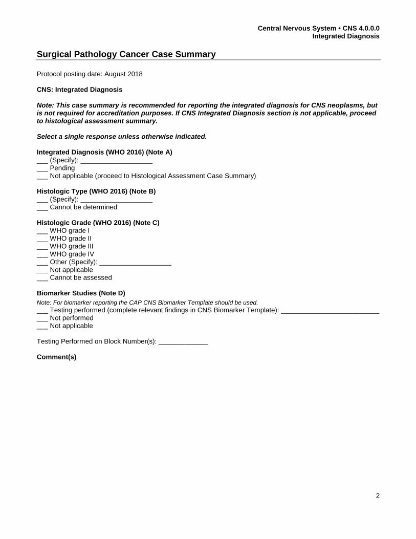
Central Nervous System • CNS 4.0.0.0 Integrated Diagnosis
Surgical Pathology Cancer Case Summary Protocol posting date: August 2018 CNS: Integrated Diagnosis Note: This case summary is recommended for reporting the integrated diagnosis for CNS neoplasms, but is not required for accreditation purposes. If CNS Integrated Diagnosis section is not applicable, proceed to histological assessment summary. Select a single response unless otherwise indicated. Integrated Diagnosis (WHO 2016) (Note A) ___ (Specify): ___________________ ___ Pending ___ Not applicable (proceed to Histological Assessment Case Summary) Histologic Type (WHO 2016) (Note B) ___ (Specify): ___________________ ___ Cannot be determined Histologic Grade (WHO 2016) (Note C) ___ WHO grade I ___ WHO grade II ___ WHO grade III ___ WHO grade IV ___ Other (Specify): ___________________ ___ Not applicable ___ Cannot be assessed Biomarker Studies (Note D) Note: For biomarker reporting the CAP CNS Biomarker Template should be used. ___ Testing performed (complete relevant findings in CNS Biomarker Template): __________________________ ___ Not performed ___ Not applicable Testing Performed on Block Number(s): _____________ Comment(s)
2

Central Nervous System • CNS 4.0.0.0 Histologic Assessment
Surgical Pathology Cancer Case Summary Protocol posting date: August 2018 CNS: Histological Assessment Note: This case summary is recommended for reporting the histologic assessment of CNS neoplasms, but is not required for accreditation purposes. Select a single response unless otherwise indicated. History of Prior Therapy for this Neoplasm (Note E) ___ Not administered ___ Not known ___ Administered (specify): _____________________ History of Previous Tumor and/or Familial Syndrome (not the current neoplasm) (Note E) ___ Not known ___ Known (specify): _______________________ ___ Not specified Neuroimaging Findings (Note F) ___ (specify):_____________ ___ Not available Procedure (Note G) ___ Open biopsy ___ Resection ___ Stereotactic biopsy ___ Other (specify): _____________________ ___ Not specified Specimen Size, gross description (Note H)# Greatest dimension (centimeters): ___ cm ___ Additional dimensions (centimeters): ___ x ___ cm ___ Cannot be determined (explain) # For fragmented tissue, an aggregate size may be given Tumor Site (select all that apply) (Note I) ___ Skull (specify precise location, if known): ___________________ ___ Dura (specify precise location, if known): ___________________ ___ Leptomeninges (specify precise location, if known): ___________________ ___ Brain ___ Cerebral lobes (specify precise location, if known): ______________________ ___ Deep grey matter (specify precise location, if known): ___________________ ___ Ventricle (specify precise location, if known): ___________________ ___ Cerebellum (specify precise location, if known): ___________________ ___ Brain stem (specify precise location, if known): ___________________ ___ Other (specify, if known): ___________________ ___ Cerebellopontine angle ___ Sellar/Suprasellar/Pituitary ___ Pineal ___ Cranial nerve (specify I–XII, if known): ___________________ ___ Spine/vertebral column (specify precise location, if known): ___________________ ___ Spinal cord (specify precise location, if known): ___________________
3
Central Nervous System • CNS 4.0.0.0 Histologic Assessment
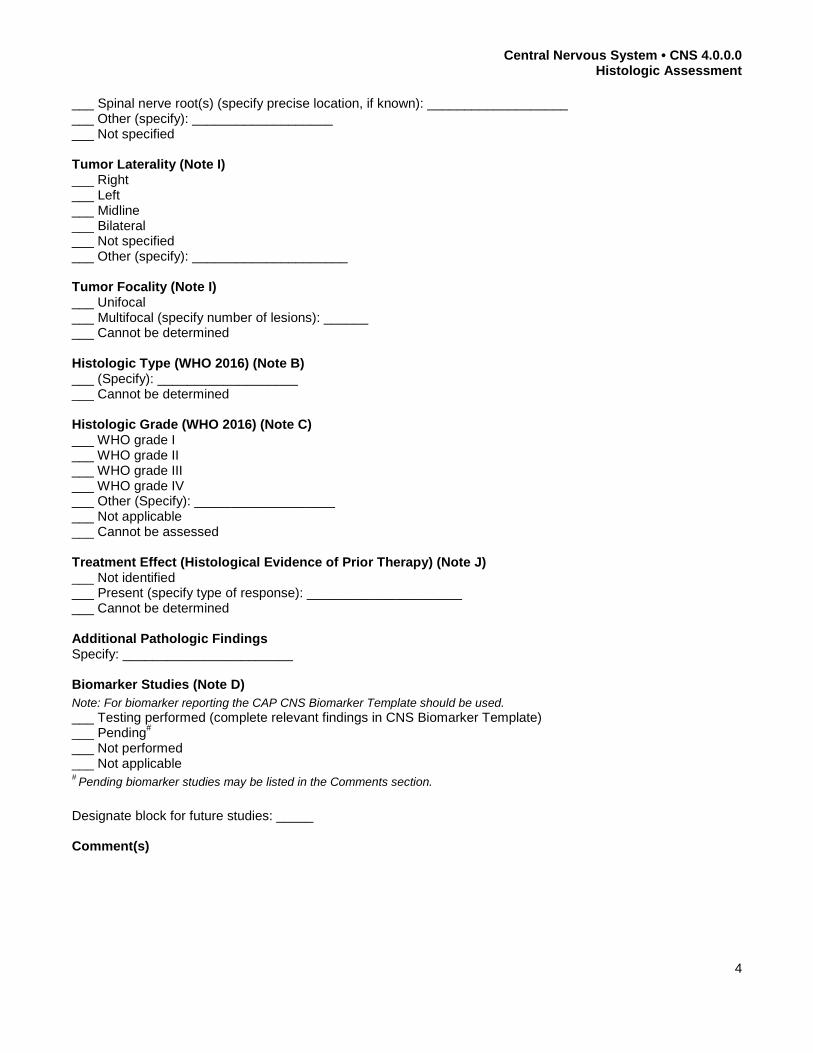
___ Spinal nerve root(s) (specify precise location, if known): ___________________ ___ Other (specify): ___________________ ___ Not specified Tumor Laterality (Note I) ___ Right ___ Left ___ Midline ___ Bilateral ___ Not specified ___ Other (specify): _____________________ Tumor Focality (Note I) ___ Unifocal ___ Multifocal (specify number of lesions): ______ ___ Cannot be determined Histologic Type (WHO 2016) (Note B) ___ (Specify): ___________________ ___ Cannot be determined Histologic Grade (WHO 2016) (Note C) ___ WHO grade I ___ WHO grade II ___ WHO grade III ___ WHO grade IV ___ Other (Specify): ___________________ ___ Not applicable ___ Cannot be assessed Treatment Effect (Histological Evidence of Prior Therapy) (Note J) ___ Not identified ___ Present (specify type of response): _____________________ ___ Cannot be determined Additional Pathologic Findings Specify: _______________________ Biomarker Studies (Note D) Note: For biomarker reporting the CAP CNS Biomarker Template should be used. ___ Testing performed (complete relevant findings in CNS Biomarker Template) ___ Pending# ___ Not performed ___ Not applicable # Pending biomarker studies may be listed in the Comments section. Designate block for future studies: _____ Comment(s)
4

Central Nervous System • CNS 4.0.0.0 Biomarker Template
CNS Biomarker Reporting Template Protocol posting date: August 2018 CNS Biomarker Reporting Template Note: This case summary is recommended for reporting biomarkers for CNS neoplasms at the completion of testing, but is not required for accreditation purposes. Select a single response unless otherwise indicated. Testing Performed on Block Number(s): _____________ Biomarker Studies (Note D) Note: Pending biomarker studies may be listed in the Comments section of this report.
ATRX
ATRX mutation ___ Absent ___ Present (specify): ________________________ ___ Cannot be determined (explain): ________________________ ATRX expression (immunohistochemistry) ___ Intact nuclear expression ___ Loss of nuclear expression ___ Cannot be determined (explain): ________________________
BRAF alterations
BRAF mutation ___ Absent ___ BRAF V600E (c.1799T>A) mutation present ___ Other BRAF mutation present (specify): ________________________ ___ Cannot be determined (explain): ________________________ KIAA:BRAF rearrangement/duplication ___ Absent ___ Present ___ Cannot be determined (explain): ________________________ BRAF V600E expression (immunohistochemistry) ___ Negative ___ Positive ___ Cannot be determined (explain): ________________________
Beta-Catenin expression / CTNNB1 mutation
Beta-catenin expression (immunohistochemistry) ___ Absence of nuclear expression ___ Positive nuclear expression ___ Cannot be determined (explain): ________________________ CTNNB1 mutation ___ Absent ___ Present
5
Central Nervous System • CNS 4.0.0.0 Biomarker Template
___ Cannot be determined (explain): ________________________
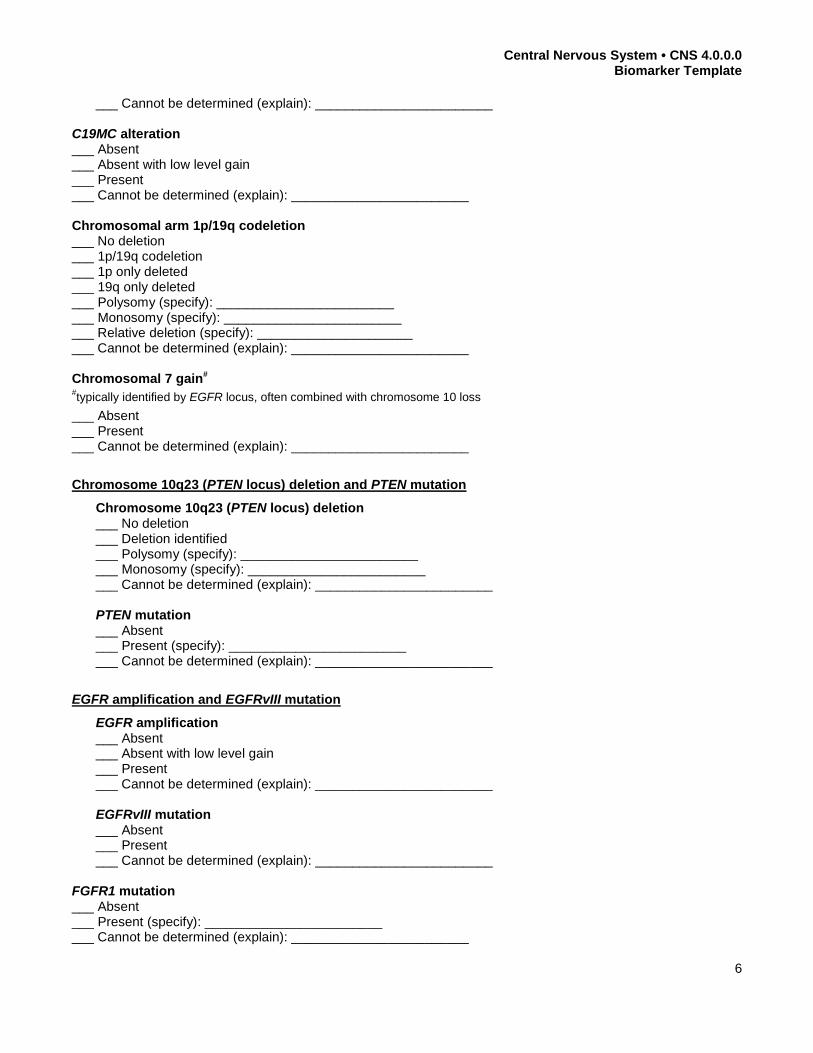
C19MC alteration ___ Absent ___ Absent with low level gain ___ Present ___ Cannot be determined (explain): ________________________ Chromosomal arm 1p/19q codeletion ___ No deletion ___ 1p/19q codeletion ___ 1p only deleted ___ 19q only deleted ___ Polysomy (specify): ________________________ ___ Monosomy (specify): ________________________ ___ Relative deletion (specify): _____________________ ___ Cannot be determined (explain): ________________________ Chromosomal 7 gain# #typically identified by EGFR locus, often combined with chromosome 10 loss ___ Absent ___ Present ___ Cannot be determined (explain): ________________________
Chromosome 10q23 (PTEN locus) deletion and PTEN mutation
Chromosome 10q23 (PTEN locus) deletion ___ No deletion ___ Deletion identified ___ Polysomy (specify): ________________________ ___ Monosomy (specify): ________________________ ___ Cannot be determined (explain): ________________________ PTEN mutation ___ Absent ___ Present (specify): ________________________ ___ Cannot be determined (explain): ________________________
EGFR amplification and EGFRvIII mutation
EGFR amplification ___ Absent ___ Absent with low level gain ___ Present ___ Cannot be determined (explain): ________________________ EGFRvIII mutation ___ Absent ___ Present ___ Cannot be determined (explain): ________________________
FGFR1 mutation ___ Absent ___ Present (specify): ________________________ ___ Cannot be determined (explain): ________________________
6
Central Nervous System • CNS 4.0.0.0 Biomarker Template
GAB1 expression (immunohistochemistry) ___ Negative ___ Positive ___ Cannot be determined (explain): ________________________
Histone H3 mutation and K27me3
H3 gene family mutation ___ Negative ___ Positive ___ Cannot be determined (explain): _______________________ Histone H3 K27M expression (immunohistochemistry) ___ Negative ___ Positive ___ Cannot be determined (explain): ________________________ H3 K27me3 expression (immunohistochemistry) ___ Intact nuclear expression ___ Loss of nuclear expression ___ Cannot be determined (explain): ________________________
IDH1/IDH2 mutation
IDH1/IDH2 mutation ___ Absent ___ Present (specify): ________________________ ___ Cannot be determined (explain): ________________________ IDH1 R132H expression (immunohistochemistry) ___ Negative ___ Positive ___ Cannot be determined (explain): ________________________
Isochromosome 17q (i17q) ___ Absent ___ Present ___ Cannot be determined (explain): ________________________ Ki-67 expression (immunohistochemistry) Hotspot percentage of positive tumor cell nuclei: ____ % L1CAM expression (immunohistochemistry) ___ Negative ___ Positive ___ Cannot be determined (explain): ________________________
7

Central Nervous System • CNS 4.0.0.0 Biomarker Template
LIN28A expression (immunohistochemistry) ___ Negative ___ Positive ___ Cannot be determined (explain): ________________________ MGMT promoter methylation ___ Absent ___ Present
If laboratory reports by level: ___ Low level
___ High level ___ Cannot be determined (explain): ________________________ Monosomy 6 ___ Absent ___ Present ___ Cannot be determined (explain): ________________________
MYC gene family amplification
MYC amplification ___ Absent ___ Present ___ Cannot be determined (explain): ________________________ MYCN amplification ___ Absent ___ Present ___ Cannot be determined (explain): ________________________
NAB2-STAT6 fusion
NAB2-STAT6 fusion ___ Negative ___ Positive ___ Cannot be determined (explain): ________________________ STAT6 expression (immunohistochemistry) ___ Absence of nuclear expression ___ Positive nuclear expression ___ Cannot be determined (explain): ________________________
Pituitary hormones and transcription factors immunohistochemistry
Tumor Cell(s) Reactivity (select all that apply) ___ Alpha subunit ___ Adrenocorticotrophic hormone (ACTH) ___ Follicular stimulating hormone (beta FSH) ___ Human growth hormone ___ Luteinizing hormone (beta LH) ___ Prolactin ___ PIT1 ___ SF1 ___ Thyroid stimulating hormone (beta TSH) ___ TPIT ___ Other (specify)
8
Central Nervous System • CNS 4.0.0.0 Biomarker Template
___ Cannot be determined (explain): ________________________ RELA fusion ___ Negative ___ Positive ___ Cannot be determined (explain): ________________________
SMARCA4/BRG1 alteration
SMARCA4/BRG1 mutation ___ Absent ___ Present (specify): ________________________ ___ Cannot be determined (explain): ________________________ BRG1 expression (immunohistochemistry) ___ Intact nuclear expression ___ Loss of nuclear expression ___ Cannot be determined (explain): ________________________
SMARCB1/INI1/HSNF5 alteration
SMARCB1/INI1/HSNF5 mutation ___ Absent ___ Present (specify): ________________________ ___ Cannot be determined (explain): ________________________ INI1 (BAF47) expression (immunohistochemistry) ___ Intact nuclear expression ___ Loss of nuclear expression ___ Cannot be determined (explain): ________________________
TERT promoter mutation ___ Absent ___ Hotspot mutation (C228T or C250T) ___ Other TERT mutation (specify): ________________________ ___ Cannot be determined (explain): ________________________
TP53 mutation
TP53 mutation ___ Absent ___ Present (specify): ________________________ ___ Cannot be determined (explain): ________________________ p53 expression (immunohistochemistry) ___ Negative or rare ___ Intermediate ___ Positive (diffuse and strong nuclear positivity) ___ Cannot be determined (explain): ________________________
YAP1
YAP1 fusion ___ Negative ___ Positive ___ Other (specify): ______________________
9
Central Nervous System • CNS 4.0.0.0 Biomarker Template
___ Cannot be determined (explain): ________________________ YAP1 expression (immunohistochemistry) ___ Negative ___ Positive ___ Cannot be determined (explain): ________________________
Other biomarker(s)
Point Mutations (specify): ______________________ Copy Number Alterations (specify): ______________________ Insertions (specify): ______________________ Deletions (specify): ______________________ Comment(s)
10

Background Documentation Central Nervous System • CNS 4.0.0.0
Explanatory Notes A. Integrated Diagnosis Historically, the diagnosis and classification of CNS tumors has been based exclusively on the histologic appearance of the tumor. In recent decades, however, our knowledge of the molecular basis of many of these tumors has increased significantly. In the updated 2016 WHO Classification of Tumours of the Central Nervous System1, molecular information is now integrated into some of the tumor diagnostic entities. In such cases, including the diffuse gliomas and embryonal tumors, the final diagnosis should reflect the integration of both histologic and molecular information. When applicable, it is suggested that all histologic and molecular information be presented in a “layered” report format as follows2: Layer 1: Integrated diagnosis (incorporating all tissue-based information) Layer 2: Histological classification Layer 3: Histologic (WHO) grade Layer 4: Biomarker studies At centers where molecular testing is not available, an NOS (not otherwise specified) designation is available for some tumor entities. The NOS designation implies that insufficient information is available to provide a more specific integrated diagnosis, and may occasionally be used for tumors that do not precisely fit into one of the defined tumor categories. References 1. Louis DN, Ohgaki H, Wiestler OD, et al. World Health Organization Classification of Tumours of the Central
Nervous System. Lyon, France: IARC Press; 2016. 2. Louis DN, Perry A, Burger P, et al. International Society of Neuropathology-Haarlem Consensus Guidelines
for Nervous System Tumor Classification and Grading. Brain Pathol. 2014;24:429-435. B. Histologic Type Classification should be made according to the WHO classification of tumors of the nervous system and the WHO classification of tumors of the endocrine organs whenever possible.1,2 The list below contains WHO 2016 diagnostic entities for which the Central Nervous System (CNS) Cancer Protocol is recommended:
Diffuse astrocytic and oligodendroglial tumors Diffuse astrocytoma, NOS Diffuse astrocytoma, IDH-mutant Diffuse astrocytoma, IDH-wildtype Gemistocytic astrocytoma, IDH-mutant Anaplastic astrocytoma, NOS Anaplastic astrocytoma, IDH-mutant Anaplastic astrocytoma, IDH-wildtype Glioblastoma, NOS Glioblastoma, IDH-mutant Glioblastoma, IDH-wildtype Epithelioid glioblastoma Giant cell glioblastoma Gliosarcoma Diffuse midline glioma, H3 K27M-mutant Oligodendroglioma, NOS Oligodendroglioma, IDH-mutant and 1p/19q-codeleted Anaplastic oligodendroglioma, NOS Anaplastic oligodendroglioma, IDH-mutant and 1p/19q-codeleted Oligoastrocytoma, NOS Anaplastic oligoastrocytoma, NOS
11
Background Documentation Central Nervous System • CNS 4.0.0.0
Other astrocytic tumors Pilocytic astrocytoma Pilomyxoid astrocytoma Subependymal giant cell astrocytoma Pleomorphic xanthoastrocytoma Anaplastic pleomorphic xanthoastrocytoma
Ependymal tumors Subependymoma Myxopapillary ependymoma Ependymoma Clear cell ependymoma Papillary ependymoma Tanycytic ependymoma Ependymoma, RELA fusion-positive Anaplastic ependymoma
Other gliomas Chordoid glioma of the third ventricle Angiocentric glioma Astroblastoma
Choroid plexus tumors Choroid plexus papilloma Atypical choroid plexus papilloma Choroid plexus carcinoma
Neuronal and mixed neuronal–glial tumors Dysembryoplastic neuroepithelial tumor Gangliocytoma Ganglioglioma Anaplastic ganglioglioma Dysplastic cerebellar gangliocytoma (Lhermitte–Duclos disease) Desmoplastic infantile astrocytoma and ganglioglioma Papillary glioneuronal tumor Rosette-forming glioneuronal tumor Diffuse leptomeningeal glioneuronal tumor Central neurocytoma Extraventricular neurocytoma Cerebellar liponeurocytoma Paraganglioma
Tumors of the pineal region Pineocytoma Pineal parenchymal tumor of intermediate differentiation Pineoblastoma Papillary tumor of the pineal region
Embryonal tumors Medulloblastomas, histologically defined Medulloblastoma, NOS Medulloblastoma, classic Medulloblastoma, desmoplastic/nodular Medulloblastoma with extensive nodularity Medulloblastoma, large cell/anaplastic Medulloblastomas, genetically defined Medulloblastoma, NOS Medulloblastoma, WNT-activated
12

Background Documentation Central Nervous System • CNS 4.0.0.0
Medulloblastoma, SHH activated Medulloblastoma, SHH activated and TP53-mutant Medulloblastoma, SHH activated and TP53-wildtype Medulloblastoma, non-WNT/non-SHH Medulloblastoma, non-WNT/non-SHH: Medulloblastoma, group 3 Medulloblastoma, non-WNT/non-SHH: Medulloblastoma, group 4 Atypical teratoid/rhabdoid tumor Embryonal tumor with multilayered rosettes, NOS Embryonal tumor with multilayered rosettes, C19MC-altered Medulloepithelioma CNS neuroblastoma CNS ganglioneuroblastoma CNS embryonal tumor, NOS CNS embryonal tumor with rhabdoid features
Meningiomas Meningioma Angiomatous meningioma Fibrous meningioma Lymphoplasmacyte-rich meningioma Meningothelial meningioma Metaplastic meningioma Microcystic meningioma Psammomatous meningioma Secretory meningioma Transitional meningioma Chordoid meningioma Clear cell meningioma Atypical meningioma Papillary meningioma Rhabdoid meningioma Anaplastic (malignant) meningioma
Mesenchymal, non-meningothelial tumors Solitary fibrous tumor/hemangiopericytoma, NOS Solitary fibrous tumor/hemangiopericytoma, grade 1 Solitary fibrous tumor/hemangiopericytoma, grade 2 Solitary fibrous tumor/hemangiopericytoma, grade 3 Hemangioblastoma
Melanocytic tumors Meningeal melanocytosis Meningeal melanocytoma Meningeal melanoma Meningeal melanomatosis
Germ cell tumors Germinoma Embryonal carcinoma Yolk sac tumor Choriocarcinoma Teratoma Mature teratoma Immature teratoma Teratoma with malignant transformation Mixed germ cell tumor
13
Background Documentation Central Nervous System • CNS 4.0.0.0
Tumors of the sellar region Craniopharyngioma Adamantinomatous craniopharyngioma Papillary craniopharyngioma Granular cell tumor of the sellar region Pituicytoma Spindle cell oncocytoma
Pituitary tumors Pituitary adenomas Pituitary adenoma Corticotroph adenoma Gonadotroph adenoma Lactotroph adenoma Somatotroph adenoma Thyrotroph adenoma Null cell adenoma Plurihormonal and double adenomas Pituitary carcinoma Pituitary carcinoma References 1. Louis DN, Ohgaki H, Wiestler OD, et al. World Health Organization Classification of Tumours of the Central
Nervous System. Lyon, France: IARC Press; 2016. 2. Lloyd RV, Osamura RY, Klöppel G, et al. WHO Classification of Tumours: Pathology & Genetics of Tumours
of Endocrine Organs. Lyon, France: IARC Press; 2017.
C. Histologic Grade Below is a list of possible WHO grades for CNS tumors.1 The WHO grading of some of the more common CNS tumors is shown in Table 1. There is no formal TNM-based classification and staging system for CNS tumors.
WHO Grades for CNS Tumors WHO grade I WHO grade II WHO grade III WHO grade IV WHO grade not assigned References 1. Louis DN, Ohgaki H, Wiestler OD, et al. World Health Organization Classification of Tumours of the Central
Nervous System. Lyon, France: IARC Press; 2016. D. Biomarker Studies Immunohistochemical and molecular genetic studies are often performed to assist with diagnosis, prognosis, or to predict therapeutic response.1 The most recent update of the World Health Organization’s Classification of Tumours of the Central Nervous System has incorporated many of these biomarkers into this formal diagnostic classification system, thereby formally encouraging their use in the evaluation of these neoplasms. Currently, the 2016 WHO Classification of Tumours of the Central Nervous System and the 2017 (WHO) Pathology & Genetics of Tumours of Endocrine Organs incorporates molecular genetic studies into several entities while the diagnosis of the majority of CNS tumors remain largely morphologic.1,2 It is expected that, as our understanding of the biology of CNS tumors improves, the list of entities requiring molecular genetic studies will continue to grow. For those defined entities, the use of the biomarker template is encouraged. Additional common ancillary molecular testing in neurooncology includes MGMT promoter methylation studies; ATRX expression/mutations; TP53 expression/mutations; copy number alterations in EGFR and PTEN; and
14
Background Documentation Central Nervous System • CNS 4.0.0.0
BRAF alterations and mutations.3-5 For medulloblastoma, assessment of MYC or MYCN amplification and beta-catenin nuclear localization has prognostic significance. In the absence of access to these biomarkers, the WHO has provided the “NOS” nomenclature appended to the end of the histologic diagnosis to indicate the absence of molecular testing on the individual case. Embryonal neoplasms may benefit from ancillary studies for proper diagnostic categorization. Assigning medulloblastomas to appropriate genetic groups may be done by immunohistochemistry in most cases: WNT-activated (group 1) cases show nuclear beta-catenin and YAP1 expression; SHH-activated (group 2) cases express markers GAB1 and YAP1; groups 3 and 4 do not express neither GAB1 nor YAP1 and exhibit only nonnuclear beta-catenin immunostaining, if any.6,7 Some copy number changes are useful for molecular grouping of medulloblastomas, but are not necessary to assess in most cases: monosomy 6 is present in the vast majority of WNT-activated cases; deletion of 9q (PTCH gene) is common in SHH-activated cases; loss of 17p and duplication of 17 (resulting in an “isochromosome 17q”) is limited to groups 3 and 4.8 SHH-activated medulloblastomas can be diagnostically segregated by TP53 mutation status; those medulloblastomas with a TP53 mutation have a much worse prognosis.9 Aberrant p53 immunostaining is an effective surrogate for the presence of a mutation, either as diffuse, strong nuclear reactivity or, less commonly, complete lack of nuclear expression in all tumor cells. Additional assessment for MYC or MYCN amplification for prognosis is indicated regardless of molecular group. Any embryonal neoplasm with lumen-forming, multilayered rosettes can be tested for amplification of the C19MC region on chromosome 19.10 The immunostain LIN28A, when strongly and diffusely positive, correlates highly with C19MC amplification, which confers a grim prognosis.11 Medulloepitheliomas have multilayered rosettes, yet may not always exhibit C19MC amplification or LIN28 expression. Such cases should be specified as non-C19MC altered. Embryonal tumors can be assessed for SMARCB1/INI1 status to identify atypical teratoid/rhabdoid tumors (AT/RT), which have a significantly different treatment regimen from other CNS embryonal malignancies. This may be effectively done by demonstrating absence of SMARCB1/INI1 nuclear immunostaining in tumor cells (for example using the BAF47 antibody).11 Morphologically rhabdoid embryonal malignancies with retained SMARCB1/INI1 nuclear expression can be assessed for loss of SMARCA4/BRG1, which is also diagnostic for AT/RT. The diagnosis "CNS embryonal tumor with rhabdoid features, NOS (WHO grade IV)" should be used when SMARCB1/INI1 or SMARCA4/BRG1 expression is retained or cannot be assessed in a malignant embryonal neoplasm with rhabdoid morphology. Pediatric embryonal tumors in the supratentorial compartment can be tested for the H3F3A K27 or G34 mutations typically found in pediatric glioblastomas, which can display embryonal, neuroblastic morphology and immunophenotype.13 Antibodies are available for immunohistochemical detection of both the H3K27M and the mutant proteins.14 H3 G34-mutant glioblastomas have high rates of ATRX loss and TP53 mutations, immunostaining for which can help distinguish them from the embryonal tumors. Supratentorial ependymomas can be tested for fusion rearrangements of the RELA gene, which are associated with a poor prognosis and constitute a separate diagnostic category in the WHO 2016 classification.15 Immunostaining for L1CAM is a surrogate marker for RELA fusion in ependymomas, although it may also be seen in other tumor types. Gain of 1q implies worse prognosis in posterior fossa ependymomas. In posterior fossa tumors, loss of H3 K27me3 staining reliably identifies the PF-A ependymomas, which have a much worse prognosis than PF-B.15 The advent of DNA next generation sequencing (NGS) techniques has led to the evaluation of many more biomarkers than can be performed one at a time in most FISH or immunohistochemical laboratories. NGS also allows the evaluation of biomarkers that are too large for routine sequencing methods such as NF1. The capture of these data may lead to the identification of less common genetic alterations that the oncologists may identify as clinically relevant, targetable pathways, particularly in the less common tumors of childhood and young adulthood.16 In such cases in which NGS analyses are obtained, we have left room at the end of the section to record the deviations found in these biomarkers. Similarly, research in brain tumor biomarkers is ongoing, making
15
Background Documentation Central Nervous System • CNS 4.0.0.0
the updating of this protocol a dynamic process. Such new discoveries can be added also in the additional spaces provided. Additional biomarker information and references developed by the International Collaboration on Cancer Reporting (ICCR) may be found at http://www.iccr-cancer.org/datasets/published-datasets/central-nervous-system.17
The ICCR Central Nervous System Molecular Notes includes an overview of selected molecular diagnostic markers for CNS tumors: Overview of selected molecular diagnostic markers for CNS tumours
The table below summarizes selected molecular diagnostic markers for CNS tumours; the list of tests is not exhaustive and other assays may be helpful in some diagnostic circumstances. In addition, the tests listed are those related to ruling in the corresponding diagnoses; however, it should be realized that the assays may also be used in particular diagnostic situations to rule out other diagnoses. An example of this would be ATRX immunohistochemistry, which is commonly used to support a diagnosis of IDH-mutant diffuse astrocytoma, but which is also used to evaluate a possible diagnosis of oligodendroglioma, IDH-mutant and 1p/19q-codeleted. Some specific tests recommended in the commentaries below represent one of several validated and equivalent approaches to the evaluation of the described molecular variable; for those tests that have multiple testing modalities (e.g., sequencing and immunohistochemistry for BRAF V600E), it is assumed that only one of these testing modalities would be used per case unless one test yields equivocal results (e.g., a result of weak immunohistochemical positivity versus nonspecific background staining should be followed by gene sequencing). For some tests, relevance may be related to the age of the patient (e.g., EGFR gene amplification in adult high-grade gliomas rather than paediatric ones).
16
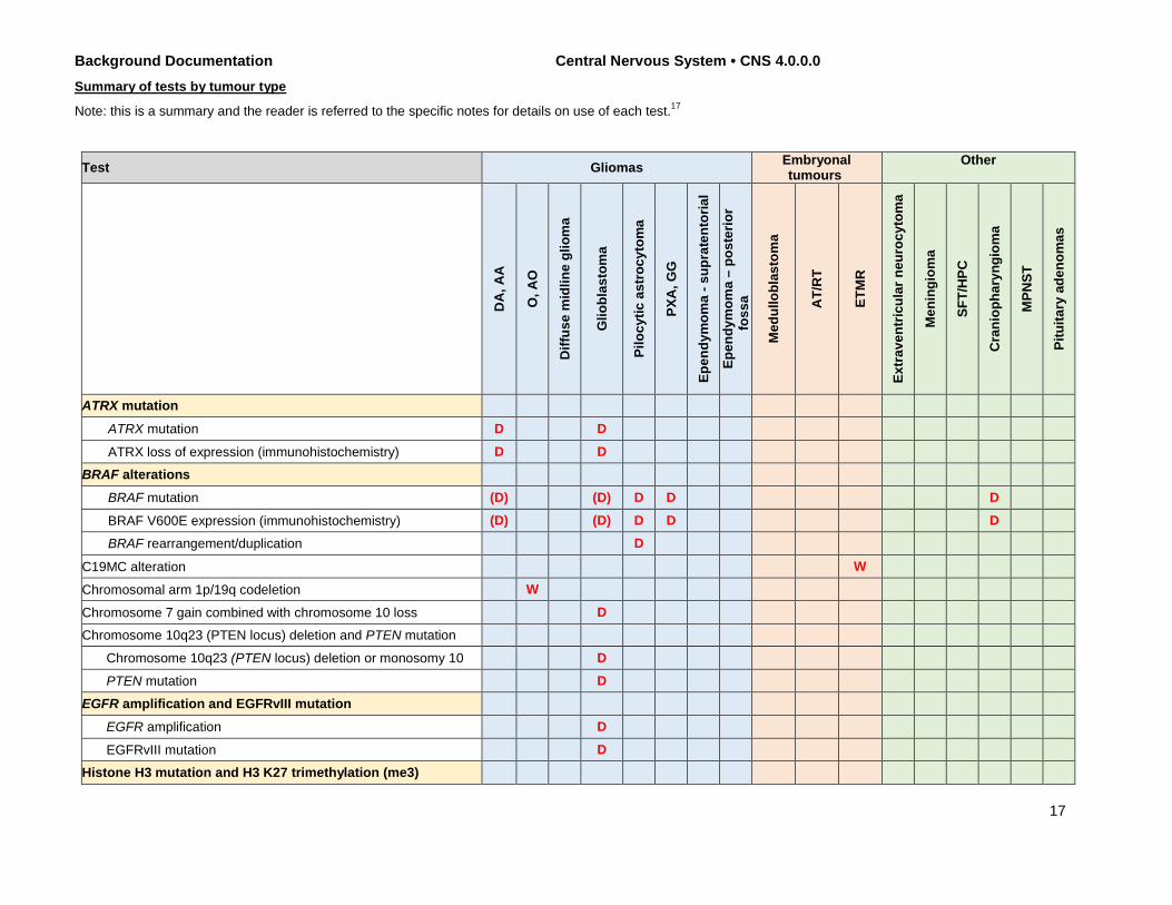
Background Documentation Central Nervous System • CNS 4.0.0.0 Summary of tests by tumour type
Note: this is a summary and the reader is referred to the specific notes for details on use of each test.17
Test Gliomas Embryonal tumours
Other
DA
, AA
O, A
O
Diff
use
mid
line
glio
ma
Glio
blas
tom
a
Pilo
cytic
ast
rocy
tom
a
PXA
, GG
Epen
dym
oma
- sup
rate
ntor
ial
Epen
dym
oma
– po
ster
ior
foss
a
Med
ullo
blas
tom
a
AT/
RT
ETM
R
Extr
aven
tric
ular
neu
rocy
tom
a
Men
ingi
oma
SFT/
HPC
Cra
niop
hary
ngio
ma
MPN
ST
Pitu
itary
ade
nom
as
ATRX mutation
ATRX mutation D D
ATRX loss of expression (immunohistochemistry) D D BRAF alterations
BRAF mutation (D) (D) D D D
BRAF V600E expression (immunohistochemistry) (D) (D) D D D BRAF rearrangement/duplication D
C19MC alteration W
Chromosomal arm 1p/19q codeletion W
Chromosome 7 gain combined with chromosome 10 loss D Chromosome 10q23 (PTEN locus) deletion and PTEN mutation
Chromosome 10q23 (PTEN locus) deletion or monosomy 10 D PTEN mutation D
EGFR amplification and EGFRvIII mutation EGFR amplification D
EGFRvIII mutation D Histone H3 mutation and H3 K27 trimethylation (me3)
17
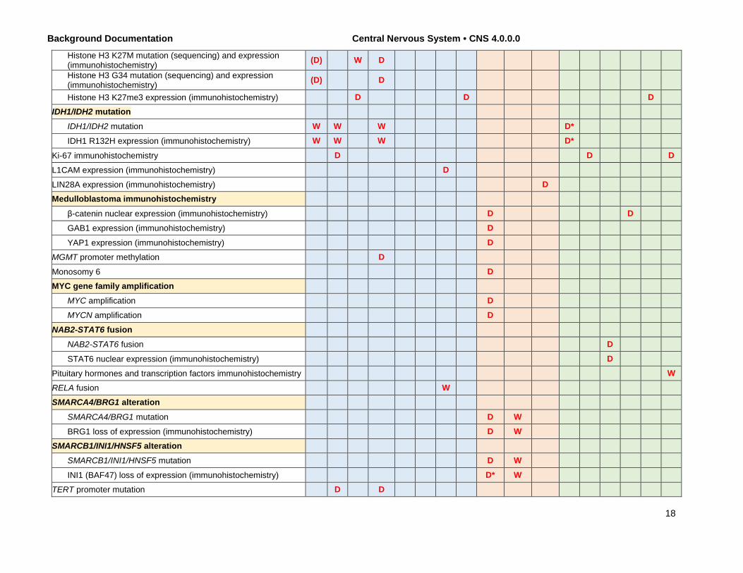
Background Documentation Central Nervous System • CNS 4.0.0.0
Histone H3 K27M mutation (sequencing) and expression (immunohistochemistry) (D) W D
Histone H3 G34 mutation (sequencing) and expression (immunohistochemistry) (D) D
Histone H3 K27me3 expression (immunohistochemistry) D D D IDH1/IDH2 mutation
IDH1/IDH2 mutation W W W D*
IDH1 R132H expression (immunohistochemistry) W W W D*
Ki-67 immunohistochemistry D D D
L1CAM expression (immunohistochemistry) D
LIN28A expression (immunohistochemistry) D Medulloblastoma immunohistochemistry
β-catenin nuclear expression (immunohistochemistry) D D
GAB1 expression (immunohistochemistry) D
YAP1 expression (immunohistochemistry) D MGMT promoter methylation D
Monosomy 6 D MYC gene family amplification
MYC amplification D MYCN amplification D
NAB2-STAT6 fusion NAB2-STAT6 fusion D
STAT6 nuclear expression (immunohistochemistry) D
Pituitary hormones and transcription factors immunohistochemistry W RELA fusion W SMARCA4/BRG1 alteration
SMARCA4/BRG1 mutation D W
BRG1 loss of expression (immunohistochemistry) D W SMARCB1/INI1/HNSF5 alteration
SMARCB1/INI1/HNSF5 mutation D W
INI1 (BAF47) loss of expression (immunohistochemistry) D* W TERT promoter mutation D D
18
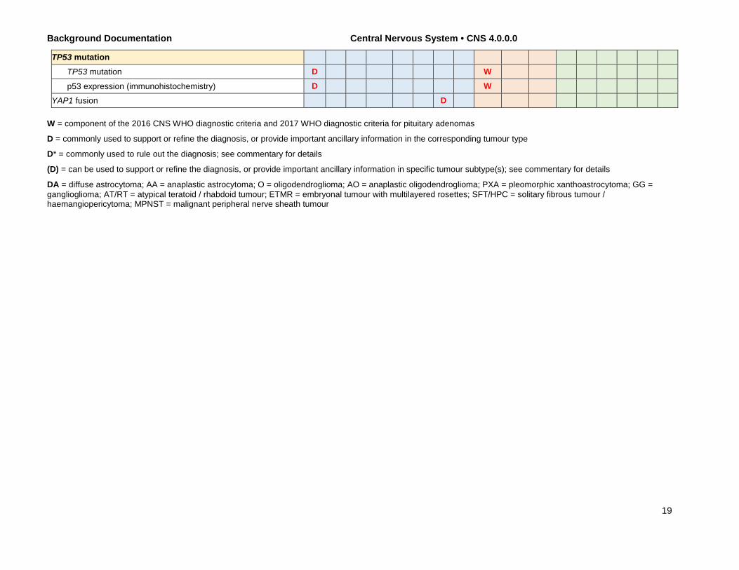
Background Documentation Central Nervous System • CNS 4.0.0.0
TP53 mutation TP53 mutation D W
p53 expression (immunohistochemistry) D W YAP1 fusion D
W = component of the 2016 CNS WHO diagnostic criteria and 2017 WHO diagnostic criteria for pituitary adenomas
D = commonly used to support or refine the diagnosis, or provide important ancillary information in the corresponding tumour type
D* = commonly used to rule out the diagnosis; see commentary for details
(D) = can be used to support or refine the diagnosis, or provide important ancillary information in specific tumour subtype(s); see commentary for details
DA = diffuse astrocytoma; AA = anaplastic astrocytoma; O = oligodendroglioma; AO = anaplastic oligodendroglioma; PXA = pleomorphic xanthoastrocytoma; GG = ganglioglioma; AT/RT = atypical teratoid / rhabdoid tumour; ETMR = embryonal tumour with multilayered rosettes; SFT/HPC = solitary fibrous tumour / haemangiopericytoma; MPNST = malignant peripheral nerve sheath tumour
19
Background Documentation Central Nervous System • CNS 4.0.0.0
References 1. Louis DN, Ohgaki H, Wiestler OD, et al. World Health Organization Classification of Tumours of the Central
Nervous System. Lyon, France: IARC Press; 2016. 2. Lloyd RV, Osamura RY, Klöppel G, et al. WHO Classification of Tumours: Pathology & Genetics of Tumours
of Endocrine Organs. Lyon, France: IARC Press; 2017. 3. Nikiforova MN, Hamilton RL. Molecular diagnostics of gliomas. Arch Pathol Lab Med. 2011;135:558-568. 4. Taylor MD, Northcott PA, Korshunov A, et al. Molecular subgroups of medulloblastoma: the current
consensus. Acta Neuropathol. 2012 ;123:465-472. 5. Nageswara Rao AA, Packer RJ. Impact of molecular biology studies on the understanding of brain tumors in
childhood. Curr Oncol Rep. 2012;14:206-212. 6. Kaur K, Kakkar A, Kumar A, et al. Integrating Molecular Subclassification of Medulloblastomas into Routine
Clinical Practice: A Simplified Approach. Brain Pathol. 2016;26:334-343. 7. Ellison DW, Dalton J, Kocak M, et al. Medulloblastoma: clinicopathological correlates of SHH, WNT, and non-
SHH/WNT molecular subgroups. Acta Neuropathol. 2011;121:381-396. 8. Kool M, Korshunov A, Remke M, et al. Molecular subgroups of medulloblastoma: an international meta-
analysis of transcriptome, genetic aberrations, and clinical data of WNT, SHH, Group 3, and Group 4 medulloblastomas. Acta Neuropathol. 2012;123:473-484.
9. Zhukova N, Ramaswamy V, Remke M, et al. Subgroup-specific prognostic implications of TP53 mutation in medulloblastoma. J Clin Oncol. 2013;31:2927-2935.
10. Korshunov A, Sturm D, Ryzhova M, et al. Embryonal tumor with abundant neuropil and true rosettes (ETANTR), ependymoblastoma, and medulloepithelioma share molecular similarity and comprise a single clinicopathological entity. Acta Neuropathol. 2014;128:279-289.
11. Korshunov A, Ryzhova M, Jones DT, et al. LIN28A immunoreactivity is a potent diagnostic marker of embryonal tumor with multilayered rosettes (ETMR). Acta Neuropathol. 2012;124:875-881.
12. Judkins AR, Mauger J, Ht A, Rorke LB, Biegel JA. Immunohistochemical analysis of hSNF5/INI1 in pediatric CNS neoplasms. Am J Surg Pathol. 2004;28:644-650.
13. Korshunov A, Capper D, Reuss D, et al. Histologically distinct neuroepithelial tumors with histone 3 G34 mutation are molecularly similar and comprise a single nosologic entity. Acta Neuropathol. 2016;131:137-46.
14. Haque F, Varlet P, Puntonet J, et al. Evaluation of a novel antibody to define histone 3.3 G34R mutant brain tumours. Acta Neuropathol Commun. 2017:5:45.
15. Pajtler KW, Witt H, Sill M, et al. Molecular Classification of Ependymal Tumors across All CNS Compartments, Histopathological Grades, and Age Groups. Cancer Cell. 2015;27:728-743.
16. Cole BL, Pritchard CC, Anderson M, et al. Targeted sequencing of malignant supratentorial pediatric brain tumors demonstrates a high frequency of clinically relevant mutations. Pediatr Dev Pathol. 2017 Jan 1:1093526617743905. doi: 10.1177/1093526617743905.
17. http://www.iccr-cancer.org/datasets/published-datasets/central-nervous-system. E. Relevant History Previous Diagnoses or CNS Biopsies Knowledge of the presence or absence of previous intracranial or extracranial disease (eg, immunosuppression, previous CNS or other primary neoplasm) is essential for specimen interpretation. If a previous tumor is included in the differential diagnosis, it is useful to have microscopic slides of the lesion available for review and comparison.1,2 Family History of Cancer or Primary CNS Tumors Several genetic conditions/syndromes are associated with an increased predisposition to the development of specific forms of CNS neoplasms (eg, neurofibromatosis types 1 and 2, Turcot/Lynch, tuberous sclerosis, von Hippel-Lindau, Cowden, Li-Fraumeni, and Gorlin syndromes).3,4 References 1. Burger PC, Scheithauer BW, Vogel FS. Surgical Pathology of the Nervous System and Its Coverings. 4th ed.
New York: Churchill Livingstone; 2002. 2. Perry A, Brat DJ. Practical Surgical Pathology: A Diagnostic Approach. Philadelphia: Elsevier; 2010.
20
Background Documentation Central Nervous System • CNS 4.0.0.0
3. McLendon RE, Rosenblum MK, Bigner DD, eds. Russell and Rubinstein's Pathology of Tumors of the Nervous System. 7th ed. New York: Hodder Arnold; 2006.
4. Burger PC, Scheithauer BW. Atlas of Tumor Pathology, Third Series. Tumors of the Central Nervous System. Washington, DC: Armed Forces Institute of Pathology; 2003.
F. Neuroimaging Findings Knowledge of neuroimaging features is extremely helpful in specimen interpretation.1 A differential diagnosis may be generated based on patient age, tumor location, and neuroimaging features. Neuroimaging also can be helpful in providing correlation with or highlighting discrepancy with pathologic diagnosis (e.g., contrast enhancement with hypocellularity). A close collaboration with the neuroradiologist and neurosurgeon is essential. References 1. Vincentelli C, Hwang SN, Holder CA, Brat DJ. The use of neuroimaging to guide the histologic diagnosis of
central nervous system lesions. Adv Anat Pathol. 2012;19:97-107. G. Procedure It is useful to know if the specimen was procured by open craniotomy or stereotactic biopsy. Since tumors may be heterogeneous, adequate sampling is an issue. The reliability of the prognostic information derived from such specimens may vary depending on how the specimen was obtained. Specimen Handling, Triage, and Special Procedures (While the reporting of specimen handling is not required in this protocol, the following information may be helpful.) It may be necessary to divide biopsy/resection tissue into portions for the following procedures:
● Squash/smear/touch preparations ● Frozen sections ● Unfrozen, routine, permanent paraffin sections (essential to avoid artifacts of freezing tissue) ● Electron microscopy (retain a small portion in glutaraldehyde, or "embed and hold" for electron
microscopy, if necessary) ● Frozen tissue, for possible molecular diagnostic studies (freeze fresh tissue as soon as possible and
store) ● Other (microbiology, flow cytometry, cytogenetics, molecular diagnostics)
Since cytologic details are essential for interpreting CNS neoplasms, previously frozen tissue with its inherent artifacts is suboptimal, especially for subclassifying and grading gliomas. Recommendations for optimal freezing and frozen sections from CNS tissue have been published.1 It is imperative to retain tissue that has not been previously frozen for permanent sections. Avoid using sponges in cassettes because they produce angular defects that resemble vascular/luminal spaces in the final sections. It is more appropriate to wrap small biopsies in lens paper or into tissue sacs prior to submitting in cassettes. If frozen and permanent sections are nondiagnostic, tissue that was retained in glutaraldehyde may be submitted for additional paraffin sections. In touch, smear, and squash preparations, the presence of cells with long delicate processes is suggestive of a primary CNS cell type. The identification of macrophages is important since a macrophage-rich lesion is more likely a subacute infarct or demyelination, rather than a neoplasm. If an infectious etiology is suspected, the neurosurgeon should be alerted to submit a fresh sample to microbiology to be processed for bacterial, fungal, and/or viral cultures. If a lymphoproliferative disorder is suspected and sufficient tissue is available, a portion of fresh tissue should be set aside for appropriate workup. References 1. Burger PC, Nelson JS. Stereotactic brain biopsies: specimen preparation and evaluation. Arch Pathol Lab
Med. 1997;121:477-480. H. Specimen Size
21

Background Documentation Central Nervous System • CNS 4.0.0.0
For most CNS tumors, specimen size is not used for staging or grading. However, in heterogeneous lesions, tissue sampling may become important, and the size of the biopsy relative to the overall size of the lesion provides useful information concerning whether the sample is representative of the overall lesion. The total specimen size may not correspond to the tumor size within the specimen, and this discrepancy should be noted. The protocol may not be applicable to biopsy specimen if the tissue sample is limited.
22
Background Documentation Central Nervous System • CNS 4.0.0.0
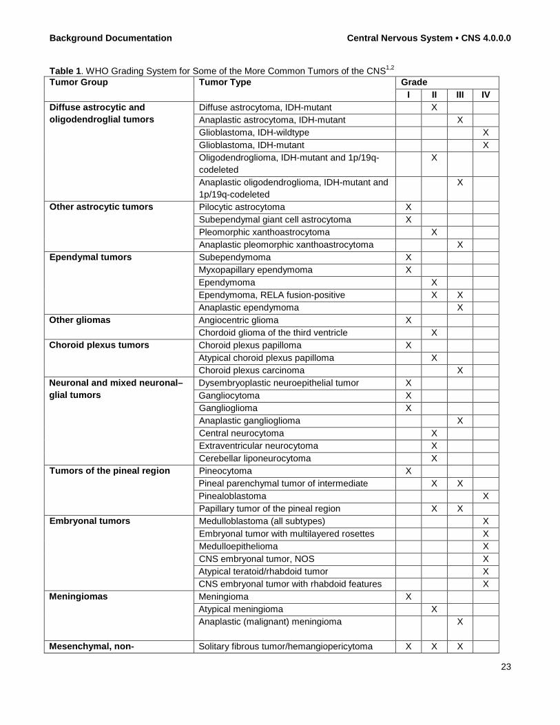
Table 1. WHO Grading System for Some of the More Common Tumors of the CNS1,2 Tumor Group Tumor Type Grade
I II III IV Diffuse astrocytic and oligodendroglial tumors
Diffuse astrocytoma, IDH-mutant X Anaplastic astrocytoma, IDH-mutant X Glioblastoma, IDH-wildtype X Glioblastoma, IDH-mutant X Oligodendroglioma, IDH-mutant and 1p/19q-codeleted
X
Anaplastic oligodendroglioma, IDH-mutant and 1p/19q-codeleted
X
Other astrocytic tumors Pilocytic astrocytoma X Subependymal giant cell astrocytoma X Pleomorphic xanthoastrocytoma X Anaplastic pleomorphic xanthoastrocytoma X
Ependymal tumors Subependymoma X Myxopapillary ependymoma X Ependymoma X Ependymoma, RELA fusion-positive X X Anaplastic ependymoma X
Other gliomas Angiocentric glioma X Chordoid glioma of the third ventricle X
Choroid plexus tumors Choroid plexus papilloma X Atypical choroid plexus papilloma X Choroid plexus carcinoma X
Neuronal and mixed neuronal–glial tumors
Dysembryoplastic neuroepithelial tumor X Gangliocytoma X Ganglioglioma X Anaplastic ganglioglioma X Central neurocytoma X Extraventricular neurocytoma X Cerebellar liponeurocytoma X
Tumors of the pineal region Pineocytoma X Pineal parenchymal tumor of intermediate X X Pinealoblastoma X Papillary tumor of the pineal region X X
Embryonal tumors Medulloblastoma (all subtypes) X Embryonal tumor with multilayered rosettes X Medulloepithelioma X CNS embryonal tumor, NOS X Atypical teratoid/rhabdoid tumor X CNS embryonal tumor with rhabdoid features X
Meningiomas Meningioma X Atypical meningioma X Anaplastic (malignant) meningioma X
Mesenchymal, non- Solitary fibrous tumor/hemangiopericytoma X X X
23

Background Documentation Central Nervous System • CNS 4.0.0.0
meningothelial tumors Hemangioblastoma X Tumors of the sellar region Craniopharyngioma X
Granular cell tumor of the sellar region X Pituicytoma X Spindle cell oncocytoma X
Tumor histology and grade are strong predictors of clinical behavior for astrocytomas and meningiomas. Tables 2 and 3 list the grading criteria for these common CNS tumor types.1 Table 2. WHO Grading System for Diffuse Infiltrating Astrocytomas
WHO Grade
WHO Designation Histologic Criteria
II Diffuse astrocytoma Nuclear atypia III Anaplastic astrocytoma Nuclear atypia and mitotic figures IV Glioblastoma Nuclear atypia, mitotic figures, and endothelial proliferation and/or necrosis
Table 3. WHO Grading of Meningiomas WHO grade I Benign meningioma
WHO grade II Atypical meningioma
Mitotic figures ≥4/10 high-power fields (HPF) or
At least 3 of 5 parameters: Sheeting architecture (loss of whorling and/or fascicles) Small cell formation Macronucleoli Hypercellularity Spontaneous necrosis
or Brain invasion
or Clear cell meningioma
or Chordoid meningioma
WHO grade III Anaplastic (malignant) meningioma
Mitotic figures ≥20/10 HPF or
Frank anaplasia (sarcoma, carcinoma, or melanoma-like histology) or
Papillary meningioma or
Rhabdoid meningioma
References 1. Louis DN, Ohgaki H, Wiestler OD, et al. World Health Organization Classification of Tumours of the Central
Nervous System. Lyon, France: IARC Press; 2016. 2. Lloyd RV, Osamura RY, Klöppel G, et al. WHO Classification of Tumours: Pathology & Genetics of Tumours
of Endocrine Organs. Lyon, France: IARC Press; 2017.
24
Background Documentation Central Nervous System • CNS 4.0.0.0
I. Primary Tumor Site, Laterality, and Focality Since the anatomic site of a neoplasm may correlate with tumor type and prognosis, it should be recorded, if known.
● For skull location, specify bone involved, such as frontal, parietal, temporal, occipital, etc, if known. The College of American Pathologists (CAP) cancer protocol for bone should be used for primary tumors of bone.1
● For dural location, indicate cerebral convexity/lobe, falx, tentorium, posterior fossa, sphenoid wing, skull base, spinal, or other, if known.
● For leptomeningeal location, indicate cerebral convexity/lobe, posterior fossa, spinal, or other, if known. ● For cerebral lobe location, indicate frontal, temporal, parietal, or occipital lobe, if known. For a deep gray
matter location, indicate basal ganglia, thalamus, or hypothalamus. ● For an intraventricular location, indicate lateral, third, fourth, or cerebral aqueduct, if known. ● For a brain stem location, indicate midbrain, pons, or medulla, if known. ● For spine (vertebral bone), spinal cord, spinal root or spinal ganglion, indicate level (eg, C5, T2, L3), if
known. The CAP cancer protocol for bone should be used for primary tumors of bone.1 The laterality of a neoplasm should be indicated as involving the left or right side of the CNS structure. In some instances, such as tumors arising in the pineal, pituitary, third ventricular, and other locations, the tumor will be situated in the midline. A tumor would be considered bilateral if it involved both sides of the brain, such as glioblastoma extending through the corpus callosum to involve the left and right hemispheres. The focality of a lesion should be indicated, if possible. Multifocality implies that multiple, noncontiguous lesions are noted on neuroimaging, such as might be seen in primary CNS lymphoma. A solitary lesion would be considered unifocal. Margins Resection margins provide no prognostic information and generally are not required for most CNS neoplasms. References 1. Laurini JA, Antonescu CR, Cooper K, et al. Protocol for the examination of specimens from patients with
tumors of bone. 2017. Available at www.cap.org/cancerprotocols. J. Preoperative Treatment and Treatment Effect Knowledge of preoperative treatment, including radiation therapy, chemotherapy, corticosteroid therapy, embolization, and other therapy, is helpful for specimen interpretation.1-3 In particular, prior radiation therapy or radiosurgery may alter the interpretation of specimens in which there are increased cellular atypia, decreased proliferative activity, or large areas of radiation-induced change (e.g., coagulative [nonpalisading] necrosis, vascular hyalinization, and gliosis). The addition of chemotherapy to radiation may further alter histomorphological appearance. For patients with malignant gliomas, the presence and degree of radiation necrosis appear to be of prognostic significance. Tumors that show evidence of radiation necrosis are associated with a longer survival, and the degree of necrosis appears to be prognostically significant.4 Corticosteroid treatment can alter the pathologic features of some CNS diseases. In particular, the treatment of primary CNS lymphoma with corticosteroids can be associated with widespread tumor necrosis or infiltration by macrophages, which may limit or misguide interpretation. Embolization of certain tumor types, especially meningiomas, may introduce histologic changes in the neoplasm. References 1. Burger PC, Scheithauer BW, Vogel FS. Surgical Pathology of the Nervous System and Its Coverings. 4th ed.
New York: Churchill Livingstone; 2002. 2. Perry A, Brat DJ. Practical Surgical Pathology: A Diagnostic Approach. Philadelphia: Elsevier; 2010. 3. McLendon RE, Rosenblum MK, Bigner DD, eds. Russell and Rubinstein's Pathology of Tumors of the
Nervous System. 7th ed. New York: Hodder Arnold; 2006. 4. Forsyth PA, Kelly PJ, Cascino TL, et al. Radiation necrosis or glioma recurrence: is computer-assisted
stereotactic biopsy useful? J Neurosurg. 1995;82:436-444.
25