proteus syndrome: evaluation of the immunological profile · progressive overgrowth over time. the...
TRANSCRIPT
LETTER TO THE EDITOR Open Access
Proteus syndrome: evaluation of theimmunological profileVassilios Lougaris1*, Vincenzo Salpietro2, Maricia Cutrupi2, Manuela Baronio1, Daniele Moratto3, M. R. Pizzino2,Kshitij Mankad4, Silvana Briuglia2, Carmelo Salpietro2 and Alessandro Plebani1
Abstract
Proteus syndrome (PS) is an extremely rare and complex disease characterized by malformations and overgrowth ofdifferent tissues. Prognosis of affected patients may be complicated by premature death, mostly due to pulmonaryembolism and respiratory failure. To date, immunological data in Proteus syndrome are scarse.We report on the novel immunologic findings of a 15 years old girl affected with PS. Detailed T and B cell evaluationrevealed maturational alterations for both subsets and functional hyperactivation for the latter. Such findings have notbeen reported previously in PS and may be the spy of more complex immune abnormalities in this syndrome.
Keywords: Proteus syndrome, Overgrowth, B cells, T cells
CorrespondenceThe Proteus syndrome (PS) is an extremely rare and com-plex disease characterized by malformations and over-growth of various tissues, mainly connective tissue, bone,skin, and central nervous system, although any tissue maybe involved [1–6]. Clinical manifestations are highly vari-able and the disproportionate overgrowth of tissue is usu-ally asymmetrical and involves the arms, legs, hands, feetand digits. The complications of PS include hyperostosis,cerebriform connective tissue progressive, skeletal de-formities, benign and malignant tumors, capillary vascularmalformations and deep venous thrombosis with pulmon-ary embolism.The syndrome has an incidence of less than 1 per
1,000.000 live births and is estimated that 120 individualswith PS are currently alive worldwide [3–5]. Newbornswith Proteus syndrome have few or no signs of the condi-tion, and overgrowth becomes apparent between 6 and18 months of life, getting more severe with age [6]. Lesionsappear to be distributed in a mosaic manner and have aprogressive evolution. Recently, a mosaic activating muta-tion in AKT1 was reported to be associated with PS [7].
Published immunological data for PS are scarse: infact, only one patient has been reported to date withmild hypogammaglobulinemia and lymphopenia leadingto reduction of total T and B cell numbers [8].
Case reportWe report the case of a 15 years old girl with Proteus syn-drome and describe novel immunological findings in PS.The index patient, born to non consanguineous parents,was born via caesarian section at 36 weeks of gestation,with normal weight and length (3500 g and 50 cm, respect-ively). Diagnosis of PS was made at the age of 2 years, withprogressive overgrowth over time. The index patient pre-sented multiple lesions affecting the central nervous andcirculatory system, lung, skeleton, limbs and abdominal or-gans. The phenotypic examination showed dolichocephaly,hyperostosis of the skull in the right fronto-parietal area, fa-cial dysmorphic features with facial asymmetry and ptosisof right eye, depressed nasal bridge, wide and antevertednares, oligodontia and multiple caries, long neck. The pa-tient presented thin skin, adipose dysregulation with poorlyrepresented subcutaneous adipose and a disproportionate,asymmetric overgrowth of the limbs. The index patient pre-sented important overgrowth of the feet (Fig. 1a, left panel)with greater growth of the calcaneus and cuboid, especiallyof the left foot (Fig. 1a, right panel). The patient also pre-sented severe kyphoscoliosis (Fig. 1b) and length discrep-ancy and macrodactyly in the III and IV fingers of right
* Correspondence: [email protected] Clinic and Institute for Molecular Medicine A. Nocivelli,Department of Clinical and Experimental Sciences, University of Brescia,Piazzale Spedali Civili 1, Brescia 25123, ItalyFull list of author information is available at the end of the article
© 2016 Lougaris et al. Open Access This article is distributed under the terms of the Creative Commons Attribution 4.0International License (http://creativecommons.org/licenses/by/4.0/), which permits unrestricted use, distribution, andreproduction in any medium, provided you give appropriate credit to the original author(s) and the source, provide a link tothe Creative Commons license, and indicate if changes were made. The Creative Commons Public Domain Dedication waiver(http://creativecommons.org/publicdomain/zero/1.0/) applies to the data made available in this article, unless otherwise stated.
Lougaris et al. Orphanet Journal of Rare Diseases (2016) 11:3 DOI 10.1186/s13023-015-0381-z

hand (Fig. 1c), emphysema, pulmonary fibrosis, syringo-myelic cavity of about 2 cm in the section C3-D4, andovarian cystadenomas. During follow-up, the patient de-veloped portal thrombosis at 12 years of age with portalsplenic and mesenteric hypertension and liver atrophy andsuffered from a transient ischemic attack (TIA) at the ageof 13. Brain magnetic resonance imaging (MRI) showedcortical dysplasia with marked thickening of the skull inthe right fronto-temporal (Fig. 1d, e and f) which in-creased in volume over 4 years reaching 44 mm with
significant compression on the midbrain, brain paren-chyma and cerebral edema, hydrocephalus and proptosisof the right eye (Fig. 1e). The significant progressionof the lesions determined respiratory failure, coma,and subsequent death of the patient.Immunological evaluation of the index patients revealed
several novel findings. Immunoglobulin serum levels werewithin normal range, in contrast with previously reporteddata [8] in the presence of valid humoral response tovaccinations (Table 1). Interestingly, both the index
Fig. 1 Clinical and radiologic presentation of the index PS patient. a Overgrowth of the feet (left panel) with radiologic confirmation of boneovergrowth, especially of the left foot (righ panel). b Severe cyphoscoliosis of the index patient. c Length discrepancy and macrodactyly in the IIIand IV fingers of right hand from the index patient. d e and f Coronal flair (d) and axial flair (e) weighted images demonstrated hyperostosis ofthe right fronto-parietal cranial vault (black asterisk) with proptosis of the right eye and extensive malformation of cortical development involvingpredominantly the right cerebral hemisphere but also part of the left cerebral hemisphere. There is an extensive white matter signal abnormalityof both hemispheres (white arrows); there is also an associated malformation of the brainstem. Coronal T2 weighted images (f) show hyperostosis ofthe right fronto-parietal cranial vault (black asterisk), extensive malformation of the right hemisphere with white matter changes in the periventricularregion (white arrows); malformation of cortex in the left perisylvian region (red arrow)
Lougaris et al. Orphanet Journal of Rare Diseases (2016) 11:3 Page 2 of 5

patient and the single patient reported previously [8]were lymphopenic (Table 1), with consequently reducedperipheral total T and B cell numbers (Table 1), withouthowever a significant history of infections. Thus, a moredetailed evaluation of the peripheral lymphocyte subsetsfrom the index PS patient was performed and revealed,within the T cell population, normal thymic output asdefined by normal naive (CD45RA+CCR7+) and recent
thymic emigrants (RTE) (CD45RA+CCR7+CD31+) CD4 Tcell percentages, an increase in the Terminally differenti-ated CD45RA+CCR7− CD4 T cells and a reduction of theCD8 T cells (Table 1). The B cell population showed areduction in the RBE (Recent Bone Marrow Emigrants:CD38hiCD21dim/loCD27−) subset and in the IgM memorysubset (IgD+CD27+), while all other subsets appearednormally distributed in terms of percentages (Table 1). No
Table 1 Immunological evaluation of the index PS patient
Patient code Index patient Normal range for age (index patient) Published patient [8]
Sex Female Male
Age (onset) 2 years n.a.
Age (evaluation) 14 years 10 years
IgG (mg/dl) 1110 (604–1909) 280
IgA (mg/dl) 100 (61–301) 40
IgM (mg/dl) 105 (59–297) 50
Anti-tetanus toxoid antibodies (UI/ml) 0,15
Anti-diphtheria antibodies (UI/ml) 0,12
Total lymphocyte count (/mm3) 736 ↓↓ (1340–3173) 470
T cell counts/ml 437 ↓↓ (954–2332) 100
B cell counts/ml 164 ↓ (173–685) 100
% (absolute cell number/μl) Normal percentage range for age(range of absolute cell number/μl for age)(index patient)
T cells (CD3+) 59,3 ↓ (437 ↓↓) 60,5–79,8 (954–2332) n.a.
CD3+CD4+ 43,2 (318 ↓↓) 30,3–48,3 (635–1334) n.a.
Naive (CD45RA+CCR7+) 49,9 (159 ↓) 34,3–74,6 (276–828) n.a.
RTE (CD45RA+CCR7+CD31+) 39,9 (127) 21,1–63,5 (21–699) n.a.
Central memory (CD45RA−CCR7+) 22,6 (72 ↓) 13,0–43,5 (107–420) n.a.
Effector memory (CD45RA−CCR7−) 19,5 (62 ↓) 8,5–28,1 (95–261) n.a.
Terminally differentiated (CD45RA+CCR7−) 7,6 ↑ (24) 0,7–6,6 (6–61) n.a.
CD3+CD8+ 9,1 ↓↓ (40 ↓↓↓) 13,8–37,5 (276–1035) n.a.
Naive (CD45RA+CCR7+) 58,5 (23,4 ↓↓↓) 26,7–72,9 (117–454) n.a.
Central memory (CD45RA−CCR7+) 10,2 (4 ↓) 1,2–11,6 (8–85) n.a.
Effector memory (CD45RA−CCR7−) 15,6 (6 ↓↓) 6,0–53,6 (19–536) n.a.
Terminally differentiated (CD45RA+CCR7−) 15,5 (6 ↓↓) 3,9–72,0 (13–699) n.a.
TCR γ/δ 3,3 (1) 0,5–21,5 n.a.
B cells (CD19+) 22,3 (164 ↓) 5,7–19,7 (173–685) n.a.
RBE (CD38hiCD21dim/loCD27−) 12,1 ↓ (20) 15,0–35,3 (20–179) n.a.
Naive (CD38dim/loCD21hiCD27−) 66,1 (108) 33,8–79,6 (40–398) n.a.
CD19hiCD21lo 6,3 (10) 1,1–10 (5–17) n.a.
Switched memory (IgD−CD27+) 8,8 (14) 2,7–20,6 (7–45) n.a.
IgM memory (IgD+CD27+) 2,5 ↓ (4 ↓↓) 3,5–24,1 (10–49) n.a.
Terminally differentiated (CD38hiCD27hiCD21lo) 0,55 (0,5) 0,16–8,70 (0,4–11) n.a.
Plasmacells (CD38hiCD20−CD138+) 0,21 (0,5) 0,04–3,20 (0,1–5,2) n.a.
NK cells (CD3−CD16+CD56+) 11,0 (81 ↓) 4,6–27,8 (111–539) n.a.
(Comparison with the only published immunological data from a single PS patient is included where data were available)
Lougaris et al. Orphanet Journal of Rare Diseases (2016) 11:3 Page 3 of 5
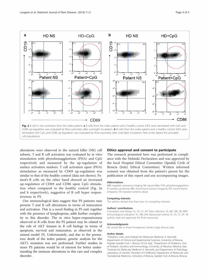
alterations were observed in the natural killer (NK) cellsubsets. T and B cell activation was evaluated by in vitrostimulation with phytohemaggluttinin (PHA) and CpGrespectively and measured by the up-regulation ofsurface activation markers. T cell activation upon (PHA)stimulation as measured by CD69 up-regulation wassimilar to that of the healthy control (data not shown). Pa-tient’s B cells on the other hand showed an increasedup-regulation of CD69 and CD86 upon CpG stimula-tion when compared to the healthy control (Fig. 2aand b respectively), suggestive of B cell hyper respon-siveness in PS.Our immunological data suggest that PS patients may
present T and B cell alterations in terms of maturationand activation. This is a novel finding in PS and, togetherwith the presence of lymphopenia, adds further complex-ity to this disorder. The in vitro hyper-responsivenessobserved in B cells from the PS patient may be related tothe role of AKT kinases in B cell biology in terms ofapoptosis, survival and maturation, as observed in theanimal model [9]. Unfortunately, and due to the prema-ture death of the index patient, genetic analysis for theAKT1 mutation was not performed. Further studies inmore PS patients would be of interest for better under-standing the immune alterations in this rare and complexdisorder.
Ethics approval and consent to participateThe research presented here was performed in compli-ance with the Helsinki Declaration and was approved bythe local Hospital Ethical Committee (Spedali Civili ofBrescia (Italy) Ethical Committee). Written informedconsent was obtained from the patient's parent for thepublication of this report and any accompanying images.
AbbreviationsMRI: magnetic resonance imaging; NK: natural killer; PHA: phytohemaggluttinin;PS: proteus syndrome; RBE: recent bone marrow emigrants; RTE: recent thymicemigrants; TIA: transient ischemic attack.
Competing interestsThe authors declare that they have no competing interests.
Authors’ contributionsConception and design: VL, VS, CS, AP; Data collection: VS, MC, MK, SB, MRP;Immunological evaluation: VL, MB, DM; Manuscript writing: VL, VS, CS, AP. Allauthors read and approved the final manuscript.
AcknowledgmentsWe would like to thank Fondazione Camillo Golgi, Brescia, Italy.
Author details1Pediatrics Clinic and Institute for Molecular Medicine A. Nocivelli,Department of Clinical and Experimental Sciences, University of Brescia,Piazzale Spedali Civili 1, Brescia 25123, Italy. 2Department of Pediatrics, Unitof Pediatric Genetics and Immunology, University of Messina, Messina, Italy.3Institute for Molecular Medicine A. Nocivelli, and Department of Pathology,Laboratory of Genetic Disorders of Childhood, Department of Molecular andTranslational Medicine, University of Brescia, Spedali Civili di Brescia, Brescia,
4100
101
102
103
104
10 4100
101
102
103
104
CD69
a bHD+CpG
Fig. 2 B cell in vitro activation from the index patient. a B cells from the index patient and a healthy control (HD) were stimulated with CpG andCD69 up-regulation was evaluated by flow-cytometry after overnight incubation. b B cells from the index patient and a healthy control (HD) werestimulated with CpG and CD86 up-regulation was evaluated by flow-cytometry after overnight incubation. Red circles depict the activatedcell populations
Lougaris et al. Orphanet Journal of Rare Diseases (2016) 11:3 Page 4 of 5

Italy. 4Department of Radiology, Great Ormond Street Hospital for ChildrenNational Health Service Foundation Trust, London, UK.
Received: 9 November 2015 Accepted: 28 December 2015
References1. Cohen MM, Hayden PW. A newly recognized hamartomatous syndrome.
Birth Defects Orig Artic Ser. 1979;15(5B):291–6.2. Temtamy SA, Rogers JG. Macrodactyly, hemihypertrophy, and connective
tissue nevi: Report of a new syndrome and review of the literature.J Pediatr. 1976;89(6):924–7.
3. Cohen Jr MM. Proteus syndrome review: molecular, clinical and pathologicfeatures. Clin Genet. 2014;85(2):111–9.
4. Biesecker LG, Happle R, Mulliken JB, Weksberg R, Graham Jr JM, Viljoen DL,et al. Proteus syndrome: diagnostic criteria, differential diagnosis, andpatient evaluation. Am J Med Genet. 1999;84(5):389–95. Review.
5. Biesecker L. The challenges of Proteus syndrome: diagnosis andamangement. Eur J Hum Genet. 2006;14(11):1151–7.
6. Thomason JL, Abramowsky CR, Rickets RR, Culbertson JH, Clifton MS,Shehata BM. Proteus syndrome: three case reports with a review of theliterature. Fetal Pediatr Pathol. 2012;31(3):145–53.
7. Lindhurst MJ, Sapp JC, Teer JK, Johnston JJ, Finn EM, Peters K, et al.A mosaic activating mutation in AKT1 associated with the Proteussyndrome. N Engl J Med. 2011;365(7):611–9.
8. Hodge D, Misbah SA, Mueller RF, Glass EJ, Chetcuti PA. Proteus syndromeand immunodeficiency. Arch Dis Child. 2000;82(3):234–5.
9. Calamito M, Juntilla MM, Thomas M, Northrup DL, Rathmell J, Birnbaum MJ,et al. Akt1 and Akt2 promote peripheral B-cell maturation and survival.Blood. 2010;115(20):4043–50.
• We accept pre-submission inquiries
• Our selector tool helps you to find the most relevant journal
• We provide round the clock customer support
• Convenient online submission
• Thorough peer review
• Inclusion in PubMed and all major indexing services
• Maximum visibility for your research
Submit your manuscript atwww.biomedcentral.com/submit
Submit your next manuscript to BioMed Central and we will help you at every step:
Lougaris et al. Orphanet Journal of Rare Diseases (2016) 11:3 Page 5 of 5