proteomic databases and tools to decipher post-translational
TRANSCRIPT
J O U R N A L O F P R O T E O M I C S 7 5 ( 2 0 1 1 ) 1 2 7 – 1 4 4
Ava i l ab l e on l i ne a t www.sc i enced i r ec t . com
www.e l sev i e r . com/ loca te / j p ro t
Review
Proteomic databases and tools to decipherpost-translational modifications
Karthik S. Kamath, Meghana S. Vasavada, Sanjeeva Srivastava⁎
Wadhwani Research Center for Biosciences and Bioengineering, Department of Biosciences and Bioengineering,Indian Institute of Technology Bombay, Mumbai 400076, India
A R T I C L E I N F O
⁎ Corresponding author. Tel.: +91 22 2576 777E-mail address: [email protected] (S. Sri
1874-3919/$ – see front matter © 2011 Elseviedoi:10.1016/j.jprot.2011.09.014
A B S T R A C T
Available online 29 September 2011
Post-translational modifications (PTMs) are vital cellular control mechanism, which affectprotein properties, including folding, conformation, activity and consequently, theirfunctions. As a result they play a key role in various disease conditions, including cancerand diabetes. Proteomics as a rapidly growing field has witnessed tremendousadvancement during the last decade, which has led to the generation of prodigiousquantity of data for various organisms' proteome. PTMs being biologically and chemicallydynamic process, pose greater challenges for its study. Amidst these complexitiesconnecting the modifications with physiological and cellular cascade of events are stillvery challenging. Advancement in proteomic technologies such as mass spectrometryand microarray provides HT platform to study PTMs and help to decipher role of some ofthe very essential biological phenomenon. To enhance our understanding of variousPTMs in different organisms, and to simplify the analysis of complex PTM data, manydatabases, software and tools have been developed. These PTM databases and toolscontain crucial information and provide a valuable resource to the research community.This article intends to provide a comprehensive overview of various PTM databases,software tools, and analyze critical information available from these resources to studyPTMs in various biological organisms.© 2011 Elsevier B.V. All rights reserved.
Keywords:Posttranslational modificationsDatabasesPredictorsPhosphorylationGlycosylation
Contents
1. Introduction . . . . . . . . . . . . . . . . . . . . . . . . . . . . . . . . . . . . . . . . . . . . . . . . . . . . . . . . . 1282. Post-translational modifications . . . . . . . . . . . . . . . . . . . . . . . . . . . . . . . . . . . . . . . . . . . . . . 1293. Techniques to study PTM . . . . . . . . . . . . . . . . . . . . . . . . . . . . . . . . . . . . . . . . . . . . . . . . . . 1304. Classification of databases . . . . . . . . . . . . . . . . . . . . . . . . . . . . . . . . . . . . . . . . . . . . . . . . . 131
4.1. Phosphorylation databases . . . . . . . . . . . . . . . . . . . . . . . . . . . . . . . . . . . . . . . . . . . . . 1324.2. Glycosylation databases . . . . . . . . . . . . . . . . . . . . . . . . . . . . . . . . . . . . . . . . . . . . . . . 1334.3. Databases of other types of PTMs . . . . . . . . . . . . . . . . . . . . . . . . . . . . . . . . . . . . . . . . . . 136
5. PTM tools . . . . . . . . . . . . . . . . . . . . . . . . . . . . . . . . . . . . . . . . . . . . . . . . . . . . . . . . . . . 1375.1. Machine learning processes for prediction . . . . . . . . . . . . . . . . . . . . . . . . . . . . . . . . . . . . . 137
9; fax: +91 22 2572 3480.vastava).
r B.V. All rights reserved.
128 J O U R N A L O F P R O T E O M I C S 7 5 ( 2 0 1 1 ) 1 2 7 – 1 4 4
5.2. Strong prediction tools by evolving models . . . . . . . . . . . . . . . . . . . . . . . . . . . . . . . . . . . . . 1395.3. Phosphorylation tools . . . . . . . . . . . . . . . . . . . . . . . . . . . . . . . . . . . . . . . . . . . . . . . . . 1395.4. Glycosylation tools . . . . . . . . . . . . . . . . . . . . . . . . . . . . . . . . . . . . . . . . . . . . . . . . . . 1395.5. Other PTM related tools . . . . . . . . . . . . . . . . . . . . . . . . . . . . . . . . . . . . . . . . . . . . . . . . 140
6. Organism specific database and tools . . . . . . . . . . . . . . . . . . . . . . . . . . . . . . . . . . . . . . . . . . . . 1407. Conclusions . . . . . . . . . . . . . . . . . . . . . . . . . . . . . . . . . . . . . . . . . . . . . . . . . . . . . . . . . . 141Acknowledgments . . . . . . . . . . . . . . . . . . . . . . . . . . . . . . . . . . . . . . . . . . . . . . . . . . . . . . . . . 141References. . . . . . . . . . . . . . . . . . . . . . . . . . . . . . . . . . . . . . . . . . . . . . . . . . . . . . . . . . . . . . 141
1. Introduction
The completion of genome projects has accelerated the anal-ysis of proteome; however, due to the complexity of proteinsits study is more challenging than any other biomolecules.This complexity arises due to the biological phenomenonsuch as gene splicing to form different isoforms and variouspost-translational modifications (PTMs), which gives rise toenormous number of proteins, about three orders of magni-tude higher than the total number of genes encoded in ge-nome [1,2]. As the name indicates for PTMs, the process ofprotein modifications takes place after translation of mRNAinto a protein. All proteins undergo appreciable amount ofPTMs to make biologically active form, and this dynamic pro-cess occurs in various cell compartments to decide the func-tion of modified protein. About 300 different types of PTMshave been reported till date and many more are still being
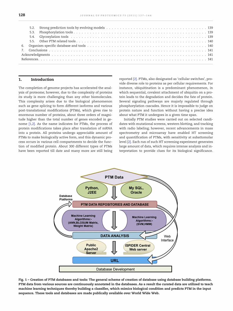
Fig. 1 – Creation of PTM databases and tools: The general schemPTM data from various sources are continuously annotated in thmachine learning techniques thereby building a classifier, whichsequence. These tools and databases are made publically availa
reported [2]. PTMs, also designated as ‘cellular switches’, pro-vide diverse role to proteins as per cellular requirements. Forinstance, ubiquitination is a predominant phenomenon, inwhich sequential, covalent attachment of ubiquitin on a pro-tein leads to the degradation and decides the fate of protein.Several signaling pathways are majorly regulated throughphosphorylation cascades. Hence it is impossible to judge onprotein nature and function without having a precise ideaabout what PTM it undergoes in a given time span.
Initially PTM studies were carried out on selected candi-dates with mutational screens, western blotting, and trackingwith radio labeling; however, recent advancements in massspectrometry and microarray have enabled HT screeningand quantification of PTMs, with sensitivity at subattomolarlevel [2]. Each run of such HT screening experiment generateslarge amount of data, which requires intense analysis and in-terpretation to provide clues for its biological significance.
e of creation of database using database building platforms.e databases. As a result the curated data are utilized to teachmimics biological condition and predicts PTM in the input
ble over World Wide Web.
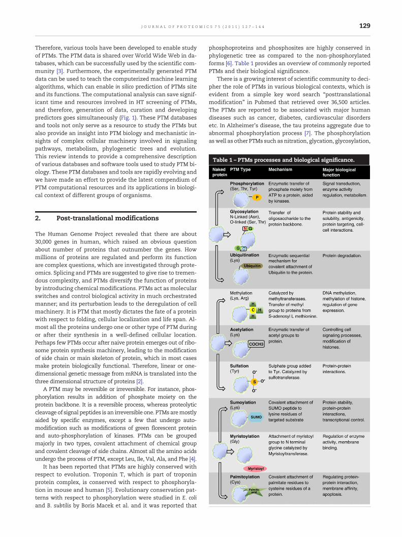
Table 1 – PTMs processes and biological significance.
129J O U R N A L O F P R O T E O M I C S 7 5 ( 2 0 1 1 ) 1 2 7 – 1 4 4
Therefore, various tools have been developed to enable studyof PTMs. The PTM data is shared over World Wide Web in da-tabases, which can be successfully used by the scientific com-munity [3]. Furthermore, the experimentally generated PTMdata can be used to teach the computerized machine learningalgorithms, which can enable in silico prediction of PTMs siteand its functions. The computational analysis can save signif-icant time and resources involved in HT screening of PTMs,and therefore, generation of data, curation and developingpredictors goes simultaneously (Fig. 1). These PTM databasesand tools not only serve as a resource to study the PTMs butalso provide an insight into PTM biology and mechanistic in-sights of complex cellular machinery involved in signalingpathways, metabolism, phylogenetic trees and evolution.This review intends to provide a comprehensive descriptionof various databases and software tools used to study PTM bi-ology. These PTM databases and tools are rapidly evolving andwe have made an effort to provide the latest compendium ofPTM computational resources and its applications in biologi-cal context of different groups of organisms.
2. Post-translational modifications
The Human Genome Project revealed that there are about30,000 genes in human, which raised an obvious questionabout number of proteins that outnumber the genes. Howmillions of proteins are regulated and perform its functionare complex questions, which are investigated through prote-omics. Splicing and PTMs are suggested to give rise to tremen-dous complexity, and PTMs diversify the function of proteinsby introducing chemical modifications. PTMs act as molecularswitches and control biological activity in much orchestratedmanner; and its perturbation leads to the deregulation of cellmachinery. It is PTM that mostly dictates the fate of a proteinwith respect to folding, cellular localization and life span. Al-most all the proteins undergo one or other type of PTM duringor after their synthesis in a well-defined cellular location.Perhaps few PTMs occur after naive protein emerges out of ribo-some protein synthesis machinery, leading to the modificationof side chain or main skeleton of protein, which in most casesmake protein biologically functional. Therefore, linear or one-dimensional genetic message from mRNA is translated into thethree dimensional structure of proteins [2].
A PTM may be reversible or irreversible. For instance, phos-phorylation results in addition of phosphate moiety on theprotein backbone. It is a reversible process, whereas proteolyticcleavage of signal peptides is an irreversible one. PTMsaremostlyaided by specific enzymes, except a few that undergo auto-modification such as modifications of green florescent proteinand auto-phosphorylation of kinases. PTMs can be groupedmajorly in two types, covalent attachment of chemical groupand covalent cleavage of side chains. Almost all the amino acidsundergo the process of PTM, except Leu, Ile, Val, Ala, and Phe [4].
It has been reported that PTMs are highly conserved withrespect to evolution. Troponin T, which is part of troponinprotein complex, is conserved with respect to phosphoryla-tion in mouse and human [5]. Evolutionary conservation pat-terns with respect to phosphorylation were studied in E. coliand B. subtilis by Boris Macek et al. and it was reported that
phosphoproteins and phosphosites are highly conserved inphylogenetic tree as compared to the non-phosphorylatedforms [6]. Table 1 provides an overview of commonly reportedPTMs and their biological significance.
There is a growing interest of scientific community to deci-pher the role of PTMs in various biological contexts, which isevident from a simple key word search “posttranslationalmodification” in Pubmed that retrieved over 36,500 articles.The PTMs are reported to be associated with major humandiseases such as cancer, diabetes, cardiovascular disordersetc. In Alzheimer's disease, the tau proteins aggregate due toabnormal phosphorylation process [7]. The phosphorylationaswell as other PTMs such asnitration, glycation, glycosylation,

130 J O U R N A L O F P R O T E O M I C S 7 5 ( 2 0 1 1 ) 1 2 7 – 1 4 4
polyamination, ubiquitination and oxidation is also reported tobe associatedwith variousneurodegenerative disorders. Andro-gen receptors,which are dynamically regulated through variousPTMs, are strongly correlated with manifestation of prostatecancer [8]. Hence studying PTMsmay enhance our understand-ing for various human diseases.
3. Techniques to study PTM
Detection of subtle changes, which occur during the PTMs,poses challenge to even advance proteomic techniques. De-termination of changes to very minute level and correlationwith biological phenomenon remain challenging for moderntechnologies. PTMs bring in either addition of chemical moie-ties or removal of few amino acids; therefore, difference inmass must be apparent when measured. For instance, in pal-mitoylation, addition of palmitic acid on the cystein residueyields the addition of 238 Da whereas; methylation of lysine
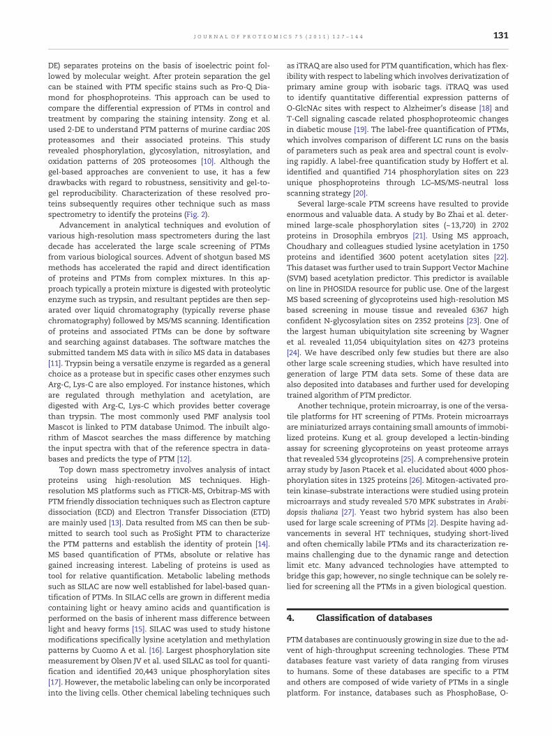
Fig. 2 – Proteomic techniques for profiling PTMs. Present day cutPTMs. Gel-based or gel-free approaches are widely used. Methodextensively used for proteome wide screening of PTMs. Similarlusing various purification strategies. Essentially shotgun gunmemass spectrometer involving soft ionization techniques such asusing high-resolution MS platforms such as FTICR for elucidating2-DE, helps in separation and visualization of proteins. After pro
residue causes the addition of 14 Da to the total mass. Thebulkier modifications such as ubiquitination may result inmass difference of about 1 kDa. There are many approachesranging fromgel-based techniques,mass spectrometry,microar-rays, peptide library screening etc. that are currently used tostudy PTMs (Fig. 2). As per the research questions, either wholeproteome or only an enriched part containing proteins withPTMs of specific interest can be screened. Affinity based enrich-ments, immunopurification and metal affinity chromatographyare commonly used strategies for the purification of proteinscontaining specific PTM. Immobilized metal affinity chromatog-raphy (IMAC) purification is a common chemical affinity strategyfor the enrichment of phosphoproteins, whereby immobilizedFe3+ ions selectively bind to the phosphorylated peptides. Othermetal oxide affinity resins such as TiO2, Fe3O4 are also commonlyused [9].
Conventional proteomic approaches such as gel-basedtechniques have been used to profile global PTMs in a givenbiological condition. Two dimensional gel electrophoresis (2-
ting edge technologies enable high throughput screening ofs such as mass spectrometry, microarray and 2-DE are
y, a subclass of proteins with specific PTM can be segregatedthod involves analysis of proteolytically digested peptide overESI and MALDI. Top-down MS technique typically involvesPTM patterns of intact proteins. Gel-based approach such astein separation gels can be stained with PTM specific stain.
131J O U R N A L O F P R O T E O M I C S 7 5 ( 2 0 1 1 ) 1 2 7 – 1 4 4
DE) separates proteins on the basis of isoelectric point fol-lowed by molecular weight. After protein separation the gelcan be stained with PTM specific stains such as Pro-Q Dia-mond for phosphoproteins. This approach can be used tocompare the differential expression of PTMs in control andtreatment by comparing the staining intensity. Zong et al.used 2-DE to understand PTM patterns of murine cardiac 20Sproteasomes and their associated proteins. This studyrevealed phosphorylation, glycosylation, nitrosylation, andoxidation patterns of 20S proteosomes [10]. Although thegel-based approaches are convenient to use, it has a fewdrawbacks with regard to robustness, sensitivity and gel-to-gel reproducibility. Characterization of these resolved pro-teins subsequently requires other technique such as massspectrometry to identify the proteins (Fig. 2).
Advancement in analytical techniques and evolution ofvarious high-resolution mass spectrometers during the lastdecade has accelerated the large scale screening of PTMsfrom various biological sources. Advent of shotgun based MSmethods has accelerated the rapid and direct identificationof proteins and PTMs from complex mixtures. In this ap-proach typically a protein mixture is digested with proteolyticenzyme such as trypsin, and resultant peptides are then sep-arated over liquid chromatography (typically reverse phasechromatography) followed by MS/MS scanning. Identificationof proteins and associated PTMs can be done by softwareand searching against databases. The software matches thesubmitted tandem MS data with in silico MS data in databases[11]. Trypsin being a versatile enzyme is regarded as a generalchoice as a protease but in specific cases other enzymes suchArg-C, Lys-C are also employed. For instance histones, whichare regulated through methylation and acetylation, aredigested with Arg-C, Lys-C which provides better coveragethan trypsin. The most commonly used PMF analysis toolMascot is linked to PTM database Unimod. The inbuilt algo-rithm of Mascot searches the mass difference by matchingthe input spectra with that of the reference spectra in data-bases and predicts the type of PTM [12].
Top down mass spectrometry involves analysis of intactproteins using high-resolution MS techniques. High-resolution MS platforms such as FTICR-MS, Orbitrap-MS withPTM friendly dissociation techniques such as Electron capturedissociation (ECD) and Electron Transfer Dissociation (ETD)are mainly used [13]. Data resulted from MS can then be sub-mitted to search tool such as ProSight PTM to characterizethe PTM patterns and establish the identity of protein [14].MS based quantification of PTMs, absolute or relative hasgained increasing interest. Labeling of proteins is used astool for relative quantification. Metabolic labeling methodssuch as SILAC are now well established for label-based quan-tification of PTMs. In SILAC cells are grown in different mediacontaining light or heavy amino acids and quantification isperformed on the basis of inherent mass difference betweenlight and heavy forms [15]. SILAC was used to study histonemodifications specifically lysine acetylation and methylationpatterns by Cuomo A et al. [16]. Largest phosphorylation sitemeasurement by Olsen JV et al. used SILAC as tool for quanti-fication and identified 20,443 unique phosphorylation sites[17]. However, the metabolic labeling can only be incorporatedinto the living cells. Other chemical labeling techniques such
as iTRAQ are also used for PTM quantification, which has flex-ibility with respect to labeling which involves derivatization ofprimary amine group with isobaric tags. iTRAQ was usedto identify quantitative differential expression patterns ofO-GlcNAc sites with respect to Alzheimer's disease [18] andT-Cell signaling cascade related phosphoproteomic changesin diabetic mouse [19]. The label-free quantification of PTMs,which involves comparison of different LC runs on the basisof parameters such as peak area and spectral count is evolv-ing rapidly. A label-free quantification study by Hoffert et al.identified and quantified 714 phosphorylation sites on 223unique phosphoproteins through LC–MS/MS-neutral lossscanning strategy [20].
Several large-scale PTM screens have resulted to provideenormous and valuable data. A study by Bo Zhai et al. deter-mined large-scale phosphorylation sites (~13,720) in 2702proteins in Drosophila embryos [21]. Using MS approach,Choudhary and colleagues studied lysine acetylation in 1750proteins and identified 3600 potent acetylation sites [22].This dataset was further used to train Support Vector Machine(SVM) based acetylation predictor. This predictor is availableon line in PHOSIDA resource for public use. One of the largestMS based screening of glycoproteins used high-resolution MSbased screening in mouse tissue and revealed 6367 highconfident N-glycosylation sites on 2352 proteins [23]. One ofthe largest human ubiquitylation site screening by Wagneret al. revealed 11,054 ubiquitylation sites on 4273 proteins[24]. We have described only few studies but there are alsoother large scale screening studies, which have resulted intogeneration of large PTM data sets. Some of these data arealso deposited into databases and further used for developingtrained algorithm of PTM predictor.
Another technique, protein microarray, is one of the versa-tile platforms for HT screening of PTMs. Protein microarraysare miniaturized arrays containing small amounts of immobi-lized proteins. Kung et al. group developed a lectin-bindingassay for screening glycoproteins on yeast proteome arraysthat revealed 534 glycoproteins [25]. A comprehensive proteinarray study by Jason Ptacek et al. elucidated about 4000 phos-phorylation sites in 1325 proteins [26]. Mitogen-activated pro-tein kinase–substrate interactions were studied using proteinmicroarrays and study revealed 570 MPK substrates in Arabi-dopsis thaliana [27]. Yeast two hybrid system has also beenused for large scale screening of PTMs [2]. Despite having ad-vancements in several HT techniques, studying short-livedand often chemically labile PTMs and its characterization re-mains challenging due to the dynamic range and detectionlimit etc. Many advanced technologies have attempted tobridge this gap; however, no single technique can be solely re-lied for screening all the PTMs in a given biological question.
4. Classification of databases
PTM databases are continuously growing in size due to the ad-vent of high-throughput screening technologies. These PTMdatabases feature vast variety of data ranging from virusesto humans. Some of these databases are specific to a PTMand others are composed of wide variety of PTMs in a singleplatform. For instance, databases such as PhosphoBase, O-
132 J O U R N A L O F P R O T E O M I C S 7 5 ( 2 0 1 1 ) 1 2 7 – 1 4 4
glycobase are specifically focused for one type of PTM, where-as Swiss-Prot, HPRD, dbPTM, RESID, PHOSIDA etc. provide de-tailed information for different types of PTMs (Table 2). Thesedatabases and resources have accelerated the analysis, visu-alization and prediction of PTMs in biological contexts. Thedata in most of the PTM databases are derived empirically orcurated manually through the literature. For example, HPRDis a manually curated database, which has more than 93,710entries and it is linked to other entities such as PhosphoMotifFinder for further reference [28]. Swiss-Prot (UniProt), one ofthe largest collections of various PTM types, is non-redundant and enables users to get amass information on asingle platform. These databases are collection of variety ofinformation in one place, thereby trying to provide completebiological information to the entries present [29]. Fig. 3 repre-sents general and PTM specific databases.
4.1. Phosphorylation databases
The PTMs act as a biological switch to activate or deactivatemolecules by signal transduction pathways. Protein phos-phorylation is one of the most-studied PTMs, which accountsfor over 30% of all PTMs. Phosphorylation process involvestransfer of phosphate moiety from ATP to a protein (serine,threonine or tyrosine residues) by enzyme kinases resultingin formation of ADP. Phosphorylation is biologically signifi-cant because this ubiquitous regulatory mechanism controlsprocesses such as cellular growth, differentiation, apoptosisand DNA repair etc. [2,30]. Many of the kinases are beingused as potential drug targets to treat some of the major dis-eases such as cancer. It is predicted that there are approxi-mately 500,000 phosphorylation sites in human proteomealone [30], which emphasizes that there is a great need to de-cipher the role of protein phosphorylation.
Mass spectrometry is widely used to study phospho-proteome. As discussed in the previous section, severallarge-scale MS based studies have identified thousands ofphosphoproteins and phosphosites. Several databases helpin mining phosphorylation data. PhosphoBase was the first
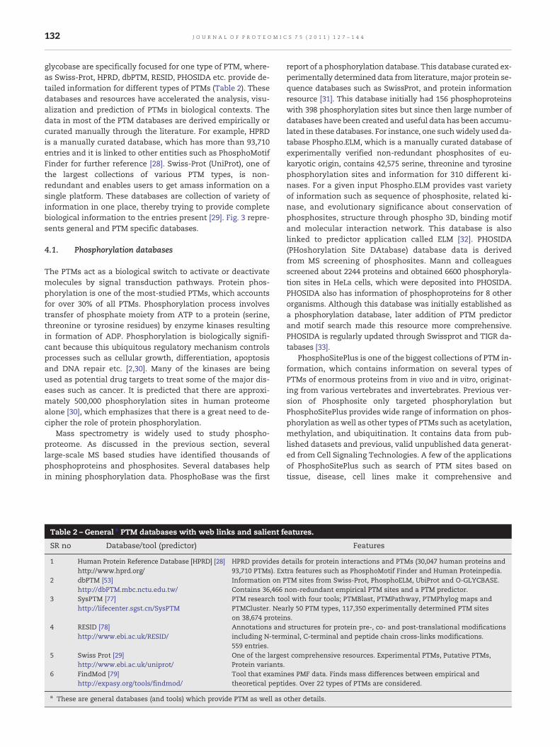
Table 2 – General a PTM databases with web links and salient f
SR no Database/tool (predictor)
1 Human Protein Reference Database [HPRD] [28]http://www.hprd.org/
HPRD provides d93,710 PTMs). Ex
2 dbPTM [53]http://dbPTM.mbc.nctu.edu.tw/
Information on PContains 36,466
3 SysPTM [77]http://lifecenter.sgst.cn/SysPTM
PTM research toPTMCluster. Neaon 38,674 protein
4 RESID [78]http://www.ebi.ac.uk/RESID/
Annotations andincluding N-term559 entries.
5 Swiss Prot [29]http://www.ebi.ac.uk/uniprot/
One of the largesProtein variants.
6 FindMod [79]http://expasy.org/tools/findmod/
Tool that examintheoretical pepti
a These are general databases (and tools) which provide PTM as well as
report of a phosphorylation database. This database curated ex-perimentally determined data from literature,major protein se-quence databases such as SwissProt, and protein informationresource [31]. This database initially had 156 phosphoproteinswith 398 phosphorylation sites but since then large number ofdatabases have been created and useful data has been accumu-lated in these databases. For instance, one suchwidely used da-tabase Phospho.ELM, which is a manually curated database ofexperimentally verified non-redundant phosphosites of eu-karyotic origin, contains 42,575 serine, threonine and tyrosinephosphorylation sites and information for 310 different ki-nases. For a given input Phospho.ELM provides vast varietyof information such as sequence of phosphosite, related ki-nase, and evolutionary significance about conservation ofphosphosites, structure through phospho 3D, binding motifand molecular interaction network. This database is alsolinked to predictor application called ELM [32]. PHOSIDA(PHoshorylation Site DAtabase) database data is derivedfrom MS screening of phosphosites. Mann and colleaguesscreened about 2244 proteins and obtained 6600 phosphoryla-tion sites in HeLa cells, which were deposited into PHOSIDA.PHOSIDA also has information of phosphoproteins for 8 otherorganisms. Although this database was initially established asa phosphorylation database, later addition of PTM predictorand motif search made this resource more comprehensive.PHOSIDA is regularly updated through Swissprot and TIGR da-tabases [33].
PhosphoSitePlus is one of the biggest collections of PTM in-formation, which contains information on several types ofPTMs of enormous proteins from in vivo and in vitro, originat-ing from various vertebrates and invertebrates. Previous ver-sion of Phosphosite only targeted phosphorylation butPhosphoSitePlus provides wide range of information on phos-phorylation as well as other types of PTMs such as acetylation,methylation, and ubiquitination. It contains data from pub-lished datasets and previous, valid unpublished data generat-ed from Cell Signaling Technologies. A few of the applicationsof PhosphoSitePlus such as search of PTM sites based ontissue, disease, cell lines make it comprehensive and
eatures.
Features
etails for protein interactions and PTMs (30,047 human proteins andtra features such as PhosphoMotif Finder and Human Proteinpedia.TM sites from Swiss-Prot, PhosphoELM, UbiProt and O-GLYCBASE.non-redundant empirical PTM sites and a PTM predictor.ol with four tools; PTMBlast, PTMPathway, PTMPhylog maps andrly 50 PTM types, 117,350 experimentally determined PTM sitess.structures for protein pre-, co- and post-translational modificationsinal, C-terminal and peptide chain cross-links modifications.
t comprehensive resources. Experimental PTMs, Putative PTMs,
es PMF data. Finds mass differences between empirical anddes. Over 22 types of PTMs are considered.
other details.

Fig. 3 – General and PTM specific databases: (A) Common PTM repositories share large amount of data pertaining to differenttypes of PTMs on a single platform. SwissProt is one of the largest continuously updated, non-redundant repositories for PTMdata. This encompasses enormous data on various types of PTMs (B) Databases for specific PTM store data with respect to onetype of PTM. One of the typical databases such as Phospho.ELM has largest collected information on phosphorylation.
133J O U R N A L O F P R O T E O M I C S 7 5 ( 2 0 1 1 ) 1 2 7 – 1 4 4
informative [34]. Structural repositories such as Phospho3Dcontain experimentally verified 3D structures of phosphoryla-tion sites. Most of the databases enable various search optionssuch as name of kinase; PDB identification code etc., whichmake the search process simple [35]. PlantsP, a plant specificdatabase, combines information derived from plant genomicsequences with experimentally derived functional genomicsdata on plant kinases and phosphatases [36]. PhosphoPep pro-vides an idea about the signal transduction pathways by link-ing kinases with their upstream and downstream moleculesin various organisms such as S. cerevisiae, C. elegans, D. melano-gaster and H. sapience. This database contains LC–MS/MS gen-erated data, which is searched from standard proteindatabase and validated further. The relevant pathways of pro-teins of interest can also be visualized [37]. LymPHOS data-base is focused on phosphoproteome of human lymphocytes
and it elucidates key role of various signal transduction path-ways and altered immune response. This database providesoptions of searching proteins and peptide sequences, withthe mass spectral information [38]. A comprehensive view ofmost widely used databases and tools for phosphorylation re-search is depicted in Table 3, Figs. 3 and 4.
4.2. Glycosylation databases
Glycosylation involves linking saccharides to proteins in pres-ence of glycosyltransferase enzymes, giving rise to a glycopro-tein. Glycosylation process mainly occurs in endoplasmicreticulum and golgi complex of the cellular compartmentand major glycoproteins are seen localized to the cell surface.Like-wise glycation, a non-enzymatic process involves attach-ment of sugar moieties to proteins specifically at lysine

Table 3 – Databases and tools to study phosphorylation.
SRno
Database/tool (predictor) Features
1 The Phosphorylation Site Database [80]http://www.phosphorylation.biochem.vt.edu/
PTMs of prokaryotes of the domains archaea and bacteria.Directly linked with RESID, O-GLYCBASE etc.
2 Phospho.ELM [32]http://phospho.elm.eu.org/
One of the largest DB of eukaryotic p-site. Contains 42,575 Serine,Threonine and Tyrosine non-redundant p-sites.
3 PhosPhAt [75]http://phosphat.mpimp-golm.mpg.de
DB: p-sites in A thaliana identified by MS.Predictor, trained on the experimental dataset. Predicts p-sites inprotein sequence.
4 PHOSIDA [33]http://www.phosida.com/
PTM DB of wide range of organisms. Phosphorylation, acetylation,N-Glycosylation information. 70,095 p-sites on 23,669 proteinsPredictor, predicts high confidence phosphosites, with the Support vector machines(SVMs) algorithm. It also predicts other types of PTMs such as acetylation.
5 PhosphoPep [37]http://www.phosphopep.org/
Phosphoproteome resource for S cerevisiae, C. elegans, D. melanogaster(Kc167 cells) and Homo sapiens. Based on MS data. 9000 identified p-sites inyeast, 10,000 phosphorylation sites of nearly 4600 phosphoproteinsD. melanogaster and 3980 p-sites in humans.
6 PhosphoPOINT [81]http://kinase.bioinformatics.tw/
Human kinase interactome and p-protein DB. 518 known humanserine/threonine/tyrosine kinases. 4195 p-proteins.
7 PhosphoNET–Human Phosphosite KnowledgeBase [82]http://www.phosphonet.ca/
Human p-sites. Over 657,391 p-sites in 23,469 represented proteins.
8 Phospho3D [35]http://cbm.bio.uniroma2.it/phospho3d
DB of 3D structures of p-sites. Retrieves data from the phospho.ELMDatabase enriches further at structural level.
9 ProMEX [83]http://promex.pph.univie.ac.at/promex/
Mass spectral reference DB for tryptic digested proteins and protein p-sitesof 14 plant species including Arabidopsis thaliana. Contains manuallyvalidated mass spectra.
10 PhosphoSitePlus [34][http://www.phosphosite.org]
One of the largest DB of protein phosphorylation majorly in humans andmouse, also has information on other organisms such as rabbit, hamster etc.Includes information on other types of PTMs such as ubiquitination etc.
11 PhosphoregDB [84]http://phosphoreg.imb.uq.edu.au/
Tissue and sub-cellular distribution of mouse protein kinasesand phosphatases.
12 PhosphoGRID [85]http://phosphogrid.org/
DB of experimentally verified in vivo p-sites in S cerevisiae.Entry over 5000 p-sites.
13 Plant Protein Phosphorylation Database (P3DB) [86]http://digbio.missouri.edu/p3db/
Plant protein phosphorylation DB. Involves data of 4 different plant species.Over 31,000 p-sites from 10,400 proteins.
14 LymPHOS [38]http://www.lymphos.org
Database of phosphoproteome of human lymphocytes. Annotates datagenerated by MS based experiments.
15 NetworKIN [87,88]http://networkin.info/search.php
Provides latest collection of phosphoproteins through connectionwith PhosphoELM.Predictor, predicts in vivo kinase–substrate relationships. Adopts inbuiltNetworKIN method for search.
16 Predikin [89]http://predikin.biosci.uq.edu.au/
Predikin predicts protein kinase peptide and likely p-sites for a specificprotein kinase, links substrates to kinase sequences.
17 Protein kinase resource [90]http://pkr.genomics.purdue.edu/pkr/Welcome.do
Integrated view of the protein kinase superfamily, structural representationalong with complete assistance for kinases study. Provides information onsignaling pathway and associated diseases.
18 Kinomer [91]http://www.compbio.dundee.ac.uk/kinomer/
Systematically classified protein kinases. Annotations of kinases for about43 organisms.
19 KinG [92]http://king.mbu.iisc.ernet.in/
Collection of protein kinases in genomes. Protein Kinases information onD. melanogaster, A. thaliana, H. sapiens, C. elegans and S. cerevisiae. In additionProtein Kinases information on 8 archaeal genomes and 27 bacterial genomes.
20 PlantsP [36]http://plantsp.genomics.purdue.edu/
DB which connects functional genomics data to sequence in context of plant(Kingdome viridiplantae) kinases and phosphatases.
21 PepCyber: P~Pep [93]http://www.pepcyber.org/PPEP/
DB of human Protein–Protein Interactions mediated by Phosphorylationbinding domains (PPBDs). 11,269 records of interactions between 387 PPBDsproteins and 1471 substrate proteins.
22 PhosSNP [94]http://phossnp.biocuckoo.org/
Database of phosphorylation related non-synonymous SNPs in Humans.
23 ProMost [95]http://proteomics.mcw.edu/promost
Tool calculating the pI and molecular weight of phosphorylated and modifiedproteins on 2D gels.
24 PhosphoScore [96]http://dir.nhlbi.nih.gov/papers/lkem/phosphoscore/
Java based software for phosphorylation site assignment tool for MS data.
25 NetPhos [97]http://www.cbs.dtu.dk/services/NetPhos/
Predicts non-kinase specific phosphorylation status based on sets ofexperimentally validated Ser, Thr and Tyr p-sites. ANN used.
26 NetPhosK [39]http://www.cbs.dtu.dk/services/NetPhosK/
Predicts kinase-specific p-sites based on sets of Ser, Thr and Tyr p-sites.ANN used.
134 J O U R N A L O F P R O T E O M I C S 7 5 ( 2 0 1 1 ) 1 2 7 – 1 4 4

Table 3 (continued)
SRno
Database/tool (predictor) Features
27 NetPhosBac [73]http://www.cbs.dtu.dk/services/NetPhosBac
Predicts Ser/Thr p-sites in bacterial proteins. ANN used.
28 NetPhosYeast [74]http://www.cbs.dtu.dk/services/NetPhosYeast/
Predicts p-sites in yeast proteins. ANN used.
29 KinasePhos [98]http://KinasePhos2.mbc.nctu.edu.tw/
SVM based predictor for protein kinase-specific p-sites prediction.SVM utilizes features such as solvent accessibility and sequences basedamino acid coupling patterns.
30 PostMod [99]http://pbil.kaist.ac.kr/PostMod
Prediction of kinase-specific p-sites. Training data was retrieved fromPhosphoELM.
31 DISPHOS [100]http://www.ist.temple.edu/disphos/
Disorder-Enhanced p-site predictor. Uses logistic regression basedlinear predictor model.
32 NetPhorest [101]http://netphorest.info/
Catalogue of linear motifs involved in phosphorylation based signaling.Information on 179 kinases.
33 pkaPS [102]http://mendel.imp.ac.at/pkaPS/
Prediction of protein kinaseA p-sites. High confidence prediction with96% sensitivity and 94% specificity.
34 Group-based Prediction system (GPS) [103]http://gps.biocuckoo.org/
Prediction of p-sites for 408 human kinases. Group based scoring system,uses BLOSUM62 for scoring.
35 Scansite [104]http://scansite.mit.edu.
Predicts cell signaling interactions using short sequence motifs withinproteins that are likely to be phosphorylated by specific protein kinases.
36 PhosphoMotif Finder [105]http://www.hprd.org/PhosphoMotif_finder
Literature based information on kinase and phosphatase substrates andbinding motifs.
37 MetaPredPS [106]http://MetaPred.biolead.org/MetaPredPS
Predicts p-sites of major S/T kinase families: CDK, CK2, PKA, and PKC.Makes use of element predictors such as GPS, KinasePhos, NetPhosK, PPSP,PredPhospho and Scansite.
38 CRPhos [107]http://www.ptools.ua.ac.be/CRPhos
Prediction of kinase-specific phosphorylation sites. Uses CRF conditionalrandom fields.
39 PhoScan [108]http://bioinfo.au.tsinghua.edu.cn/phoscan/
Prediction of kinase-specific phosphorylation sites with sequence features.Uses log-odds ratio approach.
40 Prediction of PK-Specific PhosphorylationSite (PPSP) [109]http://ppsp.biocuckoo.org/
Prediction of kinase-specific phosphorylation sites. Uses BDT.
41 PhosphoBlast [110]http://phospho.elm.eu.org/pELMBlastSearch.html
Predictor in PhosphoELM. Tool which identifies specific phosphosite mutations.Matches p-peptides sharing the p-sites within and across species.
42 Motif-X [55]http://motif-x.med.harvard.edu/
Predictor of phosphorylation short linear motif. First ever substrate drivenapproach to predict motifs.
P-site—phosphorylation site, p-proteins—phoshoproteins, DB—database, MS—mass spectrometry, p-peptides—Phosphopeptides, BDT = BaysianDecision Theory, CRF = Conditional Random Fields, SVM—Support Vector Machines, ANN = Artificial neural network.
135J O U R N A L O F P R O T E O M I C S 7 5 ( 2 0 1 1 ) 1 2 7 – 1 4 4
residue. Depending on the linkage between the amino acidand the sugar moiety, there are 4 types of glycosylations,namely; N-linked glycosylation, O-linked glycosylation, C-mannosylation and Glycophosphatidlyinositol anchored(GPI) attachments [39]. Glycosylation is involved in variouscellular events, which has implications in various biologicalfunctions such as antigenicity of immunological molecules,protein's half-life, protein folding, protein targeting, cell–cellinteractions and protein stability [39]. Aberrant forms of gly-cosylation play a major role in various human congenitaldisorders. Despite technological advances, as compared tophosphorylation, much is yet to be explored to understandthe interaction between several glycotransferases and theircorresponding substrates. Glycosylation databases providevaluable information curated out of published reports thathelps to study glycobiology and its relevance to diseases.General glycosylation databases such as GlycoSuiteDB anno-tate and collect glycan structures derived from glycoproteinsof various biological sources. It contains information ofglycan types, linkages and anomeric configurations, mass,composition and the analytical methods used to determine
the glycans structure. Current version; GlycoSuiteDB 8.0 iscomposed of 9436 entries of which 3238 are unique and 1851are completely characterized. This database is extensivelylinked with the ExPASy, GlycoMod, SWISS-PROT and PubMedand provides details on the disease relevant modifications[40].
The glycosylation screening technologies are continuouslyevolving. MS techniques and protein microarray are regularlyused to study glycosylation. One of the recent MS based data-base screening studies by Dorota et al. revealed 6367 N-glycosylation sites on 2352 proteins derived from four mousetissues and blood plasma [23]. Massive data for glycosylatedproteins thus produced are annotated in various databases.Glycodatabases, depending upon the type of chemistryinvolved in the attachment of saccharide moieties, areavailable on World Wide Web (Table 4). O-glycosylation isa process occurring in golgi apparatus, which is anenzymatic attachment of N-Acetylgalactosamine on hydroxylgroup of Ser or Thr residues in presence of enzyme N-Acetylgalactosaminyltransferase. Transfer of first sugar moie-ty leads to the sequential addition of other sugar molecules in

Fig. 4 – Different tools for PTM prediction: A number of PTM predictors are being reported every day. To date compared to toolsof other types of PTMs, phosphorylation prediction tools aremost studied. The figure segregates various tools as per the type ofPTM. Each type of PTM is presented with most popular predictors, which are available on WWW.
136 J O U R N A L O F P R O T E O M I C S 7 5 ( 2 0 1 1 ) 1 2 7 – 1 4 4
synthesis of O-linked glycoprotein [39]. O-GLYCBASE andBOLD databases provide information on O-linked glycosyla-tion [41,42]. One of the initially reported databases, O-GLYBASE contains information related to the glycosylationsites and glycoproteins in context of O-linked glycosylation.There are 242 glycoproteins and 2413 verified, non-redundant O-glycosylation sites in this database that areextracted from SWISS-PROT, PIR and cross-referred to se-quence and structural databases. Another O-Linked glycosyla-tion database, the biological O-linked database (BOLD),provides information about glycans at four levels: glycanstructure, biological sources, glycan related references, andmethods for the identification and characterization of gly-cans. Very recently Jinlian Wang et al. reported first publiclyavailable largest collection of O-GlcNAcylated proteins andsites called dbOGAP. Currently the database has entry ofabout 800 glycoproteins with experimentally established O-GlcNAcylation information. This database not only providesinformation about O-GlcNAcylation but also about associateddiseases, biological pathways, cellular components etc. [43].
N-linked glycosylation is one of the commonly observed typesof glycosylation in eukaryotic proteins than prokaryotic ones.This phenomenon is localized to endoplasmic reticulum mem-brane and involves enzymatic transfer of N-Acetylglucosamineto asperagine residue of the proteins. Further addition of different
types of the sugar moieties takes place depending on the subtypeof N-glycoproteins. N-linked glycosylation can be mainly of threetypes, namely high mannose type, hybrid and complex types.The sequence involved in this process is Asn-Xaa-Ser/Thr, whereXaa is not proline. Although this type of glycosylationwas initiallyreported in eukaryotes, it was also observed to be present in to bepresent in one of the gram-negative bacteria, Campylobacter jejuni[44]. Similar to theO-linkedglycosylation, there areN-linkedglyco-sylation databases such as UniPep, which provide information forabout 1522 unique N-linked glycosites. Since N-linked glycopro-teins are major secretory, surface linked and plasma proteins,this database intends to enable biomarker discovery. The dataoriginates from various biological sources such as plasma, liver,prostate etc. Each query submitted to the database provides infor-mation on protein, trans-membrane sequence, sub-cellular loca-tion, predicted N-glycosylated site and biological pathway.Information of sub-cellular location of the protein is provided onthe basis of signal peptide, which is present in the correspondingglycoprotein [45].
4.3. Databases of other types of PTMs
Phosphorylationandglycosylationaremajor PTMs,however, thereare other PTMs, which also play an important role in various cellu-lar events; to list a few, acetylation, ubiquitination, sulfonation,
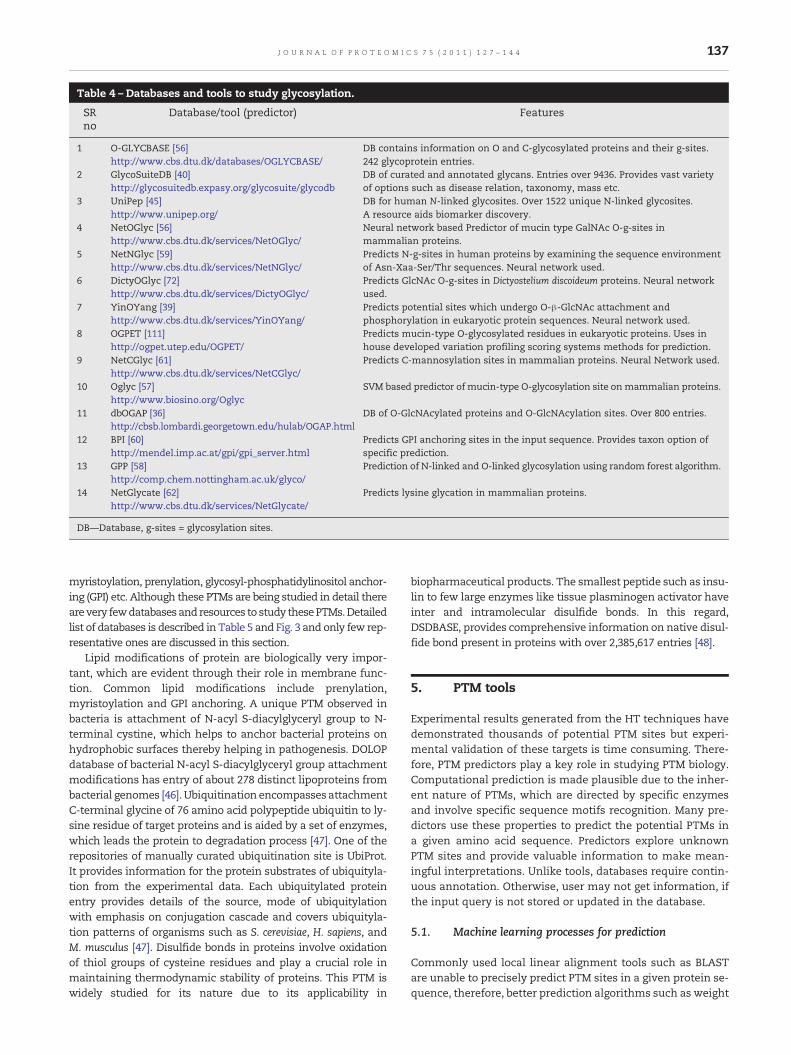
Table 4 – Databases and tools to study glycosylation.
SRno
Database/tool (predictor) Features
1 O-GLYCBASE [56]http://www.cbs.dtu.dk/databases/OGLYCBASE/
DB contains information on O and C-glycosylated proteins and their g-sites.242 glycoprotein entries.
2 GlycoSuiteDB [40]http://glycosuitedb.expasy.org/glycosuite/glycodb
DB of curated and annotated glycans. Entries over 9436. Provides vast varietyof options such as disease relation, taxonomy, mass etc.
3 UniPep [45]http://www.unipep.org/
DB for human N-linked glycosites. Over 1522 unique N-linked glycosites.A resource aids biomarker discovery.
4 NetOGlyc [56]http://www.cbs.dtu.dk/services/NetOGlyc/
Neural network based Predictor of mucin type GalNAc O-g-sites inmammalian proteins.
5 NetNGlyc [59]http://www.cbs.dtu.dk/services/NetNGlyc/
Predicts N-g-sites in human proteins by examining the sequence environmentof Asn-Xaa-Ser/Thr sequences. Neural network used.
6 DictyOGlyc [72]http://www.cbs.dtu.dk/services/DictyOGlyc/
Predicts GlcNAc O-g-sites in Dictyostelium discoideum proteins. Neural networkused.
7 YinOYang [39]http://www.cbs.dtu.dk/services/YinOYang/
Predicts potential sites which undergo O-β-GlcNAc attachment andphosphorylation in eukaryotic protein sequences. Neural network used.
8 OGPET [111]http://ogpet.utep.edu/OGPET/
Predicts mucin-type O-glycosylated residues in eukaryotic proteins. Uses inhouse developed variation profiling scoring systems methods for prediction.
9 NetCGlyc [61]http://www.cbs.dtu.dk/services/NetCGlyc/
Predicts C-mannosylation sites in mammalian proteins. Neural Network used.
10 Oglyc [57]http://www.biosino.org/Oglyc
SVM based predictor of mucin-type O-glycosylation site onmammalian proteins.
11 dbOGAP [36]http://cbsb.lombardi.georgetown.edu/hulab/OGAP.html
DB of O-GlcNAcylated proteins and O-GlcNAcylation sites. Over 800 entries.
12 BPI [60]http://mendel.imp.ac.at/gpi/gpi_server.html
Predicts GPI anchoring sites in the input sequence. Provides taxon option ofspecific prediction.
13 GPP [58]http://comp.chem.nottingham.ac.uk/glyco/
Prediction of N-linked and O-linked glycosylation using random forest algorithm.
14 NetGlycate [62]http://www.cbs.dtu.dk/services/NetGlycate/
Predicts lysine glycation in mammalian proteins.
DB—Database, g-sites = glycosylation sites.
137J O U R N A L O F P R O T E O M I C S 7 5 ( 2 0 1 1 ) 1 2 7 – 1 4 4
myristoylation, prenylation, glycosyl-phosphatidylinositol anchor-ing (GPI) etc. Although these PTMs are being studied in detail therearevery fewdatabasesand resources to study thesePTMs.Detailedlist of databases is described in Table 5 and Fig. 3 and only few rep-resentative ones are discussed in this section.
Lipid modifications of protein are biologically very impor-tant, which are evident through their role in membrane func-tion. Common lipid modifications include prenylation,myristoylation and GPI anchoring. A unique PTM observed inbacteria is attachment of N-acyl S-diacylglyceryl group to N-terminal cystine, which helps to anchor bacterial proteins onhydrophobic surfaces thereby helping in pathogenesis. DOLOPdatabase of bacterial N-acyl S-diacylglyceryl group attachmentmodifications has entry of about 278 distinct lipoproteins frombacterial genomes [46]. Ubiquitination encompasses attachmentC-terminal glycine of 76 amino acid polypeptide ubiquitin to ly-sine residue of target proteins and is aided by a set of enzymes,which leads the protein to degradation process [47]. One of therepositories of manually curated ubiquitination site is UbiProt.It provides information for the protein substrates of ubiquityla-tion from the experimental data. Each ubiquitylated proteinentry provides details of the source, mode of ubiquitylationwith emphasis on conjugation cascade and covers ubiquityla-tion patterns of organisms such as S. cerevisiae, H. sapiens, andM. musculus [47]. Disulfide bonds in proteins involve oxidationof thiol groups of cysteine residues and play a crucial role inmaintaining thermodynamic stability of proteins. This PTM iswidely studied for its nature due to its applicability in
biopharmaceutical products. The smallest peptide such as insu-lin to few large enzymes like tissue plasminogen activator haveinter and intramolecular disulfide bonds. In this regard,DSDBASE, provides comprehensive information on native disul-fide bond present in proteins with over 2,385,617 entries [48].
5. PTM tools
Experimental results generated from the HT techniques havedemonstrated thousands of potential PTM sites but experi-mental validation of these targets is time consuming. There-fore, PTM predictors play a key role in studying PTM biology.Computational prediction is made plausible due to the inher-ent nature of PTMs, which are directed by specific enzymesand involve specific sequence motifs recognition. Many pre-dictors use these properties to predict the potential PTMs ina given amino acid sequence. Predictors explore unknownPTM sites and provide valuable information to make mean-ingful interpretations. Unlike tools, databases require contin-uous annotation. Otherwise, user may not get information, ifthe input query is not stored or updated in the database.
5.1. Machine learning processes for prediction
Commonly used local linear alignment tools such as BLASTare unable to precisely predict PTM sites in a given protein se-quence, therefore, better prediction algorithms such as weight

Table 5 – Resources to study other types of PTMs.
SRno
Database/tool (predictor) Features
1 Ubiprot [47]http://ubiprot.org.ru
DB of protein substrates of ubiquitylation. Manually curated database onverified literature.
2 Saccharomyces Cerevisiae Ubiquitination Database(SCUD) [112]http://scud.kaist.ac.kr
DB with information about ubiquitinated proteins and related enzymes inS. cerevisiae. Has entry of over 940 substrates.
3 PlantsUPS [113]http://bioinformatics.cau.edu.cn/plantsUPS
DB of ubiquitin/26S proteasome system of 7 species of higher plants. Vastinformation on basic gene characterization, protein features andmicroarray information as well as BLAST hits against various DB data.
4 Database Of Bacterial Lipoproteins (DOLOP) [46]http://www.mrc-lmb.cam.ac.uk/genomes/dolop/
DB of Bacterial Lipoproteins. Probable lipoproteins from 234 bacterialgenomes.
5 Disulphide Database DSDBASE [48]http://caps.ncbs.res.in/dsdbase/dsdbase.html
DB of disulphide bonds in proteins, which provides information on nativedisulfides. Records 2385617 protein substructures that have stereochemicalcompatibility.
6 E3Miner [114]http://e3miner.biopathway.org
Text mining tool for ubiquitin-protein ligases. Extracting and managingdata from MEDLINE abstracts and relevant protein databases.
7 UbiPred [68]http://flipper.diff.org/app/tools/info/2503
Predict ubiquitylation sites in query sequence. SVM built on informativephysicochemical property mining algorithm (IPMA).
8 SulfoSite [115]http://sulfosite.mbc.nctu.edu.tw/
Tool to predict protein sulfotyrosine sites. Uses SVM.
9 Sulfinator [71]http://au.expasy.org/tools/sulfinator/
A tool predicts tyrosine sulfation sites in protein sequences. HHM used.
10 NetAcet [116]http://www.cbs.dtu.dk/services/NetAcet/
Predicts N-acetyltransferase A (NatA) substrates (in yeast andmammalian proteins). ANN used.
11 Myristoylator [64]http://web.expasy.org/myristoylator/
Predicts N-terminal myristoylation by neural networks.
12 NMT-The MYR Predictor [117]http://mendel.imp.ac.at/myristate/SUPLpredictor.htm
Predicts N-Myristoylation for higher Eukaryote, viral and fungal query se-quences. Self-consistency and Jack-knife test used.
13 LipoP [118]http://www.cbs.dtu.dk/services/LipoP/
Predict lipoprotein signal peptides in Gram-negative Eubacteria. HMMused.
14 NBA-Palm [65]http://www.bioinfo.tsinghua.edu.cn/NBA-Palm
Predicts palmitoylation. SVMs, Naïve Bayes algorithm used.
15 Clustering and scoring strategy CSS-Palm [119]http://csspalm.biocuckoo.org/online.php
Software for predicting Palmitoylation site. Clustering and scoring strategy(CSS) algorithm used.
16 Prenylation Prediction Suite (PrePS) [67]http://mendel.imp.ac.at/sat/PrePS/index.html
Predicts prenylation motifs.
17 SUMOplot [69]http://www.abgent.com/tools/sumoplot_login
Predicts SUMO protein attachment sites and scores sumoylation sites inproteins. BLOSUM62 for matrix, Matthews' correlated coefficient used.
18 SUMOsp [120]http://sumosp.biocuckoo.org/
Predicts sumoylation sites. BLOSUM62 for matrix, Matthews' correlatedcoefficient used.
19 LysAcet [121]http://www.biosino.org/LysAcet/
Performs lysine acetylation prediction. SVM used.
20 Methylation Modification Prediction Server MeMo [122]http://www.bioinfo.tsinghua.edu.cn/~tigerchen/memo.html
Tool to predict protein methylation modifications mostly Lysine andArginine. Uses SVMs
21 BPB-PPMS [123]http://www.bioinfo.bio.cuhk.edu.hk/bpbppms/intro.jsp
Predicts the methylation for lysine and arginine residues by usingBi-profile Bayes approach and SVM.
DB—Database, SVM—Support Vector Machines, NMT—N-terminal N-Myristoylation, HHM—Hidden Markov Models.
138 J O U R N A L O F P R O T E O M I C S 7 5 ( 2 0 1 1 ) 1 2 7 – 1 4 4
matrices and machine learning techniques are evolved. Ma-chine learning process involves teaching the system andbuilding the algorithm through which it is made to learn andmimic the biological phenomenon of PTMs with experimen-tally proved training dataset. Then the same algorithm istrained to predict PTMs for a test set while it is checked fortrue and false prediction. Hence, unlike simple local align-ment tool, which just aligns the input sequence with that ofthe stored sequence, the machine learning enables input ofspecific properties andmakes prediction similar to an enzymerecognizing its motif to bring in PTM.
Supervised machine learning techniques majorly involvestwo aspects; training and testing. Training the system is
done through the potential PTM datasets, which are experi-mentally derived or mined from known repositories contain-ing curated data from literature. The training dataset shouldhave optimized level of positive and negative PTM sites in it.Once a high quality dataset is ready, the input feature for pre-diction is provided and learning functions are selected. Thisprediction process is performed on training dataset to gener-ate a classifier, which can be further used for prediction oftest dataset. The established classifier is then tested for itsperformance on an independent test dataset. This is check-point for accurate prediction of PTM sites. Inaccurate predic-tion due to less sensitivity and specificity during thevalidation brings a need to check the built algorithm. It is
139J O U R N A L O F P R O T E O M I C S 7 5 ( 2 0 1 1 ) 1 2 7 – 1 4 4
very essential that the data used for training the predictor al-gorithmmust be error free, because an error will subsequentlyproduce erroneous PTM prediction. Hence, trained algorithmsprovide positive and negative prediction outputs for PTM andnon-PTM sites, respectively. Presently predictors tend to pos-sess more sensitivity than specificity but one need to balancebetween specificity and sensitivity for comprehensive predic-tion [49,50].
Supervised machine learning approaches typically usesome of the learning functions such as Artificial Neural Net-work (ANN), Support VectorMachine (SVM) and HiddenMarkovModels (HMMs) etc., which are unique to each predictor. For in-stance, the SVMmethod is designed tomaximize themargin toseparate two classes so that the trained model can be general-ized to predict the data [51]. PHOSIDA enables Support VectorMachine (SVM) based prediction of phosphorylation and acety-lation with 78% precision [33]. ANN is inspired by the biologicalneural networks, in which each data point is represented asneuron and interconnected as biological neuron. Multiple in-puts are provided to generate single output from each nodeand each point is trained to predict PTM site accurately. Net-PhosK is one such ANN based kinase specific phosphorylationsite predictor, which is based on data specific to six kinases[39].
Several prediction tools for various types of PTMs are avail-able for public use in WWW (Fig. 4). Single prediction servermay predict one or many PTMs. FindMod present in ExPASyserver predicts about 22 different types of PTMs from inputPMF data, Swiss-Prot ID and single-letter amino acid code.Neural network based tool such as NetNGlyc predicts specifi-cally N-glycosylation sites in human proteins. FengfengZhou and colleagues created a user interface, which collects32 different types of PTMs and reduces effort of searching dif-ferent resources for multiple PTM prediction [52].
5.2. Strong prediction tools by evolving models
Similar to the advancement in mass spectrometry and othertechnologies for PTM identification, the PTM prediction tools arealso evolving. The present day tools are not restricted to specificsequence motifs but are much more advanced to provide evolu-tionary aspects, kinase specific information, consideration offlanking amino acids etc. For example, NetPhosKwas introducedtopredict phosphorylation sites basedonsimple sequencemotifsbut later on kinase specific information and a concept of ‘evolu-tionary stable sites’ were introduced to make the predictionsmore precise [39]. The dbPTM considers secondary and tertiarystructures, solvent accessibility of the substrate and protein do-mains for prediction [53], whereas PHOSIDA considers structureof the motif and evolutionary conservation of phosphosites. TheBlastP was used to perform homology search of all phosphopro-teins over seventy species ranging from bacteria to mouse [33].
Organism specific predictors of PTMs can provide strongprediction model by involving and teaching the system to pre-dict the PTMs with an orientation of biological phenomenonpertaining to a particular organism. More organism specificdatabases and predictors are described in section 6. Withmore andmore global proteomes being analyzed, larger train-ing datasets are generated and it is anticipated that better pre-dictions would evolve in days to come. In the following
section, a few common PTMs and their corresponding toolsfor prediction are discussed.
5.3. Phosphorylation tools
Phosphorylation prediction tools outnumber predictors of anyother PTMs. Although there are over 500 kinases in human ge-nome, referring to several phosphorylation sites, only a fewkinases are extensively studied. Therefore, predictors of phos-phorylation sites are very informative. Initially, phosphoryla-tion site prediction was only kinase driven approach, whichfollowed screening by incubation of a specific kinase withlarge set of peptides and ATP, and resulting phosphorylationpatterns were studied to understand kinase substrate interac-tion [30,54]. Current MS based screening and in silico predic-tions are very efficient and far more rapid. The patternrecognition algorithms such as ANN, SVM etc are trained forthis purpose to recognize phosphorylation sites with highsensitivity and specificity.
A typical kinase specific phosphorylation site predictor, suchas NetPhosK, predicts the phosphorylation site using artificialnetwork and gives an option to choose from 18 different ki-nases. Another predictor YinOYang, in conjunction with Net-Phos predicts the potential sites on protein that may undergoboth glycosylation and phosphorylation [39]. A non-kinase driv-en motif prediction approach was taken in case of Motif-X,which is a substrate driven predictor for phosphorylation mo-tifs. In this regard, authors developed a statistical algorithmusing two database contemporary datasets [55].
5.4. Glycosylation tools
The glycosylation process with respect to enzymes involvedand their specificity is less understood. This is because attach-ment of saccharide chain on protein is complex as comparedto other PTMs such as methylation, acetylation or phosphory-lation, where transfer of chemical and covalent attachment ismore or less a single step process. N and O-linked glycosyla-tions are two mostly studied glycosylation types and hencemuch of predictors are centered to these types.
NetOglyc is a predictor formammalianmucin type GalNAcO-glycosylation sites. The authors showed high confidence predic-tion of glycosylation sites by training ANN with data extractedfrom O-GLYCBASE. This prediction was based on action of UDP-GalNAc:polypeptide N-acetylgalactosaminyltransferases com-plex, its specificity for Serine or Threonine and charged residuesin −1 and +3 position. ANN trained in all these features resultedin good prediction (76% prediction of glycosylated and 93% ofnon-glycosylated sites) [56]. One of the SVM based mucin-typeO-glycosylation site predictors of mammalian protein is Oglyc,which considers the sequence of O-glycosylation, physical prop-erties of amino acids and binary way of representing the se-quence. Positive and negative datasets for training SVMwere obtained from Swiss-Prot/UniProt [57]. Glycosylationprediction program (GPP) uses random forest along with in-formation about pair wise pattern to predict glycosylationsites. Training dataset was extracted from OGLYCBASE. Au-thors have made an effort to make the prediction better byintroducing further information such as hydrophobicity, pre-dicted secondary structure and predicted surface accessibility
140 J O U R N A L O F P R O T E O M I C S 7 5 ( 2 0 1 1 ) 1 2 7 – 1 4 4
[58]. NetNglyc predicts N-Glycosylation sites in human proteinsusing ANN and it was trained with 469 positive and 309 negativeglycosylation sites. During cross validation, the method yieldedidentification of 86 and 61% of glycosylated and non-glycosylated sites, respectively [59]. Other types of glycosylationsuch as GPI anchoring, C-mannsylation, and glycations havealso attracted researchers attention to develop supervised ma-chine learning based predictors. To name a few, BigPI predictsGlycosylphosphatidylinositol (GPI) attachment sites [60], NetC-Glyc predicts mammalian C-mannosylation sites [61] and Net-Glycate predicts glycation sites for mammalian proteins [62].Since studying glycosylation sites is complex, availability of gly-cosylation tools would definitely help researchers to provide pre-dictions of this crucial PTM.
5.5. Other PTM related tools
Majority of PTM tools are centered on two most commonPTMs, phosphorylation and glycosylation; however, there arearound 300 other types of PTMs, which are also biologicallyimportant. Few prominent tools are discussed in this sectionand detailed predictors for various types of PTMs are de-scribed in Table 5. Myristoylation is addition of myristoylgroup to the N-terminal of glycine residue of a protein withan enzyme N-myristoyltransferase. Apart from eukaryotes,myristoylation is also found to be associated with viral pro-teins [63]. Myristoylator, a predictor for myristoylation sitesin a given amino acid sequence, is one of the best myristoyla-tion predictors available with false discovery positive errorrate of 2.1% [64]. This predictor uses neural network, trainedwith 390 positive and 327 negative sequences. NBA-palm, apredictor of palmitoylated sites, is designed on basis ofNaïve Bayes algorithm. The algorithm was trained with
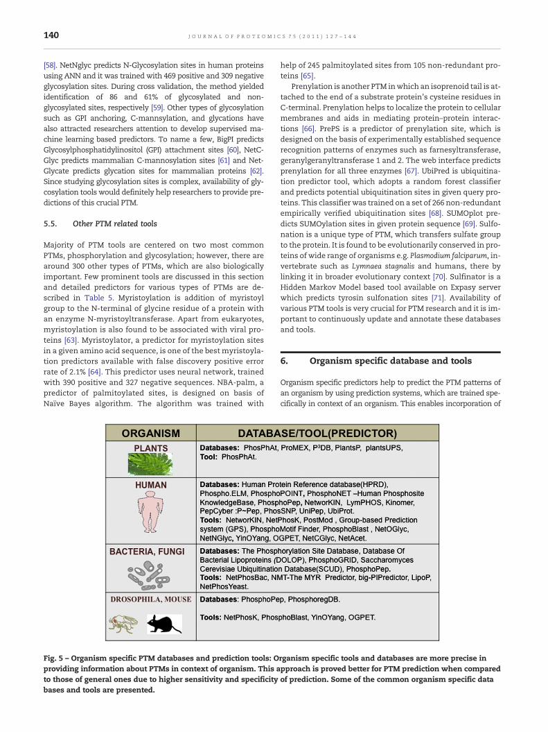
Fig. 5 – Organism specific PTM databases and prediction tools: Oproviding information about PTMs in context of organism. This ato those of general ones due to higher sensitivity and specificitybases and tools are presented.
help of 245 palmitoylated sites from 105 non-redundant pro-teins [65].
Prenylation is another PTM inwhich an isoprenoid tail is at-tached to the end of a substrate protein's cysteine residues inC-terminal. Prenylation helps to localize the protein to cellularmembranes and aids in mediating protein–protein interac-tions [66]. PrePS is a predictor of prenylation site, which isdesigned on the basis of experimentally established sequencerecognition patterns of enzymes such as farnesyltransferase,geranylgeranyltransferase 1 and 2. The web interface predictsprenylation for all three enzymes [67]. UbiPred is ubiquitina-tion predictor tool, which adopts a random forest classifierand predicts potential ubiquitination sites in given query pro-teins. This classifier was trained on a set of 266 non-redundantempirically verified ubiquitination sites [68]. SUMOplot pre-dicts SUMOylation sites in given protein sequence [69]. Sulfo-nation is a unique type of PTM, which transfers sulfate groupto the protein. It is found to be evolutionarily conserved in pro-teins of wide range of organisms e.g. Plasmodium falciparum, in-vertebrate such as Lymnaea stagnalis and humans, there bylinking it in broader evolutionary context [70]. Sulfinator is aHidden Markov Model based tool available on Expasy serverwhich predicts tyrosin sulfonation sites [71]. Availability ofvarious PTM tools is very crucial for PTM research and it is im-portant to continuously update and annotate these databasesand tools.
6. Organism specific database and tools
Organism specific predictors help to predict the PTM patterns ofan organism by using prediction systems, which are trained spe-cifically in context of an organism. This enables incorporation of
rganism specific tools and databases are more precise inpproach is proved better for PTM prediction when comparedof prediction. Some of the common organism specific data
141J O U R N A L O F P R O T E O M I C S 7 5 ( 2 0 1 1 ) 1 2 7 – 1 4 4
unique features into training dataset pertaining to a specific or-ganismandmakes the predictionmore targeted (Fig. 5). RamneekGupta and colleagues developed, DictyOGlyc,which is ANNbasedO-linked GlcNAc glycosylation predictor for secreted and mem-brane proteins of Dictyostelium discoideum. This predictor is usefulsinceDictyostelium serves as amodel organism to study glycosyla-tion of eukaryotes [72]. NetPhosBac and NetPhosYeast are twoANN-based kinase predictors for bacterial and yeast proteins, re-spectively [73,74]. Thesepredictors canbeused to screenpotentialsubstrates of protein kinases or to distinguish phosphorylatedand non-phosphorylated protein forms. PhosPhAT, a databaseof phosphorylation sites in Arabidopsis, contains data producedby MS experiments and annotations in database are linked withmass spectrum to provide information in context of their biologi-cal significance. This database also contains a predictor, whichpredicts the potential phosphorylation sites [75]. virPTM is firstvirus PTM database which contains manually curated informa-tion for 329 phosphorylation sites of 53 different human viruses[76]. A detailed list of organism-specific database and tools arelisted in Figure 5.
7. Conclusions
Proteomics has enhanced our understanding of diverse andcomplex PTMs by applying various advanced techniquessuch as mass spectrometry. Enormous data generated by var-ious HT methods are curated and shared worldwide with helpof dedicated databases. The growing number of databasesprovides researchers various resources; however, due to thecomplexity of PTMs, no database can provide a holistic solu-tion. To provide accurate information, the databases requirecontinuous update of new dataset. In this regard, Swiss Prot,HPRD, Phospho.ELM etc. provide comprehensive and latestcollection of data. Apart from the database, the PTM predic-tion tools also play an important role due to their rapidnessand accuracy of prediction. These databases and tools arepowerful resources to reduce the time and effort of re-searchers for PTM prediction since identifying PTMs in vitrois always challenging. However, by using newer and advancedmachine learning methods these prediction tools can bemade more effective. The comprehensive tables for varietyof PTMs described in this article will provide researchers a re-source for selecting PTM databases and tools best suited fortheir PTM research; however, based on the type of application,one must be very careful in choosing the right database orpredictor. The user may look into details such as the algo-rithm used for prediction, the accuracy of prediction etc. Sim-ilarly, the databases can be looked into details such as howoften and effectively they are updated. Undoubtedly, in yearsto come, the computational resources will provide greatvalue, and extend its analysis to decipher the role of PTMs insignaling pathways and related ailments.
Acknowledgments
The financial support from Ministry of Human Resource andDevelopment India (MHRD-10MHRD005), Board of Research inNuclear Sciences India (2009/20/37/4/BRNS) and Department of
Biotechnology India (BT/PR13562/MED/12/451/2010) for the pro-teomics work performed in SS laboratory is gratefully acknowl-edged. The help rendered by Renisa D'souza in drawing figuresis gratefully acknowledged.
R E F E R E N C E S
[1] Cox J, Mann M. Is proteomics the new genomics? Cell2007;130:395–8.
[2] Witze ES, Old WM, Resing KA, Ahn NG. Mapping proteinpost-translational modifications with mass spectrometry.Nat Methods 2007;4:798–806.
[3] Wojcik J, Schachter V. Proteomic databases and software onthe web. Brief Bioinform 2000;1:250–9.
[4] Walsh CT, Garneau-Tsodikova S, Gatto Jr GJ. Proteinposttranslational modifications: the chemistry of proteomediversifications. Angew Chem Int Ed Engl 2005;44(45):7342–72.
[5] Zhang J, Zhang H, Ayaz-Guner S, Chen YC, Dong X, Xu Q,et al. Phosphorylation, but not alternative splicing orproteolytic degradation, is conserved in human and mousecardiac troponin T. Biochemistry 2011;50(27):6081–92.
[6] Macek B, Gnad F, Soufi B, Kumar C, Olsen JV, Mijakovic I,et al. Mol Cell Proteomics. Phosphoproteome analysis of E.coli reveals evolutionary conservation of bacterial Ser/Thr/Tyr phosphorylation. Mol Cell Proteomics 2008;7(2):299–307.
[7] Martin L, Latypova X, Terro F. Post-translationalmodifications of tau protein: implications for Alzheimer'sdisease. Neurochem Int 2011;58(4):458–71.
[8] Gioeli D, Paschal BM. Post-translational modification of theandrogen receptor. Mol Cell Endocrinol in press. Jul 24.[PMID:21820033].
[9] Macek B, Mann M, Olsen JV. Global and site-specificquantitative phosphoproteomics: principles andapplications. Annu Rev Pharmacol Toxicol 2009;49:199–221.
[10] Zong C, Young GW, Wang Y, Lu H, Deng N, Drews O, et al.Two-dimensional electrophoresis based characterization ofpost-translational modifications of mammalian 20Sproteasome complexes. Proteomics 2008;8:5025–37.
[11] Wu CC, MacCoss MJ. Shotgun proteomics: tools for theanalysis of complex biological systems. Curr Opin Mol Ther2002;4(3):242–50.
[12] Jensen ON. Interpreting the protein language usingproteomics. Nat Rev Mol Cell Biol 2006;7:391–403.
[13] Siuti N, Kelleher NL. Decoding protein modifications usingtop-downmass spectrometry. Nat Methods 2007;4(10):817–21.
[14] Zamdborg L, LeDuc RD, Glowacz KJ, Kim YB, Viswanathan V,Spaulding IT, et al. ProSight PTM 2.0: improved proteinidentification and characterization for top down massspectrometry. Nucleic Acids Res 2007;35:W701–6.
[15] Mann M. Functional and quantitative proteomics usingSILAC. Nat Rev Mol Cell Biol 2006;7(12):952–8.
[16] Cuomo A, Moretti S, Minucci S, Bonaldi T. SILAC-basedproteomic analysis to dissect the “histone modificationsignature” of human breast cancer cells. Amino Acids2011;41(2):387–99.
[17] Olsen JV, Vermeulen M, Santamaria A, Kumar C, Miller ML,Jensen LJ, et al. Quantitative phosphoproteomics revealswidespread full phosphorylation site occupancy duringmitosis. Sci Signal 2010;3(104):ra3.
[18] Skorobogatko YV, Deuso J, Adolf-Bryfogle J, Nowak MG, GongY, Lippa CF, et al. Human Alzheimer's diseasesynaptic O-GlcNAc site mapping and iTRAQ expressionproteomics with ion trap mass spectrometry. Amino Acids2011;40(3):765–79.
[19] Iwai LK, Benoist C, Mathis D, White FM. Quantitativephosphoproteomic analysis of T cell receptor signaling in

142 J O U R N A L O F P R O T E O M I C S 7 5 ( 2 0 1 1 ) 1 2 7 – 1 4 4
diabetes prone and resistant mice. J Proteome Res 2010;9(6):3135–45.
[20] Hoffert JD, Pisitkun T, Wang G, Shen RF, Knepper MA.Quantitative phosphoproteomics of vasopressin-sensitiverenal cells: regulation of aquaporin-2 phosphorylation attwo sites. Proc Natl Acad Sci U S A 2006;103(18):7159–64.
[21] Zhai B, Villen J, Beausoleil SA, Mintseris J, Gygi SP.Phosphoproteome analysis of Drosophila melanogasterembryos. J Proteome Res 2008;7:1675–82.
[22] Choudhary C, Kumar C, Gnad F, Nielsen ML, Rehman M,Walther TC, et al. Lysine acetylation targets proteincomplexes and co-regulates major cellular functions.Science 2009;325:834–40.
[23] Zielinska DF, Gnad F, Wiśniewski JR, Mann M. Precisionmapping of an in vivo N-glycoproteome reveals rigidtopological and sequence constraints. Cell 2010;141(5):897–907.
[24] Wagner SA, Beli P, Weinert BT, Nielsen ML, Cox J, Mann M,et al. A proteome-wide, quantitative survey of in vivoubiquitylation sites reveals widespread regulatory roles.Mol Cell Proteomics in press. Sep 1. PMID: 21890473.
[25] Kung LA, Tao SC, Qian J, Smith MG, Snyder M, Zhu H. Globalanalysis of the glycoproteome in Saccharomyces cerevisiaereveals new roles for protein glycosylation in eukaryotes.Mol Syst Biol 2009;5:308.
[26] Ptacek J, Devgan G, Michaud G, Zhu H, Zhu X, Fasolo J, et al.Global analysis of protein phosphorylation in yeast. Nature2005;438:679–84.
[27] Popescu SC, Popescu GV, Bachan S, Zhang Z, Gerstein M,Snyder M, et al. MAPK target networks in Arabidopsis thalianarevealed using functional protein microarrays. Genes Dev2009;23(1):80–92.
[28] Keshava Prasad TS, Goel R, Kandasamy K, Keerthikumar S,Kumar S, Mathivanan S, et al. Human Protein ReferenceDatabase—2009 update. Nucleic Acids Res 2009;37:D767–72.
[29] Farriol MN, Garavelli JS, Boeckmann B, Duvaud S, GasteigerE, Gateau A, et al. Annotation of post-translationalmodifications in the Swiss-Prot knowledge base. Proteomics2004;4:1537–50.
[30] Lemeer S, Heck AJ. The phosphoproteomics data explosion.Curr Opin Chem Biol 2009;13:414–20.
[31] Kreegipuu A, Blom N, Brunak S. PhosphoBase, a database ofphosphorylation sites: release 2.0. Nucleic Acids Res 1999;27:237–9.
[32] Diella F, Cameron S, Gemund C, Linding R, Via A, Kuster B,et al. Phospho.ELM: a database of experimentally verifiedphosphorylation sites in eukaryotic proteins. BMCBioinformatics 2004;5:79.
[33] Gnad F, Gunawardena J, Mann M. PHOSIDA 2011: theposttranslational modification database. Nucleic Acids Res2011;39:D253–60.
[34] http://www.phosphosite.org/homeAction.do.[35] Zanzoni A, Ausiello G, Via A, Gherardini PF, Helmer-Citterich
M. Phospho3D: a database of three-dimensional structuresof protein phosphorylation sites. Nucleic Acids Res 2007;35:D229.c–31.c.
[36] Gribskov M, Fana F, Harper J, Hope DA, Harmon AC,Smith DW, et al. PlantsP: a functional genomics databasefor plant phosphorylation. Nucleic Acids Res 2001;29:111–3.
[37] Bodenmiller B, Campbell D, Gerrits B, Lam H, Jovanovic M,Picotti P, et al. PhosphoPep — a database of proteinphosphorylation sites in model organisms. Nat Biotechnol2008;26:1339–40.
[38] Ovelleiro D, Carrascal M, Casas V, Abian J. LymPHOS. Designof a phosphosite database of primary human T cells.Proteomics 2009;9:3741–51.
[39] Blom N, Sicheritz-Pontén T, Gupta R, Gammeltoft S, BrunakS. Prediction of post-translational glycosylation and
phosphorylation of proteins from the amino acid sequence.Proteomics 2004;4:1633–49.
[40] Cooper CA, Joshi HJ, Harrison MJ, Wilkins MR, Packer NH.GlycoSuiteDB: a curated relational database of glycoproteinglycan structures and their biological sources. 2003 update.Nucleic Acids Res 2003;31:511–3.
[41] Gupta R, Birch H, Rapacki K, Brunak S, Hansen JE.O-GLYCBASE Version 4.0: a revised database ofO-glycosylated proteins. Nucleic Acids Res 1999;27:370–2.
[42] Cooper CA, Wilkins MR, Williams KL, Packer NH. BOLD—abiological O-linked glycan database. Electrophoresis 1999;20:3589–98.
[43] Wang J, Torii M, Liu H, Hart GW, Hu ZZ. dbOGAP — anintegrated bioinformatics resource for proteinO-GlcNAcylation. BMC Bioinformatics 2011;12:91.
[44] Weerapana E, Imperiali B. Asparagine-linked proteinglycosylation: from eukaryotic to prokaryotic systems.Glycobiology 2006;16(6):91R–101R.
[45] Zhang H, Loriaux P, Eng J, Campbell D, Keller A, Moss P,et al. UniPep — a database for human N-linked glycosites:a resource for biomarker discovery. Genome Biol 2006;7:R73.
[46] Babu MM, Priya ML, Selvan AT, Madera M, Gough J, AravindL, et al. A database of bacterial lipoproteins (DOLOP) withfunctional assignments to predicted lipoproteins. J Bacteriol2006;188(8):2761–73.
[47] Chernorudskiy AL, Garcia A, Eremin EV, Shorina AS,Kondratieva EV, Gainullin MR. UbiProt: a database ofubiquitylated proteins. BMC Bioinformatics 2007;8:126.
[48] Vinayagam A, Pugalenthi G, Rajesh R, Sowdhamini R.DSDBASE: a consortium of native and modeled disulphidebonds in proteins. Nucleic Acids Res 2004;32:D200–2.
[49] Eisenhaber B, Eisenhaber F. Prediction of posttranslationalmodification of proteins from their amino acid sequence. In:Oliviero C, Eisenhaber F, editors. Methods in molecularbiology. Data mining techniques for the life sciences.Humana Press; 2010. p. 365–84.
[50] Liu C, Li H. In silico prediction of post-translationalmodifications. In: Bing Y, Marcus H, editors. Methods inmolecular biology. Silico tools for gene discovery. HumanaPress; 2011. p. 325–40.
[51] Yang ZR. Biological applications of support vector machines.Brief Bioinform 2004;5:328–38.
[52] Zhou F, Xue Y, Yao X, Xu Y. A general user interface forprediction servers of proteins' post-translationalmodification sites. Nat Protoc 2006;1(3):1318–21.
[53] Lee TY, Huang HD, Hung JH, Huang HY, Yang YS, Wang TH.dbPTM: an information repository of proteinpost-translational modification. Nucleic Acids Res 2006;34:D622–7.
[54] Mann M, Ong SE, Grønborg M, Steen H, Jensen ON, Pandey A.Analysis of protein phosphorylation using massspectrometry: deciphering the phosphoproteome. TrendsBiotechnol 2002;20(6):261–8.
[55] Schwartz D, Gygi SP. An iterative statistical approach to theidentification of protein phosphorylation motifs fromlarge-scale data sets. Nat Biotechnol 2005;23(11):1391–8.
[56] Julenius K, Mølgaard A, Gupta R, Brunak S. Prediction,conservation analysis, and structural characterization ofmammalian mucin-type O-glycosylation sites. Glycobiology2005;15(2):153–64.
[57] Li S, Liu B, Zeng R, Cai Y, Li Y. Predicting O-glycosylationsites in mammalian proteins by using SVMs. Comput BiolChem 2006;30(3):203–8.
[58] Hamby SE, Hirst JD. Prediction of glycosylation sites usingrandom forests. BMC Bioinformatics 2008;9:500.
[59] Julenius K, Johansen MB, Zhang Y, Brunak S, Gupta R.Prediction of glycosylation sites in proteins. In: Lieth CW,Luetteke T, Frank M, editors. Bioinformatics for glycobiology
143J O U R N A L O F P R O T E O M I C S 7 5 ( 2 0 1 1 ) 1 2 7 – 1 4 4
and glycomics: an introduction. Wiley-Blackwell; 2009.p. 163–92.
[60] Eisenhaber B, Bork P, Eisenhaber F. Prediction of potentialGPI-modification sites in proprotein sequences. J Mol Biol1999;292(3):741–58.
[61] Julenius K. NetCGlyc 1.0: prediction of mammalianC-mannosylation sites. Glycobiology 2007;17:868–76.
[62] Johansen MB, Kiemer L, Brunak S. Analysis and prediction ofmammalian protein glycation. Glycobiology 2006;16(9):844–53.
[63] Boutin JA. Myristoylation. Cell Signal 1997;9:15–35.[64] Bologna G, Yvon C, Duvaud S, Veuthey AL. N-Terminal
myristoylation predictions by ensembles of neuralnetworks. Proteomics 2004;4:1626–32.
[65] Xue Y, Chen H, Jin C, Sun Z, Yao X. NBA-Palm: prediction ofpalmitoylation site implemented in Naive Bayes algorithm.BMC Bioinformatics 2006;7:458.
[66] Hougland JL, Fierke CA. Getting a handle on proteinprenylation. Nat Chem Biol 2009;5:197–8.
[67] Maurer SS, Eisenhaber F. Refinement and prediction ofprotein prenylation motifs. Genome Biol 2005;6:R55.
[68] Radivojac P, Vacic V, Haynes C, Cocklin RR, Mohan A, HeyenJW, et al. Identification, analysis, and prediction of proteinubiquitination sites. Proteins 2010;78:365–80.
[69] Zhou F, Xue Y, Lu H, Chen G, Yao X. A genome-wide analysisof sumoylation-related biological processes and functions inhuman nucleus. FEBS Lett 2005;579:3369–75.
[70] Medzihradszky KF, Darula Z, Perlson E, Fainzilber M, ChalkleyRJ, Ball H, et al. O-sulfonation of serine and threonine Massspectrometric detection and characterization of a newposttranslationalmodification in diverse proteins throughoutthe eukaryotes. Mol Cell Proteomics 2004;3:429–43.
[71] Monigatti F, Gasteiger E, Bairoch A, Jung E. The Sulfinator:predicting tyrosine sulfation sites in protein sequences.Bioinformatics 2002;18:769–70.
[72] Gupta R, Jung E, Gooley AA, Williams KL, Brunak S, Hansen J.Scanning the available Dictyostelium discoideum proteome forO-linked GlcNAc glycosylation sites using neural networks.Glycobiology 1999;9:1009–22.
[73] Miller ML, Soufi B, Jers C, Blom N, Macek B, Mijakovic I.NetPhosBac — a predictor for Ser/Thr phosphorylation sitesin bacterial proteins. Proteomics 2009;9:116–25.
[74] Ingrell CR, Miller ML, Jensen ON, Blom N. NetPhosYeast.Prediction of protein phosphorylation sites in yeast.Bioinformatics 2007;23:895–7.
[75] Joshua LH, Pawel D, Jan H, Joachim S, Wolfram W, Dirk W,et al. PhosPhAt: a database of phosphorylation sites inArabidopsis thaliana and a plant-specific phosphorylationsite predictor. Nucleic Acids Res 2008;36:D1015–21.
[76] Schwartz D, Church GM. Collection and motif-basedprediction of phosphorylation sites in human viruses. SciSignal 2010;3(137):rs2.
[77] Li H, Xing X, Ding G, Li Q, Wang C, Xie L, et al. SysPTM: asystematic resource for proteomic research onpost-translational modifications. Mol Cell Proteomics2009;8:1839–49.
[78] Garavelli JS. The RESID Database of Protein Modifications:2003 developments. Nucleic Acids Res 2003;31:499–501.
[79] Wilkins MR, Gasteiger E, Gooley AA, Herbert BR, Molloy MP,Binz PA, et al. High-throughput mass spectrometricdiscovery of protein post-translational modifications. J MolBiol 1999;289:645–57.
[80] Wurgler MSM, King DM, Kennelly PJ. The PhosphorylationSite Database. A guide to the serine-, threonine-, and/ortyrosine-phosphorylated proteins in prokaryotic organisms.Proteomics 2004;4:1562–70.
[81] Yang CY, Chang CH, Yu YL, Lin TC, Lee SA, Yen CC, et al.PhosphoPOINT: a comprehensive human kinaseinteractome and phospho-protein database. Bioinformatics2008;24:i14–20.
[82] http://www.phosphonet.ca/Default.aspx?AspxAutoDetectCookieSupport=1.
[83] Hummel J, NiemannM,Wienkoop S, SchulzeW, SteinhauserD, Selbig J, et al. ProMEX: a mass spectral reference databasefor proteins and protein phosphorylation sites. BMCBioinformatics 2007;8:216.
[84] Forrest AR, Taylor DF, Fink JL, Gongora MM, Flegg C,Teasdale RD, et al. PhosphoregDB: the tissue andsub-cellular distribution of mammalian protein kinases andphosphatases. BMC Bioinformatics 2006;7:82.
[85] Stark C, Su TC, Breitkreutz A, Lourenco P, Dahabieh M,Breitkreutz BJ, et al. PhosphoGRID: a database ofexperimentally verified in vivo protein phosphorylationsites from the budding yeast Saccharomyces cerevisiae.Database 2010;2010:bap026 2010 Jan 28 (PMID:20428315).
[86] Gao J, Agrawal GK, Thelen JJ, Xu D. P3DB: a plant proteinphosphorylation database. Nucleic Acids Res 2009;37:D960–2.
[87] Linding R, Jensen LJ, Pasculescu A, Olhovsky M, Colwill K,Bork P, et al. NetworKIN: a resource for exploring cellularphosphorylation networks. Nucleic Acids Res 2008;36:D695–9.
[88] Linding R, Jensen LJ, Ostheimer GJ, van Vugt MA, JorgensenC, Miron IM, et al. Systematic discovery of in vivophosphorylation networks. Cell 2007;129:1415–26.
[89] Saunders NF, Brinkworth RI, Huber T, Kemp BE, Kobe B.Predikin and PredikinDB: a computational framework for theprediction of protein kinase peptide specificity and anassociated database of phosphorylation sites. BMCBioinformatics 2008;9:245.
[90] Niedner RH, Buzko OV, Haste NM, Taylor A, Gribskov M,Taylor SS. Protein kinase resource: an integratedenvironment for phosphorylation research. Proteins2006;63:78–86.
[91] Martin DM, Miranda-Saavedra D, Barton GJ. Kinomer v. 1.0: adatabase of systematically classified eukaryotic proteinkinases. Nucleic Acids Res 2009;37:D244–50.
[92] Krupa A, Abhinandan KR, Srinivasan N. KinG: a database ofprotein kinases in genomes. Nucleic Acids Res 2004;32:D153–5.
[93] Gong W, Zhou D, Ren Y, Wang Y, Zuo Z, Shen Y, et al.PepCyber:P PEP: a database of human protein proteininteractions mediated by phosphoprotein-binding domains.Nucleic Acids Res 2008;36:D679–83.
[94] Ren J, Jiang C, Gao X, Liu Z, Yuan Z, Jin C, et al. PhosSNP forsystematic analysis of genetic polymorphisms thatinfluence protein phosphorylation. Mol Cell Proteomics2010;9:623–34.
[95] Halligan BD. ProMoST: A tool for calculating the pI andmolecular mass of phosphorylated and modified proteinson 2 dimensional gels. Methods Mol Biol 2009;527:283.
[96] Ruttenberg BE, Pisitkun T, Knepper MA, Hoffert JD.PhosphoScore: an open-source phosphorylation siteassignment tool for MSn data. J Proteome Res 2008;7:3054–9.
[97] Blom N, Gammeltoft S, Brunak S. Sequence andstructure-based prediction of eukaryotic proteinphosphorylation sites. J Mol Biol 1999;294:1351–62.
[98] Wong YH, Lee TY, Liang HK, Huang CM, Wang TY, Yang YH,et al. KinasePhos 2.0: a web server for identifying proteinkinase-specific phosphorylation sites based on sequencesand coupling patterns. Nucleic Acids Res 2007;35:W588–94.
[99] Jung I, Matsuyama A, Yoshida M, Kim D. PostMod: sequencebased prediction of kinase-specific phosphorylation siteswith indirect relationship. BMC Bioinformatics 2010;11:S10.
[100] Iakoucheva LM, Radivojac P, Brown CJ, O'Connor TR, Sikes JG,Obradovic Z, et al. The importance of intrinsic disorder forprotein phosphorylation. Nucleic Acids Res 2004;32:1037–49.
[101] Miller ML, Jensen LJ, Diella F, Jørgensen C, Tinti M, Li L, et al.Linear motif atlas for phosphorylation-dependent signaling.Sci Signal 2008;1:ra2.
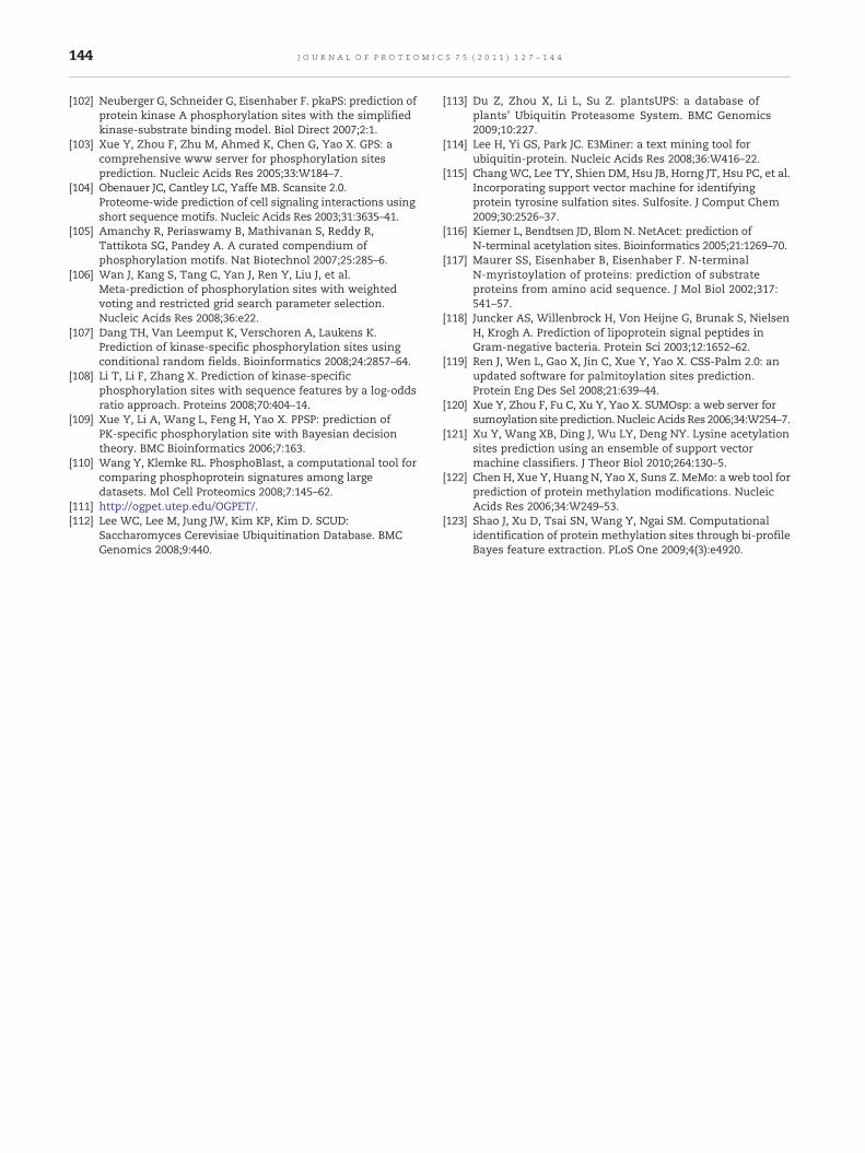
144 J O U R N A L O F P R O T E O M I C S 7 5 ( 2 0 1 1 ) 1 2 7 – 1 4 4
[102] Neuberger G, Schneider G, Eisenhaber F. pkaPS: prediction ofprotein kinase A phosphorylation sites with the simplifiedkinase-substrate binding model. Biol Direct 2007;2:1.
[103] Xue Y, Zhou F, Zhu M, Ahmed K, Chen G, Yao X. GPS: acomprehensive www server for phosphorylation sitesprediction. Nucleic Acids Res 2005;33:W184–7.
[104] Obenauer JC, Cantley LC, Yaffe MB. Scansite 2.0.Proteome-wide prediction of cell signaling interactions usingshort sequence motifs. Nucleic Acids Res 2003;31:3635–41.
[105] Amanchy R, Periaswamy B, Mathivanan S, Reddy R,Tattikota SG, Pandey A. A curated compendium ofphosphorylation motifs. Nat Biotechnol 2007;25:285–6.
[106] Wan J, Kang S, Tang C, Yan J, Ren Y, Liu J, et al.Meta-prediction of phosphorylation sites with weightedvoting and restricted grid search parameter selection.Nucleic Acids Res 2008;36:e22.
[107] Dang TH, Van Leemput K, Verschoren A, Laukens K.Prediction of kinase-specific phosphorylation sites usingconditional random fields. Bioinformatics 2008;24:2857–64.
[108] Li T, Li F, Zhang X. Prediction of kinase-specificphosphorylation sites with sequence features by a log-oddsratio approach. Proteins 2008;70:404–14.
[109] Xue Y, Li A, Wang L, Feng H, Yao X. PPSP: prediction ofPK-specific phosphorylation site with Bayesian decisiontheory. BMC Bioinformatics 2006;7:163.
[110] Wang Y, Klemke RL. PhosphoBlast, a computational tool forcomparing phosphoprotein signatures among largedatasets. Mol Cell Proteomics 2008;7:145–62.
[111] http://ogpet.utep.edu/OGPET/.[112] Lee WC, Lee M, Jung JW, Kim KP, Kim D. SCUD:
Saccharomyces Cerevisiae Ubiquitination Database. BMCGenomics 2008;9:440.
[113] Du Z, Zhou X, Li L, Su Z. plantsUPS: a database ofplants' Ubiquitin Proteasome System. BMC Genomics2009;10:227.
[114] Lee H, Yi GS, Park JC. E3Miner: a text mining tool forubiquitin-protein. Nucleic Acids Res 2008;36:W416–22.
[115] ChangWC, Lee TY, Shien DM, Hsu JB, Horng JT, Hsu PC, et al.Incorporating support vector machine for identifyingprotein tyrosine sulfation sites. Sulfosite. J Comput Chem2009;30:2526–37.
[116] Kiemer L, Bendtsen JD, Blom N. NetAcet: prediction ofN-terminal acetylation sites. Bioinformatics 2005;21:1269–70.
[117] Maurer SS, Eisenhaber B, Eisenhaber F. N-terminalN-myristoylation of proteins: prediction of substrateproteins from amino acid sequence. J Mol Biol 2002;317:541–57.
[118] Juncker AS, Willenbrock H, Von Heijne G, Brunak S, NielsenH, Krogh A. Prediction of lipoprotein signal peptides inGram-negative bacteria. Protein Sci 2003;12:1652–62.
[119] Ren J, Wen L, Gao X, Jin C, Xue Y, Yao X. CSS-Palm 2.0: anupdated software for palmitoylation sites prediction.Protein Eng Des Sel 2008;21:639–44.
[120] Xue Y, Zhou F, Fu C, Xu Y, Yao X. SUMOsp: a web server forsumoylation site prediction. Nucleic Acids Res 2006;34:W254–7.
[121] Xu Y, Wang XB, Ding J, Wu LY, Deng NY. Lysine acetylationsites prediction using an ensemble of support vectormachine classifiers. J Theor Biol 2010;264:130–5.
[122] Chen H, Xue Y, Huang N, Yao X, Suns Z. MeMo: a web tool forprediction of protein methylation modifications. NucleicAcids Res 2006;34:W249–53.
[123] Shao J, Xu D, Tsai SN, Wang Y, Ngai SM. Computationalidentification of protein methylation sites through bi-profileBayes feature extraction. PLoS One 2009;4(3):e4920.