protein phosphatase 2a catalytic subunit α plays a myd88-dependent, central role in the...
TRANSCRIPT

Please cite this article in press as: Xie et al., Protein Phosphatase 2A Catalytic Subunit a Plays a MyD88-Dependent, Central Role in the Gene-SpecificRegulation of Endotoxin Tolerance, Cell Reports (2013), http://dx.doi.org/10.1016/j.celrep.2013.01.029
Cell Reports
Report
Protein Phosphatase 2A Catalytic Subunit aPlays a MyD88-Dependent, Central Role in theGene-Specific Regulation of Endotoxin ToleranceLing Xie,1,5 Cui Liu,1,5 Li Wang,1,4 Harsha P. Gunawardena,1 Yanbao Yu,1 Ruyun Du,4 Debra J. Taxman,3 Penggao Dai,1
Zhen Yan,1 Jing Yu,1 Stephen P. Holly,1 Leslie V. Parise,1 Yisong Y. Wan,2,3 Jenny P. Ting,2,3 and Xian Chen1,2,4,*1Department of Biochemistry and Biophysics2Lineberger Comprehensive Cancer Center3Department of Microbiology and Immunology
University of North Carolina, Chapel Hill, NC 27599, USA4Department of Chemistry, Fudan University, Shanghai 20032, China5These authors contributed equally to this work
*Correspondence: [email protected]://dx.doi.org/10.1016/j.celrep.2013.01.029
SUMMARY
MyD88, the intracellular adaptor of most TLRs, medi-ates either proinflammatory or immunosuppressivesignaling that contributes to chronic inflammation-associated diseases. Although gene-specific chro-matin modifications regulate inflammation, the roleof MyD88 signaling in establishing such epigeneticlandscapes under different inflammatory statesremains elusive. Using quantitative proteomics toenumerate the inflammation-phenotypic constituentsof theMyD88 interactome,we found that inendotoxin-tolerant macrophages, protein phosphatase 2A cata-lytic subunit a (PP2Ac) enhances its association withMyD88 and is constitutively activated. Knockdownof PP2Ac prevents suppression of proinflammatorygenes and resistance to apoptosis. Through site-specific dephosphorylation, constitutively activePP2Ac disrupts the signal-promoting TLR4-MyD88complex and broadly suppresses the activities ofmultiple proinflammatory/proapoptotic pathways aswell, shifting proinflammatory MyD88 signaling to aprosurvival mode. Constitutively active PP2Ac trans-located with MyD88 into the nuclei of tolerant macro-phages establishes the immunosuppressive patternof chromatin modifications and represses chromatinremodeling to selectively silence proinflammatorygenes, coordinating the MyD88-dependent inflam-mation control at both signaling and epigenetic levelsunder endotoxin-tolerant conditions.
INTRODUCTION
Toll-like receptors (TLRs) activate the innate immune system by
mounting appropriate inflammatory responses to contain infec-
tion or repair damaged tissues (Drexler and Foxwell, 2010). To
avoid harmful effects of persistent signaling caused by the
continual presence of stimuli, cells become transiently unre-
sponsive by acquiring tolerance to chronic inflammation, leading
to a negative consequence that tumor cells can escape immuno-
surveillance (Rakoff-Nahoum and Medzhitov, 2009). In transmit-
ting inflammatory signals, by mechanisms yet to be elucidated,
the intracellular adaptor protein MyD88 (myeloid differentiation
primary response 88) of most TLRs acts as a double-edged
sword promoting both protective and harmful inflammation
(Huang et al., 2008).
Recent work has revealed that inflammation control is
achieved by a gene-specific mechanism in which distinct chro-
matin modifications contribute to selective silencing of TLR4-
induced proinflammatory or tolerizable (T class) genes under
an endotoxin tolerance (ET)-associated chronic inflammatory
state (Foster et al., 2007). Major questions remain unanswered
including the following:
(1) How doesMyD88mediate both acute and chronic inflam-
matory responses?
(2) What is the driving force for ‘‘converting’’ proinflammatory
MyD88 to an immunosuppressive mediator?
(3) How are distinct, inflammation-specific patterns of
chromatin modifications differentially established at the
T class promoters?
Emerging evidence suggests that an effective, unharmful
inflammatory response is intrinsically regulated by subtly distinct
intracellular protein interactions, i.e., ‘‘interactomes’’ (protein-
protein interaction network) involved in signal transduction
(Liew et al., 2005). During TLR signaling, MyD88 serves as a
scaffold that coordinates protein complex assembly through
sequential recruitment of specific proteins varying from TLR
molecules to downstream proteins (Dai et al., 2009; Wang
et al., 2006). Based on our previous identification of multiple
signal regulators that interact with MyD88 in a timely and orderly
manner to tightly regulate the amplitude and duration of TLR
signaling (Dai et al., 2009), we reason that MyD88 may regulate
either acute or chronic inflammation via assembling different,
inflammation-phenotypic interactomes.
Cell Reports 3, 1–11, March 28, 2013 ª2013 The Authors 1

(legend on next page)
2 Cell Reports 3, 1–11, March 28, 2013 ª2013 The Authors
Please cite this article in press as: Xie et al., Protein Phosphatase 2A Catalytic Subunit a Plays a MyD88-Dependent, Central Role in the Gene-SpecificRegulation of Endotoxin Tolerance, Cell Reports (2013), http://dx.doi.org/10.1016/j.celrep.2013.01.029

Please cite this article in press as: Xie et al., Protein Phosphatase 2A Catalytic Subunit a Plays a MyD88-Dependent, Central Role in the Gene-SpecificRegulation of Endotoxin Tolerance, Cell Reports (2013), http://dx.doi.org/10.1016/j.celrep.2013.01.029
RESULTS
As a Core Component of MyD88 Interactome in ETMacrophages, PP2Ac Is Chronically ActivatedFirst, by using our amino-acid-coded mass tagging (AACT)-
based quantitative proteomic approach (Chen et al., 2000) with
modifications for interactome screening (Figure 1A), we dis-
sected the MyD88-interacting complexes assembled in RAW
cells under different inflammatory states including (1) no stimula-
tion (N), (2) challenging with a single high LPS dose (LPS respon-
sive, NL), (3) priming with a low LPS dose (LPS tolerant, T), and
(4) challenging T cells with a high-dose LPS (TL). Compared
to NL cells, T or TL cells showed an ET-specific phenotype of
immunosuppression and resistance to apoptosis (Figure S1A).
Through phenotypic interactome analysis (Figure 1A), we found
that MyD88 interacts with different sets of proteins in NL ver-
sus TL macrophages (unpublished data): together with many
negative immune regulators (Liew et al., 2005) including a
negative TLR regulator Flii (Wang et al., 2006), protein phospha-
tase catalytic subunit a (PP2Ac) was found recruited into the
MyD88 interactome specifically in ET cells (Figures 1B and
S1B). Because approximately 20% of the components in the
TL-specific MyD88 interactome contain the domains interacting
with PP2Ac (Figure S1C) that is generally considered as a
suppressor of proinflammatory kinases (Junttila et al., 2008),
this inflammation-phenotypic proteomic finding suggested that
PP2Ac plays a central, MyD88-dependent, immunosuppressive
role during ET.
Given that neither expression nor stability of PP2Ac was
affected by LPS-induced inflammation (Figure S2A), we com-
pared the PP2Ac activity in RAW cells under different inflamma-
tory conditions. Compared to naive (N) cells, whereas its activity
was little changed under NL, PP2Ac was highly activated with
a prolonged stimulation and was sustained under TL (Figure 1C),
indicating the activity-based, inflammation-phenotypic function
of PP2Ac. To clarify the MyD88 dependence of PP2Ac activa-
tion, we measured PP2Ac activity in paired wild-type (WT) and
MyD88-depleted (MyD88�/�) (Figure S2B) bone marrow-derived
macrophages (BMDMs) under different inflammatory states in-
duced by a variety of TLR agonists. Similar to RAWs, PP2Ac
activity showed a 2-fold increase in TL WT BMDMs but less
than 10% changes in MyD88�/� BMDMs under N, NL, and
TL (Figure 1D). The TLR9 agonist CpG, which triggers only
MyD88-dependent pathways, induced a greater increase in
PP2Ac activity in TL WT BMDMs than that induced by LPS,
which activates bothMyD88-dependent and -independent path-
ways (Figure 1E), whereas no differences in PP2Ac activity were
observed between WT and MyD88�/� BMDMs when the TLR3
agonist Poly (I:C) stimulated only MyD88-independent path-
ways (Figure 1F). These agonist-specific effects indicated the
Figure 1. PP2Ac Enhances Its Association with MyD88 and Shows a M
(A) Experimental design of the AACT-based quantitative proteomics for phenoty
(B–H) Mass spectra of the AACT-containing peptides of MyD88 and indicated
representative of at least two peptides sequenced by MS/MS. The PP2Ac activ
BMDMs under N, or NL, or T, or TL (D), stimulated by CpG (E), by Poly (I:C) (F), by n
cytokine-induced, time-dependent changes of PP2Ac activity and p-p65 (H). Ea
See also Figure S1.
MyD88-dependent activation of PP2Ac in chronically inflamed
cells. Second, to clarify how PP2Ac activity is regulated in
tolerant macrophages, we respectively used either LPS-condi-
tioned medium, i.e., that separated from the LPS-pretreated
RAWs, or TNF-a or ionizing radiation (IR) to trigger inflammation
in BMDMs. Under chronic exposure, all non-TLR stimuli caused
a similar degree of tolerance-specific activation of PP2Ac in both
WT andMyD88�/� BMDMs (Figure 1G). In a coordinate manner,
unlike its LPS-inducible nature in WT but not in MyD88�/�
BMDMs (Figure 1H, left), phospho-p65 triggered by non-TLR
stimuli showed similar time-dependent induction in both types
of BMDMs (Figure 1H, right), indicating that stimuli-induced,
secondary, NFkB-regulated production of inflammatory cyto-
kines such as TNF-a causes tolerance-specific PP2Ac activa-
tion. Because TNF-a is a T class gene and its release is MyD88
promoted (Figure S2B), this secondary effect on PP2Ac activa-
tion was therefore more pronounced in the WT BMDMs directly
stimulated by the TLR agonists that trigger MyD88-dependent
pathways for producing these T class cytokines. Only during
the chronic inflammation mediated by TLR2/TLR4/TR9 is the
sustained PP2Ac activation MyD88 dependent.
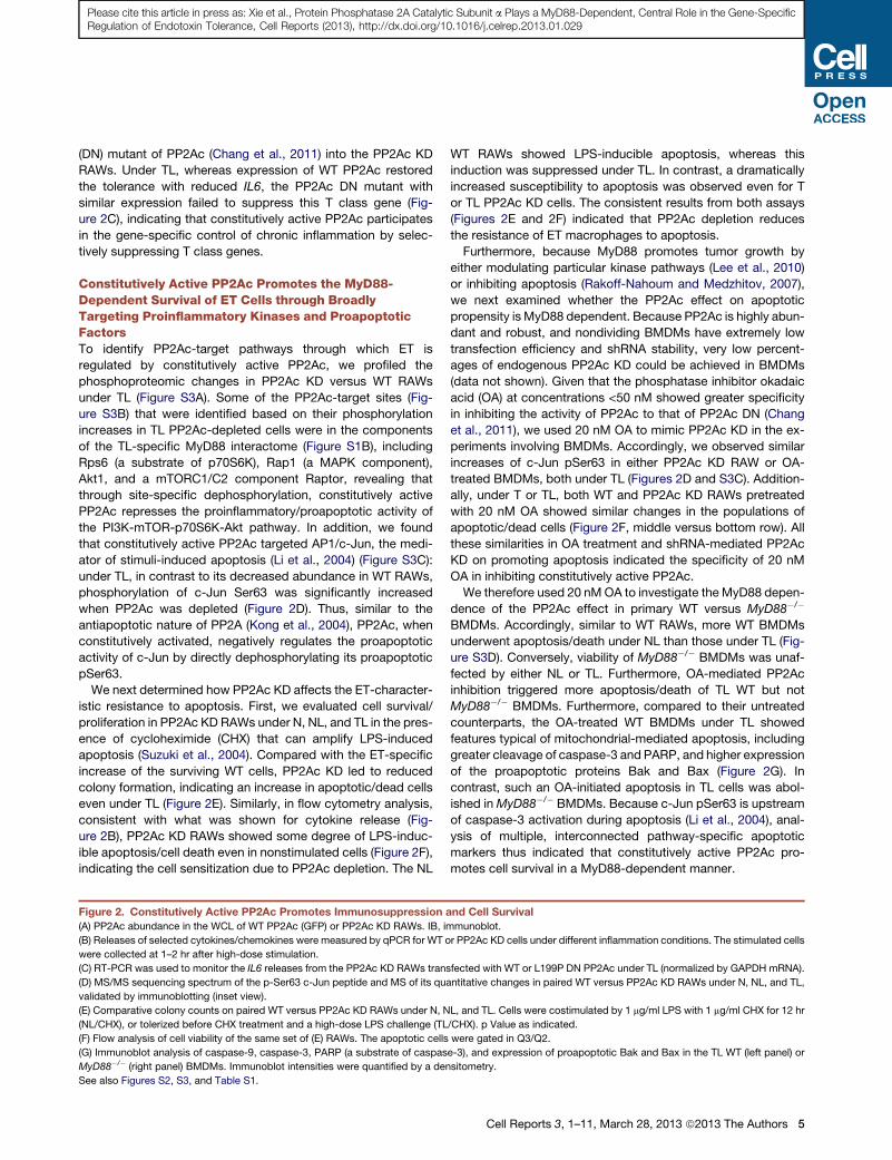
Constitutively Active PP2Ac RegulatesImmunosuppression during ETTo determine the correlation of persistently or constitutively acti-
vated PP2Ac with immunosuppression, we measured cytokine
release from stable RAW lines expressing either shRNA for
knocking down PP2Ac (PP2Ac knockdown [KD]) or shRNA for
GFP (WT control) (Figures 2A and S2C). Under NL, most cyto-
kines showed LPS-inducible release in both PP2Ac KD and
WT RAWs (Figure 2B). In contrast, compared to the LPS-toler-
ized WT RAWs showing reduced production of T class cyto-
kines, the LPS-primed PP2Ac KD cells were hyperresponsive
to high-dose LPS stimulation and released greater amounts of
cytokines. Furthermore, we determined the effect of constitu-
tively active PP2Ac on the expression of 84mouse genes encod-
ing inflammatory factors, most known to be regulated by NFkB
(Figure S2D). Compared with their levels in WT RAWs, 34 out
of 84 genes showed more than a 30% increase (Table S1),
some of which were validated by qPCR (Figure 2B). Also, T class
genes (IL6 or IL10) had dramatically enhanced expression in the
PP2Ac KD cells even under TL, reversing their suppressed
expression in TL WT cells. In contrast, under NL or TL, PP2Ac
KDhad little effect on the LPS-inducible trend of Fpr1 (Figure 2B),
a nontolerizeable gene (Foster et al., 2007), suggesting that
constitutively active PP2Ac is involved specifically in T class
gene regulation under ET.
Furthermore, to determine whether the constitutively active
form of PP2Ac directly contributes to acquiring ET, we trans-
fected constructs expressing either WT or a dominant-negative
yD88-Dependent Activation in ET Macrophages
pic compositional screening of the MyD88 interactomes under N, NL, and TL.
TL-specific MyD88 interactors (B). Shown for each protein is one spectrum
ity was measured by phosphatase assay in RAWs (C), in WT and MyD88�/�
on-TLR stimuli including LPS-conditionedmedium, TNF-a, and IR (G), agonist/
ch bar represents the mean ± SD of triplicates (same in figures below).
Cell Reports 3, 1–11, March 28, 2013 ª2013 The Authors 3

(legend on next page)
4 Cell Reports 3, 1–11, March 28, 2013 ª2013 The Authors
Please cite this article in press as: Xie et al., Protein Phosphatase 2A Catalytic Subunit a Plays a MyD88-Dependent, Central Role in the Gene-SpecificRegulation of Endotoxin Tolerance, Cell Reports (2013), http://dx.doi.org/10.1016/j.celrep.2013.01.029
Please cite this article in press as: Xie et al., Protein Phosphatase 2A Catalytic Subunit a Plays a MyD88-Dependent, Central Role in the Gene-SpecificRegulation of Endotoxin Tolerance, Cell Reports (2013), http://dx.doi.org/10.1016/j.celrep.2013.01.029
(DN) mutant of PP2Ac (Chang et al., 2011) into the PP2Ac KD
RAWs. Under TL, whereas expression of WT PP2Ac restored
the tolerance with reduced IL6, the PP2Ac DN mutant with
similar expression failed to suppress this T class gene (Fig-
ure 2C), indicating that constitutively active PP2Ac participates
in the gene-specific control of chronic inflammation by selec-
tively suppressing T class genes.
Constitutively Active PP2Ac Promotes the MyD88-Dependent Survival of ET Cells through BroadlyTargeting Proinflammatory Kinases and ProapoptoticFactorsTo identify PP2Ac-target pathways through which ET is
regulated by constitutively active PP2Ac, we profiled the
phosphoproteomic changes in PP2Ac KD versus WT RAWs
under TL (Figure S3A). Some of the PP2Ac-target sites (Fig-
ure S3B) that were identified based on their phosphorylation
increases in TL PP2Ac-depleted cells were in the components
of the TL-specific MyD88 interactome (Figure S1B), including
Rps6 (a substrate of p70S6K), Rap1 (a MAPK component),
Akt1, and a mTORC1/C2 component Raptor, revealing that
through site-specific dephosphorylation, constitutively active
PP2Ac represses the proinflammatory/proapoptotic activity of
the PI3K-mTOR-p70S6K-Akt pathway. In addition, we found
that constitutively active PP2Ac targeted AP1/c-Jun, the medi-
ator of stimuli-induced apoptosis (Li et al., 2004) (Figure S3C):
under TL, in contrast to its decreased abundance in WT RAWs,
phosphorylation of c-Jun Ser63 was significantly increased
when PP2Ac was depleted (Figure 2D). Thus, similar to the
antiapoptotic nature of PP2A (Kong et al., 2004), PP2Ac, when
constitutively activated, negatively regulates the proapoptotic
activity of c-Jun by directly dephosphorylating its proapoptotic
pSer63.
We next determined how PP2Ac KD affects the ET-character-
istic resistance to apoptosis. First, we evaluated cell survival/
proliferation in PP2Ac KD RAWs under N, NL, and TL in the pres-
ence of cycloheximide (CHX) that can amplify LPS-induced
apoptosis (Suzuki et al., 2004). Compared with the ET-specific
increase of the surviving WT cells, PP2Ac KD led to reduced
colony formation, indicating an increase in apoptotic/dead cells
even under TL (Figure 2E). Similarly, in flow cytometry analysis,
consistent with what was shown for cytokine release (Fig-
ure 2B), PP2Ac KD RAWs showed some degree of LPS-induc-
ible apoptosis/cell death even in nonstimulated cells (Figure 2F),
indicating the cell sensitization due to PP2Ac depletion. The NL
Figure 2. Constitutively Active PP2Ac Promotes Immunosuppression a
(A) PP2Ac abundance in the WCL of WT PP2Ac (GFP) or PP2Ac KD RAWs. IB, im
(B) Releases of selected cytokines/chemokines were measured by qPCR for WT o
were collected at 1–2 hr after high-dose stimulation.
(C) RT-PCR was used to monitor the IL6 releases from the PP2Ac KD RAWs trans
(D) MS/MS sequencing spectrum of the p-Ser63 c-Jun peptide and MS of its qu
validated by immunoblotting (inset view).
(E) Comparative colony counts on paired WT versus PP2Ac KD RAWs under N, N
(NL/CHX), or tolerized before CHX treatment and a high-dose LPS challenge (TL
(F) Flow analysis of cell viability of the same set of (E) RAWs. The apoptotic cells
(G) Immunoblot analysis of caspase-9, caspase-3, PARP (a substrate of caspas
MyD88�/� (right panel) BMDMs. Immunoblot intensities were quantified by a den
See also Figures S2, S3, and Table S1.
WT RAWs showed LPS-inducible apoptosis, whereas this
induction was suppressed under TL. In contrast, a dramatically
increased susceptibility to apoptosis was observed even for T
or TL PP2Ac KD cells. The consistent results from both assays
(Figures 2E and 2F) indicated that PP2Ac depletion reduces
the resistance of ET macrophages to apoptosis.
Furthermore, because MyD88 promotes tumor growth by
either modulating particular kinase pathways (Lee et al., 2010)
or inhibiting apoptosis (Rakoff-Nahoum and Medzhitov, 2007),
we next examined whether the PP2Ac effect on apoptotic
propensity is MyD88 dependent. Because PP2Ac is highly abun-
dant and robust, and nondividing BMDMs have extremely low
transfection efficiency and shRNA stability, very low percent-
ages of endogenous PP2Ac KD could be achieved in BMDMs
(data not shown). Given that the phosphatase inhibitor okadaic
acid (OA) at concentrations <50 nM showed greater specificity
in inhibiting the activity of PP2Ac to that of PP2Ac DN (Chang
et al., 2011), we used 20 nM OA to mimic PP2Ac KD in the ex-
periments involving BMDMs. Accordingly, we observed similar
increases of c-Jun pSer63 in either PP2Ac KD RAW or OA-
treated BMDMs, both under TL (Figures 2D and S3C). Addition-
ally, under T or TL, both WT and PP2Ac KD RAWs pretreated
with 20 nM OA showed similar changes in the populations of
apoptotic/dead cells (Figure 2F, middle versus bottom row). All
these similarities in OA treatment and shRNA-mediated PP2Ac
KD on promoting apoptosis indicated the specificity of 20 nM
OA in inhibiting constitutively active PP2Ac.
We therefore used 20 nMOA to investigate the MyD88 depen-
dence of the PP2Ac effect in primary WT versus MyD88�/�
BMDMs. Accordingly, similar to WT RAWs, more WT BMDMs
underwent apoptosis/death under NL than those under TL (Fig-
ure S3D). Conversely, viability of MyD88�/� BMDMs was unaf-
fected by either NL or TL. Furthermore, OA-mediated PP2Ac
inhibition triggered more apoptosis/death of TL WT but not
MyD88�/� BMDMs. Furthermore, compared to their untreated
counterparts, the OA-treated WT BMDMs under TL showed
features typical of mitochondrial-mediated apoptosis, including
greater cleavage of caspase-3 and PARP, and higher expression
of the proapoptotic proteins Bak and Bax (Figure 2G). In
contrast, such an OA-initiated apoptosis in TL cells was abol-
ished inMyD88�/� BMDMs. Because c-Jun pSer63 is upstream
of caspase-3 activation during apoptosis (Li et al., 2004), anal-
ysis of multiple, interconnected pathway-specific apoptotic
markers thus indicated that constitutively active PP2Ac pro-
motes cell survival in a MyD88-dependent manner.
nd Cell Survival
munoblot.
r PP2Ac KD cells under different inflammation conditions. The stimulated cells
fected with WT or L199P DN PP2Ac under TL (normalized by GAPDH mRNA).
antitative changes in paired WT versus PP2Ac KD RAWs under N, NL, and TL,
L, and TL. Cells were costimulated by 1 mg/ml LPS with 1 mg/ml CHX for 12 hr
/CHX). p Value as indicated.
were gated in Q3/Q2.
e-3), and expression of proapoptotic Bak and Bax in the TL WT (left panel) or
sitometry.
Cell Reports 3, 1–11, March 28, 2013 ª2013 The Authors 5

A C
B D
E
FG
H
I
TN
F-α
ove
r
Figure 3. Chronic-Active PP2Ac Dephosphorylates MyD88 for Dissociating Proinflammatory TLR4-MyD88 Complex and Promoting Its
Cotranslocation with MyD88
(A) Immunoblot analysis of the immunoprecipitates from the TLR4 stable 293 cells cotransfected with MYC-tagged PP2Ac and HA-tagged MyD88 fused with
GyrB (MyD88-GyrB) under N, NL, or TL (Left). HA-MyD88-GyrB was cotransfected with either MYCWT or DN PP2Ac construct, respectively, into the TLR4 stable
cells that were induced to TL (Right).
(B) MS/MS spectrum of the phosphorylated S242 or S244 MyD88 peptide.
(legend continued on next page)
6 Cell Reports 3, 1–11, March 28, 2013 ª2013 The Authors
Please cite this article in press as: Xie et al., Protein Phosphatase 2A Catalytic Subunit a Plays a MyD88-Dependent, Central Role in the Gene-SpecificRegulation of Endotoxin Tolerance, Cell Reports (2013), http://dx.doi.org/10.1016/j.celrep.2013.01.029

Please cite this article in press as: Xie et al., Protein Phosphatase 2A Catalytic Subunit a Plays a MyD88-Dependent, Central Role in the Gene-SpecificRegulation of Endotoxin Tolerance, Cell Reports (2013), http://dx.doi.org/10.1016/j.celrep.2013.01.029
Constitutively Active PP2Ac Dissociates theSignal-Promoting TLR4-MyD88 Complex throughDephosphorylating Key Serines in the Toll/IL-1Receptor Domain of MyD88We next investigated the role of TL-specific PP2Ac-MyD88
interaction (Figure 3A, left) in acquiring ET. First, we found that
MyD88 binds to the WT PP2Ac more tightly than to the PP2Ac
DN under TL (Figure 3A, right), indicating that MyD88 interacts
only with constitutively active PP2Ac. Furthermore, to identify
exactly where constitutively active PP2Ac targets MyD88, we
employed an AACT-based quantitative proteomics approach
where tagged MyD88 expressed in HEK TLR4 stable cells was
immunoprecipitated (IP) for MS analysis because no phosphor-
ylation on endogenousMyD88 was detected in the phosphopro-
teomic analysis above. Meanwhile, we used 20 nM OA to
highlight PP2Ac-target phospho-sites on MyD88. In MyD88
immunoprecipitates from differentially stimulated TLR4 stable
cells, with or without 20 nM OA treatment, we identified phos-
phorylation sites at Ser242/Ser244 (Figure 3B) and also found
that these residues exhibited an LPS-inducible increase in
phosphorylation, more so in NL than in TL cells (Figures S4A
and S4B). Furthermore, phosphorylation of these sites was
increased approximately 30% in the OA-treated TL cells (Fig-
ure 3C) compared to NL cells in which OA had little effect,
suggesting that constitutively active PP2Ac targets these phos-
phoserines for dephosphorylation. Thus, we generated single
(S242A and S244A) and double (S242A-S244A) nonphosphory-
latable mutants of MyD88 and found that constitutively active
PP2Ac associated more strongly with WT MyD88 than with
any nonphosphorylatable mutant (Figure 3D), accordingly vali-
dating these PP2Ac-recognizing sites in TL cells. Because these
serines reside in the TLR4-interacting MyD88 Toll/IL-1 receptor
(TIR) domain that is critical for initiating proinflammatory sig-
naling (Wang et al., 2006), we reasoned that these LPS-induc-
ible, PP2Ac-target phosphorylations are essential for stability
of the signal-promoting MyD88-TLR4 complex. Thus, we exam-
ined the LPS-induced, time-dependent TLR4-MyD88 associa-
tion, which reached its highest strength at 15 min following
LPS stimulation before returning to the basal level 24 hr after
stimulation (Figure 3E, left). We then generated an phospho-
Ser242 antibody (Figure S4C) to assay the time-dependent level
of this proinflammatory MyD88 phosphorylation (pSer242) that
also reached the highest level at 15 min and was reduced with
(C) Effects of PP2Ac inhibition on the LPS-inducible S242/S244 phosphorylation o
this peptide with or without p-S242/p-S244. (Left) The fold change of phosphory
(D) MYC-PP2Ac was cotransfected with the construct of HA-WT or a nonphosp
immunoblot analysis.
(E) (Left) LPS-induced, time-dependent bindings between YFP-tagged TLR4 an
TLR4-MyD88 complexes blotted by pSer242 antibody. (Right) Comparative bindin
stable cells following 15 min LPS stimulation.
(F) (Top) MyD88�/� BMDMs were infected by retrovirus that packaged each o
stimulated by Pam3CSK4 for 1 hr. TNF-amRNAwas measured by RT-PCR for ea
NV, noninfected MyD88�/� control. (Bottom) MyD88 expression in non- or trans
(G) Cytoplasmic and nuclear distributions of MyD88, PP2Ac, and p65 in BMDMs
(H) Immunoblot analysis of the nuclear fraction of PP2Ac KD or WT RAWs.
(I) Confocal microscopy of the subcellular colocalization of MyD88 and PP2Ac. Th
versus that in cytoplasm, ten spots with the same square of area for each comp
See also Figure S4.
prolonged LPS stimulation (Figure 3E, middle). We then
compared binding of TLR4 to WT MyD88 versus each mutant
in cotransfected cells stimulated by LPS for 15 min. Compared
with WT MyD88, binding of each nonphosphorylatable mutant
to TLR4 was not only weaker but also not LPS inducible
(Figure 3E, right). These results together revealed that site-
specific phosphorylation(s) in the MyD88 TIR is directly involved
in the proinflammatory TLR4-MyD88 interaction, also demon-
strating the physiological accuracy of our quantitative proteo-
mic method for identification of these sites with 20 nM OA
treatment.
To determine the impact of the PP2Ac-mediated MyD88 de-
phosphorylation on T class gene regulation, we transfected WT
and the nonphosphorylatable MyD88 mutants into MyD88�/�
BMDMs. Coincident with the weakened binding of MyD88
mutants to TLR4 (Figure 3E), following a high-dose Pam3CSK4
stimulation (MyD88-dependent TLR2 agonist), a 50% reduction
in TNF-a was observed for the mutant-expressing cells com-
pared to cells expressing WTMyD88 (Figure 3F). Clearly, disrup-
tion of the phosphorylation-dependent TLR4-MyD88 complex
by constitutively active PP2Ac directly contributes to suppres-
sion of T class genes.
Constitutively Active PP2Ac Colocalizes with MyD88 inthe Nuclei of Tolerant MacrophagesGiven that constitutively active PP2Ac facilitates the departure of
MyD88 from the TLR4 complex through site-specific dephos-
phorylation that often regulates protein translocation (Deribe
et al., 2010), we then determined the ET-specific distribution of
MyD88 and PP2Ac. Compared to their subcellular distributions
in proapoptotic NL cells, both MyD88 and PP2Ac showed coin-
cidently increased presence in the nuclei of the TL, prosurvival
WT BMDMs (Figure 3G), in line with a previous report that
MyD88 was preferentially in nuclei of nonapoptotic cells (Jaunin
et al., 1998). Furthermore, immunoblotting revealed that more
MyD88 and PP2Ac were retained in the nuclei of LPS-tolerant
WT BMDMs than in naive cells (Figure 3H). In contrast, in the
PP2Ac KD cells under TL, MyD88 failed to accumulate in nuclei.
Similarly, OA-mediated PP2Ac inhibition reversed survival-
characteristic retention of MyD88. Immunofluorescence staining
further indicated that more PP2Ac and MyD88 colocalized in
nuclei of TL WT macrophages than those in nonstimulated cells
(Figure 3I, yellow spots in the nuclei area), all indicating that
f the MyD88 IP from the TLR4 stable cells under NL or TL. (Right) MS spectra of
lation caused by OA (y axis) under TL calculated in Figure S3B.
horylatable mutant MyD88 into the TLR4 stable cells before TL induction for
d WT MyD88. (Middle) The level of pSer242 in LPS-induced, time-dependent
gs of TLR4 to either WT or nonphosphorylatable mutants of MyD88 in the TLR4
f the constructs containing WT, S242A, S244A, and S34A MyD88 and then
ch MyD88-expressing BMDMwithout or with stimulation. EV, empty retrovirus;
fected MyD88�/� BMDMs.
under N, NL, T, and TL; Lmnb1 as a nuclear marker.
e bar blot shows the ratio of the intensity of green or red fluorescence in nuclei
artment.
Cell Reports 3, 1–11, March 28, 2013 ª2013 The Authors 7

(legend on next page)
8 Cell Reports 3, 1–11, March 28, 2013 ª2013 The Authors
Please cite this article in press as: Xie et al., Protein Phosphatase 2A Catalytic Subunit a Plays a MyD88-Dependent, Central Role in the Gene-SpecificRegulation of Endotoxin Tolerance, Cell Reports (2013), http://dx.doi.org/10.1016/j.celrep.2013.01.029

Please cite this article in press as: Xie et al., Protein Phosphatase 2A Catalytic Subunit a Plays a MyD88-Dependent, Central Role in the Gene-SpecificRegulation of Endotoxin Tolerance, Cell Reports (2013), http://dx.doi.org/10.1016/j.celrep.2013.01.029
chronic-active PP2Ac promotes survival-specific translocation
of MyD88 to ET nuclei through MyD88 dephosphorylation.
Constitutively Active PP2Ac Plays a MyD88-DependentRole in Establishing the Gene-Specific ChromatinModifications Responsible for Acquiring ETWe next explored the impact of the nuclear MyD88/PP2Ac
accumulation on T class gene regulation. First, along with
increasing PP2Ac in ET nuclei, the LPS-inducible increase of
phospho-p65 was significantly suppressed in TL WT cells
(Figure 3G, left lanes 2–4). Either PP2Ac KD or OA treatment
dramatically reversed the TL-specific suppression of p-p65, indi-
cating that constitutively active PP2Ac specifically dephosphor-
ylates p-p65. Additionally, although p-p65 was suppressed, p65
was even higher in TL nuclei, indicating abnormal nuclear reten-
tion of nonphosphorylated p65 under ET. We then clarified the
impact of PP2Ac-mediated p65 dephosphorylation on NFkB-
regulated T class genes and its MyD88 dependence: in contrast
to the MyD88-dependent suppression of IL6/IL10 in the TL
WT BMDMs, OA inhibition of PP2Ac activity reactivated their
release under LPS stimulation, even more so with Pam3CSK4
treatment for MyD88-dependent pathways (Figure 4A), all sug-
gesting that, in a MyD88-dependent manner, constitutively
active PP2Ac renders NFkB-dependent genes unresponsive
by dephosphorylating nuclear p65 in ET macrophages.
Additionally, our phosphoproteomic findings of PP2Ac-target
chromatin assembly/remodeling factors such as SWI/SNF (Fig-
ure S3B) implied that nuclear translocated, constitutively active
PP2Ac directly participates in gene-specific chromatin modi-
fications. First, because histone H3 Ser10 phosphorylation
(H3pS10) reflects the accessibility of NFkB to T class genes
and mediates NFkB-dependent transactivation of these genes
(Saccani et al., 2002), we determined whether constitutively
active PP2Ac affects this proinflammatory epigenetic marker.
Compared with its NL-enhanced but TL-suppressed nature in
irritated WT cells, H3pS10 remained high in PP2Ac KD cells
under NL or TL (Figure 4B), indicating that constitutively active
PP2Ac specifically targets H3pS10. Furthermore, the LPS-
inducible H3pS10 under NL was fully suppressed in TL WT
BMDMs and was reversed by OA pretreatment, whereas no
such OA effect was observed for NL cells (Figure 4C). As well,
no detectable change due to OA treatment was observed in
MyD88�/� BMDMs under TL, all indicating that in a MyD88-
dependent manner, constitutively active PP2Ac dephosphory-
Figure 4. Chronic-Active PP2Ac Directly Participates in TLR-Induced,
(A) A high or priming dose of LPS (L) or Pam3CSK4 (P) was respectively used
stimulation, and IL6 and IL10 were quantified by quantitative RT-PCR and norma
(B) Immunoblot analysis of pSer10 H3 in WT or PP2Ac KD RAWs under N, NL, T
(C) LPS-induced changes of pSer10 H3 or acetylated H3 in WT or MyD88�/� BM
(D) Cells were treated by 2 mM SAHA for 24 hr prior to stimulation. Similar to (A),
BMDMs with indicated treatment(s).
(E) WT or PP2Ac KD RAWs with indicated treatment(s) were fixed with DSG/f
normalized to INPUT.
(F) WT or MyD88�/� BMDMs with indicated treatment(s) were analyzed by ChIP
(G) Location of serine phosphorylations in the MyD88 TIR domain. S242(m) and S2
(NP_002459).
(H) The mechanistic model for the MyD88-dependent, PP2Ac-coordinated contr
See also Figures S3 and S4.
lates H3pS10 to selectively restrict accessibility of NFkB to
T class gene promoters.
We then determined the link of the PP2Ac-coordinated
MyD88 signaling to two major characteristics of the gene-
specific regulation, histone deacetylation and inhibition of chro-
matin remodeling, at T class gene promoters under TL (Foster
et al., 2007). First, the reversed suppression of IL6 and IL10 in
the TL WT BMDMs pretreated with a HDAC inhibitor (SAHA)
was not observed in the TL MyD88�/� cells (Figure 4D), indi-
cating that histone deacetylation is MyD88 dependent. Second,
levels of both p-Ser10 and acetyl-lysine 9 on H3 showed similar
OA-triggered MyD88-dependent changes (Figure 4C), indicating
that in the MyD88 interactome, constitutively active PP2Ac
negatively regulates histone acetylation in a phosphorylation-
dependent manner.
In combination, our findings of MyD88-dependent, PP2Ac-
mediated p65 dephosphorylation and PP2Ac-target HDAC1
and chromatin remodeling complexes (Figure S3B) suggested
that, in a phosphorylation-dependent manner, constitutively
active PP2Ac directly regulates TLR4-induced recruitment of
chromatin remodeling complexes to T class gene promoters, a
process that is inhibited in tolerant macrophages. Thus, we
examined the effect of PP2Ac KD on recruitment of an ATP-
dependent chromatin remodeling complex, Brg1, to T class pro-
moters, which was found suppressed under TL (Foster et al.,
2007). As revealed by ChIP experiments, either PP2Ac KD or
OA-mediated PP2Ac inhibition similarly reversed the ET-specific
suppression of Brg1 recruitment to the IL6 and Lipg promoters
(Figure 4E). Furthermore, this proinflammatory recruitment was
heavily suppressed in the TL WT BMDMs but suppressed to
a much lower degree in the TL MyD88�/� BMDMs (Figure 4F).
By contrast, whereas OA had little effect on Brg1 in the NL WT
BMDMs, the TL WT BMDMs pretreated with OA showed greater
Brg1 recruitment. However, this OA-initiated Brg1 recruitment
was not observed in the TLMyD88�/� BMDMs. These results in-
dicated that constitutively active PP2Ac cotranslocated with
MyD88 negatively regulates the chromatin modification/remod-
eling required for T class gene transactivation.
DISCUSSION
Here, we have revealed a mechanism by which TLR4-
induced, constitutively active PP2Ac broadly intersects with
multiple, MyD88-dependent, interconnected signaling pathways
MyD88-Dependent, Gene-Specific Chromatin Modifications
to induce acute or chronic inflammation. Cells were harvested at 1 hr after
lized with GAPDH. p values are as indicated.
, and TL.
DMs.
IL6/IL10 mRNAs were measured for those isolated from the WT or MyD88�/�
ormaldehyde 3 hr after stimulation and analyzed by ChIP (Brg1). qPCR was
(Brg1).
44(m) are the mouse counterparts (NP_034981) of human S255(h) and S257(h)
ol of the inflammation.
Cell Reports 3, 1–11, March 28, 2013 ª2013 The Authors 9

Please cite this article in press as: Xie et al., Protein Phosphatase 2A Catalytic Subunit a Plays a MyD88-Dependent, Central Role in the Gene-SpecificRegulation of Endotoxin Tolerance, Cell Reports (2013), http://dx.doi.org/10.1016/j.celrep.2013.01.029
and epigenetic regulation programs at both signal-specific and
gene-specific levels for controlling inflammation as macro-
phages acquire ET. At the signaling level, because PP2Ac-
targeted p-Ser242/p-Ser244 localize to the highly accessible
area close to the conserved lymphoma-associated, survival-
promoting Leu-265 (Ngo et al., 2011), we rationalized that under
TL conditions with a lymphoma-mimicking chronic inflamma-
tory environment, PP2Ac-mediated dephosphorylation of these
residues could generate charge changes in the MyD88 TIR
domain (Ohnishi et al., 2009) (Figure 4G), similarly leading to
a weakened MyD88-TLR4 association and impaired proinflam-
matory and proapoptotic programs with reduced sensitivity to
apoptotic stimuli. Consequently, the dissociating MyD88 from
the TLR4 complex was translocated along with constitutively
active PP2Ac into the nuclei of tolerant macrophages, indicative
of increased cell survival (Figure 4H). At the chromatin level,
constitutively active PP2Ac initiated the removal of phos-
phorylation-dependent acetylation, established T gene-specific
chromatin modification, and suppressed the phosphorylation-
dependent, Brg-mediated nucleosome remodeling for silencing
of T class genes (Figure 4H).
From the perspective of a dynamic system, we have estab-
lished a direct link between gene-specific and signal-specific
regulation coordinately for the control of chronic inflammation,
elucidating how the differential regulation of proinflammatory
versus antimicrobial genes at the chromatin level is achieved.
Of clinical relevance, the specific protein interactions in the
tolerance-specific MyD88 interactome coordinated by con-
stitutively active PP2Ac that we have dissected may be tar-
geted to unleash or attenuate cellular inflammatory responses.
This knowledge is critical to better understand how MyD88
signaling produces diverse pathologic outcomes, including
tissue repair and carcinogenesis, under different inflammatory
conditions.
EXPERIMENTAL PROCEDURES
Proteomic Identification of TL-Specific MyD88 Interactors
The details about sample preparation, nano-LC-MS/MS analysis, and AACT-
based quantitative proteomic identifications of NL- or TL-specific MyD88-
interacting partners are given in Extended Experimental Procedures.
BMDM Stimulation and OA or SAHA Treatment
LPS stimulations of differentiated BMDMs were similar as to RAWs. In
Pam3CSK4 stimulations, the priming dose was at 50 ng/ml, and a high dose
was 500 ng/ml. SAHA was introduced into media 24 hr before stimulation,
and 20 nM OA was added along with the stimuli.
PP2A Activity Assay
PP2Acwas IP using PP2Ac antibodywith protein A agarose from 0.5–1.0mg of
each lysate of differentially stimulated cells. The phosphatase activity of IP
PP2Ac was measured by the Phosphatase Assay Kit (Millipore) and normal-
ized based on the amount of PP2Ac in the IP products.
Generation of the RAW Stable Lines with Either shRNA-GFP
or -PP2Ac
The lentiviral plasmids pLKO.1 expressing the shRNA-PP2Ac targeting se-
quences 50-CCAGATACAAATTACCTGTT-30 and 50-CGACGAGTGTTTAAG
GAAATA-30 were purchased from Thermo Scientific. To produce virus,
pLKO.1-shRNA plasmids were cotransfected into 293T cells with ViraPower
Mix (Invitrogen) by jetPRIME in vitro DNA and siRNA transfection reagent
10 Cell Reports 3, 1–11, March 28, 2013 ª2013 The Authors
(Polyplus). Pseudovirus in supernatants was collected after 48 hr following
transfection and used to transduce RAWs by spinoculation. Forty-eight hours
after transfection, 8 mg/ml puromycin was added to select puromycin-resistant
clones. Stable clones were maintained in the media containing 4 mg/ml
puromycin.
Identification of LPS-Inducible, PP2Ac-Target Phosphorylations at
MyD88
Due to the low abundance of endogenousMyD88 in RAW cells, few phosphor-
ylation sites could be identified/quantified for their LPS-inducible changes.
Therefore, quantitative proteomic identification of phosphorylation sites on
MyD88 and the analysis of LPS or OA effects on these sites were conducted
on the AACT-tagged HEK293 cells stably expressing TLR4-MD2-CD14. The
details are given in Extended Experimental Procedures.
Retrovirus Infection and Cytokine Assay
Each cDNA of WT MyD88 or a MyD88 mutant was inserted into the MIG
vector (Wang et al., 2010). Viral particles were generated in 293T cells and
infected bone marrow cells three times. Five days after infection, matured
BMDM cells were processed for LPS stimulation as described above.
Analysis of LPS-Induced Binding between TLR4 and WT or Mutant
Myd88
HEK-TLR4 stable cells were cotransfected with both YFP-TLR4 and HA-WT or
a mutant MyD88 by using jetPRIME. The cells were stimulated with 1 mg/ml
LPS for indicated time and harvested. The cells were lysed in the buffer
(50 mM Tris [pH 8.0], 150 mM NaCl, 1 mM EDTA, 1% Triton 100-X, protease
inhibitor, and phosphatase inhibitors; Sigma-Aldrich). One milligram lysate
was incubated with the anti-GFP IgG coupled on protein A magnetic beads
for coIP and immunoblotting.
All procedures involving wild-type (WT) andMyD88�/� knock-out mice have
been approved by the University of North Carolina Animal Care and Use
Committee.
SUPPLEMENTAL INFORMATION
Supplemental Information includes four figures, two tables, and Extended
Experimental Procedures and can be found with this article online at http://
dx.doi.org/10.1016/j.celrep.2013.01.029.
LICENSING INFORMATION
This is an open-access article distributed under the terms of the Creative
Commons Attribution License, which permits unrestricted use, distribution,
and reproduction in any medium, provided the original author and source
are credited.
ACKNOWLEDGMENTS
This work is supported by NIH grants to X.C. (NIH R01AI064806 and
NIH 1U24CA160035) and L.V.P. (NIH 1R01HL092544 and AHA
10GRNT3870017). We thank Dr. Howard Fried for editing the manuscript,
Dr. Hengming Ke for drawing the structure shown in Figure 4G, and Dr. S.
Zhang for technical assistance. L.X., C.L., L.W., and J.Y. performed major bio-
logical/immunological experiments. H.P.G. and Y.Y. performed quantitative
proteomics experiments. Z.Y. measured PP2Ac activity in radiation-treated
BMDMs. R.D. analyzed mass spectrometric data and network analysis.
D.J.T. and J.P.T. assisted in generating PP2Ac knockdown Raw264.7 cells.
Y.W. provided advice for BMDM transfection and helpful discussion. S.P.H.
and L.V.P. assisted in flow cytometry analysis. X.C. conceived the project, de-
signed the experiments, analyzed the results, and wrote the manuscript.
Received: May 15, 2012
Revised: November 30, 2012
Accepted: January 24, 2013
Published: February 21, 2013

Please cite this article in press as: Xie et al., Protein Phosphatase 2A Catalytic Subunit a Plays a MyD88-Dependent, Central Role in the Gene-SpecificRegulation of Endotoxin Tolerance, Cell Reports (2013), http://dx.doi.org/10.1016/j.celrep.2013.01.029
REFERENCES
Chang, H.Y., Jennings, P.C., Stewart, J., Verrills, N.M., and Jones, K.T. (2011).
Essential role of protein phosphatase 2A in metaphase II arrest and activation
of mouse eggs shown by okadaic acid, dominant negative protein phospha-
tase 2A, and FTY720. J. Biol. Chem. 286, 14705–14712.
Chen, X., Smith, L.M., and Bradbury, E.M. (2000). Site-specific mass tagging
with stable isotopes in proteins for accurate and efficient protein identification.
Anal. Chem. 72, 1134–1143.
Dai, P., Jeong, S.Y., Yu, Y., Leng, T., Wu, W., Xie, L., and Chen, X. (2009).
Modulation of TLR signaling by multiple MyD88-interacting partners including
leucine-rich repeat Fli-I-interacting proteins. J. Immunol. 182, 3450–3460.
Deribe, Y.L., Pawson, T., and Dikic, I. (2010). Post-translational modifications
in signal integration. Nat. Struct. Mol. Biol. 17, 666–672.
Drexler, S.K., and Foxwell, B.M. (2010). The role of toll-like receptors in chronic
inflammation. Int. J. Biochem. Cell Biol. 42, 506–518.
Foster, S.L., Hargreaves, D.C., and Medzhitov, R. (2007). Gene-specific
control of inflammation by TLR-induced chromatin modifications. Nature
447, 972–978.
Huang, B., Zhao, J., Unkeless, J.C., Feng, Z.H., and Xiong, H. (2008). TLR
signaling by tumor and immune cells: a double-edged sword. Oncogene 27,
218–224.
Jaunin, F., Burns, K., Tschopp, J., Martin, T.E., and Fakan, S. (1998). Ultra-
structural distribution of the death-domain-containing MyD88 protein in
HeLa cells. Exp. Cell Res. 243, 67–75.
Junttila, M.R., Li, S.P., and Westermarck, J. (2008). Phosphatase-mediated
crosstalk between MAPK signaling pathways in the regulation of cell survival.
FASEB J. 22, 954–965.
Kong, M., Fox, C.J., Mu, J., Solt, L., Xu, A., Cinalli, R.M., Birnbaum, M.J.,
Lindsten, T., and Thompson, C.B. (2004). The PP2A-associated protein alpha4
is an essential inhibitor of apoptosis. Science 306, 695–698.
Lee, S.H., Hu, L.L., Gonzalez-Navajas, J., Seo, G.S., Shen, C., Brick, J., Herd-
man, S., Varki, N., Corr, M., Lee, J., and Raz, E. (2010). ERK activation drives
intestinal tumorigenesis in Apc(min/+) mice. Nat. Med. 16, 665–670.
Li, L., Feng, Z., and Porter, A.G. (2004). JNK-dependent phosphorylation of
c-Jun on serine 63 mediates nitric oxide-induced apoptosis of neuroblastoma
cells. J. Biol. Chem. 279, 4058–4065.
Liew, F.Y., Xu, D., Brint, E.K., and O’Neill, L.A. (2005). Negative regulation
of toll-like receptor-mediated immune responses. Nat. Rev. Immunol. 5,
446–458.
Ngo, V.N., Young, R.M., Schmitz, R., Jhavar, S., Xiao, W., Lim, K.H., Kohlham-
mer, H., Xu, W., Yang, Y., Zhao, H., et al. (2011). Oncogenically active MYD88
mutations in human lymphoma. Nature 470, 115–119.
Ohnishi, H., Tochio, H., Kato, Z., Orii, K.E., Li, A., Kimura, T., Hiroaki, H.,
Kondo, N., and Shirakawa, M. (2009). Structural basis for the multiple interac-
tions of the MyD88 TIR domain in TLR4 signaling. Proc. Natl. Acad. Sci. USA
106, 10260–10265.
Rakoff-Nahoum, S., and Medzhitov, R. (2007). Regulation of spontaneous
intestinal tumorigenesis through the adaptor protein MyD88. Science 317,
124–127.
Rakoff-Nahoum, S., and Medzhitov, R. (2009). Toll-like receptors and cancer.
Nat. Rev. Cancer 9, 57–63.
Saccani, S., Pantano, S., and Natoli, G. (2002). p38-Dependent marking of
inflammatory genes for increased NF-kappa B recruitment. Nat. Immunol. 3,
69–75.
Suzuki, T., Kobayashi, M., Isatsu, K., Nishihara, T., Aiuchi, T., Nakaya, K., and
Hasegawa, K. (2004). Mechanisms involved in apoptosis of human macro-
phages induced by lipopolysaccharide from Actinobacillus actinomycetemco-
mitans in the presence of cycloheximide. Infect. Immun. 72, 1856–1865.
Wang, T., Chuang, T.H., Ronni, T., Gu, S., Du, Y.C., Cai, H., Sun, H.Q., Yin,
H.L., and Chen, X. (2006). Flightless I homolog negatively modulates the TLR
pathway. J. Immunol. 176, 1355–1362.
Wang, Y.Q., Souabni, A., Flavell, R.A., andWan, Y.Y. (2010). An intrinsic mech-
anism predisposes Foxp3-expressing regulatory T cells to Th2 conversion
in vivo. J. Immunol. 185, 5983–5992.
Cell Reports 3, 1–11, March 28, 2013 ª2013 The Authors 11