protein labeling & conugation - g-biosciences · protein labeling & conugation handbook...
TRANSCRIPT

Protein Labeling & Conjugation
Handbook & Selection Guide
c G-Biosciences • 1-800-628-7730 • www.GBiosciences.com
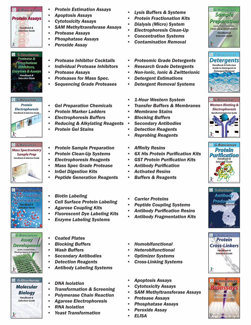
• Protein Estimation Assays• Apoptosis Assays• Cytotoxicity Assays• SAM Methyltransferase Assays• Protease Assays• Phosphatase Assays• Peroxide Assay
• Lysis Buffers & Systems• Protein Fractionation Kits• Dialysis (Micro) System• Electrophoresis Clean-Up• Concentration Systems• Contamination Removal
• Protease Inhibitor Cocktails• Individual Protease Inhibitors• Protease Assays• Proteases for Mass Spec.• Sequencing Grade Proteases
• Proteomic Grade Detergents• Research Grade Detergents• Non-Ionic, Ionic & Zwitterionic• Detergent Estimations• Detergent Removal Systems
• Gel Preparation Chemicals• Protein Marker Ladders• Electrophoresis Buffers• Reducing & Alkylating Reagents• Protein Gel Stains
• 1-Hour Western System• Transfer Buffers & Membranes• Membrane Stains• Blocking Buffers• Secondary Antibodies• Detection Reagents• Reprobing Reagents
• Protein Sample Preparation• Protein Clean-Up Systems• Electrophoresis Reagents• Mass Spec Grade Protease• InGel Digestion Kits• Peptide Generation Reagents
• Affinity Resins• 6X His Protein Purification Kits• GST Protein Purification Kits• Antibody Purification• Activated Resins• Buffers & Reagents
• Biotin Labeling• Cell Surface Protein Labeling• Agarose Coupling Kits• Fluorescent Dye Labeling Kits• Enzyme Labeling Systems
• Carrier Proteins• Peptide Coupling Systems• Antibody Purification Resins• Antibody Fragmentation Kits
• Coated Plates• Blocking Buffers• Wash Buffers• Secondary Antibodies• Detection Reagents• Antibody Labeling Systems
• Homobifunctional • Heterobifunctional• Optimizer Systems• Cross-Linking Systems
• DNA Isolation• Transformation & Screening• Polymerase Chain Reaction• Agarose Electrophoresis• RNA Isolation• Yeast Transformation
• Apoptosis Assays• Cytotoxicity Assays• SAM Methyltransferase Assays• Protease Assays• Phosphatase Assays• Peroxide Assay• ELISA
1For further details, visit GBiosciences.com
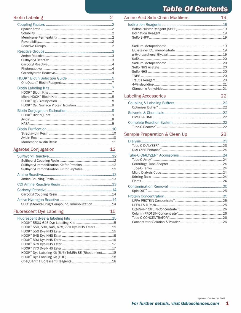
Table Of ContentsBiotin Labeling 2
Coupling Factors ...................................................................2Spacer Arms ................................................................................2Solubility ......................................................................................2Membrane Permeability .............................................................2Reversibility..................................................................................2Reactive Groups ..........................................................................2
Reactive Groups ....................................................................3Amine Reactive ............................................................................3Sulfhydryl Reactive ......................................................................3Carboxyl Reactive ........................................................................4Photoreactive ..............................................................................4Carbohydrate Reactive ................................................................4
HOOK™ Biotin Selection Guide .............................................5OneQuant™ Biotin Reagents .......................................................7
Biotin Labeling Kits ...............................................................7HOOK™ Biotin Kits .......................................................................7Micro HOOK™ Biotin Kits .............................................................8HOOK™ IgG Biotinylation .............................................................8HOOK™ Cell Surface Protein Isolation ........................................9
Biotin Conjugation Estimation ..............................................9HOOK™ BiotinQuant .....................................................................9Avidin ............................................................................................9HABA ............................................................................................9
Biotin Purification ..................................................................10Streptavidin Resin .......................................................................10Avidin Resin .................................................................................10Monomeric Avidin Resin .............................................................11
Agarose Conjugation 12Sulfhydryl Reactive ................................................................12
Sulfhydryl Coupling Resin ...........................................................12Sulfhydryl Immobilization Kit for Proteins ..................................12Sulfhydryl Immobilization Kit for Peptides .................................12
Amine Reactive ......................................................................13Amine Coupling Resin .................................................................13
CDI Amine Reactive Resin ....................................................13Carboxyl Reactive ..................................................................14
Carboxyl Coupling Resin .............................................................14Active Hydrogen Reactive .....................................................14
SDC™ (Steroid/Drug/Compound) Immobilization ......................14
Fluorescent Dye Labeling 15Fluorescent dyes & labeling kits ..........................................15
HOOK™ 550& 645 Dye Labeling Kits ........................................15HOOK™ 550, 590, 645, 678, 770 Dye-NHS Esters ..................15HOOK™ 550 Dye-NHS Ester ........................................................15HOOK™ 645 Dye-NHS Ester ........................................................16HOOK™ 590 Dye-NHS Ester ........................................................16HOOK™ 678 Dye-NHS Ester ........................................................17HOOK™ 770 Dye-NHS Ester ........................................................17HOOK™ Dye Labeling Kit (5/6) TAMRA-SE (Rhodamine) ...........18HOOK™ Dye Labeling Kit (FITC) ...................................................18OneQuant™ Fluorescent Reagents .............................................18
Amino Acid Side Chain Modifiers 19Iodination Reagents ..............................................................19
Bolton-Hunter Reagent (SHPP) ...................................................19Iodination Reagent ......................................................................19Sulfo SHPP ...................................................................................19
Sodium Metaperiodate ...............................................................19L-Cysteine·HCL, monohydrate .....................................................19p-Hydroxyphenyl Glyoxal ..............................................................19SATA ..............................................................................................20Sodium Metaperiodate ...............................................................20Sulfo NHS Acetate .......................................................................20Sulfo NHS ....................................................................................20TNBS ............................................................................................20Traut’s Reagent ...........................................................................204-Vinylpyridine .............................................................................21Citroconic Anhydride ...................................................................21
Labeling Accessories 22Coupling & Labeling Buffers .................................................22
Optimizer Buffer™ ........................................................................22Solvents & Chemicals ...........................................................22
DMSO & DMF...............................................................................22Complete Reaction System ..................................................22
Tube-O-Reactor™ ..........................................................................22
Sample Preparation & Clean Up 23Dialysis ...................................................................................23
Tube-O-DIALYZER™ .......................................................................23DIALYZER-Enhance™ ....................................................................24
Tube-O-DIALYZER™ Accessories ............................................24Tube-O-Array™ ...............................................................................24Centrifuge Tube-Adapter .............................................................24Tube-O-Tanks ..............................................................................24Micro Dialysis Cups .....................................................................24Stirring Balls ................................................................................24Floats ...........................................................................................24
Contamination Removal .......................................................25Spin-OUT™ ....................................................................................25
Protein Concentration ...........................................................25UPPA-PROTEIN-Concentrate™......................................................25UPPA-I & II Pack ...........................................................................25OrgoSol-PROTEIN-Concentrate™ .................................................26Column-PROTEIN-Concentrate™ ..................................................26Tube-O-CONCENTRATOR™ ............................................................26Concentrator Solution & Powder ................................................26
Updated: October 10, 2017

For further details, visit GBiosciences.com2
Biotin, a 244 Dalton vitamin (Vitamin H) molecule, exhibits an extraordinary binding affinity for avidin (Ka=1015M-1) and streptavidin. Biotin and avidin interaction is rapid and once the bond is established it can survive up to 3M guanidine-hydrochloride and extremes of pH. Biotin-avidin bonds can only be reversed by denaturing the avidin protein molecule with 8M guanidine-hydrochloride at pH1.5 or by autoclaving. The biotinylated molecules are efficiently probed with avidin or streptavidin conjugated to reporter molecules, such as peroxidases or phosphatases. The use of biotin for non-radioactive labeling of proteins and nucleic acids has now become an increasingly popular technique in life science research. Avidin is a glycoprotein with approximately 10% of its total mass coming from carbohydrates. Avidin has a molecular weight of 67kDa and contains four identical 128 amino acid subunits that each have a single biotin binding domain. Avidin is a basic protein with an isoelectric pH of 10-10.5 and is readily soluble in aqueous buffers containing a wide range of salt, pH, temperature and other laboratory agents. This wide range of tolerance makes avidin suitable for a wide variety of analytical applications. Streptavidin is a tetrameric protein and in many respects is similar to avidin except that it has no carbohydrate and has a slightly lower molecular weight of about 60kDa. The solubility of streptavidin (isoelectric pH5) in aqueous buffer is much lower than avidin, but the binding of streptavidin to biotin is similar to that of avidin.
S
NH
HN
O
O
HO
Figure 1: Structure of Biotin.
COUPLING FACTORS————————————————————————————
Several factors must be considered when coupling a biotin reagent to a protein to ensure a successful reaction. The primary consideration is the selection of the biotinylation reagent itself. A wide range of biotin reagents are offered that have variations in their reactive groups, spacer arm lengths, solubility, membrane permeability and reversibility. All these factors must be considered and are dependent on your protein/peptide.
Spacer Arms————————————————————————————The biotin-binding domain in avidin/ streptavidin molecules are
buried 9Å below the surface and hence, the presence of bulky groups in the vicinity of the biotin-binding site may create steric hindrances and reduce the binding efficiency and the sensitivity of detection methods. Greater binding capacity can be realized by using biotin derivatives that have large spacer arms. Extended spacer arms afford the ability to overcome steric hindrances and bind deep within the binding sites of the avidin/ streptavidin molecules.
Solubility————————————————————————————Solubility of the HOOK™-Biotin Reagents varies greatly, with some
being only soluble in organic solvents, i.e. DMSO and DMF.
Membrane Permeability————————————————————————————This has become of great interest in studies of cell surface
proteins and therefore membrane trafficking and cell signaling. The HOOK™ Biotin Reagents that are not membrane permeable are excellent candidates for labeling membrane surface proteins.
Reversibility————————————————————————————Biotin tags are often used for protein purification, however with
the biotin:avidin binding affinity being one of the strongest known it is often difficult to release the protein from the avidin. In fact, 8M guanidine at pH1.5 is often used, which has severe detrimental effects on the protein of interest. Several HOOK™ Biotin Reagents have disulfide bonds that can be reduced to release the protein of interest under mild conditions and other HOOK™ Biotin Reagents can be removed from the protein with changes in pH.
Reactive Groups————————————————————————————The reagents offered have numerous reactive groups that
can couple to amines, sulfhydryls, carboxyls and carbohydrates. Conjugation of biotin reagents to proteins and other molecules generally does not have adverse effects on the biological properties of the target molecules, unless biotin reagents are conjugating to or modifying active residues or sites of the protein. Due to this, it is important to find an appropriate biotin reagent and optimal biotin conjugation efficiency for maintaining the functional properties of the target molecules.
The conjugation efficiency of the reactions is dependent on the reaction groups and the buffers used for the reactions as many coupling reactions are sensitive to pH and chemical composition. The following section highlights the key features of the coupling reactions and important buffer information.
Based on the target reactive groups, biotin reagents can be divided into amine reactive, sulfhydryl reactive, carbohydrate reactive, and carboxyl reactive.
Photoreactive biotin reagents react non-specifically upon exposure to UV light and are used when no appropriate reactive target is available on the molecules.
Biotin Labeling

3For further details, visit GBiosciences.com
REACTIVE GROUPS————————————————————————————Amine Reactive————————————————————————————
Amines, lysine ε-amines and N-terminal α-amines, are the most abundant group in protein molecules and represent the most common target for biotinylation. For example, BSA contains 59 primary amines, of which up to 35 are available on the surface of the molecules and can be reacted with amine reactive esters.
The most widely used amine reactive biotinylation reagents are the water insoluble N-hydroxysuccinimide (NHS) esters or the water soluble N-hydroxysulfosuccinimide (sulfo-NHS) esters. The addition of a charged sulfonate (SO3-) on the N-hydroxysuccinimide ring of the sulfo-NHS esters results in their solubility in water (~10mM), but are not permeable to plasma membranes. The solubility and impermeability to plasma membranes makes them ideal for studying cell surface proteins as they will only react with the protein molecules on the outer surface of plasma membranes.
The reaction of the NHS and sulfo-NHS esters with amines are virtually identical leading to the formation of an amide bond and release of NHS or sulfo-NHS.
Both HOOK™-NHS-Biotin and HOOK™-sulfo-NHS-Biotin are available with various spacer arms (See Selection Guide). Also available is a cleavable form of HOOK™-sulfo-NHS-Biotin, HOOK™-sulfo-NHS-SS-Biotin, which has a disulfide bond in the spacer arm. The disulfide bond permits the cleavage of the biotin moiety from the protein, making its interaction with avidin/ streptavidin reversible. Disulfide bonds are cleaved under reducing conditions with 100mM mercaptoethanol, 30-50mM DTT, or 1% sodium borohydride.
HOOK™-PFP-Biotin is another reagent that reacts with amines and forms stable amide bonds. HOOK™-PFP-Biotin is more reactive than other NHS esters and can react with both primary and secondary amines at pH 7-9.REACTION CONDITIONS
NHS esters are soluble in organic solvents and DMSO or DMF are the most commonly used, which are compatible with most proteins in a 20% solution. Sulfo-NHS ester is soluble in water, up to ~10mM and should only be dissolved immediately prior to use.
Reactive pH is neutral pH and above. Competing hydrolysis of the NHS esters and Sulfo-NHS esters in aqueous solution is a major concern as the rate of hydrolysis increases with increasing pH. Half-life of 2-4 hours at pH7.0 increasing to a few minutes at pH 9.0. For optimal amine coupling conditions, use Optimizer Buffer™-I.
Reaction incubation time is a few minutes to a few hours at 4-35°C.GENERAL PRECAUTIONS
Avoid buffers containing amines such as Tris or glycine.
Sulfhydryl Reactive————————————————————————————Sulfhydryl reactive reagents are more specific and react only with
free sulfhydryl residues (-SH or thiol groups). The side chain of the amino acid cysteine is the most common source of free sulfhydryl groups. If free sulfhydryl residues are not available, they can be generated by the reduction of disulfides (-S-S-) with reducing agents such as mercaptoethylamine, or by modifying lysine ε-amines with Traut’s reagent or SATA. After reduction, excess reducing agent must be removed before coupling. In addition a metal chelating agent (EDTA) (an anti-oxidant) should be used to reduce the chances of reoxidation of sulfhydryls to disulfides.
There are three different reactions employed to couple biotin reagents to sulfhydryl residues and involve either iodoacetyl, maleimide or pyridylthiol groups.IODOACETYL REACTION CONDITIONS
HOOK™-PEG2-Iodoacetyl-biotin and HOOK™-Iodoacetyl-LC-biotin are both sulfhydryl reactive biotinylation reagents that react with thiol groups at pH7.5-8.5 and form stable thioether bonds. HOOK™-PEG2-Iodoacetyl-biotin is water soluble, due to its polyethylene glycol (PEG) spacer arm, while HOOK™-Iodoacetyl-LC-biotin must be dissolved in an organic solvent prior to use. Both may react with imidazoles at pH 6.9-7.0. For specific reaction with sulfhydryls, limit the reaction to pH 7.5-8.5 and the molar ratio of iodoacetyl-biotin to protein such that the concentration of biotin is only slightly higher than the sulfhydryl concentration. Iodoacetyl reaction should be performed in dark to limit the formation of free iodine, which has the potential to react with tyrosine, tryptophan, and histidine residues. For optimal iodoacetyl conjugation, we recommend Optimizer Buffer™-II.MALEIMIDE REACTION CONDITIONS
HOOK™-Biotin-BMMCC is a sulfhydryl reactive reagent that contains a maleimide functional group. The maleimide group is more specific for sulfhydryl residues than iodoacetyl groups, at pH7 maleimide groups are 1000 fold more reactive toward free sulfhydryls than amines. At pH > 8.5, maleimide groups favors primary amines. Conjugation is carried out at pH 6.5-7.5 for minimizing the reaction toward primary amine. At higher pH > 8.0, hydrolysis of maleimide to maleamic acid also increases, which can compete with thiol modification. Optimizer Buffer™-III provides ideal conditions for maleimide coupling reactions.PYRIDYLDITHIOL REACTION CONDITIONS
HOOK™-Biotin-PDA is a cleavable sulfhydryl reactive reagent. The reactive group is a pyridyldithiol that reacts with free sulfhydryl by disulfide exchange over a wide range of pH, forming a disulfide linkage. The optimal reaction pH is 6-9. Pyridine-2-thione is released, which absorbs light at 343nm. The coupling reaction can be monitored by measuring the absorbance of released pyridine-2-thione at 343nm. The disulfide bonds formed between HOOK™-Biotin-PDA and the protein can be cleaved with a reducing agent, generating the starting protein in its original form. This reagent is suitable for reversible applications. Optimizer Buffer™-III provides the optimized conditions.GENERAL PRECAUTIONS
Remove reducing agents from the conjugation reaction. Add metal chelating agent EDTA as an anti-oxidant.
Biotin Labeling

For further details, visit GBiosciences.com4
Carboxyl Reactive————————————————————————————HOOK™-Biotin-PEG2-Amine and its long chain form, HOOK™-Biotin-
PEG3-Amine, are carboxyl reactive biotinylation reagents. These agents contain terminal amines and react with carboxyl groups found at the carboxyl termini, aspartate, and glutamate side chains. The reaction is mediated by a water-soluble carbodiimide. The carbodiimide (EDC) activates the carboxyl group and reacts with the amines (-NH2) on the biotinylation agent to form an amide bond. This reaction is rapid and takes just a few minutes to complete. Under these conditions, hydrazide-derivatives of biotin reagents may also react with the carboxyls.REACTION CONDITIONS
The reaction is mediated by EDC, a water-soluble carbodiimide cross-linking agent. EDC activates carboxyl groups to bind with the -NH2 group from the biotin derivatives. Optimizer Buffer™-IV provides the ideal buffer for EDC and other carbodiimides.GENERAL PRECAUTIONS
EDC may crosslink protein, decreasing EDC and/or increasing biotin reagent levels minimizing conjugation.
Avoid buffers containing amines, such as Tris or glycine, or carboxyls, such as acetate, citrate, etc. These buffers react with aldehydes, quenching the reaction.
Phosphate buffers also reduce the conjugation efficiency.
Photoreactive ————————————————————————————Photoreactive agents on exposure to ultraviolet light become
active and bind non-specifically with neighboring molecules. Photoreactive reagents are suitable for labeling molecules that do not contain easily reactable functional groups. There are a variety of photoreactive biotinylation reagents for the labeling of proteins, peptides, nucleic acids, and other molecules. HOOK™-Psoralen-PEO-Biotin, a photoreactive reagent, reacts and labels nucleic acids and protein molecules. When reacted with nucleic acids, it cross-links with pyrimidine bases. Cross-linking does not interfere with hybridization applications. REACTION CONDITIONS
Photoreactive reagents contain any aryl azide group. Aryl azide groups are chemically inert until exposed to ultraviolet light. Highly reactive and short-lived aryl nitrenes are formed, which rapidly and non-specifically react with electron-rich sites by inserting into double bonds or active hydrogen bonds (insertion into C-H and N-H sites). Uncreated aryl nitrenes undergo ring expansion and become reactive toward primary amines and sulfhydryls. A wide variety of reaction buffer conditions are acceptable for photoreactive reaction, however Optimizer Buffer-I provides excellent buffer conditions.GENERAL PRECAUTIONS
Avoid acidic and reducing agents since they inactivate aryl azide groups.
Carbohydrate Reactive————————————————————————————Some biotin reagents do not bind directly to the protein itself
but conjugate to the carbohydrate residues of glycoproteins. Carbohydrate reactive biotin reagents contain hydrazides (-NH-NH2) as a reactive group. The hydrazide reactions require carbonyl groups, such as aldehydes and ketones, which are formed by oxidative treatment of the carbohydrates. Hydrazides react spontaneously with carbonyl groups, forming a stable hydrazone bond. These reagents are particularly suitable for labeling and studying glycosylated proteins, such as antibodies and receptors. HOOK™-Biotin-hydrazide and its long spacer arm equivalent, HOOK™-Biotin-LC-hydrazide, are carbohydrate reactive reagents.REACTION CONDITIONS
For reaction with glycoproteins, the first step is to generate carbonyl groups that react with hydrazide, under mild oxidizing conditions with sodium periodate (NaIO4). At 1mM periodate and at 0°C, sialic acid residues on the glycoproteins can be specifically oxidized converting hydroxyls to aldehydes and ketones. At higher concentrations of 6-10mM periodate, other carbohydrates in protein molecules will be oxidized. Such oxidation reactions are performed in the dark to minimize unwanted side reactions.
Aldehyde can also be generated by enzymatic reactions. For example, neuraminidase treatment will generate galactose groups from sialic acid residues on glycoproteins and galactose oxidase converts primary hydroxyl groups on galactose and N-acetylgalactosamine to their corresponding aldehydes. For coupling to carbohydrates, Optimizer Buffer™-V is recommended.GENERAL PRECAUTIONS
Each glycoprotein has an optimal pH for oxidation and optimal pH for the hydrazide reaction. Periodate oxidation is dependent on temperature, pH, as well as concentration. The extent of glycosylation varies for each protein; therefore, optimal condition for each protein must be determined.
Avoid buffers containing amines, such as Tris or glycine; these buffers react with aldehydes, quenching their reaction with hydrazides.
Biotin Labeling

5For further details, visit GBiosciences.com
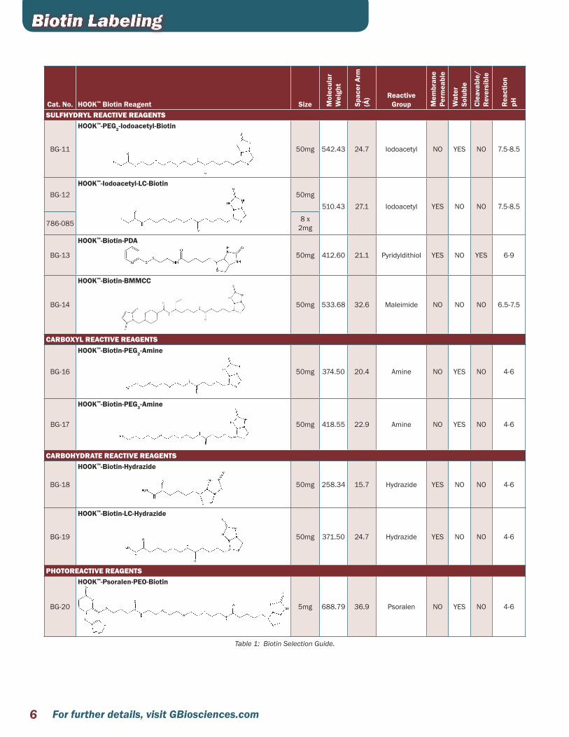
Cat. No. HOOK™ Biotin Reagent Size Mol
ecul
ar
Wei
ght
Spac
er A
rm
(Å) Reactive
Group Mem
bran
e Pe
rmea
ble
Wat
er
Solu
ble
Clea
vabl
e/
Rev
ersi
ble
Rea
ctio
n pH
BG-00
d-Biotin (vitamin H)
S
NH
HN
O
O
HO
500mg 244.32 0
AMINE REACTIVE REAGENTS
BG-01HOOK™-NHS-Biotin
50mg341.38 13.5 NHS-ester YES NO NO 7-9
786-083 8 x 2mg
BG-02
HOOK™-NHS-LC-Biotin
50mg 454.54 22.4 NHS-ester YES NO NO 7-9
BG-03
HOOK™-NHS-LC-LC-Biotin
50mg 567.70 30.5 NHS-ester YES NO NO 7-9
BG-04
HOOK™-NHS-SS-Biotin
50mg 504.65 24.3 NHS-ester YES NO YES 7-9
BG-05HOOK™ NHS-dPEG4 -Biotin
S
NHHN
O
HN
O
O OOOO
N
O
O
O
50mg
588.67 29 NHS-ester NO YES NO 7-9
786-700 8 x 1mg
BG-06 HOOK™-sulfo-NHS-Biotin 50mg443.43 13.5 sulfo-NHS
ester NO YES NO 7-9786-698 8 x
1mg
BG-07HOOK™-sulfo-NHS-LC-Biotin
50mg
556.59 22.4 sulfo-NHS ester NO YES NO 7-9
786-084 8 x 1mg
BG-08
HOOK™-sulfo-NHS-LC-LC-Biotin
50mg 669.75 30.5 sulfo-NHS ester NO YES NO 7-9
BG-09HOOK™-sulfo-NHS-SS-Biotin
50mg606.69 24.3 sulfo-NHS
ester NO YES YES 7-9786-699 8 x
1mg
BG-10
HOOK™-PFP-Biotin
50mg 410.36 9.6 Pentafluoro-phenyl ester YES NO NO 7-9
Biotin LabelingHOOK™ BIOTIN SELECTION GUIDE————————————————————————————• Reactive Group: Determines the location of the biotin moiety• Membrane Permeability: For cell surface labeling select non membrane permeable reagents• Cleavable: For easy removal from immobilized avidin or streptavidin during purification• Reversible: An alternative to cleavable reagents are reversible reagents• Steric Hinderance: Bulky groups around the binding site may require reagents with longer spacer arms
For further details, visit GBiosciences.com6
Cat. No. HOOK™ Biotin Reagent Size Mol
ecul
ar
Wei
ght
Spac
er A
rm
(Å) Reactive
Group Mem
bran
e Pe
rmea
ble
Wat
er
Solu
ble
Clea
vabl
e/
Rev
ersi
ble
Rea
ctio
n pH
SULFHYDRYL REACTIVE REAGENTS
BG-11
HOOK™-PEG2-Iodoacetyl-Biotin
50mg 542.43 24.7 Iodoacetyl NO YES NO 7.5-8.5
BG-12HOOK™-Iodoacetyl-LC-Biotin
50mg510.43 27.1 Iodoacetyl YES NO NO 7.5-8.5
786-085 8 x 2mg
BG-13
HOOK™-Biotin-PDA
50mg 412.60 21.1 Pyridyldithiol YES NO YES 6-9
BG-14
HOOK™-Biotin-BMMCC
50mg 533.68 32.6 Maleimide NO NO NO 6.5-7.5
CARBOXYL REACTIVE REAGENTS
BG-16
HOOK™-Biotin-PEG2-Amine
50mg 374.50 20.4 Amine NO YES NO 4-6
BG-17
HOOK™-Biotin-PEG3-Amine
50mg 418.55 22.9 Amine NO YES NO 4-6
CARBOHYDRATE REACTIVE REAGENTS
BG-18
HOOK™-Biotin-Hydrazide
50mg 258.34 15.7 Hydrazide YES NO NO 4-6
BG-19
HOOK™-Biotin-LC-Hydrazide
50mg 371.50 24.7 Hydrazide YES NO NO 4-6
PHOTOREACTIVE REAGENTS
BG-20
HOOK™-Psoralen-PEO-Biotin
5mg 688.79 36.9 Psoralen NO YES NO 4-6
Table 1: Biotin Selection Guide.
Biotin Labeling
7For further details, visit GBiosciences.com
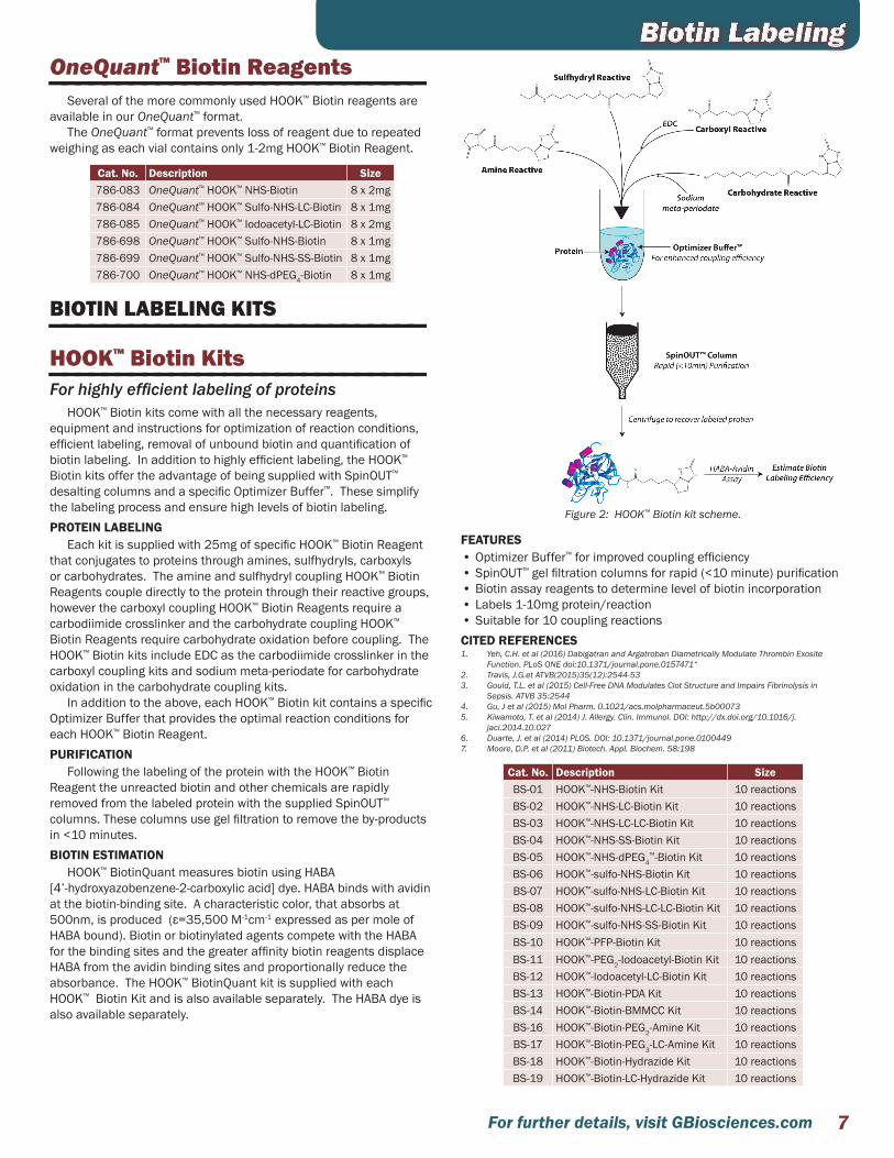
OneQuant™ Biotin Reagents————————————————————————————Several of the more commonly used HOOK™ Biotin reagents are
available in our OneQuant™ format.The OneQuant™ format prevents loss of reagent due to repeated
weighing as each vial contains only 1-2mg HOOK™ Biotin Reagent.
Cat. No. Description Size786-083 OneQuant™ HOOK™ NHS-Biotin 8 x 2mg786-084 OneQuant™ HOOK™ Sulfo-NHS-LC-Biotin 8 x 1mg786-085 OneQuant™ HOOK™ Iodoacetyl-LC-Biotin 8 x 2mg786-698 OneQuant™ HOOK™ Sulfo-NHS-Biotin 8 x 1mg786-699 OneQuant™ HOOK™ Sulfo-NHS-SS-Biotin 8 x 1mg786-700 OneQuant™ HOOK™ NHS-dPEG4-Biotin 8 x 1mg
BIOTIN LABELING KITS————————————————————————————HOOK™ Biotin Kits————————————————————————————For highly efficient labeling of proteins
HOOK™ Biotin kits come with all the necessary reagents, equipment and instructions for optimization of reaction conditions, efficient labeling, removal of unbound biotin and quantification of biotin labeling. In addition to highly efficient labeling, the HOOK™ Biotin kits offer the advantage of being supplied with SpinOUT™ desalting columns and a specific Optimizer Buffer™. These simplify the labeling process and ensure high levels of biotin labeling.PROTEIN LABELING
Each kit is supplied with 25mg of specific HOOK™ Biotin Reagent that conjugates to proteins through amines, sulfhydryls, carboxyls or carbohydrates. The amine and sulfhydryl coupling HOOK™ Biotin Reagents couple directly to the protein through their reactive groups, however the carboxyl coupling HOOK™ Biotin Reagents require a carbodiimide crosslinker and the carbohydrate coupling HOOK™ Biotin Reagents require carbohydrate oxidation before coupling. The HOOK™ Biotin kits include EDC as the carbodiimide crosslinker in the carboxyl coupling kits and sodium meta-periodate for carbohydrate oxidation in the carbohydrate coupling kits.
In addition to the above, each HOOK™ Biotin kit contains a specific Optimizer Buffer that provides the optimal reaction conditions for each HOOK™ Biotin Reagent.PURIFICATION
Following the labeling of the protein with the HOOK™ Biotin Reagent the unreacted biotin and other chemicals are rapidly removed from the labeled protein with the supplied SpinOUT™ columns. These columns use gel filtration to remove the by-products in <10 minutes.BIOTIN ESTIMATION
HOOK™ BiotinQuant measures biotin using HABA [4’-hydroxyazobenzene-2-carboxylic acid] dye. HABA binds with avidin at the biotin-binding site. A characteristic color, that absorbs at 500nm, is produced (ε=35,500 M-1cm-1 expressed as per mole of HABA bound). Biotin or biotinylated agents compete with the HABA for the binding sites and the greater affinity biotin reagents displace HABA from the avidin binding sites and proportionally reduce the absorbance. The HOOK™ BiotinQuant kit is supplied with each HOOK™ Biotin Kit and is also available separately. The HABA dye is also available separately.
Figure 2: HOOK™ Biotin kit scheme.
FEATURES• Optimizer Buffer™ for improved coupling efficiency• SpinOUT™ gel filtration columns for rapid (<10 minute) purification• Biotin assay reagents to determine level of biotin incorporation• Labels 1-10mg protein/reaction• Suitable for 10 coupling reactionsCITED REFERENCES1. Yeh, C.H. et al (2016) Dabigatran and Argatroban Diametrically Modulate Thrombin Exosite
Function. PLoS ONE doi:10.1371/journal.pone.0157471“2. Travis, J.G.et ATVB(2015)35(12):2544-533. Gould, T.L. et al (2015) Cell-Free DNA Modulates Clot Structure and Impairs Fibrinolysis in
Sepsis. ATVB 35:25444. Gu, J et al (2015) Mol Pharm. 0.1021/acs.molpharmaceut.5b000735. Kiwamoto, T. et al (2014) J. Allergy. Clin. Immunol. DOI: http://dx.doi.org/10.1016/j.
jaci.2014.10.0276. Duarte, J. et al (2014) PLOS. DOI: 10.1371/journal.pone.01004497. Moore, D.P. et al (2011) Biotech. Appl. Biochem. 58:198
Cat. No. Description SizeBS-01 HOOK™-NHS-Biotin Kit 10 reactionsBS-02 HOOK™-NHS-LC-Biotin Kit 10 reactionsBS-03 HOOK™-NHS-LC-LC-Biotin Kit 10 reactionsBS-04 HOOK™-NHS-SS-Biotin Kit 10 reactionsBS-05 HOOK™-NHS-dPEG4
™-Biotin Kit 10 reactionsBS-06 HOOK™-sulfo-NHS-Biotin Kit 10 reactionsBS-07 HOOK™-sulfo-NHS-LC-Biotin Kit 10 reactionsBS-08 HOOK™-sulfo-NHS-LC-LC-Biotin Kit 10 reactionsBS-09 HOOK™-sulfo-NHS-SS-Biotin Kit 10 reactionsBS-10 HOOK™-PFP-Biotin Kit 10 reactionsBS-11 HOOK™-PEG2-Iodoacetyl-Biotin Kit 10 reactionsBS-12 HOOK™-Iodoacetyl-LC-Biotin Kit 10 reactionsBS-13 HOOK™-Biotin-PDA Kit 10 reactionsBS-14 HOOK™-Biotin-BMMCC Kit 10 reactionsBS-16 HOOK™-Biotin-PEG2-Amine Kit 10 reactionsBS-17 HOOK™-Biotin-PEG3-LC-Amine Kit 10 reactionsBS-18 HOOK™-Biotin-Hydrazide Kit 10 reactionsBS-19 HOOK™-Biotin-LC-Hydrazide Kit 10 reactions
Biotin Labeling

For further details, visit GBiosciences.com8
Micro HOOK™ Biotin Kits————————————————————————————For highly efficient labeling of proteins
The micro HOOK™ Biotin kits are designed to label small amounts of proteins, with each kit designed for 8-10 labelings of 50-250µg protein/reaction. Each kit is supplied with all the necessary reagents for optimization of reaction conditions, efficient labeling and removal of unbound biotin. In addition to highly efficient labeling, the HOOK™ Biotin kits offer the advantage of being supplied with SpinOUT™ desalting columns and a specific Optimizer Buffer™. These simplify the labeling process and ensure high levels of biotin labeling.PROTEIN LABELING
Each kit is supplied with 8 x 1mg single use aliquots of biotin reagent to minimize waste and degradation of the NHS ester coupling reaction group. The following HOOK™ Biotin reagents are available in the micro format:• HOOK™ Sulfo-NHS-Biotin
Amine reactive reagent, shortest spacer arm• HOOK™ Sulfo-NHS-LC-Biotin
Amine reactive reagent, longer spacer arm• HOOK™ Sulfo-NHS-SS-Biotin
Cleavable, amine reactive reagent• HOOK™ NHS-dPEG4-Biotin
Amine reactive, pegylated reagent; enhances water solubilityIn addition, each HOOK™ Biotin kit contains a specific Optimizer
Buffer™ that provides the optimal reaction conditions.PURIFICATION
Following the labeling of the protein with the HOOK™ Biotin Reagent the unreacted biotin and other chemicals are rapidly removed from the labeled protein with the supplied SpinOUT™ Columns. These columns use gel filtration to remove the by-products in <10 minutes.FEATURES• Micro kit for labeling protein primary amines• Optimizer Buffer™ for improved coupling efficiency• Gel filtration columns for rapid (<10 minute) purification• Labels 50-250µg protein/reaction• Suitable for 8-10 couplings
Cat. No. Description Size786-694 HOOK™-sulfo-NHS-Biotin Kit (micro) 8-10 reactions786-695 HOOK™-sulfo-NHS-LC-Biotin Kit (micro) 8-10 reactions786-696 HOOK™ Sulfo-NHS-SS-Biotin Kit (micro) 8-10 reactions786-697 HOOK™-NHS-dPEG4-Biotin Kit (micro) 8-10 reactions
HOOK™ IgG Biotinylation————————————————————————————Rapid antibody labeling with biotin
Designed for the efficient biotinylation of IgG molecules by first immobilizing the IgG molecules on a solid support
The HOOK™ IgG Biotinylation kits offer an advantage over standard biotinylation reactions as the immobilization of the IgG to the Nickel Chelating resin allows for the rapid removal of uncoupled biotin and therefore eliminates the need for further dialysis or desalting of the biotinylated antibody.
Two kits are available for labeling antibodies through free amines or sulfhydryls. The amine kit uses NHS-dPEG4-Biotin to label free primary amines. The sulfhydryl kit uses the supplied Protein-S-S-Reductant™ to reduce the disulfide bonds of the immobilized IgG molecule. The reduced immobilized IgG molecule is then incubated with PEG2-Iodoacetyl-Biotin solution to biotinylate the free sulfhydryl groups.
The advantage of a PEG (polyethylene glycol) biotinylation reagent is that the long hydrophilic spacer arm conveys its water solubility to the antibodies and have a reduced occurrence of aggregation compared to non-PEG biotinylation reactions.
Figure 3: HOOK™ IgG Biotinylation (Amine) Scheme. The IgG antibody is first immobilized through its histidine rich domain on a nickle column. Immobilized antibody is labeled with the NHS-dPEG4-Biotin reagent that reacts with primary amines. Free biotin is washed away and the biotinylated antibody is eluted with the supplied His Elution Buffer.
FEATURES• Simpler antibody biotinylation• Solid support technology eliminates dialysis/desalting• Suitable for 1-10mg antibody• PEG Biotin reagent for reduced steric hindrance and increased
labeled antibody solubilityAPPLICATIONS• For the efficient and simple labeling of antibodies with biotinCITED REFERENCES1. Patlaka, C. et al (2014) Biochem Biophys Res Commun. DOI: 10.1016/j.bbrc.2014.10.112
Cat. No. Description Size786-728 HOOK™ IgG Biotinylation (Amine) 8 reactions786-729 HOOK™ IgG Biotinylation (Sulfhydryl) 8 reactions
Biotin Labeling

9For further details, visit GBiosciences.com
HOOK™ Cell Surface Protein Isolation————————————————————————————Complete cell surface protein labeling & isolation
Uses our proven biotin labeling and purification technology in conjunction with our Mammalian Cell PE LB™ lysis buffer to conveniently label cell surface proteins and isolate them for further analysis.
Figure 4: HOOK™ Cell Surface Protein Isolation scheme.
Mammalian cells, adherent or non-adherent, are labeled with Sulfo-NHS-SS-Biotin, an amine reactive biotinylation reagent that is soluble in water, but impermeable to plasma membranes. Sulfo-NHS-SS-Biotin has a disulfide bond in the spacer arm that permits the cleavage of the biotin moiety from the protein, making its interaction with streptavidin purification column reversible.
Cells are lysed with Mammalian Cell PE LB™ and applied to a Streptavidin agarose column. Unlabeled intracellular proteins are washed away and the biotin labeled cell surface proteins are then released by reduction of the disulfide bond with DTT.
The kit is supplied with all the necessary reagents and buffers for convenience and improved reproducibility. The kit is compatible with a wide variety of mammalian cells and can be used to compare treated and untreated cells and differences between different cell lines. This kit is supplied with sufficient reagents for five experiments, with each experiment consisting of four 90-95% confluent T-75cm2 flasks.FEATURES• Complete cell surface labeling & isolation kit• Convenient; all required reagents are included• Versatile; suitable for wide selection of mammalian cellsAPPLICATIONS• For the isolation of cell surface proteins• Study receptor:ligand interaction• Study membrane traffickingCITED REFERENCES1. Hartz, et al (2011) J. Euk. Microbio. 58:1712. Bizet, A.A. et al (2011) BBA-Mol. Cell. Res. 1813:742
Cat. No. Description Size786-316 HOOK™ Cell Surface Protein Isolation 5 Expts
BIOTIN CONJUGATION ESTIMATION————————————————————————————HOOK™ BiotinQuant————————————————————————————For the estimation of biotin conjugation
HOOK™ BiotinQuant measures biotin using HABA [4’-hydroxyazobenzene-2-carboxylic acid] dye. HABA binds with avidin at the biotin-binding site. A characteristic color, that absorbs at 500nm, is produced (ε=35,500 M-1 cm-1 expressed as per mole of HABA bound). Biotin or biotinylated agents compete with the HABA for the binding sites and the greater affinity biotin reagents displace HABA from the avidin binding sites and proportionally reduce the absorbance.
Cat. No. Description SizeBKC-01 HOOK™ BiotinQuant Kit 20 assaysBKC-03 HABA Dye 1g
Avidin————————————————————————————Affinity purified for the estimation of biotin conjugation
Avidin is a glycoprotein with approximately 10% of its total mass comes from carbohydrates. Avidin has a molecular weight of 67kDa and contains four identical 128 amino acid subunits that each have a single biotin binding domain. Avidin is a basic protein with an isoelectric pH of 10-10.5 and is readily soluble in aqueous buffers containing a wide range of salt, pH, temperature and other laboratory agents. This wide range of tolerance makes avidin suitable for a wide variety of analytical applications.
This affinity purified avidin is ideal for estimation of biotin incorporation and other applications.
Cat. No. Description Size786-581 Avidin 5mg786-582 Avidin 25mg786-583 Avidin 100mg
HABA————————————————————————————A biotin estimation dye reagent.
Cat. No. Description SizeBKC-03 HABA 1g
Biotin Labeling

For further details, visit GBiosciences.com10
BIOTIN PURIFICATION————————————————————————————Streptavidin Resin————————————————————————————High binding affinity for biotin labeled proteins & molecules
Biotin, a 244Da vitamin (Vitamin H) molecule, exhibits an extraordinary binding affinity for avidin (Ka=1015 M-1) and streptavidin (Ka=1015 M-1). Biotin and (strept)avidin interaction is rapid and once the bond is established it can survive up to 3M guanidine-hydrochloride and extremes of pH. Biotin-avidin bonds can only be reversed by denaturing the avidin protein molecule with 8M guanidine-hydrochloride at pH1.5 or by boiling in SDS Page Sample Loading Buffer.
Streptavidin is a tetrameric protein containing 4 biotin binding sites. Streptavidin in many respects is similar to avidin except that it has no carbohydrate and has a slightly lower molecular weight of about 60kDa. The solubility of streptavidin (isoelectric pH5) in aqueous buffer is much lower than avidin, but the binding of streptavidin to biotin is similar to that of avidin. The advantage of streptavidin is that the lack of carbohydrates significantly reduces the amount of non-specific binding.
The streptavidin used for immobilization on porous 6% crosslinked agarose is a recombinant form with a mass of 53kDa and near neutral pI. The streptavidin is covalently coupled to the agarose resulting in minimal leaching and is stable over pH2-11.
The Steptavidin Resin is designed for the single step small and large scale affinity purification of proteins and antibodies with a biotin tag. The resin can also be used for immunoprecipitations using biotin labeled antibodies. Supplied as a resin slurry or in a 1ml spin column format. Specific Binding and Elution Buffers are also available.
The Streptavidin Resin is available as resin alone or supplied in a kit format containing:• 5ml resin• 100ml Streptavidin Binding/Wash Buffer
(20mM NaPO4, 0.15M NaCl, pH7.5)• 100ml Streptavidin Elution Buffer (8M Guanidine.HCl pH1.5)• 5 empty 1ml spin columns • 5 empty <5ml gravity flow columns
The buffers are also available separately.FEATURES• Recombinant streptavidin covalently coupled to ~6% cross linked
agarose. Minimal Leaching• Ligand Density >1mg/ml• Binding capacity 15-30µg biotin/ml resinAPPLICATIONS• Immunoprecipitation with biotinylated antibodies• Pull down assays with biotinylated proteins• Purification of biotinylated molecules, including: proteins,
antibodies, DNA and carbohydrates
CITED REFERENCES1. Dong D. et al (2016) Human Serum Albumin and HER2-Binding Affibody Fusion Proteins for
Targeted Delivery of Fatty Acid-Modified Molecules and Therapy. Mol Pharm DOI: 10.1021/acs.molpharmaceut.6b00265
Cat. No. Description Size786-584 Streptavidin, recombinant 5mg786-585 Streptavidin, recombinant 25mg786-586 Streptavidin, recombinant 100mg786-590 Immobilized Streptavidin Resin 2ml resin786-390 Immobilized Streptavidin Resin 5ml Resin786-591 Immobilized Streptavidin Resin 10ml resin786-592 Immobilized Streptavidin Resin 5 x 1ml786-555 Streptavidin Resin Kit 1786-548 Streptavidin Binding Buffer 100ml786-549 Streptavidin Elution Buffer 100ml
Avidin Resin————————————————————————————High binding affinity for biotin labeled proteins & molecules
Biotin, a 244Da vitamin (Vitamin H) molecule, exhibits an extraordinary binding affinity for avidin (Ka=1015 M-1). Biotin and avidin interaction is rapid and once the bond is established it can survive up to 3M guanidine-hydrochloride and extremes of pH. Biotin-avidin bonds can only be reversed by denaturing the avidin protein molecule with 8M guanidine-hydrochloride at pH1.5 or by boiling in SDS Page Sample Loading Buffer.
Avidin is a glycoprotein with approximately 10% of its total mass coming from carbohydrates. Avidin has a molecular weight of 67kDa and contains four identical 128 amino acid subunits that each has a single biotin binding domain. Avidin is a basic protein with an isoelectric pH of 10-10.5 and is readily soluble in aqueous buffers containing a wide range of salt, pH (2-11), temperature and other laboratory agents. This wide range of tolerance makes avidin suitable for a wide variety of analytical applications. Avidin has extraordinary binding affinity for biotin (Ka=1015M-1).
The avidin in covalently coupled to the agarose resulting in minimal leaching and is stable over pH2-11.
The Avidin Resin is designed for the single step small and large scale affinity purification of proteins and antibodies with a biotin tag. The resin can also be used for immunoprecipitations using biotin labeled antibodies. Supplied as a 50% resin slurry.
Specific Binding and Elution Buffers are also available.FEATURES• Avidin covalently coupled to ~6% cross linked agarose. Minimal
Leaching• Binding capacity 15-20µg biotin/ml resinAPPLICATIONS• Immunoprecipitation with biotinylated antibodies• Pull down assays with biotinylated proteins• Purification of biotinylated molecules, including:
• Proteins• Antibodies• DNA• Carbohydrates
CITED REFERENCES1. Wang, Y. et al (2014) ACS Chem. Biol. DOI: 10.1021/cb400900r
Cat. No. Description Size786-593 Immobilized Avidin Resin 5ml resin786-594 Immobilized Avidin Resin 25ml Resin786-548 Streptavidin Binding Buffer 100ml786-549 Streptavidin Elution Buffer 100ml
Biotin Labeling

11For further details, visit GBiosciences.com
Monomeric Avidin Resin————————————————————————————Purification & elution of biotin labeled molecules under mild elution conditions
G-Biosciences Immobilized Monomeric Avidin Resin is designed for the simple affinity chromatography purifications of proteins, antibodies and other molecules with a biotin tag. The resin consists of monomeric subunits of avidin covalently coupled to 6% cross-linked agarose, offering a stable, reusable resin for the purification of biotinylated molecules.
Monomeric avidin offers a distinct advantage over native avidin, a tetrameric molecule, and streptavidin as it has a much lower biotin binding affinity, Kd=10-7 as opposed to Kd=10-15 for native avidin. This lower binding affinity allows elution of molecules with mild elution buffers (2mM D-Biotin in 1X PBS), as opposed to the strong denaturing buffers (8M Guanidine•HCl, pH 1.5) used with native avidin.
The covalent attachment of monomeric avidin to the agarose ensures no detectable leaching of the avidin during biotin purification and offers a wide tolerance to chemicals. This ensures the resin can be reused at least 10 times with no loss of function.
The Immobilized Monomeric Avidin Resin is available as a 50% resin slurry or as a complete kit containing a reusable monomeric avidin column and the respective buffers for successful purification of biotinylated molecules.FEATURES• Monomeric avidin covalently coupled to ~6% cross linked agarose. • Minimal Leaching• Binding capacity »1.2mg biotinylated BSA/ml resin• Non Denaturing: Elute biotinylated molecules with free biotin• Reusable: Reuse the resin at least 10 times (2.5% loss of binding/
regeneration)• Specific: Retains avidins high specificity for biotin moleculesAPPLICATIONS• Purification of biotinylated molecules, including:
• Proteins• Antibodies• DNA• Carbohydrates
Cat. No. Description Size786-595 Immobilized Monomeric Avidin 5ml resin786-596 Immobilized Monomeric Avidin 10ml resin786-597 Immobilized Monomeric Avidin Kit
Biotin Labeling

For further details, visit GBiosciences.com12
Activated resins have immobilized groups bound to agarose beads that can be used to generate specific affinity columns for protein, antibody and other molecule purification.
Activated resins offered include:• Sulfhydryl Coupling Resin: Activated iodoacetyl groups for coupling
free sulfhydryls• HOOK™ Activated Agarose (Amine Reactive): Activated aldehyde
groups for coupling primary amines• Carboxyl Coupling Resin: Immobilized DADPA
(Diaminodipropylamine) generates a free amine to conjugate carboxyls and other moieties with the aid of crosslinkers
• HOOK™ Activated Agarose (Carboxyl/Sulfhydryl Reactive): Aminoethyl groups with a free amine to conjugate carboxyls, sulfhydryls and other moieties with the aid of crosslinkers
• SDC™ Immobilization: Uses Immobilized DADPA (Diaminodipropylamine)) for the immobilization of steroids, drugs and chemical compounds that lack primary amines, sulfhydryls, carbonyls and other common coupling groups
SULFHYDRYL REACTIVE————————————————————————————Sulfhydryl Coupling Resin————————————————————————————Activated iodoacetyl group for binding free sulfhydryls
The Sulfhydryl Coupling Resin is designed for the simple and efficient coupling of peptides and proteins to a solid 6% agarose support through free sulfhydryl groups (-SH). The iodoacetyl groups of the Sulfhydryl Coupling Resin specifically react with free sulfhydryls to form covalent, permanent thioether bonds. The long spacer arm reduces steric hindrance and ensures greater binding of proteins and antibodies during affinity purification.
The Sulfhydryl Coupling Resin is available as a resin slurry or prealiquoted as five 2ml spin column format.
Figure 5: Sulfhydryl Coupling Resin scheme.
FEATURES• Stable coupling of proteins and peptides, forms covalent thioether
bonds• Couples 1-2mg peptide and 2-20mg protein/ml resinAPPLICATIONS• For the generation of affinity columns for antibody purification and
other affinity chromatography
Cat. No. Description Size 786-794 Sulfhydryl Coupling Resin 10ml resin786-795 Sulfhydryl Coupling Resin 50ml resin786-796 Sulfhydryl Coupling Resin 250ml resin786-806 Sulfhydryl Coupling Resin 5 x 2ml columns
Sulfhydryl Immobilization Kit for Proteins————————————————————————————For generation of protein affinity columns through free sulfhydryls
The Sulfhydryl Immobilization Kit for Proteins is a complete kit designed for the simple and efficient coupling of proteins to a solid agarose support. The Sulfhydryl Coupling Resin Columns utilizes iodoacetyl groups that specifically react with free sulfhydryls to form covalent, permanent thioether bonds. The long spacer arm reduces steric hindrance and ensures greater binding of proteins and antibodies during affinity purification.
Proteins, including antibodies, must have free sulfhydryls for immobilization to the resin. A mild reducing agent, 2-Mercaptoethylamine, is supplied to reduce the hinge region disulfide bonds of antibodies, while preserving the functionally crucial disulfide bonds between the heavy and light chains.
The resulting columns can be used to study protein-protein interactions or for purification, via affinity chromatography. The columns, depending on the stability of the immobilized molecule, can be used several times without significant loss of activity.FEATURES• Generates 5 reusable, spin format affinity columns• Specific conjugation through free sulfhydryls• High Capacity: 2-40mg protein/ column• Supplied with mild reducing agent for free sulfhydryls generationAPPLICATIONS• Immobilize proteins to purify interacting molecules• Immobilize antibodies in the correct orientation
Cat. No. Description Size 786-804 Sulfhydryl Immobilization Kit for Proteins For 5 x 2ml columns
Sulfhydryl Immobilization Kit for Peptides————————————————————————————For generation of peptide affinity columns through free sulfhydryls
Sulfhydryl Immobilization Kit for Peptides is designed for the simple and efficient coupling of sulfhydryl-containing peptides to a solid agarose support. The Sulfhydryl Coupling Resin Columns utilizes iodoacetyl groups that specifically react with free sulfhydryls to form covalent, permanent thioether bonds. The long spacer arm reduces steric hindrance and ensures greater binding of proteins and antibodies during affinity purification.
Peptides must have free sulfhydryls for immobilization to the resin. The supplied Protein-S-S-Reductant™ reducing agent efficiently reduces disulfide bonds and does not interfere with the iodoacetyl coupling reaction. Protein-S-S-Reductant™ offers the advantage that it does not require removal before peptide immobilization.
The resulting columns can be used for the purification of antibodies that have been raised against the specific peptide. The columns, depending on peptide stability, can be used several times.FEATURES• Generates 5 reusable, spin format affinity columns• Specific conjugation through free sulfhydryls• High Capacity: 2-4mg peptide/columnAPPLICATIONS• Immobilize peptides for antibody purification
Cat. No. Description Size 786-805 Sulfhydryl Immobilization Kit for Peptides For 5 x 2ml columns
Agarose Conjugation

13For further details, visit GBiosciences.com
Agarose ConjugationAMINE REACTIVE————————————————————————————Amine Coupling Resin————————————————————————————
The amine reactive HOOK™ Activated Agarose is 6% agarose that has been activated to generate reactive aldehyde groups. The aldehyde groups of the agarose react spontaneously with primary amines, located at the N-terminus of proteins or in lysine residues, to form intermediate Schiff Base complexes. These, in turn, are selectively reduced by reductive amination to form stable amine linkages between the agarose and the ligand.
Figure 6: Scheme for the coupling of proteins to HOOK™ Activated Agarose (Amine Reactive).
The amine reactive HOOK™ Activated agarose is also supplied in a complete kit for the generation of 5 x 2ml resins. The kit is supplied with all the necessary reagents and columns.FEATURES• Binding capacity: 20mg protein/ml resin• 6% cross-linked agaroseAPPLICATIONS • Coupling of proteins and peptides to agarose beads• Suitable for antibody purificationCITED REFERENCES1. Lai, J.C. et al (2015)Macrophage migration inhibitory factor has a permissive role
in concanavalin A-induced cell death of human hepatoma cells through autophagy. Cell Death Dis. doi:10.1038/cddis.2015.349
2. Rudolph, V. et al (2008) J. Pharmacol. Exp. Ther. 327:324
Cat. No. Description Size 786-066 HOOK™ Activated Agarose (Amine Reactive) 10ml resin
786-063 HOOK™ Activated Agarose Coupling Kit (Amine Reactive)
For 5 x 2ml columns
CDI Amine Reactive Resin————————————————————————————
G-Biosciences CDI Amine Reactive Agarose consists of 6% cross-linked agarose activated with CDI (1,1’-carbonyl diimidazole) to form reactive imidazole carbamates.
The activation of the resin occurs in solvent and to maintain its activity the resin is supplied in acetone to prevent hydrolysis. Upon reaction of the resin with primary amine containing molecules, i.e. proteins, in basic (pH8.5-10) aqueous buffers the imidazole carbamates lose the imidazole group and form carbamate linkages.
CDI Amine Reactive Agarose is ideal for immobilizing peptides, small organic molecules and certain proteins and reactions can occur in organic solvent making it ideal for water-insoluble ligands.
Figure 7: Scheme for the coupling of proteins to CDI Amine Reactive Agarose.
The amine reactive HOOK™ Activated agarose is also supplied in a complete kit for the generation of 5 x 2ml resins. The kit is supplied with all the necessary reagents and columns.FEATURES• Proven coupling chemistry• Easy to use, no secondary coupling agents required• Stable linkages• Couple in inorganic buffers for insoluble moleculesAPPLICATIONS • Couple proteins and peptides• Couple primary amine containing ligands
Cat. No. Description Size 786-404 CDI Amine Reactive Resin 10ml resin

For further details, visit GBiosciences.com14
Agarose ConjugationCARBOXYL REACTIVE————————————————————————————Carboxyl Coupling Resin————————————————————————————
Consists of 6% cross-linked agarose with covalent linked diaminodipropylamine (DADPA) to generate a free primary amine at the end of a long spacer arm.
Figure 8: Carboxyl Coupling Resin scheme.
Molecules, including proteins and peptides, are covalently coupled to the free primary amines, and the stable columns are ideal for affinity purification of antibodies and other interacting partners. Molecules can be coupled to the free amine by numerous amine-reactive methods; however the use of the carbodiimide EDC allows coupling of free carboxyl groups. The resulting amide bond is highly stable and greatly reduces the chance of leaching of the affinity tag. The long spacer arm reduces steric hindrance and ensures greater binding of proteins and antibodies during affinity purification. FEATURES• Immobilized DADPA (diaminodipropylamine)• 6% cross-linked agarose• Long spacer arm to limit steric hindrance• Couple carboxyl groupsAPPLICATIONS • Couple peptides for antibody purification• Couple peptides and proteins to purify interacting molecules
Cat. No. Description Size
786-797 Carboxyl Coupling Resin (Immobilized DADPA (Diaminodipropylamine)) 25ml resin
786-809 Carboxyl Immobilization Kit For 5 x 2ml columns
ACTIVE HYDROGEN REACTIVE————————————————————————————SDC™ (Steroid/Drug/Compound) Immobilization————————————————————————————
Designed for the immobilization of steroids, drugs and chemical compounds that lack primary amines, sulfhydryls, carbonyls and other common coupling groups to a solid-phase agarose support for the use in affinity purification. The kit uses Immobilized DADPA (diaminodipropylamine) resin to bind steroids, drugs and chemicals through their active hydrogens.
The coupling uses the Mannich reaction, which is described as the condensation of formaldehyde with ammonia, in the form of its salt, and another compound containing an active hydrogen. The SDC™ Immobilization kit replaces the ammonia with the primary amine on the DADPA and the active hydrogen is supplied by the steroid, drug or chemical to be coupled.
Figure 9: Active hydrogen containing compounds.
Figure 10: SDC™ (Steroid/ Drug/ Compound) Immobilization scheme.
FEATURES• Uses Immobilized DADPA (diaminodipropylamine) resin• Stable, covalent linkageAPPLICATIONS • Immobilization of drugs, steroids and small metabolites through
active hydrogens• Ideal for compounds lacking primary amines, sulfhydryls,
carbonyls and other common coupling groups
Cat. No. Description Size 786-271 SDC™ (Steroid/Drug/Compound) Immobilization 5 reactions
15For further details, visit GBiosciences.com
FLUORESCENT DYES & LABELING KITS————————————————————————————HOOK™ 550& 645 Dye Labeling Kits ————————————————————————————
HOOK™ 550 & 645 Dye Labeling Kits are designed with the most advanced line of fluorescent dyes and they are used for labeling antibodies and other proteins for biochemical detection assays. HOOK™ 5550 & 645 Dye Labeling Kits comprise of the proprietary dyes and the dye activator solutions. The dye activator solution is added to the dye to make it reactive in situ and the conjugation with antibodies or protein is simply attained by mixing kit components with the target antibody or protein. The volumes and amounts of protein and dye are standardized to obtain maximum fluorescence and preserve the antibody activity.FEATURES• Purity of the Dye: The reactive dye is made in situ thus allowing for
maximum efficiency in the conjugation step.• Easy -to-use formulation: The reaction is simply carried out by
mixing the supplied reagents followed by adding the target protein or antibody.
• Extended stability of the dye: The kit when stored properly will remain stable for at least four months
APPLICATIONS• HOOK™ 5550 & 645 Dye Labeling Kits are designed for labeling
antibodies and other proteins for biochemical detection assays. HOOK™ 550 Dye Labeling Kit has absorption maxima of 550 nm
and emission maxima at 567 nm and the fluorescent color is orange.
Figure 11: HOOK™ 550 Dye NHS Ester spectra
HOOK™ 645 Dye Labeling Kit has absorption maxima of 646 nm and emission maxima at 665 nm and the fluorescent color is red.
Figure 12: HOOK™ 645 Dye NHS Ester spectra
Cat. No. Description Size 786-1225 HOOK™ 550 Dye Labeling Kit For 1mg Antibody786-1226 HOOK™ 645 Dye Labeling Kit For 1mg Antibody
Fluorescent Dye LabelingHOOK™ 550, 590, 645, 678, 770 Dye-NHS Esters————————————————————————————
HOOK™ 550, 590, 645, 678, 770 Dye-NHS Esters are range of fluorescent dye-NHS esters that are amine reactive and used for used for labeling antibodies, proteins, nucleic acids via amine group for application is biochemical detection assays.
All peptides and proteins have at least one primary amino group at the N-terminus of the peptide and several very reactive ε-amino groups from lysine residues depending upon protein sequence. For coupling of the Dye-NHS esters to nucleic acids, amino group is incorporated in such a way that the functionality and activity of nucleic acid is not altered, For example, 5’-aminomodified DNA oligomers.APPLICATION(S)• A fluorescent dye-NHS ester used for labeling antibodies, proteins,
nucleic acids• Labeled antibodies and proteins are used in biochemical detection
assays including Flow Cytometry Western Blotting, Microscopy and Imaging.
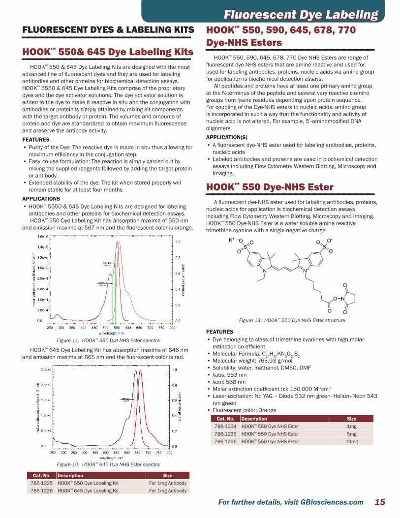
HOOK™ 550 Dye-NHS Ester————————————————————————————A fluorescent dye-NHS ester used for labeling antibodies, proteins,
nucleic acids for application is biochemical detection assays including Flow Cytometry Western Blotting, Microscopy and Imaging. HOOK™ 550 Dye-NHS Ester is a water soluble amine reactive trimethine cyanine with a single negative charge.
Figure 13: HOOK™ 550 Dye NHS Ester structure
FEATURES• Dye belonging to class of trimethine cyanines with high molar
extinction co-efficient • Molecular Formula: C35H40KN3O10S2• Molecular weight: 765.93 g/mol• Solubility: water, methanol, DMSO, DMF• λabs: 553 nm• λem: 568 nm• Molar extinction coefficient (ε): 150,000 M-1cm-1
• Laser excitation: Nd YAG – Diode 532 nm green- Helium-Neon 543 nm green
• Fluorescent color: OrangeCat. No. Description Size
786-1234 HOOK™ 550 Dye NHS Ester 1mg786-1235 HOOK™ 550 Dye NHS Ester 5mg786-1236 HOOK™ 550 Dye NHS Ester 10mg

For further details, visit GBiosciences.com16
HOOK™ 645 Dye-NHS Ester————————————————————————————A fluorescent dye-NHS ester used for labeling antibodies,
proteins, nucleic acids for application is biochemical detection assays including Flow Cytometry Western Blotting, Microscopy and Imaging. HOOK™ 645 Dye-NHS Ester is water soluble amine-reactive pentamethine cyanine with an intrinsic single negative charge.
Figure 14: HOOK™ 645 Dye NHS Ester structure
FEATURES:• Dye belonging to class of pentamethine cyanines with high molar
extinction co-efficient • Molecular Formula: C37H42N3KO10S2• Molecular weight: 791.97 g/mol• Solubility: water, methanol, DMSO, DMF• λabs: 648 nm• λem: 667 nm• Molar extinction coefficient (ε): 250,000 M-1cm-1
• Laser excitation: Krypton-Argon 647 -Diode 635 nm red• Fluorescent color: Red
Cat. No. Description Size 786-1228 HOOK™ 645 Dye NHS Ester 1mg786-1229 HOOK™ 645 Dye NHS Ester 5mg786-1230 HOOK™ 645 Dye NHS Ester 10mg
Fluorescent Dye LabelingHOOK™ 590 Dye-NHS Ester————————————————————————————
A fluorescent dye-NHS ester used for labeling antibodies, proteins, nucleic acids for application is biochemical detection assays including Flow Cytometry Western Blotting, Microscopy and Imaging. HOOK™ 590 Dye-NHS Ester is a water soluble amine reactive trimethine cyanine with a single negative charge.
Figure 15: HOOK™ 590 Dye NHS Ester structure
Figure 16: HOOK™ 590 Dye NHS Ester spectra
FEATURES• Dye belonging to class of trimethine cyanines with high molar
extinction co-efficient • Molecular Formula: C43H44KN3O10S2• Molecular weight: 866.05• Solubility: water, methanol, DMSO, DMF• λabs: 584 nm• λem: 598 nm• Molar extinction coefficient (ε): 125,000 M-1cm-1
• Laser excitation: Krypton 568 nm green- Helium- Neon 543 nm green
• Fluorescent color: RedCat. No. Description Size
786-1237 HOOK™ 590 Dye NHS Ester 1mg786-1238 HOOK™ 590 Dye NHS Ester 5mg786-1239 HOOK™ 590 Dye NHS Ester 10mg
17For further details, visit GBiosciences.com
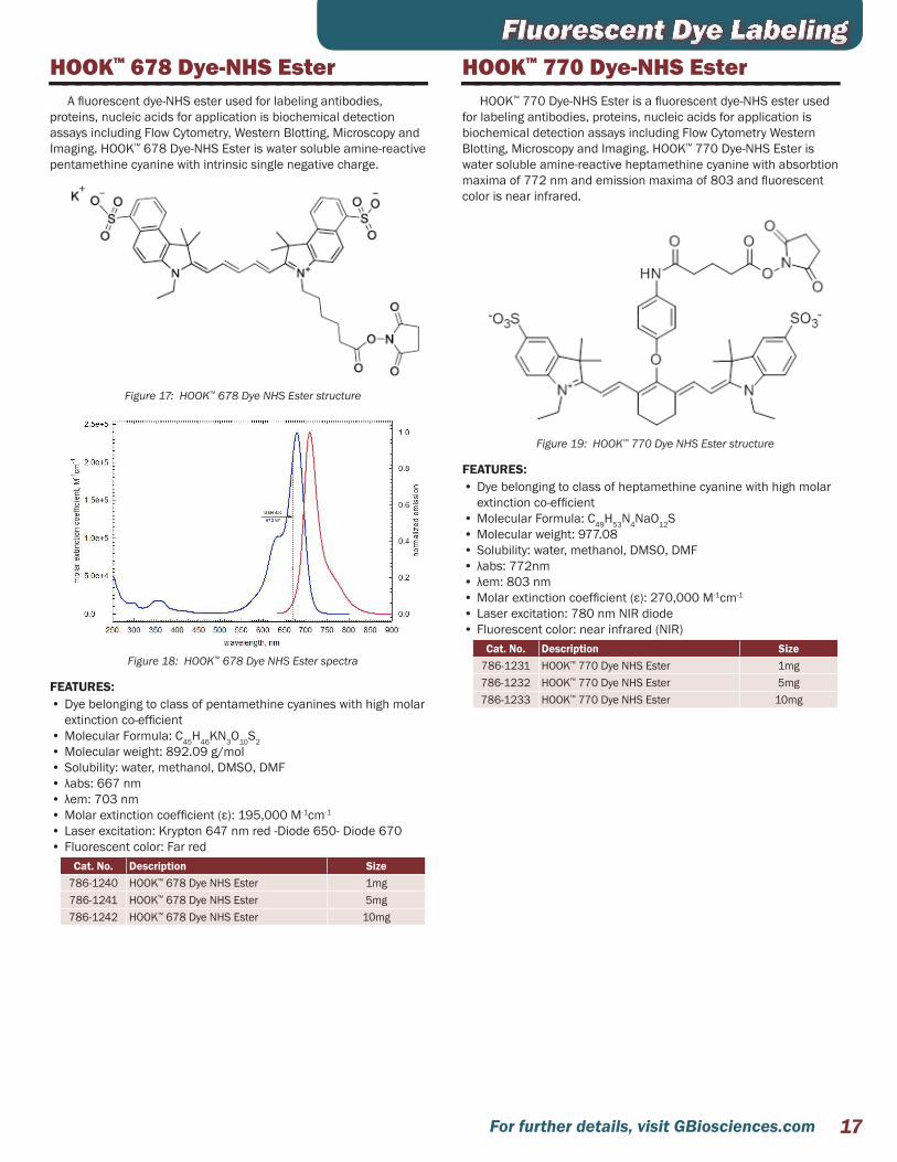
HOOK™ 678 Dye-NHS Ester————————————————————————————A fluorescent dye-NHS ester used for labeling antibodies,
proteins, nucleic acids for application is biochemical detection assays including Flow Cytometry, Western Blotting, Microscopy and Imaging. HOOK™ 678 Dye-NHS Ester is water soluble amine-reactive pentamethine cyanine with intrinsic single negative charge.
Figure 17: HOOK™ 678 Dye NHS Ester structure
Figure 18: HOOK™ 678 Dye NHS Ester spectra
FEATURES:• Dye belonging to class of pentamethine cyanines with high molar
extinction co-efficient • Molecular Formula: C45H46KN3O10S2• Molecular weight: 892.09 g/mol• Solubility: water, methanol, DMSO, DMF• λabs: 667 nm• λem: 703 nm• Molar extinction coefficient (ε): 195,000 M-1cm-1
• Laser excitation: Krypton 647 nm red -Diode 650- Diode 670• Fluorescent color: Far red
Cat. No. Description Size 786-1240 HOOK™ 678 Dye NHS Ester 1mg786-1241 HOOK™ 678 Dye NHS Ester 5mg786-1242 HOOK™ 678 Dye NHS Ester 10mg
Fluorescent Dye LabelingHOOK™ 770 Dye-NHS Ester————————————————————————————
HOOK™ 770 Dye-NHS Ester is a fluorescent dye-NHS ester used for labeling antibodies, proteins, nucleic acids for application is biochemical detection assays including Flow Cytometry Western Blotting, Microscopy and Imaging. HOOK™ 770 Dye-NHS Ester is water soluble amine-reactive heptamethine cyanine with absorbtion maxima of 772 nm and emission maxima of 803 and fluorescent color is near infrared.
Figure 19: HOOK™ 770 Dye NHS Ester structure
FEATURES:• Dye belonging to class of heptamethine cyanine with high molar
extinction co-efficient • Molecular Formula: C49H53N4NaO12S• Molecular weight: 977.08• Solubility: water, methanol, DMSO, DMF• λabs: 772nm• λem: 803 nm• Molar extinction coefficient (ε): 270,000 M-1cm-1
• Laser excitation: 780 nm NIR diode• Fluorescent color: near infrared (NIR)
Cat. No. Description Size 786-1231 HOOK™ 770 Dye NHS Ester 1mg786-1232 HOOK™ 770 Dye NHS Ester 5mg786-1233 HOOK™ 770 Dye NHS Ester 10mg

For further details, visit GBiosciences.com18
HOOK™ Dye Labeling Kit (5/6) TAMRA-SE (Rhodamine)————————————————————————————
Figure 20: Structure of (5/6) TAMRA-SE.
(5/6) TAMRA-SE (5-(and-6)- Carboxytetramethylrhodamine succinimidyl ester, mixed isomers) is based on tetramethylrhodamine, one of the most common fluorophores used in the labeling of peptides, proteins, nucleic acids and nucleotides.
(5/6) TAMRA absorbs green visible light at 546nm and emits an orange-red visible light at a maximum emission of 575nm.
The NHS ester group provides the simplest and most commonly used group for labeling proteins. The succinimidyl ester group reacts with primary amines in lysine side chains and N-terminal amines forming a stable, covalent amide bond.
This kit utilizes SpinOUT™ columns for the rapid purification of dye labeled proteins.
Figure 21: Visualization of TAMRA labeled BSA. 1μg (5/6) TAMRA-SE labeled BSA was resolved on a 4-20% SDS polyacrylamide gel.
FEATURES• Dye preweighed and supplied in single use OneQuant™ vials• Suitable for most proteins• Utilizes SpinOUT™ desalting columns to isolate labeled proteinAPPLICATIONS• Labeling of proteins, peptides and nucleic acids with a red
fluorescent dye • Suitable for antibody labeling CITED REFERENCES1. Han L. et al (2016) An injectable, low-toxicity phospholipid-based phase separation gel that
induces strong and persistent immune responses in mice. Biomaterials. 105: 1852. Sun, Z. et al (2016) A novel Nogo-66 receptor antagonist peptide promotes neurite regeneration
in vitro. Mol Cell Neurosci. 71:803. Pan, C. et al (2014) JBC 289:27764. Aktas, M. et al (2011) J Bacteriol 193:34735. Banerjee, P.S. et al (2011) J. Virol 85:75466. Sanoj Rejinold, N. et al (2010) Int. J. Biol. Macromol. 47:37
Cat. No. Description Size786-142 HOOK™ (5/6) TAMRA-SE (Rhodamine) Labeling Kit 1 kit
Fluorescent Dye LabelingHOOK™ Dye Labeling Kit (FITC)————————————————————————————
Figure 22: Structure of fluorescein isothiocyanate.
FITC (fluorescein isothiocyanate) is a commonly used fluorescent label for proteins, as it contains the groups required for conjugating to amino, sulfhydryl, imidazoyl, tyrosyl or carbonyl groups of proteins. FITC has a molecular weight of 389, and excitation and emission wavelengths of 494nm and 520nm, respectively, therefore emitting green visible light.
This kit utilizes SpinOUT™ columns for the rapid purification of dye labeled proteins.
Figure 23: Visualization of FITC Labeled Casein. Lane 1: 1μg FITC labeled casein, Lane 2-3: 1μg FITC-Casein digested with 0.2μg or 0.1μg Trypsin. Samples were resolved on a 4-20% SDS polyacrylamide gel.
FEATURES• Dye preweighed and supplied in single use OneQuant™ vials• Suitable for most proteins• Utilizes SpinOUT™ desalting columns to isolate labeled protein
APPLICATIONS• Labeling of a green fluorescent dye to proteins and peptides • Suitable for antibody labelingCITED REFERENCES1. Zheng, Y. et al (2017) Data for the effects of rLj-RGD3 on normal tissues of rats and its location
in HeyA8 cells. Data in Brief. 12:772. Afzal, A. et al (2013) BMC Plant Biol. 13:43
Cat. No. Description Size786-141 HOOK™ FITC Labeling Kit 1 kit
OneQuant™ Fluorescent Reagents————————————————————————————Both the fluorescent reagents (FITC and (5/6) TAMRA) are
available in our OneQuant™ format.The OneQuant™ format prevents loss of reagent due to repeated
weighing. Each vial also limits exposure to light.
Cat. No. Description Size786-079 OneQuant™ TAMRA 8 x 0.5mg786-080 OneQuant™ FITC 8 x 1mg

19For further details, visit GBiosciences.com
IODINATION REAGENTS————————————————————————————Bolton-Hunter Reagent (SHPP)————————————————————————————
G-Biosciences Bolton-Hunter Reagent conjugates tyrosine-like groups to end-terminal α-amino groups or ε-amino groups of lysine to increase the number of tyrosyl groups that can be iodinated by iodine-125 labeling procedures.
Radioactive iodine (125I) is routinely used by researchers to label proteins. The iodination of proteins can be performed enzymatically or chemically. The Bolton-Hunter reagent is designed to aid the labeling of proteins with radioactive iodine.
FEATURES• Optimal reaction at pH 8.5• Ideal for proteins with masked or no tyrosine residuesreductant
Cat. No. Description SizeBC84 Bolton-Hunter Reagent 1g
Iodination Reagent————————————————————————————A Solid Phase Iodination Reagent
Radioactive iodine (125I) is routinely used by researchers to label proteins. The iodination of proteins can be performed enzymatically or chemically. The Iodination reagent isdesigned to aid in the labeling of proteins with radioactive iodine.
The iodination reagent is virtually insoluble in all aqueous solutions and allows for solid phase iodination of proteins FEATURES• Chemical Name: 1,3,4,6-tetrachloro-3α-6α-diphenylglycouril• Molecular Weight: 432.09• CAS Number: 51592-06-4• InsolubleAPPLICATIONS • Iodination of tyrosyl groups in proteins and cell membranes• Iodination of phenolic groups on crosslinkers or other protein
modification reagentsCat. No. Description Size
BC93 Iodination Reagent 1g
Sulfo SHPP————————————————————————————G-Biosciences water-soluble Bolton-Hunter Reagent (Sulfo-SHPP)
conjugates tyrosine-like groups to end-terminal α-amino groups or ε-amino groups of lysine to increase the number of tyrosyl groups that can be iodinated by iodine-125 labeling procedures.
Radioactive iodine (125I) is routinely used by researchers to label proteins. The iodination of proteins can be performed enzymatically or chemically. The Bolton-Hunter reagent is designed to aid the labeling of proteins with radioactive iodine.FEATURES• Synonyms: Sulfosuccinimidyl-3-(4-hydroxyphenyl) propionate• CAS Number: 106827-57-0• Molecular Weight: 365.3• Ideal for proteins with masked or no tyrosine residues• Optimal reaction at pH 8.5• Water soluble
Cat. No. Description SizeBC92 Sulfo SHPP 100mg
Sodium Metaperiodate————————————————————————————Sodium metaperiodate, or sodium m-periodate, is a mild oxidant
that is routinely used for the conversion of cis-glycol groups in carbohydrates to reactive aldehdye groups (Figure 1). The reactive aldehyde groups are used in chemical conjugation procedures or detection of carbohydrates. For proteomic research, sodium m-periodate is used for the oxidation of the carbohydrate moiety of glycoproteins and offers the advantage of modifying the sugar side chains as opposed to critical amino acids.
The resulting aldehydes can interact with primary amines to from Schiff’s bases, which in turn can be stabilized by reduction with sodium cyanoborohydride to form covalent amide bonds. Alternatively, the aldehydes can spontaneously react with hydrazide activated molecules to form relatively stable hydrazone bonds, which again can be stabilized with sodium cyanoborohydride.FEATURES• A mild oxidizing agent that converts carbohydrates to activated
active aldehydes• Used in coupling to amines with cyanborohydride reductionAPPLICATIONS • Oxidation of glycoproteins for coupling chemistry or detection• For the generation of active aldehydes for reaction with primary
amines to form Schiff’s base• For the generation of active aldehydes to react with hydrazide
activated molecules, such as HOOK Biotin-HydrazideCITED REFERENCES1. Zhou, V. et al (2016) Direct Orthotopic Implantation of Hepatic Organoids. Journal of Surgical
Research. doi.org/10.1016/j.jss.2016.12.028
Cat. No. Description SizeBKC-12 Sodium Metaperiodate 25gBKC-15 Sodium Metaperiodate 5g
L-Cysteine·HCL, monohydrate————————————————————————————L-Cysteine hydrochloride salt is routinely used with Ellman’s
Reagent assays as a sulfhydryl standard. In addition, it is also used as a supplement for protein refolding experiments.FEATURES• CAS #: 7048-04-6• Formula: HSCH2CH(NH2)COOH•HCl•H2O• Molecular weight: 175.63• Pubchem Substance ID: 24892992
Cat. No. Description Size786-713 L-Cysteine·HCL, monohydrate 5g
p-Hydroxyphenyl Glyoxal————————————————————————————HPG (p-hydroxyphenylglyoxal) reacts to specifically modify arginine
residues under mild conditions to yield spectrophotometrically measurable signal for amino acid detectionFEATURES• Arginine-specific—reacts specifically with arginine residues under
mild conditions (pH 7 to 9, 25°C)• Quantitative—reaction follows Beer’s Law at 5 to 50 µM and can
be monitored at 340nm (pH 9)• Superior to alternatives—more resistant to oxidation than
p-nitrophenylglyoxal and more water-soluble than phenylglyoxal
Cat. No. Description SizeBC94 p-Hydroxyphenyl Glyoxal 100mg
Amino Acid Side Chain Modifiers

For further details, visit GBiosciences.com20
SATA————————————————————————————FEATURES• Chemical Name: N-Succinimidyl S-acetylthioacetate• CAS Number: 76931-93-6• Molecular Weight: 245.25xal• Reacts primarily with primary amines• Adds protected sulfhydryl residues• Sulfhydryl group can be used in coupling reactions• Soluble in DMSO• Chemical Formula: C8H9NO5S
Cat. No. Description SizeBC96 SATA 100mg
Sodium Metaperiodate————————————————————————————Sodium metaperiodate, or sodium m-periodate, is a mild oxidant
that is routinely used for the conversion of cis-glycol groups in carbohydrates to reactive aldehdye groups (Figure 1). The reactive aldehyde groups are used in chemical conjugation procedures or detection of carbohydrates. For proteomic research, sodium m-periodate is used for the oxidation of the carbohydrate moiety of glycoproteins and offers the advantage of modifying the sugar side chains as opposed to critical amino acids.
The resulting aldehydes can interact with primary amines to from Schiff’s bases, which in turn can be stabilized by reduction with sodium cyanoborohydride to form covalent amide bonds. Alternatively, the aldehydes can spontaneously react with hydrazide activated molecules to form relatively stable hydrazone bonds, which again can be stabilized with sodium cyanoborohydride.FEATURES• A mild oxidizing agent that converts carbohydrates to activated
active aldehydes• Used in coupling to amines with cyanborohydride reductionAPPLICATIONS • Oxidation of glycoproteins for coupling chemistry or detection• For the generation of active aldehydes for reaction with primary
amines to form Schiff’s base• For the generation of active aldehydes to react with hydrazide
activated molecules, such as HOOK Biotin-HydrazideCITED REFERENCES1. Zhou, V. et al (2016) Direct Orthotopic Implantation of Hepatic Organoids. Journal of Surgical
Research. doi.org/10.1016/j.jss.2016.12.028
Cat. No. Description SizeBKC-12 Sodium Metaperiodate 25gBKC-15 Sodium Metaperiodate 5g
Sulfo NHS Acetate————————————————————————————FEATURES• Chemical Name: Sulfosuccinimidyl acetate• Reacts primarily with primary amines• Water soluble• Molecular weight: 259.17APPLICATIONS • Blocks primary amines by acylationCITED REFERENCES1. Slee, J. B. et al (2016) Enhanced biocompatibility of CD47-functionalized vascular stents.
Biomaterials. DOI:10.1016/j.biomaterials.2016.02.008
Cat. No. Description SizeBC91 Sulfo NHS Acetate 100mg
Sulfo NHS ————————————————————————————FEATURES• Chemical Name: N-Hydroxysulfosuccinimide• Reacts primarily with primary amines• Water soluble• Molecular weight: 217.13APPLICATIONS • Ideal for cross-linking, chemical labeling and solid support
immobilization• Increase efficiency of EDC coupling• Convert carboxyl groups to amine reactive sulfo NHS estersCITED REFERENCES1. Afsahi, s. et al (2017) Towards Novel Graphene-Enabled Diagnostic Assays with Improved Signal-
to-Noise Ratio. MRS Advances. DOI: https://doi.org/10.1557/adv.2017.4312. Gu, J. et al (2017) Development of antibody-modified chitosan nanoparticles for the targeted
delivery of siRNA across the blood-brain barrier as a strategy for inhibiting HIV replication in astrocytes. Drug Deliv Transl Res.DOI: 10.1007/s13346-017-0368-5
3. Chen, Q. et al (2017) Probiotic E. coli Nissle 1917 biofilms on silicone substrates for bacterial interference against pathogen colonization. Acta Biomater. doi.org/10.1016/j.actbio.2017.01.011
4. Lerner, M.B. et al (2016) Large Scale Commercial Fabrication of High Quality Graphene-Based Assays for Biomolecule Detection. Sensors and Actuators B: Chemical doi10.1016/j.snb.2016.09.137
5. Andrus, L.P. et al (2015) Biosensors. 5:3986. Wongkaew, N. et al (2013) Anal. Bioanal. Chem. 405:59657. Jiang, Q, et al (2013) J. Biomed. Mater. Res. A. 101A:12378. Yang, S. et al (2013) Int. J. Nanomedicine. 8:28479. Nugen, S.R. et al (2009) Biosens. Bioelectron. 24:2428
Cat. No. Description SizeBC97 Sulfo NHS 500mg
TNBS————————————————————————————TNBS (2,4,6-trinitrobenzene sulfonic acid) is a highly sensitive
and rapid chemical used to quantitate the free amino groups. The reaction of TNBS with primary amines generates a highly chromogenic product that can be readily measured at 335nm.
Supplied as a 1% solution in methanol.FEATURES• Generates a colorimetric product, easily monitored at 335-345nmAPPLICATIONS • For colorimetric detection of primary amines
Cat. No. Description SizeBC86 TNBS 10ml
Traut’s Reagent————————————————————————————FEATURES• Chemical Name: 2-Iminothiolane hydrochloride• Molecular weight: 137.63• Mild conditions: pH 7-10, 25°C• Soluble in waterAPPLICATIONS • For the addition of sulfhydryls to primary amines• For the preparation of disulfide bridges or generation of sulfhydryl
groups for conjugation• Thiolates primary aminesCITED REFERENCES1. Nosoudi, N. et al (2015) Circ Res. 117:e802. Lei, Y. et al (2014) J Control Release 196:79
Cat. No. Description SizeBC95 Traut’s Reagent 500mg
Amino Acid Side Chain Modifiers
21For further details, visit GBiosciences.com
Amino Acid Side Chain Modifiers4-Vinylpyridine————————————————————————————
4-Vinylpyridine is used as a derivatizing reagent for free thiols such as GSH. It is used in the Glutathione colorimetric assay to remove reduced GSH, so that the oxidized Glutathione (GSSG) concentration can be measured. 4-Vinylpyridine is a better derivatizing reagent for GSH when compared to N-ethyl-maleimide (NEM) as N-ethyl-maleimide is a potent inhibitor of glutathione reductase. The treatment of samples with 4-vinylpyridine removes all the free thiols present in the sample leaving only GSSG which can be quantified in the same way as total glutathione using Ellman’s Reagent.
4-Vinylpyridine alkylates cysteine and cystine residues (after reduction) in proteins to give derivatives that are stable to acid hydrolysis and so it is used in analysis of proteins2. Its alkylating property also enables it to be used for preparation of proteins from PAGE for peptide mapping by MALDI-MS and MALDI-TOF.FEATURES• It can be used as derivatizing agent for free thiols or alkylating
agent depending upon requirements• Molecular formula: C7H7N• Molecular weight: 105.1• CAS #: 100-43-6• Density: 0.975 g/mlAPPLICATIONS • Removes GSH in samples so that oxidized glutathione
concentration can be measured • It can be used in protein structure analysis as it alkylates
cysteine and cystine residues (after reduction) in proteins to give derivatives that are stable to acid hydrolysis.
• It can be used for preparation of proteins from PAGE for maximal recovery for peptide mapping by MALDI-MS and MALDI-TOF
Cat. No. Description Size786-031 4-Vinylpyridine 1 ml
Citroconic Anhydride————————————————————————————Citraconic anhydride reacts with primary amines and blocks them
by creating an amide linkage and a terminal carboxylate.The linkage is stable at neutral to alkaline pH (pH >7) and at
acidic conditions (pH 4) the amide linkage is rapidly hydrolyzed to release the citraconic acid and free the amines. The block by citraconic anhydride can also be reveresed by treatment with hydroxylamine. This property makes citraconic anhydride a very useful tool for blocking free amines in proteins and other biomolecules.FEATURES• Reversible blocking of primary amines• Reactive towards primary amines• Synonym: 2-methylmaleic anhydride• Empirical formula:C5H4O3• CAS # 616-02-4• Molecular weight: 112.08• Form: Colorless to slight yellow, clear liquidAPPLICATIONS • Temporarily block amines to allow derivatization of other parts of
the molecule• Block removed by shifting to acidic conditions (pH3-4) or treatment
with hydroxylamine
Cat. No. Description Size786-389 Citraconic Anhydride 10g

For further details, visit GBiosciences.com22
COUPLING & LABELING BUFFERS————————————————————————————Optimizer Buffer™
————————————————————————————Optimal labeling & conjugation conditions
G-Biosciences has prepared six reaction specific buffers that provide the optimal conditions for protein labeling, modification, and cross reaction. The table below highlights the reaction each buffer is specific for:
Opt
imiz
er
Buf
fer™
Reaction Type Reactive GroupI Amine & Photoreactive Reactions NHS-ester & imidoester groupsII Sulfhydryl Reactions Iodoacetyl groupsIII Sulfhydryl Reactions Maleimides & pyridyl sulfides IV Carboxyl Reactions CarbodiimidesV Carbohydrate Reactions Hydrazide groupsVI Amine Reactions Glyoxal groups
These buffers contain optimized concentration of buffering agents, pH, and other cofactors for specific reactions. Simply exchange the buffer of your sample with a suitable Optimizer Buffer™ and you are ready for efficient reaction. Use of SpinOUT™ or Tube-O-DIALYZER™ is recommended for buffer exchange and optimal reaction results.
Each Optimizer Buffer™ is supplied as a 5X concentrated buffer.
Cat. No. Description SizeBKC-04 Optimizer Buffer™-I [5X] 2 x 25mlBKC-05 Optimizer Buffer™-II [5X] 2 x 25mlBKC-06 Optimizer Buffer™-III [5X] 2 x 25mlBKC-07 Optimizer Buffer™-IV [5X] 2 x 25mlBKC-08 Optimizer Buffer™-V [5X] 2 x 25mlBKC-09 Optimizer Buffer™-VI [5X] 2 x 25ml
SOLVENTS & CHEMICALS————————————————————————————DMSO & DMF————————————————————————————Organic solvents for HOOK™ reagents
Bottles containing anhydrous DMSO [Dimethyl sulfoxide (CH3)2SO] and DMF [N,N-Dimethylformamide (HCON(CH3)2)]. Suitable for biotinylation reaction applications.CITED REFERENCES1. Kouroklis, A. P. (2016) Substrate stiffness and matrix composition coordinately control the
differentiation of liver progenitor cells.Biomaterials. doi:10.1016/j.biomaterials.2016.05.0162. Fernandez-Gallardo et al (2015) Chem. Sci. : DOI: 10.1039/c5sc01753j
Cat. No. Description SizeBKC-16 DMF, anhydrous 50mlBKC-17 DMSO, anhydrous 50ml
Labeling AccessoriesCOMPLETE REACTION SYSTEM————————————————————————————Tube-O-Reactor™
————————————————————————————For protein cross-linking & modification reactions
Tube-O-Reactor™ is a system that allows all the key steps of cross-linking, coupling and modification of proteins and/or nucleic acids to be performed in a single tube. This minimizes the risk of sample loss, experimental time and hands-on phases.
Most of the above reactions involve three main steps:1. Equilibration of reaction conditions for optimized reactions2. Subsequent reaction with target agents (i.e. cross-linkers and
labels)3. Removal of unreacted agents and by-products
Figure 24: Tube-O-Reactor™ system.
The Tube-O-Reactor™ system is available in three MWCO sizes, 4kDa, 8kDa and 15kDa. Tube-O-Reactor™ is supplied as a Micro kit for sample sizes of 20-250µl and a Medi size for samples of 0.2-2.5ml.
Each Tube-O-Reactor™ is suitable for 5 reactions, depending on sample volumes, and is supplied with:• 5 Medi or 5 Micro Tube-O-DIALYZER™
• 5 Floats for each size of Tube-O- DIALYZER™
• 5 Micro Dialysis Reaction Chambers• Glass stirring balls
Cat. No. Description Size786-024-4K Tube-O-Reactor™ (Micro), 4kDa MWCO 5 units786-024-8K Tube-O-Reactor™ (Micro), 8kDa MWCO 5 units
786-024-15K Tube-O-Reactor™ (Micro), 15kDa MWCO 5 units786-027-4K Tube-O-Reactor™ (Medi), 4kDa MWCO 5 units786-027-8K Tube-O-Reactor™ (Medi), 8kDa MWCO 5 units
786-027-15K Tube-O-Reactor™ (Medi), 15kDa MWCO 5 units
Protein labeling and conjugation experiments often require the use of additional systems to remove the uncoupled labels and chemicals and other reaction by-products.

23For further details, visit GBiosciences.com
Sample Preparation & Clean UpDIALYSIS————————————————————————————
Dialysis is a popular technique used for the exchange of buffer medium across semi-permeable membranes. Dialysis devices are available in many configurations for research applications. We offer innovative dialysis devices and accessories for processing small samples.
Tube-O-DIALYZER™
————————————————————————————Efficient dialysis with 100% sample recovery
Small sample dialysis has become a routine and popular technique in life science research. Today’s major concern with dialysis devices is the loss of precious samples, due either to leaking or precipitation of samples during dialysis. A second concern is the efficiency and rate of dialysis. We manufacture a unique dialysis device that allows efficient dialysis and 100% sample recovery, even if your sample precipitates.
The unique tube format of Tube-O-DIALYZER™ allows for easy handling and manipulation. For sample recovery, just place the Tube-O-DIALYZER™ in a centrifuge and spin your sample to the bottom of the tube, ensuring 100% sample recovery, even if precipitation occurs.
The unique tube format also allows for easy sample loading, as simple as transferring your sample to a microcentrifuge tube. Tube-O-DIALYZER™ does not require the use of specialized loading devices or costly syringes and hazardous needles.
Tube-O-DIALYZER™ comes in two ideal sizes; the Micro unit allows efficient dialysis of 20-250µl samples and the Medi unit is optimized for 200µl-2.5ml samples. Both sizes are available with membranes with molecular weight cutoff (MWCO) of 1kDa, 4kDa, 8kDa, 15kDa and 50kDa. Tube-O-DIALYZER™ are available in packs of 20. Each Tube-O-DIALYZER™ is supplied with 6 floats and Tube-O-DIALYZER™ storage caps to allow storage of dialyzed samples. For added convenience, Tube-O-DIALYZER™ is also supplied as a mixed kit containing 10 Micro and 10 Medi Tube-O-DIALYZER™, along with the required floats and storage caps.
A graph representing the fast and highly efficient dialysis rate of the micro Tube-O-DIALYZER™ is shown. 100µl 5M NaCl was dialyzed against one liter deionized water. Samples were taken at specific times and the conductivity was measured. The graph demonstrates the fast efficiency of Tube-O-DIALYZER™, with 50% NaCl removed within 10 minutes. APPLICATIONS• Dialysis of small sample volumes• Equilibrium dialysis for buffer exchange• Concentration of samples• Dialysis for single use applications, such as radioactive samples
Pipette sample in to Tube-O-DIALYZER™ tube,
apply patented Dialysis Cap.
Centrifuge brie�y to collect sample and replace dialysis cap with supplied storage
cap and store.
Dialyze
Figure 25: A summary of the Tube-O-DIALYZER™ system.
Figure 26: Tube-O-DIALYZER™ micro (8K MWCO) Dialysis Rate. 100µl 5M sodium chloride was dialyzed against 1 liter deionized water. 50% sodium chloride is removed in the first 10 minutes.
CITED REFERENCES1. Aslanyan, L. et al (2017) Effect of Urea on G-Quadruplex Stability. J. Phys. Chem. B. DOI:
10.1021/acs.jpcb.7b034792. Han, S. et al (2017)Fluorescent nanoswitch for monitoring specific pluripotency-related
microRNAs of induced pluripotent stem cells: Development of polyethyleneimineoligonucleotide hybridization probes. Nano Res. doi:10.1007/s12274-016-1403-4
3. Kukreja, A et al (2016) Preparation of gold core-mesoporous iron-oxide shell nanoparticles and their application as dual MR/CT contrast agent in human gastric cancer cells.Journal of indus-trial and Engineering Chemistry . doi.org/10.1016/j.jiec.2016.12.020
4. Xue, H.Y et al (2016) A biodistribution study of solid lipid-polyethyleneimine hybrid nanocarrier for cancer RNAi therapy. Eur J Pharm Biopharm 108:68
5. Kim, B.G and Chalikian T.V (2016) Thermodynamic linkage analysis of pH-induced folding and unfolding transitions of i-motifs. Biophys Chem. Vol 216:19
6. Barnaba, C. et al (2016) Substrate Dependent Native Luminescence From Cytochromes P450 3A4, 2C9, and P450cam. J. Phys. Chem. B.DOI: 10.1021/acs.jpcb.5b11804
7. Marin-Argany, M. et al (2015) Protein Science. DOI: 10.1002/pro.27908. Tarasava, K. and Freisinger, E. (2015) J Inorg Biochem doi:10.1016/j.jinorgbio.2015.08.0099. Samejima, K. et al (2015) J Biol Chem 290:2146010. Gu, R. Et al (2015) Colloids and Surfaces B: Biointerfaces. 135:12611. Byul, K.G. et al (2015) Biochem. 54:342012. Kim, H. J. et al (2015) Biomaterials. 61:9513. Pathania, A and Sardesai, A (2015) J Bacteriol. doi: 10.1128/JB.02505-1414. Seleci, M. et al (2015) Langmuir. 31:454215. Sharma, P. et al (2015) Biochimie. 112:13916. Rayan, G. et al (2015) Biophysical Chemistry. 199:3417. Yoneda, A. et al (2014) PLOS One. 9: e10630618. Cottrell, J.A. et al (2014) Anal. Biochem. 469:3419. Kitajima, S., Y. Eto, and T. Tajima. MANUFACTURING METHOD FOR FOOD WITH ENHANCED
TASTE AND METHOD FOR ENHANCING TASTE OF FOOD. Ajinomoto Co, Inc., assignee. Patent 2014/0030380 A1. 30 Jan. 2014. Print.
20. Oda, M. et al (20140 Mol. Pharmacol. 85:71521. Guo, M et al (2014) JBC. 289:1055122. Ray, S. et al (2014) Mol. Biol. Cell 25:239323. Ohtake, Y. et al (2014) Biomaterials. 35:461024. Pierce, J., and K. Abbott. Glycoprotein Cancer Biomarkers. University of Georgia Research Foun-
dation, Inc., assignee. Patent 8,623,611 B2. 7 Jan. 2014. Print.25. Hevia, A. et al (2014) Biomed Res Int. http://dx.doi.org/10.1155/2014/35120426. Fritz, B. R. et al (2014) Nucl. Acids. Res. doi: 10.1093/nar/gku30727. Schoborg, J.A. et al (2014) Biotech. J. DOI: 10.1002/biot.20130038328. Son, I. et al (2014) J. Am. Chem. Soc. 136:404029. Sfeir, C. et al (2014) Acta Biomaterialia. http://dx.doi.org/10.1016/j.actbio.2014.01.00730. Siebel, A.L. et al (2013) Circ. Res. 113:16731. Poudel, K.R. et al (2013) Methods Mol. Bio. 974:23332. Kim, H. et al (2013) Macromol. Biosci. 13:74533. Hvasanov, D. et al (2013) Org. Biomol. Chem. 11:460234. Jang, E. et al (2013) J. Mater. Chem. B 1:568635. Kim, B.G. et al (2013) BioPhys. Chem. 184:95More ciatations available at www.gbiosciences.com
Cat. No. Description Size786-610 Tube-O-DIALYZER™ Micro, 1k MWCO 20786-611 Tube-O-DIALYZER™ Micro, 4k MWCO 20786-612 Tube-O-DIALYZER™ Micro, 8k MWCO 20786-613 Tube-O-DIALYZER™ Micro, 15k MWCO 20786-614 Tube-O-DIALYZER™ Micro, 50k MWCO 20786-615 Tube-O-DIALYZER™ Medi, 1k MWCO 20786-616 Tube-O-DIALYZER™ Medi, 4k MWCO 20786-617 Tube-O-DIALYZER™ Medi, 8k MWCO 20786-618 Tube-O-DIALYZER™ Medi, 15k MWCO 20786-619 Tube-O-DIALYZER™ Medi, 50k MWCO 20786-620 Tube-O-DIALYZER™ Mixed, 1k MWCO 20786-621 Tube-O-DIALYZER™ Mixed, 4k MWCO 20786-622 Tube-O-DIALYZER™ Mixed, 8k MWCO 20786-623 Tube-O-DIALYZER™ Mixed, 15k MWCO 20786-624 Tube-O-DIALYZER™ Mixed, 50k MWCO 20

For further details, visit GBiosciences.com24
Sample Preparation & Clean UpDIALYZER-Enhance™
————————————————————————————For the dialysis of up to 12 samples at one time
Dialysis is the process of separating molecules in solution by the difference in their rates of diffusion through a semi permeable membrane, such as dialysis tubing or Tube-O-DIALYZER™ dialysis caps. Molecules small enough to pass through the dialysis membrane move across the membrane in the direction of decreasing concentration, until an equilibrium has been reached. In order to remove the highest amount of small molecules as possible, the dialysis must be performed against large volumes of dialysis buffers and/or require frequent changes of buffer to shift the equilibrium. In fact, the approximate maximal extent a small molecule can be removed by dialysis is estimated by: (Vi/Vo)#C, where Vi is the volume inside a dialysis bag; Vo is the volume of dialysis buffer and #C is the number of times the buffer is changed.
DIALYZER-Enhance™ is a proprietary product that when added to the dialysis buffer shifts the equilibrium resulting in the increased removal of a wide range of small molecules. The DIALYZER-Enhance™ consists of unreactive reagents that will not interfere or modify your reagents and will not cross the dialysis membrane, ensuring a pure, clean sample at the end of dialysis.
DIALYZER-Enhance™ is designed for use with our patented Tube-O-DIALYZER™ micro dialysis devices, dialysis tubing and bags for rapid and complete dialysis. 100X concentrated suspension suitable for 5 liters of dialysis buffer.
0
10
20
30
40
50
60
70
80
90
100
0 10 20 30 40 50 60 70 80 90
Water
DIALYZER-Enhance™
Time (min)
Abs
orba
nce
(595
nm)
Figure 27: DIALYZER-Enhance™ reduces dialysis times. 0.5ml 5M NaCl was placed in a 8,000 MWCO Tube-O-DIALYZER™ dialyzed against 20ml water or 20ml water supplemented with DIALYZER-Enhance™.
FEATURES• Unreactive dialysis enhancer• Improve dialysis rates• Increase removal of small molecules• 100X suspension suitable for up to 5L dialysis bufferAPPLICATIONS• For the enhancement of diaysis rates• For the improved removal of small waste products• Fully compatible with our Tube-O-DIALYZER™ range
Cat. No. Description Size786-627 DIALYZER-Enhance™ 50ml
TUBE-O-DIALYZER™ ACCESSORIES————————————————————————————Tube-O-Array™
————————————————————————————For the dialysis of up to 12 samples at one time
This is a low cost system that allows for the rapid equilibration of samples in minimal buffer, requires minimal hands-on manipulation and can be used for 1-12 samples. Tube-O-Array™ consists of Tube-O-Array™ tray for supplied 12 Micro dialyzer cups. Simply add Tube-O-DIALYZER™ (supplied separately) and appropriate buffers.APPLICATIONS• Dialysis of multiple samples• Ideal for equilibrium dialysis
Centrifuge Tube-Adapter————————————————————————————For centrifugation of Medi and Micro Tube-O-DIALYZER™ in a bench
top centrifuge.
Tube-O-Tanks ————————————————————————————Two dialysis tanks specifically designed for use with the Tube-O-
DIALYZER™. Two sizes are available that are suitable for Micro and Medi size Tube-O-DIALYZER™.
Micro Dialysis Cups————————————————————————————For dialysis of small sample volumes, equilibrium dialysis, dialysis
of single use preparations, and other dialysis applications. The Micro Dialysis Cup has dialysis buffer capacity of 2-15 ml.
Stirring Balls————————————————————————————Recommended for use with Micro Dialysis Cups for stirring dialysis
buffer during dialysis. Supplied as 500 stirring balls.
Floats————————————————————————————Replacement Tube-O-DIALYZER™ floats are also available
separately. Floats for Tube-O-DIALYZER™ Micro and Medi sizes are available. The floats for Micro are available in two sizes: 82021-312 is designed for dialysis in Tube-O-Tanks or a beaker and 82021-336 is designed for dialysis in the Micro Dialysis Cups.
Cat. No. Description Size786-145A Tube-O-Array™ 1 kit786-145 Tube-O-DIALYZER™ Centrifuge Tube Adapter 2
786-145D Tube-O-Tanks (Small) 1786-145E Tube-O-Tanks (Large) 1786-145C Micro Dialysis Cups 12786-145B Stirring Balls 500786-141F Tube-O-DIALYZER™ Floats (Micro) 6786-149 Tube-O-DIALYZER™ Floats (Micro for Dialysis Cups) 12
786-142F Tube-O-DIALYZER™ Floats (Medi) 6

25For further details, visit GBiosciences.com
Sample Preparation & Clean UpCONTAMINATION REMOVAL————————————————————————————Spin-OUT™
————————————————————————————For desalting and buffer exchange
The SpinOUT™ GT-600 and GT-1200 columns are versatile, spin-format columns for the desalting and buffer exchange of protein and nucleic acid solutions ranging from 5µl through to 4ml sample volumes. The SpinOUT™ columns are available in two MWCO sizes. Simply apply the sample and then centrifuge to recover protein/ nucleic acids with the column retaining >95% of the salts and small molecules (<1,000Da).
Spin-OUT™ GT-600 is for the purification of proteins >6kDa and nucleic acids larger than 10bp.
Spin-OUT™ GT-1200 is for the purification of proteins >30kDa and removal of molecules >1,500Da.FEATURES• 5 sizes available for sample volumes of 5µl to 4ml• Spin format for rapid purificationCITED REFERENCES1. Yang, Y. et al (2016) Uniquely Identifiable DNA-Embedded Silica Nanotracer for Fractured
Reservoir Characterization. PROCEEDINGS, 41st Workshop on Geothermal Reservoir Engineering. Stanford University, Stanford, California
2. Wang, X. et al (2015) Zoolog Sci. 32:4193. Fedosejevs, E.T et al (2014) J Biol Chem. 289: 334124. Wickremasinghe, N. C. et al (2014) Biomacromolecules. 15:35875. Shane, M.W. et al (2013) Plant Physiol. 161:16346. Vitrac, H. et al (2013) PNAS 110:93387. Singh, J. et al (2012) Gastroenterology. 143:13088. Singh, J. et al (2009) Am.Physiol.-Gastr. L. 297:G1206
Cat. No. Description Size
Resin Bed (ml)
Sample Load (ml)
786-865 SpinOUT™ GT-100, 0.1ml 25 columns 0.1 0.005-0.02786-866 SpinOUT™ GT-100, 1ml 10 columns 1 0.05-0.1786-867 SpinOUT™ GT-100, 3ml 10 columns 3 0.1-0.5786-897 SpinOUT™ GT-100, 3ml 5 x 10/bag 3 0.1-0.5786-868 SpinOUT™ GT-100, 5ml 5 columns 5 0.5-2786-898 SpinOUT™ GT-100, 5ml 50 columns 5 0.5-2786-869 SpinOUT™ GT-100, 10ml 5 columns 10 0.5-4786-703 SpinOUT™ GT-600, 0.1ml 25 columns 0.1 0.005-0.02786-170 SpinOUT™ GT-600, 1ml 10 Columns 1 0.02-0.13786-720 SpinOUT™ GT-600, 1ml 5 x 10/bag 1 0.02-0.13786-171 SpinOUT™ GT-600, 3ml 10 Columns 3 0.1-0.5786-721 SpinOUT™ GT-600, 3ml 5 x 10/bag 3 0.1-0.5786-704 SpinOUT™ GT-600, 5ml 5 columns 5 0.5-2786-722 SpinOUT™ GT-600, 5ml 50 columns 5 0.5-2786-705 SpinOUT™ GT-600, 10ml 5 columns 10 0.5-4786-989 SpinOut™ GT-600 Spin Plate 2 plates 1 0.02-0.13786-990 SpinOut™ GT-600 Spin Plate 4 plates 1 0.02-0.13786-706 SpinOUT™ GT-1200, 0.1ml 25 columns 0.1 0.005-0.02786-172 SpinOUT™ GT-1200, 1ml 10 Columns 1 0.05-0.1786-173 SpinOUT™ GT-1200, 3ml 10 Columns 3 0.1-0.5786-707 SpinOUT™ GT-1200, 5ml 5 columns 5 0.5-2786-708 SpinOUT™ GT-1200, 10ml 5 columns 10 0.5-4786-991 SpinOut™ GT-1200 Spin Plate 2 plates 1 0.02-0.13786-992 SpinOut™ GT-1200 Spin Plate 4 plates 1 0.02-0.13
PROTEIN CONCENTRATION————————————————————————————UPPA-PROTEIN-Concentrate™
————————————————————————————Rapid precipitation & concentration
UPPA PROTEIN-Concentrate™ uses a proprietary reagent, UPPA™ (Universal Protein Precipitation Agent), to quantitatively concentrate dilute protein samples as low as 1ng/ml. Concentration is not affected by the presence of common laboratory agents, including detergents and chaotropes. After precipitation, the sample is washed to remove salts and other interfering agents; complete recovery of sample is produced. Protein samples have conductivity of ~50µS and ~100% recovery.
UPPA PROTEIN-Concentrate™ kit is available as a Micro kit for concentrating up to 10ml of dilute protein solutions; and a Medi Kit for concentrating up to 30ml of dilute protein solutions, either as a single or multiple procedures.FEATURES• Concentrate as dilute as 1ng/ml• Removes non-protein agents• Low conductivity, ~ 50µS• 100% sample recoveryAPPLICATIONS• For concentrating proteins for running gels, raising antibodies,
protein purification, protein assays, and other applications• This kit contains an acidic component and may not be suitable for
those proteins which may lose some of their biological activities when precipitated
Figure 28: Concentration of dilute mouse liver lysate. Lane 1: Protein Marker; Lane 2: 20µl dilute protein (0.05µg/µl). Lane 3: 20µl original sample treated with UPPA-PROTEIN-Concentrate™ and resuspended in 20µl. Lane 4: 40µl original sample treated with UPPA-PROTEIN-Concentrate™ and resuspended in 20µl. Comparing lanes 2 and 3 shows that there is 100% protein recovery and lane 4 shows the actual concentration of a sample.
CITED REFERENCES1. Wan, C. et al (2015) Proteome-wide dataset supporting the study of ancient metazoan
macromolecular complexes. Data Brief. doi:10.1016/j.dib.2015.11.0622. Scherp, P. et al (2011) Methds Mol. Biol. 702:1633. McFadden, N. et al (2011) PLOS. DOI: 10.1371/journal.ppat.10024134. Grote, J. et al (2006) BMC Mol. Biol. 7:485. Singleton, D. et al (2004) Microbiol 150:285More citations available at www.gbiosciences.com
Cat. No. Description Size786-120 UPPA-PROTEIN-Concentrate™ (Micro) For 10ml sample786-121 UPPA-PROTEIN-Concentrate™ (Medi) For 30ml sample
UPPA-I & II Pack————————————————————————————UPPA™ (Universal Protein Precipitation Agent) is offered separately
for the concentration of dilute (>1ng/ml) protein solutions. Concentration of proteins with UPPA™ is unaffected by chaotropes, detergents or common laboratory reagents.
Cat. No. Description Size786-122 UPPA™-I & II Pack For >80ml

For further details, visit GBiosciences.com26
Sample Preparation & Clean UpOrgoSol-PROTEIN-Concentrate™
————————————————————————————Preserve biological activity during concentration
The OrgoSol-PROTEIN-Concentration™ kit precipitates protein with a proprietary solvent buffer, OrgoSol™. The OrgoSol™ buffer has been specifically developed for efficient precipitation of protein solutions with minimal disruption to the protein structure and therefore maintains the biological activity of most proteins.
The kit has been extensively tested for the concentration of a wide selection of enzymatic proteins without the loss of their biological activity and for ~100% protein recovery. The kit is designed to precipitate up to 5ml protein solution.
The method involves mixing a protein solution with the OrgoSol™ Buffer followed by incubation, which results in quantitative precipitation of the protein. The precipitated protein is suspended in a smaller volume of an appropriate buffer and the concentrated protein solution is ready for use. FEATURES• Precipitates enzyme proteins without loss of activity• Uses a proprietary organic solvent buffer• Recovery ~100%CITED REFERENCES1. Shah, R.N. et al (2012) Adv. Hematol. http://dx.doi.org/10.1155/2012/5969252. Olbrot, M. et al (2002) PNAS 99:6737
Cat. No. Description Size786-125 OrgoSol-PROTEIN-Concentrate™ For 5ml protein
Column-PROTEIN-Concentrate™
————————————————————————————For larger volumes of dilute protein solutions
The Column-PROTEIN-Concentrate™ kit has been specifically developed for concentration of those proteins that cannot be concentrated by precipitation. The kit is based on a proprietary Protein Binding Resin that binds and immobilizes any protein in a low salt buffer between pH 2-12. The binding capacity is ~0.5mg protein/ml Protein Binding Resin.
The immobilized protein is spin-eluted in a small volume of specifically formulated elution buffer, giving several fold effective concentration. The method is gentle and protects protein from denaturation during the concentration process.
Suitable for concentration of a total of 4mg protein in either single or multiple procedures. request further information for concentration of >5mg protein.
0
100
200
300
400
500
AfterBefore
Prot
ein
Conc
entr
atio
n (μ
g/m
l)
Figure 29: Column concentration of dilute proteins. 1ml dilute protein (50µg/ml) was concentrated with Column-PROTEIN-Concentrate™ to a final volume of 110µl with a concentration of 450µg/ml.
FEATURES• Spin column format for concentration of proteins• Reusable protein binding resin (capacity ~0.5mg protein/ml)• Maintains and protects biological activity of proteins• Recovery ~ 100%CITED REFERENCES1. Taggart, C et al (2005) J. Exp. Med. 202:1659
Cat. No. Description Size786-126 Column-PROTEIN-Concentrate™ For 4mg protein
Tube-O-CONCENTRATOR™
————————————————————————————Rapid concentration of proteins >1kDa
Tube-O-CONCENTRATOR™ is a versatile concentration device that utilizes a novel, water absorbing, liquid polymer and our patented Tube-O-DIALYZER™ for the rapid concentration of dilute protein solutions with zero protein loss. The unique tube design of Tube-O-DIALYZER™ ensures that 100% sample is recovered; simple place the entire device in a bench top centrifuge and spin for a few seconds.
The Tube-O-CONCENTRATOR™ solution is a liquid polymer that rapidly absorbs water through the dialysis membrane in the Tube-O-DIALYZER™ cap, which retains all molecules with a molecular weight >1kDa.
Tube-O-CONCENTRATOR™ is available in two sizes for concentrating sample volumes of up to 250µl (Micro) or 2.5ml (Medi).
Add dilute protein solution to Tube-O-DIALYZER™ tube,
apply patented Dialysis Cap.
Centrifuge brie�y to collect sample and replace dialysis cap with supplied storage
cap and store.
Invert tube into Concentrator Solution.Water rapidly removed.
H2O
Figure 30: Tube-O-CONCENTRATOR™ scheme.
FEATURES• Rapid concentration of >1kDa proteins without protein
precipitation• Combination of a unique tube format dialysis device and a water
absorbing liquid polymer• Suitable for up to 250µl or 2.5ml protein solutions• Zero protein loss, even if protein precipitation occursCITED REFERENCES1. Ildefonso, C. J. et al (2012) J. Biol. Chem. 287:32697
Cat. No. Description Size786-625 Tube-O-CONCENTRATOR™ for 20-250µl 5 concentrations786-626 Tube-O-CONCENTRATOR™ for 0.2-2.5ml 5 concentrations
Concentrator Solution & Powder————————————————————————————Concentrator Solution is a novel liquid polymer for the rapid
concentration of dilute protein solutions with zero loss, using dialysis. Simply transfer your dilute protein solution to a dialysis bag or dialysis device, such as our patented Tube-O-DIALYZER™ and dialyze against the concentrator solution. The water will be rapidly removed through the dialysis membrane, which also retains your protein of interest and prevents the high molecular weight liquid polymer entering your solution. Once the desired volume of your solution is achieved, quickly rinse the excess concentrator solution from the dialysis bag/ membrane and recover your sample.
Concentrator Powder is a high molecular weight polymer which will not migrate across the dialysis membrane. Simply transfer your dilute protein solution to a dialysis bag or dialysis device, such as our patented Tube-O-DIALYZER™ and then cover the membrane with Concentrator Powder. Concentrator Powder rapidly absorbs water from the sample and reduces the sample volume.
Cat. No. Description Size786-143 Concentrator Solution 125ml786-144 Concentrator Powder 150g

G-Biosciences Product Line Overview
GBiosciences.com

Updated June 2, 2014
®
www.GBiosciences.com