protein degradation that targets notch for ubiquitin-mediated sel
TRANSCRIPT

10.1128/MCB.21.21.7403-7415.2001.
2001, 21(21):7403. DOI:Mol. Cell. Biol. Jan KitajewskiGurney, Adele Pauley, Inca Chui, Raymond J. Deshaies and Guangyu Wu, Svetlana Lyapina, Indranil Das, Jinhe Li, Mark Protein DegradationThat Targets Notch for Ubiquitin-Mediated SEL-10 Is an Inhibitor of Notch Signaling
http://mcb.asm.org/content/21/21/7403Updated information and services can be found at:
These include:
REFERENCEShttp://mcb.asm.org/content/21/21/7403#ref-list-1at:
This article cites 47 articles, 18 of which can be accessed free
CONTENT ALERTS more»articles cite this article),
Receive: RSS Feeds, eTOCs, free email alerts (when new
http://journals.asm.org/site/misc/reprints.xhtmlInformation about commercial reprint orders: http://journals.asm.org/site/subscriptions/To subscribe to to another ASM Journal go to:
on February 23, 2013 by P
EN
N S
TA
TE
UN
IVhttp://m
cb.asm.org/
Dow
nloaded from

MOLECULAR AND CELLULAR BIOLOGY,0270-7306/01/$04.00�0 DOI: 10.1128/MCB.21.21.7403–7415.2001
Nov. 2001, p. 7403–7415 Vol. 21, No. 21
Copyright © 2001, American Society for Microbiology. All Rights Reserved.
SEL-10 Is an Inhibitor of Notch Signaling That Targets Notchfor Ubiquitin-Mediated Protein Degradation
GUANGYU WU,1,2 SVETLANA LYAPINA,3 INDRANIL DAS,1,2 JINHE LI,4 MARK GURNEY,4
ADELE PAULEY,4 INCA CHUI,1,2 RAYMOND J. DESHAIES,3,5 AND JAN KITAJEWSKI1,2*
Departments of Pathology1 and Obstetrics and Gynecology,2 Columbia University, New York, New York 10032; Division of Biology3
and Howard Hughes Medical Institute,5 California Institute of Technology, Pasadena, California 91125; andDepartment of Neurobiology, Pharmacia & Upjohn, Kalamazoo, Michigan 490014
Received 25 January 2001/Returned for modification 23 March 2001/Accepted 19 July 2001
Notch receptors and their ligands play important roles in both normal animal development and pathogen-esis. We show here that the F-box/WD40 repeat protein SEL-10 negatively regulates Notch receptor activity bytargeting the intracellular domain of Notch receptors for ubiquitin-mediated protein degradation. Blocking ofendogenous SEL-10 activity was done by expression of a dominant-negative form containing only the WD40repeats. In the case of Notch1, this block leads to an increase in Notch signaling stimulated by either anactivated form of the Notch1 receptor or Jagged1-induced signaling through Notch1. Expression of dominant-negative SEL-10 leads to stabilization of the intracellular domain of Notch1. The Notch4 intracellular domainbound to SEL-10, but its activity was not increased as a result of dominant-negative SEL-10 expression. SEL-10bound Notch4 via the WD40 repeats and bound preferentially to a phosphorylated form of Notch4 in cells. Wemapped the region of Notch4 essential for SEL-10 binding to the C-terminal region downstream of the ankyrinrepeats. When this C-terminal fragment of Notch4 was expressed in cells, it was highly labile but could bestabilized by the expression of dominant-negative SEL-10. Ubiquitination of Notch1 and Notch4 intracellulardomains in vitro was dependent on SEL-10. Although SEL-10 interacts with the intracellular domains of bothNotch1 and Notch4, these proteins respond differently to interference with SEL-10 function. Thus, SEL-10functions to promote the ubiquitination of Notch proteins; however, the fates of these proteins may differ.
Notch/LIN-12 receptors regulate cell fate decisions duringnormal animal development and pathogenesis. For example, inCaenorhabditis elegans, LIN-12 ensures that only one of twoundifferentiated gonadal cells develops into an anchor cell,while the other cell becomes a ventral uterine precursor cell(8). Notch genes have been linked to several human patholog-ical conditions, including cancer (21), vascular failure (13), andschizophrenia (41).
The Notch/LIN-12 signaling pathway is activated when aligand-receptor interaction induces a proteolytic cleavageevent that releases the intracellular domain of the receptorfrom the cell membrane (25, 32, 35). Release of the intracel-lular domain of Notch is dependent on presenilins (5, 35).Mammalian Notch ligands are membrane-tethered proteinsreferred to as Jagged and Delta-like. The signaling module ofa Notch/LIN-12 receptor is the intracellular domain that trans-locates to the nucleus to modulate gene expression (11, 34).The nuclear activity of Notch/LIN-12 receptors relies on theinteraction between the intracellular domain of a Notch/LIN-12 receptor and the transcription factor suppressor ofhairless (Su[H]) in Drosophila, Lag-1 in C. elegans (9, 37, 42),or CBF-1 or RBP-J� in mammals (7, 11). The complex ofSu(H) and the Notch/LIN-12 intracellular domain is a tran-scriptional activator and induces genes with a regulatory se-quence recognized by the Su(H) DNA binding domain.
Less is known about the subsequent down-regulation of
Notch signaling. Insight into this aspect of Notch signalingcame from identification of the C. elegans gene sel-10, whichwas first identified in a genetic screen as a negative regulator ofthe Notch/LIN-12 signaling pathway (36). SEL-10 is related tothe budding yeast protein CDC4 (10). Members of the CDC4family are characterized by an F-box domain (43) and sevenWD40 repeats, both protein-protein interaction motifs. In pre-vious studies, CDC4 family proteins have been shown to me-diate target protein ubiquitination and degradation. Specifi-cally, the WD40 repeats bind to the target protein in aphosphorylation-dependent manner, while the F-box domaintethers the protein to the SCF ubiquitin ligase complex viabinding to the SKP1 adapter (4, 6, 16, 33). C. elegans sel-10 wasshown to functionally reduce lin-12 activity, and coimmuno-precipitation studies demonstrated that C. elegans SEL-10 pro-tein can associate with LIN-12 or murine Notch4 protein (10).Based on this precedent, we have proposed that SEL-10 is aconserved F-box/WD40 repeat protein that negatively regu-lates Notch/LIN-12 signaling by targeting the intracellular do-main of Notch/LIN-12 receptors for ubiquitin-mediated pro-tein degradation (10).
To elucidate the mechanism by which SEL-10 regulatesNotch/LIN-12 signaling, we analyzed the function of a humanhomologue of C. elegans sel-10 in mammalian cells. We dem-onstrate that human SEL-10 (hSEL-10) binds mammalianNotch proteins in a domain-specific manner. We also show thatNotch proteins are phosphorylated and that the interactionbetween SEL-10 and Notch proteins is phosphorylation depen-dent. Through an in vitro ubiquitination assay, we show thatSEL-10 can mediate Notch protein ubiquitination and thatNotch proteins are degraded by the 26S proteasome in the cell.
* Corresponding author. Mailing address: Departments of Pathol-ogy and Obstetrics and Gynecology, Columbia University, 630 West168 St., New York, NY 10032. Phone: (212) 305-3624. Fax: (212)305-3624. E-mail: [email protected].
7403
on February 23, 2013 by P
EN
N S
TA
TE
UN
IVhttp://m
cb.asm.org/
Dow
nloaded from

The proposed role of SEL-10 in Notch ubiquitination anddegradation is further supported by data showing that aSEL-10 deletion mutant containing only the WD40 repeats canstabilize Notch proteins by competing with wild-type SEL-10for binding to Notch. In principle, Notch down-regulation bySEL-10 may be physiologically important for sensitizing cells toincoming signals from Notch ligands; alternatively, SEL-10may provide a general mechanism for preventing excess Notchsignaling.
MATERIALS AND METHODS
Cell lines and media. Bosc23 cells (26) were maintained in Dulbecco’s mod-ified Eagle’s medium (DMEM) supplemented with 10% fetal bovine serum andpenicillin-streptomycin. Sf9 insect cells were maintained in Gibco BRL SF900IImedium. Hi5 insect cells were maintained in Ex-Cell 400 medium (JRH Bio-sciences). Bacterial strain DH10Bac was purchased from Gibco BRL.
Plasmids and vectors. The following plasmids were constructed by use ofpQNCXII (14), a retrovirus vector that drives gene expression under the controlof a cytomegalovirus (CMV) promoter. pQNClacZ contains the bacterial lacZgene. pQNCint-3HAHis expresses the entire Int-3 protein (amino acids 1412 to1964 of the mouse Notch4 protein), whose C terminus is fused to hemagglutinin(HA) and six-His tags. pQNCint-3CHAHis expresses a C-terminal fragment ofthe mouse Notch4 protein (amino acids 1789 to 1964) with HA and six-His tagsat the end. pQNCNotch1ICHAHis expresses the rat Notch1 intracellular domain(amino acids 1747 to 2531) with HA and six-His tags at its C terminus.
The following plasmids were constructed by use of pLNCX (24), a retrovirusvector that drives gene expression under the control of a CMV promoter. Theseplasmids express different regions of the Int-3 protein and have been describedpreviously (40). pLNCint-3HA contains cDNA corresponding to the Notch4region expressed in the Int-3 insertion, beginning at amino acid 1411; theNotch4(int-3) protein includes the entire intracellular domain of Notch4 andadditional sequences. The entire protein is HA tagged at the C terminus.pLNCint-3�NHA expresses an Int-3 protein lacking the region upstream of theCDC10/ankyrin repeats. pLNCint-3�CHA expresses an Int-3 protein lacking theregion distal to the CDC10/ankyrin repeats. pLNCint-3�N�CHA expresses theCDC10/ankyrin repeat region of Int-3. pLNCint-3�CDCHA expresses an Int-3protein lacking the CDC10/ankyrin repeats. All of the above Int-3 proteins havean in-frame HA tag at the C terminus. pHyTC-Jagged1 is described elsewhere(38) and drives the expression of full-length Jagged1 from the CMV promoter.
The following plasmids were constructed by use of pCS2-MT6 (30), a vectorthat drives gene expression under the control of a CMV promoter. There are sixmyc tags upstream of the polyclonal sites. pCS2hSEL-10myc expresses full-lengthhSEL-10 protein with six myc epitope tags at the N terminus. pCS2hSEL-10WDmyc expresses the WD40 repeat region (amino acids 184 to 540) ofhSEL-10 with six myc epitope tags at the N terminus. pCS2hSEL-10Fmyc ex-presses the F-box region (amino acids 1 to 207) of hSEL-10 with six myc epitopetags at the N terminus.
Plasmid pCS2HA-HsSKP1, which expresses HA-tagged full-length humanSKP1, was obtained from Mike Tyers, Mt. Sinai Hospital, Toronto, Canada.
The following plasmids were constructed by use of pFastBac (Gibco BRL), ashuttle vector for making baculoviruses overexpressing proteins in insect cells.pFastBacInt-3HA encodes the entire mouse Notch4(int-3) sequence fused at itsC terminus to an HA tag. pFastBacInt-3CHAHis encodes the C-terminal region(amino acids 1789 to 1964) of mouse Notch4(int-3) fused at its C terminus to HAand six-His tags. pFastBacN1ICHAHis encodes the intracellular domain (aminoacids 1747 to 2531) of rat Notch1 fused at its C terminus to HA and six-His tags.pFastBachSEL-10myc expresses full-length hSEL-10 with six N-terminal mycepitope tags. pFastBachSEL-10WDmyc expresses the WD40 repeat region (ami-no acids 184 to 540) of hSEL-10 with six N-terminal myc tags.
Luciferase reporter assays. Transient transfections were performed by calciumphosphate precipitation. For assessment of activated Notch signaling, HeLa cells(1.2 � 105) plated 1 day earlier in six-well plates were transfected in triplicatewith 50 ng of pQNCN1ICHA or 50 ng of pLNCX in combination with 670 ng ofluciferase vector (pGA981-6) (15) and 160 ng of pLNClacZ (control for trans-fection efficiency) and with or without 500 ng of pCS2hSEL-10WDmyc. Todetermine ligand-induced Notch signaling, coculture assays were performed us-ing HeLa and Bosc23 cells. HeLa cells (1.2 � 106) plated 1 day earlier in 10-cmplates were transfected with 7.5 �g of pBOS Notch1 (20), 4 �g of pGA981-6, and1 �g of pLNClacZ and with or without 1.5 �g of pCS2hSEL-10WDmyc. Bosc23cells (3 � 106) plated 1 day earlier in 10-cm plates were transfected with either
25 �g of pHyTCJagged1 (38) or 25 �g of pLNCX. One day after transfection, theHeLa and Bosc23 cells were cocultured in triplicate (1:1) on six-well plates for24 h. Luciferase activity was determined 2 days posttransfection using an en-hanced luciferase assay kit (BD PharMingen), and �-galactosidase activity wasdetermined using a Galacto-Light Plus kit (PE Biosystems) and a Bertholddual-injection luminometer.
Transfection, immunoprecipitation, and Western blot analysis. For transienttransfection, a confluent plate of Bosc23 cells was split 1:3 on the day prior totransfection. For each 60-mm plate of cells, 4 �g of each plasmid DNA wastransfected using the calcium phosphate precipitation method. The total amountof DNA was kept constant by supplementation with lacZ-containing plasmids.
Two days after transfection, cells were harvested and lysed in TENT buffer (50mM Tris-Cl [pH 8.0], 2 mM EDTA, 150 mM NaCl, 1% Triton X-100) containingprotease inhibitors (2 �g of aprotinin/ml, 2 �g of leupeptin/ml, 2 �g of pepstatin/ml, 0.5 mM phenylmethylsulfonyl fluoride [PMSF]). Lysates were clarified bycentrifugation at 10,000 � g for 10 min, and protein content was determinedusing a Bio-Rad protein assay kit. Equal amounts of extracts from samples wereprecleared with Sepharose CL-4B beads, incubated with antibodies (50 �l of12CA5 anti-HA supernatant, 200 �l of 9E10 anti-myc supernatant, or 2 �l ofanti-FLAG antibody) for 2 h at 4°C, and then incubated with 40 �l of a 50%slurry of protein A-Sepharose for 1 h at 4°C. The protein A-Sepharose beadswere washed with TENT buffer three times by vortexing for 5 min each time. Thebeads were boiled in 30 �l of protein gel loading buffer (50 mM Tris-Cl [pH 6.8],100 mM dithiothreitol, 2% [wt/vol] sodium dodecyl sulfate [SDS], 0.1% brom-phenol blue, 10% [wt/vol] glycerol), electrophoresed on an SDS-polyacrylamidegel, and transferred to a polyvinylidene difluoride membrane.
A Western blot was blocked overnight at 4°C with TBST (10 mM Tris [pH 8.0],150 mM NaCl, 0.2% Tween 20) containing 1% bovine serum albumin (BSA).The blot was then incubated with primary antibody diluted (1:50 for 12CA5; 1:10for 9E10; 1:2,000 for anti-FLAG) in TBST-BSA for 1 h, washed three times for5 min each time with TBST, and incubated with secondary antibody (anti-mouseimmunoglobulin–horseradish peroxidase, 1:10,000; Amersham) in TBST-BSAfor 1 h. After three washes, the signal was visualized by chemiluminescence(Amersham ECL kit).
12CA5 anti-HA antibody was obtained from BabCo., Richmond, Calif. 9E10anti-myc antibody was prepared from culture supernatants of the 9E10 hybrid-oma (18). Anti-FLAG antibody was purchased from Sigma.
Dephosphorylation of proteins with CIP. Immunoprecipitation was carriedout to obtain the protein to be treated with calf intestinal phosphatase (CIP). Atthe end of the immunoprecipitation, the protein A-Sepharose beads were thor-oughly washed with TENT buffer, and the solution was completely removed byaspiration. The beads were then suspended in 30 �l of buffer containing 2 �l ofCIP (New England Biolabs) and incubated at 37°C for 2 h. Ten microliters of 4�protein gel loading buffer was added to the reaction mixture. The sample wasboiled, loaded onto an SDS-polyacrylamide gel, and subjected to Western blotanalysis.
Pulse-chase labeling assay. Bosc23 cells were transfected with plasmid DNAas described above. Two days after transfection, the cells were washed andincubated in DMEM lacking methionine (Met) and cysteine (Cys) for 0.5 h todeplete Met and Cys. Cells were then incubated for 0.5 h in labeling DMEM,containing 35S-labeled Met and Cys at 0.5 mCi/ml. The labeling medium wasthen replaced with regular DMEM. Cells were harvested every 0.5 h during thechase period for up to 2.5 h. Lactacystin (10 �M) was added to both pulse-labeling and pulse-chase media.
The harvested cells were lysed and immunoprecipitated as described above.The precipitates were separated on an SDS-polyacrylamide gel. The gel was fixedfor 30 min in isopropanol-water-acetic acid (25:65:10), stained for 30 min withAmplify (Amersham), vacuum dried, and exposed to X-ray film to visualize andquantitate the signal.
Generation of baculovirues and in vitro ubiquitination assays. Baculovirusesused in the in vitro ubiquitination assays were generated with the Gibco BRLFastBac system by following the manufacurer’s protocols. Hi5 insect cell lysateswere prepared 48 h postinfection from cells coinfected with baculoviruses thatexpressed SEL-10myc or SEL-10WDmyc plus hCUL1, hSKP1, and hHRT1. Celllysis was achieved by incubating cells in buffer containing 20 mM HEPES (pH7.4), 150 mM NaCl, 50 mM NaF, 60 mM �-glycerophosphate, 0.3% Triton X-100100 �M N-acetyl-Leu-Leu-norleucinal (LLnL), and protease inhibitor cocktail(Sigma). Crude Hi5 cell lysates (500 �g) were incubated with 10 �l of anti-mycbeads for 2 h at 4°C to allow binding of myc-tagged SEL-10 subunits. Beads werewashed three times with lysis buffer and incubated with 500 �g of crude lysatesprepared from Hi5 cells infected with baculoviruses that expressed eitherNotch4(int-3)HA, Notch4(int-3)CHAHis6, or N1ICHAHis6 to allow substratebinding to SCF. Beads were washed three times with lysis buffer, washed two
7404 WU ET AL. MOL. CELL. BIOL.
on February 23, 2013 by P
EN
N S
TA
TE
UN
IVhttp://m
cb.asm.org/
Dow
nloaded from

times with 20 mM HEPES (pH 7.4)–100 mM potassium acetate–1 mM dithio-threitol, and incubated with the following ubiquitination reaction components:50 ng of His6yUBA1, 500 ng of hCDC34, 1 �l of 10� ATP-regenerating system,1 �l of 10� reaction buffer (6), and 5 �g of either ubiquitin or methylubiquitin(Boston Biochem). Ubiquitination reactions were carried out for 60 min at 30°Cand terminated by the addition of protein gel loading buffer. The samples werefractionated by SDS-polyacrylamide gel electrophoresis (PAGE), and Notchproteins were visualized by Western blotting with anti-HA antibodies.
RESULTS
hSEL-10 is an F-box/WD40 protein that inhibits Notch sig-naling. Figure 1A presents a schematic of the identified do-mains of the hSEL-10 protein. The full coding sequence forhuman SEL-10 translates into a 540-amino-acid protein (Gen-Bank accession no. AY008274). Like its homologue in C. el-egans, hSEL-10 contains an N-terminal F-box domain followedby seven WD40 repeats. The predicted protein sequences of C.elegans Sel-10 and hSEL-10 show 47.6% identity and approx-imately 57% similarity. Higher conservation is observed in theWD40 repeats (60% identity) than in the N terminus andF-box domains (30 and 35% identities, respectively). The hu-man Sel-10 gene reported here is similar to FLJ117071 of theNew Energy and Industrial Development Organization(NEDO) human cDNA sequencing project and is also referredto as FBXW6.
To explore the activity and binding potential of hSEL-10, thecoding sequence for six consecutive myc epitope tags wasadded to the 5� end of SEL-10 (Fig. 1B). We also generatedtwo variants of SEL-10; SEL-10Fmyc contains the N terminusthrough the F-box and terminates just prior to the first WD40repeat, and SEL-10WDmyc contains the WD40 repeats butnot the F-box or any sequences upstream of the F-box (Fig.1B). By analogy to mammalian F-box/WD40 repeat familymembers, we predict that the F-box mediates association withthe ubiquitination machinery and that the WD40 repeats pro-vide a binding domain for substrates.
We were interested in how SEL-10 might interact, bothphysically and functionally, with a classic Notch intracellulardomain protein, Notch1IC, and an oncogenic form of theNotch4 protein. We used a variant of the Notch4 protein,originally referred to as int-3, and we refer to this variant asNotch4(int-3) (39). It contains a short region of the extracel-lular sequence, the transmembrane domain, and the intracel-lular domain of mouse Notch4 (see Fig. 3) but does not containa signal sequence. Notch4(int-3) behaves as a gain-of-functionmutant of Notch4 (12, 38, 40) and promotes mammary tumor-igenesis in mice.
Engineered versions of F-box/WD40 proteins, such as�-TRCP, which regulates �-catenin stability, that contain onlythe WD40 repeats function as dominant-negative proteins (17,19, 46). We asked whether a variant of SEL-10 containing onlythe WD40 repeats (SEL-10WDmyc; Fig. 1B) could inhibitendogenous SEL-10 function. Using an in vitro culture system,we examined how the overexpression of SEL-10WDmyc influ-ences Notch receptor signaling. Receptor activation was as-sessed with a luciferase reporter assay that responds to thetranscriptional activation of a downstream Notch signalingcomponent, RBP-J�. The expression of an activated Notch1receptor (Notch1IC) in HeLa cells induced reporter expres-sion greater than 500-fold relative to that in control HeLa cells
FIG. 1. hSEL-10 and its effects on Notch signaling. (A) Compar-ison of C. elegans SEL-10 and hSEL-10 proteins. Percentages indicateamino acid identities between homologous domains of the two pro-teins. (B) Epitope-tagged hSEL-10 proteins used in biochemical stud-ies. Six myc epitope tags (6Xmyc) were engineered to fuse in frame tothree SEL-10 variants at their N termini. (C and D) A SEL-10 variantexpressing only the WD40 repeats upregulates signaling of activatedNotch1 (C) and Jagged1-induced signaling of full-length Notch1 (D).Notch signaling was assessed by a luciferase assay using a reporterconstruct containing the RBP-J� binding sites. In panel C, an activatedNotch1 construct (Notch1IC) and the reporter construct were trans-fected with or without SEL-10WDmyc into HeLa cells. Reporter genetransactivation was measured 2 days posttransfection, and the foldinduction of luciferase activity was calculated relative to that in HeLacells that were transfected with an empty vector (pLNCX) and thereporter construct. In panel D, Jagged1 or pLNCX (control) wastransiently expressed in Bosc23 cells. These cells were cocultured withHeLa cells transiently expressing the full-length Notch 1 receptor,luciferase reporter, and SEL-10WDmyc. Luciferase activity was mea-sured 1 day after coculturing. Each bar represents the mean fromexperiments done in triplicate. Error bars represent standard devia-tions.
VOL. 21, 2001 SEL-10 NEGATIVELY REGULATES Notch BY TURNOVER 7405
on February 23, 2013 by P
EN
N S
TA
TE
UN
IVhttp://m
cb.asm.org/
Dow
nloaded from

transfected with the reporter and an empty vector (Fig. 1C).Coexpression of the SEL-10WDmyc construct with activatedNotch1 increased reporter expression by approximately two-fold (Fig. 1C), indicative of enhanced Notch signaling.Notch4(int-3) expression induced reporter expression greaterthan 400-fold; however, coexpression of the SEL-10WDmycconstruct with Notch4(int-3) only minimally increased reporteractivity (data not shown). Thus, Notch1- and Notch4-inducedsignaling responded differently to interference with endoge-nous SEL-10 function. We also overexpressed full-lengthSEL-10 with Notch proteins and did not find a significant effecton signaling via either Notch1IC or Notch4(int-3) (data notshown), suggesting that sufficient SEL-10 activity is presentwithin cells to mediate its activity.
We determined whether the SEL-10WDmyc construct couldaugment ligand-induced activation of Notch1 signaling. Forthese experiments, we used a coculture reporter assay with onecell type expressing the Notch ligand, Jagged1, and another celltype expressing full-length Notch1 and a reporter. Bosc23 cells(a derivative of human HEK 293 cells) expressing eitherJagged1 or an empty vector were cocultured with HeLa cellsexpressing full-length Notch1, the reporter, and SEL-10WD-myc (Fig. 1D). We observed approximately a fourfold induc-tion in reporter activation in cocultures of cells expressingJagged1 and Notch1 relative to that in cocultures in whichNotch1 was expressed without exogenous Jagged1. In cocul-tures of cells expressing Jagged1 with cells expressing bothNotch1 and SEL-10WDmyc, a further 2.5-fold increase in re-porter induction was observed. These results demonstrate thatwith activated Notch1 signaling and with Jagged1-inducedNotch1 signaling, expression of the SEL-10WDmyc proteinenhanced Notch signaling. Thus, the truncated SEL-10 proteinacts in a dominant-negative manner, potentially by competingwith endogenous SEL-10 for binding to Notch. We postulatethat the augmented Notch1 signaling that we observed is dueto a blockade of SEL-10-mediated degradation of the Notch1protein.
SEL-10 binds Notch through the WD40 domain and SKP1through the F-box domain. To investigate whether hSEL-10 isinvolved in Notch protein ubiquitination and degradation, wefirst studied the physical interaction between hSEL-10 andmouse Notch4 proteins using coimmunoprecipitation assays.Previous reports suggested that detecting F-box/WD40-targetprotein interactions is difficult due to the extreme lability of thetarget protein after ubiquitination. In initial studies we foundthat hSEL-10 binding to the oncogenic Notch4(int-3) variantcould be detected, and we thus focused our analysis on definingthis interaction in detail. Binding assays were conducted aftercoexpression of the myc-tagged variants of hSEL-10 and HA-tagged Notch4(int-3) (40). Bosc23 cells were used for transienttransfections with various expression constructs. Cell extractswere prepared and immunoprecipitated with either anti-HA oranti-myc antibodies. Western blotting of the immunoprecipi-tates demonstrated that Notch4(int-3)HA (Fig. 2A, bottompanel) and all three SEL-10 variants (Fig. 2A, second panelfrom the top) were recovered at comparable levels. By probingthe anti-myc immunoprecipitates with anti-HA antibody, wedemonstrated that Notch4(int-3)HA associated with eitherfull-length hSEL-10 (Fig. 2A, top panel, lane 6) or the WD40domain (Fig. 2A, top panel, lane 8) but not with the F-box
domain (Fig. 2A, top panel, lane 7). We confirmed the inter-action between Notch4(int-3) and SEL-10 proteins by immu-noprecipitating Notch4(int-3)HA with anti-HA antibody andthen probing for myc-tagged SEL-10 proteins. As shown in Fig.2A (third panel from the top), full-length hSEL-10 (lane 6) butnot the F-box domain (lane 7) complexes with Notch4(int-3)HA; SEL-10WDmyc (lane 8) comigrated with the heavychain of immunoglobulin and therefore could not be visualizedin this experiment. In conclusion, hSEL-10 physically associ-ates with mouse Notch4(int-3) through the WD40 domain,whereas the F-box domain is not required for this interaction.
Based on previous studies of F-box/WD40 proteins, we pre-dict that the F-box domain of SEL-10 should interact withother components of the ubiquitination machinery, such asSKP1. We tested this possibility with coimmunoprecipitationassays using HA-tagged full-length human SKP1 and myc-tagged SEL-10 proteins. As shown in Fig. 2B (top panel, lanes6, 7, and 8), SKP1HA interacted with full-length hSEL-10 andthe F-box domain but not with the WD40 domain. Consistentwith this observation, we were able to detect full-lengthSEL-10 and the F-box domain in the immunoprecipitates ofSKP1 (Fig. 2B, third panel from the top, lanes 6 and 7). Thisobserved interaction implies that SEL-10, like other F-box/WD40 family proteins, is part of an E3 ubiquitin ligase thatmediates the ubiquitination and degradation of target proteins.
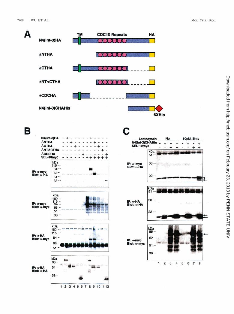
The Notch4 C-terminal domain binds to hSEL-10. To mapthe domains of Notch4(int-3) that are required for the physicalinteraction between Notch4 and SEL-10, we tested a series ofNotch4(int-3) deletion variants (schematized in Fig. 3A) fortheir ability to complex with full-length hSEL-10. We chose theNotch4(int-3) proteins to map domains of interaction becauseof the ease of detection of Notch–SEL-10 complexes.
Coexpression of myc-tagged SEL-10 and HA-tagged Notch4(int-3) variants was monitored by immunoprecipitation ofSEL-10 with anti-myc antibody; the immunoprecipitates werethen probed with anti-HA antibody. This coimmunoprecipita-tion assay revealed that N4(int-3), N4(int3)�N, and N4(int-3)�CDC proteins interacted with SEL-10 (Fig. 3B, top panel,lanes 8, 9, and 12). When the coimmunoprecipitation wasconducted in a complementary fashion by immunoprecipi-tating with anti-HA antibody for Notch4 proteins and thenprobing with anti-myc antibody for SEL-10, we detectedSEL-10 in the immune complexes of N4(int-3) and N4(int-3)�N (Fig. 3B, third panel from the top, lanes 8 and 9). Allthree of the Notch4(int-3) variants that interact with SEL-10contain the C-terminal domain, and removal of this domainabolishes the interaction between Notch4(int-3) and SEL-10. These results suggest that the C-terminal domain ofNotch4(int-3), distal to the CDC10/ankyrin repeats, bindsSEL-10.
We found that the C-terminal domain of Notch4(int-3)alone was sufficient to bind SEL-10 in coimmunoprecipitationassays using full-length hSEL-10 and a Notch4(int-3) fragmentcontaining only the C-terminal domain, N4(int-3)C. N4(int-3)Ccould be detected after expression in Bosc23 cells (Fig. 3C,middle panel, lanes 2, 4, 6, and 8). However, when coimmu-noprecipitations were carried out under standard conditions,as described above, we failed to detect a complex betweenNotch4(int-3)C and hSEL-10. To prevent ubiquitin-mediatedturnover, we treated cells with a specific proteasome inhibitor,
7406 WU ET AL. MOL. CELL. BIOL.
on February 23, 2013 by P
EN
N S
TA
TE
UN
IVhttp://m
cb.asm.org/
Dow
nloaded from

lactacystin, before harvest. After lactacystin treatment, we de-tected N4(int-3)C proteins in the immunoprecipitates of hSEL-10(Fig. 3C, top panel, lane 8). Thus, under normal conditions, theinteraction between N4(int-3)C and hSEL-10 is probably tran-sient and difficult to detect due to proteasome-dependent turn-over of SEL-10-bound Notch4(int-3)C.
SEL-10 binds to phosphorylated forms of Notch4(int-3) pro-teins. Upon Western blot analysis of Notch4(int-3) variants, wenoted that some of the Notch4(int-3) proteins appeared asmultiple bands (Fig. 3B, bottom panel, lanes 9 and 12), whichmight represent Notch protein modification by phosphoryla-tion. To address this question, we determined if the pattern of
FIG. 2. hSEL-10 interacts with Notch through the WD40 repeats and SKP1 through the F-box. (A) Physical interactions between hSEL-10 andmouse Notch4. HA-tagged Notch4(int-3) protein was coexpressed with three six-myc-epitope-tagged SEL-10 variants in Bosc23 cells by transienttransfection. Immunoprecipitation (IP) and Western blotting were performed using either anti-HA or anti-myc antibody to demonstrate that theproteins are well expressed and properly immunoprecipitated (second panel from the top and bottom panel). Anti-HA antibody was then used toprobe the anti-myc antibody immunoprecipitates to reveal Notch4(int-3)HA protein in immune complexes of either SEL-10myc or SEL-10WDmyc(top panel). Similarly, anti-myc antibody was used to probe anti-HA antibody immunoprecipitates to reveal SEL-10 proteins associated withNotch4(int-3)HA (third panel from the top). (B) Physical interactions between hSEL-10 and human SKP1. HA-tagged SKP1 was coexpressed withthree SEL-10 variants in Bosc23 cells. Immunoprecipitation followed by Western blotting using anti-HA or anti-myc antibody shows that theproteins are well expressed and precipitated efficiently (second panel from the top and bottom panel). Anti-myc antibody immunoprecipitates wereprobed with anti-HA antibody (top panel) to reveal SKP1HA associated with SEL-10 proteins, and anti-myc antibody was used to detect SEL-10proteins in immunoprecipitates of SKP1HA (third panel from the top). Arrows indicate the signal of SKP1HA, which comigrates with the lightchain of the antibody.
VOL. 21, 2001 SEL-10 NEGATIVELY REGULATES Notch BY TURNOVER 7407
on February 23, 2013 by P
EN
N S
TA
TE
UN
IVhttp://m
cb.asm.org/
Dow
nloaded from
7408 WU ET AL. MOL. CELL. BIOL.
on February 23, 2013 by P
EN
N S
TA
TE
UN
IVhttp://m
cb.asm.org/
Dow
nloaded from

migration could be altered by phosphatase treatment of im-munoprecipitated Notch4(int-3) proteins. We focused on theN4(int-3)�N variant, which contains the CDC10 repeats andthe C-terminal region (Fig. 3A). SEL-10myc and N4(int-3)�NHA were coexpressed in Bosc23 cells. When immune com-plexes containing N4(int-3)�NHA were probed with anti-HAantibody, three bands, ranging from 48 to 51 kDa, were de-tected (Fig. 4, lane 1). After treatment with CIP, the top twobands were significantly diminished, indicating that they hadbecome dephosphorylated (Fig. 4, lane 2).
We next determined which forms of N4(int-3)�N were as-sociated with SEL-10. The same cell lysate was immunopre-cipitated with anti-myc antibody to isolate SEL-10myc andthen probed with anti-HA antibody. The slowest migratingform of N4(int-3)�NHA protein was the predominant formassociated with SEL-10 (Fig. 4, lane 3). This immune complexwas treated with CIP, and the slower migrating forms werediminished at the expense of the faster migrating forms (Fig. 4,lane 4). This observation suggests that hSEL-10 preferentiallybinds to phosphorylated forms of Notch4(int-3) and is consis-tent with the behavior of CDC4-like proteins, which bind tophosphorylated target proteins. The bulk population of N4(int-3)�N molecules was efficiently dephosphorylated by CIP,whereas those bound to SEL-10 were not (Fig. 4, comparelanes 2 and 4). We speculate that the direct interaction ofSEL-10 with phosphate residues on N4(int-3)�N may haveshielded N4(int-3)�N from access to CIP. We also noted thatNotch4 variants that contain the C terminus [N4(int-3)�N,N4(int-3)�CDC, and N4(int-3)C] typically migrated as severalspecies. These other Notch4(int-3) variants were also tested inthis assay, and the slower migrating forms of these proteinswere also diminished after phosphatase treatment (data notshown). On the basis of these results and the results shownin Fig. 3B and C, we conclude that the C terminus ofNotch4(int-3) is a site of phosphorylation and is the domainrequired for the Notch4–SEL-10 interaction.
Proteasome inhibitors and a dominant-negative form ofhSEL-10 stabilize Notch proteins. To study whether Notchproteins are degraded by the ubiquitin/proteasome pathway,cells expressing Notch proteins were treated with proteasomeinhibitors. After treatment, Western blot analysis was used tomeasure the changes in the steady-state levels of these pro-teins. A protein containing the C-terminal tail of Notch4(int-3)was expressed poorly in transfected Bosc23 cells (Fig. 5A,zero-hour time point), but the levels of this protein were in-creased after lactacystin treatment (Fig. 5A), indicating turn-over by the proteasome. Notch4(int-3) proteins were expressedwell in transfected Bosc23 cells (Fig. 2A, bottom panel), and
the levels of these proteins were not significantly increasedafter lactacystin treatment (data not shown). Thus, theNotch4(int-3) protein was not efficiently processed by the pro-teasome, whereas a fragment containing the C-terminal tail ofNotch4(int-3) was. A protein containing the entire intracellulardomain of murine Notch1, N1CHAHis, was stabilized by treat-ment with another specific proteasome inhibitor, MG132 (Fig.5B). Steady-state levels of several intracellular Notch proteinsincreased upon treatment with proteasome inhibitors, indicat-ing that they are targeted for degradation via the proteasomepathway.
To determine whether the increased steady-state levels ofNotch proteins were due to increased stability, we carried outpulse-chase analysis to assess the half-life of the N4(int-3)Cprotein, the C terminus of N4(int-3). Transfected cells werepulse-labeled, cell extracts were prepared and immunoprecipi-tated, and the intensities of immunoprecipitated protein bandswere quantified to compare the relative Notch protein levels.
FIG. 4. SEL-10 binds to phosphorylated Notch. Notch4(int-3)�NTHA and SEL-10myc were coexpressed in Bosc23 cells by transienttransfection. Immunoprecipitation (IP) was performed with either an-ti-HA or anti-myc antibody. Each precipitate was divided into twotubes; one was treated with CIP, and one was left untreated. The blotwas probed with anti-HA antibody to visualize Notch4(int-3)�NTHAprecipitated either directly by anti-HA antibody or indirectly by anti-myc antibody. Arrows indicate the three species of Notch4(int-3)�NTHA. Stars indicate the heavy and light chains of immunoglobulin.
FIG. 3. The C-terminal domain of Notch4 mediates the interaction of Notch4 and SEL-10. (A) Epitope-tagged Notch4(int-3) deletion variants.The HA tag is labeled in yellow, and six-His tags are labeled in red. (B) Physical interactions between SEL-10 and Notch4 deletion variants.Six-myc-epitope-tagged full-length hSEL-10 was coexpressed with HA-tagged Notch4(int-3) deletion variants in Bosc23 cells. All the proteins werewell expressed (second panel from the top and bottom panel), except for �NT�CTHA (bottom panel, lanes 5 and 11). Anti-HA antibody was usedto detect Notch4 proteins in the immunoprecipiates of SEL-10myc (top panel), and anti-myc antibody was used to detect SEL-10myc in theimmunoprecipitates of Notch4 proteins (third panel from the top). (C) A C-terminal fragment of Notch4 complexes with SEL-10 in the presenceof a proteasome inhibitor. Notch4(int-3)CHAHis, a fragment of Notch4 containing only the C-terminal domain, was tested for its ability tocoimmunoprecipitate with SEL-10. Before the coimmunoprecipitation assays, cells were either treated with lactacystin, a specific proteasomeinhibitor, or left untreated. The bottom two panels show that both Notch4(int-3)C and SEL-10 are well expressed. Notch4(int-3)C can be detectedby anti-HA antibody in the immune complex of SEL-10myc (top panel). Arrows in the top two panels indicate the signal of Notch4(int-3)CHAHis,and the arrow in the bottom panel indicates the major species of SEL-10myc protein. IP, immunoprecipitation.
VOL. 21, 2001 SEL-10 NEGATIVELY REGULATES Notch BY TURNOVER 7409
on February 23, 2013 by P
EN
N S
TA
TE
UN
IVhttp://m
cb.asm.org/
Dow
nloaded from

Figure 5C shows that in the absence of lactacystin, more thanhalf of the N4(int-3)C protein was turned over after approxi-mately 1.5 h of chase. In contrast, after treatment with 10 �Mlactacystin, the amount of Notch4(int-3)C was diminished onlyslightly throughout the 2.5-h chase. Based on these results, theincrease in Notch4(int-3)C levels after lactacystin treatment(Fig. 5A) was likely due to decreased degradation by the pro-teasome pathway.
To determine whether endogenous SEL-10 was required totarget Notch proteins for turnover, we used the dominant-negative form of SEL-10, which interferes with endogenousSEL-10 function (SEL-10WDmyc; Fig. 1B). We tested theability of SEL-10WDmyc, which expresses only the WD40 re-peats and not the F-box, to interfere with endogenous SEL-10function by coexpressing SEL-10WDmyc with eitherNotch4(int-3)C or N1ICHA in Bosc23 cells. Two days aftertransfection, cells were harvested and the steady-state levels ofNotch proteins were examined by Western blot analysis. Theexpression of SEL-10WDmyc resulted in increased steady-state levels of N4(int-3)C (Fig. 6A) and of Notch1IC (Fig. 6B).The increased levels of these proteins were apparent after
equivalent levels of the plasmids were used in transfection, andthe levels increased in a dosage-dependent manner as moreSEL-10WDmyc plasmid was used. The increased expression ofN4(int-3)C upon coexpression of SEL-10WDmyc was reflectedin increased stability of the protein after pulse-chase analysisand quantitation (Fig. 6C). In contrast, the expression of full-length SEL-10 did not have a significant effect on steady-statelevels of Notch1IC (data not shown). Thus, the WD40 repeatregion of SEL-10 functions as a dominant-negative form ofSEL-10, and the expression of this form results in decreasedturnover of Notch proteins. In contrast, expression of the SEL-10WDmyc plasmid did not significantly alter the levels ofoncogenic Notch4(int-3) protein (data not shown). This resultis consistent with the lack of an appreciable increase in signal-ing mediated by Notch4(int-3) when coexpressed with SEL-10WDmyc (data not shown).
SEL-10 mediates Notch protein ubiquitination in vitro. Toexamine whether SEL-10 functions as part of an SCF ubiquitinligase that can target Notch proteins for ubiquitin-dependentdegradation, we first tested whether SEL-10myc and SEL-10WDmyc assembled into SCF complexes. Both proteins were
FIG. 5. Notch proteins are stabilized by proteasome inhibitor treatment. (A) Two micrograms of a plasmid expressing Notch4(int-3)CHAHiswas transiently transfected into Bosc23 cells on a 60-mm plate. The cells were treated with 10 �M lactacystin for the indicated number of hoursbefore harvest. Western blotting using anti-HA antibody was carried out to assess the steady-state levels of expression of Notch4(int-3)CHAHis(arrow). (B) A Notch1 fragment containing the intracellular domain tagged with HA and hexahistidine was expressed in Bosc23 cells by transienttransfection. Cells were treated with a proteasome inhibitor, MG132, for 12 h before harvest. Dimethyl sulfoxide (DMSO) was used as a negativecontrol. The arrow indicates the Notch1 protein revealed by Western blotting using anti-HA antibody. (C) Proteasome inhibitor treatment leadsto a longer half-life for the Notch protein. Bosc23 cells were transfected to express Notch4(int-3)CHAHis. Two days after transfection, cells werepulse-labeled with 35S-labeled methionine and cysteine for 30 min and chased with regular DMEM for up to 2.5 h. Samples were harvested every0.5 h and then immunoprecipitated using anti-HA antibody. The immunoprecipitates were separated by SDS-PAGE and autoradiographed toreveal the amount of labeled Notch4(int-3)CHAHis. For cells treated with 10 �M lactacystin, a proteasome inhibitor was added to bothpulse-labeling and pulse-chase media. Arrows indicate the two bands representing Notch4(int-3)CHAHis.
7410 WU ET AL. MOL. CELL. BIOL.
on February 23, 2013 by P
EN
N S
TA
TE
UN
IVhttp://m
cb.asm.org/
Dow
nloaded from

coexpressed in insect cells with hCUL1, hSKP1, and hHRT1,all components of the SCF complex. SEL-10 protein com-plexes were retrieved by immunoprecipitation with anti-mycantibody beads. Full-length SEL-10 efficiently coprecipitatedhCUL1, hSKP1, and hHRT1, but the WD40 repeat domain byitself was unable to recruit the other SCF subunits (Fig. 7A);these results provide an explanation for the observed domi-nant-negative effect of SEL-10WDmyc in transfected cells(Fig. 1C and E and Fig. 6).
We next tested whether Notch proteins were ubiquitinatedby recombinant SCFSEL-10. As shown in Fig. 7B, high-molec-ular-weight (HMW) forms of coprecipitated Notch4(int-3),Notch4(int-3)CHAHis, and N1ICHAHis proteins were gener-
ated in the presence of SEL-10myc immunoprecipitates thatcontained all subunits of SCF but not in the presence of SEL-10WDmyc immunoprecipitates. Formation of the HMW formsof Notch4 proteins was dependent on the presence of ubiq-uitin, confirming that Notch4 was ubiquitinated in the presenceof SCFSEL-10. We next examined in more detail N1ICHAhisubiquitination in the presence or absence of ubiquitin and itschain-terminating derivative, methyl-ubiquitin. In the presenceof ubiquitin and SCFSEL-10, SCF-bound N1ICHAHis was com-pletely converted into HMW conjugates. These conjugates mi-grated as an apparent band instead of as a more characteristicsmear, because they were compressed at the interface of thestacking and running gels. Substituting ubiquitin with methyl-
FIG. 6. Expression of the WD40 repeats of SEL-10 stabilizes Notch proteins. (A) Plasmids expressing Notch4(int-3)CHAHis and SEL-10WDmyc were cotransfected into Bosc23 cells. The amounts of plasmids used are indicated at the top of the panel. Two days after transfection,cells were harvested and subjected to Western blotting. The steady-state levels of the HA-tagged Notch protein are indicated by the arrows. (B)SEL-10WDmyc was coexpressed with N1ICHAHis, a Notch1 fragment containing the intracellular domain. Two days later, the steady-state levelsof expression of N1ICHAHis (arrow) were assessed by Western blotting. (C) Overexpression of the WD40 repeats of SEL-10 results in anincreased half-life for Notch4(int-3)CHAHis. A pulse-chase labeling experiment was done in the absence or presence of SEL-10WDmyc todetermine the half-life of Notch4(int-3)CHAHis. Metabolically labeled Notch4(int-3)CHAHis (arrows) was visualized by immunoprecipitationfollowed by autoradiography.
VOL. 21, 2001 SEL-10 NEGATIVELY REGULATES Notch BY TURNOVER 7411
on February 23, 2013 by P
EN
N S
TA
TE
UN
IVhttp://m
cb.asm.org/
Dow
nloaded from

FIG. 7. SCFSEL-10 mediates Notch ubiquitination in vitro. (A) Full-length SEL-10 but not the WD40 repeat domain interacts with other SCFcomponents. Hi5 insect cells were coinfected with a cocktail of recombinant baculoviruses that expressed hCUL1, hSKP1, and hHRT1 plus eitherfull-length SEL-10myc or SEL-10WDmyc. Crude lysates and anti-myc immunoprecipitates prepared from infected cells were fractionated bySDS-PAGE and evaluated by Western blotting with anti-myc, anti-CUL1, anti-SKP1, and anti-HRT1 antibodies. (B) SCFSEL-10 mediatesubiquitination of Notch proteins in vitro. Bead-bound SCF complexes, prepared as described for panel A, were incubated with crude lysates of Hi5insect cells infected with recombinant baculoviruses that expressed Notch4(int-3)HA, Notch4(int-3)CHAHis6, and N1ICHAHis6 to allow sub-strate binding. Beads were washed three times with lysis buffer and incubated with six-His-tagged yeast UBA1, hCDC34, and ATP-regeneratingsystem (Rxn). Ubiquitin (Ub) was either included in or omitted from the reactions. Following incubation for 60 min at 30°C, the samples werefractionated by SDS-PAGE, and Notch4 proteins were visualized by Western blotting with anti-HA antibody. Arrows designate extensivelyubiquitinated forms of Notch proteins. (C) N1ICHAHis6 ubiquitination by SCFSEL-1 and SCFSEL-10WDmyc. Ubiquitination reactions were per-formed as described for panel B, except that methylubiquitin (Me Ub) was used where indicated to inhibit multiubiquitin chain formation. Arrowsdesignate multiubiquitinated forms of Notch proteins. IP, immunoprecipitation.
7412
on February 23, 2013 by P
EN
N S
TA
TE
UN
IVhttp://m
cb.asm.org/
Dow
nloaded from

ubiquitin dramatically reduced the apparent size of ubiquiti-nated N1ICHAHis, whereas the omission of ubiquitin from thereaction completely abolished the formation of HMW conju-gates. These results confirm that the HMW forms observedwere ubiquitinated forms of N1ICHAHis. As expected, immu-noprecipitates that contained full-length SEL-10 but not thosethat contained SEL-10WD retained N1ICHAHis ubiquitina-tion in vitro.
DISCUSSION
Although the mechanisms of Notch/LIN-12 signal transmis-sion are being defined through genetic and biochemical anal-yses, little is known about the mechanisms involved in down-regulating the Notch signal. Notch signaling mediatesnumerous key developmental decisions in both vertebrates andinvertebrates. As such, mechanisms to down-regulate Notchsignaling are likely critical to maintain proper developmentalprograms or to prevent oncogenic functions of Notch proteins.sel-10 was originally identified genetically in C. elegans as anegative regulator of lin-12 activity (10). The fact that theSEL-10 protein is related to the F-box/WD40 repeat family ofproteins suggested that SEL-10 down-regulates Notch/LIN-12signaling by targeting these proteins for ubiquitin-mediatedprotein turnover (10). This proposed function of SEL-10 wouldrepresent a key mechanism by which Notch signaling is re-duced in physiological settings. Based on the paradigm estab-lished by analysis of budding yeast Cdc4, F-box/WD40 proteinsare predicted to bind their target proteins in a phosphoryla-tion-dependent fashion.
Here, we demonstrate that Notch1 signaling is negativelyregulated by SEL-10. Interference with SEL-10 function byexpression of the WD repeat region enhances steady-statelevels of Notch1 proteins by reducing the rate of turnover, thusincreasing Notch1-mediated signaling, indicating that SEL-10is directly involved in mediating Notch ubiquitination and deg-radation. We also found the Notch4 proteins interact efficientlywith SEL-10 but that the levels and activity of the intracellulardomain of Notch4, Notch4(int-3), are relatively resilient tointerference with SEL-10 function. Thus, these two Notch pro-teins behave differently in response to blocking of SEL-10function.
The ease with which we could detect Notch4(int-3)–SEL-10protein complexes prompted us to choose Notch4(int-3) pro-teins as a focus for detailed binding studies. This proved aneffective way of dissecting the biochemical interactions ingreater detail than could be achieved with substrates thatwould be tremendously labile when complexed to SEL-10. Wedemonstrated that the WD40 repeats of SEL-10 bind to theC-terminal domain of Notch4, a domain important for Notch4phosphorylation. Moreover, SEL-10 binds preferentially tophosphorylated forms of Notch4 and shields the phosphategroups from nonspecific dephosphorylation by CIP, suggestingthat the interaction is directly mediated by phosphorylatedamino acids within the C-terminal domain of Notch4. We alsofound that several forms of Notch proteins, Notch1ICD andNotch4(int-3)C, are very unstable as a result of rapid turnovervia the proteasome pathway. However, full-length SEL-10 ex-pression did not have an appreciable effect on Notch proteinlevels or activities (data not shown). This result may indicate
that sufficient SEL-10 activity exists in these cells to mediateNotch protein turnover.
Finally, recombinant SEL-10 assembles into SCF ubiquitinligase complexes in insect cells. These complexes bind coex-pressed Notch and mediate highly processive (and efficient)ubiquitination of bound Notch proteins. Given that other SCFcomplexes (including SCFCdc4, SCFGrr1, SCFSkp2, and SCF�-
TRCP) have been shown to be extremely selective for phosphor-ylated substrates, we presume that recombinant Notch proteinsare targeted to SCFSEL-10 by an endogenous protein kinase ininsect cells. Although the exact mechanism by which Notchproteins are targeted for ubiquitination remains unclear, it isevident from the in vitro experiments that Notch proteins canserve as excellent substrates for SCFSEL-10. Given the extraor-dinary substrate specificity that is evinced by all other SCFubiquitin ligases that have been evaluated to date, the efficientand highly processive in vitro ubiquitination that we observed(Fig. 6C) indicates that Notch is a physiological substrate forSCFSEL-10. Based on all of the data, the most parsimonioushypothesis is that the phosphorylation of Notch by an uniden-tified protein kinase targets it to SCFSEL-10, which in turnextensively ubiquitinates Notch as a prelude to its degradation.Conclusive proof of this hypothesis in vivo will ultimately re-quire the mapping of phosphorylation sites and the construc-tion of nonphosphorylatable point mutant versions of Notch.
We conclude that the C-terminal domain of Notch4 distal tothe CDC10/ankyrin repeats is a negative regulatory domainbecause it is responsible for interactions with SEL-10. Thisnotion is consistent with the fact that the C-terminal domaincontains a PEST sequence, which is characteristic of manyshort-lived proteins and which is thought to be a target forphosphorylation and ubiquitination (28). It has also been ob-served that a C-terminal deletion can activate GLP-1, a C.elegans Notch protein (22). The C-terminal domain of Notchproteins is also where some other regulatory proteins bind. Forexample, Drosophila protein Dishevelled has been reported tobind to this region and may thus mediate the interaction be-tween the Wingless and Notch signaling pathways (2). Ourresults predict that Notch levels and activity may be controlledby a kinase(s) that phosphorylates the C terminus of Notchproteins. This phosphorylation would mediate SEL-10 bindingand thus ubiquitination and degradation by the 26S protea-some. Little is known about kinases that phosphorylate andregulate Notch, but one would predict that the kinase thatphosphorylates the C terminus has a negative regulatory func-tion in Notch signaling.
An interesting observation is that Notch4(int-3) proteinsshow strong and specific interactions with SEL-10 but do notseem to be readily degraded by the proteasome pathway, incontrast to Notch1IC. Overexpression of the WD40 repeatregion or treatment by lactacystin failed to increase the steady-state levels of Notch4(int-3) (data not shown). Pulse-chaseanalysis indicated that Notch4(int-3) has a much longer half-life in cells than does Notch4(int-3)C, the C-terminal domainof Notch4(int-3) (data not shown). However, Notch4(int-3)still seems to be ubiquitinated in cells because a Western blotof Notch4(int-3) often displays a very high-molecular-weightsmear in addition to the main Notch4(int-3) signal at the pre-dicted molecular weight (unpublished observations). Thissmear can be seen even without proteasome inhibitor treat-
VOL. 21, 2001 SEL-10 NEGATIVELY REGULATES Notch BY TURNOVER 7413
on February 23, 2013 by P
EN
N S
TA
TE
UN
IVhttp://m
cb.asm.org/
Dow
nloaded from

ment and is very typical of proteins that are ubiquitinated.Notch4(int-3) also serves as a substrate for SEL-10-dependentin vitro ubiquitination (Fig. 7A). The fact that Notch4(int-3)protein levels are relatively unaffected by interference withSEL-10 activity is consistent with the fact that in signalingassays, Notch4(int-3) activity was not elevated by coexpres-sion with a dominant-negative form of SEL-10 (data notshown). One possible explanation for these observations isthat the extracellular sequence, the transmembrane domain,or the ankyrin repeats in Notch4(int-3) can function to pre-vent the protein from being degraded by the proteasomeeven after ubiquitination. The resultant increased stabilityof Notch4(int-3) may also contribute to the potent oncogenicactivity of this variant of Notch4, whose gene was originallydefined as a mammary oncogene (12, 29). In contrast, ourstudies show that Notch4(int-3)C and Notch1IC, both lackinga transmembrane domain and extracellular sequence, can bereadily stabilized by proteasome inhibitors or overexpressionof the WD40 repeat region of SEL-10. This issue can be fur-ther addressed by biochemical studies using a Notch4(int-3)fragment containing only the intracellular domain or possiblychimeric proteins of Notch4(int-3) and Notch1IC.
Other reports also suggest that Notch proteins are likelyturned over by ubiquitination. For example, it has been re-ported that the steady-state level of the Notch1 intracellulardomain can be elevated by lactacystin, a proteasome inhibitor(32). In addition, Notchless, a novel Drosophila gene identifiedas a modulator of Notch activity, encodes a WD40 repeat-containing protein that binds to the intracellular domain ofNotch (31). However, the function of Notchless is not clearbecause both loss-of-function mutations and overexpression ofthe gene lead to increased Notch activity. These results, onceagain, suggest that regulation of the Notch pathway is verycomplex. A recent report suggests that Notch proteins aretargets for ubiquitination and provides biochemical evidencethat the Itch protein may participate in mediating Notch ubiq-uitination (27). However, this study did not establish that Itchis responsible for or participates in the ubiquitination of Notchin vivo or that Notch ubiquitination, stability, or activity isaltered in mice with the Itch mutation.
Ubiquitin-mediated protein degradation is a highly regu-lated and selective process used to down-regulate several sig-naling pathways (1, 3). F-box/WD40 family proteins can bindto multiple target proteins (23, 44, 46). We report that Notchsignaling also utilizes ubiquitin-mediated protein turnover todown-regulate the Notch/LIN-12 signal. This is evident both inC. elegans (10) and in Notch signaling in mammalian cells (Fig.1C and D). Thus, clear evidence from sequence homology,functional studies, and binding studies points to the high levelof conserved function of SEL-10 as a negative regulator ofNotch signaling from worms to humans. SEL-10 also interactsgenetically and physically with the C. elegans presenilin,SEL-12 (45), and may target both Notch and presenilin fordegradation. The data presented here suggest that hSEL-10may serve a similar role in the down-regulation of presenilinfunction as in the down-regulation of Notch. Presenilin is re-quired for the activation of Notch, probably by mediating theproteolytic cleavage of the transmembrane domain of Notch,enabling the nuclear access of the Notch intracellular domain(5, 35, 47). It is not clear whether SEL-10 has effects on the
presenilin-Notch interaction or whether it targets each proteinseparately. It will be interesting to define how SEL-10 is di-rected to distinct targets, such as Notch and presenilins. Un-derstanding how Notch proteins are regulated by SEL-10 mayalso provide new approaches to controlling Notch activity. Forexample, as constitutive activation of Notch can lead to tumor-igenesis, SEL-10 activity could be used to reverse Notch activ-ity in these circumstances.
ACKNOWLEDGMENTS
We are grateful to Yuko Takayasu, Liz Munoz, and Khaled Zeitounfor technical assistance. We thank Iva Greenwald, Martin Julius, andRichard Kessin for comments on the manuscript. We also thank G.Weinmaster, R. Kopan, P. Sorger, P. Jackson, and Y. Xiong for gen-erously providing Jagged1 and Notch1 plasmids, Notch1�E plasmid,hSKP1 baculovirus, and anti-hSKP1 and anti-HRT1 antibodies, re-spectively.
This work was supported by grants to J.K. from the NIH (RO1HL62454 and RO1 CA75353) and the Marilyn Bokemeier SperryFund, by a grant to R.J.D. from the NIH (GM52466), and by a Bur-roughs-Wellcome Young Investigator in the Pharmacological Sciencesaward given to R.J.D. G.W. was supported by a predoctoral fellowshipfrom the Department of Defense Breast Cancer Research program(DAMD17-97-1-7291), and I.D. was supported by an NIH traininggrant (2T32 DK07328).
REFERENCES
1. Aberle, H., A. Bauer, J. Stappert, A. Kispert, and R. Kemler. 1997. �-Cateninis a target for the ubiquitin-proteasome pathway. EMBO J. 16:3797–3804.
2. Axelrod, J. D., K. Matsuno, S. Artavanis-Tsakonas, and N. Perrimon. 1996.Interaction between Wingless and Notch signaling pathways mediated bydishevelled. Science 271:1826–1832.
3. Chen, Z., J. Hagler, V. J. Palombella, F. Melandri, D. Scherer, D. Ballard,and T. Maniatis. 1995. Signal-induced site-specific phosphorylation targets Ikappa B alpha to the ubiquitin-proteasome pathway. Genes Dev. 9:1586–1597.
4. Deshaies, R. J. 1999. SCF and Cullin/Ring H2-based ubiquitin ligases. Annu.Rev. Cell Dev. Biol. 15:435–467.
5. De Strooper, B., W. Annaert, P. Cupers, P. Saftig, K. Craessaerts, J. S.Mumm, E. H. Schroeter, V. Schrijvers, M. S. Wolfe, W. J. Ray, A. Goate, andR. Kopan. 1999. A presenilin-1-dependent gamma-secretase-like proteasemediates release of Notch intracellular domain. Nature 398:518–522.
6. Feldman, R. M., C. C. Correll, K. B. Kaplan, and R. J. Deshaies. 1997. Acomplex of Cdc4p, Skp1p, and Cdc53p/cullin catalyzes ubiquitination of thephosphorylated CDK inhibitor Sic1p. Cell 91:221–230.
7. Fortini, M. E., and S. Artavanis-Tsakonas. 1994. The suppressor of hairlessprotein participates in notch receptor signaling. Cell 79:273–282.
8. Greenwald, I. 1998. LIN-12/Notch signaling: lessons from worms and flies.Genes Dev. 12:1751–1762.
9. Henderson, S. T., D. Gao, E. J. Lambie, and J. Kimble. 1994. lag-2 mayencode a signaling ligand for the GLP-1 and LIN-12 receptors of C. elegans.Development 120:2913–2924.
10. Hubbard, E. J. A., G. Wu, J. Kitajewski, and I. Greenwald. 1997. sel-10, anegative regulator of lin-12 activity in C. elegans, encodes a member of theCDC4 family of proteins. Genes Dev. 11:3182–3193.
11. Jarriault, S., O. Le Bail, E. Hirsinger, O. Pourquie, F. Logeat, C. F. Strong,C. Brou, N. G. Seidah, and A. Israel. 1998. Delta-1 activation of notch-1signaling results in HES-1 transactivation. Mol. Cell. Biol. 18:7423–7431.
12. Jhappan, C., D. Gallahan, C. Stahle, E. Chu, G. H. Smith, G. Merline, andR. Callahan. 1992. Expression of an activated Notch-related int-3 transgeneinterferes with cell differentiation and induces neoplastic transformation inmammary and salivary glands. Genes Dev. 6:345–355.
13. Joutel, A., C. Corpechot, A. Ducros, K. Vahedi, H. Chabriat, P. Mouton, S.Alamowitch, V. Domenga, M. Cecillion, E. Marechal, J. Maciazek, C. Vays-siere, C. Cruaud, E. A. Cabanis, M. M. Ruchoux, J. Weissenbach, J. F. Bach,M. G. Bousser, and E. Tournier-Lasserve. 1996. Notch3 mutations in CA-DASIL, a hereditary adult-onset condition causing stroke and dementia.Nature 383:707–710.
14. Julius, M. A., Q. Yan, Z. Zheng, and J. Kitajewski. 2000. Q vectors, bicis-tronic retroviral vectors for gene transfer. BioTechniques 28:702–708.
15. Kato, H., Y. Taniguchi, H. Kurooka, S. Minoguchi, T. Sakai, S. Nomura-Okazaki, K. Tamura, and T. Honjo. 1997. Involvement of RBP-J in biolog-ical functions of mouse Notch1 and its derivatives. Development 124:4133–4141.
16. King, R. W., R. J. Deshaies, J. M. Peters, and M. W. Kirschner. 1996. How
7414 WU ET AL. MOL. CELL. BIOL.
on February 23, 2013 by P
EN
N S
TA
TE
UN
IVhttp://m
cb.asm.org/
Dow
nloaded from

proteolysis drives the cell cycle. Science 274:1652–1659.17. Kitagawa, M., S. Hatakeyama, M. Shirane, M. Matsumoto, N. Ishida, K.
Hattori, I. Nakamichi, A. Kikuchi, and K. Nakayama. 1999. An F-box pro-tein, FWD1, mediates ubiquitin-dependent proteolysis of beta-catenin.EMBO J. 18:2401–2410.
18. Kolodziej, P. A., and R. A. Young. 1991. Epitope tagging and protein sur-veillance. Methods Enzymol. 194:508–519.
19. Latres, E., D. S. Chiaur, and M. Pagano. 1999. The human F box proteinbeta-Trcp associates with the Cul1/Skp1 complex and regulates the stabilityof beta-catenin. Oncogene 18:849–854.
20. Lindsell, C. E., C. J. Shawber, J. Boulter, and G. Weinmaster. 1995. Jagged:a mammalian ligand that activates Notch1. Cell 80:909–917.
21. Luo, B., J. C. Aster, R. P. Hasserjian, F. Kuo, and J. Sklar. 1997. Isolationand functional analysis of a cDNA for human Jagged2, a gene encoding aligand for the Notch1 receptor. Mol. Cell. Biol. 17:6057–6067.
22. Mango, S. E., E. M. Maine, and J. Kimble. 1991. Carboxy-terminal trunca-tion activates glp-1 protein to specify vulval fates in Caenorhabditis elegans.Nature 352:811–815.
23. Margottin, F., S. P. Bour, H. Durand, L. Selig, S. Benichou, V. Richard, D.Thomas, K. Strebel, and R. Benarous. 1998. A novel human WD protein,h-beta TrCp, that interacts with HIV-1 Vpu connects CD4 to the ER deg-radation pathway through an F-box motif. Mol. Cell 1:565–574.
24. Miller, A. D., and G. J. Rosman. 1989. Improved retroviral vectors for genetransfer and expression. BioTechniques 7:980–990.
25. Mumm, J. S., E. H. Schroeter, M. T. Saxena, A. Griesemer, X. Tian, D. J.Pan, W. J. Ray, and R. Kopan. 2000. A ligand-induced extracellular cleavageregulates gamma-secretase-like proteolytic activation of Notch1. Mol. Cell5:197–206.
26. Pear, W. S., G. P. Nolan, M. L. Scott, and D. Baltimore. 1993. Production ofhigh-titer helper-free retroviruses by transient transfection. Proc. Natl. Acad.Sci. USA 90:8392–8396.
27. Qiu, L., C. Joazeiro, N. Fang, H. Y. Wang, C. Elly, Y. Altman, D. Fang, T.Hunter, and Y. C. Liu. 2000. Recognition and ubiquitination of Notch byItch, a Hect-type E3 ubiquitin ligase J. Biol. Chem. 275:35734–35737.
28. Rechsteiner, M., and S. W. Rogers. 1996. PEST sequences and regulation byproteolysis. Trends Biochem. Sci. 21:267–271.
29. Robbins, J., B. J. Blondel, D. Gallahan, and R. Callahan. 1992. Mousemammary tumor gene int-3: a member of the notch gene family transformsmammary epithelial cells. J. Virol. 66:2594–2599.
30. Roth, M. B., A. M. Zahler, and J. A. Stolk. 1991. A conserved family ofnuclear phosphoproteins localized to sites of polymerase II transcription.J. Cell Biol. 115:587–596.
31. Royet, J., T. Bouwmeester, and S. M. Cohen. 1998. Notchless encodes a novelWD40-repeat-containing protein that modulates Notch signaling activity.EMBO J. 17:7351–7360.
32. Schroeter, E. H., J. A. Kisslinger, and R. Kopan. 1998. Notch-1 signalling
requires ligand-induced proteolytic release of intracellular domain. Nature393:382–386.
33. Skowyra, D., K. L. Craig, M. Tyers, S. J. Elledge, and J. W. Harper. 1997.F-box proteins are receptors that recruit phosphorylated substrates to theSCF ubiquitin-ligase complex. Cell 91:209–219.
34. Struhl, G., and A. Adachi. 1998. Nuclear access and action of notch in vivo.Cell 93:649–660.
35. Struhl, G., and I. Greenwald. 1999. Presenilin is required for activity andnuclear access of Notch in Drosophila. Nature 398:522–525.
36. Sundaram, M., and I. Greenwald. 1993. Suppressors of a lin-12 hypomorphdefine genes that interact with both lin-12 and glp-1 in Caenorhabditiselegans. Genetics 135:765–783.
37. Tax, F. E., J. J. Yeargers, and J. H. Thomas. 1994. Sequence of C. eleganslag-2 reveals a cell-signalling domain shared with Delta and Serrate ofDrosophila. Nature 368:150–154.
38. Uyttendaele, H., V. Closson, G. Wu, F. Roux, G. Weinmaster, and J. Kita-jewski. 2000. Notch4 and Jagged-1 induce microvessel differentiation of ratbrain endothelial cells. Microvasc. Res. 60:91–103.
39. Uyttendaele, H., G. Marazzi, G. Wu, Q. Yan, D. Sassoon, and J. Kitajewski.1996. Notch4/int-3, a mammary proto-oncogene, is an endothelial cell-spe-cific mammalian Notch gene. Development 122:2251–2259.
40. Uyttendaele, H., J. V. Soriano, R. Montesano, and J. Kitajewski. 1998.Notch4 and Wnt-1 proteins function to regulate branching morphogenesis ofmammary epithelial cells in an opposing fashion. Dev. Biol. 196:204–217.
41. Wei, J., and G. P. Hemmings. 2000. The NOTCH4 locus is associated withsusceptibility to schizophrenia. Nat. Genet. 25:376–377.
42. Wilkinson, H. A., K. Fitzgerald, and I. Greenwald. 1994. Reciprocal changesin expression of the receptor lin-12 and its ligand lag-2 prior to commitmentin a C. elegans cell fate decision. Cell 79:1187–1198.
43. Winston, J. T., D. M. Koepp, C. Zhu, S. J. Elledge, and J. W. Harper. 1999.A family of mammalian F-box proteins. Curr. Biol. 9:1180–1182.
44. Winston, J. T., P. Strack, P. Beer-Romero, C. Y. Chu, S. J. Elledge, and J. W.Harper. 1999. The SCFbeta-TRCP-ubiquitin ligase complex associates spe-cifically with phosphorylated destruction motifs in IkappaBalpha and beta-catenin and stimulates IkappaBalpha ubiquitination in vitro. Genes Dev.13:270–283.
45. Wu, G., E. J. Hubbard, J. K. Kitajewski, and I. Greenwald. 1998. Evidencefor functional and physical association between Caenorhabditis elegans SEL-10, a Cdc4p-related protein, and SEL-12 presenilin. Proc. Natl. Acad. Sci.USA 95:15787–15791.
46. Yaron, A., A. Hatzubai, M. Davis, I. Lavon, S. Amit, A. M. Manning, J. S.Andersen, M. Mann, F. Mercurio, and Y. Ben-Neriah. 1998. Identification ofthe receptor component of the IkappaBalpha-ubiquitin ligase. Nature 396:590–594.
47. Ye, Y., N. Lukinova, and M. E. Fortini. 1999. Neurogenic phenotypes andaltered Notch processing in Drosophila presenilin mutants. Nature 398:525–529.
VOL. 21, 2001 SEL-10 NEGATIVELY REGULATES Notch BY TURNOVER 7415
on February 23, 2013 by P
EN
N S
TA
TE
UN
IVhttp://m
cb.asm.org/
Dow
nloaded from