protein chemistry-ii anu
TRANSCRIPT
PROTEIN CHEMISTRY - II
By Dr ANURAG YADAV
Proteins are the building blocks of body.
They are linear polymer made of amino acids sequence.
They may be monomeric protein with single chain or
Oligomeric with many polypeptide chain.
Abnormal in protein structure will lead to molecular disease with
profound alteration in metabolic function.
Proteins are made up of carbon, hydrogen, oxygen, nitrogen as major
& posphate, sulphur as minor component .
STRUCTURAL HIERARCHY
PRIMARY STRUCTURE OF PROTEIN
Formation of peptide bond.
Peptide nomenclature.
Geometry of polypeptide backbone.
Features of peptide bond.
primary structure of protein means the order of amino acids in the
polypeptide chain and the location of disulfide bonds, if any.
Primary structure denotes the number and sequence of amino
acids in the protein.
The higher level of organisation is decided by primary structure.
Each polypeptide chain has a unique amino acid sequence
decided by the genes.
The primary structure is maintained by the covalent bonds of
peptide linkage.
FORMATION OF PEPTIDE BOND
-Amino acids are linked by peptide bonds
-α carboxyl group of one amino acid reacts with α-amino group of
another amino acid to form peptide bond
Or called as CO-NH bridge.
Proteins are made up of polymerisation of amino acid through
peptide bond.
PEPTIDE NOMENCLATURE
by conventionally the peptide bond chain with free amino end
i.e N-terminal end on left side, & the free carboxyl end C-
terminal end at right.
Incidently the protein synthesis also begin from N-terminal.
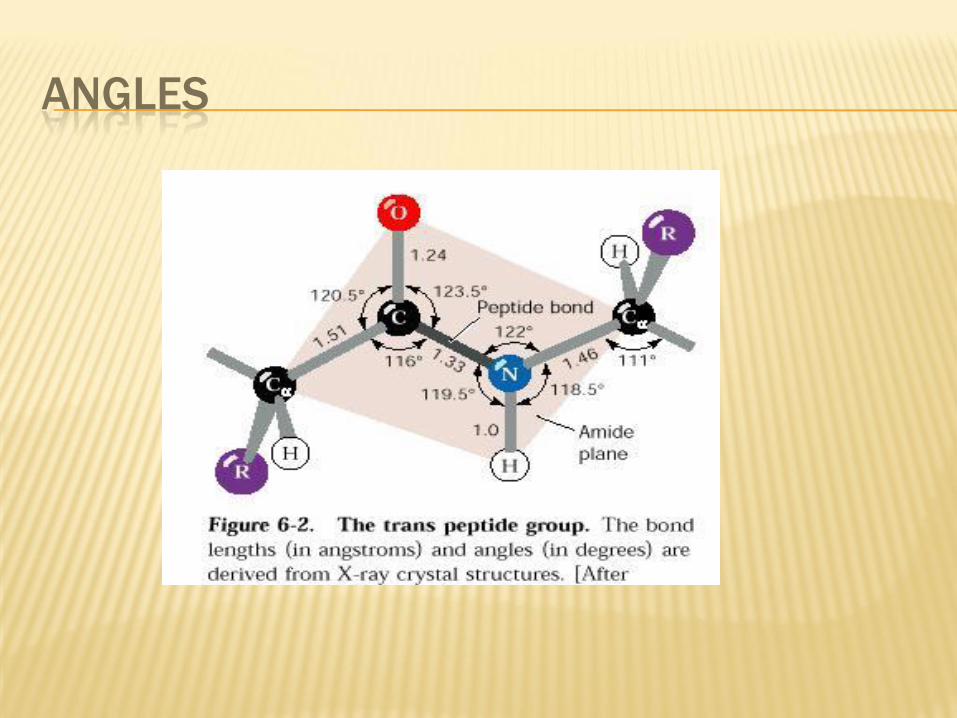
GEOMETRY OF POLYPEPTIDE BACKBONE
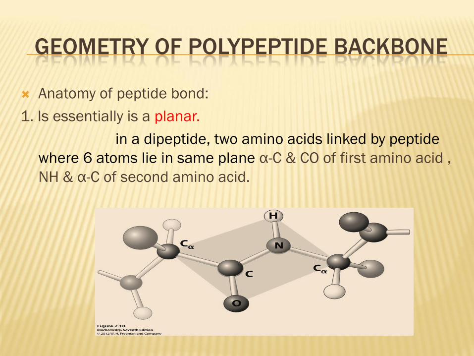
Anatomy of peptide bond:
1. Is essentially is a planar.
in a dipeptide, two amino acids linked by peptide
where 6 atoms lie in same plane α-C & CO of first amino acid ,
NH & α-C of second amino acid.
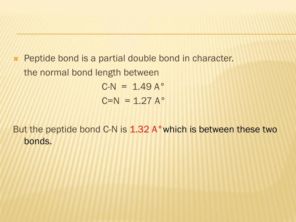
Peptide bond is a partial double bond in character.
the normal bond length between
C-N = 1.49 A°
C=N = 1.27 A°
But the peptide bond C-N is 1.32 A°which is between these two
bonds.

Considerably double bond character of peptide linkage prevent
rotation around axis of this bond.
Hence a peptide bond is considered to be rigid.
Peptide bond is a polar covalent bond:
the sharing electron reside close to the oxygen conferring
negative charge over the oxygen & partial positive charge over
nitrogen.
thus the bond carries net no charge.
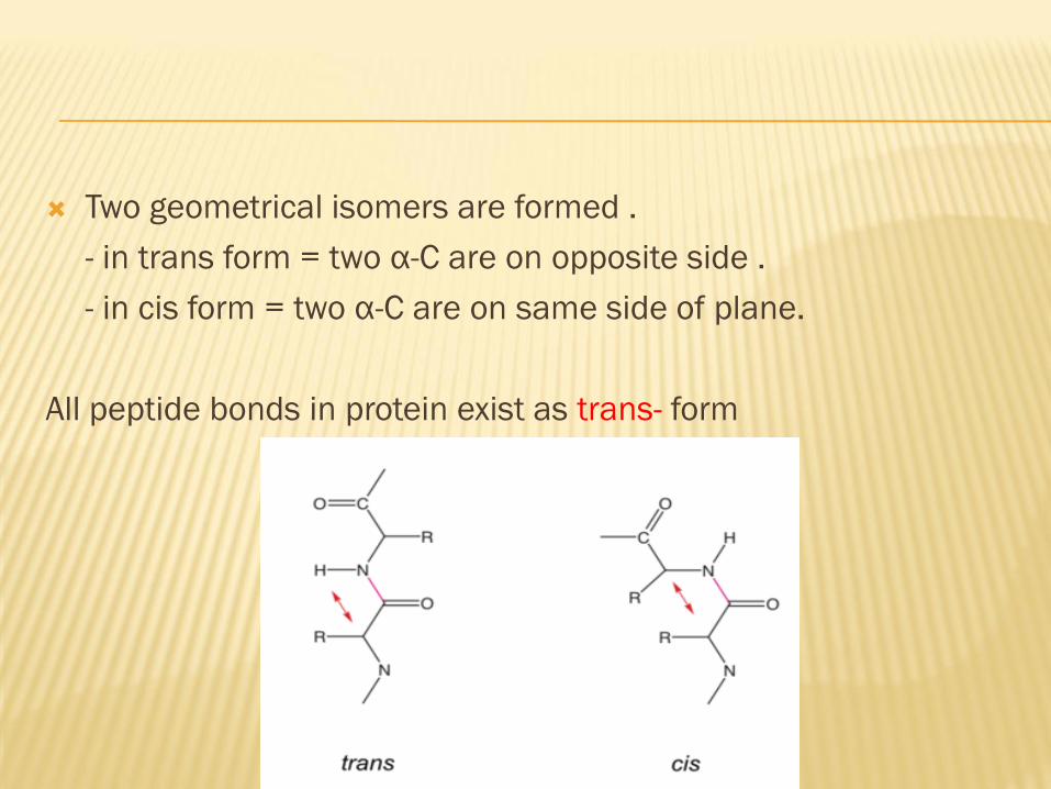
Two geometrical isomers are formed .
- in trans form = two α-C are on opposite side .
- in cis form = two α-C are on same side of plane.
All peptide bonds in protein exist as trans- form
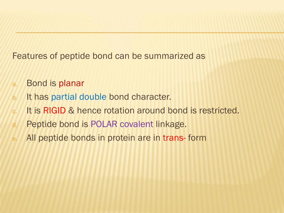
Features of peptide bond can be summarized as
a. Bond is planar
b. It has partial double bond character.
c. It is RIGID & hence rotation around bond is restricted.
d. Peptide bond is POLAR covalent linkage.
e. All peptide bonds in protein are in trans- form

DISULPHIDE BOND
Disulfide are usually formed between two cysteine residue to
form cystine.
Both contribute to structural strength & stability of protein.
BONDS & ANGLES ADJACENT TO THE PEPTIDE
LINKAGE
Unlike rigid peptide bond which does not allow rotation of bonds
adjacent to it between
a. α-carbon & carbonyl group
b. amino group & α-carbon are flexible and purely single bond.
The free of rotation around these two bonds of each amino acid
enable protein to fold in different ways .
Rotations about these bonds is called as dihedral/ tortion angle &
measured between -180°to +180°
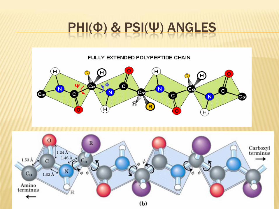
PHI(Φ) & PSI(Ψ) ANGLES
ANGLES
Not all conformation structure is possible with rotation of the
phi & psi angles.
Ramachandran showed with his plot , more than 75% of it are
not favourable/forbidden bcz of local steric clashes b/w
atoms.
INSULIN
Ex for primary structure
Sanger described structure 1955
β cells of pancreas
Hypoglycemic hormone
STRUCTURE OF INSULIN
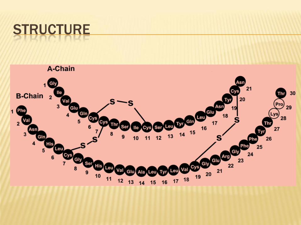
Composed of 2 chains
A and B chain
A chain – glycine chain , 21 AA
B chain – phenyl alanine chain 30 AA
Intra and inter disulphide bonds
STRUCTURE
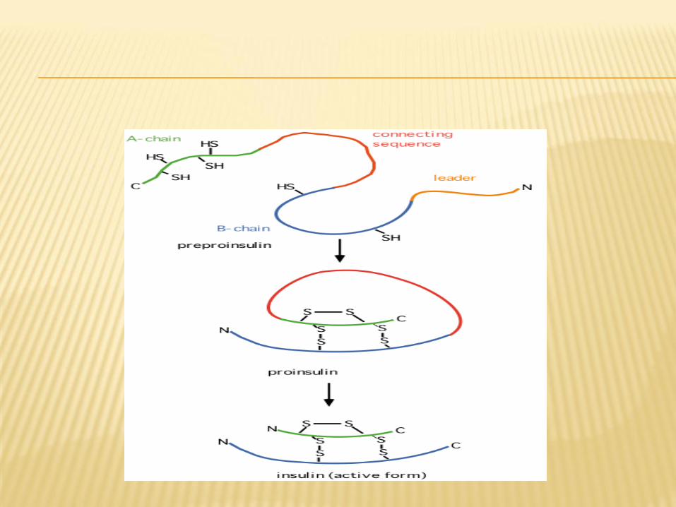
MATURATION OF INSULIN
proinsulin insulin
86 AA 51 AA
Species variation
A -8 , 9 , 10 . B– C terminal
Human insulin
Porcine insulin
Bovine insulin
HIGHER LEVEL OF PROTEIN STRUCTURE
They include :
Secondary
Tertiary
Quaternary structure of protein
CONFIGURATION & CONFORMATION
Configuration : refer to geometric relationship among given set
of atoms.
conversion to different configurational alternative into one
another is possible only by breaking & making covalent bond.
Conformation : refer to spatial relationship of every atom to all
other in three dimensional structure of protein.
the interconversion occur not by disruption of covalent
bond but rupture & reinstallation of relatively weak non-
covalent forces.
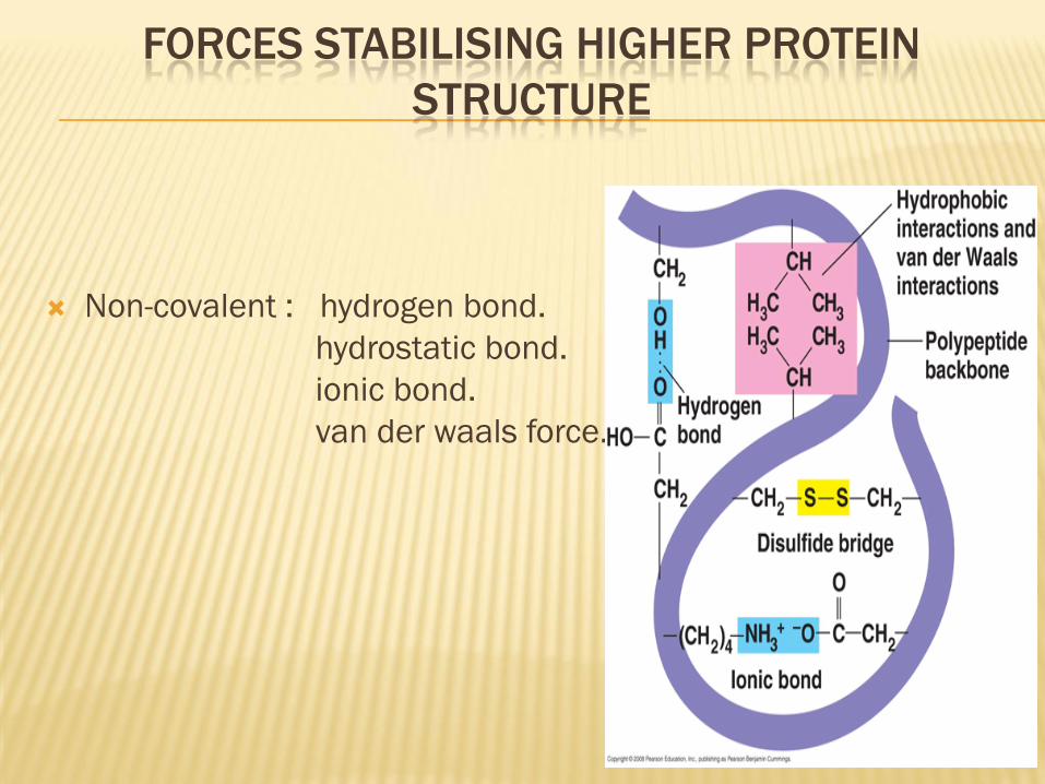
FORCES STABILISING HIGHER PROTEIN
STRUCTURE
Non-covalent : hydrogen bond.
hydrostatic bond.
ionic bond.
van der waals force.
Secondary structure is the steric relationship of amino acids
close to each other.
It denotes configurational relationship b/w residues which are
about 3-4 amino acid apart in linear sequence.
Stabilizing force: non-covalent forces (hydrogen bond, ionic
bond, hydrophobic and van der waals forces)
SECONDARY STRUCTURE OF PROTEIN:
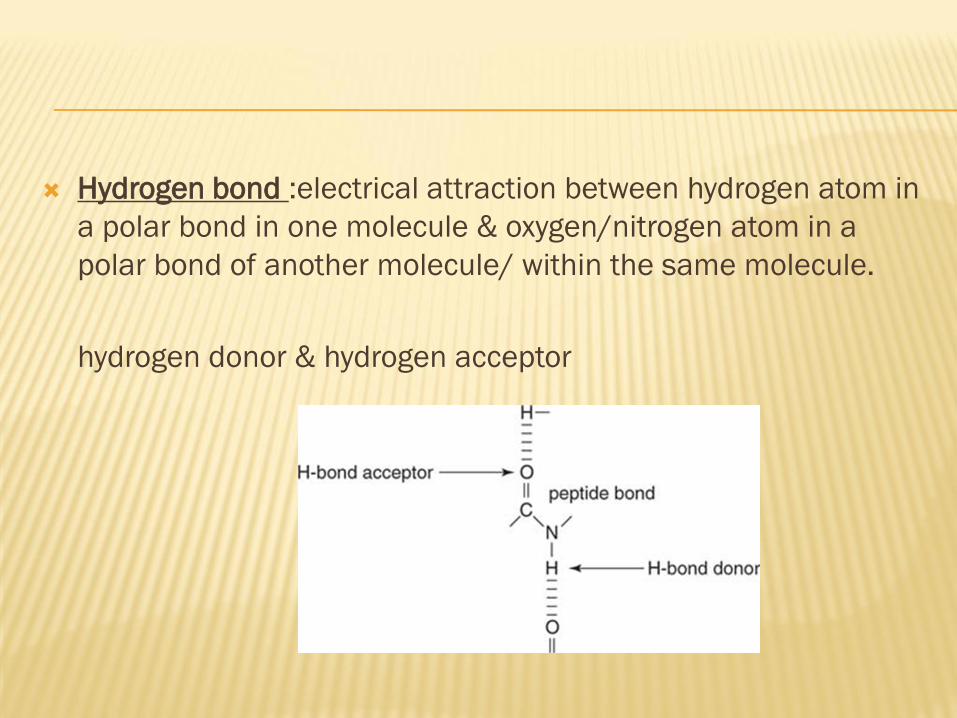
Hydrogen bond :electrical attraction between hydrogen atom in
a polar bond in one molecule & oxygen/nitrogen atom in a
polar bond of another molecule/ within the same molecule.
hydrogen donor & hydrogen acceptor
Since AA can rotate around Φ & Ψ , peptide chain is flexible &
can be bent into number of conformation.
Polypeptide chain folded into
regular α-helix ,β-sheet.
irregular forms- turns & loops.
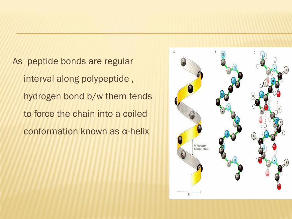
As peptide bonds are regular
interval along polypeptide ,
hydrogen bond b/w them tends
to force the chain into a coiled
conformation known as α-helix
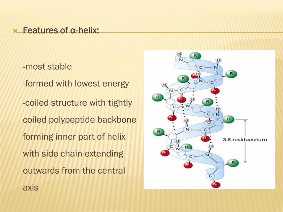
Features of α-helix:
-most stable
-formed with lowest energy
-coiled structure with tightly
coiled polypeptide backbone
forming inner part of helix
with side chain extending
outwards from the central
axis
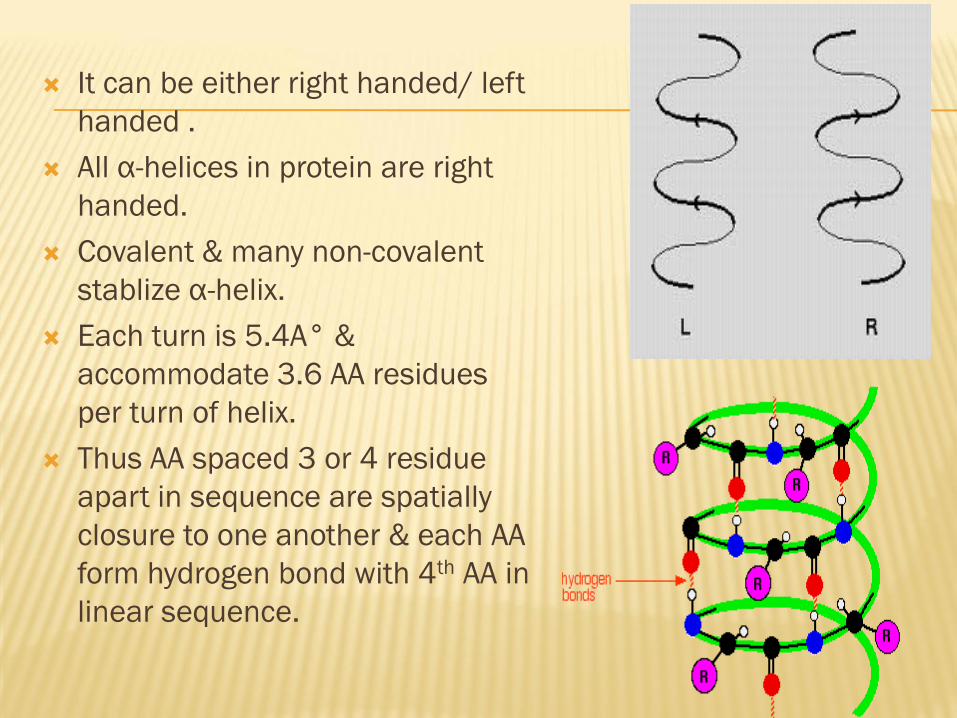
It can be either right handed/ left
handed .
All α-helices in protein are right
handed.
Covalent & many non-covalent
stablize α-helix.
Each turn is 5.4A° &
accommodate 3.6 AA residues
per turn of helix.
Thus AA spaced 3 or 4 residue
apart in sequence are spatially
closure to one another & each AA
form hydrogen bond with 4th AA in
linear sequence.
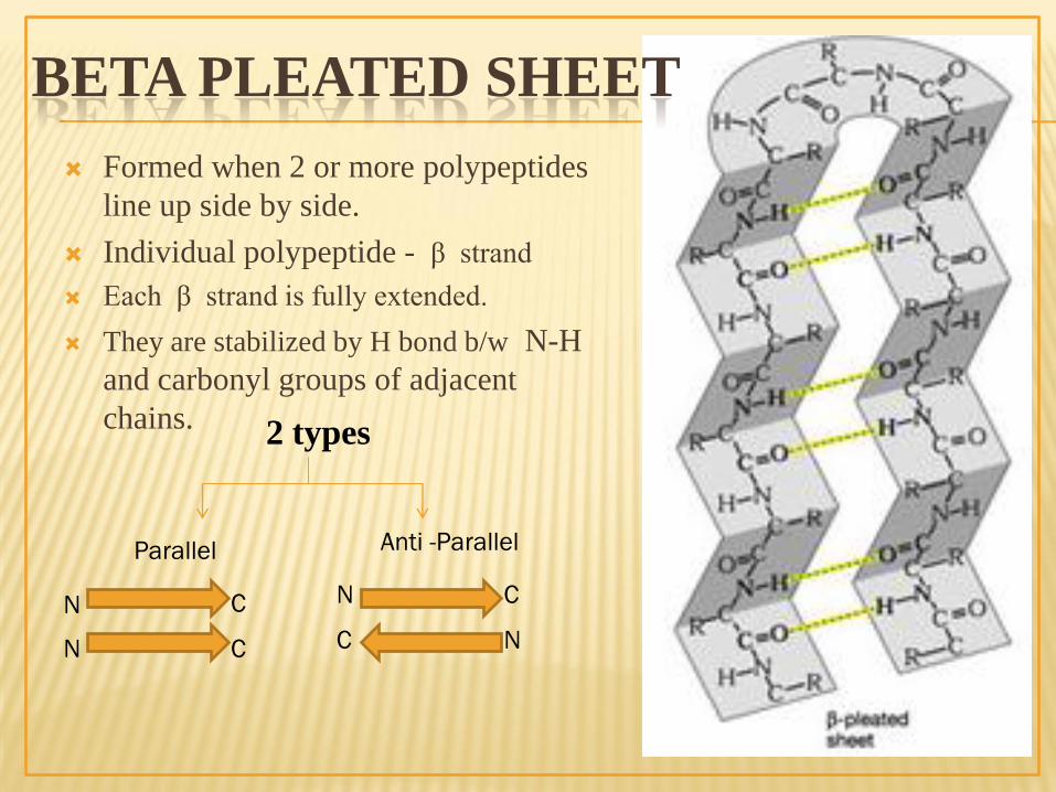
Formed when 2 or more polypeptides
line up side by side.
Individual polypeptide - β strand
Each β strand is fully extended.
They are stabilized by H bond b/w N-H
and carbonyl groups of adjacent
chains.
BETA PLEATED SHEET
2 types
Parallel Anti -Parallel
N C N
N N C
C
C
Features of β-pleated sheet:
- Second type of regular repetitive pattern
- Peptide backbone of these sheet are partly extended with
pleated appearance.
- Distance btwn AA along β-strand is 3.5A°( 1.5A° in α-helix)
- Side chain of adjacent AA orient opposite directions.
- Sheets are stabilized by extension hydrogen bond.
Anti-parallel sheet: hydrogen bonds b/w NH & CO group
connect each AA to single AA on an adjacent strand.
Eg; Silk fibroin
Parallel sheet: hydrogen bond connect each AA on one strand
with two different AA on adjacent strand.
Eg; Flavodoxin
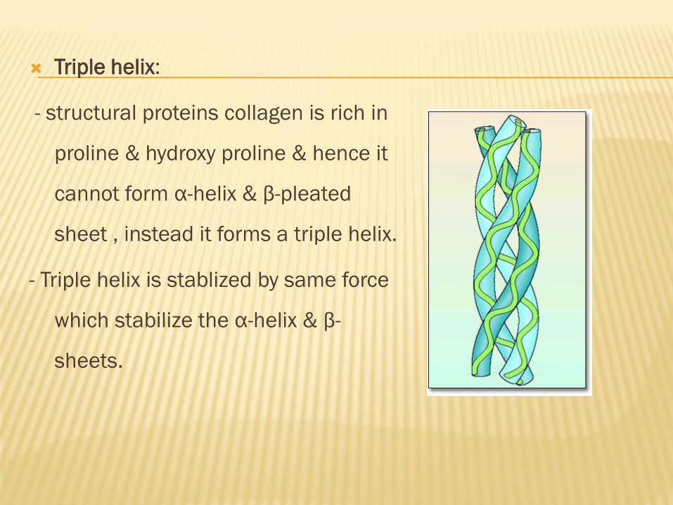
Triple helix:
- structural proteins collagen is rich in
proline & hydroxy proline & hence it
cannot form α-helix & β-pleated
sheet , instead it forms a triple helix.
- Triple helix is stablized by same force
which stabilize the α-helix & β-
sheets.
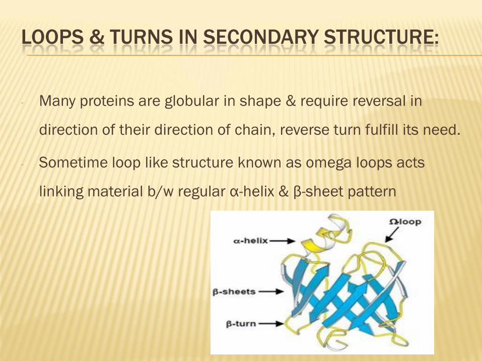
LOOPS & TURNS IN SECONDARY STRUCTURE:
- Many proteins are globular in shape & require reversal in
direction of their direction of chain, reverse turn fulfill its need.
- Sometime loop like structure known as omega loops acts
linking material b/w regular α-helix & β-sheet pattern
OMEGA LOOP
Compact annular bend (Reverse turns of the peptide back
bone)
One or more loops join successive beta sheets and alpha helix
Present over the surface of the proteins (>60AA ) to avoid steric
hindrance
Each omega loop consists of 5 to 15 AA residue
R group densely crowding in the core of the protein
These loops contribute to functional site of the protein
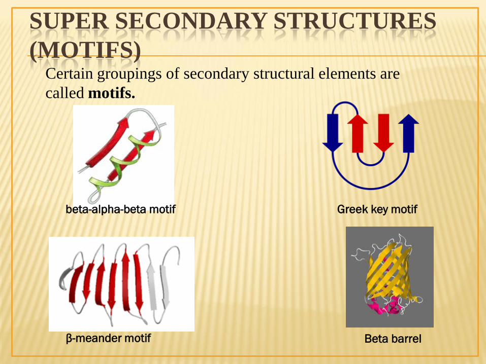
SUPER SECONDARY STRUCTURES
(MOTIFS)
Beta barrel β-meander motif
beta-alpha-beta motif Greek key motif
Certain groupings of secondary structural elements are
called motifs.
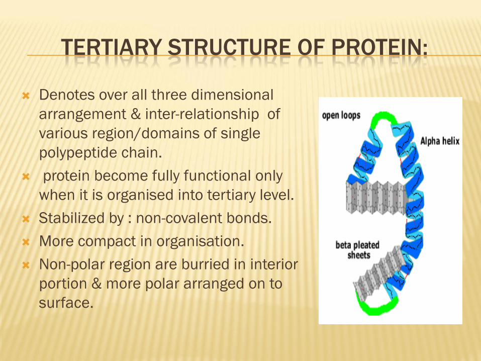
TERTIARY STRUCTURE OF PROTEIN:
Denotes over all three dimensional
arrangement & inter-relationship of
various region/domains of single
polypeptide chain.
protein become fully functional only
when it is organised into tertiary level.
Stabilized by : non-covalent bonds.
More compact in organisation.
Non-polar region are burried in interior
portion & more polar arranged on to
surface.
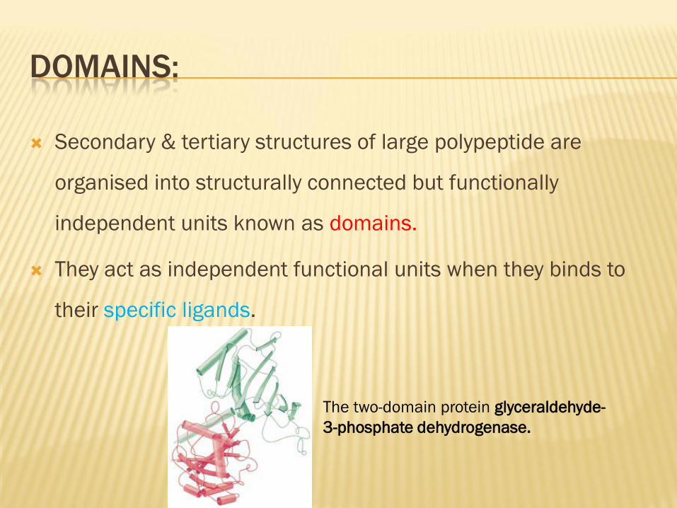
DOMAINS:
Secondary & tertiary structures of large polypeptide are
organised into structurally connected but functionally
independent units known as domains.
They act as independent functional units when they binds to
their specific ligands.
The two-domain protein glyceraldehyde-
3-phosphate dehydrogenase.
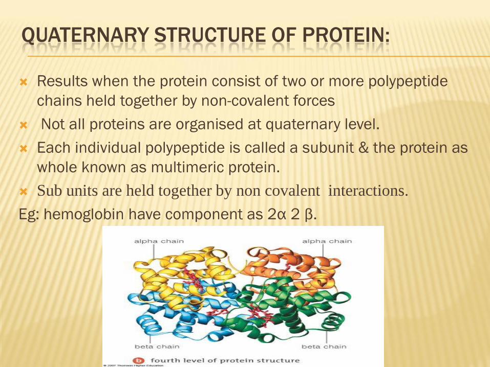
QUATERNARY STRUCTURE OF PROTEIN:
Results when the protein consist of two or more polypeptide
chains held together by non-covalent forces
Not all proteins are organised at quaternary level.
Each individual polypeptide is called a subunit & the protein as
whole known as multimeric protein.
Sub units are held together by non covalent interactions.
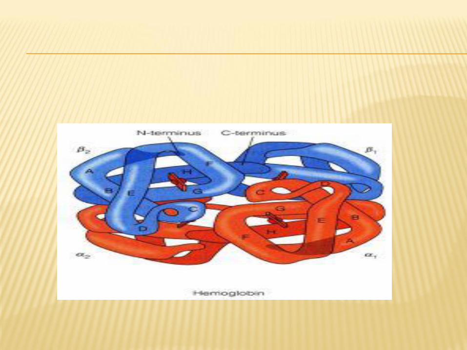
Eg: hemoglobin have component as 2α 2 β.


Hydrogen bond :electrical attraction between hydrogen atom in
a polar bond in one molecule & oxygen/nitrogen atom in a
polar bond of another molecule/ within the same molecule.
hydrogen donor & hydrogen acceptor

Hydrophobic interaction:
-Occur when interatomic distance as low as 3 to 4 A°
-b/w hydrophobic side chain of non-polar AA that reside close
to each other in the interior of protein structure.
-This interaction is not bcz of any attraction b/w non-polar
groups , but due to property of water molecule surrounding
them which push them together resulting in hydrophobic
interaction.
Electrostatic interaction;
-Occur b/w oppositely charged groups such as COO- & NH3+ of
basic amino acid
or
-b/w amino terminal & carboxyl group of protein which remain
on surface of protein donot interact with other charged group
from protein bcz of high dielectric constant of water molecule
near by.
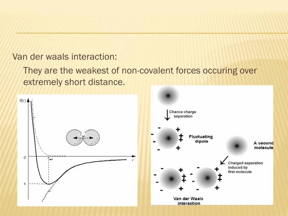
Van der waals interaction:
They are the weakest of non-covalent forces occuring over
extremely short distance.
The distance at which the attractive force b/w two atom is
maximal & repulsive force in minimal is termed as van der waal
contact distance which is sum of van der waal radii of two
atom.
They contribute for structural stability of protein bcz of cumulative
effect.