protective role of vitamin c and thyme extract (thymus
TRANSCRIPT

www.academicjournals.com

OPEN ACCESS Asian Journal of Animal and Veterinary Advances
ISSN 1683-9919DOI: 10.3923/ajava.2021.1.13
Research ArticleProtective Role of Vitamin C and Thyme Extract (Thymus vulgaris)on Chromium-Induced Toxicity in Catfish (Clarias gariepinus)1Gehan B.A. Youssef, 2Eman I. Soror, 3Seham Y. Abo Kora, 4Shimaa A.E. Atwa and 5Ahlam F. Hamouda
1,5Department of Forensic Medicine and Toxicology, Veterinary Teaching Hospital, Faculty of Veterinary medicine, Benha University, Egpyt2Department of Aquatic Animal Diseases and Management, Veterinary Teaching Hospital, Faculty of Veterinary Medicine, Benha University, Egypt3Department of Pharmacology, Veterinary Teaching Hospital, Faculty of Veterinary Medicine, Benha University, Egypt4Department of Biochemistry, Veterinary Teaching Hospital, Faculty of Veterinary Medicine, Benha University, Egypt
AbstractBackground and Objective: Chromium toxicity in aquatic environment may occur through industrial pollutions. The impact of its toxicitymay represent health hazards for aquatic environments and public health. Therefore, Clarias gariepinus were used to investigate thechromium toxicity and assess the ameliorative effect of thyme and vitamin C. Materials and Methods: A total of 90 fish were divided intosixequal groups (n = 15). Group 1 (control negative) was fed on basal diet without chromium in water. Group 2 (control positive) was fedon basal diet with Potassium Dichromate (PD) at dose 3.6 mg LG1 dissolved in water. Group 3 was fed on diet with vitamin C at dose200 mg kgG1 of diet without PD in water. Group 4 was fed on vitamin C diet with PD in water. Group 5 was fed on diet supplemented withThyme extract at dose 10 g kgG1 dry food without PD in water. Group 6 was fed on thyme diet with PC in water. The experiment was donefor a period of 30 days. Analyses of blood, antioxidant in tissues, micronucleus test, chromium tissue residues, comet assay andhistopathology were done. Results: There was a significant decrease (p<0.001) in RBCs count, Hb content, PCV% and WBCs count withsignificant increase (p<0.001) in liver and kidney function. There were increased activities of SOD, CAT, GSH and MDA and chromium tissueresidues. DNA damage and histopathological changes occurred in groups with chromium when compared to control negative group.Vitamin C and thyme extract decreased the toxic effect of chromium and improved biochemical parameters, with reduction of chromiumresidues in gills and muscles. Conclusion: Therefore, vitamin C and thyme extract can be used as ameliorative factors against chromiumtoxicity in aquaculture.
Key words: Catfish, chromium toxicity, oxidative damage, biochemical changes, vitamin C and thyme extract
Citation: Youssef, G.B.A., E.I. Soror, S.Y. Abo Kora, S.A.E. Atwa and A.F. Hamouda, 2021. Protective role of vitamin c and thyme extract (Thymus vulgaris)on chromium-induced toxicity in catfish (Clarias gariepinus). Asian J. Anim. Vet. Adv., 16: 1-13.
Corresponding Author: Eman I. Soror, Department of Aquatic Animal Diseases and Management, Veterinary Teaching Hospital, Faculty of Veterinary Medicine, Benha University, Egypt
Copyright: © 2021 Gehan B.A. Youssef et al. This is an open access article distributed under the terms of the creative commons attribution License, whichpermits unrestricted use, distribution and reproduction in any medium, provided the original author and source are credited.
Competing Interest: The authors have declared that no competing interest exists.
Data Availability: All relevant data are within the paper and its supporting information files.

Asian J. Anim. Vet. Adv., 16 (1): 1-13, 2021
INTRODUCTION
Fish are delicious foods that provide humans with highquality protein, various minerals, poly unsaturated fatty acidsand vitamins. In addition, Fish are considered as one of thevaluable bioindicators for pollution in the aquatic habitats asa result of their lower detoxification enzymes than those inmammals and thereby allowing a higher bioaccumulation fortoxicants1,2. The hazardous compounds that accumulate intheir tissues are directly or indirectly consumed by human andcan transform xenobiotic compounds into carcinogenic andmutagenic metabolites3. The Nile in Egypt facing majorenvironmental problems associated with the dispersal ordisposal of agricultural, industrial and urban wastes generatedby human activities4. These pollutants cause serious damageto aquatic life5. Toxic metals are widely found in aquaticenvironments. These compounds have been found to behighly toxic, genotoxic, mutagenic and carcinogenic. Amongthem, water pollution due to chromium (Cr) is regarded as oneof the main and most severe environmental problems6.
Chromium is considered as a heavy metal andenvironmental pollutant as well as an essential micronutrient7.In nature chromium does not exist as elementary form (Cr (0))but exists in Earth Crust in many oxidation states. Chromiumoxidation states range from divalent (Cr(II)) to hexavalent(Cr (VI)) but is most frequently found in the environment in thetrivalent (Cr (III))and hexavalent (Cr (VI))oxidation states. Thetrivalent and hexavalent forms are the most importantbecause other forms are unstable and are rapidly converted totrivalent chromium, which in turn is oxidized to hexavalentchromium8.
In aquatic environment chromium is found as Cr (III) andCr (VI) as water soluble complex anions. Cr has truly uniquetoxicological characteristics. Toxicological impact can resultfrom the action of Cr (VI) itself as an oxidizing agent9. Also, Cr(VI) is more toxic due higher solubility and mobility, ability topenetrate the cell membrane and strong oxidizing ability.The hexavalent form of chromium exists in the form ofchromate, hydro chromate or dichromate in dissolvedcondition as a component of a complex anion10. Potassiumdichromate (K2Cr2O7) is an inorganic soluble hexavalentchromium compound that is widely used in severalindustries11. Fish upon exposure to pollutants (Cr) can elicitthe production of Reactive Oxygen Species (ROS) likesuperoxide, hydrogen peroxide and hydroxyl radical as theROS levels increase, the biological system develops a firstline defense mechanism by modulating the activities ofantioxidants such as catalase (CAT), Superoxide Dismutase(SOD) and glutathione related enzymes12. The genotoxicity of
Cr(VI) has been reported in human and animals13,14. There aremany methods like micronucleus test and DNA damage assaysas Comet assay have been used for assessing genotoxicity ofvarious chemicals in different animals15,16.
Because of the hazardous effect of Cr in aquaculture,several studies were designed to find out protective agents.Vitamin C (Ascorbic acid) is an important antioxidantvitamin that is essential nutrient for optimum growth andmaintenance17. Another beneficial effect of vitamin C, whichhas been established in several animal species, including fish,is its role in enhancing the non-specific immune response18. Italso prevents oxidative renal and brain damage induced bystress and secures the body tissues against toxic effects ofheavy metals by efficiently metabolizing these toxicants.Ascorbic acid can reduce chromium-induced toxicity on liverand kidney19.
Thyme (Thymus vulgaris) is herbaceous plant of theplatoon species, grows in mountainous areas, used as abeverage instead of or with tea and added to some food togive it an acceptable flavor. the plant is used in folk medicinefrequently where it is prescribed to treat mouth infections,stomach, intestine and airways, coughing and gastroenteritisand expel intestinal worms as well as to strengthen theheartcontractility20. Fresh Thyme herb has one of the highestantioxidant levels among herbs. It is packed with minerals andvitamins that are essential for optimum health. Its leaves areone of the richest sources of potassium, iron, calcium,manganese, magnesium and selenium21. Extracts from Thymehave been used in traditional medicine for the treatment ofseveral respiratory diseases like asthma and bronchitis andfor the treatment of other pathologies thanks to severalproperties such as antiseptic, antispasmodic, antitussiveantimicrobial, antifungal, antioxidative and antiviral22.
Because Cr exposure is more likely to occur in aquaculturedue to heavy industrial pollution, it was very crucial toinvestigate some of natural protective agents that reduce itdeleterious effects. Therefore, the present study aimed toevaluate the toxic effect of potassium dichromate in catfishand amelioration and antagonizing this toxic effect by usingvitamin C and thyme extract.
MATERIALS AND METHODS
Study area and location: Clarias gariepinus, commonlyknown as African catfish was selected as an experimentalmodel. The fish within average weight 150±20 g werecollected from River Nile (El Rayah El Tewfik) of Qalubia, Egyptand transported alive to the laboratory of PhysiologyDepartment of Faculty of Veterinary Medicine, Benha
2

Asian J. Anim. Vet. Adv., 16 (1): 1-13, 2021
University where the experimental procedures were donefrom August 1-31, 2018. The fish were placed in large glasstanks disinfected with potassium permanganate to preventfungal infection and washed thoroughly prior to introductionof fish23. Fish were distributed equally to six groups and keptin glass tanks with tap water with a photoperiod 12 hrs lightand 12 hrs dark was maintained during the experiment24.Thefish were acclimated to the laboratory conditions and waterparameters for two weeks prior to the experiment. The tankwater was partially changed every day with aerated tap water.The water was continuously aerated. All groups were fed onbasal diet twice a day at a daily rate of 4% body weightthorough out the acclimatization period. The experiment wasconducted according to the rules of the Ethical Committee ofFaculty of Veterinary Medicine, Benha University.
Experimental design: The experiment was done on 90 fish ofClaris gariepinus, fish were divided into 6 groups (15 fish pergroup) as following: Group 1 represented as control groupthat fed only on basal diet. (Control negative), group 2 (controlpositive) was fed on basal diet and exposed to PD (Cr VI) (AlGomhorria Company, Egypt) dissolved in water at dose3.6 mg LG1 (1|10th of LC50) according to the median lethalconcentration dose25. Group 3 was fed on dietsupplemented with vitamin C (Al Kahera Pharm Company) at dose 200 mg kgG1 of diet as previously recorded26. Group 4was exposed to both vitamin C and Potassium dichromate atthe same doses described above, group 5 was fed on dietsupplemented with Thyme extract (Thymus vulgaris) at dose10 g kgG1 dry food27. Group 6 was exposed to both thymeextract and potassium dichromate at the same doses. Theoverall period of experiment was 30 days.
Preparation of the plant extracts (aqueous and alcoholic):Thymus vulgaris extract was prepared as previously reported28.The thyme extract was added to the fish diet as 10 g kgG1 dryfood.
Blood and tissue sampling: The blood samples were collectedat the end of the experiment (30 days), about 1 mL of bloodwas collected from the caudal vein of fish by caudal venouspuncture using sterile syringes29. Apart of the collected bloodplaced in tubes with EDTA used as anticoagulant while theother part of blood placed in sterile centrifuge tubes for serumseparation by centrifugation at 3500 rpm for 15 min. Thewhole blood was collected and immediately processed forMicronucleus test. On the final day of the experimental period,fish were sacrificed and the tissues dissected for variousanalysis as follows, Liver and kidney tissues samples were
collected and preserved in -80EC for determination ofantioxidant parameters and Comet assay. Muscles and gillssamples collected and preserved in deep freeze fordetermination of chromium residues. Tissue sample slicesfrom liver, kidney and gills were kept in 10% formalin forhistopathological examination.
Hematological and biochemical analysis: Hematologicalparameters were measured and analyzed including RedBlood Cells (RBCs) count, white blood cells (WBCs) count,Hemoglobin (Hb) and Packed cell volume (PCV%) Using theroutine methodology of fish hematology30,31. Biochemicalparameters were measured in the collected serum samplesincluding alanine aminotransferase (ALT), aspartateaminotransferase (AST)32, alkaline phosphatase (ALP), totalprotein, albumin and cholesterol33 and urea, creatinine anduric acid34. Antioxidant parameters were measured in liverand kidney tissues including SOD35, CAT activity36, ReducedGlutathione (GSH)37 and Malondialdehyde (MDA)24. Thehematological and biochemical analysis were done at theCentral Lab of Faculty of Veterinary Medicine, BenhaUniversity.
Micronucleus test: The peripheral blood samples weresmeared onto the clean glass slides from each group. Theslides were air dried for 24 hrs after fixation in pure ethanol for20 min. The smeared slide was then stained with 6% Giemsasolution for 25 min, then washed and dried. The slides wereexamined under microscope using oil immersion lens(100×magnification). A total of 2000 erythrocyte cells werescored from each slide for presence or absence of micronuclei.Small, extra-nuclear, circular or ovoid bodies were scored asmicronuclei6. Micronucleus test was done at veterinaryTeaching Lab of the Faculty of Veterinary Medicine, BenhaUniversity.
Chromium tissue residues: The samples were taken fromtissues of muscles and gills. About 5 g of tissue samples werewrapped in aluminum foil and stored on ice until transfer tofreezer and later on analyzed using atomic absorptionspectrophotometer in Soil, Water and Environment ResearchInstitution (SWERI) as recorded38 for detection of Cr residuesconcentration in tissues.
Comet assay: Comet assay was performed in liver tissues39 fordetection of DNA damage induced by hexavalent Cr. TailLength (TL) and Tail Moment (TM) were analyzed anddocumented. An increase in TL/TM on the slides is indicativefor the occurrence of direct DNA damage.
3

Asian J. Anim. Vet. Adv., 16 (1): 1-13, 2021
Histopathology: Different parts of liver, kidney and gills weretaken from the different groups. The specimens were fixedin Bouin's solution. After fixation, the specimens weredehydrated in ascending grades of ethanol, cleared in xyleneand embedded in paraffin wax. Paraffin sections were stainedas previously reported40.
Statistical analysis: Statistical analysis of data was performedand expressed by Means±SE using one way analysis ofvariance (ANOVA) to determine the significant differencesbetween the control and experimental groups. The differenceswere considered significant when p<0.001.
RESULTS
Clinical findings: After the end of the experiment, fish wereexamined for any abnormal appearance. During theexamination of fish, it was observed that there were manychanges on the external morphological appearance of theexamined cat fish, these changes were the presence of manyerosions in fins and fin rays in fish that exposed to potassiumdichromate.
Hematological findings: The data presented in Table 1showed that exposure to chromium toxicity in catfishdecreased the mean values of RBCs (106 mmG3), Hb (g dLG1),PCV% and WBCs (106 mmG3) count from 3.7, 7.66, 31.66 and
37.77 to 2.6, 6.1, 28.6, 32.93, respectively. Addition of vitaminC increased mean values of RBCs, Hb, PCV% and WBCs countcompared to group 2 exposed to chromium. Addition ofthyme extract increased RBCs, Hb, PCV% and WBCs countvalues compared to group 2 exposed to chromium group.
Biochemical analysis: Data presented in Table 2 showed thatexposure to chromium in catfish (group 2) increased valuesof liver function [AST (µ LG1), ALT(µ LG1), ALP(µ LG1) andcholesterol (mg dLG1)] and kidney function [urea, uric acid andcreatinine (mg dLG1)] from 61.44, 23.696, 32.69, 136.016,13.87, 3.28, 0.196 to 133.3, 40.31, 97.71, 293.33, 24.276, 4.69and 0.563, respectively. Addition of vitamin C significantlydecreased these values . The albumin, total protein IgA andIgG decreased in group 2 chromium exposed catfish.Whereas addition of vitamin C to chromium exposed groupsignificantly elevated the mean values of these parameters.Also, Addition of thyme extract significantly increased themean values of these parameters.
Antioxidant assays: The data presented in Table 3 showedthat exposure of catfish to chromium (group 2) increasedvalues of liver SOD (U gG1), CAT(U gG1), GSH(mg gG1) andMDA (nmol gG1) from 84.0±2.31, 4.35±0.030, 12.60±0.07,164.53±5.67 to 99.72±7.35, 4.95±0.03, 20.05±0.89,465.3±2.86, respectively. Addition of vitamin C to chromiumexposed group reduced these values to 98.48±0.37,
Table 1: Hematological findings of control and examined groups of catfishParameters G1 (n = 15) G2 (n = 15) G3 (n = 15) G4 (n = 15) G5 (n = 15) G6 (n = 15)RBCs count (106 mmG3) 3.7±0.057a 2.6±0.057d 3.5±0.115b 2.9±0.15cd 3.7±0.057c 3.0±0.057d
Hb (g dLG1) 7.66±0.33b 6.1±0.057d 8.26±0.375a 7.26±0.128ba 7.7±0.288ba 7.1±0.346ba
PCV (%) 31.66±0.837ab 28.6±0.208d 33.8±1.732a 30.00±1.558b 30.56±0.088b 29.86±0.260b
WBCs count (106 mmG13) 37.77±5.46b 32.93±2.97d 37.9±2.07a 34.8±0.866c 36.166±1.18b 35.10±1.44b
G1 (control negative): Basal diet without chromium in water, G2 (control positive): Basal diet with PD, G3: Diet with vitamin C without PD in water, G4: Diet with VitaminC with PD in water, G5: Diet with Thyme extract without PD in water, G6: Diet with vitamin Thyme extract with PD in water, means with different superscript letters inthe same row are significantly different at p<0.001
Table 2: Biochemical results of control and examined groups of catfishParameters G1 (n = 15) G2 (n = 15) G3 (n = 15) G4 (n = 15) G5 (n = 15) G6 (n = 15)AST (u LG1) 61.44±9.33d 133.3±4.9a 78.59±5.55cd 92.04±3.02bc 75.66±10.39b 109.00±5.04bcd
ALT (u LG1) 23.696±1.95b 40.31±3.92a 24.46±1.84b 28.39±3.12b 22.66±2.02b 32.74±5.87ab
ALP (u LG1) 32.69±3.83c 97.71±17.31a 54.16±3.06bc 98.66±18.52a 38.66±0.88c 75.60±6.35ab
Chol (mg dLG1) 136.016±8.14b 293.33±26.034a 155.74±13.21b 244.66±2.0a 173.22±8.90b 176.00±8.04b
Alb (mg mLG1) 30.33±1.039a 13.58±1.44c 20.19±1.605b 21.29±0.35b 15.14±1.47c 32.69±2.21aTP (mg mLG1) 50.55±5.164a 31.03±4.36b 45.51±2.28ab 37.04±6.69ab 43.88±4.02ab 51.77±3.91a
Urea (mg dLG1) 13.87±0.278b 24.276±2.94a 12.74±0.855b 14.92±2.61b 14.55±2.25b 16.56±2.80b
Uric acid (mg dLG1) 3.28±0.275b 4.69±0.514a 2.823±0.08bc 3.29±0.162b 2.21±0.22c 3.503±0.26b
Creatinine (mg dLG1) 0.196±0.023b 0.563±0.044a 0.15±0.047b 0.158±0.04b 0.13±0.026b 0.276±0.09b
IgA (mg mLG1) 4.65±0.475a 2.856±0.401b 4.186±0.210ab 3.40±0.61ab 4.046±0.36ab 3.763±0.35ab
IgG (mg mLG1) 3.06±0.313a 1.876±0.265b 2.753±0.140ab 1.99±0.48b 3.133±0.23a 2.66±0.24ab
Means with different superscript letters in the same row are significantly different at p<0.001
4

Asian J. Anim. Vet. Adv., 16 (1): 1-13, 2021
Table 3: Antioxidant results of liver of control and examined groups of catfishParameters G1 (n = 15) G2 (n = 15) G3 (n = 15) G4 (n = 15) G5 (n = 15) G6 (n = 15)SOD (U gG1) 84.00±2.31b 99.72±7.35a 103.68±2.49b 98.48±0.37b 84.0±2.31b 86.00±1.15b
CAT (U gG1) 4.35±0.030bc 4.95±0.03a 4.78±0.05ab 3.39±0.27d 4.01±0.17bc 3.73±0.12cd
GSH (mg gG1) 12.60±0.07b 20.05±0.89a 8.78±0.19c 12.52±0.28b 11.59±0.66b 12.61±0.46b
MDA (nmol gG1) 164.53±5.67d 465.3±2.86a 125.82±7.01e 268.89±1.78b 123.72±1.71d 226.38±8.54c
Means with different superscript letters in the same row are significantly different at p<0.001
Table 4: Antioxidant results of kidneys of control and examined groups of catfishParameters G1 (n = 15) G2 (n = 15) G3 (n = 15) G4 (n = 15) G5 (n = 15) G6 (n = 15)SOD (U gG1) 38.00±3.46c 88.00±2.3a 70.00±1.15b 56.00±9.24b 26.00±3.46c 62.50±0.88b
CAT (U gG1) 3.53±0.14cb 4.93±0.53a 3.84±0.09bc 4.07±0.22ba 4.28±0.18ba 2.83±0.51c
GSH (mg gG1) 31.27±0.18a 32.52±0.29a 28.47±2.08a 12.35±2.93b 22.53±6.06a 25.98±3.21a
MDA (nmol gG1) 127.00±11.67b 370.60±50.14a 188.29±7.12b 212.31±23.56b 190.57±4.1a 316.50±36.27b
Means with different superscript letters in the same row are significantly different at p<0.001
Table 5: Chromium residues in muscles and gills of control and examinedcatfish
Parameters G2 G4 G6Muscle (mg kgG1) 533.0±14.17b 189.1±8.180a 184.0±4.50c
Gills (mg kgG1) 3021.0±01.59a 2494.0±2.05b 1066.0±76.02c
Means with different superscript letters in the same row are significantly differentat p<0.001
3.39±0.27, 12.52±0.28, 268.89±1.78, respectively. Additionof thyme extract to chromium exposed group decreasedthese values to 86.0±1.15, 3.73±0.12, 12.61±0.46 and226.38±8.54, respectively.The data presented in Table 4 showed that exposure to
chromium in catfish (group 2) increased mean values ofkidney SOD (U gG1), CAT (U gG1), GSH (mg gG1) andMDA (nmol gG1) to 88.0, 4.93, 32.52, 370.60, respectively.Addition of vitamin C to chromium exposed group reducedthese values to 56.0, 4.07, 12.35 and 212.31, respectively.Addition of thyme extract to chromium exposed groupdecreased the mean values of these parameters.
Micronucleus test: The micronucleus was demonstrated inRBCs as shown in Fig. 1a and b. The occurrence ofmicronucleus was increased from 2.0±0.288 to 23.0±1.58after exposure to chromium. Addition of vitamin C tochromium exposed group reduced the micronucleus to12.66±1.45. Addition of thyme extract to chromium exposedgroup decreased the micronucleus to 13.0±1.154.
Chromium residues: The data presented in Table 5 showedthat addition of vitamin C and thyme extract to chromiumexposed catfish reduced the chromium residues in musclefrom and in gills.
Comet assay: The data presented in Table 6 showed that catfish exposed to chromium had increase in tail length, DNA%,Tail moment and olive moment from. Addition of vitamin C to
Fig. 1(a-b): (a) Normal RBCs without micronucleus and (b)RBCs with micronucleus (arrow)
chromium exposed catfish reduced these values. Also,addition of thyme extract to chromium exposed catfishreduced the mean values of these measurements.
5
(a)
(b)

Asian J. Anim. Vet. Adv., 16 (1): 1-13, 2021
Fig. 2(a-e): Ethidium bromide images of comet assay (a) G1 (Control) showing normal intact cells, (b) G2 (PD-treated) showing DNA damage in PD groups, (c) G2 (PD-treated) showing DNA damage with highmagnification, (d) G4 (vitamin C-PD group) showing slightly damaged DNA, (e and f) G6 (thyme-PD group) showing slightly damaged DNA
Table 6: Comet assay finding of liver of control and examined catfishParameters G1 (n = 15) G2 (n = 15) G3 (n = 15) G4 (n = 15) G5 (n = 15) G6 (n = 15)Tail length 6.75±0.09a 4.03±1.16a 6.32±0.65a 4.56±0.24a 7.23±1.13a 4.53±0.54a
DNA (%) 6.25±1.19b 5.94±0.67b 6.45±0.25b 4.72±0.53b 9.06±0.56a 5.03±0.45b
Tail moment 0.57±0.214ba 0.26±0.061cb 0.34±0.014ba 0.19±0.040c 0.76±0.074a 0.30±0.046cb
Olive moment 0.81±0.061b 0.67±0.0896b 0.87±0.003b 0.46±0.0200c 1.62±0.118a 0.73±0.338b
Means with different superscript letters in the same row are significantly different at p<0.001
In comet assay, the slides were stained with ethidiumbromide and examined under fluorescent microscope anddemonstrated the presence of damage in DNA (Fig. 2). Thisdamage differs in different groups. Group 1 (control) showednormal intact cells and no damage of DNA reported (Fig. 2a).While group 2 (PD-treated) showed presence of damagein DNA appeared in form of tail length (Fig. 2b and c, lowand high magnification, respectively). While group 4(Fig. 2d and 6 (Fig. 2e and f) showed slight damage in DNA.
Histopathological findings: Figure 3 showed thehistopathological changes in liver tissues. In group1 (control),liver tissue consisted of central vein which lined by simplesquamous cells, from the central vein numerous radiatinghepatic cords which appeared as polygonal in shape with
deep basophilic centrally located nuclei and faint acidophiliccytoplasm (Fig. 3a). While in group 2, liver tissue wascharacterized by thickened wall central vein, the hepatocytesshowed disruption with pyknotic nuclei and disintegratedcytoplasm. The hepatic blood vessels showed vascularchanges include hemorrhage and edema (Fig. 3b). Liver tissuein group 3 and 5 appeared as normal structure and normalarchitecture as in control group (Fig. 3c and e). Group 4showed thick wall and hemorrhage in the central vein(Fig. 3d). Group 6 showed thick wall central vein and increasedconnective tissue fibers between hepatic cords (Fig. 6f).Histopathological findings of kidney tissues were shown
in Fig. 4. In group 1, renal tissue consisted of numerousnephrons which consisted of glomeruli, proximal convolutedtubules, distal convoluted tubules and collecting tubules, in
6
(a) (b) (c)
(d) (e) (f)

Asian J. Anim. Vet. Adv., 16 (1): 1-13, 2021
Fig. 3(a-f): Photomicrograph of liver tissue (a) Normal hepatic tissue with central vein (C), (b) Pyknotic nuclei (p) and disintegrated cytoplasm (d), (c) Normal central vein (c) (d) Hemorrhage in centralwall (h), (e) Normal central vein C and (f) Increased CT between hepatic cords (d). H and E ×10 all while No.2 ×40
between them the haemopoietic tissue was located(Fig. 4a). While in group 2, kidney showed destruction andnecrosis in renal tubules and spread of patches ofeosinophilic exudate between renal tissues (Fig. 4b). Renaltissue in group 3 and 5 consisted of normal structure asin control group with prominent haemopoietic tissue
(Fig. 4c and e). Kidney in group 4 showed desquamation ofsome epithelial tissue and pyknosis of some nuclei whilethe other renal tissues appeared normal (Fig. 4d). Group 6showed desquamation of epithelial tissue of renal tubuleswith prominent haemopoietic tissue distributed betweenthem (Fig. 4f).
7
(a) (b)
(c) (d)
(e) (f)

Asian J. Anim. Vet. Adv., 16 (1): 1-13, 2021
Fig. 4(a-f): Photomicrograph of renal tissue(a) Normal renal tissues consisted of glomeruli (g), proximal convoluted tubules (p) and distal convoluted tubules (d), (b) Eosinophilic exudate betweenrenal tissues (C), (c) Normal structure consisted of glomeruli (g), (d) Proximal convoluted tubules (p) and distal convoluted tubules (d), (e) Desquamationof some renal tissues (d) and pyknotic nuclei (p) and (f) Prominent haemopoietic tissues (arrow). H and E×20
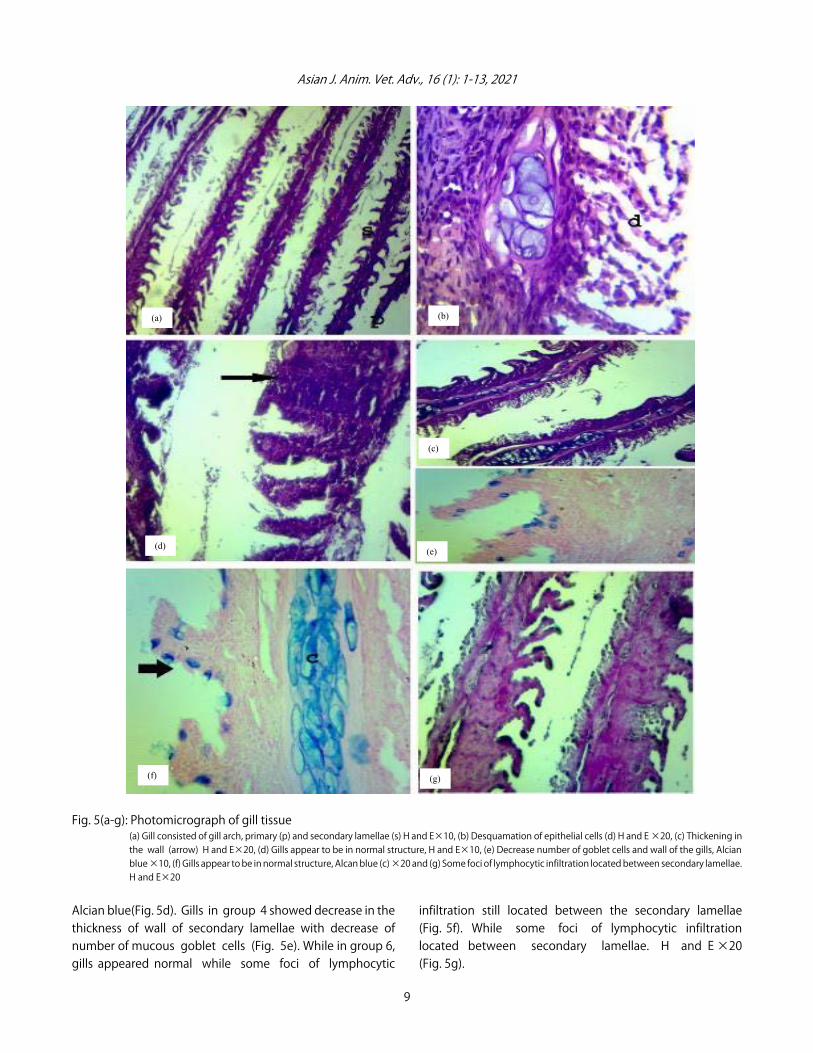
Histopathological results of gills were shown in Fig. 5. Gills ingroup 1 showed gill arch, primary and secondary lamellae, thesecondary lamellae lined by mucous cells and pillar cells(Fig. 5a). While in group 2, gills showed desquamation theepithelial cells, thickening in the wall (hyperplasia) of
secondary lamellae and circumscribed area of lymphocyticinfiltration was located between the secondary lamellae which destruct the underlying structures (Fig. 5b, c). Ingroup 3 and 5 gills appeared as normal structure as incontrol group with numerous goblet cells which gave positive
8
(a) (b)
(c) (d)
(e) (f)
Asian J. Anim. Vet. Adv., 16 (1): 1-13, 2021
Fig. 5(a-g): Photomicrograph of gill tissue (a) Gill consisted of gill arch, primary (p) and secondary lamellae (s) H and E×10, (b) Desquamation of epithelial cells (d) H and E ×20, (c) Thickening inthe wall (arrow) H and E×20, (d) Gills appear to be in normal structure, H and E×10, (e) Decrease number of goblet cells and wall of the gills, Alcianblue ×10, (f) Gills appear to be in normal structure, Alcan blue (c) ×20 and (g) Some foci of lymphocytic infiltration located between secondary lamellae.H and E×20
Alcian blue(Fig. 5d). Gills in group 4 showed decrease in thethickness of wall of secondary lamellae with decrease ofnumber of mucous goblet cells (Fig. 5e). While in group 6,gills appeared normal while some foci of lymphocytic
infiltration still located between the secondary lamellae(Fig. 5f). While some foci of lymphocytic infiltrationlocated between secondary lamellae. H and E ×20(Fig. 5g).
9
(a) (b)
(d) (e)
(f) (g)
(c)

Asian J. Anim. Vet. Adv., 16 (1): 1-13, 2021
DISCUSSION
Chromium is one of the important pollutants that affectaquatic environment. It exerts its adverse effect directly or indirectly on human health. The more toxic form of chromiumis known as hexavalent chromium10. Catfish was chosen as amodel for the experimental study because it can tolerate andsurvive in many of experimental conditions, it can also bedistributed in wide areas of water41. In this study chromiumexposure for 30 days leads to changes in fish appearance,morphology, blood analysis, cytology and histology42.Presence of erosions in fins and fin rays was the predominantclinical signs appeared on fish exposed to chromium. Thesesymptoms are supported by the different previous studies10.Blood analysis of fish differs according to fish species, sex,
age and condition of health43. The hematological andbiochemical studies in fish are considered as importantparameters for evaluation of health status of fish exposed tometallic stress44. The chromium induced toxicity in this studyresults characterized by decrease in RBCs, Hb, PCV and WBCs.These results agreed with other different studies45-48. Theyadded that the decrease in hematological parameters may beattributed to the toxic effect of chromium that affect themetabolic and hematopoietic system. The liver and kidneyfunction tests showed different changes due to the toxicityinduced by chromium. The increase in AST and ALT in thepresent study is augmented by previous study47. The increasein cholesterol may be attributed to prolonged exposure ofchromium that cause difficult adaptation and weaknessbecause of metallic stress44. The decrease in protein andalbumin could be attributed to the stress of toxin causing liverhypofunction or impairment in protein synthesis48,49,10. The useof vitamin C and thyme extract in our study decrease thetoxicity of chromium by enhancing the fish hematology26 andimproving the liver and kidney function50. The protein andalbumin were increased after adding of vitamin C and thymeextract in diet and this due to serum protein are sensitive tonutritional influences51,52.The immunity of fish in this study was affected by long
exposure to chromium and manifested in form of decreasingin immunoglobulin and this result is agreed with otherstudies42,53. The decrease in immunity may be attributed totoxin stress that lead to production of oxidative effect whichin turn causing reduction or suppression of primary antibodyresponse. While vitamin C and thyme extract in our studyelevated the immunoglobulin levels and improve immuneresponse of catfish54-57.The present study revealed that the exposure to
chromium for 30 days caused an increase in metallic stress in
catfish and this stress lead to production of ROS the body ofthe fish cause a defense mechanism against this oxidativestress via production of antioxidant enzymes which work toremove the damage effect of this stress, so this explained theincrease of SOD, CAT, GSH and MDA in our study whichshowed agreement with58,12,59. Vitamin C and thyme extractwhen given in this study act as antioxidant by increasingantioxidant activities and decreasing the oxidative stress inform of decrease MDA which agreed with studies41,60.The micronucleus test was used for detection of fish
genotoxicity in polluted aquatic area61. In the present study,there was significant increase in micronuclei in fisherythrocytes and this result coincided with previous studies6,62.Accumulation of chromium was recorded by detection ofchromium residues in fish tissues. In this study, the highestconcentration of chromium residues in fish gills while thelowest levels of residues were founded in fish muscles. Thisresult is augmented by the other studies8,9. Vitamin C andthyme extract in current results decreased chromium residuesby acting as reducing agent, decrease accumulation andpersistence of chromium in tissues as previously explained63,64.Comet assay was used for detection of DNA damage65. In thepresent study, there was damage in DNA appeared inchromium group and this genotoxic damage was similar tothat previously reported66,24. They added that the chromiuminduced metallic stress which causing induction of relativeoxygen species which attack DNA and enhancing DNAdamage. Vitamin C and thyme extract decreased oxidativestress and decrease DNA damage in tissues and this issupported by the findings of other studies67,68.
Histopathological examination of organs was used as abiomarker for evaluation of the toxicity of differentpollutants69,70. Chromium in this study induced markedchanges and damage in liver, kidney and gills. These changesin form of degeneration and necrosis of tissues may be due toaccumulation of inflammatory cells associated with chromiumtoxicity. These results are lined with previous studies71,72, 73.Theaddition of vitamin C and thyme extract had ameliorativeeffect since it minimized the hazard effect of chromium on thetissues.
CONCLUSION
Chromium exposure in catfish induced clinical,hematological, biochemical and histopathological changes,especially in liver, kidney and gills. The use of vitamin C andthyme extract have a protective and ameliorative effectagainst toxicity induced by potassium dichromate incatfish.
10

Asian J. Anim. Vet. Adv., 16 (1): 1-13, 2021
SIGNIFICANCE STATEMENT
This study discovered the addition of vitamin C or thymeextract can be beneficial for protection of catfish fromchromium toxicity by potentiating the liver and kidneyfunction and decreasing the oxidant stress. This study will helpthe researchers to uncover the critical areas where chromiumtoxicity may occur that many researchers were not able toexplore. Thus a new theory on DNA adducts and micronucleuschanges in cat fish caused by chromium exposure in industrialarea may be arrived at.
ACKNOWLEDGMENT
The authors would like to thank the staff members ofcentral lab, physiological lab and veterinary teaching lab ofFaculty of Veterinary Medicine, Benha University for theirhelping in analyses.
REFERENCES
1. Sankar, T.V., A.A. Zynudheen, R. Anandan andP.G. Viswanathan Nair, 2006. Distribution of organochloridepesticides and heavy metal residues in fish and shellfish fromCalicut region, Kerala, India. Chemosphere, 65: 583-590.
2. Davodi, M., A. Esmaili-Sari and N. Bahramifarr, 2011.Concentration of polychlorinated biphenyls andorganochlorine pesticides in some edible fish species fromthe Shadegan Marshes (Iran). Ecotoxicol. Environ. Saf.,74: 294-300.
3. Ergene, S., T. ÇavaÕ, A. Çelik, N. Köleli and C. Aymak, 2007.Evaluation of river water genotoxicity using the piscinemicronucleus test. Environ. Mol. Mutagen., 48: 421-429.
4. Abdel-Mohsien, H.S. and M.A. Mahmoud, 2015. Accumulationof some heavy metals in Oreochromis niloticus from the Nilein Egypt: Potential hazards to fish and consumers. J. Environ.Protect., 6: 1003-1013.
5. Karbassi, A.R., I. Bayati and F. Moattar, 2006. Origin andchemical partitioning of heavy metals in riverbed sediments.Int. J. Environ. Sci. Technol., 3: 35-42.
6. Kumar, P., R. Kumar, N.S. Nagpure, P. Nautiyal, A. Dabas,B. Kushwaha and W.S. Lakra, 2012. Genotoxic and mutagenicassessment of hexavalent chromium in fish following in vivochronic exposure. Hum. Ecol. Risk Assess.: Int. J., 18: 855-870.
7. Aye, B.Y., T. Cemal and T. Tahsin, 2010. Uptake anddistribution of hexavalent chromium in tissues (gill, skinand muscle) of a freshwater fish, Tilapia, Oreochromis aureus.J. Environ. Chem. Ecotoxicol., 2 : 28-33.
8. Pourahmad, J., M. Rabiei, F. Jokar and P.J. O’Brien, 2005. Acomparison of hepatocyte cytotoxic mechanisms forchromate and arsenite. Toxicology, 206: 449-460.
9. Popa, M.O., 2006. Chromium impact on marine ecosystem.Buletin USAMV-CN, 63: 379-384.
10. Bakshi, A. and A.K. Panigrahi, 2018. A comprehensive reviewon chromium induced alterations in fresh water fishes.Toxicol. Rep., 5: 440-447.
11. Arivarasu, N.A., S. Fatima and R. Mahmood, 2008. Oraladministration of potassium dichromate inhibits brush bordermembrane enzymes and alters anti-oxidant status of ratintestine. Arch. Toxicol., 82: 951-958.
12. Roberts, A.P. and J.T. Oris, 2004. Multiple biomarker responsein rainbow trout during exposure to hexavalent chromium.Comp. Biochem. Physiol. C: Toxicol. Pharmacol., 138: 221-228.
13. Maeng, S.H., H.W. Chung, K.J. Kim, B.M. Lee, Y.C. Shin,S.J. Kim and I.J. Yu, 2004. Chromosome aberration and lipidperoxidation in chromium-exposed workers. Biomarkers,9: 418-434.
14. Patlolla, A.K., C. Barnes, C. Yedjou, V.R. Velma andP.B. Tchounwou, 2009. Oxidative stress, DNA damage andantioxidant enzyme activity induced by hexavalent chromiumin Sprague-Dawley rats. Environ. Toxicol., 24: 66-73.
15. Cavas, T. and S.E. Gazukara, 2005. Induction of micronucleiand nuclear abnormalities in Oreochromis niloticus followingexposure to petroleum refinery and chromium processingplant effluents. Aquatic Toxicol., 74: 264-271.
16. Vinod, T. and R.R. Kanhere, 2014. Comparative study onthe protective role of vitamin C and vitamin E on mercuryinduced toxicity in Heteropneusts fossilis. Int. Res. J. Sci. Eng.,2 : 37-43.
17. Dhawan, A., M. Bajpayee and D. Parmar, 2009. Comet assay:A reliable tool for the assessment of DNA damage in differentmodels. Cell Biol. Toxicol., 25: 5-32.
18. Mazik, P.M., J.R. Tomasso and T.M. Brandt, 1987. Effects ofdietary vitamin C on growth, caudal fin development andtolerance of aquaculture related stressors in channel catfish.Progressive Fish-Culturist, 49: 13-16.
19. Tewary, A. and B.C. Patra, 2008. Use of vitamin C as animmunostimulant. effect on growth, nutritional quality andimmune response of Labeo rohita (Ham.). Fish Physiol.Biochem., 34: 251-259.
20. Pechova, A. and L. Pavlata, 2007. Chromium as an essentialnutrient: A review. Vet. Med., 52: 1-18.
21. Mohamed, A., A. Mohamed and A.A. Omar, 2013. A study tofind thyme oil dose that kill 50% of mice and minimal dosethat kill all mice and maximum nonlethal dose. Nat. Sci.,11: 52-53.
22. Sharangi, A.B. and S. Guha, 2013. Wonders of leafy spices:Medicinal properties ensuring human health. Sci. Int.,1: 312-317.
23. Ocaña, A. and G. Reglero, 2012. Effects of thyme extract oils(from Thymus vulgaris, Thymus zygis and Thymus hyemalis)on cytokine production and gene expression of oxLDL-stimulated THP-1-macrophages. J. Obesity, 2012: 1-11.
11

Asian J. Anim. Vet. Adv., 16 (1): 1-13, 2021
24. Ali, D., N.S. Nagpure, S. Kumar, R. Kumar, B. Kushwaha andW.S. Lakra, 2009. Assessment of genotoxic and mutageniceffects of chlorpyrifos in freshwater fish channa punctatus(bloch) using micronucleus assay and alkaline single-cell gelelectrophoresis. Food Chem. Toxicol., 47: 650-656.
25. Velma, V. and P.B. Tchounwou, 2010. Chromium-inducedbiochemical, genotoxic and histopathologic effects in liverand kidney of goldfish, Carassius auratus. Mutat. Res. Genet.Toxicol. Environ. Mutagen., 698: 43-51.
26. Johnson, C. and M.V. Radhakrishnan, 2015. Estimation ofacute toxicity of chromium to the fresh water catfish Clariasbatrachus (Linn.). Int. J. Res., 1 : 30-37.
27. Asaikkutti, A., P.S. Bhavan, K. Vimala, M. Karthik andP. Cheruparambath, 2016. Effect of different levels dietaryvitamin C on growth performance, muscle composition,antioxidant and enzyme activity of freshwater prawn,Macrobrachium malcolmsonii. Aquacult. Rep., 3: 229-236.
28. Y2lmaz, S., S. Ergün and E.Ô. Çelik, 2013. Effect of dietaryherbal supplements on some physiological conditions of seabass Dicentrarchus labrax. J. Aquat. Anim. Health, 25: 98-103.
29. Fayad, N.K., O.H.S. Al-Obaidi, T.H. Al-Noor and M.O. Ezzat,2013. Water and alcohol extraction of thyme plant (Thymusvulgaris) and activity study against bacteria, tumors and usedas anti-oxidant in margarine manufacture. Innovat. Syst.Design Eng., 4: 41-51.
30. Bucker, A., M. Carvalho, M. Conceição and J. Alves-Gomes,2012. Micronucleus test and comet assay in erythrocytes ofthe Amazonian electric fish Apteronotus bonapartii exposedto benzene. Ecotoxicol. Environ. Contam., 7 : 65-73.
31. Svobodova, Z., 2001. Stress in fishes (a review). Bull VURHVodnany, 4 : 169-191.
32. Vinodhini, R. and M. Narayanan, 2009. The impact of toxicheavy metals on the hematological parameters in commoncarp (Cyprinus carpio L.). Iran. J. Environ. Health Sci. Eng.,6: 23-28.
33. Huang, X.J., Y.K. Choi, H.S. Im, O. Yarimaga, E. Yoon andH.S. Kim, 2006. Aspartate aminotransferase (AST/GOT) andalanine aminotransferase (ALT/GPT) detection techniques.Sensors, 6: 756-782.
34. Young, D.S. and R.B. Friedman, 2001. Effects of Disease onClinical Laboratory Tests. 4th Edition, Vol. 1, 2. AACC Press,Washington, DC. USA., ISBN-13: 9781890883454, pp: 682-683.
35. Nkeck, J.R., M. Singwé-Ngandeu, V.A. Moor, J.P. Chedjou,M.S. Doualla and W.F. Mbacham, 2017. Evaluation of oxidativestatus of patients with gout attack in a cameroonian urbanhospital. Open Journal Rheumatol. Autoimmune Dis.,07: 111-119.
36. Sun, Y., H. Yu, J. Zhang, Y. Yin, H. Shen, H. Liu and X. Wang,2006. Bioaccumulation and antioxidant responses ingoldfish Carassius auratus under HC orange No. 1 exposure.Ecotoxicol. Environ. Saf., 63: 430-437.
37. Mahmoud, H.H., 2016. New method for assessment of serumcatalase activity. Indian J. Sci. Technol., Vol. 9 10.17485/ijst/2016/v9i4/80499.
38. Lahouel, M., S. Boulkour, N. Segueni and J.P. Fillastre, 2004.The flavonoids effect against vinblastine, cyclophosphamideand paracetamol toxicity by inhibition of lipid-peroxydationand increasing liver glutathione concentration. PathologieBiologie, 52: 314-322.
39. Malik, N., A.K. Biswas, T.A. Qureshi, K. Borana and R. Virha,2010. Bioaccumulation of heavy metals in fish tissues of afreshwater lake of Bhopal. Environ. Monit. Assess.,160: 267-276.
40. Figueiredo-Fernandes, A., J.V. Ferreira-Cardoso, S. Garcia-Santos, S.M. Monteiro, J. Carrola, P. Matos and A. Fontainhas-Fernandes, 2007. Histopathological changes in liver and gillepithelium of Nile tilapia, Oreochromis niloticus, exposed towaterborne copper. Pesq. Vet. Bras., 27: 103-109.
41. Bancroft, J.D. and M. Gamble, 2008. Theory and Practice ofHistological Techniques. 6th Edn., Elsevier Health Sciences,Philadelphia, PA., ISBN-13: 9780443102790, Pages: 725.
42. Adham, K.G., H.O. Hashem, M.B. Abu-Shabana and A.H. Kamel,2000. Vitamin C deficiency in the catfish Clarias gariepinus.Aquacult. Nutr., 6: 129-139.
43. Arunkumar, R.I., P. Rajasekaran and R.D. Michael, 2000.Differential effect of chromium compounds on the immuneresponse of the African mouth breeder Oreochromismossambicus (Peters). Fish Shellfish Immunol., 10: 667-676.
44. Dorojan, O.G.V., V. Cristea, M. CreÛu, L. Dediu, A.I. Docan andM.T. Coad|, 2015. The effect of thyme (Thymus vulgaris) andvitamin E on the Acipenser stellatus juvenile welfare,reared in a recirculating aquaculture system. AACL Bioflux,8: 150-158.
45. Javed, M. and N. Usmani, 2019. An overview of the adverseeffects of heavy metal contamination on fish health.Proc. Natl. Acad. Sci. India, Sect. B Biol. Sci., 89: 389-403.
46. Venkatachalam, T. and A.V. Natarajan, 2014. Haematologicalinvestigation on fresh waterteleostLabeorohita (Ham.)following aquatictoxicities of Cr (III) and Cr (VI). Int. J. Res.Biosci., 3: 1-14.
47. Naz, S., R. Hussain, Q. Ullah, A.M.M. Chatha, A. Shaheen andR.U. Khan, 2020. Toxic effect of some heavy metals onhematology and histopathology of major carp (Catla catla).Environ. Sci. Pollut. Res., Vol. 2020. 10.1007/s11356-020-10980-0.
48. Vutukuru, S.S., N.A. Prabhat, M. Raghavender and A. Yerramilli,2007. Effect of arsenic and chromium on the serum amino-transferases activity in Indian major carp, Labeo rohita. Int. J.Environ. Res. Public Health, 4: 224-227.
49. Bernet, D., H. Schmidt, T. Wahli, P. Burkhardt-Holm, 2001.Effluent from a sewage treatment works causes changes inserum chemistry of brown trout (Salmo trutta L.). Ecotoxicol.Environ. Saf., 48: 140-147.
12

Asian J. Anim. Vet. Adv., 16 (1): 1-13, 2021
50. Madhavan, P. and K.Elumalai, 2016. Effects of chromium (VI)on haematological parameters in catfish, clariasbatrachus(Linnaeus, 1758) (Actinopterygii: Siluriformes). Int. J. Adv. Res.Biol. Sci., 3: 62-70.
51. Vutukuru, S.S., 2003. Chromium induced alterations insome biochemical profiles of the Indian major carp,Labeorohita (Hamilton). Bull. Environ. Contam. Toxicol.,70: 118-123.
52. Saleh, H., J.K. Azizollah, H. Ahmadreza, A. Raham, 2015. Theapplication of Thymus vulgaris in traditional and modernmedicine: A review. Global J. Pharmacol., 9: 260-266.
53. Tollba, A.A.H., S.A.M. Shabaan and M.A.A. Abdel, 2010.Effects of using aromatic herbal extract and blended w2thorganic acids on productive and physiological performanceof poultry 2 - The growth during cold winter stres. EgyptPoult. Sci., 30: 229-248.
54. Prabakaran, M., C. Binuramesh, D. Steinhagen andR.D. Michael, 2007. Immune response in the tilapia,Oreochromis mossambicus on exposure to tannery ef uent.Ecotoxicol. Environ. Saf., 68: 372-378.
55. Murai, T., J.W. Andrews and J.C. Bauernfeind, 1978. Use ofL-ascorbic acid, ethocel coated ascorbic acid and ascorbate2-sulfate in diets for channel catfish, Ictalurus punctatus.J. Nutr., 108: 1761-1766.
56. Emeish, W.F.A. and A.G.S. El-deen, 2016. Immunomodulatoryeffects of thyme and fenugreek in sharptooth catfish,Clarias gariepinus. Assiut Vet. Med. J., 62: 1-7.
57. Hansen, B.H., O.A. Garmo, P.A. Oisvik and R.A. Anderson, 2007.Gill metal binding and stress gene transcription in browntrout (Salmo trutta) exposed to metal environments: Theeffect of pre-exposure in natural populations. Environ. ToxicolChem., 26: 944-953.
58. Ritola, O., D.R. Livingstone, L.D. Peters and P. Lindstrom-Seppa, 2002. Antioxidant processes are affected in juvenilerainbow trout (Oncorhynchus mykiss) exposed to ozone andoxygen supersaturated water. Aquaculture, 210: 1-19.
59. Rajeshkumar, S., Y. Liu, J. Ma, H.Y. Duan and X. Li, 2017. Effectsof exposure to multiple heavy metals on biochemical andhistopathological alterations in common carp, Cyprinuscarpio L. Fish Shellfish Immunol., 70: 461-472.
60. Komes, D., A. Belscak Cvitanovic, D. Horzic, G. Rusak,S. Likic and M. Berendika, 2011. Phenolic composition andantioxidant properties of some traditionally used medicinalplants affected by the extraction time and hydrolysis.Phytochem. Anal., 22: 172-180.
61. Kushwaha, B., N.S. Nagpure, S. Srivastava, K. Ravindra andM.S. Verma, 2003. Variation of micronuclei in peripheralblood cells of Channa punctatus. Indian J. Anim. Sci.,73: 1192-1193.
62. Arunachalam, K.D., S.K. Annamalai and J.K. Kuruva, 2013.In-vivo evaluation of hexavalent chromium induced dnadamage by alkaline comet assay and oxidative stress incatla catla. Am. J. Environ. Sci., 9: 470-482.
63. Poljšak, B., Z. Gazdag, Š. Jenko-Brinovec, Š. Fujs andM. Pesti et al., 2005. Prooxidative vs antioxidative propertiesof ascorbic acid in chromium(VI)- induced damage: An in vivoand in vitro approach. J. Appl. Toxicol., 25: 535-548.
64. Reddy, V.P., K.R. Vital, P.V. Varsha and S. Satyam, 2014.Review on Thymus vulgaris traditional uses andpharmacological properties. Med. Aromat Plants, Vol. 3.10.4172/2167-0412.1000164.
65. Buschini, A., P. Carboni, A. Martino, P. Poli and C. Rossi, 2003.Effect of temperature on baseline and genotoxicant-iducedDNA Damage in haemocyte of Dreissena polymorpha. Mutat.Res./Genet.Toxicol. Environ. Mutagen., 537: 81-92.
66. Ahmad, I., V.L. Maria, M. Oliveira, M. Pacheco and M.A. Santos,2006. Oxidative stress and genotoxic effects in gill and kidneyof Anguilla anguilla L. exposed to chromium with or withoutpre-exposure to $-naphthoflavone. Mutat. Res./Genet.Toxicol. Environ. Mutagen., 608: 16-28.
67. Dauqan, E.M.A., A. Abdullah and H. Abdullah, 2012. Effect ofdifferent concentrations of red palm olein and differentvegetable oils on antioxidant enzymes in normal and stressedrat. BoD-Books on Demand, Norderstedt, Germany, ISBN:9789535107897 Pages: 412.
68. Ibrahim, S.S, A.M. Said and M. Aboubakr, 2018. Ameliorativeeffect of ascorbic acid and/or ginseng extract against thyroidgland toxicity induced by potassium dichromate in rats. J.Pharmacol. Clin. Res., Vol. 5. 10.19080/JPCR.2018.05.555655.
69. Adams, S.M., 2002. Biological Indicators of Aquatic EcosystemStress. American Fisheries Society, Bethesda, MD., USA.,pp: 644.
70. Mela, M., M.A.F. Randi, D.F. Ventura, C.E.V. Carvalho,E. Pelletier and C.A.O. Ribeiro, 2007. Effects of dietary methylmercury on liver and kidney histology in the neotrophical fishHoplias malabaricus. Ecotox. Environ. Saf., 68: 426-435.
71. Amakiri, E.U., G.N.O. Ezeri and U.U. Gabriel, 2007. Liverand kidney histopathology: Biomakers of No. 1 Fuel toxicosisin African catfish, Clarias gariepinus. J. Anim. Vet. Adv.,6: 379-384.
72. Kurtovic, B., E. Teskerezi and Z. Teskerezi, 2008. Histologicalcomparison of spleen and kidney tissue from farmed andwild European sea bass (Dicentrarchus labrax L.). Acta Adriat.,49: 147-154.
73. Mohamed, F.A.S., 2008. Bioaccumulation of selected metalsand histopathological alterations in tissues of Oreochromisniloticus and Lates niloticus from Lake Nasser, Egypt. GlobalVet., 2: 205-218.
13