prostate mri case study - diagnostic radiologists pc€¦ · prostate mri mri is precision medicine...
TRANSCRIPT
Prostate MRI
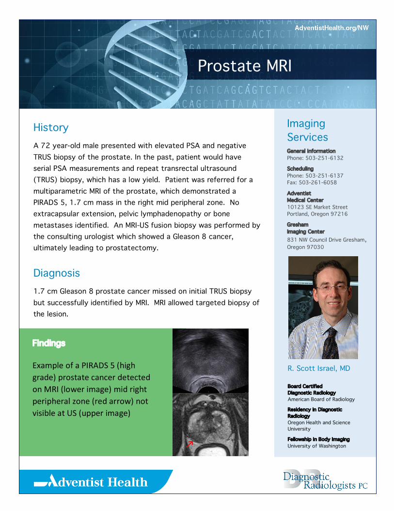
Findings ExampleofaPIRADS5(highgrade)prostatecancerdetectedonMRI(lowerimage)midrightperipheralzone(redarrow)notvisibleatUS(upperimage)
Imaging Services General information Phone: 503-251-6132
Scheduling Phone: 503-251-6137 Fax: 503-261-6058
Adventist Medical Center 10123 SE Market Street Portland, Oregon 97216
Gresham Imaging Center 831 NW Council Drive Gresham, Oregon 97030
R. Scott Israel, MD Board Certified Diagnostic Radiology American Board of Radiology
Residency in Diagnostic Radiology Oregon Health and Science University
Fellowship in Body Imaging University of Washington
History A 72 year-old male presented with elevated PSA and negative TRUS biopsy of the prostate. In the past, patient would have serial PSA measurements and repeat transrectal ultrasound (TRUS) biopsy, which has a low yield. Patient was referred for a multiparametric MRI of the prostate, which demonstrated a PIRADS 5, 1.7 cm mass in the right mid peripheral zone. No extracapsular extension, pelvic lymphadenopathy or bone metastases identified. An MRI-US fusion biopsy was performed by the consulting urologist which showed a Gleason 8 cancer, ultimately leading to prostatectomy.
Diagnosis 1.7 cm Gleason 8 prostate cancer missed on initial TRUS biopsy but successfully identified by MRI. MRI allowed targeted biopsy of the lesion.
Prostate MRI
MRIisprecisionmedicineforprostatecancer.ConventionalassessmentforanelevatedPSAisdigitalrectalexaminationandaTRUSbiopsy,typically12 or more biopsies obtained in a systematic but blind fashion. Although US is used to direct the biopsy to a region of the prostate, most tumors are not US visible.
TRUS biopsy misses 30 percent of significant cancers, particularly those located in the anterior gland and apex. MRI has high tissue discrimination, so that high histologic grade (Gleason score) tumors that are clinically significant can be identified with high sensitivity and specificity. Low-grade tumors that are less likely to be clinically significant are not detected.
MRI detected lesions can be biopsied under direct visualization by fusing the MRI data to real time US imaging. An electronic cursor marks the lesion which can be biopsied with high accuracy. MRI examinations are performed on our 3T MRI using a state of the art pelvic phased array coil. MRI is being used at AMC to:
1. Detect tumor in the setting of rising PSA and negative TRUS;
2. Follow patients expectantly who have low grade disease;
3. Stage patients with known prostate cancer;
4. Detect recurrence in patients with increasing PSA following definitive treatment.
The availability of prostate MRI and the ability to target MRI-detected lesions alters the discussion about prostate cancer screening. MRI, used in conjunction with PSA screening, TRUS, and urological consultation, gives us an enhanced ability to identify patients with higher grade, potentially actionable prostate cancer, and more confidently assign others to active surveillance.