properties of cutaneous mechanoreceptors - in the … in mammals including ... the first systematic...
TRANSCRIPT

Human Neurobiol(1984) 3:3-14
Properties of cutaneous mechanoreceptors in the human hand - related to touch sensation A. B. Vallbo' and R. S. ~ohansson~ * 'Nobel Institute for Neurophysiology, Karolinska Institutet, S-104 01 Stockholm, and 'Department of Physiology, University of UmeH, S-90187 Umei, Sweden
Summary. Recordings from single peripheral nerve fibres made it possible to analyse the functional properties of tac- tile afferent units supplying the glabrous skin of the human hand and to assess directly the relation between impulse dis- charge and perceptive experiences. The 17,000 tactile units in this skin area of the human hand are of four different types: two fast adapting types, FA I and FA I1 (formerly RA and PC), and two slowly adapting types, SA I and SA 11. The receptive field characteristics and the densities in the skin of the type I units (FA I and SA I) indicate that these account for the detailed spatial resolution that is of paramount im- portance for the motor skill and the explorative role of the hand. The relationship between the stimulus amplitude and perceived intensity during sustained skin indentations did not match the corresponding stimulus response functions of SA units suggesting non-linear transformations within the central nervous system. These transformations, in turn, appear to vary between subjects. A single impulse in a single FA I unit may be felt when originating from the most important tactile regions of the hand, indicating that the psychophysical detection may be set by the threshold of the sense organs. Moreover, no significant noise seems to be superimposed in the respective central sensory pathways.
Key words: Psychophysics - Cutaneous mechanoreceptors - Tactile sensibility - Human hand
Neural mechanisms involved in the sensation of touch have been studied extensively in subhuman species since the 1920s when Adrian and Zotterman (1926) first demonstrat- ed the basic nature of the message originating from cut- aneous mechanoreceptors in the frog. Neurophysiological studies in mammals including primates have supplied a wealth of experimental data, concepts, and theories, which together constitute the essential basis of our current under- standing of neural mechanisms responsible for tactile sensibil- ity. It is, however, beyond the scope of the present account to summarize all this work and the reader is referred to ap- propriate reviews (e.g. Burgess and Per1 1973; Iggo 1974, 1977).
Tactile sensibility has also long been studied intensively from a totally different angle, i.e. psychophysics, which
* to whom correspondence should be sent
dates back to the pioneering work by Weber (1835) and Fechner (1860). Psychophysics is concerned with the relationship between quantities of physical stimuli and properties of percepts as revealed by behavioural responses of human subjects in carefully designed test situations. Thus it does not focus on the functions and the limitations of the constituent mechanisms at the various levels of the sensory system but rather on the whole chain of events from the stimulation of the receptors to the behavioural response as one unit.
It is striking that, in recent decades, these two lines of in- vestigation have come closer together in the study of tactile mechanisms. Neurophysiological and psychophysical methods were intimately combined by Mountcastle and coworkers in their analyses of tactile sensitivity in the primate hand (e.g. Werner and Mountcastle 1965; Mountcastle et al. 1966; Mountcastle 1967; Talbot et al. 1968; Mountcastle et al. 1969). Neural responses were analysed in single nerve cells, mostly either first order afferents dissected from peripheral nerves or cortical cells recorded from anaesthetized monkeys. In parallel experiments psychophysical responses to identical stimuli were analysed in human subjects, and in some re- spects with monkeys as well (Mountcastle et al. 1972; LaMotte and Mountcastle 1975). These studies clarified a number of fundamental relations and advanced our under- standing in a most dramatic way. Here attention will be drawn only to one conclusion of general nature. It was in- ferred that the sensory capacity of a human observer as re- vealed in psychophysical experiments is set - to a much greater extent than was previously thought - by functional properties of the sense organs in the skin, rather than by mechanisms within the central nervous system. This result provided strong stimulus to further investigations of func- iional of cutaneous sense organs and of psycho- physics of tactile sensibility.
A possibility to combine, even more directly than in ~ountcastle's work, the two main approaches to the study of tactile mechanisms was offered when the microneurographic technique was introduced (Vallbo and Hagbarth 1968). This method not only allowed peripheral tactile mechanisms in man to be analysed with a precision and a resolution that was previously available only in animal experiments, but it also provided the possibility to assess directly in the human sub- ject the relation between impulse activity in primary affer- ents and perceptive experience as tested with psychophysical methods. Moreover, as recently demonstrated (Torebjork

Fibre diameter. pm
Conduction velocity, m/s
Fig. 1. A Diameter distribution of the myelinated nerve fibres in the median nerve at the wrist based on specimens from two neurologically healthy adults. For each bin, the vertical bar gives the range. B Conduction velocities of tactile afferent units terminating in the glabrous skin of the human hand. C Types of tactile afferent units in the glabrous skin of the human hand and some of their distinguishing properties. Graphs show schematically the impulse discharge (lower trace) to perpendicular ramp indentation of the skin (upper trace) for each unit type. (From Johansson and Vallbo 1983, with permission)
c
and Ochoa 1980; Konietzny et al. 1981 ; Vallbo 1981; Ochoa and Torebjork 1983) the microelectrode inserted in the peripheral nerve may be used not only for recording unitary nerve impulses, but also for stimulation of the same fibre to elucidate the properties of the percept induced by a de- fined series of impulses in an identified sensory unit.
The first systematic study on the functional properties of cutaneous mechanoreceptors in man appeared in 1970 (Knibestol and Vallbo 1970). Since then, a large number of investigations using the microneurographic technique in the study of tactile sensibility have been published. The majority of these studies are concerned with the glabrous s b n in the hand, i.e. the hairless skin of the palm and the palmar aspect of the fingers. This skin is characterized by a thick epidermal layer which forms patterns of ridges and grooves on the sur- face and, in the depth, an elaborate system of ridges and pegs where separate types of sensory endings occupy particular locations.
The present account which is mainly based on the publi- cations from the Ume% group is a survey concerning tactile mechanisms in the glabrous skin of the human hand. I t emphasizes correlative psychoneuronal analyses, whereas the functional properties of the afferent units are briefly reviewed in the first section. The reader may consult original reports as well as a recent survey for further information (Johansson and Vallbo 1983).
RECEPTIVE FIELDS
0 2 Z
k a
Tactile units in the glabrous skin of the human hand
Small, sharp borders
FA I
n - SA I
n - no statlc Fast'
response
slow. statlc
response present
Number and types of tactile units
Large. obscure borders
FA II
n - SA II
n - -
The term tactile unit refers to a primary afferent neuron whose sensory endings are primarily responsive to light skin deformations and are mostly located in the dermis. It should
be emphasized, however, that other types of afferent units, e.g. joint and muscle receptors, may also have very important roles in tactile sensibility taken in a broader sense.
The number of tactile units supplying the glabrous skin area of one hand has been estimated as 17,000 (Johansson and Vallbo 1979a). The figure is based upon actual measure- ments and counts of fibres in the median nerve at the wrist where the vast majority (ca. 90%) of the myelinated fibres are distributed to the glabrous skin. Figure 1A shows the calibre spectrum of these fibres. The diagram is based on whole nerve specimens taken from two middle-aged human subjects. It is obvious that the fibres fall into the two clas- sical groups, the smaller A6 and the larger Aa fibres. (Not seldom the latter are inappropriately denoted A0 fibres, cf. Burgess and Perl 1973.) Figure 1B shows the conduction velocities of 61 tactile units sampled from the median nerve in acute experiments on alert human subjects. A comparison
*
between the two histograms clearly indicates that this sample corresponds quite well to the Aa fibres. A "conversion factor" very close to 6 was used here to make the two diagrams , match (cf. Hursh 1939; Burgess and Perl 1973). There is strong albeit indirect evidence from a number of studies that all tactile units in the glabrous skin of the hand are found among the Aa fibres whereas the A6 and the un- myelinated C fibres belong to nociceptive and thermosensi- tive units (e.g. Burgess and Perl 1973; Darian-Smith et al. 1973; Torebjork and Hallin 1974; Georgopoulos 1976).
The 17,000 tactile units supplying the glabrous skin of one hand are of four main types. These differ with regard to functional properties such as sensitivity to static and dynamic events, and the size and structure of their receptive fields. In addition they differ with regard to their numbers I
and densities within the separate subregions of the glabrous I
skin area, and their perceptive effects. Their functional pro- perties correspond well to the properties of the four catego-
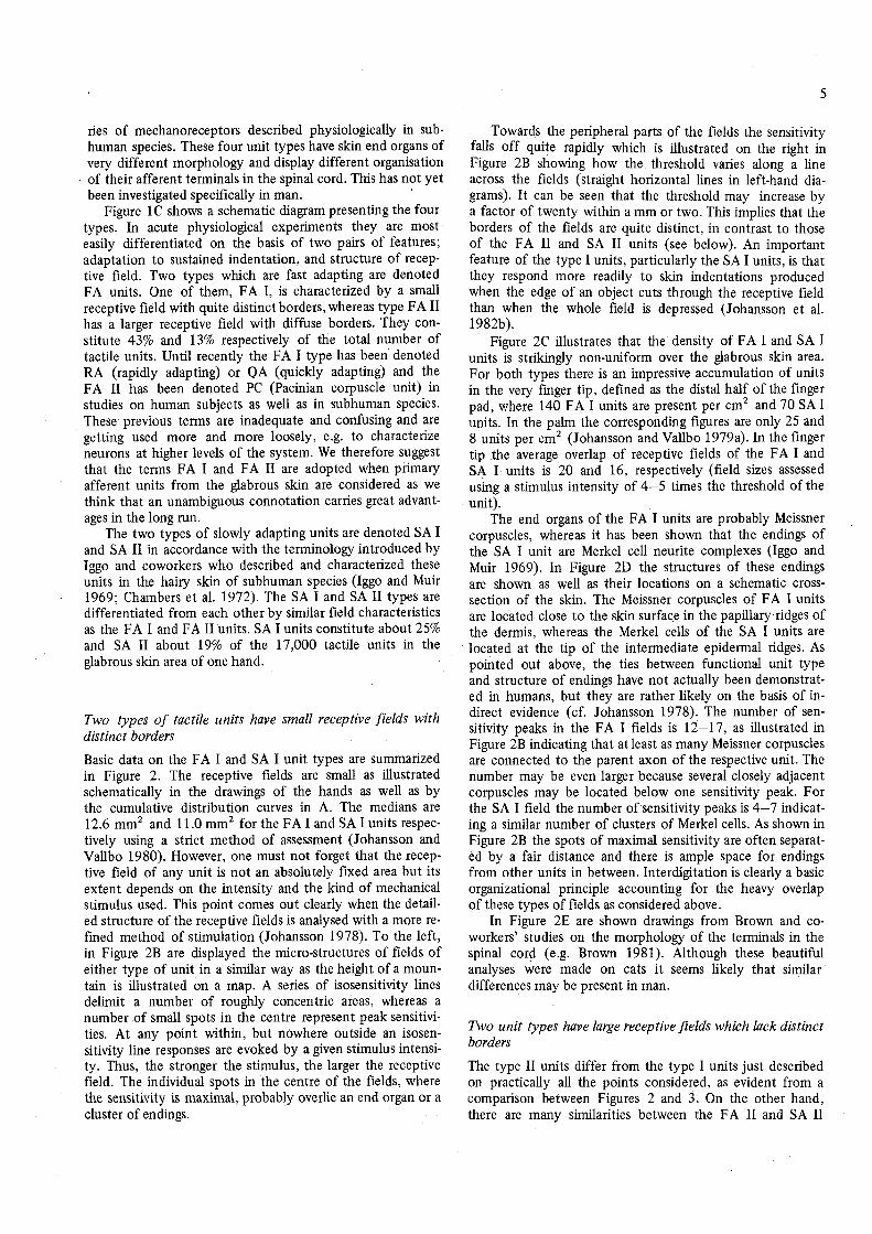
ries of mechanoreceptors described physiologically in sub- human species. These four unit types have skin end organs of very different morphology and display different organisation of their afferent terminals in the spinal cord. This has not yet been investigated specifically in man.
Figure 1 C shows a schematic diagram presenting the four types. In acute physiological experiments they are most easily differentiated on the basis of two pairs of features; adaptation to sustained indentation, and structure of recep- tive field. Two types which are fast adapting are denoted FA units. One of them, FA I, is characterized by a small receptive field with quite distinct borders, whereas type FA I1 has a larger receptive field with diffuse borders. They con- stitute 43% and 13% respectively of the total number of tactile units. Until recently the FA I type has been denoted RA (rapidly adapting) or QA (quickly adapting) and the FA I1 has been denoted PC (Pacinian corpuscle unit) in studies on human subjects as well as in subhuman species. These previous terms are inadequate and confusing and are getting used more and more loosely, e.g. to characterize neurons at higher levels of the system. We therefore suggest that the terms FA I and FA I1 are adopted when primary afferent units from the glabrous skin are considered as we think that an unambiguous connotation carries great advant- ages in the long run.
The two types of slowly adapting units are denoted SA I and SA I1 in accordance with the terminology introduced by Iggo and coworkers who described and characterized these units in the hairy skin of subhuman species (Iggo and Muir 1969; Chambers et al. 1972). The SA I and SA I1 types are differentiated from each other by similar field characteristics as the FA I and FA I1 units. SA I units constitute about 25% and SA I1 about 19% of the 17,000 tactile units in the glabrous skin area of one hand.
Two types of tactile units have small receptive fields with distinct borders
Basic data on the FA I and SA I unit types are summarized in Figure 2. The receptive fields are small as illustrated schematically in the drawings of the hands as well as by the cumulative distribution curves in A. The medians are 12.6 mm2 and 1 1 .O mm2 for the FA I and SA I units respec- tively using a strict method of assessment (Johansson and Vallbo 1980). However, one must not forget that the recep- tive field of any unit is not an absolutely fixed area but its extent depends on the intensity and the kind of mechanical stimulus used. This point comes out clearly when the detail- ed structure of the receptive fields is analysed with a more re- fined method of stimulation (Johansson 1978). To the left, in Figure 2B are displayed the micro-structures of fields of either type of unit in a similar way as the height of a moun- tain is illustrated on a map. A series of isosensitivity lines delimit a number of roughly concentric areas, whereas a number of small spots in the centre represent peak sensitivi- ties. At any point within, but nowhere outside an isosen- sitivity line responses are evoked by a given stimulus intensi- ty. Thus, the stronger the stimulus, the larger the receptive field. The individual spots in the centre of the fields, where the sensitivity is maximal, probably overlie an end organ or a cluster of endings.
Towards the peripheral parts of the fields the sensitivity falls off quite rapidly which is illustrated on the right in Figure 2B showing how the threshold varies along a line across the fields (straight horizontal lines in left-hand dia- grams). I t can be seen that the threshold may increase by a factor of twenty within a mm or two. This implies that the borders of the fields are quite distinct, in contrast to those of the FA I1 and SA I1 units (see below). An important feature of the type I units, particularly the SA I units, is that they respond more readily to skin indentations produced when the edge of an object cuts through the receptive field than when the whole field is depressed (Johansson et al. 1982b).
Figure 2C illustrates that the density of FA I and SA I units is strikingly non-uniform over the glabrous skin area. For both types there is an impressive accumulation of units in the very finger tip, defined as the distal half of the finger pad, where 140 FA I units are present per cm2 and 70 SA I units. In the palm the corresponding figures are only 25 and 8 units per cm2 (Johansson and Vallbo 1979a). In the finger tip the average overlap of receptive fields of the FA I and SA I units is 20 and 16, respectively (field sizes assessed using a stimulus intensity of 4-5 times the threshold of the unit).
The end organs of the FA I units are probably Meissner corpuscles, whereas it has been shown that the endings of the SA I unit are Merkel cell neurite complexes (Iggo and Muir 1969). In Figure 2D the structures of these endings are shown as well as their locations on a schematic cross- section of the skin. The Meissner corpuscles of FA I units are located close to the skin surface in the papillary ridges of the dermis, whereas the Merkel cells of the SA I units are located at the tip of the intermediate epidermal ridges. As pointed out above, the ties between functional unit type and structure of endings have not actually been demonstrat- ed in humans, but they are rather likely on the basis of in- direct evidence (cf. Johansson 1978). The number of sen- sitivity peaks in the FA I fields is 12-17, as illustrated in Figure 2B indicating that at least as many Meissner corpuscles are connected to the parent axon of the respective unit. The number may be even larger because several closely adjacent corpuscles may be located below one sensitivity peak. For the SA I field the number of sensitivity peaks is 4-7 indicat- ing a similar number of clusters of Merkel cells. As shown in Figure 2B the spots of maximal sensitivity are often separat- ed by a fair distance and there is ample space for endings from other units in between. Interdigitation is clearly a basic organizational principle accounting for the heavy overlap of these types of fields as considered above.
In Figure 2E are shown drawings from Brown and co- workers' studies on the morphology of the terminals in the spinal cord (e.g. Brown 1981). Although these beautiful analyses were made on cats it seems likely that similar differences may be present in man.
Two unit types have large receptive fields which lack distinct borders
The type I1 units differ from the type I units just described on practically all the points considered, as evident from a comparison between Figures 2 and 3. On the other hand, there are many similarities between the FA I1 and SA I1

0
B fn 10 200 g C
' 0 2 4 6 H 0 2 4 6 1 mm Distance. mm 1 mm Distance, mm
k-1, Innervation density, units'/cmz
80 120 2 nsity. units/cm
Fig. 2A-E. Characteristics of FA I and SA I units. A Receptive field size. The black patches of the drawing of the hands indicate receptive fields of 15 FA I and 15 SA I units as measured with von Frey hairs providing a force of 4-5 times the threshold force of the individual unit. Graphs indicate cumulative distribution curves of receptive field sizes for the four unit types (data based on a total sample of 255 units). Solid curves refer to the FA I and SA I units respectively. Most of the fields are circular or oval and they usually have a size between 3 mm2 and 50 mm2, which correspond to circular areas of 2-8 mm diameter. B Microstructure of receptive fields. Sensitivity maps of single FA I and SA I fields. Thin lines mark the grooves between the papillary ridges whereas the closed lines are isosensitivity lines enclosing skin areas where the unit re- sponds at given skin displacements delivered with a small pointed probe. The graphs illustrate the threshold variations along the straight line shown in the map. The ordinates give the indentation amplitude divided into multiples of the lowest threshold (T) as well as into actual indenta-

units. The receptive fields (Fig. 3A) are often very large as tested with crude methods. When assessed with a standard- ized stimulus (intensity 4-5 times threshold) they are 101 mm2 for the FA I1 and 59 mm2 for the SA I1 units (median values). With the FA I1 units stimuli that contain fast transients and high frequency compounds are very effective also when applied remotely from the receptor location. In other species, the response properties of similar units have been described in detail where they are usually denoted PC (e.g. Talbot et al. 1968). All available evidence indicates identical properties in man including a high sensitiv- ity to vibrations with a peak at 200-300 Hz (Johansson et al. 1982a). With regard to the SA I1 units it is striking that they strongly respond not only to indentation but also to stretching of the skin as originally emphasized in studies on subhuman species (Chambers et al. 1972). Moreover, many of them are continuously active without any indentation of the skin. Another interesting property, first demonstrated on humans, is that the SA I1 units often exhibit a directional sensitivity, e.g. their discharge may increase for a stretch in one direction and decrease for a stretch at right angles or in the opposite direction (Knibestol and Vallbo 1970; Knibestol 1975; Johansson 1978). Thus the population of SA I1 units constitutes a set of sense organs that provide highly dis- criminative information about direction and magnitude of lateral tension in the skin, as well as between skin and deeper structures (Johansson 1976). Information of this nature may be of significance to monitor not only joint position (see below) but also shearing forces between skin surface and hand held objects. This might be significant for a proper control of the finger muscles in order to compensate for continuously varying forces which tend to make a tool, e.g. a hammer, slip out of the hand during work. The receptive field characteristics and related properties of the type I1 units probably account for their high responsiveness to joint movements as well as to different joint positions (Hulliger et al. 1979). Psychophysical findings also indicate that this response might be highly relevant for kinesthesia and posi- tion sense in the human hand (Burgess et al. 1982).
Analyses of the micro-structure of the fields revealed that there is only one single point of maximal sensitivity (Fig. 3B), suggesting that a single end organ, or a group of closely adjacent end organs, are connected to the stem fibre. Most striking is the total lack of borders delimiting anything like a central receptive field area. Rather the sensitivity falls off gently with distance at a semiconstant slope (Fig. 3B). As a consequence it may be inferred that a localized mechanical event on the skin surface would ex- cite endings within a large area, granted with a gradually decreasing effectiveness from the target point. Hence, the population input to the brain from these units would provide much less distinct information on the stimulus' spatial structures compared to the information from the FA I and SA I units.
The density of the type I1 units is not only generally lower compared with the type I units but the pattern of distribution within the separate skin regions is also very different (Fig. 3C) as the units are almost uniformly distribut- ed in the whole glabrous skin area.
The endings of the FA I1 units (Fig. 3D) are very likely Pacinian corpuscles located in the subcutaneous tissue (e.g. Lindblom 1965) as well as the smaller and simpler lamellated Golgi-Mazzoni bodies in the dermis (Chouchkov 1973; Iggo and Andres 1982). The SA I1 units probably terminate with the spindle shaped Ruffini ending hooked up to dermal structures with collagen fibres extending from its poles (Chambers et al. 1972), a morphological characteristic that fits well with the directional sensitivity of the unit as de- scribed above. Thus the endings of the FA I1 and SA I1 units are located further below the skin surface than the type I endings, which are both just at the epidermal-dermal border. In the cat, it has been shown that the central termi- nals within the spinal cord of these two kinds of units have distinctive morphologies (Brown 198 1 ; Fig. 4E).
Activity in tactile units related to sensation
Intensity of sensation
It is a trivial experience that the intensity of a sensation is higher the stronger the stimulus, but it has been a long stand- ing problem in psychophysics to define how the sensation grows when the stimulus is increased. Stevens demonstrated (1957, 1967) that when a subject is asked to estimate and re- port the intensity simply by assigning a number to his sen- sation (magnitude estimation), very consistent results emerge, at least when scatter is eliminated by considering averaged data from groups of subjects. It was found that the sensation grows with the stimulus along a curve that can be adequately described by a mathematical power function, usually denot- ed the psychophysical function. However, the shape of the curve may vary between modalities. The power function has the general form R = a Sb, where R is the intensity of the sensation as estimated and reported by the subject and S is the stimulus magnitude (strictly, the stimulus magnitude minus the threshold value), whereas a and b are constants. The exponent b, whose value depends on the modality and quality of the sensation, gives the shape of the curve. An ex- ponent below unity indicates that the curve is decelerating on linear coordinates, i.e. the slope is successively decreasing, whereas an exponent above unity indicates that the curve is accelerating, i.e. the slope is successively increasing and more so the larger the exponent. A linear relation is indicat- ed by an exponent of unity. On logarithmic coordinates all power functions come out as straight lines with slopes given by the exponent b. These findings are summarized in Stevens' power law. It implies that anywhere along the intensive con-
tion amplitudes (pm), whereas the abscissa is a distance scale with arbitary origin. (From Johansson 1976, with permission). C Average density of FA I and SA I units within different glabrous skin areas. Each dot in the drawings of the hand represents a single sensory unit innervating the skin area. Histograms give the density of innervation in the following three skin regions: the tip of finger, the rest of finger and the palm. D Struc- ture and location of sensory endings. Vertical section through the glabrous skin of the human hand (center) schematically demonstrating the locations of organized nerve terminals found in this area. The Meissner corpuscle and the Merkel cell neurite complex are shown in the expanded drawings to the left and right. (From Anders and v. Diiring 1973 and Iggo and Muir 1969, with permission.) E The three-dimensional organiza- tion of the collaterals from single identified FA I and SA I mechanoreceptive afferents in the cat's lumbosacral spinal cord according to Brown (1981, with permission)

P
A 0.5 . C
a
5 o - 0 1 10 100 lo00 1 10 100 1000
Receptive field size, mm2 Receptive field size, mm2
B 1000 3 -
' 0 2 4 6 8 1 mm Oo 2 4
Distance. nlm H
Distance. mm E 1 mm no-
= ' + -i
1 1 3' Innervation density, units/cm2 lnnervation density.
Fig. 3A-E. Characteristics of FA I1 and SA I1 units. A Receptive field size. The dashed areas in the drawings of the hand illustrate the receptive fields of two FA I1 units and four SA I1 units as determined by manually delivered stimuli, i.e. taps with a small glass rod and skin stretch respec- tively. The location of the zone of maximal sensitivity is indicated by dots. As to the SA I1 units, the arrows indicate directions of skin stretch which gave rise to an increase of the discharge. Solid curves of the graphs indicate cumulative distributions of receptive field sizes of the FA I1 and SA I1 units respectively (cf. Fig. 2A). B Microstructure of receptive fields. Sensitivity map of a FA I1 and a SA I1 receptive field constructed as described in Fig. 2B. (From Johansson 1976, with permission.) C Average density of FA I1 and SA I1 units within different glabrous skin areas, presented as in Fig. 2C. D Structure and location of the sensory endings as in Fig. 2D. Pacinian corpuscle and Ruffini ending (from Chambers et al. 1972 with permission) are shown on expanded drawings. E The three-dimensional organization of the collaterals from single identified FA I1 and SA I1 mechanoreceptive afferents in the cat's lumbosacral spinal cord according to Brown (1981, with permission)

Power function exponent
Fig. 4. Comparison between psychophysical magnitude estimation functions (top, P) and neural stimulus-response functions of slowly adapting tactile units (bottom, N). Histograms give the exponents of the mathematical power functions fitted to the experimental data. The exponents indicate the curvature of the functions (see text). The two sets of data were collected on stimulation at the same target points in glabrous skin of the same subjects. (Modified from Knibestol and Vallbo 1980, with permission)
tinuum, equal ratios between stimulus magnitudes give rise to equal ratios between perceptive intensities provided that the stimulus is defined as the magnitude exceeding the threshold stimulus.
In relation to Stevens' power law it is highly interesting to explore the relation between quantities of neural activity on the one hand, and intensities of sensations, on the other, as it might shed light on the rules that the brain is following when producing within the mind the dimension of intensity of sensation. With regard to tactile sensitibility a linear rela- tion (exponent equal to unity) has been demonstrated be- tween the amplitude of a sustained indentation and sensa- tion in the glabrous skin of the hand, whereas a decelerating relation (exponent below unity) has been found in the hairy skin (Mountcastle 1967; Harrington and Merzenich 1970). Most interestingly, a similar difference has been found be- tween the two skin regions with regard to the stimulus- response relation of slowly adapting tactile units in the monkey (Mountcastle et al. 1966; Harrington and Merzenich 1970). This striking correspondence found between data from man and monkey led Mountcastle (1967) to formulate an explicit hypothesis with regard to the nature of intensity coding in afferent systems. He proposed the generalization that the particular shape of the psychophysical function is determined at the receptor level and that transformations at more central levels in the nervous system are in sum linear.
The hypothesis that the intensity dimension is essentially set by the properties of the end organs was tested on human subjects using the microneurographic method of recording from single afferents of alert volunteers when indentations of various amplitudes were delivered (Knibestol and Vallbo 1980). The subjects estimated the magnitude of their sensa- tion in psychophysical tests either simultaneously with the nerve recordings or to identical stimuli shortly afterwards. It was found that the stimulus response relations of the slow-
Indentation amplitude, rnm
Fig. 5. Comparison between neural stimulus-response functions (N) and psychophysical magnitude-estimation functions (P). Data from one subject are extracted at three different test points (1, 2, 3) locat- ed in the centre of the receptive fields of three SA I units (indicated by black dots). It is obvious that there is a pronounced discrepancy between the psychophysical functions which were all accelerating and the neural functions which were decelerating. (From Knibestol and Vallbo 1980, with permission)
ly adapting mechanoreceptors, mostly SA I units, in the glabrous skin of the hand varied considerably but were most often decelerating (average exponent 0.7, range 0.2-1.4). In contrast, the relation between stimulus magnitude and intensity of sensation was on the average linear, whereas again, the interindividual variations were considerable (aver- age exponent 1.0, range 0.3-2.1). Figure 4 demonstrates the discrepancy between the two sets of data extracted from the same target points. The findings imply that the average psychophysical function is not, in contrast to Mountcastle's hypothesis, determined at the receptor level of the single slowly adapting afferent.
Although it is obvious from Figure 4 that a close similari- ty does not exist between the psychophysical stirnulus- response functions and the corresponding neuronal functions, it may still be asked whether a weaker correlation is present, such that low exponents in the neuronal functions are asso- ciated with relatively low exponents of the psychophysical function and vice versa. A correlation of this kind would sug- gest that the transformations in the central nervous system are, if not linear, at least roughly uniform, among subjects. However, the experimental data clearly indicated that the shapes of the two types of curves were totally unrelated in this sense. A striking example is presented in Figure 5 show- ing data from a subject who produced similar and accelerat- ing psychophysical functions when tested at three different points, whereas the stimulus response functions of three slowly adapting units at these very points were strongly decelerating. Thus it seems hardly possible to maintain the hypothesis that the magnitude of sensation associated with sustained skin indentation is simply determined by the im- pulse rate in single primary afferents of SA types. On the contrary, it appears that the central nervous system plays a major role in shaping the psychophysical functions. More- over, different subjects seem to experience relative intensi-

ties in a different way probably because the intrinsic pro- perties of their brains differ.
Sensory threshold
The lower end of the intensive continuum is set by the sen- sory threshold. Over the years many theories about the nature of the sensory threshold have been advanced and it has been debated whether central or peripheral mechanisms set the limit of psychophysical detection (cf. Green and Swets 1966). Any theory must explain the general experi- mental finding that the psychometric function, i.e. the curve describing the relation between stimulus amplitude and prob- ability of detection, is not a step function but the curve is S-shaped. Psychophysicists have claimed that the subjects' detection threshold is higher than the threshold of their sense organs and have postulated that a substantial noise, which is assumed to intrude at higher levels of the sensory system, ac- counts for the S-shape of the psychometric function (Green and Swets 1966). Neurophysiologists, on the other hand, have often arrived at an almost opposite conclusion, i.e. "that the detection . . . appears to be set by the peripheral neuronal threshold; it is not elevated by any higher central threshold" (Mountcastle 197.5) and that "it is the stimulus which is variable and . . . the properties of its variation deter- mine the fluctuations found between response and stimulus" (Hecht et al. 1942).
Using the microneurographic method these two issues concerning the threshold concept were explored with tactile stimuli in the human hand (Vallbo and Johansson 1976; Johansson and Vallbo 1979b). The fact that both natural and psychophysical responses could be recorded in the same test with this approach eliminated a number of uncertainties and ambiguities attached to many previous studies, e.g. whe- ther a difference between the two kinds of thresholds was genuine or due to an uncontrollable variation in stimulus amplitude. A strict control of stimulus amplitude and of the exact target point on the skin was adopted with the purpose to excite single tactile units in isolation (Westling et al. 1976). There was strong indication that this was achieved when the most sensitive units were explored (Johansson and Vallbo 1976, 197913; Johansson 1978). Figure 6 illustrates that the psychophysical detection thresholds agree with the thresh- olds of the FA units in most regions of the hand. Thus tactile detection was dependent on these units whereas the SA units were irrelevant because they produced no response whatso- ever at the stimulus level of the psychophysical detection. Direct evidence indicated that the FA I units provided the decisive sensory input whereas the FA I1 did not. In fact, several kinds of test demonstrated that a single impulse in a single FA I unit was often detected perceptually. Moreover, when the psychometric function was compared with the threshold curve of an FA I unit an almost exact correspond- ence was found (Fig. 7A) with regard to the location of the curve as well as its slope.
These two sets of findings allow two important conclu- sions. First, that the detection capacity is set by the sensitiv- ity of the sense organs and not by central mechanisms. Se- cond, that there is no central noise added to the afferent signal at higher levels of the somatosensory system when the subject is maximally attending to the task.
However, these conclusions are not generally valid. In
Neuronal thresholds
- 4- Psychophysical thresholds
C m T
Fig. 6 . Relations between psychophysical detection threshold and thresholds of tactile units. The upper histogram shows thresholds to triangular skin indentations (4 mm/s) for evoking a single nerve impulse in afferent units in various regions of the glabrous skin area (totally 128 units). The FA I and FA I1 are pooled since they have about the same threshold distribution for this type of stimulus. Also, the slowly adapting units, which have much higher thresholds are pooled. The lower histogram shows psychophysical thresholds in cor- responding skin regions to identical stimuli (162 test points). From left to right, the columns give data from the terminal phalanx, the rest of the finger, the peripheral part of the palm, the central part of the palm, and to the extreme right, data from the lateral aspects of the fingers and the regions of the creases taken together. Column heights give medians and bars 25th and 75th percentiles. (Modified from Johansson and Vallbo 1979b, with permission)
the centre of the palm and some other regions the findings indicated that detection is in fact set by central mechanisms (Fig. 6). The data also suggest that a substantial noise is present in those parts of the somatosensory system that process the afferent information from these skin regions (Fig. 7B). It is interesting that this more crude performance of the somatosensory system was found for the particular skin regions of the hand that presumably have a less pro- minent role in tactile sensibility.
A general conclusion is that the nature of the absolute threshold is evidently different within different segments of the somatosensory system. In the most important tactile regions it is set by the sensitivity of the sense organs imply- ing that a minimal signal, i.e. a single nerve impulse in one afferent fibre, reaches consciousness, whereas in other regions

0 50 100 150 200
Indentation amplitude, pm Fig. 7. Neural and psychophysical threshold curves from two test points. In A the stimuli were delivered to one of the highly sensitive zones of a FA I unit located at the tip of the thumb, whereas in B the stimuli were delivered to one of the highly sensitive zones of a FA I unit located in the central part of the palm. Curves represent the best fitting cumulative normal distribution function (maximum likelihood estimation). (From Vallbo and Johansson 1976, with permission)
the subjects' detection threshold is set to higher levels by central mechanisms. One might speculate that optimal in- formation processing has been reserved for particular skin areas during the development of the brain whereas this could not be afforded for all skin regions.
Spatial resolution
The FA I and SA I units constitute a peripheral organization made up of a fine grain in therms of small and distinctly de- fined receptive fields. It would therefore seem reasonable that these units account for our ability to localize stimuli correctly on the skin surface and appreciate delicate details of objects that we touch and manipulate, an ability that probably is of major significance for manual skill. Spatial discrimination is one aspect of this general function. It is usually measured with the well known two-point threshold test, which defines the minimal inter-point distance at which the subject is able to perceive a gap between two points in- denting the skin.
If the two-point threshold were set by the peripheral organization one would expect that this distance is related to size of receptive fields as well as density of units. It would therefore be interesting to study these properties in different slun regions known to have different two-point thresholds. When the size of receptive fields was explored only minor
Spatial resolution
Density of FA I and SA I units
Density of FA II and SA II units
Fig. 8. Spatial resolution capacity in psychophysical tests (two point discrimination) compared to density of tactile units in three glabrous skin regions; finger tip, rest of finger, and palm, respectively. The three columns referring to one skin area represent, from left to right, (i) the inverse of the two point threshold in units of mm-' , (ii) the average density of small field units taken together (FA I and SA I units) and (iii) the average density of large field units (FA I1 and SA 11). Note that the spatial resolution increases reasonably pan passu with the increase in density of the FA I and SA I units
differences were found between different skin regions of the hand (Vallbo and Johansson 1978; Johansson 1979; Johansson and Vallbo 1980; cf. Schady 1983). The smallest fields were less than 2 mm in diameter while the largest extended up to about 15 mm for the FA I and SA I units, whereas the type I1 units generally had considerably larger fields (cf. Figs. 2A and 3A). With regard to unit densities, on the other hand, there were interesting differences between separate skin regions as well as between unit types as shown in Figures 2C and 3C. The type I1 units were present in about equal den- sities in the whole glabrous skin area. The type I units, by contrast, were much more numerous in the very finger tip than in the rest of the finger and more numerous in the finger than in the palm. When the spatial discrimination capacity was tested it turned out that this capacity defined as the inverse of the two-point threshold varied between skin regions in close parallelism to the density of type I units (Fig. 8). In the fingertip, the average two-point threshold was only 1.6 mm, which is close to the diameter of the smallest fields among the type I units. In the palm the two-point threshold was about five times higher (Vallbo and Johansson 1978). These findings support the conclusion that the type I units account for spatial acuity, suggested already by the structures of their receptive fields.
The significance of unit density for spatial resolution may schematically be conceived as follows. A high unit density

is associated with a high probability for a small field to be located between the target points in the two-point threshold test. When the density of fields is lower, on the other hand, small fields are rare and they do not cover the whole skin sur- face (Vallbo and Johansson 1978; Johansson 1979; Johansson and Vallbo 1980). Therefore the two target points must often be wider apart to accommodate the fields of silent units between the points.
Many objects manipulated and explored by the hand have approximately flat surfaces demarcated by well defined edges. In psychophysical studies it has been observed that the sen- sations evoked by such objects indenting the skin are more intense at the edges than at the flat surfaces. This edge enhancement, which probably is akin to the Mach band phenomenon in vision, has been explained on the basis of information processing within the central nervous system (surround inhibition) (e.g. Bekesy 1967). However, the pro- nounced edge sensitivity of the FA I and particularly the SA I units (Johansson et al. 1982b) indicates that there is a marked edge enhancement already at the level of the afferent response patterns. Hence, mechanisms accounting for enhancements of spatial contrasts appear to occur not only in the central nervous system as proposed previously but also at the level of the tactile afferent units.
Microstimulation
Tactile mechanisms have recently been explored with the approach of microstimulation, first reported by Torebjork and Ochoa (1980) and independently by other groups short- ly afterwards (Konietzny et al. 1981; Vallbo 1981). When a single nerve fibre belonging to an identified tactile unit in the glabrous skin is stimulated with a train of impulses the subject often reports a sensation whose quality is char- acteristic of the unit type (Ochoa and Torebjork 1983; Schady 1983; Vallbo et al. 1984). The sensation is clearly that of mechanical deformation although it is often slight- ly odd in quality. It differs drastically from the sensation associated with a local indentation with a pointed object as the sensation on electrical microstimulation is felt uni- formly within a small area, i.e. 3 mm2 and 6 mm2 for the FA I and SA I units respectively (median values) (cf. Fig. 9). The spatial extent of the area where the subject feels the sensation is often identical with that of the receptive field of the stimulated unit with regard to location, size, shape, and orientation. With the SA I units the sensation usually has the quality of a faint but strikingly uniform pressure, e.g. like a water color brush being held steadily against the skin (Fig. 9). This difference in spatial extent of the sensa- tions on single unit stimulation and on local indentation in- dicates a mechanism that suppresses, within the mind, the representations of the non-overlapping regions of the ex- cited fields when a group of units is stimulated with a mech- anical point stimulus.
The field of microstimulation is further elaborated in another article of this issue (Torebjork et al. 1984). Only one aspect, which is related to the threshold concept, will be considered here. When a single FA 1 unit was stimulated it was found that one single pulse was often sufficient to give rise to a sensation, whereas a train of impulses was required to produce a sensation from an FA I1 or an SA I unit (Torebjork and Ochoa 1980; Vallbo 1981 ; Ochoa and
Fig. 9. Electrical micro-stimulation. Excitation of an identified mechanoreceptive unit (FA I and SA I) by a train of current pulses delivered through the recording microelectrode usually evokes a sen- sation of a weak mechanical deformation projected within the recep- tive field of the unit. With the SA I units, the sensation may be de- scribed as that of a soft water-colour brush steadily held against the skin (upper diagram). This sensation of a uniform pressure within a small area contrasts strikingly to the sensation when the skin is mechanically stimulated with a pointed object, e.g. a tip of a pencil (lower diagram)
Torebjork 1983; Vallbo et al. 1984). Thus the method of microstimulation provides an independent confirmation of the conclusion reached in previous studies that the FA I units have unique connections to the perceptive levels of the somatosensory system because an indivisible neural quantum, i.e. a single impulse from a single FA I unit, elicits a sensa- tion of touch.
Conclusion
In the last decades our knowledge concerning the function of the human somatosensory system has expanded considerably thanks to the microneurographic method.
Not only tactile sensation, which is dependent on the large myelinated nerve fibres, has been explored. Also pain and temperature sensibility, which are dependent on small myelinated and unmyelinated fibres, have been studied (e.g. Torebjork and Hallin 1974; Adriaensen et al. 1983).
With regard to tactile sensibility, the functional properties of human mechanoreceptors have been subjected to detailed analyses. In some respects these have been carried further than corresponding studies on subhuman species. However, only limited groups of units have been explored and many areas remain to be investigated before it can be assessed to what extent it is justifiable to draw on animal models when the sensation of touch in human subjects is discussed.
The particular advantage of studying correlations be- tween neural and perceptive events in human subjects has been exposed in the present report. Needless to say there remain a large number of important problems to be cleared up in this field using either of the two main approaches: mechanical deformations of the skin or electrical micro- stimulation of single nerve fibres in combination with strict psychophysical tests. The role of touch receptors in kina- esthesia and in control of motor activities are promising

fields which have just been opened (Hulliger et al. 1979; Westling and Johansson 1983; Johansson and Westling 1984), whereas the exciting line of active touch implying parallel studies of motor, sensory and psychophysical events in the attending human subject has not yet been explored. But this explbration will certainly come.
It seems likely that in the future we will arrive at a deep- er understanding of the tactile system in its proper context in the behaving human subject, i.e. not only in its more passive role as a system receiving and processing information concerning externally imposed skin deformations, but also as an integral part of the complex chain of neural mech- anisms controlling muscle contractions and movements as well as in tactile exploration of the immediate surroundings.
Acknowledgements. This study was supported by the Swedish Medical Research Council (project 3548).
References
Andres KH, During M von (1973) Morphology of cutaneous receptors. In: Iggo A (ed) Somatosensory system. Springer, Berlin Heidelberg New York (Handbook of sensory physiology, vol 11, pp 3-28)
Adriaensen H, Gybels J , Handwerker HO, Hees J van (1983) Response properties of thin myelinated (Ad) fibres in human skin nerves. J Neurophysiol49: 111 -122
Adrian ED, Zotterman Y (1926) The impulses produced by sensory nerve endings. Pt 111. Impulses set up by touch and pressure. J Physiol (Lond) 61:464-483
Bekesy GV (1967) Sensory inhibition. Princeton University Press, Princeton
Brown AG (1981) The termination of cutaneous nerve fibres in the spinal cord. Trends Neurosci 4:64-67
Burgess PR, Perl ER (1973) Cutaneous mechanoreceptors and noci- ceptors. In: Iggo A (ed) Somatosensory system. Springer, Berlin Heidelberg New York (Handbook of sensory physiology, vol 11, - - -- pp 29-76
Burgess PR, Wei JK, Clark FJ, Simon J (1982) Signaling of kinesthetic information by peripheral sensory receptors. Ann Rev Neurosci - - 5:171-187
Chambers MR, Andres KH, During M von, Iggo A (1972) The structure and function of the slowly adapting type I1 mechanoreceptor in hairy skin. Q J Exp Physiol57:417-445
Chouchkov CN (1973) The fine structure of small encapsulated re- ceptors in human digital glabrous skin. J Anat Lond 114:25-33
Darian-Smith I, Johnson KO, Dykes R (1973) "Cold" fibre popula- tion innervating palmar and digital skin of the monkey: responses to cooling pulses. J Neurophysiol36:325-346
Fechner GT (1860) Elemente der Psychophysik. Breitkopf and Hartel, Leipzig
Georgopoulos AP (1976) Functional properties of primary afferent units probably related to pain mechanisms in primate glabrous skin. J Neurophysiol 39:71-83
Green DM, Swets JA (1966) Signal detection theory and psycho- physics. John Wiley and Sons, New York
Harrington T, Merzenich MM (1970) Neural coding in the sense of touch: human sensation of skin indentation compared with the responses of slowly adapting mechanoreceptive afferents inner- vating the hairy skin of monkeys. Exp Brain Res 10:251-264
Hecht S, Shlaer S, Pirenne MH (1942) Energy, quanta and vision. J Gen Physiol25:819-940
Hulliger M, Nordh E, Thelin AE, Vallbo AB (1979) The responses of afferent fibres from the glabrous skin of the hand during volun- tary finger movements in man. J Physiol (Lond) 29 1:233-249
Hursh JB (1939) Conduction velocity and diameter of nerve fibres. Am J Physiol127:131-139
Iggo A (1974) Cutaneous receptors. In: Hubbard JI (ed) Peripheral nervous system. Plenum Press, New York, pp 347-404
Iggo A (1977) Cutaneous and subcutaneoussense organs. Br Med Bull 33:97-102
Iggo A, Andres KH (1982) Morphology of cutaneous receptors. Ann Rev Neurosci 5: 1-3 1
Iggo A, Muir AR (1969) The structure and function of a slowly adapt- ing touch corpuscle in hairy skin. J Physiol (Lond) 200:763-796
Johansson RS (1976) Skin mechanoreceptors in the human hand: receptive field characteristics. In: Zotterman Y (ed) Sensory func- tions of the skin in primates, with special reference to man. Pergamon, Oxford, pp 159-170
Johansson RS (1978) Tactile sensibility in the human hand: receptive field characteristics of mechanoreceptive units in the glabrous skin area. J Physiol (Lond) 281: 101-123
Johansson RS (1979) Tactile afferent units with small and well de- marcated receptive fields in the glabrous skin area of the human hand. In: Kenshalo DR (ed) Sensory functions of the skin of humans. Plenum Press, New York, pp 129-145
Johansson RS, Landstrom U, Lundstrom R (1982a) Responses of mechanoreceptive afferent units in the glabrous skin of the human hand to sinusoidal skin displacements. Brain Res 244: 17-25
Johansson RS, Landstrom U, Lundstrom R (1982b) Sensitivity to edges of mechanoreceptive afferent units innervating the glabrous skin of the human hand. Brain Res 244:27-32
Johansson RS, Vallbo AB (1976) Skin mechanoreceptors in the human hand: an inference of some population properties. In: Zotterman Y (ed) Sensory functions of the skin in primates, with special reference to man. Pergamon, Oxford, pp 171-184
Johansson RS, Vallbo AB (1979a) Tactile sensibility in the human hand: relative and absolute densities of four types of mechano- receptive units in glabrous skin. J Physiol (Lond) 286:283-300
Johansson RS. Vallbo AB (1979b) Detection of tactile stimuli. Thresh- olds of afferent units related to psychophysical thresholds in the human hand. J Physiol (Lond) 297:405-422
Johansson RS, Vallbo AB (1980) Spatial properties of the population of mechanoreceptive units in the glabrous skin of the human hand. Brain Res 184:353-366
Johansson RS, Vallbo AB (1983) Tactile sensory coding in the glabrous skin of the human hand. Trends in Neurosci 6:27-32
Johansson RS, Westling G (1984) Influences of cutaneous sensory input on the motor coordination during precision manipulation. . In: Ottoson D, Franzen 0 (eds) Somatosensory mechanisms. Macmillan Press, London (in press)
Knibestol M (1975) Stimulus-response functions of slowly adapting mechanoreceptors in the human glabrous skin area. J Physiol (Lond) 245:63-80
Knibestol M, Vallbo AB (1980) Intensity of sensation related to activity of slowly adapting mechanoreceptive units in the human hand. J Physiol (Lond) 300:25 1-267
Knibestol M, Vallbo AB (1970) Single unit analysis of mechano- receptor activity from the human glabrous skin. Acta Pysiol Scand 80: 178-195
Konietzny F, Perl ER, Trevino D, Light A, Hensel H (1981) Sensory experiences in man evoked by intraneural electrical stimulation of intact cutaneous afferent fibres. Exp Brain Res 42:219-222
LaMotte RH, Mountcastle VB (1975) Capacities of humans and monkeys to discriminate between vibratory stimuli of different frequency and amplitude: a correlation between neural events and psychophysical measurements. J Neurophysiol38:539-555
Lindblom U (1965) Properties of touch receptors in distal glabrous skin of monkey. J Neurophysiol28:966-985
Mountcastle VB (1967) The problem of sensing and the neural coding of sensory events. In: Quarton GC, Melnichichk T, Schmidt FO (eds) The neurosciences. Rockefeller University Press, New York, pp 393-408
Mountcastle VB (1975) The view from within: pathways to the study of perception. The Johns Hopkins Medical Journal 136: 109-131
Mountcastle VB, LaMotte RG, Carli G (1972) Detection thresholds for stimuli in humans and monkeys: comparison with thresholds events in mechanoreceptive afferent nerve fibers innervating in the monkey hand. J Neurophysiol35:122-136
Mountcastle VB, Talbot WH, Kornhuber HH (1966) The neural trans- fol'mation of mechanical stimuli delivered to the monkey's hand. In: de Reuck AVS, Knight J (eds) Touch, heat and pain. Ciba Foundation, Churchill, London, pp 325-345
Mountcastle VB, Talbot WH, Sakata H, Hyvirinen J (1969) Cortical neuronal mechanisms in flutter-vibration studied in unanesthetiz- ed monkeys. Neuronal periodicity and frequency discrimination. J Neurophysiol32:452-484

Ochoa J, Torebjork E (1983) Sensations evoked by intraneural micro- stimulation of single mechanoreceptor units innervating the human hand. J Physiol (Lond) 342:633-654
Schady W (1983) Projections of sensory fibres in human median nerve. Acta Univ Upsaliensis, Abstr Uppsala Diss 457
Stevens SS (1957) On the psychophysical law. Physiol Rev 64:153- 181
Stevens SS (1967) Intensity functions in sensory systems. Int J Neuro16/2:202-209
Talbot WH, Darian-Smith I, Kornhuber HH, Mountcastle VB (1968) The sense of flutter-vibration: comparison of the human capacity with response patterns of mechanoreceptive afferents from the monkey hand. J Neurophysiol31:301-334
Torebjork HE, Hallin RG (1974) Identification of afferent C units in intact human skin nerves. Brain Res 67:387-403
Torebjork HE, Ochoa JL (1980) Specific sensations evoked by activi- ty in single identified sensory units in man. Acta Physiol Scand 110:445-447
Torebjork HE, Schady W, Ochoa J (1984) Sensory correlates of somatic afferent fibre activation. Human Neurobiol 3: 15-20
Vallbo AB (1981) Sensation evoked from the glabrous skin of the human hand by electrical stimulation of unitary mechanosen- sitive afferents. Brain Res 215: 359-363
Vallbo AB, Hagbarth KE (1968) Activity from skin mechanore- ceptors recorded percutaneously in awake human subjects. Exp Neuro121:270-289
Vallbo AB, Johansson RS (1976) Skin mechanoreceptors in the human hand: neural and psychophysical thresholds. In: Zotter- man Y (ed) Sensory functions of the skin in primates, with spatial reference to man. Pergamon, Oxford, pp 185-199
Vallbo AB, Johansson RS (1978) The tactile sensory innervation of the glabrous skin of the human hand. In: Gordon G (ed) Active touch: the mechanisms of recognition of objects by manipulation. A multidisciplinary approach. Pergamon Press, Oxford, pp 29-54
Vallbo AB, Olsson KA, Westberg KG, Clark F (1984) Micro-stimula- tion of single tactile afferents from the human hand: sensory attributes related to unit type and properties of receptive fields. Brain (in press)
Weber EH (1835) Uber den Tastsinn. Arch Anat Physiol Wissen- schaftl Med, pp 152-159
Werner G, Mountcastle VB (1965) Neural activity in mechanoreceptive cutaneous afferents: stimulus-response relations, Weber functions, and information transmission. J Neurophysiol28:359-397
Westling G, Johansson RS, Vallbo AB (1976) A method for mechanic- al stimulation of skin receptors. In: Zotterman Y (ed) Sensory functions of the skin in primates, with special reference to man. Pergamon Press, Oxford, pp 15 1-153
Westling G, Johansson RS (1983) Factors influencing the force con- trol during precision grip. Exp Brain Res 53:277-284