proof of concept in utilizing in-trans surface display
TRANSCRIPT

RESEARCH ARTICLE Open Access
Proof of concept in utilizing in-trans surfacedisplay system of Lactobacillus plantarumas mucosal tuberculosis vaccine via oraladministration in miceAnhar Danial Mustafa1, Jeevanathan Kalyanasundram1, Sarah Sabidi1, Adelene Ai-Lian Song2, Maha Abdullah3,Raha Abdul Rahim1,4 and Khatijah Yusoff2,4,5*
Abstract
Background: Tuberculosis is one of the most common and deadliest infectious diseases worldwide affecting almost athird of the world’s population. Although this disease is being prevented and controlled by the Bacille Calmette Guérin(BCG) vaccine, the protective efficacy is highly variable and substandard (0–80%) in adults. Therefore, novel and effectivetuberculosis vaccine that can overcome the limitations from BCG vaccine need to be developed.
Results: A novel approach of utilizing an in-trans protein surface display system of Lactobacillus plantarum carrying anddisplaying combination of Mycobacterium tuberculosis subunit epitope antigens (Ag85B, CFP-10, ESAT-6, Rv0475 andRv2031c) fused with LysM anchor motif designated as ACERL was constructed, cloned and expressed in Esherichia coliRossetta expression host. Subsequently the binding capability of ACERL to the cell wall of L. plantarum was examined viathe immunofluorescence microscopy and whole cell ELISA where successful attachment and consistent stabilityof cell wall binding up to 4 days was determined. The immunization of the developed vaccine of L. plantarumsurface displaying ACERL (Lp ACERL) via the oral route was studied in mice for its immunogenicity effects. LpACERL immunization was able to invoke significant immune responses that favor the Th1 type cytokine responseof IFN-γ, IL-12 and IL-2 as indicated by the outcome from the cytokine profiling of spleen, lung, gastrointestinaltract (GIT), and the re-stimulation of the splenocytes from the immunized mice. Co-administration of an adjuvantconsisting of Lactococcus lactis secreting mouse IL-12 (LcIL-12) with Lp ACERL was also investigated. It was shownthat the addition of LcIL-12 was able to further generate significant Th1 type cytokines immune responses, similaror better than that of Lp ACERL alone which can be observed from the cytokine profiling of the immunizedmice’s spleen, lung and GIT.
Conclusions: This study represents a proof of concept in the development of L. plantarum as a carrier for anon-genetically modified organism (GMO) tuberculosis vaccine, which may be the strategy in the future fortuberculosis vaccine development.
Keywords: M. tuberculosis vaccine, Surface display vaccine, L. plantarum, In-trans approach
* Correspondence: [email protected] of Microbiology, Faculty of Biotechnology and BiomolecularSciences, Universiti Putra Malaysia, 43400 Serdang, Selangor Darul Ehsan,Malaysia4Institute of Bioscience, Universiti Putra Malaysia, 43400 Serdang, SelangorDarul Ehsan, MalaysiaFull list of author information is available at the end of the article
© The Author(s). 2018 Open Access This article is distributed under the terms of the Creative Commons Attribution 4.0International License (http://creativecommons.org/licenses/by/4.0/), which permits unrestricted use, distribution, andreproduction in any medium, provided you give appropriate credit to the original author(s) and the source, provide a link tothe Creative Commons license, and indicate if changes were made. The Creative Commons Public Domain Dedication waiver(http://creativecommons.org/publicdomain/zero/1.0/) applies to the data made available in this article, unless otherwise stated.
Mustafa et al. BMC Biotechnology (2018) 18:63 https://doi.org/10.1186/s12896-018-0461-y
BackgroundTuberculosis has latently affected almost a third of theworld’s population with Mycobacterium tuberculosis, thecausative agent for tuberculosis. It was estimated that in2016, out of the 10.4 million infected cases, 1.3 milliontuberculosis related-deaths were reported worldwide [1];over 95% of which occur in low- and middle-incomecountries, predominantly in Africa, South East Asia andEastern Europe [2]. The disease is being prevented andcontrolled by the Bacille Calmette Guérin (BCG) vaccine,the only viable vaccine approved for human use since1921. Nevertheless, the protective efficacy of the BCG vac-cine has been highly variable and sub-standard (0–80%) inadults especially in endemic tropical and sub-tropicalregions, due to pre-existing immune responses to the vac-cine [3]. Therefore, there is a strong need for a new andeffective tuberculosis vaccine to be developed either as areplacement or as a booster in order to overcome the limi-tations from the current BCG vaccine.At present, research for novel tuberculosis vaccination
strategies are intensely focused on the attenuated myco-bacterial vaccines as well as subunit and viral vector vac-cines [4]. Among these approaches, the subunit vaccineapproach has shown to give the most promising outcomeas indicated by its success in stage I or II clinical trials [5].This approach is regarded to have a higher safety approvalbut typically suffers from the lack of any significant pro-tective immune response and requires adjuvant in orderto improve dramatically the immunogenicity response [6].Meanwhile, a different approach towards developing aneffective tuberculosis vaccine is to use the probiotic lacticacid bacteria (LAB) as a mucosal delivery vehicle, whichexhibits the antigenic protein(s) on their cell surface. Thisstrategy has already been successfully performed withpromising results in giving protective immunity againstrotavirus, group B streptococcus, human papillomavirusvirus 16-induced tumors, tetanus, chicken anemia virusand enterovirus [7]. The growing interest in the use of theLAB protein surface display system for mucosal vaccin-ation purposes is due to strong requirement for effectivestrategies in delivering vaccine antigens, microbiocidesand therapeutics to the mucosal tissues [7] in which theM. tuberculosis infection target entry site is at the mucosallining of the respiratory tract. By focusing on this routefor vaccine administration, effective protection gain by themucosal cells against the pathogen can be achieved pri-marily by the enhancement of mucosal cells to vaccineinteraction whilst having reduced potential side effectswhen compared to systemic routes of administration [8].This is because studies have shown that protection againstmycobacterial infection were conferred by mucosal im-munity that induces both mycobacterial-specific T helpercells-1 (Th-1) and secretory IgA responses which arethe key immune response against M. tuberculosis [9, 10].
Moreover, this approach provides painless and easy vac-cine administration with higher compliance rate than theother known invasive administration methods.In this study, candidate mucosal tuberculosis vaccine
of ACERL utilizing in-trans protein surface display sys-tem of Lactobacillus plantarum was constructed, clonedand expressed in Esherichia coli Rossetta expression host.Subsequently, this expressed fusion protein was harvestedand purified before being externally attached onto the cellwall of L. plantarum. This strategy of using an independ-ent expression host for protein expression and external at-tachment of the expressed protein onto the cell wall of theintended bacterial carrier is referred as the in-trans surfacedisplay system concept. The recombinant ACERL proteinwas studied for its functionality in its binding capability tothe cell wall of L. plantarum and its immunogenicity ef-fects based on mouse animal model. The effect of addingadjuvant consisting of Lactococcus lactis secreting IL-12co-administered with the ACERL fusion antigen was alsodetermined as it has the potential to induce a more fa-vorable condition than that of ACERL only for furthergenerating significant and improved protective immuneresponses. Thus, this study is intended to determinethe potential response using LAB of L. plantarum as anon-GMO vaccine carrier via the in-trans surface dis-play system for the development of candidate mucosaltuberculosis vaccine.
Results and discussionConstruction of ACERL fusion antigen for display onto L.plantarum cell surfaceLysM anchor motif was used for the surface display ofthe ACER fusion antigen, and a schematic outline of thegenerated expression vector is provided in Fig. 1. Theexpression plasmid was constructed initially with theconstruction of pRSF:LysM as the plasmid backbonebefore the insertion of the ACER gene that formed thedesired ACERL fusion gene. The fusion enables ACERprotein to non-covalently attach to the cell wall of L.plantarum via the N-terminal cell wall anchor of LysM.The display of proteins on LAB using LysM anchor
domain for vaccine development is well known and hasbeen extensively reviewed [7, 11]. For example, Moeiniand colleagues have utilized LysM anchor domain to de-velop an oral chicken anemia virus vaccine based on livenon-recombinant L. acidophilus [12] and Varma andco-workers also used the same motif to develop an oralcandidate vaccine against hand-foot-and-mouth disease[11]. These studies showed promising results in achievingsignificant levels of antigen-specific serum antibodies inthe animal models. However, the novelty of the presentstudy lies in the application of the in-trans surface displayconcept where the non-covalently anchor protein of LysMdomain was fused to the recombinant M. tuberculosis
Mustafa et al. BMC Biotechnology (2018) 18:63 Page 2 of 17

antigen of ACER to enable extracellular attachment to liveL. plantarum. This strategy ensures that a non-GMO exhi-biting a M. tuberculosis antigen on its cell surface [13] wasachieved. To date, only Kuczkowska and colleagues haveinvestigated the potential of using LAB surface displayingthe tuberculosis antigen for mucosal immunization [14].However, their approach did not utilize the in-trans surfacedisplay concept as presented in this study. Instead, theyhave engineered the L. plantarum to surface display the tu-berculosis antigen via intracellular attachment to the bac-terial cell wall. Although the results indicate significantinduction of antigen-specific proliferative responses in lym-phocytes purified from tuberculosis-positive donors, thisstrategy still involved a genetically engineered L. plantarumcarrier, which is a GMO and may not be acceptable tosome.In this study, different types of M. tuberculosis anti-
gens of Ag85B, CFP-10, ESAT-6, Rv2031 and Rv0475were chosen since these antigens are considered immu-nodominant proteins with ability to induce protectiveimmune responses in several different animal models[15, 16] as well as in human beings [17]. The combina-tions of these immunodominant antigens as multipleantigen fusion protein, similar to the approach of thecurrent tuberculosis subunit trial vaccines [18], can repre-sent a broad epitopic repertoire that leads to improved ac-tivation of helper as well as cytotoxic T cell responses.These combinations have been shown to significantlyimprove the immunogenicity and protective efficacy ofsubunit vaccines [19]. Moreover, previous study byPiubelli and co-workers indicated that a multiple im-munogenic antigen of a subunit vaccine was morelikely to be immunoreactive than a single immunogenicprotein [20], hence better immune responses can beaccomplished.
Expression of recombinant clonesThe recombinant E.coli Rossetta (DE3) pLysS cells har-boring the constructed plasmid pRSF:ACERL was in-duced with 1 mM/mL IPTG and verified by SDS-PAGEapproach. SDS-PAGE analysis showed that the ACERLfusion protein was over-expressed in E. coli as indicatedin Fig. 2a where a prominent protein band correspond-ing to the predicted molecular masses of approxi-mately 64 kDa was observed. As expected, the negativecontrol which was E. coli Rossetta (DE3) pLysS harboringpRSFDuet-1 plasmid did not show any over-expressedprotein band. Unfortunately, the expressed ACERL fusionprotein was observed to be localized in the inclusion bod-ies (IBs) of the host. This can be due to the sub-optimalexpression parameters used or the characteristics of theprotein themselves that have a high tendency to form IBswhen expressed in a foreign host [21]. Thus, a simpler andfaster way of solubilizing the IBs in E. coli was performedinstead, which utilizes the N-laurlyl sarcosine treatment.The NLS treatment approach in solubilizing ACERL local-ized in the IBs of the host system was achieved by the useof 1% NLS for 24 h at 20 °C (see Fig. 2b). This treatmentwas able to solublize the IBs without any denaturation andrenaturation process. The supernatants containing the sol-ubilized recombinant proteins of interest were collectedand purified on Ni2+ affinity columns of His-spin trap col-umn (Merck, USA). The NLS treated recombinant pro-teins of ACERL was purified and concentrated from 0.1 gcell wet mass with protein recovered of 0.84 ± 0.02 mg(Table 1) and protein recovery and extractability of 38.2%.However, the purity was not 100% as there were few faintprotein bands shown in SDS-PAGE gel of Fig. 2b.The overly expressed ACERL was shown to be deposited
and accumulated into the inclusion bodies (IBs) of the E.coli host (Fig. 2) which are dense and porous particles
Fig. 1 Cloning strategy using pRSF:LysM (a) as plasmid backbone for constructing the desired expression plasmid of pRSF:ACERL (b). PlasmidpRSF:LysM was constructed initially before the fusion and inclusion of antigen of interest ACER gene was performed
Mustafa et al. BMC Biotechnology (2018) 18:63 Page 3 of 17

containing almost exclusively over-expressed and aggre-gated proteins [21]. This aggregation of proteins can bedue to the nature of the over-expressed and non-nativeproteins being partially folded, misfolded or a combinationof both [22]. Most the M. tuberculosis antigens which arecurrently being explored for the development of the tuber-culosis subunit vaccine are commonly purified from suchIBs groups [20, 23, 24]. This is most probably due to thenature of the M. tuberculosis antigen which has a higherinclination for incorrect protein folding and aggregationwhen over-expressed in a heterologous expression systemsuch as E. coli [20]. To overcome this issue, the use ofNLS, an ionic and non-denaturant detergent, has beenshown to effectively solubilize IBs composed of variousprotein types [22, 25, 26]. The NLS penetrates the IBsthrough pores, disrupting the aggregated proteins frombinding to their hydrophobic patches, thereby preventinginteraction and aggregation between these proteins [26].
Overall recovery of the purified recombinant antigens ofinterest using this single-step purification method was ap-proximately > 98% pure as shown in SDS-PAGE analysis(Fig. 2b). Meanwhile the protein recovery yield was 38.2%,for ACERL (see Table 1). Extractability of ACERL via theNLS treatment was shown to be better than the typical de-naturation and renaturation processed which commonlyyielded 15–25% protein recovery from IBs [21].
Anchoring and stability of recombinant M. tuberculosis-LysMprotein onto L. plantarum cell wallThe growth study of L. plantarum interacting with therecombinant antigen ACERL, was performed in a periodof 30 h as shown in Fig. 3. This growth study on post-interaction with the recombinant antigen was performedin order to determine the effect of cell wall binding byrecombinant antigen ACERL on the L. plantarumbacterial development and survivability. Based on thegrowth profile, the wild-type L. plantarum Pa21 (Lp)has the shorter doubling time of 34.4 min while the L.plantarum with surface displayed antigen of ACERLwas shown to have higher doubling time of 39.5 min.Based on the increase in doubling time of L. plantarumcarrier cell, the surface display attachments of the recom-binant proteins did influence the physiological aspect ofthe carrier cell. It was postulated that larger protein size ofACERL when attached onto the cell surface of L. plan-tarum may induce significant stresses to the L. plantarumcell that leads to a reduction in cell division and growth.This result was in accordance with a study by Fredriksen
Fig. 2 Detection of ACERL produced from E. coli. a Over-expression of recombinant fusion proteins of ACERL expressed from E. coli Rossetta(DE3) host via SDS-PAGE analysis. Cells were induced with 1 mM/ml IPTG, agitated for 6 h at 30 °C.Label 1–3 indicates the protein of interestACERL expression profiles whereLane M: PageRuler™ PrestainedPlus Protein Ladder (Fermentas, Canada); Lane 1: Uninduced ACERL; Lanes 2:Induced ACERL from the intracellular fraction; Lanes 3: Induced ACERL from the lysate pellet fraction. ACERL was observed in the lysate pelletfraction only as indicated by the arrow. b SDS-PAGE of soluble fraction of ACERL treated with 5% NLS. Lane m: PageRuler™ Unstained ProteinLadder (Fermentas, Canada); Lane 1: ACERL intracellular fraction; Soluble recombinant ACERL proteins was achieved and detected at ~ 64 kDarespectively as indicated by arrow
Table 1 Recombinant antigen protein recovery of ACERL frominclusion bodies (IBs) by 5% (w/v) N-lauroyl sarcosine (NLS) andits purification by Ni2+-NTA affinity chromatography
Stages of solubilization /purification Protein (mg)
Recombinant antigen ACERL
Cell lysate (0.1 g cell wet mass) 3.74 ± 0.08
Insoluble fraction 2.20 ± 0.1
Soluble fraction (after 5% NLS treatment) 1.31 ± 0.1
Purified (after Ni2 + -NTA purification) 0.84 ± 0.02
Extractability (%) 38.2%
Mustafa et al. BMC Biotechnology (2018) 18:63 Page 4 of 17

and co-workers that showed surface displayed Yersiniapseudotuberculosis invasin antigens onto the cell wall of L.plantarum via several types of anchor motif includingLysM exhibited significant reduction in the growth ratetowards the bacterial carrier from strains harboring plas-mids with Lp_1452- or Lp_1568-derived anchors [27].They discovered that both the over-expression and thetypes of recombinant antigens used may provide signifi-cant burden and stress on the host physiological state thatleads to the detrimental effect on the host growth rate.Nonetheless, no significant adverse effects were found onthe growth and survivability of L. plantarum surface dis-playing ACERL as indicated by the steady increased of celldensity until 18 h of incubation followed by a plateau ofgrowth trend (stationary phase) from the period of 18 to30 h of incubation.Immunofluorescence staining was used to validate the
binding capability of the fusion proteins onto the cell wallof L. plantarum. This analysis enabled the visualizationfor confirming the protein binding structure of LysMfused with ACER was functional and correctly expressed.By using mouse anti-his monoclonal as primary antibodyand secondary antibody of goat anti-mice conjugatedFITC, the bacterial cells incubated with the fusion proteinof ACERL was shown to exhibit consistent green fluores-cence signal as shown in Fig. 4a. The green fluorescencesignals which were overlapped with the correspondinglactobacilli cells in the phase contrast image indicated thepresence of the recombinant proteins of interest on theL. plantarum cell, thus validating the binding capabilityof the recombinant protein of interest that were shownto anchor onto the cell wall surface of L. plantarum.
Meanwhile, the control bacterial cells showed no ob-servable green fluorescence signal (Fig. 4b).Investigation on the stability of ACERL fusion protein
to be able to anchor and display on the L. plantarumcell surface was performed via whole cell ELISA for overa 4-day period as shown in Fig. 5. The absorbance values(OD450 nm) correlated, relatively, to the frequency of thefusion proteins that had attached onto the cell wall of L.plantarum, thus reflecting the protein binding stabilityeach day from Day 0 to 4. The L. plantarum with at-tached ACERL showed consistent binding stability pat-terns with gradual decrease of absorbance values untilDay 4, a significant reduction of binding frequency byabout 40% from the initial binding of the attached fusionprotein until Day 4 (P < 0.001) as observed in Fig. 5.Based on the results in Fig. 4 and 5, both methods cor-roborated with the findings that the attachment of therecombinant protein of interest to the L. plantarum cellwall was successfully achieved without detrimentally af-fecting the growth of the L. plantarum as its bacterialcarrier (Fig. 3).Interestingly, the frequency of ACERL that was attached
to the L. plantarum cell wall based on the immunofluor-escence and whole cell ELISA analysis showed similar pat-tern to previous studies [12, 28] where consistent bindingwithin a period of 2 days was observed. However, asshown in Fig. 4, gradual decrease in the absorbance valuesover a period of 4 days indicated that the binding stabilityof the attached recombinant antigen maybe reduced withlonger periods of incubation time. This reduction in thebinding may be due to proteolytic effect of Lactobacillihousekeeping proteases such as HtrA [29] which cleaves
Fig. 3 Growth profiles of L. plantarum Pa21 carrying ACERL antigen in MRS broth without antibiotic. Interaction for binding on L. plantarum wasperformed at OD600 0.5. The doubling time of Lp was the highest compared to L. plantarum carrying antigens ACERL or referred as Lp ACERL.Meanwhile Lp Pa21 was the wild-type host control. Each sample was analyzed in triplicates
Mustafa et al. BMC Biotechnology (2018) 18:63 Page 5 of 17

the cell wall attached with the recombinant antigen of inter-est. Moreover, the occurrence of cell division may also causethe detachment of bound recombinant antigen of interest toL. plantarum cell wall; thereby contributing to the decreasein recombinant antigen of interest binding stability.
Immunogenicity effects of Lp ACERL and Lp ACERL/IL-12on mouse animal modelThe potential use of the LAB for live vaccine delivery ofsurface displayed recombinant tuberculosis antigen ofACERL with or without IL-12 adjuvant was evaluated
Fig. 4 Immunofluorescence microscopy of L. plantarum interacted with ACERL protein (a) and 1× PBS as negative control (b). (i) FITC conjugatedsecondary antibody staining for L. plantarum interacted with ACERL protein or 1× PBS while (ii) is phase contrast outcome for L. plantarum interactedwith ACERL protein (a) or 1× PBS (b). The positive FITC signalling by L. plantarum cells interacted with ACERL protein indicated successful docking ofthe target protein to live L. plantarum cell wall. Note that L. plantarum displayed ACERL was probed with mouse anti-his primary antibody and stainedwith anti-mouse IgY secondary antibody conjugated with FITC. (Images were observed under: × 100 oil immersion objective)
Fig. 5 Analysis of the binding of the fusion proteins onto the cell surface L. plantarum via whole cell ELISA for a four-day period. D0: initial values;D1 – D4: the days of assay. L. plantarum attached with ACERL was subjected to ELISA using mouse monoclonal anti-His as primary and HRPconjugated anti-mouse as secondary antibody. Negative control was set to be L. plantarum incubated with 1× PBS. Each groups were analyzed intriplicates. Results are presented as mean ± SD of the mean (Abs) with mean values of p < 0.001 = *** between the groups of Day 4 with thegroups of Day 0 are significantly different
Mustafa et al. BMC Biotechnology (2018) 18:63 Page 6 of 17

via the oral route administration using a mouse animalmodel. The oral delivery is advantageous considering itssuperior patient compliance and easy administrationwith the ability to induce both mucosal and systemicimmunities [30, 31]. In addition, L. plantarum is also anatural resident of the intestinal microflora thus; it wouldbe the most optimal route for this food-grade bacterium.The strategy utilized in this study offers two significantadvantages: first, L. plantarum plays a key role as thebacterial carrier for protection against environmentaldegradation and can act as adjuvant that modulates theimmune response toward a favorable Th1 response[32–34]. Secondly, the extracellular attachment approachfrom E. coli expression host onto L. plantarum cell wallensures that no implementation of GMO was required.The immunogenicity of the recombinant Lp ACERL
and Lp ACERL/IL-12 was determined based on whetherthe immune responses were skewing to a Th1 or Th2mediated response. Most of the M. tuberculosis antigensare known to illicit an unfavorable response of mix Th1and Th2 type-cytokines instead of a dominant Th1 re-sponse [20] that is more beneficial in developing protectivememory and inducing the required phagocytic responseto counter the tuberculosis infection. Ghadimi and co-workers used the probiotic L. plantarum to act as anadjuvant for modulating the T-helper cells (Th1, Th2, Th3,and Treg) with their associated cytokine profiles to beingskewed to a Th1 mediated response [33]. Meanwhile,Niers and co-workers reported that various specificstrains of the lactobacilli and bifidobacteria were ableto reduce the production of Th2 type-cytokines (IL-5and IL-13) [35] while Pochard and others also claimedthat the LAB of different strains help to modulate theTh1/Th2 balance by increasing the production of Th1type-cytokine such as IFN-γ whilst reducing Th2 type-cytokine (IL-4 and IL-5) production from allergicPBMCs specifically re-stimulated with the dust mite al-lergen [36]. Although the actual mechanisms by whichLAB mediate adjuvant activity are yet to be discoveredin detail, the effect of LAB in functioning as adjuvantmaybe due to its ability to improve antigen presentationand support preferential differentiation of mucosal lym-phocytes for the production of protective antibody [37].This effect by LAB has enhanced the activation of im-mune responses especially at the mucosal sites such asthe NALT and GALT where frequent encounter of anti-gen occurs. Mohamadzadeh and colleagues demon-strated that the LAB such as L. plantarum, L. johnsonii,and L. gasseri affected the up-regulation of secretion ofTh1 type-cytokines (IL-12, IL-18, IFN-γ) but not Th2(IL-4, IL-13) [38], suggesting a dominant Th1 polarization.Therefore, a more in depth understanding on the compre-hensive mechanisms of action of the LAB is needed to en-sure that a more efficient strategy that focuses on vital
immune pathways for the development of LAB-typevaccine can be unitized.Investigations on the total serum IgG antibody for the
oral group were performed to determine the initial im-pact on the immune response of Lp ACERL, Lp ACERL/LcIL-12 and its control after several immunizations. Itwas postulated that a more effective response for LpACERL might be achieved by the co-administration withan adjuvant, specifically the bacterial adjuvant L. lactissecreting mouse cytokine IL-12 (LcIL-12). The rationalein utilizing LcIL-12 was based on the premise of previousstudies that showed potent adjuvant and immunomodula-tory effect by LcIL-12 to improve the vaccine outcome[39–41]; IL-12 cytokine is involved in the stimulation ofIFN-γ secretion, inhibition of Th2 and stimulation of Th1immune responses [41] which are key features in enablinglong term protective immunity against tuberculosis. Thesample bleeds at days 0, 21, 33 and 45 were examined andmeasured the next day after each immunization session.In general, serum analysis based on Fig. 6, showed thatthe Lp ACERL/LcIL-12 group achieved higher IgG titrelevel after the third immunization compared to the LpACERL group with 215.2 ± 23.7 pg/ml and 179.7 ±16.1 pg/ml respectively. This indicates that higher re-activity was achieved for Lp ACERL/LcIL-12 as com-pared to Lp ACERL. It was observed that the trends forIgG titre level from serum samples for treatment groups(Lp ACERL and Lp ACERL/LcIL-12) had showed signifi-cant increase (P < 0.01) after the third immunization incomparison to the second immunizations. The trendshowed that Lp ACERL and Lp ACERL/IL-12 was able toinduce a significant increase of the IgG level (P < 0.01) ob-served after the third immunization. This trend is typicalfor vaccination due to the adaptation of the immune sys-tem with the vaccine where after the third immunizationthe reactivity reaction is far improved due to its previousrecognition based on the first and second immunization[42]. Therefore, several booster immunizations are re-quired to enable sufficient recognition of the vaccine bythe host immune system. The test also confirmed that LpACERL as being immunogenic and reactive via the oralroute.The analysis of cytokine production in the spleen, lung
and GIT was performed in order to establish the effective-ness of Lp ACERL and Lp ACERL/IL-12 immunizationvia oral route on stimulating the T-cell production inthose organ sites which ultimately leads to the activationof memory B cells, the main effector cell responsible forproviding the protective memory against tuberculosis. Thecytokines that are indicative of Th1 (IFN-y, IL-2 andIL-12) or Th2 (IL-4, IL-6 and TNF-a)-type responses weredetermined via ELISA from the spleen, lung and GIT sam-ples of the immunized mice. The results from Lp ACERLand Lp ACERL/IL-12 of the orally immunized mice were
Mustafa et al. BMC Biotechnology (2018) 18:63 Page 7 of 17

shown to induce significant increase in IFN-γ (P < 0.001)and IL-12(P < 0.01) production in spleen (Fig. 7). How-ever, from the lung samples, the changes and increasein IFN-γ production (P < 0.01) was observed (Fig. 8) forLp ACERL/IL-12 but not for Lp ACERL. Althoughwithout the increase or changes in IFN-γ, Lp ACERL oforal group only showed an increase in IL-12 and TNF- αwithout showing strong correlation for either Th1 or Th2response. Thus, it was postulated that a balance mix ofTh1/Th2 responses were induced. Meanwhile, based onthe GIT samples (Fig. 9), Lp ACERL and Lp ACERL/IL-12of orally immunized mice showed significant increase inIFN-γ, IL-2, IL-12 and TNF-α production, a strong indica-tor of Th1 type-cytokines response. From these findings,Lp ACERL and Lp ACERL/IL-12 immunization via theoral route was shown to illicit strong mucosal and sys-temic immune responses in spleen, lung and GIT. How-ever, comparison between the cytokine profiling outcomeof Lp ACERL with Lp ACERL/LcIL-12 on spleen, lungand GIT samples of orally immunized mice showed LpACERL/LcIL-12 improved the tendency for Th1 responseas compared to Lp ACERL only. Analysis on the spleensamples (Fig. 7) revealed higher (P < 0.05) IFN-γ and IL-2production in Lp ACERL/LcIL-12 than in Lp ACERL im-munized mice group indicating the positive effect of theadjuvant. Lung samples (Fig. 8) showed similar results toLp ACERL but with one major difference; there was sig-nificant increase (P < 0.01) in IFN-γ production comparedto Lp ACERL and the control groups. This indicatesthat although different mucosal sites of lung and GITare spatially compartmentalized, they are immunologicallyconnected, where immune response induction at particu-lar mucosal site is also similarly affected in another distantmucosal tissue [43]. Moreover, the GIT samples (Fig. 9)from Lp ACERL/LcIL-12 showed significant increase(P < 0.05) in IFN-γ production while having reduction
(P < 0.001) inIL-4 and IL-6 production as compared tothe Lp ACERL group. It seems that the addition ofLcIL-12 had either improved the Th1-type cytokineproduction or it had reduced the Th2-type cytokines in-stead. Nonetheless, the effect of LcIL-12 as adjuvant inpromoting a bias Th1 response was observed based onthe cytokine profiling analysis. Based on these outcomes,it was postulated that the addition of LcIL-12 had im-proved the Th1-type cytokine production whilst reducingthe Th2-type cytokines production.In order to analyze the T-helper type of immune response
induced in the different groups of immunized mice, evalu-ation of the cytokines secreted in cell culture supernatantsderived from splenocytes after in- vitro re-stimulation withthe specific antigen (purified ACERL protein) were per-formed via ELISA analysis. The secreted levels of the cyto-kines; IFN-γ, IL-4, IL-2, IL-6, IL-12 and TNF-α as indicatedin Fig. 10 showed that significant increase of IFN-γ secre-tion were detected from orally immunized mice with LpACERL (P < 0.01) and Lp ACERL/LcIL-12 (P < 0.001), inwhich the latter group possessed the higher IFN-γ level.The IL-2 cytokine level showed similar trend to that of theIFN-γ level production with Lp ACERL (P < 0.01) and LpACERL/LcIL-12 (P < 0.001) having the highest IL-2 levelproduction. Analysis on the cytokine level for IL-4 and IL-6revealed that no significant changes were observed whencompared to its control groups. Levels of IL-12 secreted insplenocyte supernatants increased slightly (P < 0.05) formice group from Lp ACERL and Lp ACERL/IL-12 whencompared to the control groups. High levels of TNF-α werealso observed in mice immunized with ACERL (P < 0.05)and LcIL-12 (P < 0.01) groups with the latter group pos-sessing significantly higher TNF-α level. Although thecomparison between Lp ACERL/LcIL-12 and Lp ACERLfor splenocytes re-stimulation was not statistically signifi-cant, Lp ACERL/LcIL-12 showed higher titre values for
Fig. 6 Time-course of the serum total IgG antibody response against Lp ACERL, Lp ACERL/LcIL-12, ACERL, LcIL-12, Lp and PBS immunization. Balb/cmice were immunized thrice via oral administration according to the immunization table of Table 4. Mice were sample bled at 21, 35 and 42 days fordetermining the total IgG antibodies. Control mice of Balb/c mice immunized with PBS and L. plantarum were also sample bled. Each sample wasanalyzed in triplicates. Results are presented as mean ± SD of the mean (pg/ml) with mean values of p < 0.01 = ** between the groups of thriceimmunized mice (ACERL, Lp ACERL and Lp ACERL/LcIL-12 group) with the groups of twice immunized mice are significantly different
Mustafa et al. BMC Biotechnology (2018) 18:63 Page 8 of 17

IFN-γ, IL-2 and IL-12 production as opposed to LpACERL of orally immunized mice. Overall, the adjuvanteffects of LcIL-12 in improving Lp ACERL to be morereactive and to increase the tendency for a bias Th1 re-sponses were successfully achieved. Furthermore, distalimmune response was also observed with the help ofLcIL-12 adjuvant.
ConclusionsThe Lp ACERL immunization in mice via the oral ad-ministration route was shown to invoke strong Th1-typeimmune response. The combined results from these stud-ies showed that the candidate vaccine of Lp ACERL hadthe potential for the rational development and design ofeffective mucosal tuberculosis vaccine which targets theoral route for vaccine administration. Moreover, compari-son between Lp ACERL and Lp ACERL/LcIL-12 of theoral group demonstrated that the positive effect of theLcIL-12 adjuvant is beneficial in improving the Th1 bias
response as indicated from the immune response outcomefrom the cytokine profiling of spleen, lung and GIT thatis important for developing protective memory againsttuberculosis. The strategy explored here provides a non-invasive mucosal immunization that exploits the benefitsfrom L. plantarum that has GRAS status and possessednatural immune-modulating properties. Additional modi-fications and improvements, such as co-administrationwith other adjuvants and investigation using variouscombinations of antigens should be elucidated furtherto improve this LAB delivery system. The present studyprovides a proof of concept for a non-GMO tuberculosisvaccine, which may be the strategy in the future for tuber-culosis vaccine development.
MethodsBacterial strains and growth conditionsThe strains and plasmids used in this study are shown inTable 2. The bacterium L. plantarum Pa21 (ATCC 14917)
a b
c d
e f
Fig. 7 Cytokine response of spleen from orally immunized Balb/c mice. a IFN-γ (pg/ml), b IL-4 (pg/ml) c IL-2 (pg/ml), d IL-6 (pg/ml), e IL-12 (pg/ml)and f TNF-α (pg/ml) detection of spleen from the immunized mice of oral groups were performed using ELISA. Each sample was analyzed intriplicates. Results are presented as mean ± SD of the mean. Mean values of P < 0.05 = *, P < 0.01 = ** and P < 0.001 = *** between the treatmentgroups and the treatment groups with the PBS and/or Lp control groups were of significant values
Mustafa et al. BMC Biotechnology (2018) 18:63 Page 9 of 17

[44] was used as the bacterial delivery vehicle to displaythe protein of interest on its cell wall. L. plantarum Pa21was propagated without shaking at 37 °C under anaerobicconditions in MRS (Difco, Detroit, MI) broth or on MRSwith 1.5% agar. The E. coli strain Rossetta (DE3) pLysSwas used as the expression host for the intracellular pro-duction of candidate vaccine proteins and was propagatedat 37 °C in Luria-Bertani, LB (EMD BioSciences, SanDiego, CA) or in Terrific broth, TB (EMD BioSciences,San Diego, CA) with shaking at 190 rpm or on LB and TBmedia supplemented with 1.5% agar. The antibiotic kana-mycin (100 μg/ml) was added to broth or agar media usedfor growing E. coli Rossetta harbouring pRSF:Duet-1 ex-pression plasmid. Genetically modified L. lactis NZ9000strains [38] containing mouse IL-12 in the pNZ8048 wasobtained from the lab stock and was a generous courtesyfrom Mr. Jeevanathan Kalyanasundram (this lab). TheL. lactis/IL-12 uses nisin induction of the PnisA promoter
on the pNZ8048 plasmid to enable the expression and se-cretion of IL-12. The L. lactis strains were typically grownat 30 °C as a standing culture in M17 broth or agar [45]supplemented with 0.5% (w/v) glucose (GM17) and7.5 μg/ml chloramphenicol whenever necessary. Whenscreening for L. lactis transformants, M17 agar supple-mented with 0.5% (w/v) glucose, 0.5 M sucrose and7.5 μg/ml chloramphenicol was used.
DNA manipulations and plasmid constructionIn order to construct pRSF:ACERL, the LysM anchormotif, ACE and R0R2 genes were first amplified throughthe use of pre-designed primers as indicated in Table 2whilst the basic outline of the constructed expressionvectors is depicted in Fig. 1. The optimum annealingtemperature for LysM, ACE and R0R2 genes were deter-mined by the annealing temperature gradient PCR reac-tion. The respective sets of primers were expected to
a b
c d
e f
Fig. 8 Cytokine response of lung from orally immunized Balb/c mice. a IFN-γ (pg/ml), b IL-4 (pg/ml) c IL-2 (pg/ml), d IL-6 (pg/ml), e IL-12 (pg/ml)and f TNF-α (pg/ml) detection of spleen from the immunized mice of oral groups were performed using ELISA. Each sample was analyzed intriplicates. Results are presented as mean ± SD of the mean. Mean values of P < 0.05 = *and P < 0.01 = ** between the treatment groups and thetreatment groups with the Lp control group were of significant values
Mustafa et al. BMC Biotechnology (2018) 18:63 Page 10 of 17

amplify the LysM (600 bp), ACE (754 bp) and R0R2(405 bp) genes from genomic DNA of L. lactis NZ9000,extracted using the HiYield Plasmid Mini Kit (YeasternBiotech, Taiwan) according to manufacturer’s protocolwith minor modifications, pJET:ACE and pJET:R0R2plasmids respectively. Initially plasmid backbone wasconstructed by the cloning of PCR amplified LysM geneinto pRSF:Duet-1 via plasmid-insert digestion (PstI/NotI)followed by ligation with a 1:4 ratio of plasmid to insert,yielding pRSF:LysM. Subsequently, gel purified PCR prod-ucts of ACE and R0R2 were digested with KpnI, then puri-fied and ligated together with 1:1 ratio using T4 ligase toproduce the ACER fusion gene. This ligation mixturewas used as template for PCR amplification of ACERfusion gene using the respective pre-designed primersas in Table 3. The amplified ACER gene was purified andcloned into pRSF:LysM, both insert and plasmid DNAswere initially double digested with BamHI/PstI and puri-fied before they were ligated with T4 ligase at 14 °C
overnight with a 1:4 ratio of plasmid to insert. The ligatedproduct was then transformed into E. coli Rossetta pLysS(DE3) host by using heat shock method. Following over-night incubation, several putative positive transformantswere selected by resistance to kanamycin and were rap-idly screened via colony PCR and was further con-firmed with its plasmid subjected to double digestionusing BamHI and NotI restriction enzymes, after whichthe PCR-amplified fragment were verified by sequencing.Plasmid pRSF:LysM and pRSF:ACERL were purified usinga Wizard™ SV Gel and PCR Clean Up System (Promega,USA) according to the manufacturer’s protocol.
Protein expression and extractionInoculation of E. coli Rossetta harbouring pRSF:ACERLor empty pRSF:Duet-1 (as negative control) into freshTB medium was performed using overnight cultures andgrown to an OD 600nm of 0.5. The recombinant E. coli wereexpressed for 6 h with 1 mM/mL IPTG at 30 °C with
a b
e f
c d
Fig. 9 Cytokine response of GIT from orally immunized Balb/c mice. a IFN-γ (pg/ml), b IL-4 (pg/ml) c IL-2 (pg/ml), d IL-6 (pg/ml), e IL-12 (pg/ml)and f TNF-α (pg/ml) detection of spleen from the immunized mice of oral groups were performed using ELISA. Each sample was analyzed intriplicates. Results are presented as mean ± SD of the mean. Mean values of P < 0.05 = *, P < 0.01 = **and P < 0.001 = *** between the treatmentgroups and the treatment groups with the PBS and/or Lp control groups were of significant values
Mustafa et al. BMC Biotechnology (2018) 18:63 Page 11 of 17

Table 2 Bacterial strains and plasmids used/or developed in this study
Strain/Plasmid Characteristics Source
Strains
L. lactis NZ9000 MG1363 pepN::nisRK, host strain for all lactococcal plasmids [44]
E. coli Rossetta (DE3) pLysS Expression host for all E. coli vectors Novagen
Plasmids
pRSF:Duet-1 ~ 3.8 kb, KanR, T7 promoter Invitrogen
pNZ8048 ~ 3.3 kb, CamR [44]
pJet:ACE ~ 3.6 kb, ApR, pJET1.2 vector derivatives containing fusion M. tuberculosis antigen of Ag85B,CFP10 and ESAT6 (ACE) with corresponding RE sites for sub-cloning purpose
IDT
pJet:R0R2 ~ 3.3 kb, ApR, pJET1.2 vector derivatives containing fusion M. tuberculosis antigen of Rv0475and Rv2031c (R0R2) with corresponding RE sites for sub-cloning purpose
IDT
pNZ:mIL-12 ~ 5.0 kb, CmR, pNZ8048 vector derivatives containing mouse IL-12 gene downstream of PnisA Gift from JK,IBS, UPM
pRSF:LysM ~ 5.6 kb, KanR, pRSFDuet-1 vector derivatives containing LysM anchor motif with correspondingRE sites for sub-cloning purpose downstream of T7 promoter
This study
pRSF:ACERL ~ 5.6 kb, KanR, pRSFDuet-1 vector derivatives containing fusion M. tuberculosis antigen gene ofAg85B, CFP10, ESAT6 Rv0475, Rv2031c and LysM (ACERL) with corresponding RE sites for sub-cloning purpose downstream of T7 promoter
This study
a b
c d
e f
Fig. 10 Cytokine response of re-stimulated splenocytes with ACERL antigen after 48 h from orally immunized Balb/c mice groups. a IFN-γ (pg/ml),b IL-4 (pg/ml) c IL-2 (pg/ml), d IL-6 (pg/ml), e IL-12 (pg/ml) and f TNF-α (pg/ml) detection of re-stimulated splenocytes from the immunized mice ofthe oral groups were measured using ELISA. Each sample was analyzed in triplicates. Results are presented as mean ± SD of the mean. Meanvalues of P< 0.05 = *, P< 0.01 = ** and P< 0.001 = *** between the immunized mice and the PBS and/or Lp control groups were of significant values
Mustafa et al. BMC Biotechnology (2018) 18:63 Page 12 of 17
shaking at 200 rpm. The cells were then harvested bycentrifugation at 8000×g for 10 min at 4 °C. At thispoint, if required, the cells can be stored at -80 °C priorto protein extraction. For extraction, the cell pelletscontaining the protein of interest were prepared for sonic-ation treatment using the Omni Ruptor 4000 (Omni Inter-national, USA) set to 40% power and pulsed for 8 min persample. After that, the sonicated cell suspensions werecentrifuged at 12000×g, for 20 min, at 4 °C. Both super-natant and pellet samples were isolated and further veri-fied for the presence of the protein of interest viaSDS-PAGE analysis before proceeding with the proteinextraction and purification steps. Later, it was determinedthat all of the expressed proteins of interest were availableonly in the cell pellet or inclusion bodies (IB). Thus, fur-ther steps to extract the protein of interest from IB weretaken using the N-lauryl sarcosine (NLS) treatment.The insoluble fraction containing IBs was re-suspended
in 10 ml solubilizing buffer, 40 mM Tris–HCl, pH 8 withNLS concentrations of 0.5%, 1%, 3% or 5% (w/v). The sus-pension was agitated at 180 rpm for 24 h at 20 °C,followed by centrifugation at 4400×g for 20 min at 4 °C.Subsequently, the supernatant was then filter sterilizedwith 0.45 μm membrane filter before the addition of bind-ing buffer (20 mM imidazole, 40 mM Tris-HCl) to achievefinal NLS concentration of 0.1% (w/v). The pH of thesupernatant was adjusted to pH 7.4 and was loaded onto aNi2+–NTA column (GE Healthcare, USA) which wasequilibrated with the binding buffer prior to use. The pro-tein was finally eluted out with elution buffer containing500 mM imidazole. The concentration of the eluted pro-tein was determined using Bradford assay and its purityanalyzed by SDS-PAGE analysis.Once the IB (cell pellet) treated with NLS, the soluble
form of protein of interest was further purified withHis-SpinTrap (GE Healthcare, USA) according to themanufacturer’s protocol. However, before the suspensioncan applied to the His-Spin Trap column, it needed tobe filtered again with 0.4 μm filter (Millipore, Molsheim,
France) before the addition of imidazole to a final con-centration of 20 mM to prevent unspecific binding of thehost protein to the column. The columns used were ini-tially equilibrated with 5–10 column volumes of BindingBuffer (20 mM Na2HPO4, 500 mM NaCl) of 1× PBS con-taining 20 mM imidazole at pH 7.4. This His-SpinTrapcolumn was used to bind and trap recombinant proteinthat possessed the His-tag terminal. The column wascentrifuged at 100×g for 30 s at 4 °C for each equilibra-tion step. The equilibrated column was loaded with500–700 μl of the protein mixture at one time and wasrepeated until all of the protein mixture has been appliedthrough the column. Afterward, a 5 column volumes ofBinding Buffer was applied to the column as washingstep before a 300 μl Elution Buffer (20 mM Na2HPO4,500 mM NaCl) of 1× PBS containing 500 mM imidazoleat pH 7.4 was added to the column as final protein elutionstep. The eluted proteins were then quantified using theBradford assay and analyzed via and SDS-PAGE.
Growth studyCultivation of L. plantarumPa21 surface displayed thefusion antigen of interest was performed on MRS media.The growth curves were produced by plotting the ab-sorbance values (OD600nm) versus times (h) for 30 h. Cellstability for each construct was determined based on thedoubling time of post-induction absorbance reading.The calculation of the cell doubling time was performedbased on the formula of log10N2-log10N1 = k(t2-t1)/2.303,where N2 is cell population at later time (t2), N1 is thecell population at initial time (t1) and k is the growthrate. Once the growth rate is determined, the doublingtime or generation time (g) is then identified using theformula of g = ln2/k = 0.693 k [46].
Preparation of the vaccineA single colony of L. plantarum was inoculated into5 ml MRS broth supplemented with 0.5% glucose (GMRS)followed by overnight incubation at 37 °C. Fresh GMRS(10 ml) was inoculated with the overnight culture (0.1 ml)and grown at 37 °C until the culture reached to OD600nm
of 0.5–0.7. The cells (1 x 10⁹cfu/ml) were then harvestedat 2000×g for 5 min and the cell pellet was re-suspendedin 600 μl of fresh GMRS broth. The bacterial suspensionwas then mixed with 300 μl of the purified recombinantproteins (0.5 mg/ml) and incubated at 30 °C for 3 h. Thecells were precipitated at 2000×g for 5 min and thenwashed with 1× PBS, 3 times. The binding was analyzedusing whole cell ELISA and immunofluorescence stainingapproaches. Lactobacillus cells mixed with 200 μl of 1×PBS was used as controls.Candidate vaccines were prepared from L. plantarum
that have undergo protein fusion antigen binding proced-ure where the vaccine concentration used throughout this
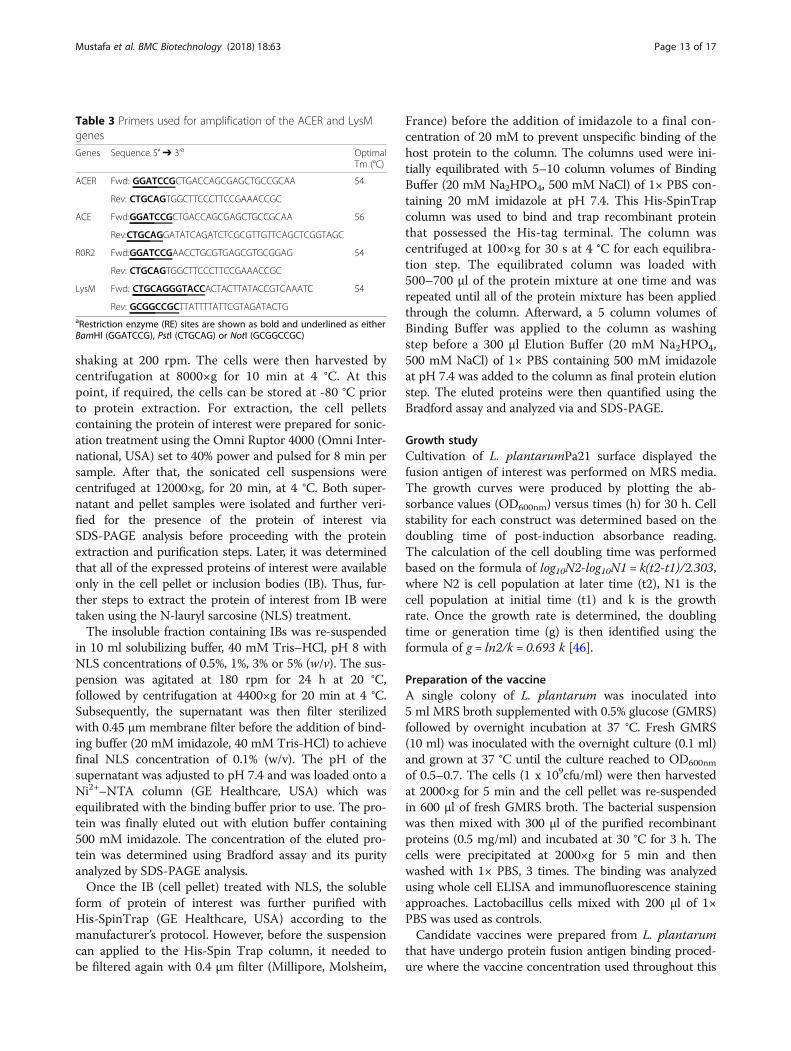
Table 3 Primers used for amplification of the ACER and LysMgenes
Genes Sequence 5′ ➔ 3’a OptimalTm (°C)
ACER Fwd: GGATCCGCTGACCAGCGAGCTGCCGCAA 54
Rev: CTGCAGTGGCTTCCCTTCCGAAACCGC
ACE Fwd:GGATCCGCTGACCAGCGAGCTGCCGCAA 56
Rev:CTGCAGGATATCAGATCTCGCGTTGTTCAGCTCGGTAGC
R0R2 Fwd:GGATCCGAACCTGCGTGAGCGTGCGGAG 54
Rev: CTGCAGTGGCTTCCCTTCCGAAACCGC
LysM Fwd: CTGCAGGGTACCACTACTTATACCGTCAAATC 54
Rev: GCGGCCGCTTATTTTATTCGTAGATACTGaRestriction enzyme (RE) sites are shown as bold and underlined as eitherBamHI (GGATCCG), PstI (CTGCAG) or NotI (GCGGCCGC)
Mustafa et al. BMC Biotechnology (2018) 18:63 Page 13 of 17

study was 1 × 10⁹ cfu/ml attached with 100 μl 0.5 mg/mlpurified antigen of interest. Once prepared, the candidatevaccines were used immediately. Similarly, preparation fora mixture of the candidate vaccine with L. lactis secretingmIL-12 was also performed. The recombinant L. lactis(LcIL12) was grown in GM17 at 30 °C without shaking.To optimize the nisin promoter stimulation, strains weregrown until OD600nm reached 0.6, followed by inductionwith 10 ng/ml nisin (Sigma-Aldrich) for 6 h. Cellularpellets were then harvested by centrifugation at 4000×gat 4 °C and washed three times with 1× PBS. The pelletwas re-suspended in 1× PBS to a final concentration of1 × 109 CFU before the final step of mixing with 1 × 10⁹ CFUof candidate vaccine.
Binding stability analysis and verificationTo confirm the binding of the expressed recombinantprotein of ACERL onto cell wall of L. plantarum, wholecell ELISA was utilized. The lactobacilli cells which wereformerly incubated with the recombinant proteins ofinterest were harvested by centrifugation at 2000×g for10 min and then fixed with 4% (w/v) paraformaldehydefor 20 min at RT, followed by washing step. The cellswere then incubated with blocking solution [3% (w/v)BSA in 1× PBS] for 30 min at RT. Then, the cells wereincubated with anti-his monoclonal (Novagen, USA) oranti-ACER specific antibody (Calbiochem, USA) as pri-mary antibody at a ratio of 1:200 in 1% BSA in 1× PBS,followed by 1 h incubation at RT. The cells were thenharvested and then incubated with horseradish peroxid-ase conjugated with anti-chicken antibody as secondaryantibody at a ratio of 1:200 in 1% BSA in 1× PBS for 1 hat RT. Following three times washing with 1× PBS, thecells were harvested and finally re-suspended in 200 μlof 1× PBS. Ten μl of the bacterial suspension and 50 μlof substrate (BM Blue, Roche, USA) was mixed in thewells of the ELISA plate and then incubated at RT for20 min. Finally 50 μl of the stop solution (1 M H2SO4)was added to end the reaction. The absorbance wasmeasured at OD450 nm by using ELISA reader (MultiscanMCC/340 MK II, Lab System). Controls were set usinglactobacilli cells incubated with BSA or 1× PBS.The binding of ACERL to L. plantarum was visualized
by immunofluorescence microscopy using 20 μl mouse
anti-his monoclonal antibody [0.2 μg/μl of His∙Tag® Mono-clonal Antibody (Novagen, USA)] as primary antibody andGoat Anti-Mouse Flourescein Conjugated Antibody(Calbiochem, USA) (1 μg/μl) as secondary antibodiesprepared by 1:200 dilution in 1% BSA (dissolved in 1×PBS). After the binding procedure with ACERL to L. plan-tarum, the attached lactobacilli cells were harvested bycentrifugation at 2000 x g for 10 min; washed with 1×PBS and then re-suspended in 200 μl of 1× PBS. About20–30 μl of the cells (1 × 10¹⁰ cfu/ml) was dropped onslides coated with poly-L-lysine; air dried and then washedwith 1× PBS. The attached cells were fixed with 4% (w/v)paraformaldehyde for 20 min at RT, followed by 1× PBSwashing. In the next step, the cells were incubated with3% (w/v) BSA in 1× PBS for 30 min at RT, to blocknon-specific sites. After washing with 1× PBS, the cellswere overlaid with primary polyclonal antibody diluted atratio 1:200 in 1% BSA in 1× PBS followed by incubation atRT for 60 min. The cells were washed with 1× PBS andthen incubated with FITC anti-goat conjugated antibody,diluted at 1:200, at RT for 1 h. Following 3 times washingwith 1× PBS, the slide was dried and 10 μl of Flourguar-dAntifade Mounting Solution (Sigma-Aldrich, USA) wasadded to keep the fluorescence dye from fading. The slidewas viewed and analyzed under phase contrast and fluor-escence microscope (Nikon ® Eclipse E200 fluorescencemicroscope) with immersion oil at 100× magnification.
Immunization protocolAfter the completion of the mice acclimatization period,mice were separated into 6 groups of 7 mice as indicatedin Table 4. All mice were immunized by oral gavage 3times at week 1 (days 1, 2, 3), week 4 (days 27, 28, 29) andweek 5 (days 40, 41, 42) with 100 μl of 1 × 10⁹ CFU of thevaccine. The dosage of the vaccine was adapted based onChilders and colleagues [47] while the immunizationschedule was based on Corthésy and co-workers [48].Control mice received identical quantities of PBS, L. plan-tarum or L. lactis/mIL-12 like the treatment groups.Blood sample from each group was taken by submandibu-lar bleeding during the immunization period at day 0, 21,33 and 45. All of the mice were euthanized 13 daysafter the last immunization. The control groups for oralgroup were divided into two sets which were referred
Table 4 Immunization strategy via oral administration in mice
Group Mouse Strain Immunized with: Dose Route No of mice
1 Balb/c PBS 100 μl Oral 7
2 Balb/c L. plantarum (Lp) 100 μl of 1 × 10⁹ cfu/ml Oral 7
3 Balb/c Lp ACERL 50 μg antigen + 100 μl of 1 × 10⁹ cfu/ml Oral 7
4 Balb/c Lp ACERL + mIL-12 50 μg antigen + 50 μl of 1 × 10⁹ cfu/ml + 50 μl of 1 × 10⁹ cfu/ml Oral 7
5 Balb/c mIL-12 100 μl of 1 × 10⁹ cfu/ml Oral 7
6 Balb/c ACERL 50 μg antigen Oral 7
Mustafa et al. BMC Biotechnology (2018) 18:63 Page 14 of 17

as the baseline set of Group 1 (PBS) and the referenceset of Group 2 (L. plantarum) and Group 5 (L. lactis/mIL-12). Meanwhile the treatment groups were Group3 (Lp ACERL), Group 4 (LP ACERL + Lc mIL-12), andGroup 6 (ACERL).
Determination of specific antibody IgGThe titre level of IgG total from blood sera of day 0, 21,33 and 45 was determined using eBioscience Mouse IgGtotal ready-SET-Go (eBioscience, Austria). Briefly, Corn-ing Costar 9018 ELISA plate was coated with 100 μl/wellof antigen ACERL in coating buffer, seal and incubateovernight at 4 °C. The wells were aspirated and washedtwice with 250 μl/well wash buffer (1× PBS, 0.05%Tween-20) and the wells blocked with 200 μl of blockingbuffer at room temperature for 2 h. The wells were aspi-rated and washed three times with wash buffer and100 μl reconstituted standard and sample in assay bufferwas added in triplicate into well and incubated for 2 h atroom temperature. The wells were aspirated and washedthree times with wash buffer and 100 μl/well of diluteddetection antibody was added to all wells for 1 h at RT.Then, the wells were aspirated again and washed threetimes with wash buffer and 100 μl/well of substrate solu-tion was added to each well and incubated at RT for ap-proximately 15 min and before 100 μl of Stop Solutionwas added to each well and the plate read at 450 nm.The plate was read using the Bio-Plex Suspension ArraySystem model Bio-Plex 100 System (BioRad, USA) andanalysed using the Bio-Plex Manager 4.0 (BioRad, USA)software.
Re-stimulation of splenocytes from the immunized miceApproximately 2 × 10⁷ cells/ml of splenocytes from the im-munized mice were stimμlated with 100 μl of 0.5 mg/mlACERL antigen protein in 24-well plates (Nunc, Denmark)at 37 °C and 5% CO2 for 48 h. Complete RPMI mediumcontaining only mouse splenocytes cells were included asnegative control. Once the incubation has completed, 10 μlof cell culture medium was used for cell viability assay.The rest of the cell culture supernatants was centrifuged at1200×g for 20 min at 4 °C before being sterilized throughfilter passage of a 0.2 μm pore size filter (Millipore,Germany) and stored at − 80 °C until needed for cytokineanalysis. All experiments were performed in triplicate.
Cytokine assessmentSamples from the supernatant of the induced splenocytes,sera, spleen, feaces, small intestine and lungs were col-lected and pooled from each group were quantified forcytokine production of IL-2, IL-4, IL-6, IL-12, TNF-α, andIFN-γ via Sandwich ELISA (BD OptEIA™ set mouse IL-2,IL-4, IL-6, IL-12, TNF-α, and IFN-γ, Germany) method.The detection limit of the assay was 6.25 pg/ml for IL-2,
0.5 pg/ml for IL-4, 5.25 pg/ml for IL-6, 6.25 pg/ml forIL-12, 1 pg/ml for IFN-γ, and 20 pg/ml for TNF- α.Optical density values of the samples were read at 450and 570 nm on an ELISA plate reader (Molecular Devices,Munich, Germany). Experiments were run in triplicate andrepeated at least twice, according to the manufacturer’sprotocol. Briefly, ELISA plates (Costar, UK) were coatedwith capture antibody of either goat anti-mouse IL-2, IL-4,IL-6, IL-12, TNF-α, or IFN-γ per well in a 0.1 M carbonatebuffer, pH 9.6, at 4 °C overnight. Following three washeswith PBS–Tween-20 (PBST) buffer (PBS pH 7·4 and 0.05%Tween-20), plates were incubated with blocking buffer[PBS, 0.1%, Tween-20, 1% bovine serum albumin (BSA)]for 2 h at 37°. After three washes, the plates were incubatedwith the serially diluted sera in a blocking buffer at 37 °Cfor 2 h. Subsequently using the washing buffer, the plateswere washed thrice before biotinylated secondary antibodyrabbit anti-mouse was added and incubated at 37° for 1 h.Then, streptavidin–peroxidase conjugate (1:10000 dilution)was added after washing and incubated at 37° for 1 h. Fi-nally, the plates were washed thrice and color developed bythe addition of tetra methyl benzidine TMB (Pharmingen,USA) at RT for 30 min and absorbance was read at450 nm. The plate was read using the Bio-Plex SuspensionArray System model Bio-Plex 100 System (BioRad, USA)and analsyed using the Bio-Plex Manager 4.0 (BioRad,USA) software. The production and ratio between cyto-kines of IL-2, IL-4, IL-6, IL-12, TNF-α and IFN-γ wascalculated based on the maximum production of eachcytokine observed in culture via ELISA. The samplesthat produce the most significant detection of cytokines(P < 0.05, P < 0.01 or P < 0.001) as compared to the con-trol of Lp or/and PBS groups were investigated.
Statistical analysisAll animal experiments were independently replicated atleast thrice. Statistical significance between two groupswas evaluated via the student T-test function of the Stat-View program, version 5.0 (SAS Institute Inc.; Cary,USA) and the graphs were prepared using GraphPadPrism version 7.00 (La Jolla; California, USA). The resultswere expressed as the mean ± standard deviation (S.D.) ofthe mean of combined data from the replicate experi-ments. To measure the differences among groups, the re-sults were tested for significance by a non-parametric way.Results having P-values of less than 0.05 were consideredstatistically significant.
Additional files
Additional file 1: Table S1. Data set of L. plantarum Pa21 carryingACERL antigen growth profiles. (XLSX 25 kb)
Additional file 2: Table S2. Data set of whole cell ELISA of L. plantarumattached with ACERL for a four-day period. (XLSX 13 kb)
Mustafa et al. BMC Biotechnology (2018) 18:63 Page 15 of 17

Additional file 3: Table S3. Data set from ELISA for time-course of theserum total IgG antibody response against Lp ACERL, Lp ACERL/LcIL-12,ACERL, LcIL-12, Lp and PBS immunization. (XLSX 16 kb)
Additional file 4: Table S4. Data set from cytokine response profiling ofspleen for IFN-γ (pg/ml), IL-4 (pg/ml), IL-2 (pg/ml), IL-6 (pg/ml), IL-12 (pg/ml)and TNF-α (pg/ml). (XLSX 47 kb)
Additional file 5: Table S5. Data set from cytokine response profiling oflung for IFN-γ (pg/ml), IL-4 (pg/ml), IL-2 (pg/ml), IL-6 (pg/ml), IL-12 (pg/ml)and TNF-α (pg/ml). (XLSX 37 kb)
Additional file 6: Table S6. Data set from cytokine response profiling ofGIT for IFN-γ (pg/ml), IL-4 (pg/ml), IL-2 (pg/ml), IL-6 (pg/ml), IL-12 (pg/ml)and TNF-α (pg/ml). (XLSX 148 kb)
Additional file 7: Table S7. Data set from cytokine response profiling ofre-stimulated splenocytes with ACERL antigen after 48 h for IFN-γ (pg/ml),IL-4 (pg/ml), IL-2 (pg/ml), IL-6 (pg/ml), IL-12 (pg/ml) and TNF-α (pg/ml).(XLSX 41 kb)
AbbreviationsACERL: Ag85B-CFP10-ESAT-6-Rv0475-Rv203; BCG: Bacilli Calmette–Guérin;GIT: Gastrointestinal tract; GMO: Genetically modified organism; IB: Inclusionbody; IFN-γ: Interferon gamma; IL-12: Interleukin 12; IL-2: Interleukin 2;IL-4: Interleukin 4; IL-6: Interleukin 6; LAB: Lactic acid bacteria; LcIL-12: L. lactissecreting IL-12; Lp ACERL: L. plantarum ACERL; LysM: Lysin motif; NLS:N-lauryl sarcosine; Th1: Type 1 T helper; Th2: Type 2 T helper; TNF-α: Tumornecrosis factor alpha
AcknowledgementsThe authors wish to thank Yeap Swee Keong and Fuzina for assistance inanimal ethics application and approval.
FundingThis work was supported by a research grant from the Ministry of HigherEducation of Malaysia (MOHE) under the grant number 700–2/1/LRGS/5525500.
Availability of data and materialsThe authors declare that [the/all other] data supporting the findings of thisstudy are available within the article [and its Additional files 1, 2, 3, 4, 5, 6, 7].
Authors’ contributionsAM helped design the experiments, performed experimental work, analyzeddata and drafted the manuscript. SB and JK performed experimental workand analyzed data. MA, RR and KY helped to design the experiments and toanalyze and interpret data. All authors read and approved the final manuscript.
Ethics approvalAnimals were sourced, housed and treated in accordance with institutionalguidelines approved by the Institutional Animal Care and Use Committee,Universiti Putra Malaysia (UPM/IACUC/AUP - R072/2014).
Consent for publicationNot applicable.
Competing interestsThe authors declare that they have no competing interests.
Publisher’s NoteSpringer Nature remains neutral with regard to jurisdictional claims inpublished maps and institutional affiliations.
Author details1Department of Cell and Molecular Biology, Faculty of Biotechnology andBiomolecular Sciences Universiti Putra Malaysia, 43400 Serdang, SelangorDarul Ehsan, Malaysia. 2Department of Microbiology, Faculty ofBiotechnology and Biomolecular Sciences, Universiti Putra Malaysia, 43400Serdang, Selangor Darul Ehsan, Malaysia. 3Department of Pathology, Facultyof Medicine and Health Science, Universiti Putra Malaysia, 43400 Serdang,Selangor Darul Ehsan, Malaysia. 4Institute of Bioscience, Universiti Putra
Malaysia, 43400 Serdang, Selangor Darul Ehsan, Malaysia. 5Malaysia GenomeInstitute, 43000 Kajang, Selangor Darul Ehsan, Malaysia.
Received: 4 May 2018 Accepted: 10 August 2018
References1. World Health Organization. Global Tuberculosis Report 2017. WHO/HTM/TB/
2017.23. Geneva: WHO; 2017.2. Pareek M, Greenaway C, Noori T, Munoz J, Zenner D. The impact of
migration on tuberculosis epidemiology and control in high-incomecountries: a review. BMC Med Central. 2016;14:48.
3. Zhang L, Ru H, Chen F, Jin C, Sun R, Fan X, Guo M, Mai J, Xu W, Lin Q, Liu J.Variable virulence and efficacy of BCG vaccine strains in mice andcorrelation with genome polymorphisms. Mol Ther. 2016;24:398–405.
4. Delogu G, Fadda G. The quest for a new vaccine against tuberculosis.J Infect Dev. 2009;3
5. Sarhan M a a, Musa M, Zainuddin ZF. Cloning, protein expression anddisplay of synthetic multi-epitope mycobacterial antigens on Salmonellatyphi Ty21a cell surface. Indian J Exp Biol. 2011;49:645–53.
6. Mohan T, Verma P, Rao DN. Novel adjuvants & delivery vehicles for vaccinesdevelopment: a road ahead. Indian J MedRes. 2013;138:779–95.
7. Wells JM, Mercenier A. Mucosal delivery of therapeutic and prophylacticmolecules using lactic acid bacteria. Nat Rev Microbiol. 2008;6:349–62.
8. Holmgren J, Czerkinsky C. Mucosal immunity and vaccines. Nat Med. 2005;119. Badell E, Nicolle F, Clark S, Majlessi L, Boudou F, Martino A, Winter N.
Protection against tuberculosis induced by oral prime with Mycobacteriumbovis BCG and intranasal subunit boost based on the vaccine candidateAg85B-ESAT-6 does not correlate with circulating IFN-γ producing T-cells.Vaccine. 2009;27:28–37.
10. Hokey D, Misra A. Aerosol vaccines for tuberculosis: a fine line betweenprotection and pathology. Tuberculosis (Edinb). 2011;91:82–5.
11. Varma NRS, Toosa H, Foo HL, Alitheen NBM, Nor Shamsudin M, Arbab AS.Display of the viral epitopes on Lactococcus lactis: a model for food gradevaccine against EV71. Biotechnol Res Int. 2013;431315
12. Moeini H, Rahim RA, Omar AR, Shafee N, Yusoff K. Lactobacillus acidophilusas a live vehicle for oral immunization against chicken anemia virus. ApplMicrobiol Biotechnol. 2011;90:77–88.
13. Steen A, Steen A, Buist G, Buist G, Kok J, Kok J, Leenhouts K. Novel surfacedisplay system for proteins on non-genetically modifed gram-positivebacteria. Society. 2006;72:880–9.
14. Kuczkowska K, Kleiveland CR, Minic R, Moen LF, Øverland L, Tjåland R, EijsinkVGH. Immunogenic properties of Lactobacillus plantarum producing surface-displayed Mycobacterium tuberculosis antigens. Appl Environ Microbiol. 2017;83
15. Langermans J a M, Doherty TM, Vervenne R a W, van der Laan T,Lyashchenko K, Greenwald R, Andersen P. Protection of macaques againstMycobacterium tuberculosis infection by a subunit vaccine based on a fusionprotein of antigen 85B and ESAT-6. Vaccine. 2005;23:2740–50.
16. Li W, Deng G, Li M, Zeng J, Zhao L, Liu X, Wang Y. A recombinantadenovirus expressing CFP10, ESAT6, Ag85A and Ag85B of Mycobacteriumtuberculosis elicits strong antigen-specific immune responses in mice. MolImmunol. 2014;62:86–95.
17. Lewinsohn DA, Swarbrick GM, Park B, Cansler ME, Null MD, Toren KG,Lewinsohn DM. Comprehensive definition of human immunodominantCD8 antigens in tuberculosis. Vaccine. 2017;2(1):8.
18. Xin Q, Niu H, Li Z, Zhang G, Hu L, Wang B, Li J, Yu H, Liu W, Da Z, Li R, XianQ, Wang Y, Zhang Y, Jing T, Ma X, Zhu B. Subunit vaccine consisting ofmulti-stage antigens has high protective efficacy against Mycobacteriumtuberculosis infection in mice. PLoS One. 2013;8:e72745.
19. Nagy G, Emo”dy L, Pál T. Strategies for the development of vaccinesconferring broad-spectrum protection. Int J Med Microbiol. 2008;298:379–95.
20. Piubelli L, Campa M, Temporini C, Binda E, Mangione F, Amicosante M,Pollegioni L. Optimizing Escherichia coli as a protein expression platform toproduce Mycobacterium tuberculosis immunogenic proteins. Microb CellFactories. 2013;12:115.
21. Singh SM, Panda AK. Solubilization and refolding of bacterial inclusion bodyproteins. J Bio Bioeng. 2005;99:303–10.
22. Francis VG, Majeed MA, Gummadi SN. Recovery of functionally activerecombinant human phospholipid scramblase 1 from inclusion bodiesusing N-lauroyl sarcosine. J Ind Microbiol Biotechnol. 2012;39:1041–8.
Mustafa et al. BMC Biotechnology (2018) 18:63 Page 16 of 17

23. Mir SA, Sharma S. Cloning, expression and N-terminal formylation of ESAT-6of Mycobacterium tuberculosis H37Rv. Protein Expr Purif. 2013;92:223–9.
24. Shi S, Yu L, Sun D, Liu J, Hickey AJ. Rational design of multiple TB antigensTB10.4 and TB10.4-Ag85B as subunit vaccine candidates againstMycobacterium tuberculosis. Pharm Res. 2010;27:224–34.
25. Peternel S, Grdadolnik J, Gaberc-Porekar V, Komel R. Engineering inclusionbodies for non denaturing extraction of functional proteins. Microb CellFactories. 2008;7:34.
26. Tao H, Liu W, Simmons BN, Harris HK, Cox TC, Massiah M. Purifying nativelyfolded proteins from inclusion bodies using sarkosyl, triton X-100, andCHAPS. BioTechniques. 2010;48:61–4.
27. Fredriksen L, Kleiveland CR, Olsen Hult LT, Lea T, Nygaard CS, Eijsink VGH,Mathiesen G. Surface display of N-terminally anchored invasin by Lactobacillusplantarum activates NF-κB in monocytes. Appl Environ Microbiol. 2012;78:5864–71.
28. Raha AR, Varma NR, Yusoff K, Ross E, Foo HL. Cell surface display system forLactococcus lactis: a novel development for oral vaccine. Appl MicrobiolBiotechnol. 2005;68 https://doi.org/10.1007/s00253-004-1851-8.
29. Poquet I, Saint V, Seznec E, Simoes N, Bolotin A, Gruss A. HtrA is the uniquesurface housekeeping protease in Lactococcus lactis and is required fornatural protein processing. MolMicrob. 2000;35:1042–51.
30. Lavelle EC, O’Hagan DT. Delivery systems and adjuvants for oral vaccines.Expert Opin Drug Deliv. 2006;3:747–62.
31. Marasini N, Skwarczynski M, Toth I. Oral delivery of nanoparticle-basedvaccines. Expert Rev Vaccines. 2014;13:1361–76.
32. Qiao H, Duffy LC, Griffiths E, Dryja D, Leavens A, Rossman J, Locniskar M.Immune responses in rhesus rotavirus-challenged Balb/c mice treated withbifidobacteria and prebiotic supplements. Pediatr Res. 2002;51:750–5.
33. Ghadimi D, de Vrese M, Heller KJ, Schrezenmeir J. Lactic acid bacteriaenhance autophagic ability of mononuclear phagocytes by increasing Th1autophagy-promoting cytokine (IFN-gamma) and nitric oxide (NO) levelsand reducing Th2 autophagy-restraining cytokines (IL-4 and IL-13) inresponse to Mycobacterium. Int Immunopharmacol. 2010;10:694–706.
34. Smelt MJ, de Haan BJ, Bron P a, van Swam I, Meijerink M, Wells JM, de VosP. L. plantarum, L. salivarius, and L. lactis attenuate Th2 responses andincrease Treg frequencies in healthy mice in a strain dependent manner.PLoS One. 2012;7:e47244.
35. Niers LEM, Timmerman HM, Rijkers GT, van Bleek GM, van Uden NOP, KnolEF, Hoekstra MO. Identification of strong interleukin-10 inducing lactic acidbacteria which down-regulate T helper type 2 cytokines. Clin Exp Allergy.2005;35:1481–9.
36. Pochard P, Gosset P, Grangette C, Andre C, Tonnel A-B, Pestel J, MercenierA. Lactic acid bacteria inhibit TH2 cytokine production by mononuclear cellsfrom allergic patients. J Allergy Clin Immunol. 2017;110:617–23.
37. Scaldaferri F, Gerardi V, Lopetuso LR, Del Zompo F, Mangiola F, Boškoski I,Gasbarrini A. Gut microbial flora, prebiotics, and probiotics in IBD: theircurrent usage and utility. Biomed Res Int. 2013;3:55–64.
38. Kuipers OP, De Ruyter PGGA, Kleerebezem M, De Vos WM. Quorum sensing-controlled gene expression in lactic acid bacteria. J Biotechnol. 1998;64:15–21.
39. Cortes-Perez NG, Ah-Leung S, Bermudez-Humaran LG, Corthier G, Wal JM,Langella P, Adel-Patient K. Intranasal co-administration of live lactococciproducing interleukin-12 and a major cow’s milk allergen inhibits allergicreaction in mice. Clin Vaccine Immunol. 2007;14(3):226–33.
40. Sáez D, Fernández P, Rivera A, Andrews E, Oñate A. Oral immunization of micewith recombinant Lactococcus lactis expressing Cu,Zn superoxide dismutase ofBrucella abortus triggers protective immunity. Vaccine. 2012;30:1283–90.
41. Hugentobler F, Di Roberto RB, Gillard J, Cousineau B. Oral immunizationusing live Lactococcus lactis co-expressing LACK and IL-12 protects BALB/cmice against Leishmania major infection. Vaccine. 2012;30:5726–32.
42. Pichichero ME. Booster Vaccinations: Can immunologic memory outpacedisease pathogenesis? Pediatrics. 2009;124:1633 LP–1641.
43. Pavot V, Rochereau N, Genin C, Verrier B, Paul S. New insights in mucosalvaccine development. Vaccine. 2012;30:142–54.
44. Maidin MST, Song AA-L, Jalilsood T, Sieo CC, Yusoff K, Rahim RA. Construction ofa novel inducible expression vector for Lactococcus lactis M4 and Lactobacillusplantarum Pa21. Plasmid. 2014;74:32–8.
45. Terzaghi BE, Sandine WE. Improved medium for lactic streptococci and theirbacteriophages. Appl Microbiol. 1975;29:807–13.
46. Frederick C, Neidhardt, Ingraham MS JL. Physiology of the Bacterial Cell: AMolecular Approach. Biochem Educ; 1990. p. 2.
47. Childers NK, Miller KL, Tong G, Llarena JC, Greenway T, Ulrich JT, MichalekSM. Adjuvant activity of monophosphoryl lipid a for nasal and oralimmunization with soluble or liposome-associated antigen. Infect Immun.2000;68:5509–16.
48. Corthésy B, Boris S, Isler P, Grangette C, Mercenier A. Oral immunization ofmice with lactic acid bacteria producing Helicobacter pylori urease B subunitpartially protects against challenge with Helicobacter felis. J Infect Dis.2005;192:1441–9.
Mustafa et al. BMC Biotechnology (2018) 18:63 Page 17 of 17