prolonging nerve grafts using chemical extracted … jemnoschi hreniuc 12 17.pdf2936 http: //...
TRANSCRIPT
http://www.revistadechimie.ro REV.CHIM.(Bucharest)♦ 68♦ No. 12 ♦ 20172936
Prolonging Nerve Grafts Using Chemical ExtractedMuscle-in-vein with Vein Window Method
Chemical acellular nerve grafts
IRINA MIHAELA JEMNOSCHI HRENIUC1*, CAMELIA TAMAS1, SORIN AURELIAN PASCA2, BOGDAN CIUNTU3*, ROXANA CIUNTU5*,ROBERT D. NEGRU6 , CARMEN ZAMFIR3, TEODOR STAMATE1
1 Grigore T. Popa University of Medicine and Pharmacy, Faculty of Medicine, Department of Plastic Surgery, Iasi, 16 UniversitatiiStr., 700115, Romania2 Ion Ionescu de la Brad University of Agricultural Sciences and Veterinary Medicine, Faculty of Veterinary Medicine, Departmentof Veterinary Morphopathology, 3 Mihail Sadoveanu Alley, 700490, Iasi, Romania1 Grigore T.Popa University of Medicine and Pharmacy, Faculty of Medicine, Department of General Surgery, 16 Universitatii Str.,700115, Iasi, Romania1 Grigore T.Popa University of Medicine and Pharmacy, Faculty of Medicine, Department of Ophtalmology, 16 Universitatii Str.,700115, Iasi, Romania1 Grigore T. Popa University of Medicine and Pharmacy,Faculty of Medicine, Ist Medical Department, 16 Universitatii Str.,700115,Iasi, Romania1 Grigore T. Popa University of Medicine and Pharmacy, Faculty of Medicine, Department of Histology, 16 Universitatii Str.,700115,Iasi, Romania
Nerve injuries are a common pathology in hand trauma. The consequences are drastic both for patients anddoctors/medical system. In many cases direct coaptation is impossible. A nerve graft should be used in thecase of a neuroma, trauma or tumor, for restoration of nervous influx. The aim of this study is demonstratethat by grafting restant nerve stumps with muscle-in-vein nerve grafts we obtain good result in terms offunctional and sensibility recovery and also our method ‚window-vein’ is a good way of prolonging nervegrafts. The method of study is experimental. We worked in the laboratory in optimal conditions for carryingout of muscles-in-vein nerve grafts (nerve grafts size 1.5 cm-3 cm). We used acellular muscle grafts with thechemical extraction method.The study was conducted on experimental animals (Wistar male rats).Weused 30 experience animals in 3 equal groups (classical group and muscle-in-vein nerve grafts-2 nervegrafts of 1,5 cm central sutured and the third group with muscle-in-vein nerve grafts, window-vein method,3 cm). At 4 and respectively 6 weeks postoperative at the quality tests we observed the progress with thefootprint test. The operated hind in comparison with the healthy hind was 86% recovered and similar withclassic nerve grafts. Quantitatively the number of regenerated axons in the group with muscle-in-vein nervegrafts was significant bigger in comparison with the classical group (15%).The method using muscle-in-vein nerve graft with windows-vein it’s a good alternative for nerve grafting in comparison with classicalnerve grafting. When the local possibilities are limited, this method is good for prolonging the grafts. Therelationship between cost and benefit in this case it’s an advantage because we use the local resources ofthe affected area. The motor results of nerve grafting ingroup 2 in comparison with group 3 were similar andin some cases better in group 1. Grafting with MVNG offers a better alternative for donor site regeneration incomparison with classical nerve grafts. This method is useful to prolong nerve grafts without adding morbidity.
Keywords: nerve grafting, regeneration, axons, window-vein, acellular
Peripheral nerve injuries are a common entity in thetreatment of hand disorders. They are most commonlyrelated to direct mechanical trauma, and can be associatedwith other soft and bone tissue lesions [1)].
Transection of axons implicates in morphologic andmetabolic changes throughout the whole length of theneuron. From the moment of injury, the cell body issignalized and the regeneration process starts [2]. Whilethe distal nerve stump undergoes Wallerian degeneration,the proximal stump retracts and the Schwann cellsundergo apoptosis [3]. The axon sprouts and a growth coneis formed at the tip of each sprout, interacting withactivated and proliferating Schwann cells [4]. The nerveinjury itself results in decreasing of motoneurons in thespinal cord. In an experimental study with Wistar rats, thenumber of motoneurons labeled in the spinal corddecreased an average of 20% due to an isolated tibial nerveinjury, with no technical interference by the surgeon [5].
Exploring peripheral nerve injuries was since the earlymedicine a controversy issue. In our days the most frequent
* email: [email protected]; [email protected]; [email protected]
problem is nerve repairing and functional outcome. Duringtime, different types of nerve grafting had success. Themost common is allograft with contralateral sural nerve.But in this situation the local possibilities become limitedand it adds morbidity. That’s how the need for safe graftingappeared [6]. Muscle-in-vein method is used since the early2000s with good results in specific clinical cases. Battistonet al (2000) published their result on 21 patients with arange of success of 85%. The length of the nerve graft wasfrom 0.5 to 6 cm. Later, in other clinical cases it wasobserved that grafts longer than 3-4 cm have the bestfunctional outcome in comparison with longer than 5 cmnerve grafts. This gives the hypothesis that we need to findout another convenient method of peripheral nervegrafting. The functional results are comparative with directcoaptation, the cost are small and the second donor sitemorbidity is avoided [7].
Sometimes the local possibilities of nerve grafting usingthe sural nerve are limited, so the muscle-in-vein nervegrafts are the proper solution [8].

REV.CHIM.(Bucharest)♦ 68♦ No. 12 ♦ 2017 http://www.revistadechimie.ro 2937
Direct coaptation of restant nerve stumps is a problemoften because the surgical intervention is made later andthe nerves have the property to retract. In this case a nervegraft is absolutely necessary. The possibilities are allografts,synthetic nerve conduits to conduct axons regeneration ornatural conduit filled with muscles, nerve growth factors,fat, collagen.
Every method has advantages and disadvantages, butthe cost and site morbidity is often named.
Peripheral nerves are regenerating with approximatively1 mm per day. This means that a big trauma, with largenerve gaps need more time to heal. The best timing forsurgery is 6 months after the trauma but sometimes atthis point the resources of direct coaptation are almostinexistent so we need to use other surgical techniquesand among them is nerve grafting on different size defects[9].
Experimental partMaterial and method
Animals are anesthetized for surgery using ketamine(100 mg/kg) and xylazine (10 mg/kg) via intramuscularinjection. Each animal also receives a subcutaneousinjection of meloxicam (10 mg/kg) to minimize post-operative pain and also bupivacaine (1µg/kg) at the site ofsurgery.
Both hindquarters are carefully shaved using a surgicalclippers and depilation is completed with hair removalcream.
Skin is cleansed using sterile cotton tipped applicatorsand betadine surgical scrub.
Ophthalmic ointment is applied to the eyes using sterilecotton tipped applicators.
The rat is placed on a clean stainless steel plate, underwhich has been placed on the surgical table. Animaltemperature is maintained at 37°C .
All limbs are taped down, with care taken to positionthe hind limbs symmetrically so that the knee joint makesa right angle with the body. A syringe was placed under theleft foot to emphasis the sciatic nerve intraoperatively.
The surgical field is covered with a sterile drape. Allinstruments are sterilized by autoclave or hot beadsterilization, and we wear a mask, gown, and sterile gloves.
After preparation, a semi-circular incision across midlineis made in the skin. The skin is gently dissected from theunderlying musculature, and folded over to remain out ofthe way during the procedure. It is kept moist usingapplications of 0.1 mL sterile saline during the procedure.
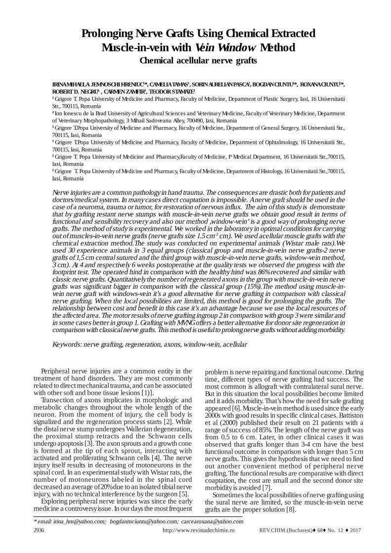
Opening the fascial plane between the gluteus maximusand the anterior head of the biceps femoris reveals thesciatic nerve (fig. 1). For a surgical control, the contralateralsciatic nerve should be exposed and mobilized, but leftintact. The gluteal musculature is then re-opposed andsutured using a 6-0 nylon, non-absorbable sutures [10]
The experimental sciatic nerve is then exposed in thesame fashion, with retractors in place to ease visualization.The retractors are sterilized prior to use. The sciatic nerveis then gently freed from the surrounding connective tissueusing iridectomy scissors.
Using a fine forceps, the nerve is placed on the bottomjaw of a super-fine hemostatic forceps. The three fasciclesare sequentially aligned, not on top of each other. Thehemostatic forceps have been engraved with a mark at1.5 mm from their tip. The outermost portion of the sciaticis placed in line with this mark before crush. This ensuresa crush of uniform width, and that the nerve does not extendbeyond the jaws of the hemostatic forceps when flatteneddue to the crushing force. If the nerve extends beyond theforceps tip the nerve will only be partially crushed [11].
For our experiment we cut the nerve so we are able tograft it after.
The gluteal musculature is re-opposed and sutured inthe same way as the contralateral side.
Finally, the skin incision is closed using 4/0 Nylon sutures.On this model of injury we graft the nerves.
Using the muscle around and a segment from tibial veinwe built the muscle-in-vein nerve grafts (Figure 4). On aforceps we glide the entire vein after we washed it in salinesolution and heparin. With the tip of the forceps we grabthe muscle and glide it into the vein.
The muscle fibers were treated chemically with TritonX-100 and deoxycholate in proportion 2/1 for 5 minutes (5mL Triton X-100 and 2.5 mL deoxycholate) before clearingit with saline solution and introducing then into the vein.
Triton X-100 and deoxycholate are 2 detergents used inexperimental laboratories to create acellular grafts orsegments of tissues. The outcomes after using it in nervegrafts were conclusive in comparison with other methods(e.g. freezing-dehydratation method) and this is the reasonwe used it.
In group 1 we cut the nerve and suture it back afterreversing it with 8/0 Premilene under 8x magnificationloupes.
In group 2 we built a 3 cm long muscle-in-vein nervegraft from 2 nerve grafts of 1.5 cm centrally sutured.
In group 3 we built a 3 cm long single unit muscle-in-vein nerve graft using ‚window-vein’ method.
This nerve graft is 1.5 long for the second group and 3cm long for the 3rd group but instead of suturing 2 grafts of1.5 cm centrally, we built a single unit graft using thewindows-vein method: in the central part of the vein wemade a small perpendicular incision into the vein, then weglide the vein starting from this window on the forceps andwith the end of the forceps we grab the muscle and pull ituntil the end. In the same manner we proceed with thesecond half of the vein. At the end we suture the windowand our nerve graft is ready for the final suture.
Fig. 1. Actual imagine of thesciatic nerve before
neurorraphy. (8mp Samsungcamera)
Post-Operative CareFollowing the procedure, animals are placed on a
heating pad at 37°C until they show signs of movement.They are then moved back to their home cage, where
water and food are readily accessible on the floor in theform of Hydrogel and wetted food.
Histology analysis: preservation of the sciatic nervesegments.
Nerve specimens were buffered initially in formaldehyde37% (12 h). Than we proceed the deparaffinization withxylene I (5 min), xylene II (5 min), 96% ethanol (3-5 min),70% ethanol (3-5 min) and distilled water (5 min). Afterthis the staining was made with hematoxylin (3-5 min),rinse in water (10-15 min), 70% ethanol (3-5 min) then ineosin (1-3 min). The dehydration was made with 96%ethanol (2 min) and carbon-xylene (3-5 min). The lastprocedure was clearing with xylene I (5 min) and xylene II(5 min). We mounted with mounting medium and examinethe blades is microscope.
Proximal, middle and distal segments of nerve tissue
http://www.revistadechimie.ro REV.CHIM.(Bucharest)♦ 68♦ No. 12 ♦ 20172938
were examined, revealing a number of myelinated fibersthat were counted under the microscope and analyzedusing computer imaging.
The footprint testAt 12 weeks we performed the footprint test. We marked
the back hind of the Wistar rat with methylene blue andput them into a cage of 60x90 cm to walk over a piece ofspecial paper. At the end we measured the size of the backhinds in comparison with each other.
Footprints were evaluated by three parameters: 1)distance from the heel to the third toe, print length; 2)distance from the first to the fifth toes, TS; and 3) distancefrom the second to the fourth toes, intermediary TS. Allthree measurements were taken from the experimental(E) and normal (N) sides. Functional recovery wasassessed by calculating the Sciatic Functional Index (SFI)value. A value close to 0 indicate a good recovery and avalue close to 100 means the opposite [12].
Results and discussionsA complete investigation of post injury regeneration
combines functional, electrophysiological, andmorphological assessments. Morphological assessmentsfollowing regeneration are the ones we are interested inour study.
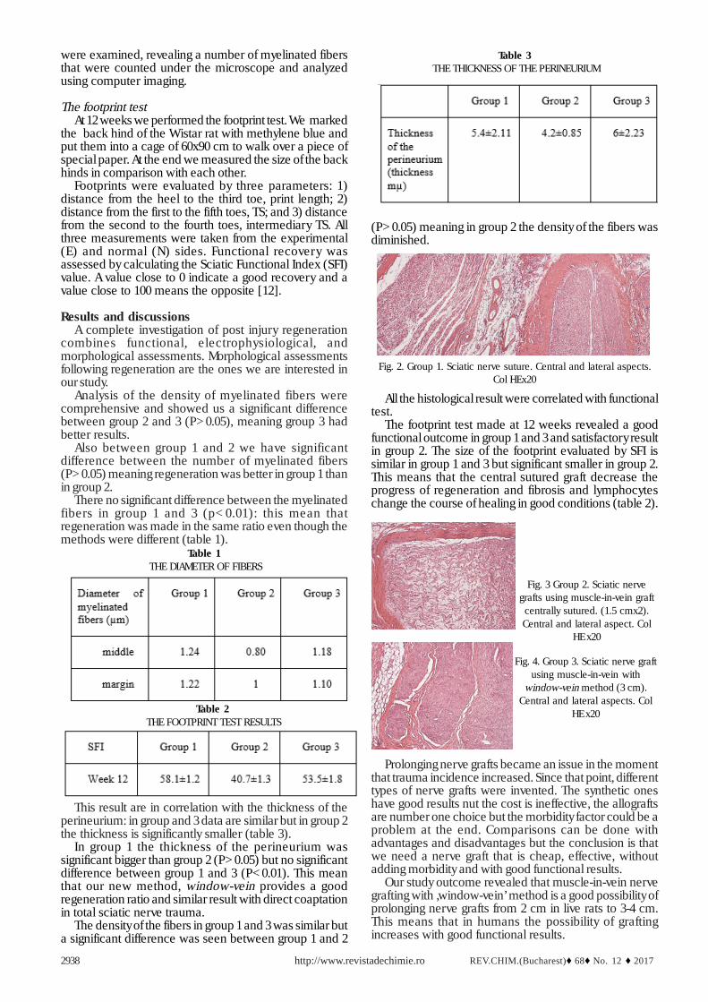
Analysis of the density of myelinated fibers werecomprehensive and showed us a significant differencebetween group 2 and 3 (P>0.05), meaning group 3 hadbetter results.
Also between group 1 and 2 we have significantdifference between the number of myelinated fibers(P>0.05) meaning regeneration was better in group 1 thanin group 2.
There no significant difference between the myelinatedfibers in group 1 and 3 (p<0.01): this mean thatregeneration was made in the same ratio even though themethods were different (table 1).
(P>0.05) meaning in group 2 the density of the fibers wasdiminished.
Table 1 THE DIAMETER OF FIBERS
This result are in correlation with the thickness of theperineurium: in group and 3 data are similar but in group 2the thickness is significantly smaller (table 3).
In group 1 the thickness of the perineurium wassignificant bigger than group 2 (P>0.05) but no significantdifference between group 1 and 3 (P<0.01). This meanthat our new method, window-vein provides a goodregeneration ratio and similar result with direct coaptationin total sciatic nerve trauma.
The density of the fibers in group 1 and 3 was similar buta significant difference was seen between group 1 and 2
Table 2 THE FOOTPRINT TEST RESULTS
Table 3 THE THICKNESS OF THE PERINEURIUM
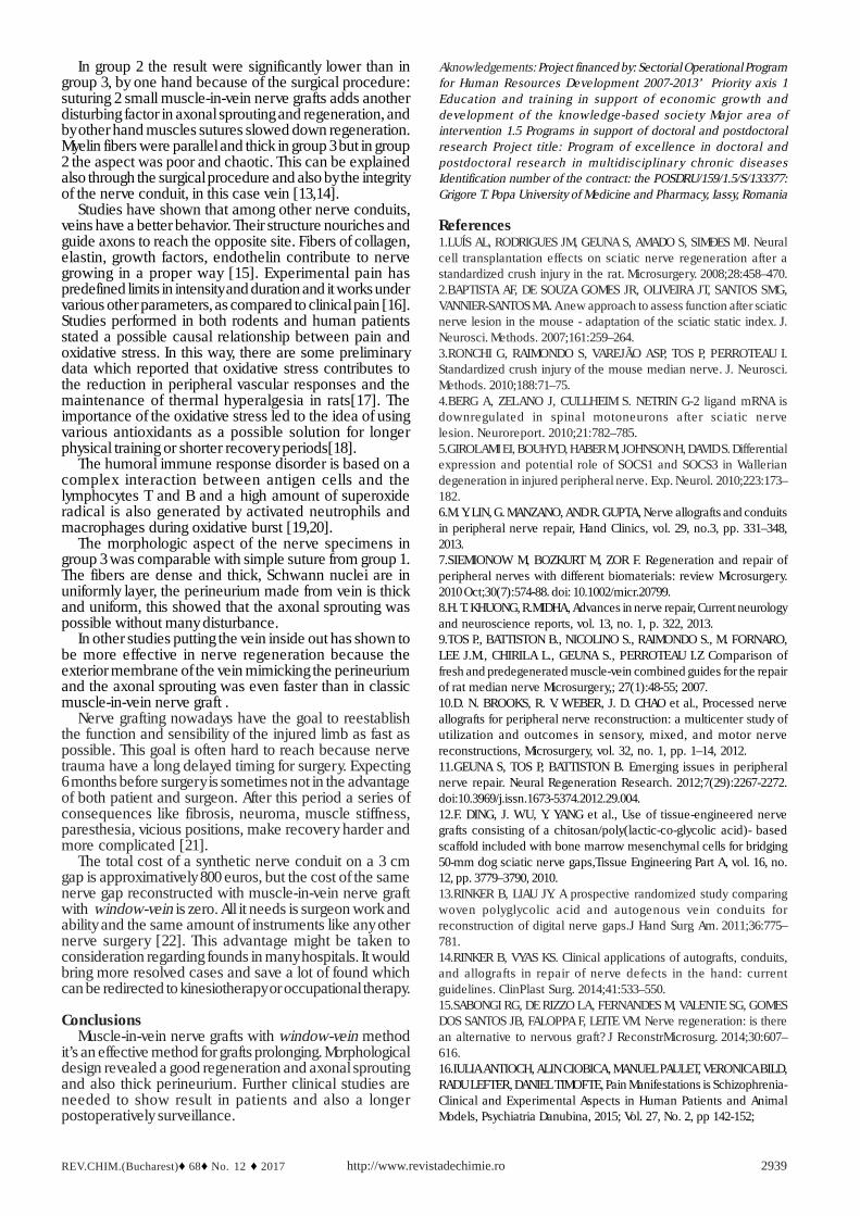
Fig. 2. Group 1. Sciatic nerve suture. Central and lateral aspects.Col HEx20
All the histological result were correlated with functionaltest.
The footprint test made at 12 weeks revealed a goodfunctional outcome in group 1 and 3 and satisfactory resultin group 2. The size of the footprint evaluated by SFI issimilar in group 1 and 3 but significant smaller in group 2.This means that the central sutured graft decrease theprogress of regeneration and fibrosis and lymphocyteschange the course of healing in good conditions (table 2).
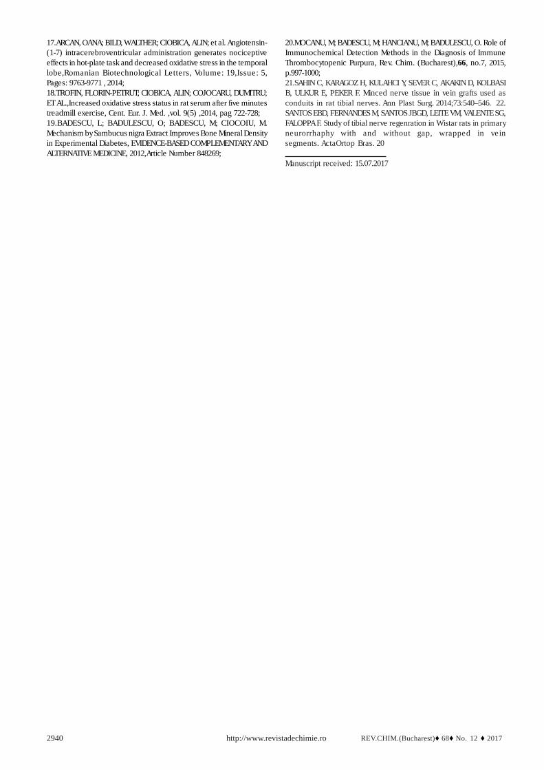
Fig. 3 Group 2. Sciatic nervegrafts using muscle-in-vein graft
centrally sutured. (1.5 cmx2).Central and lateral aspect. Col
HE x20
Fig. 4. Group 3. Sciatic nerve graftusing muscle-in-vein with
window-vein method (3 cm).Central and lateral aspects. Col
HE x20
Prolonging nerve grafts became an issue in the momentthat trauma incidence increased. Since that point, differenttypes of nerve grafts were invented. The synthetic oneshave good results nut the cost is ineffective, the allograftsare number one choice but the morbidity factor could be aproblem at the end. Comparisons can be done withadvantages and disadvantages but the conclusion is thatwe need a nerve graft that is cheap, effective, withoutadding morbidity and with good functional results.
Our study outcome revealed that muscle-in-vein nervegrafting with ‚window-vein’ method is a good possibility ofprolonging nerve grafts from 2 cm in live rats to 3-4 cm.This means that in humans the possibility of graftingincreases with good functional results.
REV.CHIM.(Bucharest)♦ 68♦ No. 12 ♦ 2017 http://www.revistadechimie.ro 2939
In group 2 the result were significantly lower than ingroup 3, by one hand because of the surgical procedure:suturing 2 small muscle-in-vein nerve grafts adds anotherdisturbing factor in axonal sprouting and regeneration, andby other hand muscles sutures slowed down regeneration.Myelin fibers were parallel and thick in group 3 but in group2 the aspect was poor and chaotic. This can be explainedalso through the surgical procedure and also by the integrityof the nerve conduit, in this case vein [13,14].
Studies have shown that among other nerve conduits,veins have a better behavior. Their structure nouriches andguide axons to reach the opposite site. Fibers of collagen,elastin, growth factors, endothelin contribute to nervegrowing in a proper way [15]. Experimental pain haspredefined limits in intensity and duration and it works undervarious other parameters, as compared to clinical pain [16].Studies performed in both rodents and human patientsstated a possible causal relationship between pain andoxidative stress. In this way, there are some preliminarydata which reported that oxidative stress contributes tothe reduction in peripheral vascular responses and themaintenance of thermal hyperalgesia in rats[17]. Theimportance of the oxidative stress led to the idea of usingvarious antioxidants as a possible solution for longerphysical training or shorter recovery periods[18].
The humoral immune response disorder is based on acomplex interaction between antigen cells and thelymphocytes T and B and a high amount of superoxideradical is also generated by activated neutrophils andmacrophages during oxidative burst [19,20].
The morphologic aspect of the nerve specimens ingroup 3 was comparable with simple suture from group 1.The fibers are dense and thick, Schwann nuclei are inuniformly layer, the perineurium made from vein is thickand uniform, this showed that the axonal sprouting waspossible without many disturbance.
In other studies putting the vein inside out has shown tobe more effective in nerve regeneration because theexterior membrane of the vein mimicking the perineuriumand the axonal sprouting was even faster than in classicmuscle-in-vein nerve graft .
Nerve grafting nowadays have the goal to reestablishthe function and sensibility of the injured limb as fast aspossible. This goal is often hard to reach because nervetrauma have a long delayed timing for surgery. Expecting6 months before surgery is sometimes not in the advantageof both patient and surgeon. After this period a series ofconsequences like fibrosis, neuroma, muscle stiffness,paresthesia, vicious positions, make recovery harder andmore complicated [21].
The total cost of a synthetic nerve conduit on a 3 cmgap is approximatively 800 euros, but the cost of the samenerve gap reconstructed with muscle-in-vein nerve graftwith window-vein is zero. All it needs is surgeon work andability and the same amount of instruments like any othernerve surgery [22]. This advantage might be taken toconsideration regarding founds in many hospitals. It wouldbring more resolved cases and save a lot of found whichcan be redirected to kinesiotherapy or occupational therapy.
ConclusionsMuscle-in-vein nerve grafts with window-vein method
it’s an effective method for grafts prolonging. Morphologicaldesign revealed a good regeneration and axonal sproutingand also thick perineurium. Further clinical studies areneeded to show result in patients and also a longerpostoperatively surveillance.
Aknowledgements: Project financed by: Sectorial Operational Programfor Human Resources Development 2007-2013’ Priority axis 1Education and training in support of economic growth anddevelopment of the knowledge-based society Major area ofintervention 1.5 Programs in support of doctoral and postdoctoralresearch Project title: Program of excellence in doctoral andpostdoctoral research in multidisciplinar y chronic diseasesIdentification number of the contract: the POSDRU/159/1.5/S/133377:Grigore T. Popa University of Medicine and Pharmacy, Iassy, Romania
References1.LUÍS AL, RODRIGUES JM, GEUNA S, AMADO S, SIMÐES MJ. Neuralcell transplantation effects on sciatic nerve regeneration after astandardized crush injury in the rat. Microsurgery. 2008;28:458–470.2.BAPTISTA AF, DE SOUZA GOMES JR, OLIVEIRA JT, SANTOS SMG,VANNIER-SANTOS MA. A new approach to assess function after sciaticnerve lesion in the mouse - adaptation of the sciatic static index. J.Neurosci. Methods. 2007;161:259–264. 3.RONCHI G, RAIMONDO S, VAREJÃO ASP, TOS P, PERROTEAU I.Standardized crush injury of the mouse median nerve. J. Neurosci.Methods. 2010;188:71–75. 4.BERG A, ZELANO J, CULLHEIM S. NETRIN G-2 ligand mRNA isdownregulated in spinal motoneurons after sciatic nervelesion. Neuroreport. 2010;21:782–785. 5.GIROLAMI EI, BOUHY D, HABER M, JOHNSON H, DAVID S. Differentialexpression and potential role of SOCS1 and SOCS3 in Walleriandegeneration in injured peripheral nerve. Exp. Neurol. 2010;223:173–182. 6.M. Y. LIN, G. MANZANO, AND R. GUPTA, Nerve allografts and conduitsin peripheral nerve repair, Hand Clinics, vol. 29, no.3, pp. 331–348,2013.7.SIEMIONOW M, BOZKURT M, ZOR F. Regeneration and repair ofperipheral nerves with different biomaterials: review Microsurgery.2010 Oct;30(7):574-88. doi: 10.1002/micr.20799.8.H. T. KHUONG, R.MIDHA, Advances in nerve repair, Current neurologyand neuroscience reports, vol. 13, no. 1, p. 322, 2013.9.TOS P., BATTISTON B., NICOLINO S., RAIMONDO S., M. FORNARO,LEE J.M., CHIRILA L., GEUNA S., PERROTEAU I.Z Comparison offresh and predegenerated muscle-vein combined guides for the repairof rat median nerve Microsurgery,; 27(1):48-55; 2007.10.D. N. BROOKS, R. V. WEBER, J. D. CHAO et al., Processed nerveallografts for peripheral nerve reconstruction: a multicenter study ofutilization and outcomes in sensory, mixed, and motor nervereconstructions, Microsurgery, vol. 32, no. 1, pp. 1–14, 2012.11.GEUNA S, TOS P, BATTISTON B. Emerging issues in peripheralnerve repair. Neural Regeneration Research. 2012;7(29):2267-2272.doi:10.3969/j.issn.1673-5374.2012.29.004.12.F. DING, J. WU, Y. YANG et al., Use of tissue-engineered nervegrafts consisting of a chitosan/poly(lactic-co-glycolic acid)- basedscaffold included with bone marrow mesenchymal cells for bridging50-mm dog sciatic nerve gaps,Tissue Engineering Part A, vol. 16, no.12, pp. 3779–3790, 2010.13.RINKER B, LIAU JY. A prospective randomized study comparingwoven polyglycolic acid and autogenous vein conduits forreconstruction of digital nerve gaps.J Hand Surg Am. 2011;36:775–781. 14.RINKER B, VYAS KS. Clinical applications of autografts, conduits,and allografts in repair of nerve defects in the hand: currentguidelines. ClinPlast Surg. 2014;41:533–550. 15.SABONGI RG, DE RIZZO LA, FERNANDES M, VALENTE SG, GOMESDOS SANTOS JB, FALOPPA F, LEITE VM. Nerve regeneration: is therean alternative to nervous graft? J ReconstrMicrosurg. 2014;30:607–616. 16.IULIA ANTIOCH, ALIN CIOBICA, MANUEL PAULET, VERONICA BILD,RADU LEFTER, DANIEL TIMOFTE, Pain Manifestations is Schizophrenia-Clinical and Experimental Aspects in Human Patients and AnimalModels, Psychiatria Danubina, 2015; Vol. 27, No. 2, pp 142-152;
http://www.revistadechimie.ro REV.CHIM.(Bucharest)♦ 68♦ No. 12 ♦ 20172940
17.ARCAN, OANA; BILD, WALTHER; CIOBICA, ALIN; et al. Angiotensin-(1-7) intracerebroventricular administration generates nociceptiveeffects in hot-plate task and decreased oxidative stress in the temporallobe,Romanian Biotechnological Letters, Volume: 19,Issue: 5,Pages: 9763-9771 , 2014;18.TROFIN, FLORIN-PETRUT; CIOBICA, ALIN; COJOCARU, DUMITRU;ET AL.,Increased oxidative stress status in rat serum after five minutestreadmill exercise, Cent. Eur. J. Med. ,vol. 9(5) ,2014, pag 722-728;19.BADESCU, L; BADULESCU, O; BADESCU, M; CIOCOIU, M.Mechanism by Sambucus nigra Extract Improves Bone Mineral Densityin Experimental Diabetes, EVIDENCE-BASED COMPLEMENTARY ANDALTERNATIVE MEDICINE, 2012,Article Number 848269;
20.MOCANU, M; BADESCU, M; HANCIANU, M; BADULESCU, O. Role ofImmunochemical Detection Methods in the Diagnosis of ImmuneThrombocytopenic Purpura, Rev. Chim. (Bucharest),66, no.7, 2015,p.997-1000;21.SAHIN C, KARAGOZ H, KULAHCI Y, SEVER C, AKAKIN D, KOLBASIB, ULKUR E, PEKER F. Minced nerve tissue in vein grafts used asconduits in rat tibial nerves. Ann Plast Surg. 2014;73:540–546. 22.SANTOS EBD, FERNANDES M, SANTOS JBGD, LEITE VM, VALENTE SG,FALOPPA F. Study of tibial nerve regenration in Wistar rats in primaryneurorrhaphy with and without gap, wrapped in veinsegments. ActaOrtop Bras. 20
Manuscript received: 15.07.2017