progress of liquid chromatography–mass spectrometry...
TRANSCRIPT

Progress of Liquid Chromatography–Mass Spectrometry inClinical and Forensic Toxicology
Pierre Marquet
Department of Pharmacology and Toxicology, University Hospital, Limoges, France
Summary: The use of liquid chromatography–mass spectrometry (LC-MS) has re-cently exploded in various analytic fields, including toxicology and therapeutic drugmonitoring (although still far behind pharmacokinetics). There is no doubt that LC-MSis currently competing with gas chromatography (GC)–MS for the status of the ref-erence analytic technique in toxicology. This review presents, for the nonspecialistreader, the principles, advantages, and drawbacks of LC-MS systems using atmo-spheric pressure interfaces. It also gives an overview of the analytic methods forxenobiotics that could be set up with these instruments for clinical or forensic toxi-cology. In particular, as far as quantitative techniques are concerned, this review triesto underline the large number and variety of drugs or classes of drugs (drugs of abuse,therapeutic drugs) or toxic compounds (e.g., pesticides) that can be readily determinedusing such instruments, the respective merits of the different ionization sources, andthe improvements brought about by tandem MS. It also discusses new applications ofLC-MS in the field of toxicology, such as “general unknown” screening proceduresand mass spectral libraries using LC–atmospheric pressure ionization (API)–MS orMS-MS, presenting the different solutions proposed to overcome the naturally lowfragmentation power of API sources. Finally, the opportunities afforded by the mostrecent or proposed instrument designs are addressed. Key Words: Liquid chromatog-raphy–mass spectrometry—Electrospray—Atmospheric pressure chemical ioniza-tion—Therapeutic drug monitoring—General unknown screening.
Writing a review article is never easy, but it is par-ticularly difficult when at least five such articles havebeen written in the past 4 years (1–5). The application ofliquid chromatography–mass spectrometry (LC-MS) totoxicology has been a hot topic in recent years becausethe technology is now mature and validated and hashelped to resolve some acute analytic problems in thefield of toxicology, while a substantial number of otheranalytic methods, some very exciting, were developedas alternatives to gas chromatography (GC)–MS or high-performance liquid chromatography (HPLC) techniques.
The history of LC-MS coupling, as well as the
principles, advantages, and drawbacks of the various andsuccessive interfaces/ionization sources used, were de-tailed in most of the reviews cited above. In this paper, Iwill focus on the electrospray (ES) and atmospheric pres-sure chemical ionization (APCI) sources that equip themajority of the instruments sold recently and represent alarge majority of the applications reported.
However, the main purpose of this new review, ratherthan listing and detailing all the analytic methods devel-oped in toxicology, is to summarize what the reader whois not yet involved in this technique would be able to do,or not do, after acquiring such an instrument. For moredetailed comparison of the different methods reported forthe same application, or the same classes of toxic com-pounds, the reader is invited to refer to the reviews citedabove and then, for a complete description of the meth-ods most suited to his or her needs, to refer to the originalexperimental articles.
Received January 24, 2001; accepted September 30, 2001.Address correspondence and reprint requests to Dr. Pierre Marquet,
Service de Pharmacologie et Toxicologie, CHU Dupuytren, 87042Limoges, France; E-mail: [email protected]
Therapeutic Drug Monitoring24:255–276 © 2002 Lippincott Williams & Wilkins, Inc., Philadelphia
255

Finally, a new topic dealt with only briefly in a pre-vious review article from my group (4)—general un-known screening procedures using LC-MS—will be dis-cussed here in more detail.
PRINCIPLES, ADVANTAGES, ANDDRAWBACKS OF LC-MS COUPLING VIAATMOSPHERIC PRESSURE INTERFACES
The first description of atmospheric pressure ioniza-tion (API) sources for MS was made in the late 1950s,but the first systems were commercialized only in the1970s. Nevertheless, it was not until the late 1980s thatthese coupling techniques really began to spread to thevarious fields of science, while the first applications intoxicology appeared in the early 1990s. API interfacesmainly comprise different versions of ES and APCIinterfaces/sources. The common features of these inter-faces can be described as a combination of a liquid in-troduction device, an API source, an ion sampling aper-ture, an interface between atmospheric pressure and highvacuum, and an ionic optical system, all of which meetdifferent specifications, depending on the differentmakers (6). API interfaces and the related ionizationsources described below were used for a large majorityof the applications in forensic and clinical toxicologypublished in the past 3 years, and probably equip an evenlarger proportion of the instruments sold in toxicologylaboratories.
ES Interfaces
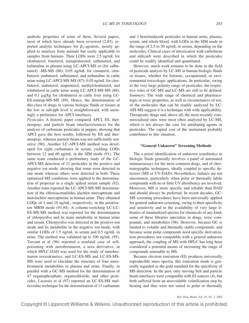
In genuine ES sources, the liquid flow was nebulizedexclusively by means of an intense electric field betweenthe tip of the capillary inlet and the aperture, which wascompatible only with very low flow rates (1–10 �L/min)(7). In 1987, Bruins et al (8) described a pneumaticallyassisted ES interface, which they called ionspray, thataccepted flow rates up to 200 �L/min with no loss insensitivity, contrary to most of the other high-flow ratesolutions proposed previously, which resulted in higherdetection limits (9). Pneumatically assisted ES sourcescombine the principles of ion evaporation (10) and ES,
which are close ionization mechanisms (Fig. 1). Sincethe advent of IonSpray™ (named after Sciex LC-MSinstruments) (Applied-Biosystems/MDS-Sciex; Con-cord, Ontario, Canada), similar solutions have been pro-posed by different manufacturers under various names(e.g., high-flow or megaflow ES). Further steps in ac-commodating high flow rates have consisted of an addi-tional drying of the spray by an orthogonal, heated ni-trogen beam, used in the Turbo-IonSpray™ interface(Sciex); or an orthogonal positioning of the ES withrespect to the ion sampling orifice, using different tech-nical solutions (Agilent Technologies orthogonal ES sys-tem (Agilent Technologies; Palo Alto, CA) or MicromassZ-spray™ ES source (Micromass; Manchester, UK)).
In the spray, the droplet size decreases rapidly, owingto an initial desolvation in the atmospheric pressurechamber, assisted in some instruments by a countercur-rent of pure nitrogen. Therefore, the electrical field at thedroplet surface increases until the droplets explode, giv-ing smaller droplets that will undergo the same fate. Theions are extracted from the spray, which is frequentlyoff-axis with respect to the orifice, by an electrical fielddirected toward the intermediate- or low-pressure cham-ber, where the residual clusters are broken down, owingto their acceleration through the electrical field. If thisacceleration is increased, dissociation of ions can occurthrough collision with the residual solvent and gas mol-ecules, and the fragments produced by this collision-induced dissociation (CID), called “in-source CID,” canbe used as confirmation ions for quantitation methods inthe selected ion monitoring (SIM) mode, or for structureelucidation. This interface allows the analysis of moder-ately nonpolar to highly polar compounds, even thosethat are thermally labile or those with a high molecularweight. In particular, it gives multicharged ions frompeptides and proteins, making it possible to analyze themusing single quadrupole instruments, as the m/z ratiosresulting from molecules up to 30 to 200 kDa generallyfall into their usual m/z range. Electrospray is surpassedin this application only by the more expensive matrix-assisted laser desorption ionization (MALDI) source.Other advantages of ES are that it is compatible with
FIG. 1. A pneumatically assisted elec-trospray (ES) ionization source andtriple-quadrupole mass spectrometer(MS). API, atmospheric pressureionization.
P. MARQUET256
Ther Drug Monit, Vol. 24, No. 2, 2002

chromatographic gradients and can be coupled with cap-illary electrophoresis through a different introduction de-vice. Its main drawbacks are a significant chemical noisein the low mass range (although lower than with ther-mospray ionization sources); “ion suppression” problemswhen different compounds enter the source at the sametime and compete for ionization; low fragmentation(which can be enhanced afterwards by CID); and a lim-ited admissible flow rate except, maybe, with some ofthe most recent types of sources. Concerning this lastpoint, most users choose to use chromatographic col-umns with a 2.1-mm I.D. and a mobile phase flow rate ofabout 200 �L/min, with or without postcolumn splitting.Nevertheless, as previously noticed when reviewing themethods published for the same compound or class ofcompounds (4), the lower the column I.D. and mobilephase flow rate, the lower the limits of detection (LOD)or quantitation (LOQ) obtained: narrow-bore columns ofabout 1 mm I.D. with flow rates of 40 to 50 �L/mingenerally give the best results, inasmuch as the sensitiv-ity of ES-type sources seems to be related to the concen-tration of the compounds of interest in the chromato-graphic effluent rather than to the amount per unit time(mass flow rate) admitted in the source. Indeed, this isattributed to a higher evaporation rate of mobile phasefor low flow rates and, thus, to a better transmission ofthe ions formed toward the mass spectrometer (6).
APCI Interfaces
Horning et al (11) developed the APCI source in theearly 1970s. Since then, different technical solutions anddesigns have been proposed (9), some of which are com-mercially available. Schematically, the chromatographiceffluent flows through a heated nebulizer, either pneu-matically assisted and housed in a quartz tube in whichflows a make-up gas (Fig. 2), or assisted by a piezoelec-tric ultrasonic or a sonic spray device. Alternately, heat is
not applied to the three concentric tubes (chromato-graphic effluent, nebulizing and make-up gases) but in a“vaporization zone” in the atmospheric pressure inter-face. However, the mixture of hot liquid and vapors ex-pands into the atmospheric pressure interface, where it isionized by a corona discharge. The ions formed from thesolvent molecules transfer a charge to the analytes. As inthe ES source, droplets can be further desolvated and ionclusters broken down when crossing a countercurrentstream of gas. The pumping stages, intermediate-pressure chamber, ion optic, and in-source CID possi-bilities are the same as those of ES, as both are generallyavailable on the same instruments. However, although itcan admit higher liquid flow rates, fewer applicationswere reported with this type of source than with ES, atleast in the fields of clinical and forensic toxicology. Thereasons for this are probably its more limited polarityrange, partly overlapping that of GC-MS; its relative in-compatibility with thermally labile compounds; and thehigher background noise produced compared with ES. Itsmain advantage is that it extends the polarity range ofcompounds amenable to LC-API-MS toward less polarmolecules. APCI sources can be either more or less sen-sitive than ES sources, depending on the type of sourceor manufacturer (more sensitive on LC-MSD fromAgilent Technologies and less sensitive than Turbo-IonSpray™ sources from Applied Biosystem-Sciex).
APPLICATION OF LC-API-MSTO TOXICOLOGY
Quantitative Methods
The extensive use of LC-MS and LC-MS-MS by thepharmaceutical industry in the past few years and thehuge volume of related literature have shown the poten-tial value of these techniques for the analysis of smallmolecules, typically therapeutic drugs and metabolites,
FIG. 2. An atmospheric pressure chemicalionization (APCI) source.
LC-MS IN TOXICOLOGY 257
Ther Drug Monit, Vol. 24, No. 2, 2002

whether qualitatively (metabolic studies) or quantita-tively (pharmacokinetic studies). The same applies totoxicology, which shares a large number of compoundsof interests with pharmacology. At the beginning of theexpansion of LC-API-MS into toxicology laboratories,the most frequently analyzed compounds were those notamenable to GC-MS (e.g., LSD and glucuronides) or forwhich no sufficiently sensitive analytic technique wasavailable so far (e.g., colchicine, cardiac glycosides).LC-API-MS and even LC-API-MS-MS are now appliedin toxicology to compounds previously analyzed by lessspecific HPLC techniques or even by GC-MS owing tothe simpler sample preparation generally needed. Itwould appear now that virtually all xenobiotics are ame-nable to LC-MS. Investigating this hypothesis is one ofthe aims of this review.
Quantitative Methods for Drugs of Abuse
Opiates and Opioids. The detection and quantitation ofopiates in biologic fluids by LC-MS has been the objectof many papers since 1994, most of which have alreadybeen analyzed in recent review papers (3–5). An ESsource was used in at least eight methods (12–19) and anAPCI source in at least three others (20–22). One methodelicited the direct determination of heroin in mouse se-rum by LC-APCI-MS with a limit of detection of0.5 ng/mL (14). Most others concerned heroin metabo-lites 6-monoacetylmorphine (6-MAM) and morphine, aswell as codeine and the metabolites of morphine, namelyglucuro-3- and glucuro-6-morphine (M3G and M6G, re-spectively). Overall results for 6-MAM showed LOQsequal to 4 ng/mL using ES and 0.5 to 2 ng/mL usingAPCI. For morphine they were 0.84 to 10 ng/mL with ESand 0.1 to 30 ng/mL with APCI; for codeine they were 5and 4 ng/mL, respectively. Finally, the LOQs of M3Gwere 1 to 100 ng/mL and 1 to 30 ng/mL, respectively;those of M6G were 2 to 50 ng/mL and 2 to 5 ng/mL.Morphine glucuronides can be directly and accuratelydetermined using LC-MS, in contrast to GC-MS, wherethey have to be hydrolyzed (with variable and often in-complete recovery) and their global concentration de-duced from the difference between total and free mor-phine concentrations. The conclusion that can be drawnfrom these studies is that heroin, morphine, codeine, andtheir metabolites can be determined by LC-API-MS witha sensitivity at least equal to that of GC-MS (and maybeeven better with APCI, although only a few methodswere reported with such sources). In general, simplersample preparation (no hydrolysis, one-step extraction,no derivatization) was used. For both these criteria ofsensitivity and simplicity, as well as for its increased
selectivity, tandem MS is undoubtedly a further improve-ment, as shown by four recent papers using ES ioniza-tion. In the first, morphine and codeine, as well as otherdrugs of abuse, were screened for and quantitatedin serum and urine in a concentration range of 10 to1,000 ng/mL using flow-injection analysis (FIA)-ES-MS-MS operated in the multiple reaction monitoring(MRM) mode (19). The second paper reported the fullyvalidated determination of 6-MAM, morphine, codeine,norcodeine, pholcodine, and codethyline in whole blood,plasma, and urine in the concentration range of 10 to1,000 ng/mL, using nalorphine as internal standard andreversed-phase chromatography (RP-HPLC)-ES-MS-MS in the MRM mode (16). Third, Slawson et al (18)reported a validated method for the determinationof morphine (0.25–10 ng/mL) and M3G and M6G(0.5–10 ng/mL) in plasma using morphine-d3 and M3G-d3 as internal standards and RP-HPLC and acquisition inthe MRM mode. Finally, Naidong et al (17) reported afully validated method for the determination of morphine(concentration range 0.5–50 ng/mL) and its two glucuro-nides (M6G, 1–100 ng/mL; M3G, 10–100 ng/mL) inhuman plasma using the respective deuterated internalstandard, ion-pairing solid-phase extraction (SPE), chro-matography on a nongrafted silica column using a water-acetonitrile mobile phase, and the MRM mode. For adefinitive identification of the compounds detected, al-though MS-MS in the MRM mode is relatively specific,full mass spectra should be obtained in the product-ionscan mode, in which all the fragments generated by thedissociation in the collision cell of each given pseudo-molecular ion selected in the first quadrupole are re-corded. However, whether in the MRM or product-ionscan mode, good chromatographic separation betweenmorphine and its glucuronides seems an important point,because the glucuronides are often partly fragmented tomorphine in the ionization source, even at low fragmen-tation voltage. However, a fully validated method includ-ing all the above-mentioned opiates and metabolites andall the respective deuterated standards is needed, to beproposed as an alternative gold standard to GC-MS.
LC-MS was also shown to be a convenient techniquefor the sensitive and specific determination of syntheticor semisynthetic opioids: dihydromorphine and dihydro-codeine (23); buprenorphine and norbuprenorphine, forwhich it is now considered the gold standard (24,25);methadone in hair (26) and different biofluids (23); anddespropionyl-bezitramide, the active metabolite of theprodrug bezitramide (27). Methods using only old typesof ionization sources/interfaces were reported for dextro-moramide and dextropropoxyphene (thermospray [TSP]
P. MARQUET258
Ther Drug Monit, Vol. 24, No. 2, 2002

ionization) and dextromethorphan (continuous-flow fast-atom bombardment). GC-MS was often used for thephenylperidines fentanyl, alfentanyl, mirfentanyl, sufen-tanyl, or remifentanyl (28–30), whereas, except for theLC-TSP-MS determination of mirfentanil in plasma(with a 0.4–100-ng/mL quantitation range) (31), no pa-per concerning the LC-MS analysis of these drugs hasbeen published so far. Nevertheless, LC-MS emerges asthe first choice for several synthetic opioids, even morethan for opiates (5).Cocaine and Metabolites. At least a dozen papers, mostof which were reviewed recently (5), reported LC-API-MS analytic methods for cocaine and its metabolites,mainly using tandem MS. Using an ES ionization source,Weinmann et al (19) presented the FIA-MS-MS analysisof benzoylecgonine in addition to that of morphine, co-deine, and amphetamine in serum and urine after mixed-mode SPE. This previously mentioned study yielded aLOD of 2 ng/mL for benzoylecgonine and was linear inthe concentration range of 10 to 1,000 ng/mL (19); pos-sible matrix effects (ion suppression) were taken in ac-count by the use of deuterated analogs of all the mol-ecules of interest. However, benzoylecgonine has thesame molecular mass as norcocaine (289 u), an activemetabolite of cocaine (5) with which it also shares thesame most intense fragment at m/z 168; therefore, them/z 290 → 168 transition used by these authors shouldhave been avoided (even if norcocaine concentrations inplasma or urine are generally much lower than benzoy-lecgonine concentrations) because no chromatographicseparation was used in this method. Several other studiesused LC-ES-MS-MS for the determination of cocaineand various metabolites. Clauwaert et al (32) designed aconfirmation method for the detection of cocaine, benz-oylecgonine, and cocaethylene in hair, for which unfor-tunately they did not report a LOD. In urine, Jeanville etal (33) obtained an LOQ of 7.5 ng/mL for cocaine andbenzoylecgonine, using a simple filtration as samplepreparation. Needham et al (34) obtained an LOQ of1.0 ng/mL for the same molecules after a simple 1:10dilution of urine samples and using a pentafluorophen-ylpropyl (PFPP) bonded silica chromatographic columnto increase the retention of ecgonine methyl ester (EME)and to be able to use a high enough concentration oforganic solvent in the mobile phase, thereby increasingthe ES-MS signal because of more efficient desolvata-tion. Cailleux et al (16) obtained LOQs of 10 ng/mL forcocaine, benzoylecgonine, EME, cocaethylene and an-hydroecgonine methylester after liquid-liquid extraction.These latter authors obtained the same LOQs in plasmaand whole blood and validated their technique in boththese matrices, whereas Srinivasan et al (35) designed a
validated method for the determination of cocaine and 12metabolites in blood, amniotic fluid, and placental andfetal tissues with LOQs of 10 ng/mL, using SPE on un-derivatized silica columns. Wang and Bartlett (36) re-ported a single quadrupole method for the determinationin plasma of cocaine-N-oxide, a thermally labile metabo-lite that converts to cocaine when heated, with an LOQof 10 ng/mL. Finally, Jeanville et al (37) reported thefirst use of a quadrupole orthogonal acceleration time-of-flight (Q-TOF) instrument operated in the MS-MSmode for the determination of cocaine and EME in ratplasma. The procedure had a high-throughput, total turn-over time of 3.2 minutes owing to on-line SPE and rapidchromatography using a cyanopropyl-based column withretention characteristics similar to the PFPP stationaryphase cited above (i.e., high retention for the extremelypolar EME). Relying on the resolving ability of TOFinstruments (high discrimination of mass differences andcollection of a full-scan mass spectrum every 10–100 �s)to provide specificity, simple extraction and separationconditions could be used. Using the postacquisition,pseudo-MRM mode (TOF detectors can be operated onlyin the full scan mode), the LODs were 0.5 ng/mLfor cocaine (m/z 304.2 → m/z 182.2 transition) and0.05 ng/mL for EME (m/z 200.2 → m/z 182.2 transi-tion). Moreover, despite the inherent limited dynamicrange of TOF instruments (typically two orders of mag-nitude), modifications of the detection system and theuse of correction algorithms improved the procedure sothat it was linear over a large range (5.0–10,000 ng/mLfor cocaine, 0.5–10,000 ng/mL for EME) and showedgood intra- and interday precision and accuracy, usingtrideuterated cocaine as internal standard and three rep-licate injections of each sample (which tends to reducethe gain in analytic time of this technique).
Besides these methods, using ES-type sources, severalgroups proposed using APCI sources for the determina-tion of cocaine and/or metabolites. The first combineddouble SPE, a wide-bore steric-exclusion chromato-graphic column, and SIM mode on a single-quadrupoleinstrument for the determination of cocaine, benzoylec-gonine, EME, norcocaine, and ecgonine in urine, yield-ing LODs of 50 to 800 ng/mL (20). More recently, Bo-gusz et al (23) reported another SPE, single quadrupole,SIM method for the determination of cocaine, benzoy-lecgonine, and EME (in addition to opiates and LSD) inurine, blood, and serum using deuterated analogs of thecompounds as internal standard, that yielded much betterLODs of 0.2 ng/mL for benzoylecgonine and EME and0.5 ng/mL for cocaine. The method was linear in therange 1 to 200 ng/mL but used two different chromato-graphic conditions, one for cocaine and benzoylecgonine
LC-MS IN TOXICOLOGY 259
Ther Drug Monit, Vol. 24, No. 2, 2002

and another for EME, confirming the particular chro-matographic behavior of this last compound. A methodwas reported for the determination of benzoylecgonine indried blood spots collected from newborns, using benz-oylecgonine-d3 as internal standard, two serial C18 wide-bore analytic columns, and MS/MS in the MRM mode,with an LOD of 2 ng/mL (despite typical sample vol-umes of 12 �L) and linearity up to 166 ng/mL (38).Finally, Singh et al (39) designed a LC-APCI-MS-MSmethod for the determination of cocaine, benzoylec-gonine, EME, and norcocaine in plasma using simpleprotein precipitation, gradient elution-reversed-phasechromatography on a wide-bore column, deuterated ana-logs as internal standard, and acquisition in the MRMmode. This method was linear in the range of 2 to1,000 ng/mL for all four compounds. From all thesestudies, it is obvious that cocaine and its main metabo-lites can be easily determined using LC-MS or betterLC-MS/MS, simple sample preparation (in particular, noderivatization required), and either ES or APCI sources.The other two conclusions that can be drawn from thesepapers are that the sensitivity of the detection of the mostpolar metabolites of cocaine, such as EME, is related tothe proportion of organic solvent used to elute them, thusnecessitating adapted chromatographic stationaryphases; and that although providing more specificity andselectivity (higher signal-to-noise ratios [S/N]), MS-MSdoes not necessarily yield lower LODs than single-quadrupole MS detectors.Amphetamines and Related Compounds. At least 10 pa-pers have reported the LC-API-MS analysis of amphet-amines and related compounds, some of which werealready analyzed in previous review papers (2,5). Am-phetamines are volatile but polar compounds that need tobe derivatized before GC-MS analysis, which is probablywhy several teams developed LC-MS techniques. Unlikecocaine and its metabolites, many of these methods usedsingle-quadrupole MS instruments, possibly due to thelow molecular weight of these compounds, meaning thatthe daughter ions generated by LC-MS-MS are in aneven lower m/z range, at which high background noise isunavoidable. All these techniques used the positive-iondetection mode. The first published method using an ES-type source, although not validated, yielded LODs in thehigh pg/mg to low ng/mg range for methylenedioxy-methamphetamine (MDMA) and its metabolite methy-lenedioxyamphetamine (MDA) in rabbit blood and post-mortem hepatic tissue, as well as in fly larvae andchitinized insect tissues, using a triple-quadrupole MSoperated in the single-quadrupole SIM mode (three ionsselected per compound). Sample preparation was by liq-uid-liquid extraction and MDMA-d5 was used as internal
standard (40). Katagi et al (41) developed a direct-injection, column-switching LC-ES-MS method for thedetermination of methamphetamine and amphetamineenantiomers in urine, using a �-cyclodextrin phenylcar-bamate bonded silica column. Differentiating the enan-tiomers of amphetamines can be useful because D and Lenantiomers have different pharmacologic properties,and in certain countries D-enantiomers are used in legalmedicat ions . The LOD was 0.5 to 1 ng/mLand the linearity range was 1 to 5,000 ng/mL for allcompounds. The same team proposed an LC-ES-MSmethod for the determination of dimethylamphetamine(DMA) and its metabolites dimethylamphetamine-N-oxide (DMAO), amphetamine, and methamphetamine inurine (42). This study was initiated because DMA usecan sometimes be difficult to differentiate from metham-phetamine use in urine if DMA is totally metabolized.With that aim, the detection of DMAO, a thermally labilecompound, is of primary importance, justifying the useof LC-MS. Indeed, GC-MS analysis of DMAO requiresits prior reduction to DMA followed by a derivatizationstep, or alternately direct derivatization to trifluoroace-tyl-methamphetamine, both pretreatments that render thesimultaneous determination of the three compounds im-possible. SPE and chromatographic separation were per-formed using a strong cation-exchanger phase to obtainsufficient retention for the more polar compounds. Thequadrupole MS was operated in the positive-ion full-scanmode between m/z 100 and 500 for drug identification(LOD � 50 ng/mL for DMA and DMAO, 100 ng/mLfor methamphetamine, 500 ng/mL for amphetamine) andin the SIM mode using the protonated molecular ion ofeach compound for drug quantitation (LODs 10 timesless, respectively). The method was linear from twice theLODs in the SIM mode up to 5,000 ng/mL and wasapplied to the urine elimination study of DMA, showingthat DMAO was detectable up to 3 to 5 days after intake.Kataoka et al (43) also used LC-ES-MS in the SIM modefor the determination of amphetamine, methamphet-amine, MDA, MDMA, and MDEA in urine. After highdilution of urine in water and automated in-tube solid-phase microextraction (SPME) using a relatively polarcapillary, they obtained LODs of 0.38 to 0.82 ng/mL(seemingly using standard solutions) owing to concen-tration of the amphetamines in the capillary during sev-eral draw/eject cycles, and LOQs of 32 to 79 ng/mL inurine, although the criteria used were unclear. In theirpreviously cited study of FIA-ES-MS-MS analysis ofdrugs of abuse, Weinmann and Svoboda (19) reportedthe determination of amphetamine in urine with an S/Nratio of 4.4 at 2 ng/mL, following the m/z 136 → 91
P. MARQUET260
Ther Drug Monit, Vol. 24, No. 2, 2002

transition. Finally, Clauwaert et al (44) reported a LC-ES-Q-TOF method in the pseudo-MRM mode (one par-ent ion and three extracted daughter ions) for the deter-mination of MDMA in whole blood, serum, and vitreoushumor, using MDEA as internal standard and liquid-liquid extraction. The quantitative potential of Q-TOFMS was evaluated by comparison with HPLC withfluorescence detection, showing a very low LOD of0.25 ng/mL and a three-decade dynamic range (1–1000 ng/mL) for the Q-TOF, superseding that of fluo-rescence detection. However, the choice of MDEA asinternal standard was probably not the best one, becausea significant percentage of tablets sold as Ecstasy in Eu-rope contain MDEA in addition to (or instead of) MDMA.
Using an APCI ionization source and single-quadrupole MS detection in the SIM mode (followingtwo or three ions per molecule), Bogusz et al (45) de-signed a quantitative method for amphetamine, metham-phetamine, MDA, MDMA, and MDEA in serumand urine that involved a phenylisothiocyanate derivati-zation of the compounds, mostly justified by the HPLC-diode-array UV method used as a comparison with LC-APCI-MS. Highly deuterated analogs of amphetamine,methamphetamine, MDEA, and MDMA were used asinternal standard and LODs between 1 ng/mL (metham-phetamine, MDMA, MDEA) and 5 ng/mL (amphet-amine, MDA) were reached. Later, the same team re-ported an alternative LC-APCI-MS method for thedetermination of the same molecules using the same in-ternal standard as well as 11 related compounds forwhich no validation data were provided (46). Moreover,although not clearly stated, it appears that different chro-matographic conditions were applied to analyze some ofthese additional molecules. The main differences withthe previous method were SPE instead of LLE and theabsence of derivatization, resulting in quasimolecularions and fragments of lower masses. However, the LODsreached were similar or slightly better (1–2 ng/mL forthe five principal analytes), whereas the linearity rangewas seemingly limited at its upper end (5–500 ng/mLinstead of 1–1,000 ng/mL). Ramos et al (47) reported anLC-APCI-MS-MS method for the determination ofmethylphenidate, an amphetamine-related stimulant drugused in the treatment of attention deficit hyperactivitydisorder and narcolepsy. This method used a high-throughput sample preparation procedure based on semi-automated liquid-liquid extraction in a 96-well plate for-mat, a very short chromatographic elution (TR # 1.6 min)using a wide-bore column and detection in the MRMmode, using one transition. The LOQ was 50 pg/mL andthe method was linear and validated in the range of 0.05to 100 ng/mL.
Despite this apparent diversity of analytic methods foramphetamines, ES ionization sources and, as alreadymentioned, single-quadrupole MS were used by mostteams. Moreover, although the low molecular weights ofunderivatized amphetamines are not optimal for LC-MSdetection, LOQs better than those reported for GC-MSmethods (classically, in the range of 10–50 ng/mL) couldbe attained while using simpler sample preparation pro-cedures. However, the formal identification of the com-pounds detected is difficult using such types of instru-ments. In this respect, triple-quadrupole or even betterQ-TOF mass spectrometers provide more selectivity andspecificity and have proved to be more sensitive thanquadrupoles.Cannabinoids. It was not until 1999 that the first report ofan LC-API-MS method for the determination of canna-binoids in biologic fluids appeared (48), to be followed ayear later by a second paper (49). In the first, the authorsdeveloped a LC-ES-MS method for the urinary determi-nation of 11-Nor-�9-tetrahydrocannabinol-9-carboxylicacid (THC-COOH), the main urinary metabolite of 11-Nor-�9-tetrahydrocannabinol (THC), the major psycho-active component of Cannabis sativa. BecauseTHC-COOH is excreted mainly as its glucuronide inurine, the authors used basic hydrolysis of conjugates inurine samples before SPE (48). After gradient elutionchromatography, THC-COOH and THC-COOH-d3 (in-ternal standard) were detected in the positive-ion SIMmode following, respectively, three and two m/z ratios.The LOD was 2 ng/mL for the quantitation ion ofTHC-COOH, 15 ng/mL when taking into account itsqualifying ions and their relative intensity with respect tothat of the quantitation ion (accepting ± 20% variability).Considering only the quantifying m/z ratio, the methodwas linear in the concentration range of 2.5 to125 ng/mL, much lower than the concentrations found inmany positive urine samples. However, such sensitivetechniques are required to confirm or disprove the posi-tive results obtained with immunologic techniques in-volving antibodies that partly cross-react with other can-nabinoids or cannabinoid metabolites, in addition toTHC-COOH and its glucuronide. The two questionsraised by this report were already discussed in VanBocxlaer et al’s review article (5): the relevance ofTHC-COOH-glucuronide hydrolysis when this moleculeis theoretically directly amenable to LC-ES-MS (as dem-onstrated later on) and the questionable use of the posi-tive ionization mode when THC-COOH is under anionicform in solution. Answers to the first question were pro-vided both by the authors themselves (48), who advo-cated a variable conjugation of THC-COOH, and by theabove-mentioned review article (5), which underlined
LC-MS IN TOXICOLOGY 261
Ther Drug Monit, Vol. 24, No. 2, 2002

the need for correct interpretation of the concentrationmeasured with respect to regulatory cutoff levels. As forthe second question, this previous review paper regrettedthat negative ionization was not used, owing to a sup-posed increase of sensitivity, which in my experience isseldom achieved. Indeed, although the S/N ratio is lower,the chromatographic signal is often of much lower in-tensity in the negative-ion mode than in the positivemode, because the chromatographic mobile phases aregenerally acidic. Therefore, it is not uncommon to usethe positive-ion mode for those acidic compounds thatcan be easily protonated. As a confirmation, in the sec-ond paper dealing with cannabinoids, both THC-COOHand THC-COOH-glucuronide were analyzed in urine byLC-ES-MS-MS in the positive-ion mode (49). However,because no reference compound was available forthe latter, only the former could be quantitated (usingTHC-COOH-d3 as internal standard), urine samples fromcannabis users being used for defining the analytic con-ditions for the glucuronide. After liquid-liquid extrac-tion, the compounds of interest were separated using gra-dient elution reversed-phase chromatography on a 2-mmI.D. column and detected in the MRM mode by selectionof the protonated molecules in the first quadrupole and ofone major fragment in the third quadrupole (and two ofthe THC-COOH-glucuronides). Indeed, because themajor fragment of THC-COOH-glucuronide is theTHC-COOH moiety, obtained in the first quadrupole byin-source CID even at low fragmentation voltage, thetransition selected for THC-COOH also allowed the con-firmation of THC-COOH glucuronide at its respectiveretention time. The LOD of THC-COOH was estimatedto be less than 10 ng/mL, whereas no validated LOQwas reported. Linearity was assessed between 10 and10,000 ng/mL. Interestingly, the authors compared THC-COOH concentrations in urine samples before and aftercomplete enzymatic hydrolysis (as could be verified bythe disappearance of the glucuronide mass signal). Fromthis experiment it was possible to deduce the concentra-tion of the glucuronide and to relate it to the area of itschromatographic peak in the nonhydrolyzed sample, orto its peak area ratio with the internal standard.
Another improvement would be a LC-API-MSmethod for the determination of THC, 11-hydroxy-THC(the main blood metabolite of THC), THC-COOH, andeventually THC-COOH-glucuronide in blood, with lowenough LODs and without a derivatization step, to becompetitive with GC-MS.LSD, Metabolites, and Epimers. All the methods usingLC-API-MS for the determination of LSD and its me-tabolites and epimers were previously reviewed (4,5),
except one (50). Most of these methods used ES-typesources and single-quadrupole MS and most were lim-ited to LSD in urine, with LODs ranging from 0.3 (51) to0.5 ng/mL (52–54). Still using LC-ES-MS, my groupdesigned validated methods for the determination ofLSD and N-demethyl-LSD (or nor-LSD) in urine withLODs of 0.025 and 0.035 ng/mL and LOQs of 0.05 and0.10 ng/mL, respectively (55). For the determination ofLSD, nor-LSD, iso-LSD, and isonor-LSD in serum andblood, our LODs were 0.02 to 0.05 ng/mL and LOQs0.05 to 0.10 ng/mL (56). Using LC-ES-MS-MS in theMRM mode, De Kanel et al (57) designed an analyticmethod for LSD and nor-LSD in urine, blood, serum, andplasma, using two or three transitions per compound andyielding LODs of 25 pg/mL for both compounds. Thismethod was validated in these three matrices for LSD,with an LOQ of 50 pg/mL and a calibration range of 0.05to 5 ng/mL, with good within-run and between-run pre-cision and accuracy. The method was suitable only forqualitative identification of nor-LSD due to higher be-tween-run variability. Cai and Henion (58,59) designedan LC-ES-MS-MS technique for LSD, nor-LSD, iso-LSD, nor-iso-LSD, and methysergide in liver micro-somes and urine, using SPE, with an LOD of 50 pg/mL,as well as a second technique for LSD, nor-LSD, iso-LSD, iso-nor-LSD, de-ethyl-LSD, and nor-allyl-LSD inurine using immunoaffinity extraction, reaching a veryimpressive LOD of 2.5 pg/mL. However, no validationdata were reported in these two papers.
Only three papers have reported analytic methods forLSD and/or metabolites using APCI sources. The firstwas a study by Henion’s team in 1992 (60) concerningthe feasibility of LSD analysis by LC-MS in the scan andSIM modes using an ancestor of APCI interfaces. Usingstandard solutions of LSD, an LOD of 2 ng/mL wasobtained in the scan mode. In their multielement LC-APCI-MS method for drugs of abuse in urine, Bogusz etal (23) obtained an LOD of 0.5 ng/mL and a linearityrange of 0.5 to 10 ng/mL for LSD. More recently, LC-APCI-iontrap-MS-MS was used for the determination ofnor-LSD and, principally, 2-oxo-3-hydroxy-LSD (O-H-LSD), the main urinary metabolite of LSD, whereas LSDand iso-LSD were determined by GC-MS (50). Althoughsomewhat clumsy (e.g., an m/z 338 → 338 transition wasused for O-H-LSD and quantitation was made by single-point calibration), this method was partially validated forO-H-LSD, using 2-oxo-3-hydroxy-LAMPA as internalstandard. It showed an LOQ of 0.4 ng/mL and a linearityrange up to 200 ng/mL. More importantly, this methodwas applied to the confirmation analysis of 74 positiveurine specimens, showing that the O-H-LSD urine con-centration was much higher than that of the other
P. MARQUET262
Ther Drug Monit, Vol. 24, No. 2, 2002

compounds, with a mean O-H-LSD/LSD concentrationratio of 42.9 (range 1–778), emphasizing the importanceof this metabolite for the detection window of LSDabuse.
From this review, it is clear that LC-MS is the methodof choice for LSD analysis, eliciting the simultaneousdetermination of different metabolites and epimers inbiofluids with high sensitivity and specificity, using ei-ther ES or APCI, MS or MS-MS. As already empha-sized, tandem MS does not seem to yield lower LODsthan single-quadrupole MS, but it is, nevertheless, animprovement in terms of specificity and S/N ratio. How-ever, here again a complete and fully validated techniqueable to determine all of these compounds (or at least allthose of clinical interest) in one run is still needed.Summary. If virtually all drugs of abuse can be sensi-tively and specifically determined using LC-API-MStechniques, the status of gold standard in this field is stillthe prerogative of GC-MS, due to wide acceptance, iden-tity of the analytic conditions required for the differentfamilies (e.g., no change of chromatographic column ormobile phase required), and reliability and affordabilityof instruments.
Quantitative Methods for Other Drugs andToxic Compounds
A large volume of literature has been published on thedetermination of therapeutic drugs in biologic fluids(mainly by the pharmaceutical industry but also in thefield of therapeutic drug monitoring) (6), as well as forthe detection and quantitation of pesticides and industrialor environmental pollutants, all of which are of clinicalor forensic relevance. It is beyond the scope of a singlepaper to review all these studies, and I will limit the taskto discussing some particularly interesting classes ofdrugs.Central Nervous System Drugs. Several LC-MS tech-niques have been reported for benzodiazepines with ex-cellent sensitivity, and have been the subject of previousreview articles (2,4,5), but few of these involved an APIinterface. Using LC-APCI-MS, LODs of 2 ng/mL wereobtained for midazolam in pig plasma (61), whereas us-ing LC-ES-MS, LODs of 0.2 ng/mL for midazolam and0.5 ng/mL for its metabolite 1-hydroxymidazolam couldbe reached in human serum, with LOQs of 0.5 ng/mL forboth (validated method) (62). LC-APCI-MS was used forthe determination of triazolam in plasma and urine withan LOD of 0.02 ng/mL (63), and of oxazepam glucuro-nide in urine (LOD � 50 ng/mL) (64). Bogusz et al (65)described the determination of flunitrazepam and its
metabolites 7-aminoflunitrazepam (7-AF), N-desmethyl-flunitrazepam (N-DT), and 3-hydroxyflunitrazepam(3-HF) in urine, serum, and blood using macroboreHPLC coupled to either ES-MS or APCI-MS (65). Thecomparison of ES and APCI in the positive-ion SIMmode, following a single ion per compound, showed thatAPCI was 7 times more sensitive for flunitrazepam,about 20 times more for N-DF, and 40 times more for3-OHF compared with ES, but that there was no differ-ence for 7-AF. The LODs were 0.2 ng/mL for flunitra-zepam and 7-AF and 1 ng/mL for N-DF and 3-OHF, andthe method was linear over a range of 1 to 500 ng/mLwith the APCI source. This method is highly sensitive,but the use of a single ion for the identification of eachcompound limits its specificity. Despite the limited num-ber of papers, LC-API-MS appears to be at least as sen-sitive as, but easier to handle than, GC-NICI-MS for thedetermination of benzodiazepines.
Four papers have reported analytic techniques for an-tidepressants in plasma. The first concerned the analysisof nortriptyline and its metabolite 10-hydroxynor-triptyline in plasma using LC-APCI-MS-MS. LODs of0.2 ng/mL were achieved, and the assay was linear in therange of 0.8 to 32 ng/mL (66). The second reported thedetermination of nefazodone and its metabolites inplasma by LC-ES-MS, achieving LODs of 4 and2 ng/mL, respectively (67). Third, doxepin and desmeth-yldoxepin were determined in plasma by LC-ES-MS-MS, with respective LOQs of 0.320 and 0.178 ng/mL(68). Finally, Zhang et al (69) reported a LC-ES-TOFtechnique for doxepin, desipramine, imipramine, ami-triptyline, and trimipramine, using imipramine-d3 as in-ternal standard, that showed, in the extracted-ion mode,LOQs of 2 ng/mL for desipramine and 1 ng/mL forthe other drugs. The method was linear in ratherrestricted concentration ranges (respectively, 2–100 and1–50 ng/mL).
Several papers have reported LC-API-MS techniquesfor the determination of commercialized antipsychotic(or neuroleptic) drugs, whereas others have reported ana-lytic techniques for candidate drugs under evaluation(e.g., sertindole, pramipexole, iloperidone). The butyro-phenone haloperidol and its reduced metabolite were de-termined in serum by LC-ES-MS with LODs of 0.075and 0.1 ng/mL and LOQs of 0.1 and 0.25 ng/mL, respec-tively (70). The thienobenzodiazemine olanzapine wasdetermined in human plasma and serum using LC-APCI-MS-MS in the positive-ion MRM mode, with an LOQ of0.25 ng/mL and linearity up to 50 ng/mL (71). A LC-ES-MS-MS technique was designed for the pharmacoki-netic study and therapeutic drug monitoring of risperi-done, a benzisoxazole derivative. Risperidone and its
LC-MS IN TOXICOLOGY 263
Ther Drug Monit, Vol. 24, No. 2, 2002

metabolite 9-hydroxy risperidone were determined inplasma with an LOQ of 0.1 ng/mL and linearity up to100 ng/mL (72). The phenothiazine trimeprazine and itsmain metabolites were determined in rat serum usingLC-ES-MS with LODs of 0.4 ng/mL, and linearity wasverified in the range 0.5 to 40 ng/mL for all compounds(73). In a recent paper, McClean et al (74) described anLC-ES-iontrap-MS-MS method for chlorpromazine, tri-fluoperazine, flupenthixol, and risperidone in a hairsample from a patient under clinical treatment for schizo-phrenia. Calibration and validation data were obtainedfrom blank hair samples spiked with standard solutionsof the drugs of interest, showing LODs in the range of10−9 to 10−8 mol/L (i.e., roughly 0.3–3 ng/mL) and lin-earity in the range 5 × 10⁻⁷ and 1 × 10⁻⁵ mol/L for allcompounds except risperidone, for which the range was4.9 × 10−7 to 9.6 × 10⁻⁶ mol/L (mol/L is a rather sur-prising unit for a solid matrix such as hair). Finally,Kumazawa et al (75) reported a method for the determi-nation of 11 phenothiazine antipsychotics in humanwhole blood and urine using LC-ES-MS-MS. The au-thors compared the single-quadrupole SIM and triple-quadrupole MRM modes, showing much lower LODswith the latter, due to much better S/N ratios (and not, asmisinterpreted by the authors, higher sensitivity, becausesignal intensity was actually decreased). Using SPMEand detection in the MRM mode, the LODs obtainedwere between 0.2 (flupentixol) and 200 ng/mL (clospi-razine) in whole blood and between 4 (thioridazine) and22 pg/mL (propericiazine, clospirazine) in urine; themethod was linear and validated over wide concentrationranges.
Again, owing to the high sensitivity and specificity ofthe methods published, it is probable that LC-API-MS isgoing to be a technique of choice for antipsychotic drugsin toxicology.Cardiac Drugs. At least two papers reported the determi-nation of cardiac glycosides in human plasma or serum,both using LC-ES-MS and both applied to clinical andforensic toxicology. The first concerned digoxin, digi-toxin, lanatoside C, and acetyldigoxin with LODs be-tween 0.15 ng/mL (acetyldigitoxin) and 0.60 ng/mL (la-natoside C) and linearity within the range 1 to 100 ng/mL(76). The second paper concerned 17 cardiac glycosidesand metabolites and showed LODs between 1 ng/mL(deslanoside, digoxin, methyldigoxin) and 10 ng/mL(acetyldigoxin, gitaloxin) and linearity up to 100 ng/mLfor all compounds (77). For the first time, these tech-niques allowed the MS confirmation of intoxication withcardiac glycosides, whereas it was previously possible toconfirm the results obtained only using immunologictechniques, mainly radioimmunoassay.
Only one recent paper concerned the LC-API-MSanalysis of beta-blocking drugs, but only in pharmaceu-tical preparations (78). However, the concentration rangestudied using pure solutions (100–300 ng/mL) suggeststhat such a technique could be applied to biologic fluidswith sufficient sensitivity, provided a convenient extrac-tion/concentration procedure was developed.
Several reports concerned the determination of cal-cium antagonists by LC-API-MS, mainly applied topharmacokinetic studies. LOD and LOQ were respec-tively 0.25 and 0.5 ng/mL for both nimodipine enantio-mers by LC-ES-MS/MS (79) and 0.08 and 0.24 ng/mLfor nifedipine and dehydronifedipine by LC-APCI-MS/MS (80). An LOQ of 0.03 ng/mL was achieved forbarnidipine using LC-ES-MS/MS (81). Obviously, al-though there are still no published techniques allowingthe simultaneous detection and quantitation of all, ormost, of the calcium antagonists, these data suggest thatLC-API-MS could be a convenient analytic technique forthese drugs too.Doping Agents. The detection and identification of en-dogenous and exogenous anabolic steroids and their me-tabolites are of primary importance in sports doping con-trol. The sulfate and glucuronide conjugates oftestosterone and epitestosterone were determined in hu-man urine using LC-ES-MS-MS in the MRM mode.LODs “in the low nanomolar range” were achieved forthese steroid conjugates, except that of testosterone sul-fate, in urine from a normal man. This method wasclaimed to be convenient for the direct and precise de-termination of the testosterone/epitestosterone metabo-lite ratio in doping control, as an alternative to the classictestosterone/epitestosterone ratio obtained by GC-MS af-ter enzymatic hydrolysis of glucuroconjugated metabo-lites (82). Even better, an LOD of 25 pg for epitestos-terone glucuronide could be obtained using a 300 �mI.D. packed capillary HPLC column and similar MS con-ditions (83). Recently, Kuuranne et al (84) reported astudy of the best ionization conditions of eight anabolicsteroid glucuronides (of testosterone, epitestosterone,nandrolone, androsterone, and four new compounds syn-thesized in their laboratory), using ES or APCI in thepositive or negative mode and triple-quadrupole MS. Us-ing direct injections of standard compounds into thesource, they concluded that the ES source and the posi-tive-ion mode were the most promising for further de-velopment of LC-MS methods for anabolic steroid com-pounds (which confirms that the positive-ion mode isoften more sensitive than the negative ion mode, even foracidic compounds).
Beta-agonists are increasingly used by athletes, pre-sumably because of their bronchodilator as well as the
P. MARQUET264
Ther Drug Monit, Vol. 24, No. 2, 2002
anabolic properties of some of them. Several papers,most of which have already been reviewed (2,85), re-ported analytic techniques for �2-agonists, mostly ap-plied to matrices from animals but easily applicable tosamples from humans. Their LODs were 2.5 ng/mL forclenbuterol, fenoterol, metaproterenol, salbutamol, andterbutaline in plasma using LC-APCI-MS or (for salbu-tamol) -MS-MS (86); 0.05 ng/mL for cimaterol, clen-buterol, mabuterol, salbutamol, and terbutaline in cattleurine using LC-APCI-MS-MS (87); 0.05 ng/mL for clen-buterol, mabuterol, mapenterol, methylclenbuterol, andtolubuterol in cattle urine using LC-APCI-MS-MS (88);and 0.1 �g/kg for clenbuterol in cattle liver using LC-ES-iontrap-MS-MS (89). Hence, the determination ofthis class of drugs in various biologic fluids or tissues atthe low or sub-ppb level is straightforward, with seem-ingly a preference for APCI interfaces.Pesticides. A historic paper compared APCI, ES, ther-mospray, and particle beam interfaces/sources for theanalysis of carbamate pesticides in pepper, showing thatAPCI gave the best results, followed by ES and ther-mospray, whereas particle beam was not sufficiently sen-sitive (90). Another LC-APCI-MS method was devel-oped for eight carbamates in serum, yielding LODsbetween 12 and 60 ng/mL in the SIM mode (91). Thesame team conducted a preliminary study of the LC-APCI-MS detection of 21 pesticides in the positive andnegative ion mode, showing that some were detected inone mode whereas others were detected in both. Theseoptimized MS conditions were applied to the determina-tion of propoxur in a single spiked serum sample (92).Another team reported the LC-APCI-MS-MS determina-tion of the chloroacetanilides alachlor mercapturate andmetolachlor mercapturate in human urine. They obtainedLOQs of 1 and 10 ng/mL, respectively, in the positive-ion MRM mode (93,94). A column-switching LC-LC-ES-MS-MS method was reported for the determinationof chlorpyrifos and its main metabolite in human urineand serum. Chlorpyrifos was detected in the positive-ionmode and its metabolite in the negative ion mode, withsimilar LODs of 1.5 ng/mL in serum and 0.5 ng/mL inurine. The method was validated up to 100 ng/mL (95).Turcant et al (96) reported a nonfatal case of self-poisoning with metobromuron, a urea derivative, inwhich HPLC-DAD was used for the study of metobro-muron toxicokinetics, and LC-ES-MS and LC-ES-MS-MS were used to elucidate the structure of four meto-bromuron metabolites in plasma and urine. Finally, inparallel with a GC-MS method for the determination of47 organophosphate, organochloride, and other pesti-cides, Lacassie et al (97) reported an LC-ES-MS mul-tiresidue technique for the determination of 11 carbamate
and 3 benzimidazole pesticides in human urine, plasma,serum, and whole blood, with LODs in the SIM mode inthe range of 2.5 to 50 ng/mL in serum, depending on themolecules. Clinical cases of intoxication with carbofuranand aldicarb were described in which the pesticidescould be readily identified and quantitated.
However, much work remains to be done in the fieldof pesticide analysis by LC-MS in human biologic fluidsor tissues, whether for forensic, occupational, or envi-ronmental toxicologic applications. In particular, owingto the very large polarity range of pesticides, the respec-tive roles of GC-MS and LC-MS are still to be defined.Summary. The wide range of chemical and pharmaco-logic or toxic properties, as well as circumstances of use,of the molecules that can be readily analyzed by LC-API-MS suggest it is a technique with wide applicability.Therapeutic drugs and, above all, the most recently com-mercialized ones were most often analyzed by LC-MS,which is not always the case for antidoping agents orpesticides. The capital cost of the instrument probablycontributes to this situation.
“General Unknown” Screening Methods
The a priori identification of unknown xenobiotics inbiologic fluids generally involves a panel of automatedimmunoassays for the most common drugs, and of chro-matographic techniques, ideally coupled to specific de-tectors (MS or UV-DAD). Nevertheless, failures are notuncommon, particularly when polar or thermally labilecompounds with no or little UV absorbency are involved.Moreover, MS is more specific and reliable than DADand should always be preferred. In recent decades, GC-MS screening procedures have been universally appliedfor general unknown screening, owing to their specificityand sensitivity and to the availability of very large li-braries of standardized spectra for chemicals of any kind;some of these libraries specialize in drugs, toxic com-pounds, and metabolites (98). However, because GC islimited to volatile and thermally stable compounds, andbecause some polar compounds need specific derivatiza-tion procedures not compatible with a general unknownapproach, the coupling of MS with HPLC has long beenconsidered a potential means of increasing the range ofcompounds amenable to MS.
Because electron ionization (EI) produces universallyreproducible mass spectra, this ionization mode is gen-erally regarded as the gold standard for the specificity ofMS detection. In the past, only moving belt and particlebeam interfaces were compatible with EI sources (4), butboth suffered from an unavoidable volatilization step byheating and thus were not suited to polar or thermally
LC-MS IN TOXICOLOGY 265
Ther Drug Monit, Vol. 24, No. 2, 2002

labile compounds. Nevertheless, one group recently pro-posed an application combining a double liquid-liquidextraction (for acidic and neutral compounds on the onehand and for alkaline compounds on the other) and agradient chromatographic elution on a C18 column(chromatographic time 70 minutes), the first detectionstep using a DAD, followed by a second using MS (99).The authors used a particle beam interface and electronionization. Mass spectra were acquired in the range of 50to 400 u, then three different libraries were searched: acustomized in-house library, the Pfleger-Maurer-Weberlibrary, and the Wiley 138K library.
With this procedure, the researchers analyzed about150 compounds, mainly drugs, with a LOD varying from20 to more than 1,000 ng/mL in whole blood, corre-sponding to extraction recovery values between a fewpercent to 99%. Interestingly, many compounds with thehighest LODs were polar compounds, such as morphine,benzoylecgonine, or ibuprofen, although it was attributedto low extraction recovery for the first two, amphotericcompounds. However, the main drawback of particlebeam interfaces is that they are limited to rather nonpolarcompounds that can be desolvated and transferred in thegas phase to the mass spectrometer. Moreover, virtuallyno manufacturer still commercializes particle beam in-terfaces. This procedure has been used to solve forensiccases with multidrug intoxication.
Though ES and APCI have superseded all the othertypes of interfaces/ionization sources for LC-MS, theyare not compatible with EI. On the contrary, they involvea soft ionization process. This limitation can be bypassedby using CID, which provides thorough fragmentation ofthe compounds. CID consists of accelerating the ionsgenerated and making them collide with molecules of aneutral gas, either in a specialized collision cell or in theintermediate-pressure part of the MS, between the atmo-spheric pressure source and the high vacuum of the massanalyzer (in-source CID). The first solution, necessitat-ing ion-trap or tandem MS of any sort, supposes that alimited number of parent ions are selected in the first MSstage and submitted to fragmentation in the collision celland that the resulting fragments are analyzed in the sec-ond MS stage. It can be used easily, with or withoutchromatographic separation, to confirm the identity ofsuspected compounds as long as fragmentation energy isstandardized (in terms of nature and pressure of collisiongas, and ion kinetic energy) and libraries of mass spectraof compounds of interest are built for each different typeof MS. Weinmann et al (100) developed an MS-MS li-brary of more than 500 therapeutic or illicit drugs usingLC-ES-QqQ (API 365, Sciex) in the product-ion scanmode. Four different positive product-ion spectra were
recorded in the library for each compound, correspond-ing to four different collision energies (101). The librarysearching algorithm used was that included in the Mul-tiview 1.3 software (Sciex). The authors showed thatthese product-ion spectra were also helpful for identify-ing unknown metabolites of known drugs because manyof them share common fragments with the parent drug(102). Using the same type of instrument equipped witha Turbo-IonSpray™ source, Gergov et al (103) built alibrary of MS-MS spectra from about 400 therapeuticand illegal drugs. The spectra were generally obtainedusing a collision energy of 35 eV, except that additionalspectra were acquired at 20 or 50 eV for compoundsgiving no informative spectrum at 35 eV. For the detec-tion of the compounds of interest, a first injection in thesingle-quadrupole SIM mode was performed and, if anyof the [M+H]+ ions of these compounds gave a chro-matographic peak at their expected retention time, anautomatic procedure created new experimental condi-tions for the next injection of the extract in which theseions were selected as parent ions for product-ion scan-ning and library searching. However, it seems a rathercomplicated process when direct product-ion scanning ofthe 17 �-blocking drugs studied could probably havebeen applied, eventually using different time windows tolimit the number of parent ions selected at any time.Moreover, in no way could this be described as generalunknown screening, because only a limited number ofselected compounds were screened for. Baumann et al(104) built an MS-MS library of 517 spectra using anion-trap MS (LCQ, Thermoquest (Thermo Finnegan; SanJose, CA)) and either an ES or an APCI source operatedin the positive-ion mode. Helium was used as collisiongas in the trap. To obtain rich product ion spectra, wide-band excitation was used (i.e., resonance excitation at 20u below the parent ion selected) to further dissociate the[M+H-H2O]+ ions generally produced by the rather softfragmentation process involved in ion traps. Moreover,as fragmentation energy decreases linearly when massincreases, a mass-dependent correction was automati-cally applied to the collision energy (“normalized colli-sion energy”). Owing to both these improvements, suf-ficiently specific MS-MS spectra of different drugs, aswell as endogenous compounds, could be recorded. Li-brary searching was possible owing to the NIST algo-rithm implemented in the Xcalibur data system (ThermoFinnegan) used. The examples given include the perfectdistinction between the diastereoisomeric corticosteroidsdexamethasone and betamethasone, injected as pure so-lutions; the identification of morphine-glucuronides in aurine sample of a drug addict, owing to a rather compli-cated MS(3) process involving the successive selection
P. MARQUET266
Ther Drug Monit, Vol. 24, No. 2, 2002

of the parent ion of the glucuronides, then of morphine,then the full-scan monitoring of the fragments furthergenerated from the morphine moiety (m/z 462 → m/z286 → full scan). Although theoretically interesting,these applications are obviously of little use in forensicor clinical toxicology, where simple multiple-ion moni-toring (MRM) experiments would be sufficient to iden-tify the suspected compounds (using relative retentiontime and one or two specific ion transitions) and to quan-titate them, provided calibration samples are prepared inparallel.
To be able to use MS-MS spectra library searching forgeneral unknown screening, it is necessary to use anautomatic process, called data-dependent acquisition orinformation-dependent acquisition, to select the parentions of interest, totally unexpected by definition, and todissociate them and monitor their fragments. This ap-proach was used by Decaestecker et al (105), who useda Q-TOF instrument equipped with a Z-spray source,operated in the positive ES ion mode using a single set ofvoltage conditions. During the chromatographic run, thequadrupole initially transmitted all masses in the range of50 to 450 u to the TOF detector (pass-band mode) untilone or more ions reached a predefined threshold. In-stantly, the quadrupole selectively transmitted thesehigh-intensity ions (maximum of four ions) to the colli-sion cell, and the resulting fragments were analyzed bythe TOF detector. Then, the instrument switched back tothe initial conditions after 4 seconds, except that a2-minute refractory period was applied to the last se-lected ions. Seventeen common drugs were used to op-timize the whole procedure, in particular fragmentationenergy. This procedure was applied to the analysis ofurine samples from toxicology cases, in which all thecompounds previously detected by enzyme-multipliedimmunoassay technique (EMIT) and HPLC-DAD werealso identified by LC-MS. However, no details weregiven about the mass spectra library used (or the condi-tions in which such a library was built), nor about thelibrary searching facilities available. Moreover, the verysmall number of compounds studied makes this reportonly preliminary. Data-dependent acquisition was alsoused by Fitzgerald et al (106), who used a particularinstrumental setting, coupling the sample prepara-tion/chromatographic separation parts of the REMEDIHS instrument from Biorad Diagnostic (Bio-Rad Labo-ratories; Hercules, CA) to a Finnigan (Thermo Finnegan)ion-trap mass spectrometer via an ES source. A postcol-umn splitting system was used, directing 5% of the mo-bile phase to the MS and 95% to waste. After directinjection of urine samples in the column switching sys-tem, the purified and separated analytes were ionized
using a single set of voltage conditions and analyzed inthe full-scan mode between 50 and 500 u. When an ionexceeded a preset threshold, this ion was selected andfragmented by CID in the ion trap, and the resultingfragments were recorded in the product-ion scan mode.Finally, the MS reverted back to the full-scan mode. Thisprocedure was tested on urine samples spiked with only17 drugs again, resulting in only a very preliminarystudy, inasmuch as no precise CID conditions were pro-vided, nor any fit values of CID spectra obtained fromthese spiked samples and from pure standards.
The major advantage of such techniques is their highspecificity and selectivity, as the spectra recorded comefrom a single parent ion. However, their main drawbackis that the setting of a given intensity threshold is hardlycompatible with the intense and, above all, highlyvariable background noise produced by extracts ofreal samples (in particular forensic samples), or by gra-dient chromatographic elution, except if this threshold isgiven a high value (which would result in very poorsensitivity).
Finally, both these papers reported quantitative datafor test compounds, the principle of which is question-able because the reliability of quantitation depends onextraction recovery, which is low for many compoundswhen using a standard extraction procedure. In my opin-ion, general unknown screening procedures should neverbe used as a quantitation tool, whatever the analytic tech-nique used, except maybe in an emergency when noother method is available, and provided that the quanti-tative results are delivered with caution. Here again, ifone is equipped with tandem MS, quantitation shouldbetter be performed in the MRM mode, using a dedicatedmethod.
An alternative to the use of MS-MS spectra for generalunknown screening is single MS spectra, providedenough fragment ions can be produced. Although ES andAPCI generate very few fragments, fragmentation can beobtained by in-source CID, whose principle is similar tothat used in collision cells. The molecular or pseudomo-lecular ions produced by the ionization process are ac-celerated through an ambiance of neutral gas with themolecules of which they will collide. In-source CID in-volves an acceleration potential, the name of which var-ies depending on the manufacturer (orifice voltage forSciex (Applied Biosystems/MDS-Sciex), cone voltagefor Waters-Micromass (Waters; Milford, MA), octapoleoffset voltage for Finnigan, and fragmentor voltage” forAgilent Technologies). Nitrogen and residual vaporsfrom the mobile phase are as the ambient gas, in a zonecorresponding roughly to the transition between theatmospheric to intermediate-pressure parts of the
LC-MS IN TOXICOLOGY 267
Ther Drug Monit, Vol. 24, No. 2, 2002

instrument. The fragments produced by in-source CIDare generally the same as those produced by conven-tional CID in the collision cell of an MS-MS instrument,but not necessarily with the same intensity. As shown bymost papers analyzed in the previous section, in-sourceCID is routinely used to generate confirmation ions forquantitative methods using single-quadrupole instru-ments in the selected ion monitoring mode. My groupreported the first application of in-source CID for generalunknown screening using an ionspray interface (107).Acquisition was performed in the full-scan mode, from100 to 1,100 u, with a step of 0.2 u. In-source CID wasperformed at four, continuously alternated orifice volt-ages, both in the positive- and negative-ion modes: posi-tive-ion mode with low CID energy (20 eV); positive-ionmode with high CID energy (80 eV); negative-ion modewith low CID energy (−20 eV); and negative-ion modewith high CID energy (−80 eV). The resulting recordingswere automatically separated into four different chro-matograms with respect to polarity and orifice voltagevalue. To obtain both fragment-ions and the protonatedmolecule (in the positive mode) or molecular ion (in thenegative mode) for the majority of the compounds tested,weakly and highly fragmented spectra of the same po-larity were added within each acquisition time. The resultwas a pair of full mass reconstructed spectra used aslibrary entries, a positive and a negative one, obtained byadding spectra at +20 and +80 V on one hand and spectraat −20 and −80 V on the other. The total turnover time of5.8 seconds for looping these four acquisition conditionsstill corresponded to a convenient time resolution withrespect to HPLC peak width, even with the narrow-borecolumn used. Indeed, reversed-phase chromatographicseparation of extracted chemicals was performed on aNucleosil C18, 5 �m (150 × 1 mm ID) column (LC-Packings; Amsterdam, The Netherlands), using a gradi-ent of acetonitrile (5–95% in 50 minutes) in5 mmol/L, pH 3 ammonium formate as mobile phase(flow rate: 40 �L/min). A narrow-bore column and arather long chromatographic separation with gradientelution were chosen because unlike MS-MS, where aprecursor ion is selected before fragmentation, in-sourceCID needs to be preceded by an efficient chromato-graphic separation procedure for good selectivity (no in-terference), good sensitivity (no ion suppression), andreproducible fragmentation (fragmentation efficiencybeing dependent on the ion density in the transitionzone (9)).
We showed that in-source CID fragmentation was de-pendent on the distance the accelerated ions have to crossbefore entering the high-vacuum region (108). This is
partly dependent on the geometry of the instrument (ion-ization source as well as transition zone between pump-ing stages), but it can be adjusted by means of the dis-tance between the ionization needle and the MS entranceorifice, which is variable in certain types of instrument(109). In our experiments, this distance was finely tunedbetween about 5.8 and 6.2 mm to obtain a standard frag-mentation pattern for glafenine (test compound). We andothers (109,110) showed that such a tuning procedureensured reproducible in-source CID spectra, whether us-ing a single or different instruments, even with differentconfigurations (e.g., ionization sources, spray mode,pumping stages).
The reconstructed CID-MS spectra thus obtained com-pared favorably with LC-MS-MS spectra in terms ofnumber and intensity of ions in the spectrum, due tomolecular or pseudomolecular ions of higher intensityand sometimes to the presence of high-mass adducts(109). They were even as rich as EI spectra. However,when using ES ionization in the positive mode, pseudo-molecular ions and fragments are often protonated, oreven form adducts with sodium or potassium, whereaswhen using EI, molecular ions and fragments are ob-tained rather in the M+ form. The mixture of soft andharder fragmentation conditions used in the present pro-cedure generally enables the simultaneous detection ofmolecular or pseudomolecular ions as well as fragments,which is not always the case with EI (because of exces-sively strong fragmentation conditions) or with MS-MS(because no single fragmentation energy would provideboth molecular or pseudomolecular ions and fragmentsfor most molecules). Moreover, the use of both positiveand negative ionization in the same run provided signifi-cantly more information than either of them alone forthose compounds giving both positive and negative ions,and allowed the detection of compounds giving onlynegative ions. Less than half the molecules were ionizedin the negative mode, but most acidic compounds, hardlyamenable to GC-MS without convenient derivatization,were detected using this polarity.
Under these chromatographic and mass spectral con-ditions, one library containing about 1,100 reconstructedmass spectra was built in the positive ionization mode,and another library containing about 500 reconstructedspectra was built in the negative ionization mode. Theselibraries include spectra from about 1,300 therapeuticdrugs, drugs of abuse, pesticides, plants, and industrialand domestic toxicants; they are updated regularly.
In addition, software was developed in cooperationwith the manufacturer to automatically reconstruct suchspectra and compare each pair of positive and negativespectra, together with their retention time (in optional,standardized chromatographic conditions) to those in thelibraries (Fig. 3) (107).
P. MARQUET268
Ther Drug Monit, Vol. 24, No. 2, 2002

The intra- and interassay variability of reconstructedmass spectra was studied for nine compounds. Intra-assay precision coefficients of variation (n � 6) of therelative intensity of a significant ion with respect to themost intense ion in the spectrum was less than 25% forall nine compounds, whether in the positive or the nega-tive mode, and was less than 10% for most of them.Interassay precision coefficients of variation over 6 con-secutive days were between 5.8% and 22.0% in the posi-tive-ion mode and between 11.9% and 33.3% in thenegative mode, and were generally high for the low ionratios. This variability, including that of the chromato-graphic background noise, was found acceptable for cor-rect identification of compounds, inasmuch as a lowweight was attributed to this intensity ratio in the librarysearch procedure. Moreover, visual inspection of thespectra recorded showed a satisfactory reproducibility ofthe spectrum pattern, including m/z ratios of low inten-sity. Even Bogusz et al (111), who reported a poor in-terlaboratory reproducibility of in-source CID massspectra with a different type of instrument and ES andAPCI sources (no standardization of fragmentation usinga test compound was applied), found a satisfactory short-term and long-term intra-laboratory reproducibility whenusing ES sources. These authors (111) as well as others(110,112) confirmed that the mobile-phase compositionin terms of ionic strength, pH, or organic solvent contenthad little or no influence on fragmentation. The optimalformate concentration for the intensity of the MS signalwas found to be 2 (109) to 2.5 mmol/L (112). Further, itwas verified that the reconstructed mass spectra werenot dependent on concentration in a wide range (e.g.,10 ng/mL to 10 �g/mL (108)), except when saturation ofthe channel electron multiplier occurred (110). Finally,the last problem to solve with this procedure was todetect even small signals against a high background
noise. After testing different solutions, such as back-ground subtraction, examination of reconstructed masschromatograms in small pass-bands (m/z ranges),contour mapping (or “eagle’s view”) of the three-dimensional traces (time, m/z, and intensity), it wasfound that the best results by far were obtained using analgorithm present in the standard software provided withthe instrument, the so-called enhance procedure. Thissignal processing algorithm is similar to that developedby Visentini et al (113) and called TICfilt. It eliminatesthe contributions of solvent and buffer ions to the ac-quired mass spectra, recognizes and removes noisespikes caused by experimental variations, and extractsweak eluent peaks containing significant ions from theoverall total iron chromatogram (TIC) trace, based on thepremise that the occurrence of background ions is morefrequent than that of ions due to analytes. An example ofthe efficiency of this algorithm is presented in Figure 4,in which the “enhanced” mass chromatogram is super-imposed over the raw mass chromatogram. Chromato-graphic peaks undetectable on the latter trace can bedetected easily (and even resolved, for overlapping com-pounds) using the former trace.
In a preliminary experiment, the whole procedure de-scribed above was compared with GC-MS and HPLC-DAD for the analysis of clinical serum samples afternonselective SPE. It was found that LC-ES-CID-MS wasable to detect approximately 44% of all the compoundsdetected and that 20% of all compounds were detectedonly by this procedure, either because their concentrationwas too low or because they were not detectable byGC-MS or HPLC-DAD under the conditions used (108).The inconveniences of this are summarized as follows:
• A rather long chromatographic time (50 min), giv-ing a total turnover time of about 1 h 30 min for one
FIG. 3. Principles of the identificationof an unknown compound from a typical“screening” chromatogram obtained byanalyzing an extract of a patient’s serumsample. RT, retention time; TIC, total ionchromatogram.
LC-MS IN TOXICOLOGY 269
Ther Drug Monit, Vol. 24, No. 2, 2002

compound (this may be even longer than GC-MSafter short microwave-assisted derivatization, aprocedure still not widespread in toxicologylaboratories)
• Relatively high LODs (100 ng/mL in the best casesat this stage) not convenient for drugs of abuse, somebenzodiazepines, or antipsychotic drugs (108).
However, polar drugs often present higher therapeuticand/or toxic thresholds than nonpolar drugs due to theirlow capacity to cross cell membranes. For example, dur-ing our preliminary experiments for determining the lim-its of detection of about 150 drugs and toxic compoundsas test molecules, this procedure allowed the detection ofsubtoxic levels of oral anticoagulants (acenocoumarol),rodenticides (crimidine, chloralose), or antibiotics (e.g.,ciprofloxacin, cephalosporins, aztreonam) that were notaccessible to GC-MS. For a comprehensive overview ofthe efficiency of this procedure, extraction recoveriesand LODs should be determined for a wider range ofcompounds of different lipid solubility, polarity, pKa,molecular mass, and so forth, and mainly for polar com-pounds, toxic at relatively low concentrations, becausethey are not accessible to GC-MS. Moreover, systematiccomparison of LC-MS, GC-MS, and HPLC-DAD gen-eral unknown screening procedures should be conductedon a larger number of clinical samples. However, this isprobably too much work for a single laboratory, in ad-dition to the building of large libraries of mass spectra.Collaborative studies are now necessary, provided it isfirst verified that this procedure is robust enough to betransferred to other laboratories, equipped with othertypes of instruments.
Using a triple-quadrupole instrument from the samemanufacturer, Gergov et al (103) also built a library of
reconstructed CID-MS spectra obtained by the additionof spectra recorded respectively at +25 and +90 V, inaddition to a library of single CID-MS spectra obtainedat +40 V orifice voltage. Each contained mass spectrafrom about 400 therapeutic or illicit drugs. The recon-structed mass spectra were claimed to maximize the in-formation content of the library entry. The reproducibil-ity coefficient of variation of the reconstructed spectra of17 �-blocking drugs over a 1-week period was found tobe 0.9%. No other details were given with respect to theuse of these procedures and libraries, except that thelibrary search results for spiked urine samples were bet-ter using the previously mentioned MS-MS library thanthis reconstructed spectra CID-MS library. However, ap-parently no attempt was made to standardize the frag-mentation energy or to extract the significant signal fromnoise.
Other teams used three different fragmentation volt-ages, in the positive-ion mode only for all of them (102,110,114,115) but one (112). Moreover, as no final spec-tra were reconstructed by addition of the different spectragenerated for a given compound, different libraries cor-responding to the different fragmentation conditions hadto be built and searched, sometimes leading to confusingresults. In particular, Weinmann et al (102) used frag-mentation energies (OR voltages) of +20, +50, and+80 V in the in-source CID mode and built a mass spec-tra library for about 400 compounds, including thesethree spectra per compound. In my experience, the spec-tra obtained at +50 V added no further useful informationto that already brought by the pair of spectra obtained at+20 and +80 V. The rest of their experimental conditionswere very similar to those of my own group, except thatthey used a chromatographic column of larger ID
FIG. 4. Influence of signal processing(“enhance” procedure) on the total ionchromatogram (TIC) of an extract ofspiked serum.
P. MARQUET270
Ther Drug Monit, Vol. 24, No. 2, 2002

(2 mm), on-line DAD detection up-front to ES-MS, aTurbo-Ion Spray™ source and a triple-quadrupole instru-ment operated in the single-quadrupole mode (102).Fragmentation was standardized using haloperidol as thetest compound (100). The reported method was used forthe identification of maprotiline, citalopram, and theirdesmethyl-metabolites, as well as pipamperone in hairextracts; of sildenafil in urine; and of lorazepam andlorazepam-glucuronide in serum. In particular, loraze-pam-glucuronide showed almost the same mass spectraas lorazepam, except for the presence of the pseudomo-lecular ion of the conjugate with the lowest collisionenergy, because the bond between the glucuronide moi-ety and lorazepam was broken when the highest frag-mentation energy was applied (102). This is a good il-lustration of the potential of LC-MS general unknownscreening procedures to directly identify conjugated me-tabolites of drugs, opening a new detection window (i.e.,for almost totally metabolized compounds). However,the whole procedure can hardly be called general un-known screening because the authors applied differentand sometimes very selective extraction procedures, aswell as different internal standards depending on thedrugs analyzed (102), and even selective ion extractionto locate the compounds in the background noise of thechromatogram (110,114). When interfering compoundsor background noise impeded the identification of thesuspected compounds, they used MS-MS fragmentationof selected ions and MS-MS library searching (102,114).Although the combination of in-source CID-MS andMS-MS library searching is obviously a valuable tool, itis a rather time-consuming and cumbersome procedurefor it to be used routinely as a complement to GC-MSand HPLC-DAD in forensic toxicology, as claimed bythe authors (102). Finally, they admitted that for acidiccompounds, analysis should be conducted in the nega-tive-ion mode, but they did not actually apply it (110).
Schreiber et al (112) used similar conditions to buildspectral libraries for 42 pesticides and 48 explosive resi-dues, to be applied to the analysis of environmentalsamples. They used a Sciex API 100 LC-MS system, ESas well as APCI sources, in-source CID in the positive-and negative-ion modes, and three fragmentation (OR)voltages in each mode: ± 10, ± 50, and ± 100 V. Themass spectral conditions were optimized and the libraryentries obtained by flow injection (i.e., no chromato-graphic separation) of pure solutions of the compoundsof interest. They found that APCI gave slightly differentmass spectra than ES because it induces more fragmen-tation, probably owing to heating. Explosives weremainly detected in the negative-ion mode, whereas pes-ticides gave negative or positive mass spectra, or both.
The intensity of MS signals was dependent on the num-ber and position of functional groups, and only a fewsubstances were not detectable in either mode. Finally,the authors pointed out the difficulty in differentiatingisomeric compounds by their respective mass spectra,emphasizing the need for their chromatographic separa-tion. Although not applied to human samples, this pro-cedure confirms the reproducibility of mass spectra andthe potential efficiency of LC-ES-CID-MS as a generalunknown screening technique, provided all the chainlinks are optimized and standardized, beginning withchromatography. However, in addition to the lack of anystandardization of the fragmentation energy, some of thepreviously mentioned limitations appear in this work,compared with the first method reported (107). Theseinclude the complexity of handling several mass spectrafor each compound, when each spectrum of an unknownhas to be compared with the library entries acquired withthe same respective fragmentation energy, and the diffi-culty of detecting chromatographic peaks against thebackground noise.
Finally, Hough et al (115) reported a preliminary studythat concerned an in-source CID-MS library of 22 ben-zodiazepines and 16 sulfonylurea herbicides for whichthey applied a tuning of the instrument using a referencecompound to define what they called “performance-based conditions.” However, the relative intensity crite-ria presented seemed somewhat imprecise (e.g., intensityof m/z 290 >80% and intensity of m/z 260 <20% withrespect to the reference ion m/z 586). Moreover, theauthors actually found a range of fragmentation voltagesin which the tuning criteria for each level were accept-able and chose the median of this range as a standardsetting for each energy level, a procedure they had pre-viously reproached others for using. They argued thattheir “performance-based” optimization was the solutionto transferring the technique from one instrument to an-other of a different type, but the tuning compound theyused was only identified as “the Hewlett-Packard ESITune Mixture.” Benzodiazepines were analyzed usingflow-injection-ES-MS and sulfonylurea using LC-ES-MS, but no chromatogram was shown. Mass spectrawere acquired in the m/z 50 to 800 range, with an un-specified step size (probably 1 u based on the mass spec-tra presented), at three collision energies, giving rise tothree libraries. A positive identification was defined by asimilar search result with high confidence for at least twoof the three spectra generated for each peak. The searchalgorithm used was derived from the NIST Mass Spec-tral Search Program version 1.6, but no indication wasgiven with respect to the relative weights of the nature
LC-MS IN TOXICOLOGY 271
Ther Drug Monit, Vol. 24, No. 2, 2002

and intensity of m/z ratios used by this algorithm. Fi-nally, aside from the small number of mass spectra en-tered in the libraries, this paper brought very little towhat had been published previously, other than showingthe feasibility of in-source CID and library searchingusing an instrument from another maker.
What is needed now is a complete, standardized, au-tomated, and routinely applicable procedure to verify ona large number of biologic samples of toxicologic inter-est the real place and interest of LC-ES-MS as a generalunknown screening procedure, with respect to GC-MSand HPLC-DAD. GC-EI-MS is still the only techniqueavailable today offering robust and reproducible spec-trum generation and identification, large standardized li-braries, and a large number of studies or case reportsshowing its efficiency in toxicology.
DISCUSSION
Within the past 5 years, the potential and then theroutine applicability of LC coupled to MS via an APIinterface with ES or an APCI source have been shown.The relative merits of ES and APCI are not so clear-cut,except for the most polar (for ES) or nonpolar (for APCI)compounds. However, as far as the most hydrophobiccompounds are concerned, such as some pesticides (e.g.,organophosphorus pesticides), very few or even no con-venient LC-MS technique could be found in the litera-ture, as is of course the case for the analysis of gases. Atthe other end of the polarity range, some difficulties areencountered when one wants to analyze very polar com-pounds such as phosphorylated metabolites of nucleosideanalogs (e.g., some anticancer or antiviral drugs), forexample, which may be due to chromatographic separa-tion issues rather than to MS detection. Indeed, the mo-bile-phase composition compatible with API sources isvery restrictive, with neither nonvolatile salts or counter-ions nor aggressive modifiers such as alkylamines beingacceptable. In such cases, capillary electrophoresiscoupled to ES-MS could be an interesting solution.
The potential of triple-quadrupole or Q-TOF MS-MSdetection over single MS is largely exemplified in thepresent review. MS-MS in toxicology brings higherspecificity and selectivity (higher S/N) as well as morestructural information when an unknown chromato-graphic peak is to be explored. However, its sensitivity(as number of counts per second) is generally less thanthat of MS due to low ion transmission through the threestages, and the limits of detection, resulting from bothsensitivity and S/N, can be either increased or decreased,depending on the compounds and applications. Someworkers have attempted to use the separation power of
the first MS step to dispense with chromatography. Asshown by a few applications reviewed here, flow-injection analysis can sometimes be used successfully forparticular drugs and matrices (mainly urine), provideddeuterated analogs that compensate for any ionizationcompetition phenomena are available. Indeed, infusion-or flow-injection-API-MS is subject to the ion-suppression phenomenon, the main reason why attemptsare being made to use very rapid chromatographic tech-niques, mainly intended to separate the compounds ofinterest from salts and proteins, preceded by automatedor even no sample preparation.
In this review I did not insist on the validation criteriaof the quantitative methods analyzed because the datawere often incomplete. Indeed, the novelty of the variousapplications probably allowed publication of many ofthese methods despite the lack of careful validation. Thisapproach is actually changing as the technique is gainingin maturity, and this will help promote LC-API-MS tothe status of the gold standard as an alternative toGC-MS.
As far as instrumental improvements are concerned,manufacturers continuously optimize the design of theirinterfaces with respect to the shape of the ion-focusingparts or to prevent source contamination (6). One of therecent improvements is the development of multi-ESsources (i.e., with multiple ES needles, each receivingthe effluent of a different chromatographic column), al-lowing multiplexed (or time-shared) acquisition of sev-eral analyses, performed in parallel, using a single MS.This will obviously benefit from the speed of TOF andQ-TOF scanning. Despite various attempts, to date theredoes not seem to be any new type of ionization sourcethat can compete with ES or APCI. Indeed, the mainimprovements are now expected from the MS part of thecoupling (6). In particular, in addition to the impressivemass precision achieved, the “all-ion-detection” capabil-ity of orthogonal-acceleration reflectron TOF (oa-TOF)mass spectrometers provides a 20- to 100-fold improve-ment in sensitivity compared with the triple-quadrupolesystems used in the product ion scan mode, whereas thelatter will provide better sensitivity in the MRM mode, atthe expense of the information content. The combinationof a quadrupole, a collision cell, and an oa-TOF (Q-TOF)leads to unsurpassed sensitivity and specificity thatgreatly facilitates the identification of unknown com-pounds. This, combined with signal processing and data-dependent acquisition (i.e., software improvements todetect a chemical signal in the chromatographic noiseand switch from single to tandem MS modes), will prob-ably help design even better general unknown screeningprocedures, thus truly combining the relative universality
P. MARQUET272
Ther Drug Monit, Vol. 24, No. 2, 2002

of both HPLC and MS. However, a limitation of oa-TOFalready alluded to here is its limited dynamic range,which could be compensated for in the future by hard-ware and software developments. In the meantime, QqQand Q-TOF are bound to coexist, and QqQ instrumentswill still offer the largest span of applications to toxi-cologists interested by LC-MS.
CONCLUSIONS
A review paper from my group in 1999 concluded thatthe potential of LC-MS in human toxicology had beenshown during the previous few years, mainly using API.During the past 2 years, the situation has evolved towardthe disappearance of other types of interfaces/ionizationsources and the exponential increase in the number ofapplications in the field of clinical or forensic toxicology.Almost all drugs of abuse can be (and were) analyzed inbiologic fluids or tissues with LC-MS or LC-MS-MSinstruments, with equal or better specificity and gener-ally better sensitivity than GC-MS, using simpler samplepreparation. The number of therapeutic drugs to whichLC-MS was applied in the past 5 years, mainly by thepharmaceutical industry, probably exceeds that of drugsfor which GC-MS methods were published in the pastfew decades. However, only a limited number of LC-MSmethods were published for the analysis of pesticides inbiologic samples, maybe due to the high lipid solubilityof many pesticides. Indeed, despite the complementari-ties of ES and APCI sources, LC-MS is not always con-venient for very lipophilic or very hydrophilic com-pounds (and of course not for the analysis of the gases ofhighly volatile compounds).
LC-API-MS is now a mature technique, not expectedto be revolutionized in the near future. The only limita-tion to its widespread use in toxicology laboratories isstill its high price, which, in the best case, is twice that ofGC-MS instruments (and much more for tandem massspectrometers). It is probably only a matter of time be-fore these prices decrease owing to the large number ofinstruments being sold today (mainly in the richer ana-lytic fields) and the competition between manufacturers.
REFERENCES
1. Hoja H, Marquet P, Verneuil B, et al. Applications of liquidchromatography-mass spectrometry in analytical toxicology: areview. J Anal Toxicol 1997;21:116–26.
2. Maurer H. Liquid chromatography-mass spectrometry in forensicand clinical toxicology. J Chromatogr B 1998;713:3–25.
3. Pichini S, Altieri I, Pellegrini M, et al. The role of liquid chro-matography-mass spectrometry in the determination of heroinand related opioids in biologic fluids. Mass Spectrom Rev 1999;18:119–30.
4. Marquet P, Lachâtre G. Liquid chromatography-mass spectrom-etry: potential in forensic and clinical toxicology. J ChromatogrB 1999;733:93–118.
5. Van Bocxlaer JF, Clauwaert KM, Lambert WE, et al. Liquidchromatography-mass spectrometry in forensic toxicology. MassSpectrom Rev 2000;19:165–214.
6. Niessen WMA. State-of the-art in liquid chromatography-massspectrometry. J Chromatogr B 1999;856:179–97.
7. Whitehouse CM, Dreyer RN, Yamashita M, et al. Electrosprayinterface for liquid chromatographs and mass spectrometers. AnalChem 1985;57:675–9.
8. Bruins AP, Covey TR, Henion JD. Ion spray interface for com-bined liquid chromatography/atmospheric pressure ionizationmass spectrometry. Anal Chem 1987;58:2642–6.
9. Niessen WMA. Advances in instrumentation in liquid chroma-tography-mass spectrometry and related liquid-introduction tech-niques. J Chromatogr A 1998;794:407–35.
10. Thomson BA, Iribarne JV, Dziedick PJ. Liquid ion evapor-ation/mass spectrometry/mass spectrometry for the determinationof polar and labile molecules. Anal Chem 1982;54:2219–24.
11. Horning EC, Carroll DI, Dzidic I, et al. Atmospheric pressureionization (API) mass spectrometry. Solvent-mediated ionizationof samples introduced in solution and in a liquid chromatographeffluent stream. J Chromatogr Sci 1974;99:725–9.
12. Pacifici R, Pichini S, Altieri I, et al. High-performance liquidchromatographic-ES mass spectrometric determination of mor-phine and its 3- and 6-glucuronides: application to pharmacoki-netic studies. J Chromatogr B 1995;664:329–34.
13. Tyrefors N, Hyllbrant B, Ekman L, et al. Determination of mor-phine, morphine-3-glucuronide and morphine-6-glucuronide inhuman serum by solid-phase extraction and liquid chromatogra-phy-mass spectrometry with ES ionisation. J Chromatogr A1996;729:279–85.
14. Zuccaro P, Ricciarello R, Pichini S, et al. Simultaneous determi-nation of heroin, 6-monoacetylmorphine, morphine, and its gluc-uronides by liquid chromatography-atmospheric pressure ion-spray-mass spectrometry. J Anal Toxicol 1997;21:268–77.
15. Zheng M, McErlane KM, Ong MC. High-performance liquidchromatography-mass spectrometry analysis of morphine andmorphine metabolites and its application to a pharmacokineticstudy in male Sprague-Dawley rats. J Pharmaceut Biomed Anal1998;16:971–80.
16. Cailleux A, Le Bouil A, Bonsergent G, et al. Determination ofopiates and cocaine and its metabolites in biologic fluids by high-performance liquid chromatography with ES tandem mass spec-trometry. J Anal Toxicol 1999;23:620–4.
17. Naidong W, Lee JW, Jiang X, et al. Simutaneous assay of mor-phine, morphine-3-glucuronide and morphine-6-glucuronide inhuman plasma using normal-phase liquid chromatography-tandem mass spectrometry with a silica column and an aqueousorganic mobile phase. J Chromatogr B 1999;735:255–69.
18. Slawson MH, Crouch DJ, Andrenyak DM, et al. Determination ofmorphine, morphine-3-glucuronide, and morphine-6-glucuronidein plasma after intravenous and intrathecal morphine administra-tion using HPLC with ES ionization and tandem mass spectrom-etry. J Anal Toxicol 1999;23:468–73.
19. Weinmann W, Svoboda M. Fast screening for drugs of abuse bysolid-phase extraction combined with flow-injection ionspray-tandem mass spectrometry. J Anal Toxicol 1998;22:319–28.
20. Nishikawa M, Nakajima K, Tsatsuno K, et al. The analysis ofcocaine and its metabolites by liquid chromatography/at-mospheric pressure chemical ionization-mass spectrometry. Fo-rensic Sci Int 1994;66:149–58.
21. Bogusz MJ, Maier RD, Erkens M, et al. Determination of mor-phine and its 3- and 6-glucuronides, codeine-glucuronide and6-monoacetylmorphine in body fluids by liquid chromatographyatmospheric pressure chemical ionization mass spectrometry. JChromatogr B 1997;703:115–27.
LC-MS IN TOXICOLOGY 273
Ther Drug Monit, Vol. 24, No. 2, 2002

22. Bogusz MJ, Maier RD, Driessen S. Morphine, morphine-3-glucuronide, morphine-6-glucuronide and 6-monoacetylmor-phine determined by means of atmospheric pressure chemicalionization-mass spectrometry-liquid chromatography in body flu-ids of heroin victims. J Anal Toxicol 1997;21:346–55.
23. Bogusz MJ, Maier RD, Krüger KD, et al. Determination of com-mon drugs of abuse in body fluids using one isolation procedureand liquid chromatography-atmospheric pressure chemical-ionization mass spectrometry. J Anal Toxicol 1998;22:549–58.
24. Hoja H, Marquet P, Verneuil B, et al. Determination of buprenor-phine and norbuprenorphine in whole blood by liquid chroma-tography-mass spectrometry. J Anal Toxicol 1997;21:160–5.
25. Tracqui A, Kintz P, Mangin P. HPLC/MS determination of bu-prenorphine and norbuprenorphine in biologic fluids and hairsamples. J Forensic Sci 1997;42:111–4.
26. Kintz P, Eser HP, Tracqui A, et al. Enantioselective separation ofmethadone and its main metabolite in human hair by liquidchromatography/ionspray-mass spectrometry. J Forensic Sci1997;42:291–5.
27. De Baere SM, Lambert WE, Esmans EL, et al. Identification andquantitation of despropionyl-bezitramide in postmortem samplesby liquid chromatography coupled to ES ionization tandem massspectrometry. Anal Chem 1999;71:2908–14.
28. Labroo R, Kharasch ED. Gas chromatographic-mass spectromet-ric analysis of alfentanil metabolites. Application to human livermicrosomal alfentanil biotransformation. J Chromatogr B 1994;660:85–94.
29. Mautz DS, Labroo R, Kharasch ED. Determination of alfentaniland noralfentanil in human plasma by gas chromatography-massspectrometry. J Chromatogr B 1994;658:149–53.
30. Fryirs B, Woodhouse A, Huang JL, et al. Determination of sub-nanogram concentrations of fentanyl in plasma by gas chroma-tography-mass spectrometry: comparison with standard radioim-munoassay. J Chromatogr B 1997;688:79–85.
31. Bugge CJ, Tucker MD, Garcia DB, et al. Determination of mir-fentanil hydrochloride (A-3508.HCl) in human plasma by liquidchromatography-mass spectrometry. J Pharmaceut Biomed Anal1993;11:809–15.
32. Clauwaert KM, Van Bocxlaer JF, Lambert WE, et al. Narrow-bore HPLC in combination with fluorescence and ES mass spec-trometric detection for the analysis of cocaine and metabolites inhuman hair. Anal Chem 1998;70:2336–44.
33. Jeanville PM, Estapé ES, Needham SR, et al. Rapid confir-mation/quantitation of cocaine and benzoylecgonine in urine uti-lizing high performance liquid chromatography and tandem massspectrometry. J Am Soc Mass Spectrom 2000;11:257–63.
34. Needham SR, Jeanville PM, Brown PR, et al. Performance of apentafluorophenylpropyl stationary phase for the ES ionizationhigh-performance liquid chromatography-mass spectrometry-mass spectrometry assay of cocaine and its metabolite ecgoninemethyl ester in human urine. J Chromatogr B 2000;748:77–87.
35. Srinivasan K, Wang P, Eley AT, et al. Liquid chromatography-tandem mass spectrometry analysis of cocaine and its metabolitefrom blood, amniotic fluid, placental and fetal tissues: study onthe metabolism and distribution of cocaine in pregnant rats. JChromatogr B 2000;745:287–303.
36. Wang P, Bartlett MG. Identification and quantitation of cocaineN-oxide: a thermally labile metabolite of cocaine. J Anal Toxicol1999;23:62–6.
37. Jeanville PM, Wood JH, Baird III TJ, et al. Direct determinationof ecgonine methyl ester and cocaine in rat plasma, utilizingon-line sample extraction coupled with rapid chromato-graphy/quadrupole orthogonal acceleration time-of-flight detec-tion. J Pharmaceut Biomed Anal 2000;23:897–907.
38. Sosnoff CS, Ann Q, Bernet JTJ, et al. Analysis of benzoylec-gonine in dried blood spots by liquid chromatography-atmospheric pressure chemical ionization tandem mass spectrom-etry. J Anal Toxicol 1996;20:179–84.
39. Singh G, Arora V, Fenn PT, et al. A validated stable isotopedilution liquid chromatography tandem mass spectrometry assayfor the trace analysis of cocaine and its major metabolites inplasma. Anal Chem 1999;71:2021–7.
40. Goff ML, Miller ML, Paulson JD, et al. Effects of 3,4-methylenedioxymethamphetamine in decomposing tissues on thedevelopment of Parasarcophaga ruficornis (Diptera: Sar-cophagidae) and detection if the drug in postmortem blood, livertissue, larvae, and puparia. J Forensic Sci 1997;42:276–80.
41. Katagi M, Nishikawa M, Tsatsuno M, et al. Direct analysis ofmethamphetamine and amphetamine enantiomers in human urineby semi-column HPLC-ES ionization mass spectrometry. Jpn JToxicol Environ Health 1998;44:107–15.
42. Katagi M, Tsatsuno M, Miki A, et al. Discrimination of dimeth-ylamphetamine and methamphetamine use: simultaneous deter-mination of dimethylamphetamine-N-oxide and other metabolitesin urine by high-performance liquid chromatography-ES ioniza-tion mass spectrometry. J Anal Toxicol 2000;24:354–8.
43. Kataoka H, Lord HL, Pawliszyn J. Simple and rapid determina-tion of amphetamine, methamphetamine, and their methylenedi-oxy derivatives in urine by automated in-tube solid-phase micro-extraction coupled with liquid chromatography-ES ionizationmass spectrometry. J Anal Toxicol 2000;24:257–65.
44. Clauwaert KM, Van Bocxlaer JF, Major HJ, et al. Investigation ofthe quantitative properties of the quadrupole orthogonal accelera-tion time-of-flight mass spectrometer with ES ionisation using3,4-methylenedioxymethamphetamine. Rapid Commun MassSpectrom 1999;13:1540–5.
45. Bogusz MJ, Kala M, Maier RD. Determination of phenylisothio-cyanate derivatives of amphetamine and its analogues in biologicfluids by HPLC-APCI-MS or DAD. J Anal Toxicol 1997;21:59–69.
46. Bogusz MJ, Krüger KD, Maier RD. Analysis of underivatizedamphetamines and related phenetylamines with high-perfor-mance liquid chromatography-atmospheric pressure chemicalionization mass spectrometry. J Anal Toxicol 2000;24:77–84.
47. Ramos L, Bakhtiar R, Tse FL. Liquid-liquid extraction using96-well plate format in conjunction with liquid chromatog-raphy/tandem mass spectrometry for quantitative determinationof methylphenidate (Ritalin) in human plasma. Rapid CommunMass Spectrom 2000;14:740–5.
48. Breindahl T, Andreasen K. Determination of 11-nor-delta9-tetrahydrocannabinol-9-carboxilic acid in urine using high-performance liquid chromatography and ES ionization mass spec-trometry. J Chromatogr B 1999;732:155–64.
49. Weinmann W, Vogt S, Goerke R, et al. Simultaneous determi-nation of THC-COOH and THC-COOH-glucuronide in urinesamples by LC/MS/MS. Forensic Sci Int 2000;113:381–7.
50. Poch GK, Klette KL, Hallare DA, et al. Detection of metabolitesof lysergic acid diethylamide (LSD) in human urine specimens:2-oxo-3-hydroxy-LSD, a prevalent metabolite of LSD. J Chro-matogr B 1999;724:23–33.
51. White S, Catterick T, Harrison ME, et al. The determination oflysergide in urine by high-performance liquid chromatographycombined with ES ionisation mass spectrometry. J Chromatogr B1997;689:335–40.
52. Rule GS, Henion JD. Determination of drugs from urine by on-line immunoaffinity chromatography-mass spectrometry. J Chro-matogr B 1992;582:103–12.
53. Webb KS, Baker PB, Cassells NP, et al. The analysis of lysergide(LSD): development of novel enzyme immunoassay and immu-noaffinity extraction procedures together with an HPLC-MS con-firmation procedure. J Forensic Sci 1996;41:938–46.
54. White S, Kidd AS, Webb KS. The determination of lysergide(LSD) in urine by high performance liquid chromatography-isotope dilution mass spectrometry (IDMS). J Forensic Sci 1999;44:375–9.
55. Hoja H, Marquet P, Verneuil B, et al. Determination of LSD andN-demethyl-LSD in urine by liquid chromatography coupled to
P. MARQUET274
Ther Drug Monit, Vol. 24, No. 2, 2002

ES ionization mass spectrometry. J Chromatogr B 1997;692:329–35.
56. Sauvage MF, Marquet P, Ragot S, et al. Dosage du LSD et detrois de ses métabolites ou isomères dans le sérum et le sang totalpar LC-ES-MS. Toxicorama 1998;10:73–9.
57. De Kanel J, Vickery WE, Waldner B, et al. Automated extractionof lysergic acid diethylamide (LSD) and N-demethyl-LSD fromblood, serum, plasma and urine samples using the RapidTrace™with LC/MS/MS confirmation. J Forensic Sci 1998;43:622–5.
58. Cai J, Henion J. Elucidation of LSD in vitro metabolism by liquidchromatography and capillary electrophoresis with tandem massspectrometry. J Anal Toxicol 1996;20:27–37.
59. Cai J, Henion J. On-line immunoaffinity extraction-coupled col-umn capillary liquid chromatography/tandem mass spectrometry:trace analysis of LSD analogs and metabolites in human urine.Anal Chem 1996;68:72–8.
60. Duffin KL, Wachs T, Henion JD. Atmospheric pressure ion-sampling system for liquid chromatography/mass spectrometryanalyses on a benchtop mass spectrometer. Anal Chem 1992;64:61–8.
61. Kanazawa H, Nishimura R, Sasaki N, et al. Determination ofmedetomidine, atipamezole and midazolam in pig plasma by liq-uid chromatography-mass spectrometry. Biomed Chromatogr1995;9:188–91.
62. Marquet P, Baudin O, Gaulier JM, et al. Sensitive and specificdetermination of midazolam and 1-hydroxymidazolam in humanserum by liquid chromatography-ES mass spectrometry. J Chro-matogr B 1999;734:137–44.
63. Senda N, Kohta K, Takahashi T, et al. A highly sensitive methodto quantify triazolam and its metabolites with liquid chromatog-raphy-mass spectrometry. Biomed Chromatogr 1995;9:48–51.
64. Tatsuno M, Nishikawa M, Katagi M, et al. Determination ofoxazepam glucuronide in human urine by liquid chroma-tography/APCI-mass spectrometry. Jpn J Toxicol Environ Health1992;20:281–6.
65. Bogusz MJ, Maier RD, Krüger KD, et al. Determination of flu-nitrazepam and its metabolites in blood by high-performance liq-uid chromatography-atmospheric pressure chemical ionizationmass spectrometry. J Chromatogr B 1998;713:361–9.
66. Tybring G, Nordin J, Widén J. High-performance liquid chroma-tography-mass spectrometry for the quantification of nortriptylineand 10-hydroxynortriptyline in plasma. J Chromatogr B 1998;716:382–6.
67. Yao M, Shah VR, Shyu WC, et al. Sensitive liquid chromato-graphic-mass spectrometric assay for the simultaneous quantita-tion of nefazodone and its metabolites hydroxynefazodone m-chlorophenylpiperazine and triazole-dione in human plasma us-ing single-ion monitoring. J Chromatogr B 1998;718:77–85.
68. Badenhorst D, Stherland FCW, de Jager AD, et al. Determinationof doxepin and desmethyldoxepin in human plasma using liquidchromatography-tandem mass spectrometry. J Chromatogr B2000;742:91–8.
69. Zhang H, Heining K, Henion J. Atmospheric pressure ionizationtime-of-flight mass spectrometry coupled with fast liquid chro-matography for quantitation and accurate mass measurement offive pharmaceutical drugs in human plasma. J Mass Spectrom2000;35:423–31.
70. Hoja H, Marquet P, Verneuil B, et al. Determination of haloper-idol and its reduced metabolite in human plasma by liquid chro-matography-mass spectrometry with ES ionization. J Chro-matogr B 1997;688:275–80.
71. Berna M, Shugert R, Mullen J. Determination of olanzapine inhuman plasma and serum by liquid chromatography/tandem massspectrometry. J Mass Spectrom 1998;33:1003–8.
72. Aravarigi M, Marder SR. Simultaneous determination of risperi-done and 9-hydroxyrisperidone in plasma by liquid chroma-tography/ES tandem mass spectrometry. J Mass Spectrom 2000;35:718–24.
73. Sauvage MF, Marquet P, Rousseau A, et al. Determination of
trimeprazine and its main metabolites in mouse serum and thyroidby liquid chromatography-ES-mass spectrometry. J Liq Chro-matogr 1998;21:3173–85.
74. McClean S, O’Kane EJ, Smyth WF. Electrospray ionisation-massspectrometry characterisation of selected anti-psychotic drugsand their detection and determination in human hair samples byliquid chromatography-tandem mass spectrometry. J ChromatogrB 2000;740:141–57.
75. Kumazawa T, Seno H, Watanabe-Suzuki K, et al. Determinationof phenothiazines in human body fluids by solid-phase microex-traction and liquid chromatography/ tandem mass spectrometry. JMass Spectrom 2000;35:1091–9.
76. Tracqui A, Kintz P, Ludes B, et al. High-performance liquidchromatography-ionspray mass spectrometry for the specific de-termination of digoxin and some related cardiac glycosides inhuman plasma. J Chromatogr B 1997;692:101–9.
77. Lacassie E, Marquet P, Martin-Dupont S, et al. A non-fatal caseof intoxication with foxglove, documented by means of liquidchromatography-ES-mass spectrometry. J Forensic Sci 2000;45:1154–8.
78. Abdel-Hamid ME. Comparative LC-MS and HPLC analyses ofselected antiepileptics and beta-blocking drugs. Il Farmaco 2000;55:136–45.
79. Mück WM. Enantiospecific determination of nimodipine in hu-man plasma by liquid chromatography-tandem mass spectrom-etry. J Chromatogr A 1995;712:45–53.
80. Streel B, Zimmer C, Sibenaler R, et al. Simultaneous determina-tion of nifedipine and dehydronifedipine in human plasma byliquid chromatography-tandem mass spectrometry. J ChromatogrB 1998;720:119–28.
81. Pawula M, Watson D, Teramura T, et al. Sensitive and selectiveliquid chromatographic-tandem mass spectrometric assay forbarnidipine in human plasma. J Chromatogr B 1998;719:113–23.
82. Bean KA, Henion JD. Direct determination of anabolic steroidconjugates in human urine by combined high-performance liquidchromatography and tandem mass spectrometry. J Chromatogr B1997;690:65–75.
83. Bowers L, Sanaullah. Direct measurement of steroid sulfate andglucuronide conjugates with high-performance liquid chromatog-raphy-mass spectrometry. J Chromatogr B 1996;687:61–8.
84. Kuuranne T, Vahermo M, Leinonen A, et al. Electrospray andatmospheric pressure chemical ionization tandem mass spectro-metric behavior of eight anabolic steroid glucuronides. J Am SocMass Spectrom 2000;11:722–30.
85. Polettini A. Bioanalysis of beta-2-agonists by hyphenated chro-matographic and mass spectrometric techniques. J Chromatogr B1996;687:27–42.
86. Doerge DR, Bajic S, Blankenship LR, et al. Determination of�-agonist residues in human plasma using LC-APCI/MS. J MassSpectrom 1995;30:911–6.
87. Van Vyncht G, Preece S, Gaspar P, et al. Gas and liquid chro-matography coupled to tandem mass spectrometry for the mul-tiresidue analysis of beta-agonists in biologic matrices. J Chro-matogr A 1996;750:43–9.
88. Cai J, Henion J. Quantitative multi-residue determination of beta-agonists in bovine urine using on-line immunoaffinity extraction-coupled column packed capillary liquid chromatography-tandemmass spectrometry. J Chromatogr B 1997;691:357–70.
89. De Wasch K, De Brabander H, Courtheyn D. LC-MS-MS todetect and identify four beta-agonists and quantify clenbuterol inliver. Analyst 1998;123:2701–5.
90. Pleasance S, Anacleto JF, Bailey MR, et al. An evaluation ofatmospheric pressure ionization techniques for the analysis ofN-methyl carbamate pesticides by liquid chromatography massspectrometry. J Am Soc Mass Spectrom 1992;3:378–92.
LC-MS IN TOXICOLOGY 275
Ther Drug Monit, Vol. 24, No. 2, 2002

91. Kawasaki S, Nogumo F, Ueda H, et al. Simple, rapid and simul-taneous measurement of eight different types of carbamate pes-ticides in serum using liquid chromatography-atmospheric pres-sure chemical ionization mass spectrometry. J Chromatogr B1993;620:61–71.
92. Itoh H, Kawasaki S, Tadano J. Application of liquid chromatog-raphy-atmospheric-pressure-chemical-ionization mass spectrom-etry to pesticide analysis. J Chromatogr A 1996;754:61–76.
93. Driskell WJ, Hill Jr RH, Shealy DB, et al. Identification of amajor human urinary metabolite of alachlor by LC-MS/MS. BullEnviron Contam Toxicol 1996;56:853–9.
94. Driskell WJ, Hill Jr. RH. Identification of a major human urinarymetabolite of metolachlor by LC-MS/MS. Bull Environ ContamToxicol 1997;58:929–33.
95. Sancho JV, Pozo OJ, Hernandez F. Direct determination of chlor-pyrifos and its main metabolite 3,5,6-trichloro-2-pyridinol in hu-man serum and urine by coupled-column liquid chromatog-raphy/ES-tandem mass spectrometry. Rapid Commun Mass Spec-trom 2000;14:1485–90.
96. Turcant A, Cailleux A, Le Bouil A, et al. Acute metobromuronpoisoning with severe associated methemoglobinemia. Identifi-cation of four metabolites in plasma and urine by LC-DAD, LC-ESI-MS and LC-ESI-MS-MS. J Anal Toxicol 2000;24:157–64.
97. Lacassie E, Marquet P, Gaulier JM, et al. Sensitive and specificmultiresidue methods for the determination of pesticides of vari-ous classes in clinical and forensic toxicology. Forensic Sci Int2001 (in press).
98. Pfleger K, Maurer HH, Weber A, eds. Mass Spectral Library ofDrugs, Poisons, Pesticides, Pollutants and their Metabolites, 3ded. Palo Alto, CA: Agilent Technologies, 2000.
99. Spratt E, Vallaro GM. LC/MS with a particle beam interface inforensic toxicology. Clin Lab Med 1998;18:651–63.
100. Weinmann W, Lehmann N, Renz M, et al. Screening for drugs inserum and urine by LC/ESI/CID-MS and MS/MS with librarysearching. Probl Forensic Sci 2000;22:202–8.
101. Renz M, Lehmann N, Eppinger B, et al. Use of ionspray-MS/MSlibrary for drug screening. IATDMCT; 1999 September; Cairns,Australia.
102. Weinmann W, Lehman N, Müller C, et al. Identification of lor-azepam and sildenafil as examples for the application ofLC/ionspray-MS and MS-MS with mass spectra library searchingin forensic toxicology. Forensic Sci Int 2000;113:339–44.
103. Gergov M, Robson JN, Duchoslav E, et al. Automated liquidchromatographic/tandem mass spectrometric method for screen-ing beta-blocking drugs in urine. J Mass Spectrom 2000;35:912–8.
104. Baumann C, Cintora MA, Eichler M, et al. A library of atmo-spheric pressure ionization daughter ion mass spectra based onwideband excitation in an ion trap mass spectrometer. RapidCommun Mass Spectrom 2000;14:349–56.
105. Decaestecker TN, Clauwaert KM, Van Bocxlaer JF, et al. Evalu-ation of automated single mass spectrometry to tandem massspectrometry function switching for comprehensive drug profil-ing analysis using a quadrupole time-of-flight mass spectrometer.Rapid Commun Mass Spectrom 2000;14:1787–92.
106. Fitzgerald RL, Rivera JD, Herold DA. Broad spectrum drug iden-tification directly from urine, using liquid chromatography-tandem mass spectrometry. Clin Chem 1999;45:1224–34.
107. Marquet P, Dupuy JL, Lachâtre G, et al. Development of a gen-eral unknown screening procedure using liquid chromatography-ionspray-mass spectrometry. Presented at American Society ofMass Spectrometry, June 1–3, 1998, Orlando, FL.
108. Marquet P, Venisse N, Gaulier JM, et al. “General unknown”screening of xenobiotics in serum by LC-ES-MS. Abstracts of the35th TIAFT Meeting, September 6–10, 1999, Cracow, Poland.
109. Marquet P, Venisse N, Lacassie E, et al. In-source CID massspectral libraries for the “general unknown” screening of drugsand toxicants. Analysis 2000;28:41–50.
110. Weinmann W, Wiedemann A, Eppinger B, et al. Screening fordrugs in serum by ES ionization/collision-induced dissociationand library searching. J Am Soc Mass Spectrom 1999;10:1028–37.
111. Bogusz MJ, Maier RD, Krüger KD, et al. Poor reproducibility ofin-source collisional atmospheric pressure ionization mass spec-tra in toxicologically relevant drugs. J Chromatogr A 1999;844:409–18.
112. Schreiber A, Efer J, Engewald W. Application of spectral librar-ies for high performance liquid chromatography-atmosphericpressure ionisation mass spectrometry to the analysis of pesticideand explosve residues in environmental samples. J Chromatogr A2000;869:411–25.
113. Visentini J, Kwong EC, Carrier A, et al. Comparison of softwaresused for the detection of analytes present at low levels in liquidchromatographic-mass spectrometric experiments. J ChromatogrA 1995;712:31–43.
114. Müller C, Vogtn S, Goerke R, et al. Identification of selectedpsychopharmaceuticals and their metabolites in hair by LC/ESI-CID/MS and LC/MS/MS. Forensic Sci Int 2000;113:415–21.
115. Hough JM, Haney CA, Voyksner RD, et al. Evaluation of EStransport CID for the generation of searchable libraries. AnalChem 2000;72:2265–70.
P. MARQUET276
Ther Drug Monit, Vol. 24, No. 2, 2002