professor, radiologia de diagnóstico md anderson cancer ... · final desta palestra, os...
TRANSCRIPT

1
PowerPoint Slides English Brazilian Portuguese Translation
Inflammatory Breast Cancer: Diagnostic Imaging Video Transcript
Carcinoma inflamatório de mama: diagnóstico por imagem Transcrição de vídeo
Professional Oncology Education Inflammatory Breast Cancer: Diagnostic Imaging Time: 23:41
Educação profissional em oncologia Carcinoma inflamatório de mama: diagnóstico por imagem Duração: 23:41
Wei Tse Yang, M.D. Professor, Radiologia de Diagnóstico MD Anderson Cancer Center da Universidade do Texas
Dra. Wei Tse Yang, M.D. Professora de Radiologia de Diagnóstico MD Anderson Cancer Center da Universidade do Texas
Good afternoon. It’s my pleasure to be presenting on Inflammatory Breast Cancer: Diagnostic Imaging this afternoon. I’m Wei Yang, Professor in the Department of Diagnostic Radiology, Chief, Section of Breast Imaging, and Medical Director, Ben and Julie Rogers Breast Diagnostic Clinic at The University of Texas MD Anderson Cancer Center.
Boa tarde. É com prazer que apresento o tema Carcinoma inflamatório de mama: diagnóstico por imagem esta tarde. Meu nome é Wei Yang, sou professora do Departamento de Radiologia Diagnóstica, chefe da Seção de Imagem da Mama e diretora médica da Ben and Julie Rogers Breast Diagnostic Clinic no MD Anderson Cancer Center da Universidade do Texas.

2
The objectives of this presentation are to ensure that at the completion of this lecture, participants will be able to understand the clinical presentation and conventional imaging features including mammography, ultrasound, and MRI of inflammatory breast cancer, also known as IBC; participants should be able to consider imaging differences between IBC and locally advanced breast cancer; and be familiar with advanced imaging tools that may be used to monitor therapy response of this disease.
Os objetivos desta apresentação são tais que, ao final desta palestra, os participantes possam: compreender a apresentação clínica e os recursos de imagem convencionais, inclusive mamografia, ultrassonografia e RM do carcinoma inflamatório de mama, também conhecido como CIM, considerar as diferenças em imagens entre o CIM e o câncer de mama localmente avançado; e estar familiarizados com as ferramentas avançadas de imagem que possam ser utilizadas para monitorar a resposta terapêutica desta doença.
This study was funded in part by the Morgan Welch Inflammatory Breast Cancer Research Program and the State of Texas Rare and Aggressive Breast Cancer Research Program.
Este estudo foi financiado em parte pelo Morgan Welch Inflammatory Breast Cancer Research Program e o State of Texas Rare and Aggressive Breast Cancer Research Program.

3
The outline of this presentation includes the definition of inflammatory breast cancer, diagnosis and staging using imaging methods, such as mamm --- mammography, ultrasound, MRI, PET-CT, and Positron Emission Mammography; the differential diagnosis to be considered in the event of an inflammatory breast cancer patient; the biology of this dreaded disease; imaging-guided biopsy processes; the monitoring of response with this of --- of patients with this condition; and future advances.
O esquema desta apresentação inclui a definição de carcinoma inflamatório de mama, diagnóstico e estadiamento por métodos de imagem, como mamografia, ultrassonografia, RM, PET-TC e mamografia por emissão de pósitrons, o diagnóstico diferencial a ser considerado no caso de uma paciente com carcinoma inflamatório de mama, a biologia desta doença temida, processos de biópsia guiada por imagem, o monitoramento da resposta de pacientes com esta condição e os avanços futuros.
Inflammatory breast cancer is an extremely rare disease that accounts for 1-5% of all breast cancers. It is, however, aggressive and frequently lethal. The clinical presentations of this condition include diffuse erythema, edema, and peau d’orange changes of the skin in the breast, with a rapid onset that occurs over a period of three months frequently without an underlying palpable mass.
O carcinoma inflamatório de mama é uma doença extremamente rara que é responsável por 1 a 5% de todos os cânceres de mama. É, no entanto, agressivo e frequentemente letal. As apresentações clínicas dessa doença incluem eritema difuso, edema e alterações em “casca de laranjaˮ da pele da mama com início rápido que ocorre durante um período de três meses, frequentemente sem massa subjacente palpável.

4
Here is an example of a patient with bilateral inflammatory breast cancer with characteristic erythema, induration, and skin changes on both breasts. There has been an incisional skin biopsy performed in the inferior aspect of the left breast of this patient.
Eis aqui um exemplo de paciente com carcinoma inflamatório de mama bilateral com eritema característico, endurecimento e alterações da pele em ambas as mamas. Houve uma biópsia incisional da pele realizada na face inferior da mama esquerda desta paciente.
A second patient with right inflammatory breast cancer demonstrates skin changes and swelling and engorgement of the right breast, together with characteristic peau d’orange changes.
A segunda paciente com carcinoma inflamatório da mama direita apresenta alterações na pele e edema e ingurgitamento da mama direita, juntamente com a alteração em casca de laranja característica.

5
Demographics of inflammatory breast cancer: 20-40% of patients with IBC have gross distant metastasis at the time of diagnosis. Of these, approximately 70% will have distant recurrence. The mean five-year overall survival rate with current multidisciplinary therapy approaches 40%. It is important to note that 5% of non-IBC Stage III patients have distant metastasis at diagnosis and approximately 40% of these patients will have distant recurrence. The median five-year overall survival for IBC patients is 60-70%.
Demografia do carcinoma inflamatório de mama: 20 a 40% de pacientes com CIM apresentam metástases distantes macroscópicas à época do diagnóstico. Destas, aproximadamente 70% terão recidiva a distância. A taxa média de sobrevida global de cinco anos à terapia multidisciplinar atual alcança 40%. É importante notar que 5% das pacientes com CIM de estádio III apresentam metástases distantes no momento do diagnóstico e, aproximadamente, 40% destas pacientes terão recidiva a distância. A sobrevida média de cinco anos em pacientes com CIM é de 60 a 70%.
IBC is defined as a Stage T4d or Stage IIIB breast cancer, pending nodal and distant metastasis status. The issue with these patients is that obtaining histological diagnosis for marker evaluation as well as diagnosis is not as simple as with patients with common garden invasive ductal carcinoma. Staging of these patients ushing --- using imaging methods is necessary to increase the efficacy of systemic treatments.
O CIM é definido como câncer de mama em estádio T4d ou estádio IIIB enquanto se aguarda o status de metástases nodais e distantes. O problema com essas pacientes é que a obtenção do diagnóstico histológico para avaliação de marcador, bem como o diagnóstico, não é tão simples como com pacientes com carcinoma ductal invasivo comum. O estadiamento destas pacientes por meio de métodos de imagem é necessário para aumentar a eficácia dos tratamentos sistêmicos.

6
Enclosed are photomicrographs of inflammatory breast cancer from a core biopsy specimen which shows the tumor foci of invasive ductal cancer.
Em anexo, fotomicrografias de carcinoma inflamatório de mama de um espécime de biópsia de fragmento (core-biopsy) que mostra os focos tumorais do câncer ductal invasivo.
These aspects of cancer are often not detectable by both mammography, and 20 years ago, by ultrasound. Imaging methods have since evolved. With high resolution ultrasound, the success rates are significantly higher today. I would also like to discuss emerging, more sophisticated ima --- imaging techniques such as MRI and Positron Emission Tomography, and their role in patients with IBC. The advantages of these sophisticated techniques are that they enable direct visualization of functional disease processes; they allow for the quantitation of functional changes over time; and they represent noninvasive techniques that enable the assessment of response which may potentially obviate the need for repeated tissue sampling for biomarker evaluation.
Muitas vezes, estes aspectos de câncer não são detectáveis por mamografia, e 20 anos atrás, por ultrassonografia. Os métodos de imagem evoluíram muito. Com ultrassonografia de alta resolução, atualmente, as taxas de sucesso são significativamente mais altas. Eu também gostaria de discutir técnicas de imagem emergentes mais sofisticadas, como RM e tomografia por emissão de pósitrons e seu papel em pacientes com CIM. As vantagens destas técnicas sofisticadas são tais que permitem a visualização direta dos processos funcionais das doenças, permitem a quantificação de alterações funcionais ao longo do tempo e representam técnicas não invasivas que possibilitam a avaliação da resposta que pode, potencialmente, evitar a necessidade de repetir a amostragem de tecido para avaliação de biomarcador.

7
I would like to next discuss diagnosis and staging at the time of diagnosis.
Eu gostaria de discutir, agora, o diagnóstico e o estadiamento no momento do diagnóstico.
This table illustrates the findings using mammography in patients with inflammatory breast cancer over the past decade, starting from 2000 to 2008, with varied number of patients.
Esta tabela ilustra os resultados obtidos usando a mamografia em pacientes com carcinoma inflamatório de mama na última década, começando em 2000 até 2008, com número variado de pacientes.

8
The striking overall significant findings in patients with inflammatory breast cancer when imaged with mammography are skin thickening and trabecular thickening. One also notes that the prevalence of masses or abnormal malignant calcifications in this subpopulation is not as high as the findings of skin, trabecular thickening, and axillary adenopathy.
Os achados significativos globais notáveis em pacientes com carcinoma inflamatório de mama quando a imagem é obtida por mamografia são o espessamento da pele e o espessamento trabecular. Observa-se também que a prevalência de massas ou calcificações anormais malignas nesta subpopulação não é tão alta quanto a dos achados de pele, espessamento trabecular e adenopatia axilar.
Here are two images that illustrate the findings of inflammatory breast cancer on the left breast where there is striking global skin thickening and striking global trabecular thickening that occupies the entire left breast. One can make out a small area of architectural distortion in the left upper outer quadrant as well as left axillary adenopathy.
Aqui estão duas imagens que ilustram os resultados de carcinoma inflamatório de mama na mama esquerda, onde há um espessamento da pele global marcante e um espessamento trabecular global notável que ocupa toda a mama esquerda. Pode-se imaginar uma pequena área de distorção arquitetural no quadrante superior esquerdo externo, bem como adenopatia axilar esquerda.

9
Looking at the same table, then,… Olhando para a mesma tabela, em seguida,…
…we move onto a second patient that shows mammography of inflammatory breast cancer where we see to better effect the skin thickening occupying the entire left breast, and a small area of architectural distortion in the upper breast with left adenopathy.
…passamos para a segunda paciente em cuja mamografia de carcinoma inflamatório de mama vemos melhor o efeito do espessamento da pele ocupando toda a mama esquerda e a pequena área de distorção arquitetural na face superior da mama com adenopatia esquerda.

10
In a single institution study that was published in 2008, the mammography findings in 75 IBC patients showed that of the significant findings as a primary breast lesion, masses was still the most common, occurring in 32% of patients followed by architectural distortion. And calcifications was seen in just under half of these patients. It --- there was a propensity for the disease to be multifocal or multicentric up to at least 33%. And as one notes, trabecular distortion and skin thickening are common features. Axillary adenopathy was seen in up to half of the patients in this single institution study.
Em um estudo de uma única instituição publicado em 2008, os resultados de mamografia em 75 pacientes com CIM mostraram que, dos achados significativos, como lesão primária da mama, as massas ainda eram as mais comuns, ocorrendo em 32% das pacientes, seguidas de distorção arquitetural. E foram observadas calcificações em pouco menos da metade destas pacientes. Houve uma propensão para a doença ser multifocal ou multicêntrica de até pelo menos 33%. E, como se nota, a distorção trabecular e o espessamento da pele são características comuns. Foi observada adenopatia axilar em até metade das pacientes neste estudo de uma única instituição.
Here we have inflammatory breast cancer of the right breast, again with skin global trabecular thickening, marked by these large arrows, and a unifocal mass with a small lymph node peeping out of the right top hand corner.
Aqui temos carcinoma inflamatório de mama da mama direita, novamente com espessamento [de pele e] trabecular global, marcado por essas setas grandes, e uma massa unifocal com um linfonodo pequeno aparecendo no canto superior direito.

11
“What about the ultrasound findings in patients with inflammatory breast cancer?” review of six studies performed in the last decade with the largest series describing 142 patients and the smallest series describing nine patients. One notes that skin thickening is still highly prevalent in more than 90% of patients. And with the evolution of high resolution ultrasound machines, one notes that masses are seen in the majority of patients, more than 50% of patients in all studies, and in some series approaching 80%.
“E quanto aos achados ultrassonográficos em pacientes com carcinoma inflamatório de mama?ˮ Uma revisão de seis estudos realizados na última década com a maior série descrevendo 142 pacientes e a menor série descrevendo nove pacientes. Observa-se que o espessamento da pele é ainda altamente prevalente em mais de 90% das pacientes. E com a evolução dos equipamentos de ultrassonografia de alta resolução nota-se que as massas são vistas na maior parte das pacientes, mais de 50% das pacientes em todos os estudos e, em algumas séries, se aproximam de 80%.
Axillary adenopathy is once again a very common finding.
Novamente, a adenopatia axilar é um achado muito comum.

12
Here are examples of inflammatory breast cancer and metastatic adenopathy in a patient with IBC where we note the diffuse global skin thickening and areas of parenchymal distortion with posterior acoustic shadowing as well as metastatic as --- axillary adenopathy in this single patient.
Aqui estão alguns exemplos de carcinoma inflamatório de mama e adenopatia metastática em uma paciente com CIM nos quais notamos o espessamento global difuso da pele e áreas de distorção parenquimatosa com sombra acústica posterior, além de adenopatia axilar metastática nesta única paciente.
Here is a close-up view of a different patient where we see an irregular hypoechoic mass with indistinct margins representing the primary parenchymal lesion in an IBC patient.
Aqui está um close-up de outra paciente no qual vemos uma massa hipoecoica irregular com margens indistintas que representam a lesão primária do parênquima em uma paciente com CIM.

13
In a single institution study where we described ultrasound findings in 76 IBC patients, we see that architectural distortion was less common than masses. And multifocal, multicentric disease was fairly common as is axillary as well as infraclavicular adenopathy.
Em um estudo realizado de uma única instituição do qual descrevemos os achados ultrassonográficos em 76 pacientes com CIM, vemos que a distorção arquitetural foi menos comum do que as massas. E a doença multifocal e multicêntrica era bastante comum assim como a adenopatia axilar e infraclavicular.
Here are further images of a patient with extensive tumoral involvement throughout the entire lateral breast, in the upper outer and lower outer quadrants, as well as multiple abnormal lymph nodes in the infraclavicular nodal basin.
Aqui estão imagens adicionais de uma paciente com comprometimento tumoral extenso ao longo de toda a face lateral da mama, nos quadrantes superior externo e inferior externo, bem como vários linfonodos anormais na cadeia linfonodal infraclavicular.

14
Here, we talk about multifocal, multicentric disease. Aqui, nos referimos a uma doença multifocal e multicêntrica.
And this is a different patient where the panoramic or extended field of view image shows thickened skin, dilated lymphatics, and multiple masses throughout the parenchyma of the breast as well as multiple lesions within different aspects of the breast consistent with multicentric disease.
E esta é outra paciente, na qual o campo panorâmico ou prolongado de imagem mostra espessamento da pele, vasos linfáticos dilatados e várias massas em todo o parênquima da mama, bem como várias lesões nas diferentes faces da mama, coerentes com doença multicêntrica.

15
Third, we’ll move on to MRI findings in IBC patients. And here we have a total of eight studies over the last 14 years with different numbers of patients, with the largest study published in 2008. And the frequent findings in --- on MRI are once again skin thickening, skin enhancement, and breast masses.
Em terceiro lugar, prosseguiremos com os achados de RM em pacientes com CIM. E aqui temos um total de oito estudos nos últimos 14 anos com diferentes números de pacientes; o maior estudo foi publicado em 2008. E os achados frequentes na RM são, mais uma vez, espessamento da pele, realce da pele e massas mamárias.
Here, we see that in the two largest studies, skin thickening was highly, highly prevalent, up to 90%. And breast masses were seen again in a high proportion of patients, 70-80%, with architectural distortion being a secondary parameter. And axillary adenopathy was once again extremely common.
Aqui, vemos que nos dois estudos maiores, o espessamento da pele foi altamente prevalente, até 90%. E as massas mamárias foram vistas novamente em uma alta proporção de pacientes, 70 a 80%; a distorção arquitetural foi um parâmetro secundário. E a adenopatia axilar foi mais uma vez extremamente comum.

16
In a single institutional study published recently in the past year, the MRI features of inflammatory breast cancer were described.
Em um estudo de uma única instituição, publicado recentemente no ano passado, foram descritas as características de RM do carcinoma inflamatório de mama.
And here we see that breast enlargement was a significant finding as was a mass lesion. So there were significant findings together with skin thickening and abnormal skin enhancement. Non-mass-like lesion was significantly less common.
E aqui vemos que o aumento de tamanho da mama foi um achado significativo, pois era uma lesão de massa. Portanto, houve achados significativos em conjunto com o espessamento da pele e realce anormal da pele. A lesão não nodular foi frequentemente menos comum.

17
And here we see a patient with an extremely larged --- enlarged right breast with global skin thickening and dense packing of the right breast --- breast tissue that enhances abnormally, consistent with right IBC.
E aqui vemos uma paciente com a mama direita muito aumentada, com espessamento global da pele e densificação do tecido da mama direita que se acentua de forma anormal, coerente com o CIM direito.
A different patient with a sagittal T2 weighted image showing intense parenchymal edema in the prepectoral space, retropectoral space as well as encompassing the muscle. There is also global skin thickening and trabecular distortion throughout the right breast.
Outra paciente com uma imagem ponderada sagital T2, mostrando edema parenquimatoso intenso no espaço pré-peitoral, espaço retropeitoral, bem como englobando o músculo. Existe também espessamento global da pele e distorção trabecular em toda a mama direita.

18
Other features in MRI, looking at 80 IBC patients, emphasizes the --- the fact that unifocal masses are seen much less common, about 18% of patients, while multiple masses was much more frequent, approximately 82% in this study.
Outras características na RM, examinando 80 pacientes com CIM, enfatizam o fato de que as massas unifocais são menos vistas, cerca de 18% das pacientes, enquanto várias massas eram muito mais frequentes, aproximadamente em 82% neste estudo.
And here we see a patient with multiple abnormal irregular enhancing masses throughout the breast which are largely occult on mammography of the same patient but also fairly evident on ultrasound.
E aqui vemos uma paciente com várias massas anormais irregulares que se realçam ao longo da mama, que, em grande parte, estão ocultas na mamografia da mesma paciente, mas também são bem evidentes na ultrassonografia.

19
Here is a different patient with multiple different tumors in the superior right breast, consistent with multifocal breast cancer, some of which show a very mild delay plateau wash-out with rapid initial enhancement.
Esta é outra paciente com vários tumores diferentes na mama superior direita, coerente/congruente com câncer de mama multifocal, alguns dos quais mostram um wash-out em platô com retardo bastante moderado e com realce inicial rápido.
Moving on to the fourth modality, which is Positron Emission Tomography with Computer Tomography, also known as PET-CT. A study was published in 2009 that retrospectively evaluated the 18Fluorine-FDG PET-CT findings for the diagnosis of IBC. And this study in --- in --- utilized integrated PET-CT as co-registered images that were reviewed in standard planes with maximum intensity whole body coronal projection images on a separate workstation.
Passamos à quarta modalidade, que é a tomografia por emissão de pósitrons com tomografia computadorizada, também conhecida como PET-CT. Em 2009, foi publicado um estudo que, retrospectivamente, avaliava os achados de Flúor18-FDG PET-CT para o diagnóstico de CIM. E neste estudo usou-se a PET-CT integrada com imagens corregistradas que foram analisadas em planos padrão, com imagens de projeção coronal de corpo inteiro com intensidade máxima em equipamento separado.
20
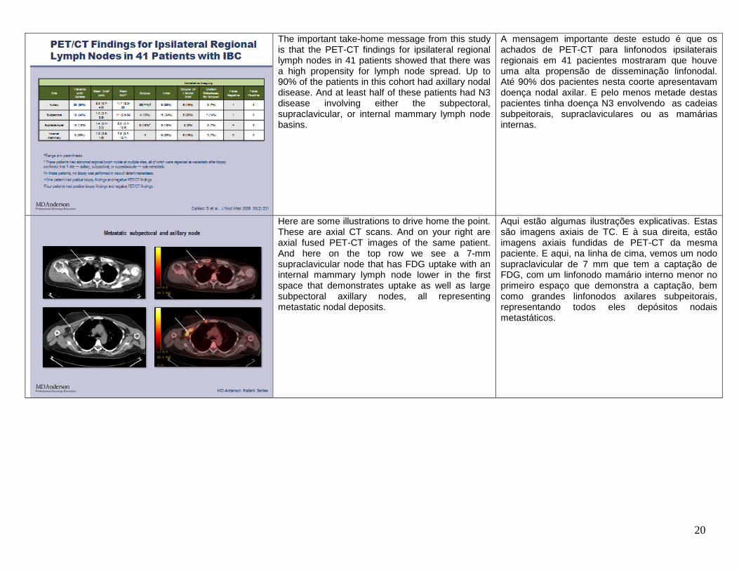
The important take-home message from this study is that the PET-CT findings for ipsilateral regional lymph nodes in 41 patients showed that there was a high propensity for lymph node spread. Up to 90% of the patients in this cohort had axillary nodal disease. And at least half of these patients had N3 disease involving either the subpectoral, supraclavicular, or internal mammary lymph node basins.
A mensagem importante deste estudo é que os achados de PET-CT para linfonodos ipsilaterais regionais em 41 pacientes mostraram que houve uma alta propensão de disseminação linfonodal. Até 90% dos pacientes nesta coorte apresentavam doença nodal axilar. E pelo menos metade destas pacientes tinha doença N3 envolvendo as cadeias subpeitorais, supraclaviculares ou as mamárias internas.
Here are some illustrations to drive home the point. These are axial CT scans. And on your right are axial fused PET-CT images of the same patient. And here on the top row we see a 7-mm supraclavicular node that has FDG uptake with an internal mammary lymph node lower in the first space that demonstrates uptake as well as large subpectoral axillary nodes, all representing metastatic nodal deposits.
Aqui estão algumas ilustrações explicativas. Estas são imagens axiais de TC. E à sua direita, estão imagens axiais fundidas de PET-CT da mesma paciente. E aqui, na linha de cima, vemos um nodo supraclavicular de 7 mm que tem a captação de FDG, com um linfonodo mamário interno menor no primeiro espaço que demonstra a captação, bem como grandes linfonodos axilares subpeitorais, representando todos eles depósitos nodais metastáticos.

21
The PET-CT findings in the same cohort of patients with IBC, describing distant metastasis, show that metastasis occurred in the bone, in the liver, in the abdomen, and also in the mediastinum
Os achados de PET-TC na mesma coorte de pacientes com CIM, descrevendo metástases distantes, mostram que a metástase ocorreu no osso, fígado, abdômen e também no mediastino
Here is an example of a coronal MIP image showing the primary inflammatory breast cancer on the left, with associated regional lymph nodes in the subpectoral axillary area. Unfortunately, the patient also has disseminated metastasis at diagnosis involving the skeletal system and the liver. This is an axial fused PET-CT image of the same patient showing the left primary inflammatory breast cancer and skeletal mets as well as liver lesions.
Aqui está um exemplo de uma imagem coronal MIP mostrando o carcinoma inflamatório de mama primário do lado esquerdo, com os linfonodos regionais associados na região axilar subpeitoral. Infelizmente, a paciente também apresentou metástase disseminada no diagnóstico que invadiram/atingiram o sistema esquelético e o fígado. Esta é uma imagem de PET-CT axial fundida da mesma paciente que mostra carcinoma inflamatório primário e metástase esquelética da mama esquerda, bem como lesões hepáticas.
22
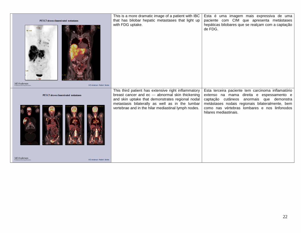
This is a more dramatic image of a patient with IBC that has bilobar hepatic metastases that light up with FDG uptake.
Esta é uma imagem mais expressiva de uma paciente com CIM que apresenta metástases hepáticas bilobares que se realçam com a captação de FDG.
This third patient has extensive right inflammatory breast cancer and ec --- abnormal skin thickening and skin uptake that demonstrates regional nodal metastasis bilaterally as well as in the lumbar vertebrae and in the hilar mediastinal lymph nodes.
Esta terceira paciente tem carcinoma inflamatório extenso na mama direita e espessamento e captação cutâneos anormais que demonstra metástases nodais regionais bilateralmente, bem como nas vértebras lombares e nos linfonodos hilares mediastinais.

23
In summary, CT together with whole body bone scan, ultrasound, and MR are currently used for staging IBC. In this new decade of the millennium PET-CT has developed an emerging role in the comprehensive staging of breast cancer with its ability to co-register anatomic and functional information on one image. It may potentially allow for more aggressive and diagnostic imaging strategies that will enable the detection of microscopic disease.
Em resumo, a TC, em conjunto com a cintilografia óssea do corpo todo, a ultrassonografia e a RM, é utilizada atualmente para o estadiamento da CIM. Nesta nova década do milênio, a PET-CT tem tido um papel emergente no estadiamento completo do câncer de mama por sua capacidade de corregistrar informações anatômicas e funcionais em uma [única] imagem. Ela pode, potencialmente, permitir estratégias mais agressivas de diagnóstico por imagem que permitirão a detecção de doença microscópica.
\
Moving on to differential diagnosis of patients with IBC.
Passemos para o diagnóstico diferencial de pacientes com CIM.

24
Here is a patient with a neglected locally advanced breast cancer which was neglected for more than two years. This is not similar to a patient with inflammatory breast cancer.
Aqui está uma paciente com câncer de mama localmente avançado, que não foi percebido por mais de dois anos. Não é semelhante a uma paciente com carcinoma inflamatório de mama.
This next image shows an abnormal mass in the right breast with global skin thickening and trabecular distortion.
Esta imagem ao lado mostra uma massa anormal na mama direita com espessamento global da pele e distorção trabecular.

25
Ultrasound of the same patient shows an abnormal mass and an abnormal lymph node.
A ultrassonografia da mesma paciente mostra uma massa anormal e um linfonodo anormal.
And PET-CT shows the breast mass as well as disseminated metastasis. And this patient ended up having a primary colorectal carcinoma metastatic to the breast,…
E a PET-CT mostra a massa mamária, bem como a metástase disseminada. Esta paciente acabou por apresentar um carcinoma colorretal primário com metástase mamária,…

26
…rather than an inflammatory breast cancer. …em vez de um carcinoma inflamatório de mama.
This is a third patient with very, very distorted parenchyma, trabecular distortion and skin thickening as well as enlarged lymph nodes.
Esta é uma terceira paciente com parênquima muito distorcido, distorção trabecular e espessamento da pele, bem como gânglios linfáticos aumentados.

27
The ultrasound is dramatic for global skin thickening and abnormal echoes throughout the right breast.
A ultrassonografia é expressiva para o espessamento global da pele e ecos anômalos em toda a mama direita.
And MR is striking for abnormal enhancement throughout the breast with wash-out kinetics. And the final diagnosis for this patient was idiopathic granulomatous mastitis…
A RM é surpreendente para o realce anormal em toda a mama com cinética de wash-out. E o diagnóstico final para esta paciente foi mastite granulomatosa idiopática…

28
….with wash-out kinetics. …. com cinética de wash-out.
“What about imaging-guided biopsies?” “E quanto a biópsias guiadas por imagens?”

29
Most of the pathologic characteristics of IBC are under the categories of invasive ductal carcinoma which is by far the commonest, followed by invasive lobular carcinoma. Most of the cancers are high grade or intermediate grade, and a high proportion of cancers of angiolymphatic invasion, also known as LVI, as well as dermal lymphatic invasion, which is why skin punch biopsy is an important diagnostic tool in these patients. We have discussed axillary adenopathy and regional adenopathy. The immuno-biomarker distribution in terms of ER, PR, and HER-2 status is fairly similar to regular invasive carcinomas.
A maioria das características patológicas do CIM corresponde às categorias de carcinoma ductal invasivo, que é, de longe, o mais comum, seguido pelo carcinoma lobular invasivo. A maioria dos cânceres é de alto grau ou grau intermediário e uma alta proporção de cânceres de invasão angiolinfática, também conhecida como LVI, bem como invasão linfática dérmica, razão pela qual a biópsia percutânea é uma ferramenta de diagnóstico importante nestas pacientes. Discutimos adenopatia axilar e adenopatia regional. A distribuição do imuno- biomarcador em termos do status de ER, PR e HER-2 é bastante semelhante à dos carcinomas invasivos regulares.
So this slide highlights the most pertinent features of the histological characteristics of breast cancer in IBC patients…
Portanto, este slide destaca os aspectos mais pertinentes das características histológicas do câncer de mama em pacientes com CIM…

30
…as well as the metastatic and the biomarker status of such patients.
…bem como o status metastático e do biomarcador de tais pacientes.
This slide illustrates a process of obtaining an ultrasound-guided core biopsy specimen of the breast in a patient with IBC where we have the transducer and the ultrasound probe within the breast.
Este slide ilustra um processo de obtenção de espécime para biópsia de fragmento da mama guiada por ultrassonografia de uma paciente com CIM em que temos o transdutor e a sonda de ultrassonografia dentro da mama.

31
And here is a pre-fire and post-fire image showing collection of the tissue samples from the primary breast mass,…
E aqui está uma imagem pré-biópsia e pós-biópsia mostrando a coleta das amostras de tecidos de massa de mama primária,…
…with the cores that are to be sent for histological evaluation.
…com os fragmentos a serem enviados para avaliação histológica.

32
The hematoxylin and eosin stains of the core needle biopsy of the breast are important to determine tumor cellularity, invasive and in situ component, lymphovascular emboli components.
Os corantes de hematoxilina e eosina da biópsia por agulha grossa da mama são importantes para determinar a celularidade do tumor, componente invasivo e in situ, os componentes de embolia linfovascular.
And here are examples of core specimens which show continuous tumor emboli…
E aqui estão exemplos de amostras dos fragmnetos que mostram êmbolos tumorais contínuos…

33
…versus discontinuous tumor emboli. And these have important implications in terms of biomarker assessments as well as molecular high throughput genetic studies down the line, in terms of research potential using tissues samples from such patients.
…versus êmbolos tumorais descontínuos. E estes têm consequências importantes em termos de avaliação de biomarcadores, bem como estudos genéticos de alto rendimento molecular no futuro, em termos de potencial de pesquisa usando amostras de tecidos de tais pacientes.
The next topic is monitoring response and, “How do the imaging methods contribute to this important function?”
O próximo tópico é o monitoramento da resposta e, “Como os métodos de imagem contribuem para esta importante função?”

34
Monitoring of response is particularly important throughout the patient’s course of treatment and especially at the time prior to surgery in order to delineate and delimit the disease extent for appropriate and accurate surgical planning.
Monitorar a resposta é particularmente importante em toda a evolução de tratamento da paciente e, em especial, no momento antes da cirurgia para fins de delinear e delimitar a extensão da doença para o planejamento cirúrgico adequado e preciso.
So here is the patient at the time of presentation with skin erythema and after treatment for 12 weeks, the follow-up shows significant resolution of clinically evident disease.
Aqui está a paciente no momento do diagnóstico com eritema cutâneo e após o tratamento durante 12 semanas. O acompanhamento mostra resolução significativa da doença clinicamente evidente.

35
And in terms of assessment of repo --- response, the final arbiter is by the pathologist whereby pathologic complete response is defined as proof of no residual invasive cancer. And in order to perform this task adequately, it requires that the identification of the tumor bed has to be assessed accurately to allow for adequate sampling in the microscopic component of this process.
Em termos de avaliação da resposta, o árbitro final é o patologista; a resposta patológica completa é definida como prova de ausência de câncer invasivo residual. E, para executar esta tarefa adequadamente, é necessário que a identificação do leito tumoral seja feita com precisão para permitir a amostragem adequada no componente microscópico deste processo.
Here are several images to illustrate the monitoring of response in a 44-year-old female with IBC that is HER2-positive that was on a trial study treated with targeted lapatinib. And here we see the mammogram with the diffuse skin thickening and the abnormal mass.
Aqui estão várias imagens para ilustrar o monitoramento da resposta em uma paciente de 44 anos com CIM positivo para HER-2, que estava em um estudo experimental tratada com lapatinibe direcionado. E aqui vemos a mamografia com o espessamento cutâneo difuso e a massa anormal.

36
And four months after treatment, there is still residual skin thickening and trabecular thickening but the mass has resolved.
E quatro meses após o tratamento, existe ainda o espessamento residual da pele e do trabéculo, mas a massa foi resolvida.
Similarly, in the same patient, there is not just one mass but multiple masses throughout the breast. We see this giant mass occupying most of the breast with subcutaneous as well as parenchymal deposits throughout the breast with skin thickening.
Do mesmo modo, na mesma paciente, não existe apenas uma massa, mas várias massas ao longo da mama. Vemos esta massa gigante ocupando a maior parte da mama com depósitos subcutâneos e parenquimatosos em toda a mama com espessamento da pele.

37
And four months after treatment, we see that most of the masses have resolved, but there is residual skin and trabecular thickening.
E quatro meses após o tratamento, vemos que a maioria das massas foi resolvida, mas há espessamento residual da pele e do trabéculo.
The most dramatic of this imaging techniques is MRI of the same patient which shows in one snapshot extensive tumor tissue throughout the breast with skin thickening. And three to four months after therapy, there is absolutely no enhancement, which indicates complete response by imaging with MR.
A mais impressionante destas técnicas de imagem é a RM da mesma paciente, que mostra um detalhe de tecido tumoral extenso em toda a mama com espessamento da pele. E três a quatro meses após a terapia, não há absolutamente nenhum realce, o que indica resposta completa por imagem com RM.

38
And similarly, with the coronal MIP PET image of this patient, we have dramatic disease in the left breast and the left regional nodes, with absence of uptake indicating complete response by PET with the --- the false positives of brown fat noted in each supraclavicular fossa.
E da mesma forma, com a imagem coronal MIP PET desta paciente, temos doença expressiva na mama esquerda e nos nódulos regionais esquerdos, com ausência de captação, indicando resposta completa na PET com os falsos positivos de gordura castanha observados em cada fossa supraclavicular.
At surgery, the patient’s left breast and axillary conte --- content from a modified radical mastectomy procedure showed no invasive carcinoma, stromal fibrosis in the breast, with 20 axillary lymph nodes negative for carcinoma, consistent with pathologic complete response.
Na cirurgia, a mama esquerda da paciente e o conteúdo axilar originado de um procedimento de mastectomia radical modificado não apresentaram carcinoma invasivo, fibrose do estroma mamário, com 20 linfonodos axilares negativos para carcinoma, coerente/congruente com a resposta patológica completa.

39
In summary, the initial staging of a patient with inflammatory breast cancer is optimal using breast ultrasound and MRI. And PET-CT has a role in the detection of distant metastasis. Imaging-guided biopsy using ultrasound or less commonly with MRI is essential for diagnosis and in biomarker evaluation. Neoadjuvant therapy is the standard of care for IBC patients. Therefore, early monitoring of patients using imaging is critical in the detection of non-responders. This has a huge role as potential surrogates for the prediction of pathologic complete response, as we know that patients who achieve pathologic complete response have improved outcomes.
Em resumo, o estadiamento inicial de uma paciente com carcinoma inflamatório de mama é ideal com o emprego de ultrassonografia mamária e ressonância magnética. E a PET-CT desempenha um papel na detecção de metástases a distância. A biópsia guiada por imagem usando ultrassonografia, ou mais raramente, com RM, é essencial para o diagnóstico e na avaliação de biomarcadores. A terapia neoadjuvante é o tratamento padrão para pacientes com CIM. Portanto, o monitoramento precoce com imagem de pacientes é fundamental para detectar não responsivos. Cumpre um papel muito importante como substitutos potenciais para o prognóstico/a previsão de resposta patológica completa, pois sabemos que os pacientes que atingem resposta patológica completa têm melhores resultados.
On this note, I would like to thank you for your attention and would welcome further queries and any questions regarding the presentation today. Thank you very much.
Com isto, gostaria de agradecer sua atenção. Suas consultas e questões sobre a apresentação de hoje serão bem-vindas. Muito obrigada.