production, purification and characterization of thermostable protease...
TRANSCRIPT
PRODUCTION, PURIFICATION AND
CHARACTERIZATION OF THERMOSTABLE
PROTEASE FROM ALKALIPHILIC AND
THERMOPHILIC Geobacillus sp.
A Thesis Submitted to
the Graduate School of Engineering and Sciences of
İzmir Institute of Technology
in Partial Fulfillment of the Requirements for the Degree of
MASTER OF SCIENCE
in Chemistry
by
Seden GÜRACAR
July 2011
İZMİR
We approve the thesis of Seden GÜRACAR
______________________________
Assist. Prof. Dr. Gülşah ŞANLI
Supervisor
______________________________
Prof. Dr. Ahmet YEMENİCİOĞLU
Committee Member
______________________________
Assoc. Prof. Dr. Talat YALÇIN
Committee Member
6 July 2011
______________________________ __________________________
Prof. Dr. Serdar ÖZÇELİK Prof. Dr. Durmuş Ali DEMİR
Head of the Department of Chemistry Dean of the Graduate School
Engineering and Sciences

ACKNOWLEDGEMENTS
Firstly, I would like to thank to my supervisor Assist. Prof. Dr. GülĢah ġANLI
MOHAMED for her supervision, support, valuable help, encouragement, confidence
and endless patience throughout my study.
I also would like to thank to Dane RUSÇUKLU, Yekta GÜNAY OĞUZ and
Evrim BALCI in Izmir Institute of Technology, Biotechnology and Bioengineering
Central Research Laboratories for all their advice, confidence, help and support during
my experiments.
I want to thank Assist. Prof. Dr. Ali Fazıl YENĠDÜNYA and Elif YAVUZ for
the kind supply of the Alkaliphilic and Thermophilic Geobacillus strains.
I would like to express my sincere thanks to Hande GENÇKAL for having
shared her experiences with me, her advices and kind helps.
I especially thank Prof. Dr. Ahmet YEMENĠCĠOĞLU and Assoc. Prof. Dr. Talat
YALÇIN for their careful reading of my manuscript and their advices.
I want to express my thankfulness to my close friends Nur ARGUN, Emine
DEĞĠRMENCĠ, Yelda KADIOĞLU, Gözde M. KAMER, Deniz BÖLEK, Hatice
ANDIRIN ÇĠÇEK and Nazlı DEĞĠRMENCĠ for their all kind of support, understanding
and friendship.
Finally, I am grateful to my mother Ruhsel GÜRACAR, my father Hüseyin
GÜRACAR, my sister Seven GÜRACAR and my cousin Arda Oğuzhan SAYIN for
their endless support, understanding and love during my thesis as in all stages of my
life. My special thanks are for Olgun BAYKARA for his endless love and
encouragement.

iv
ABSTRACT
PRODUCTION, PURIFICATION AND CHARACTERIZATION OF
THERMOSTABLE PROTEASE FROM ALKALIPHILIC AND
THERMOPHILIC Geobacillus sp.
Proteases are the hydrolase enzymes that catalyze the hydrolysis of the peptide
bonds in the primary structure of proteins and peptids. They are used to cleave the
proteins specifically to produce useful peptides in the processes. Proteases are present in
a wide variety of living organisms and they also show different physicological,
physicochemical, biological, chemical functions on the earth. They are the most important
enzymes in the industry, accounting for 60% of the total enzyme scales in the world.
The microorganisms that were previously isolated and characterized as a
Bacillus sp. from Balçova Geotermal region in Ġzmir were used in the experiments. The
aim of this study was to produce the protease enzyme from alkaliphilic and thermophilic
Bacillus sp., purify and determine the properties of the enzyme with the characterization
steps. When the screening studies and growth conditions were investigated, it was
understood that the alkaliphilic and thermophilic Bacillus sp. produced extracellular
protease enzyme. This extracellular protease enzyme was purified by ammonium
sulphate precipitation and ion exchange chromatography chromatograpy. The yield and
purification fold after purification of the enzyme were 33% and 1.41, respectively.
In the characterization studies, the results indicated that the protease enzyme had
highest activity at pH 8.0 and 55 ºC. The protease enzyme lost 20% of its activity at pH
4.0 and it lost 10% of its activity at pH 10.0. The protease enzyme at temperatures
below 55 ºC lost 15% of its activity and also the protease enzyme at temperatures above
55 ºC lost 25% of its activity. The protease enzyme was stable at different pH values
during 3 hours and at different temperature values during 6 hours. When compared the
substrates, casein showed higher activity. The effect of organic solvents and surfactants
on protease activity was investigated and the results indicated that the protease enzyme
was stable in the presence of 10% of the organic solvents and 1% of the surfactants.
PMSF and the protease inhibitor coctail decrease the activity of the protease.
v
ÖZET
ALKALĠFĠLĠK VE TERMOFĠLĠK Geobacillus sp.’den TERMAL
KARARLI PROTEAZ ENZĠMĠNĠN ÜRETĠLMESĠ, SAFLAġTIRILMASI
VE KARAKTERĠZASYONU
Proteazlar proteinler ve peptitlerdeki peptit bağlarının hidrolizini katalizleyen
hidrolaz enzimlerdir. Proseslerde yararlı peptitleri üretmek için proteinleri spesifik bir
Ģekilde kesmede kullanılırlar. Proteazlar büyük çeĢitlilik gösteren canlı organizmalarda
bulunurlar ve aynı zamanda yeryüzünde farklı fiziksel, fizikokimyasal, biyolojik, kimyasal
fonksiyonlara sahiptirler. Dünya enzim skalasının %60’ını oluĢturan, endüstrideki en
önemli enzimlerdir.
Ġzmir Balçova Jeotermal bölgesinden daha önce izole edilen ve Basillus sp.
olarak karakterize edilen mikroorganizmalar deneylerimizde kullanıldı. Bu çalıĢmanın
amacı, alkalifilik ve termofilik Basillus sp.’den protease enzimini üretmek, saflaĢtırmak
ve karakterizasyon basakları ile enzimin özelliklerini belirlemektir. Görüntüleme
çalıĢmaları ve büyüme koĢulları incelendiğinde, alkalifilik ve termofilik Basillus sp.’nin
ekstraselüler proteaz enzimi ürettiği anlaĢılmıĢtır. Bu ekstraselüler proteaz enzimi
amonyum sülfat çökürmesi ve iyon değiĢtirici kromatografi ile saflaĢtırılmıĢtır.
SaflaĢtırma sonrası verim ve saflık katsayısı sırasıyla %33 ve 1.41’dir.
Karakterizasyon çalıĢmalarında, sonuçlar enzimin pH 8.0 ve 55 ºC’de en yüksek
aktiviteye sahip olduğunu göstermiĢtir. Enzim pH 4.0’te aktivitesinin %20’sini ve pH
10.0’da aktivitesinin %20’sini kaybetmiĢtir. 55 ºC’nin altında proteaz enzimi
aktivitesinin %15’ini kaybetmiĢtir ve aynı zamanda 55 ºC’nin üzerinde proteaz enzimi
aktivitesinin %25’ini kaybetmiĢtir. Proteaz enzimi farklı pH değerlerinde 3 saat ve
farklı sıcaklık değerlerinde 6 saat boyunca stabildir. Substratlar kıyaslandığında, kazein
daha yüksek aktivite göstermiĢtir. Organik çözücülerin ve yüzey aktif maddelerin
proteaz aktivitesi üzerine etkisi incelenmiĢtir ve sonuçlar proteaz enziminin %10’luk
organik çözücü ve %1’lik yüzey aktif madde varlığında stabil olduğunu göstermiĢtir.
PMSF ve proteaz inhibitör kokteyli protease activitesini düĢürmüĢtür.
vi
TABLE OF CONTENTS
LIST OF FIGURES ......................................................................................................... ix
LIST OF TABLES ............................................................................................................ x
LIST OF ABBREVIATIONS .......................................................................................... xi
CHAPTER 1. INTRODUCTION.................................................................................... 1
1.1. Proteases (EC 3.4) ....................................................................................... 1
1.2. Classification of Proteases ......................................................................... 1
1.2.1. Endopeptidases (Proteinases) (E.C. 3.4.21-99) .................................... 4
1.2.1.1. Serine Proteases (E.C. 3.4.21)……………………………….5
1.2.1.2. Cysteine/Thiol Proteases (E.C. 3.4.22)………...……………8
1.2.1.3. Aspartic Proteases (E.C. 3.4.23)………………………..….. 8
1.2.1.4. Metalloprotease (E.C. 3.4.24)….……………….……………... 8
1.2.2. Exopeptidases (Peptidases) (E.C. 3.4.11-19) ....................................... 9
1.2.2.1. Aminopeptidases…………….…………………………….. 10
1.2.2.2. Carboxypeptidases………………………………………… 10
1.3. Industrial Applications of Proteases .......................................................... 10
1.4. Thermophiles ............................................................................................. 17
1.4.1. Thermophilic and Alkaliphilic Microorganisms ................................ 17
1.4.1.1. Thermophilic Bacillus…………………………………….. 18
1.4.1.2. Alkaliphilic Bacillus………………………….…………… 20
1.4.2. Thermophilic Enzymes ....................................................................... 22
1.4.2.1. Thermophilic and Thermostable Proteases……………….. 23
1.4.3. The Applications of Thermophiles in Biotechnology and
Industry ............................................................................................ 24
CHAPTER 2. MATERIALS AND METHODS .......................................................... 26
2.1. Materials .................................................................................................... 26
2.2. Bacterial Strain and Growth Conditions ................................................... 26
2.3. Screening of Protease Activity in Thermophilic Bacillus sp. ................... 27
vii
2.4. Determination of Enzyme Activity ........................................................... 27
2.5. Determination of Protein Concentration ................................................... 29
2.6. Preparation of Crude Enzyme Extract ...................................................... 29
2.7. Enzyme Purification Procedure ................................................................. 30
2.8. Electrophoretic Studies (SDS- PAGE) ..................................................... 30
2.9. Characterization Studies ............................................................................ 31
2.9.1. Effect of pH on Enzyme Activity and Stability ................................. 31
2.9.2. Effect of Temperature on Enzyme Activity and Stability.................. 32
2.9.3. Substrate Specificity of Enzyme ........................................................ 32
2.9.4. Effect of Metal Ions on Enzyme Activity .......................................... 32
2.9.5. Effect of Various Agents on Enzyme Activity .................................. 33
2.9.5.1. Effect of Organic Solvents on Enzyme Activity………….. 33
2.9.5.2. Effect of Surfactants on Enzyme Activity………………… 33
2.9.6. Effect of Inhibitors on Enzyme Activity ............................................ 33
CHAPTER 3. RESULTS AND DISCUSSION ........................................................... 35
3.1. Screening of Protease Activity in Thermophilic Bacillus sp. ................... 35
3.2. Purification of Protease ............................................................................. 36
3.3. Electrophoretic Studies (SDS-PAGE) ...................................................... 39
3.4. Characterization Studies ............................................................................ 42
3.4.1. Effect of pH on Enzyme Activity and Stability ................................. 42
3.4.2. Effect of Temperature on Enzyme Activity and Stability.................. 45
3.4.3. Substrate Specificity of Enzyme ........................................................ 48
3.4.4. Effect of Metal Ions on Enzyme Activity .......................................... 49
3.4.5. Effect of Various Agents on Enzyme Activity .................................. 51
3.4.5.1. Effect of Organic Solvents on Enzyme Activity………….. 51
3.4.5.2. Effect of Surfactants on Enzyme Activity………………… 52
3.4.6. Effect of Inhibitors on Enzyme Activity ............................................ 53
CHAPTER 4. CONCLUSION ...................................................................................... 56
REFERENCES ............................................................................................................... 58

viii
APPENDICES
APPENDIX A. PREPARATION OF BRADFORD REAGENT, PROTEIN
STANDARTS AND STANDART CURVE FOR BRADFORD
ASSAY ................................................................................................. 68
APPENDIX B. PROTEOLYTIC ACTIVITY DETERMINATION STANDARTS
AND STANDART CALIBRATION CURVE FOR L-TYROSINE
ASSAY ................................................................................................. 71
APPENDIX C. MEDIA, BUFFERS, REAGENTS AND SOLUTIONS ....................... 73
ix
LIST OF FIGURES
Figure Page
Figure 1.1. The mechanism of protease hydrolysis ............................................................. 1
Figure 1.2. The classification of proteases .......................................................................... 4
Figure 1.3. The schematic representation of the serine protease amino acid residues
involved in the proteolytic scission. ................................................................ 7
Figure 1.4. The distribution of enzyme sales .................................................................... 10
Figure 1.5. Phylogenetic tree based on 16S rRNA gene alignments ................................. 19
Figure 3.1. Screening of the protease activity ................................................................ 35
Figure 3.2. The concentration of the protease enzyme fractions after ion exchange
chromatography ........................................................................................... 37
Figure 3.3. SDS-PAGE image with silver staining under UV light. .............................. 40
Figure 3.4. SDS-PAGE image with silver staining........................................................ 40
Figure 3.5. SDS-PAGE image with colloidal coomassie staining................................. 40
Figure 3.6. Effect of pH on protease activity .................................................................. 43
Figure 3.7. pH stability of protease (at pH 4-7-10) ........................................................ 45
Figure 3.8. Effect of temperature on protease activity ................................................... 46
Figure 3.9. Thermal stability of protease ........................................................................ 48
Figure 3.10. Effect of metal ions on protease activity .................................................... 50
Figure 3.11. Effect of inhibitors on protease activity ..................................................... 54
x
LIST OF TABLES
Table Page
Table 1.1. The characteristic features of endopeptidases .................................................... 5
Table 1.2. Optimized production conditions for alkaline proteases ................................. 7
Table 1.3. Different applications of some industrally important proteases .................... 11
Table 1.4. Application of proteases in industry .............................................................. 12
Table 1.5. Serin proteases (Subtilisin) used in detergents ................................................. 13
Table 1.6. Commercial bacterial alkaline proteases, sources, applications and their
industrial suppliers ......................................................................................... 13
Table 1.7. Description of the Geobacillus genus ............................................................ 20
Table 1.8. Properties of some alkaline proteases from different microbial sources ....... 21
Table 1.9. Bioconversion reactions and applications of thermostable enzymes ............ 22
Table 1.10. Source microorganisms,properties of thermostable proteolytic enzymes ... 22
Table 1.11. Main advantages of high temperature and thermostable enzymes .............. 22
Table 2.1. Inhibitors of the endopeptidases ...................................................................... 34
Table 3.1. Purification of protease from alkaliphilic and thermophilic Bacillus sp. ...... 38
Table 3.2. Substrate specificity of protease .................................................................... 48
Table 3.3. Effect of organic solvents on protease activity .............................................. 52

xi
LIST OF ABBREVIATIONS
APS Ammonium persulfate
BSA Bovine serum albumin
CBB Commasie brilliant blue
DMSO Dimethyl sulfoxide
DIFP Diisopropylmethylfluoro phosphate
DTT Dithiothreitol
EDTA Ethylenediamine tetra acetic acid
LB Luria-Bertani broth
PAGE Polyacrylamide gel electrophoresis
PMSF Phenylmethylsulphonyl fluoride
Rpm Revolutions per minute
SDS Sodium dodecyl sulfate
sp. Species
TCA Trichloroacetic acid
TEMED Tetramethylethylenediamine
TLCK Tosyl-L-lysine chloromethyl ketone
UV Ultraviolet
μl Microliter
ml Milliliter
mM Millimolar
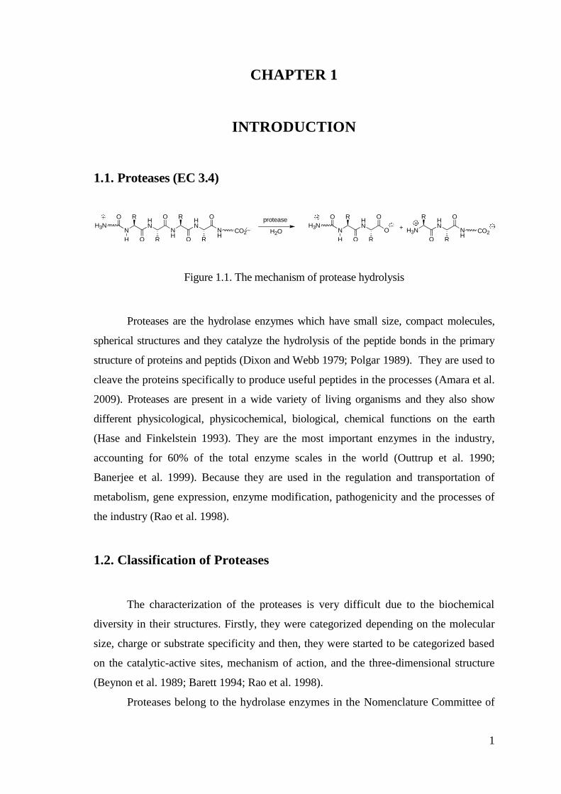
1
NH
HN
NH
O
RO
HN
N
O R
RO
R
H
O protease
H2OH3N
CO2 NH
HN
H3N
O
RO
HN
N
O R
RO
R
H
O
H3NCO2O +
CHAPTER 1
INTRODUCTION
1.1. Proteases (EC 3.4)
Figure 1.1. The mechanism of protease hydrolysis
Proteases are the hydrolase enzymes which have small size, compact molecules,
spherical structures and they catalyze the hydrolysis of the peptide bonds in the primary
structure of proteins and peptids (Dixon and Webb 1979; Polgar 1989). They are used to
cleave the proteins specifically to produce useful peptides in the processes (Amara et al.
2009). Proteases are present in a wide variety of living organisms and they also show
different physicological, physicochemical, biological, chemical functions on the earth
(Hase and Finkelstein 1993). They are the most important enzymes in the industry,
accounting for 60% of the total enzyme scales in the world (Outtrup et al. 1990;
Banerjee et al. 1999). Because they are used in the regulation and transportation of
metabolism, gene expression, enzyme modification, pathogenicity and the processes of
the industry (Rao et al. 1998).
1.2. Classification of Proteases
The characterization of the proteases is very difficult due to the biochemical
diversity in their structures. Firstly, they were categorized depending on the molecular
size, charge or substrate specificity and then, they were started to be categorized based
on the catalytic-active sites, mechanism of action, and the three-dimensional structure
(Beynon et al. 1989; Barett 1994; Rao et al. 1998).
Proteases belong to the hydrolase enzymes in the Nomenclature Committee of
2
the International Union of Biochemistry and Molecular Biology (International Union of
Biochemistry 1992). According to the committee, proteases have four classes with the
six families. They are serine I, serine II, cysteine (plants), cysteine (animals), aspartic
and metalloproteases (Kumar and Savitri 2008).
In the literature, you can review the studies about spesific capabilities in the
hydrolytic mechanism, specificity for a particular site, maximum activity under particular
conditions (temperature, salt concentration, endo/exopeptidase ratio, activity in organic
solvents) (García-Carreńo 1992).
Proteases are divided into three groups depends on their origin. They are animal-origin
proteases, plant-origin proteases and microbial-origin proteases. Recently, the
reserchers are interested in the microbial-origin proteases in the genus Bacillus because
of the fact that they are of vital importance in the industry (Priest 1977).
o Plant proteases: The places for cultivation and the climatic circumstances
affect the growing of the plant and production of the enzyme. The
disadvantage of the production of the enzyme is losing time. For example;
papaya, pineapple, papain, bromelain, keratinases, ficin (Rao et al. 1998).
o Animal Proteases: The political and agricultural strategies affect the obtaining
the animals and production of the enzyme from them. For example;
pancreatic trypsin, chymotrypsin, pepsin, and rennins (Rao et al. 1998).
o Microbial Proteases: The use of microbial proteases are generally common
(nearly 40% of the total enzyme sales) in comparison to plant, animal and
fungal proteases, due to having improved biological, biochemical and
molecular diversity in the nature (Godfrey and West 1996; Rao et al. 1998).
For examples; Bacteria, fungi, viruses. Microbial proteases are of the vital
importance because of the biochemical diversity, the rapid growth of the
microorganisms and the limited space required for cell cultivation. Bacterial
neutral proteases (pH 5-8, low thermotolerance, includes metallopreotease and
serine protease) and bacterial alkaline proteases (pH 10, high thermotolerance,
used in detergent technology) are the best known examples of the microbial
proteases (Rao et al. 1998). The microbial preteases play a crucial role in not
only cellular metabolic processes, but also industrial processes (Mahendran et
al. 2010). In the literature, you can examine the microbial proteases
(especially serine and metalloprotease) isolated by Bacillus subtilis, B.
amyloliquefaciens, Pseudomonas sp., Lysobacter enzymogenes and
3
Escherichia coli (Fujishige et al. 1992). It is important to identify and
characterize the microbial proteases with the sensitive experimental designs
in order to understand their role and improve their applications in the
industry (Lantz and Ciborowski 1994).
Proteases are divided into three groups depends on the secretion of the enzyme from
the microorganism. They are intracellular-protease (in the cell), periplasmic (with
the cell wall) and extracellular-protease (in the media) (Kohlmann et al. 1991; El-
Safey et al. 2004; do Nascimento and Martins 2004). Intracellular proteases are of
vital importance in the cellular, metabolic and regulatory processes. Extracellular
proteases are of vital importance in the hydrolysis of proteins in cell-free
environments, obtaining hydrolytic products in the commercial processes (Kalisz
1988; Kumar and Takagi 1999; Gupta and Beg 2002). Recently, not only the
extracellular protease enzyme but also the intracellular protease enzyme are
investigated due to the important roles in metabolic and regulatory processes in
the industries (Zeigler 2001).
Proteases are divided into four groups depends on the optimum pH. They are acidic
protease, neutral protease, alkaline protease and high-alkaline protease (Guangrong et
al. 2006). Acidic proteases are found in animal cells, moulds, yeasts and rarely
bacteria. A few of them include aspartic acid residue. The aromatic and bulky side
chains at the cleaving bonds reveal the specificity of the acidic proteases. Neutral
proteases contain the cystein proteases (Papain, bromelain, ficin) isolated from
botanical origin and the metalloproteases (Sumantha et al. 2006). The production of
bacterial neutral proteases is commonly occured at neutral (pH 5-8) conditions. They
have low thermo tolerance, so they control their activity and stability in the food
hydrolysis (Rao et al. 1998; Siddalingeshwara et al. 2010). Alkaline proteases are
produced at alkaline conditions and also they have wide application spectra because
of having their catalytic nature (Asokan et al. 2010).
Proteases are divided into different groups depends on the substrate specificity. They
are collagenases, keratinases, elastases, etc (Sumantha et al. 2006).
According to Enzyme Commission (EC) classification, proteases are divided into
two groups depends on the site of action and the cleavage sites within the target
molecule in the catalytic mechanism. They are exopeptidases or endopeptidases
(García-Carreńo 1991; García-Carreńo 1993; Liao and McCallus 1998).
4
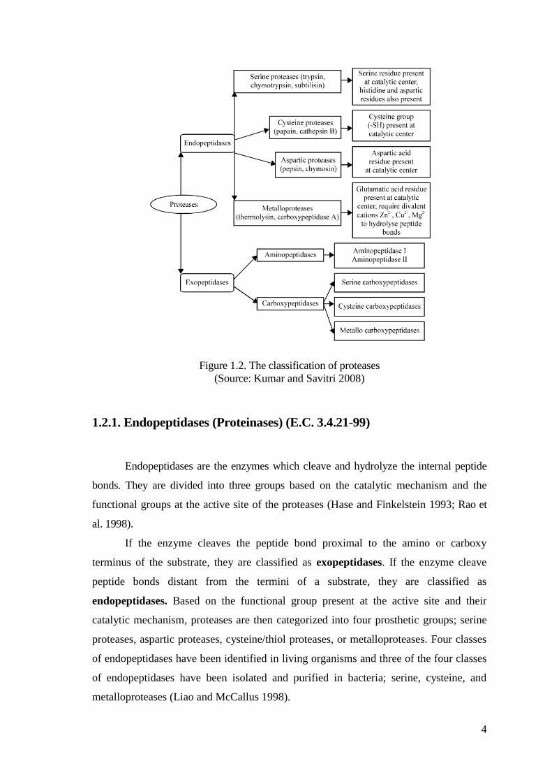
Figure 1.2. The classification of proteases
(Source: Kumar and Savitri 2008)
1.2.1. Endopeptidases (Proteinases) (E.C. 3.4.21-99)
Endopeptidases are the enzymes which cleave and hydrolyze the internal peptide
bonds. They are divided into three groups based on the catalytic mechanism and the
functional groups at the active site of the proteases (Hase and Finkelstein 1993; Rao et
al. 1998).
If the enzyme cleaves the peptide bond proximal to the amino or carboxy
terminus of the substrate, they are classified as exopeptidases. If the enzyme cleave
peptide bonds distant from the termini of a substrate, they are classified as
endopeptidases. Based on the functional group present at the active site and their
catalytic mechanism, proteases are then categorized into four prosthetic groups; serine
proteases, aspartic proteases, cysteine/thiol proteases, or metalloproteases. Four classes
of endopeptidases have been identified in living organisms and three of the four classes
of endopeptidases have been isolated and purified in bacteria; serine, cysteine, and
metalloproteases (Liao and McCallus 1998).
5
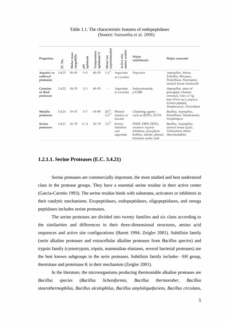
Table 1.1. The characteristic features of endopeptidases
(Source: Sumantha et al. 2006)
1.2.1.1. Serine Proteases (E.C. 3.4.21)
Serine proteases are commercially important, the most studied and best understood
class in the protease groups. They have a essential serine residue in their active center
(García-Carreńo 1993). The serine residue binds with substrates, activators or inhibitors in
their catalytic mechanisms. Exopeptidases, endopeptidases, oligopeptidases, and omega
peptidases includes serine proteases.
The serine proteases are divided into twenty families and six clans according to
the similarities and differences in their three-dimensional structures, amino acid
sequences and active site configurations (Barett 1994; Zeigler 2001). Subtilisin family
(serin alkaline proteases and extracellular alkaline proteases from Bacillus species) and
trypsin family (cymotrypsin, tripsin, mammalian elastases, several bacterial proteases) are
the best known subgroups in the serin proteases. Subtilisin family includes –SH group,
thermitase and proteinase K in their mechanism (Zeigler 2001).
In the literature, the microorganisms producing thermostable alkaline proteases are
Bacillus species (Bacillus licheniformis, Bacillus thermoruber, Bacillus
stearothermophilus, Bacillus alcalophilus, Bacillus amyloliquefaciens, Bacillus circulans,
6
Bacillus firmus, Bacillus intermedius, Bacillus lentus, Bacillus proteolyticus, Bacillus
pumilus, Bacillus sphaericus, Bacillus subtilis, Bacillus thuringiensis) (Kumar and Takagi
1999).
The stability of the alkaline proteases is provided via the immobilization
methods in a good way. The methods are based on the ground of the binding between
enzymes and carriers by carrier binding (physical adsorpsion, ionic bonding, covalent
bonding), cross-linking with bifunctional reagents, entrapping into gels / beads / fibres
(lattice-matrix type, microencapsule type). Thanks to the immobilization methods, the
enzymes are re-used and stabilized easily for a long time in the applications in the
industry (Anwar and Saleemuddin 1998).
The production of serine proteases is commonly occured at neutral (pH 7) and
alkaline (pH 11) conditions. They have broad substrate specificities. The molecular mass
of serin proteases generally changes from 18 kDa to 35 kDa and the isoelectric point of
them changes from pH 4 to pH 6. Bacillus species are the best known producer of serine
protease in comparison with the other microorganisms (Rao et al.1998). Also, they are
inhibited by PMSF (phenylmethylsulfonyl fluoride), DIFP (diisopropylfluoro phosphate),
3,4-dichloroisocoumarin (3,4-DCI), L-3-carboxytrans 2,3-epoxypropyl-leucylamido (4-
guanidine) butane (E.64), tosyl-L-lysine chloromethyl ketone (TLCK) and they are
active at alkaline pH (9 – 11) (Priest 1989; Kamal 1993; Rao et al. 1998).
In some of the serin proteases, the peptide bonds are attacked by the hydroxyl
group of a serine residue which carries a proton donor/general based on the serin
protease enzyme. On the other hand, it is called nucleophilic attact in the catalytic
mechanism. The proton donor should be a histidine residue which includes imidazolium
ring. But, an aspartate or an histidine residue is required because of the catalytic triad.
In some of the serin proteases, the peptide bonds are attacked by the catalytic
dyad of a serine residue which carries a proton donor/general base on the serin protease
enzyme. The proton donor should be a lysine residue and there is no need another
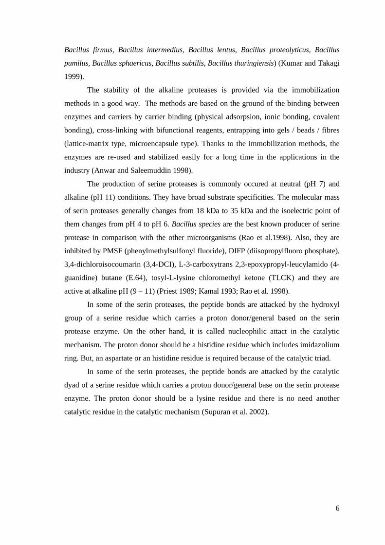
catalytic residue in the catalytic mechanism (Supuran et al. 2002).
7
Figure 1.3. The schematic representation of the serine protease amino acid residues
involved in the proteolytic scission. (Source: Supuran et al. 2002) A: Catalytic triad (chymotrypsin numbering)
B: Catalytic dyad (a lysine residue activates the hydroxyl group of serine residue essential for
catalysis)
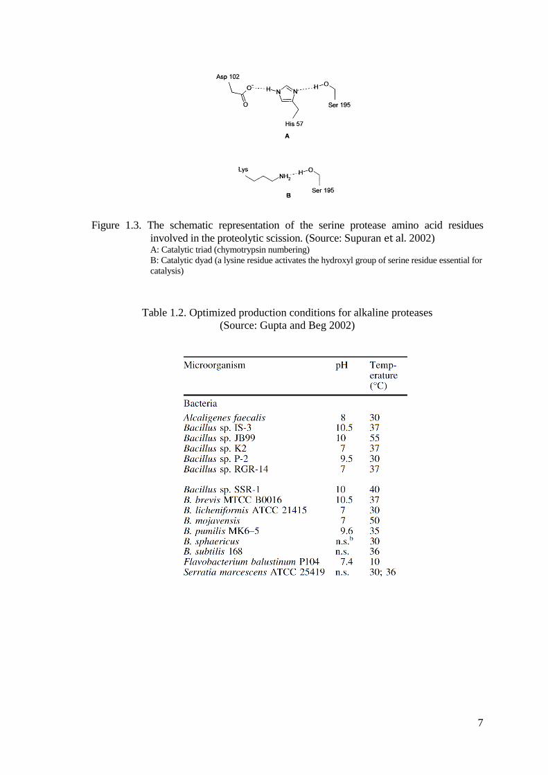
Table 1.2. Optimized production conditions for alkaline proteases
(Source: Gupta and Beg 2002)
8
1.2.1.2. Cysteine/Thiol Proteases (E.C. 3.4.22)
Cysteine proteases are produced by prokaryotes and eukaryotes. The cystein
proteases have cysteine (SH-) and histidin groups in their active sites (Garcia-Carreno
1993). They are divided into twenty families depends on the order differences between
cysteine and histidin residues (Barett 1994). On the other hand, they are categorized into
four groups. They are papain-like, trypsin-like, glutamic acid-like and others. The
production of cystein proteases is commonly occured at neutral conditions. The molecular
mass of serin proteases generally changes from 32 kDa to 50 kDa and the isoelectric point
of them changes from pH 4.9 to pH 8.4 (Rao et al. 1998). Also, they are inhibited by thiol
reagents (heavy metals, alkylating-oxidizing agents), sulphydryl reagents, (p-
chloromercuribenzoate and iodoacetamide) (Kamal 1993; Zeigler 2001).
The peptide bonds are attacked by the –SH group of a cysteine residue which
carries a nucleophile and a proton donor/general base on the serin protease enzyme. On
the other hand, it is called nucleophilic attact in the catalytic mechanism. The proton
donor should be a His residue which includes imidazolium ring (Supuran et al. 2002).
1.2.1.3. Aspartic Proteases (Acidic Proteases) (E.C. 3.4.23)
Aspartic proteases have an aspartic acid residue in the active sites (Garcia-Carreno
1993). The aspartic proteases are divided into three families. They are pepsin, retropepsin
and enzymes from pararetroviruses. The production of aspartic proteases is commonly
occured at acidic conditions owing to the fact that the aspartic proteases show maximum
activity at acidic pH. The molecular mass of serin proteases generally changes from 30
kDa to 45 kDa and the isoelectric point of them changes from pH 3 to pH 4.5. Also, they
are inhibited by pepstatin, diazoacetyl norleucine methyl ester (DAN), and 1,2-epoxyp-
nitrophenoxy propane (EPNP) (Rao et al. 1998; Zeigler 2001).
1.2.1.4. Metalloproteases (E.C. 3.4.24)
Metalloproteases are the enzymes which cleave the peptide bonds thanks to the
nucleophilic attact of a water molecule which is coordinated to a divalent metal ion
9
(catalytic zinc, manganese, cobalt, nickel or copper) or bridged to a dimetallic center.
The metal ion is complexed by three conserved amino acid residues that can be glutamic
acid (Glu), aspartic acid (Asp), histidin (His) or lysin (Lys) in their active sites (Hase and
Finkelstein 1993; Supuran et al. 2002; Mansfeld 2007).
The properties of catalytic and structural metal-binding sites are defined by the
x-ray crystallographic analyses of the metalloproteases. The most studied
metalloprotease, zinc-containing metalloproteases have a catalytic zinc atom
(coordinated to three amino acid residues) and an active water molecule in their cristal
structures. Also, the residues (His, Glu, Asp, Cys) coordinates the catalytic zinc atom,
the tridentate active zinc site occures and the coordination sphere is completed by the
activated water (Vallee and Auld 1990).
The metalloproteases are divided into thirty families and the families are
categorized into fourteen different clans according to the amino acid sequences and the
relation between the aminoacids and the metal binding sites. Clan MA, MB, MD, ME,
MJ, MK, MM, MO and MP include only one divalent cation and clan MF, MG, MH,
MN and MQ include two divalent cations acted co-catalytically on the substrate
(Mansfeld 2007). The clan MA has the HEXXH-E sequence (for exp: thermolysin) and
the clan MB has the HEXXH-H sequence.
On the other hand, the metalloproteases are divided into four groups according
to the catalitic action definitely. They are neutral, alkaline, Myxobacter I and
Myxobacter II. The production of metalloproteases is commonly occured at neutral
conditions (Rao et al. 1998). The pH optima of metalloproteases is commonly in the range
of 5-9 (Zeigler 2001). Also, the metalloproteases are inhibited by chelating agents (for
example: EDTA, 1,10-phenantroline) (Matsubara and Feder 1971; Kamal 1993).
1.2.2. Exopeptidases (Peptidases) (E.C. 3.4.11-19)
Exopeptidases are the enzymes which cleave and hydrolize the external peptide
bonds in the terminal amino end or carboxylic end of the substrate (Garcia-Carreno 1993).
They are divided into two subgroups based on the cleavage sites.
10
1.2.2.1. Aminopeptidases
Aminopeptidases are the enzymes that hydrolize and split off the N-terminal
residues (Kamal 1993). They are generally intracellular enzymes.
1.2.2.2. Carboxypeptidases
Carboxypeptidases are the enzymes that hydrolize and split off the C-terminal
residues (Kamal 1993). Carboxypeptidases are divided into three groups due to having
different amino acid residues at the active site of the proteases. They are serine
carboxypeptidases, metallocarboxypeptidases, and cysteine carboxypeptidases.
1.3. Industrial Applications of Proteases
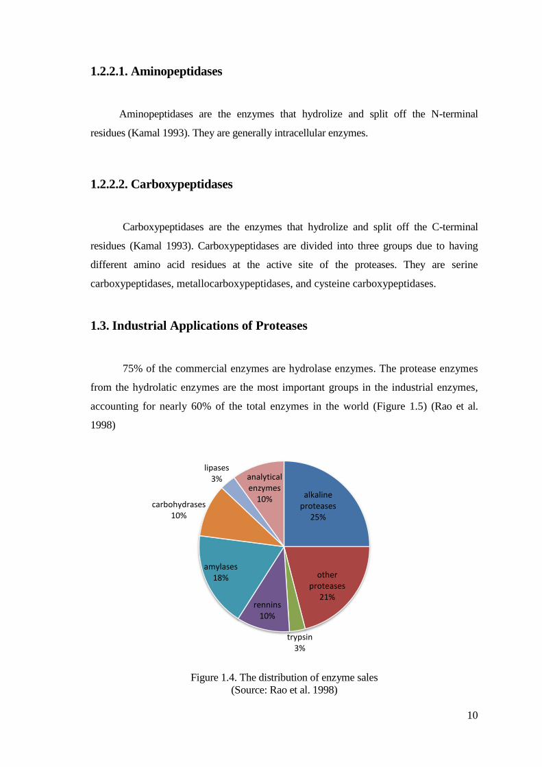
75% of the commercial enzymes are hydrolase enzymes. The protease enzymes
from the hydrolatic enzymes are the most important groups in the industrial enzymes,
accounting for nearly 60% of the total enzymes in the world (Figure 1.5) (Rao et al.
1998)
Figure 1.4. The distribution of enzyme sales
(Source: Rao et al. 1998)
alkaline proteases
25%
other proteases
21%
trypsin3%
rennins10%
amylases18%
lipases3% analytical
enzymes10%
carbohydrases 10%
11
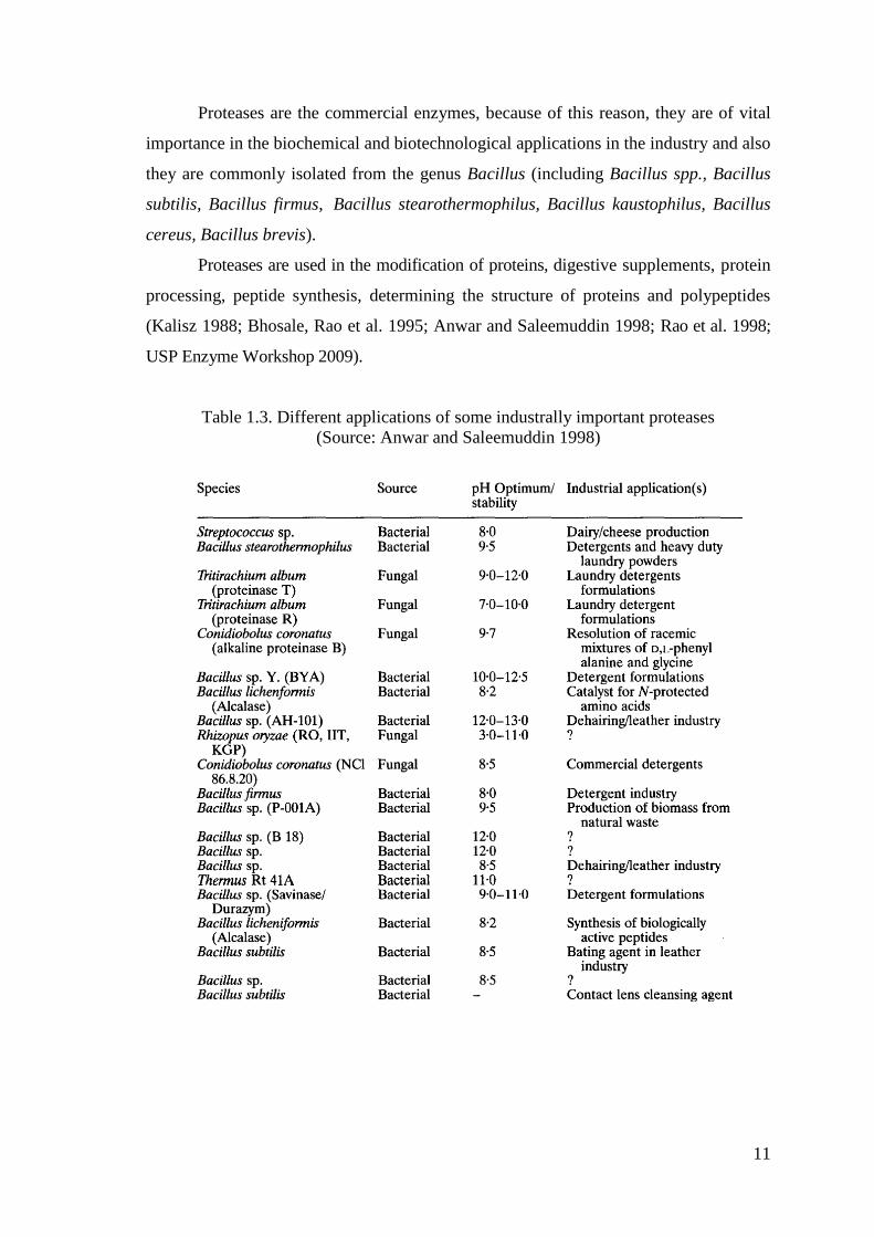
Proteases are the commercial enzymes, because of this reason, they are of vital
importance in the biochemical and biotechnological applications in the industry and also
they are commonly isolated from the genus Bacillus (including Bacillus spp., Bacillus
subtilis, Bacillus firmus, Bacillus stearothermophilus, Bacillus kaustophilus, Bacillus
cereus, Bacillus brevis).
Proteases are used in the modification of proteins, digestive supplements, protein
processing, peptide synthesis, determining the structure of proteins and polypeptides
(Kalisz 1988; Bhosale, Rao et al. 1995; Anwar and Saleemuddin 1998; Rao et al. 1998;
USP Enzyme Workshop 2009).
Table 1.3. Different applications of some industrally important proteases
(Source: Anwar and Saleemuddin 1998)
12
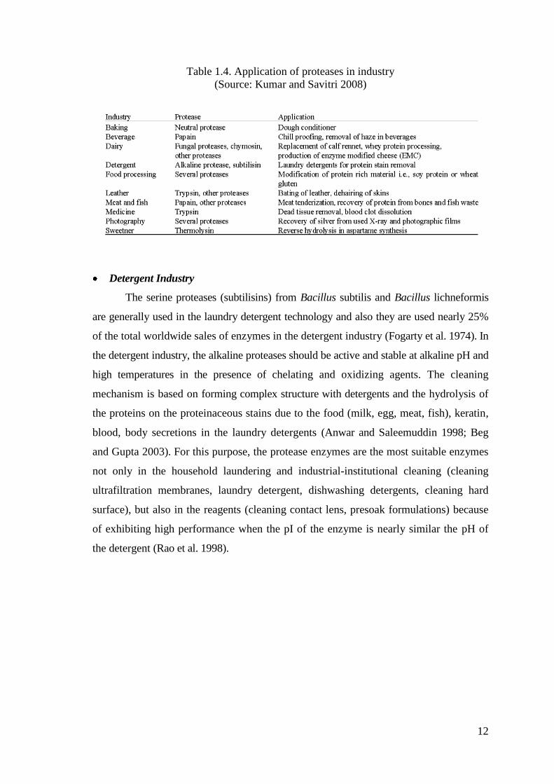
Table 1.4. Application of proteases in industry
(Source: Kumar and Savitri 2008)
Detergent Industry
The serine proteases (subtilisins) from Bacillus subtilis and Bacillus lichneformis
are generally used in the laundry detergent technology and also they are used nearly 25%
of the total worldwide sales of enzymes in the detergent industry (Fogarty et al. 1974). In
the detergent industry, the alkaline proteases should be active and stable at alkaline pH and
high temperatures in the presence of chelating and oxidizing agents. The cleaning
mechanism is based on forming complex structure with detergents and the hydrolysis of
the proteins on the proteinaceous stains due to the food (milk, egg, meat, fish), keratin,
blood, body secretions in the laundry detergents (Anwar and Saleemuddin 1998; Beg
and Gupta 2003). For this purpose, the protease enzymes are the most suitable enzymes
not only in the household laundering and industrial-institutional cleaning (cleaning
ultrafiltration membranes, laundry detergent, dishwashing detergents, cleaning hard
surface), but also in the reagents (cleaning contact lens, presoak formulations) because
of exhibiting high performance when the pI of the enzyme is nearly similar the pH of
the detergent (Rao et al. 1998).
13
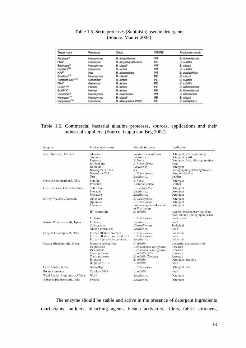
Table 1.5. Serin proteases (Subtilisin) used in detergents
(Source: Maurer 2004)
Table 1.6. Commercial bacterial alkaline proteases, sources, applications and their
industrial suppliers. (Source: Gupta and Beg 2002)
The enzyme should be stable and active in the presence of detergent ingredients
(surfactants, builders, bleaching agents, bleach activators, fillers, fabric softeners,
14
detergent composition, formulation aids) and the different conditions (pH of detergent,
ionic strength, wash temperature, mechanical handling) (Anwar and Saleemuddin 2000;
Beg and Gupta 2003).
The thermostable proteases show high activity and stability in the high
temperatures, so using them in the detergents is of vital importance because of having
high yield and low cost. They also used in the dishwashing and the laundry detergents
in the hospitals, due to the fact that the sterilization at high temperatures is too
important to overcome the microbial contaminations (Zeigler 2001).
Food Industry
Proteases are very significant in the food technology because of the fact that they
are used as a catalyst and they try to convert the untreated materials to the developed
food materials. The importance of substrate specificity, kinetic parameters, catalitic
activity should be taken into the consideration while doing the experiments under the
different conditions (García-Carreńo 1991).
The researchers’ aim is to save time, enegy, money and to reach high yield, high
catalytic activiy, high substrate specificty, large amount of product. Because of this
reason, they tried to find new approaches (fermantation processes, protein engineering,
synthetic enzymes, three dimensional structures) in food technology (Wasserman 1990).
For example; dairy industry, baking, cheese production, gluten development, whipping,
preparation of soya hydrolysates, aspartame synthesis, meat tenderization, chill-haze
prevention, brewing, dietetic, health products (Rao et al. 1998).
Further, proteases play a prominent role in meat tenderization, especially of beef.
Thermophilic alkaline proteases are used in the meat tenderization by hydrolyzing the
tissue proteins. Firstly, the enzyme was given into the meat by injecting. Nowadays, the
enzyme is given into the circulatory system of the animal (Kumar and Takagi 1999).
Hydrolysis of the proteins is required in the nutritions and also it is used in
different situations such as food formulations, therapeutic dietary foods and the addition
to the fruit juices and soft drinks (Neklyudov et al. 2000; Gupta and Beg 2002).
Proteases are useful addition matters in the food industry because of having
improved digestibility, solubility, flavor & palatability, processing (viscosity reduction,
improved drying), modified functional properties (emulsification, fat-binding, water-
binding, foaming properties, gel strength, whipping properties) (USP Enzyme Workshop
2009).
15
o Dairy Industry: The improvements are freezing storage of raw-pasteurized
milk for a long time, staying at higher temperatures in the pasteurization and
obtaining high quality of milk in the dairy industry (Meer et al. 1991; Meer,
Wodburn et al. 1993; Stadhouders 1992; Baker and Griffiths, 1993). The
proteases are used as milk-clotting agents for cheese production in the dairy
industry (Sumantha et al. 2006).
o Baking industry: The neutral proteases from Bacillus spesies are generally
used in baking industry. The soft dough in the biscuit and crackers are taken
into consideration not to be confronted with a problem about bending and
wrinkling in the oven (Bryce 1966). Thanks to using the protease enzyme, the
mixing time is reduced and the loaf volumes are increased (Rao et al. 1998).
o Brewing industry: Proteases from the Bacillus species are generally used in
brewing industry in order to cleave the peptide bonds in the proteins. If you
don’t want to be confronted with a problem about turbidity in the beverages,
you should add the protease into the cold English and German beer. (Fogarty
et al. 1974)
o Animal protein processing includes animal feed additives, improving
digestibility, reducing allergenicity, improving flavor and meat tenderization
in the food industry. In the meat tenderization, if the protease enzyme should
be active and have thermal stability during the cooking and storage
processes, good tenderization and developed taste of the meat will be
obtained in these applications (Wilson et al. 1992; Zeigler 2001). In the fish
industry, proteases from the Bacillus species are generally used in the fish
industry. The aim is to obtain fishmeal and enhanced oil recovery from the
uneatable fish (Lovern 1955).
Leather Industry
Initial soaking (neutral proteases), de-hairing of animal hair, hides and skin
(alkaline proteases), bating (acidic proteases), tanning and removing the waste materials
(animal glue) are the stages of leather preparation. The animal hair, hides and skin include
the materials with protein. The proteases are used instead of hazardous chemicals (sodium
sulfide) to prevent the pollution problems, obtain high quality, easy control, speed up
dehairing and reduce the waste materials in the leather industry. The most improtant
advantage is being eco-friendly (Grimm 1958; Rao et al. 1998; Gupta and Beg 2002) The
16
unwanted proteinaceous pigments are removed by using protease enzyme and the clean
skin-hide are obtained. The alkaline conditions should be choosen because it is easy to
remove the hair for the alkali protease enzyme (Varela et al. 1997; Gupta and Beg 2002).
Silver Recovery
The photographic films and the X-ray films which are used before include silver
(1.5-2.0%) in their gelatin layers. Because of this reason, alkaline proteases are used in
order to hydrolise the gelatin layers on the films and release of the silver materials in the
industrial applications (Kumar and Takagi 1999). There are lots of advantages in
recovering silver from photographic films by digestion of gelatin emulsion coating. The
pollution of silver is prevented and the silver is used in another purposes after
recovering in the environment. Thus, recovering silver is very important in the silver
industry (Gupta and Beg 2002).
Medical and Pharmaceutical Industry
Proteases are used in the medical and pharmaceutical industry because of having
broad diversity and specificity. The usage of this enzyme is common in developing
therapeutic agents, preparation of medicines, medical diagnosis, biopharmaceutical
products (contact-lens enzyme cleaners, enzymatic debriders) and cosmetics (skin care
ointments) (Anwar and Saleemuddin 2000). In the treatment of burns, wounds,
carbuncles, furuncles and abscesses, the subtilisin (or clostridial collagenase) is chosen
to be used with the advanced antibiotics (Rao et al. 1998; Gupta and Beg 2002).
Waste Treatment (Industrial and household)
Alkaline proteases are used to solubilize and hydrolyze the proteins in the
wastes. The wastes include proteinaceous materials which come from not only the
industries but also the house (Kumar and Takagi 1999). The aim is to obtain
bioconversion of the proteinaceous waste into the useful biomass and to decrease the
biological oxygen demand of aquatic systems in the biotechnological hydrolising and
bioremediation prosesses (deproteinization of shrimp and crabshell waste) (Yang et al.
2000; Gupta et al. 2002). Conversion the waste materials which include fibrous proteins
(horn, feather, wool, nail, hair) into the useful biomass is of vital importance in the waste
treatment industry (Venugopal et al. 1989).
17
1.4. Thermophiles
Microorganisms are categorized into three groups depends on the optimal
growth temperatures. They are psychrophiles (below 20°C), mesophiles (moderate
temperatures) and thermophiles (high temperatures, above 55°C) (Brock 1986; Turner
et al. 2007).
Thermophiles are adapted to live at high temperatures. The enzymes isolated from
thermophiles are used in most of the commercial applications due to their thermostability
and thermoactivity. Thus, the isolation, identification and characterization of the
thermophiles from natural sources are of vital significance in terms of discovering new
commercial enzymes (Yavuz et al. 2004).
All of the microorganisms have adaptation mechanism to the environment which
they live. Thus, thermophiles are used to live at high temperatures and need this
condition to survive. They are isolated from terrestrial, subterranean and submarines.
Thermophiles include lots of proteins and produce lots of enzymes, so they have
resistance to denaturation and proteolysis (Kumar and Nussinov 2001).
Thermophiles are divided into three groups depends on their minimal and maximal
growth temperatures. They are moderate thermophiles (35 - 70 ºC), extreme
thermophiles (55 - 85 ºC) and hyperthermophiles (75 - 113 ºC) (Baker et al. 2001).
Thermophiles are also classified into three different groups based on the growth and
not growth temperatures. They are obligate thermophiles (optimum growth temperatures
between 65-75 ºC, do not grow below 40 ºC), facultative thermophiles (optimum growth
temperatures between 50-60 ºC and around 37 ºC), thermotolerant thermophiles
(optimum growth temperatures between 45-50 ºC and 30 ºC) (Hughes and Williams 1977).
1.4.1. Thermophilic and Alkaliphilic Microorganisms
Thermophilic and alkaliphilic bacillus produce thermophilic protease enzyme
which has resistance to high temperature, pH, organic solvents, detergents, oxidizing
and denaturing agents (Johnvesly and Naik 2001; Hawumba 2002).
18
1.4.1.1. Thermophilic Bacillus
Bacteria of the genus Bacillus is aerobic, facultatively anaerobic, endospore-
forming, Gram-positive and rod-shaped. The representatives of this genus are widely
distributed in soil and water (deep layers in the ocean, arctic regions, etc.). The ability
of certain strains to tolerate high or low temperatures and extreme pHs and to secrete
high levels of proteins has made bacilli important as sources of commercial enzymes
(Norris et al. 1981). Strains of Bacillus are used for the production of enzymes,
antibiotics, fine biochemicals and insecticides (Harwood 1989). Bacillus species
produce several extracellular proteases. For example; Bacillus cereus, Bacillus
sterothermophilus, Bacillus pumilus, Bacillus licheniformis, Bacillus mojavensis,
Bacillus megaterium and Bacillus subtilis (Mahmoud et al.; Ammar et al. 1991;
Sookkheo et al. 2000; Beg and Gupta 2003; Banik and Prakash 2004; Gerze et al. 2005;
Soares et al. 2005).
The genus Bacillus is divided into different groups based on the different
conditions in the environments. They are temperature (thermophilic and psychrophilic
bacteria), pH (acidophilic and alkalophilic bacteria) and carbon sources (freshwater and
halophilic bacteria) (Nazina et al. 2001).
The genus Bacillus species are categorized into seven phylogenetic groups that
are Alicyclobacillus, Paenibacillus, Brevibacillus, Aneurinibacillus, Virgibacillus
Salibacillus and Gracilibacillus (Wisotzkey et al. 1992; Ash et al. 1993; Heyndrickx et
al. 1997; Wainø et al. 1999; Sung et al. 2002).
The thermophilic Bacillus species are divided into different groups depends on
the temperature range (45-70°C) in their growing conditions. They are genera Bacillus,
Alicyclobacillus, Brevibacillus, Aneurinibacillus, Sulfobacillus, Thermoactinomyces and
Thermobacillus (Sneath 1986; Wisotzkey et al. 1992; Dufresne et al. 1996; Heyndrickx
et al. 1997; Touzel et al. 2000, Nazina et al. 2001).
The groups in the termophilic Bacillus species are transferred to the new genus
Geobacillus. The Geobacillus are isolated from the geothermal areas, oilfield, shallow
marine vents-hot springs, deepsea hydrothermal vents, artificial hot environments (hot
water pipelines, heat exchangers, waste treatment plants, burning coal refuse piles and
bioremediation biopiles (McMullan et al. 2004).
19
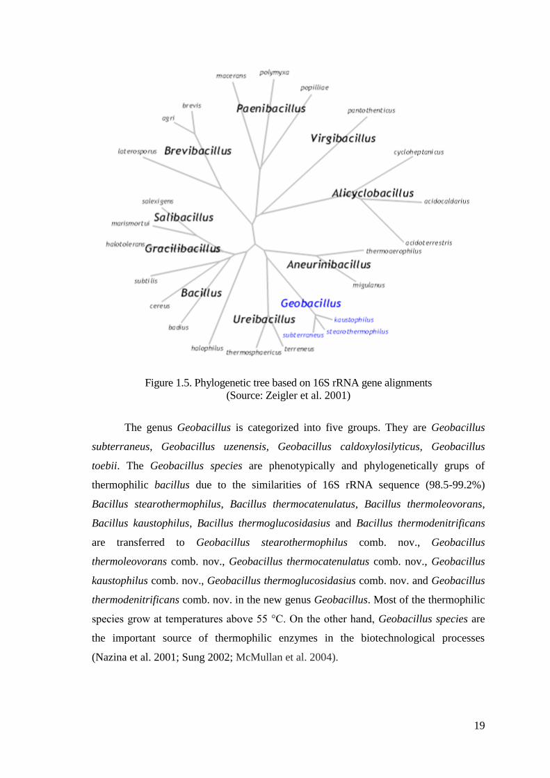
Figure 1.5. Phylogenetic tree based on 16S rRNA gene alignments
(Source: Zeigler et al. 2001)
The genus Geobacillus is categorized into five groups. They are Geobacillus
subterraneus, Geobacillus uzenensis, Geobacillus caldoxylosilyticus, Geobacillus
toebii. The Geobacillus species are phenotypically and phylogenetically grups of
thermophilic bacillus due to the similarities of 16S rRNA sequence (98.5-99.2%)
Bacillus stearothermophilus, Bacillus thermocatenulatus, Bacillus thermoleovorans,
Bacillus kaustophilus, Bacillus thermoglucosidasius and Bacillus thermodenitrificans
are transferred to Geobacillus stearothermophilus comb. nov., Geobacillus
thermoleovorans comb. nov., Geobacillus thermocatenulatus comb. nov., Geobacillus
kaustophilus comb. nov., Geobacillus thermoglucosidasius comb. nov. and Geobacillus
thermodenitrificans comb. nov. in the new genus Geobacillus. Most of the thermophilic
species grow at temperatures above 55 °C. On the other hand, Geobacillus species are
the important source of thermophilic enzymes in the biotechnological processes
(Nazina et al. 2001; Sung 2002; McMullan et al. 2004).
20
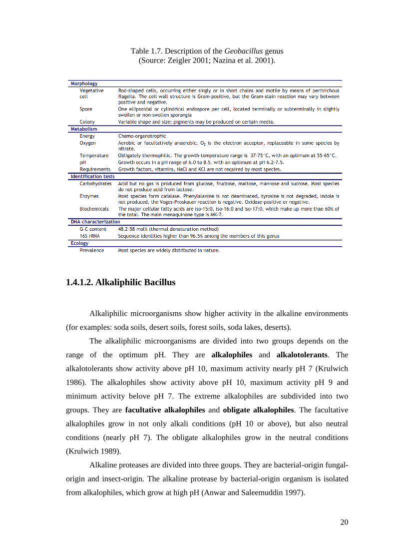
Table 1.7. Description of the Geobacillus genus
(Source: Zeigler 2001; Nazina et al. 2001).
1.4.1.2. Alkaliphilic Bacillus
Alkaliphilic microorganisms show higher activity in the alkaline environments
(for examples: soda soils, desert soils, forest soils, soda lakes, deserts).
The alkaliphilic microorganisms are divided into two groups depends on the
range of the optimum pH. They are alkalophiles and alkalotolerants. The
alkalotolerants show activity above pH 10, maximum activity nearly pH 7 (Krulwich
1986). The alkalophiles show activity above pH 10, maximum activity pH 9 and
minimum activity belove pH 7. The extreme alkalophiles are subdivided into two
groups. They are facultative alkalophiles and obligate alkalophiles. The facultative
alkalophiles grow in not only alkali conditions (pH 10 or above), but also neutral
conditions (nearly pH 7). The obligate alkalophiles grow in the neutral conditions
(Krulwich 1989).
Alkaline proteases are divided into three goups. They are bacterial-origin fungal-
origin and insect-origin. The alkaline protease by bacterial-origin organism is isolated
from alkalophiles, which grow at high pH (Anwar and Saleemuddin 1997).
21
When the enzimatic and physicochemical properties are compared in genus
Bacillus strains, it is understood that they are distinct from eachother. The genus
Bacillus and the alkaliphilic Bacillus spp. are prolific producers of extracellular
proteolytic enzymes and they are the best known source of alkaline proteases and the
secretion of these enzymes are used in vaarious industrial applications (Horlkoshi 1971;
Kobayashi et al. 1996; Kumar and Takagi 1999).
Alkaline proteases are of vital significance due to being active and stable at
higher pH and temperature values (Ward 1993). They also withstand to harsh
conditions. That’s why, they are chosen to be used in the industrial applications.
With a view to develop an economically feasible technology, research efforts are
mainly focused on the improvement in the yields of alkaline proteases and the
optimization of the fermentation medium and production conditions (Kumar and Takagi
1999).
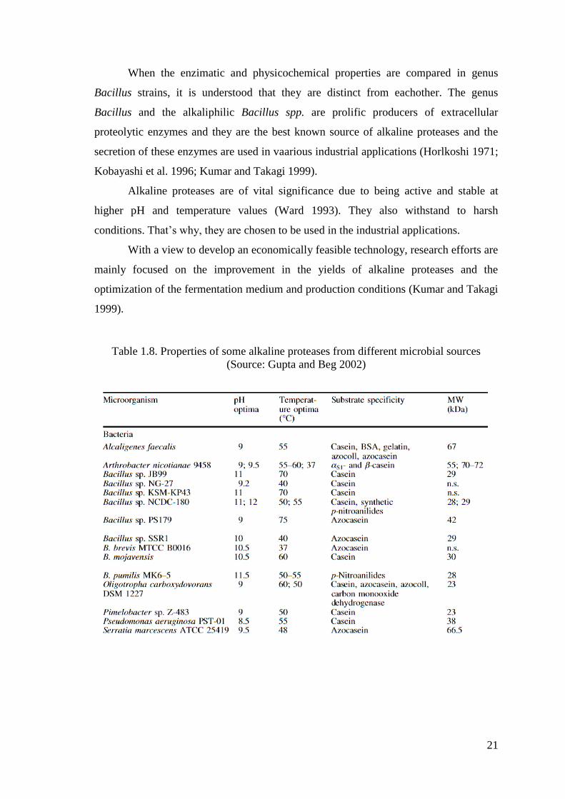
Table 1.8. Properties of some alkaline proteases from different microbial sources
(Source: Gupta and Beg 2002)
22
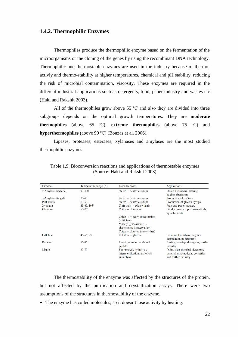
1.4.2. Thermophilic Enzymes
Thermophiles produce the thermophilic enzyme based on the fermentation of the
microorganisms or the cloning of the genes by using the recombinant DNA technology.
Thermophilic and thermostable enzymes are used in the industry because of thermo-
activiy and thermo-stability at higher temperatures, chemical and pH stability, reducing
the risk of microbial contamination, viscosity. These enzymes are required in the
different industrial applications such as detergents, food, paper industry and wastes etc
(Haki and Rakshit 2003).
All of the thermophiles grow above 55 ºC and also they are divided into three
subgroups depends on the optimal growth temperatures. They are moderate
thermophiles (above 65 ºC), extreme thermophiles (above 75 ºC) and
hyperthermophiles (above 90 ºC) (Bouzas et al. 2006).
Lipases, proteases, esterases, xylanases and amylases are the most studied
thermophilic enzymes.
Table 1.9. Bioconversion reactions and applications of thermostable enzymes
(Source: Haki and Rakshit 2003)
The thermostability of the enzyme was affected by the structures of the protein,
but not affected by the purification and crystallization assays. There were two
assumptions of the structures in thermostability of the enzyme.
The enzyme has coiled molecules, so it doesn’t lose activity by heating.
23
The enzyme has a rigid structure, so it doesn’t lose activity by temperature, organic
solvents or etc. (Cambell 1955).
The studies of the thermophilic enzymes in the literature, the concentration
methods (alcohol-acetone-ammonium sulphate precipitations, ultrafiltration,
lyophilization), the purification methods (ion-exchange, gel-filtration, size-exclusion,
affinity, hydrophobic interaction, dye ligand chromatography) and the characterization
(temperature, pH, molecular, weight, isoelectric point, effects of inhibitors, various
agents, metal ions, kinetic parameters, Km-Vmax) are investigated.
1.4.2.1. Thermophilic and Thermostable Proteases
Thermophilic and thermostable proteases are used in most applications due to the
fact that they show high activity and stability at high temperatures (Zeigler 2001).
Reaching higher temperatures, faster reaction rates, increasing products in the solubility,
and reducing risks of microbial contamination are the advantages of the thermostable
proteases (do Nascimento 2004). That’s why, the thermophilic proteases have
increasingly usages in a wide range of commercial applications of the industry
(Wasserman 1984; Cowan et al. 1985; Gusek et al. 1988; Sookkheo et al. 2000).
In the biological diversity of the Bacillus species. There is no exact evidence that
the thermophilic and thermostable proteases are produced not only the the thermophilic
microorganisms but also the other species (Rahman 1994; Kaur et al. 2001). The best
known sources of thermostable proteases are the thermophilic bacteria as Bacillus
thermoproteolyticus, Thermus aquaticus spp., Bacillus stearothermophilus in thermal
environments.
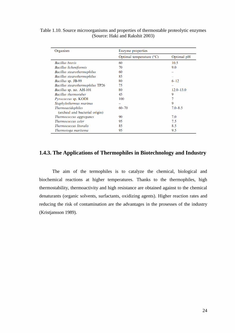
24
Table 1.10. Source microorganisms and properties of thermostable proteolytic enzymes
(Source: Haki and Rakshit 2003)
1.4.3. The Applications of Thermophiles in Biotechnology and Industry
The aim of the termophiles is to catalyze the chemical, biological and
biochemical reactions at higher temperatures. Thanks to the thermophiles, high
thermostability, thermoactivity and high resistance are obtained against to the chemical
denaturants (organic solvents, surfactants, oxidizing agents). Higher reaction rates and
reducing the risk of contamination are the advantages in the prosesses of the industry
(Kristjansson 1989).
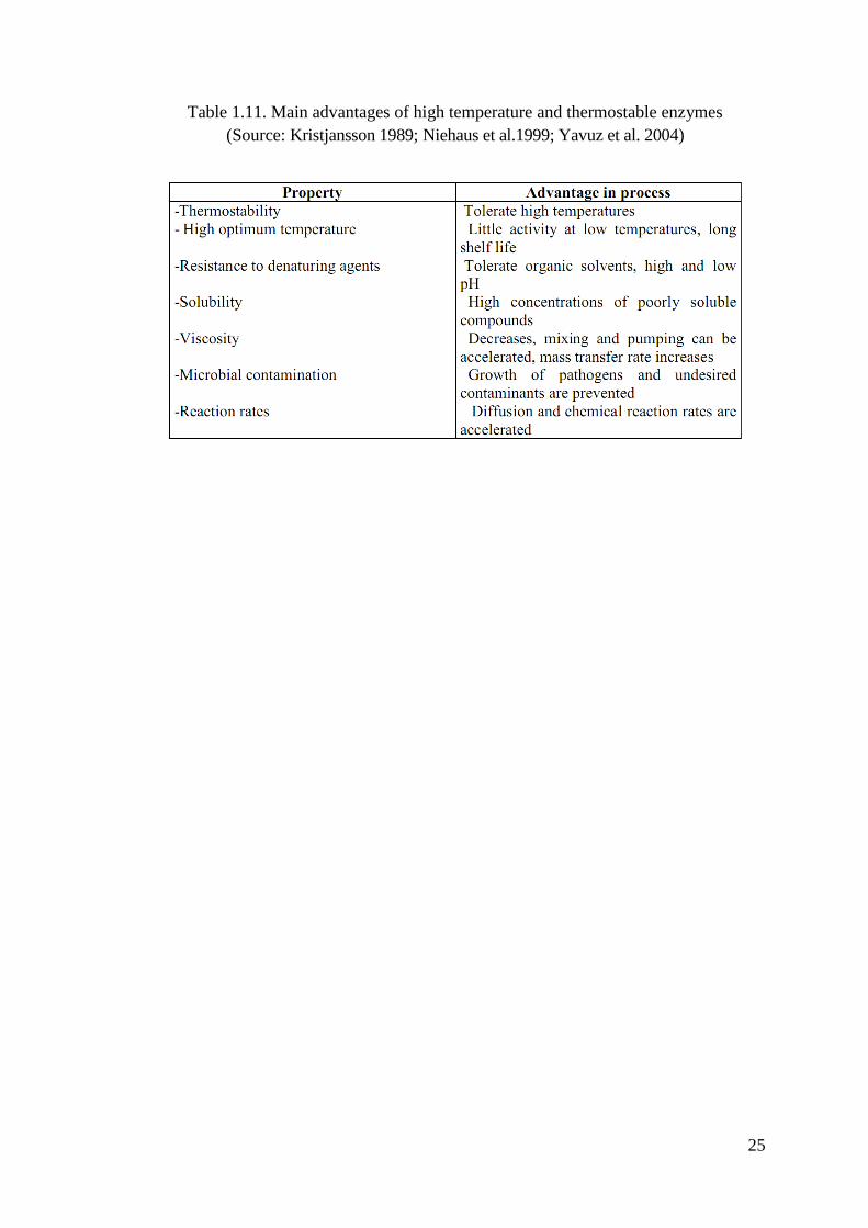
25
Table 1.11. Main advantages of high temperature and thermostable enzymes
(Source: Kristjansson 1989; Niehaus et al.1999; Yavuz et al. 2004)
26
CHAPTER 2
MATERIALS AND METHODS
2.1. Materials
Casein, Folin & Ciocalteu’s Phenol reagent, L-tyrosine and Coomassie Brilliant
Blue G-250 were purchased from Merck. TCA was purchased from Sigma Aldrich.
Cellulose Ion Exchanger, DEAE-Cellulose resin was purchased from Servacel and the
other chemicals for electrophoresis and characterization studies were purchased from
Sigma Chem. Co.
All of the analyses were carried out triplicate and mean values were reported.
The preparation of the buffers, reagents, solutions, the procedure of the assays and the
standart curves are given in Appendix A, Appendix B and Appendix C.
2.2. Bacterial Strain and Growth Conditions
In this study, the bacterial strain (alkaliphilic and thermophilic Geobacillus sp.)
was isolated from an uncontrolled thermal leak of Balçova Geothermal Region in Ġzmir
(Yavuz et al. 2004). In biochemistry research laboratory, the optimum growth
conditions of these thermophilic bacterial strains and their bacterial growth curves were
determined. The bacterial strain number-53 which has been stored at -86 °C for nearly 6
years was used in this study.
Two different media was used in order to compare the secretion of the proteins
in the bacterial strain.
Alkaliphilic and thermophilic Geobacillus was cultivated overnight in 250 ml
Erlenmayer flask containing 200 ml LB media, at 55 ºC and 200 rpm. Luria-Bertani
broth medium contains 10.0 g/L tryptone, 5.0 g/L yeast extract and 5.0 g/L NaCl per
liter with a final pH of 7.0. The medium was sterilised by autoclaving at 121°C for
15 minutes under high pressure.
27
Alkaliphilic and thermophilic Geobacillus was cultivated overnight in 250 ml
Erlenmayer flask containing 200 ml complex media, at 55 ºC and 200 rpm.
Complex medium contains 10.0 g/L glucose, 5.0 g/L yeast, 2.5 g/L peptone, 2.5 g/L
casein, 0.3 g/L MgSO4, 0.02 g/L FeSO4, 0.2 g/L ZnSO4, 1.0 g/L CaSO4, 1.0 g/L
KH2PO4, 1.0 g/L K2HPO4 per liter with a final pH of 7.0. The medium was
autoclaved for sterilization for 15 minutes at 121 °C under high pressure.
2.3. Screening of Protease Activity in Thermophilic Bacillus sp.
There are two methods to exhibite the protease activity in the bacterial strains.
Firstly, the media which consists of 8 g/L nutrient broth, 10 g/L skim milk and 15
g/L agar agar, was prepared. The skim milk was dissolved in deionized water by
putting a magnet into the bottle to prevent the precipitation and autoclaved at 110 °C
for 5 minutes (two times) and added to the medium. Nutrient broth and agar agar
were dissolved in deionized water by putting a magnet into the bottle to prevent the
precipitation and they sterilised by autoclaving at 121 °C for 15 minutes. When the
bottles’ temperatures became nearly 50-55 °C, the skim milk was poured into the
nutrient-agar agar. The mixture mixed without heating. The prapared medium was
poured into the plates quickly and the plates were put into the refrigerator at +4 °C
(Priest et al. 1989).
The bacterial culture number-53 stored at -86 °C was inoculated onto the plate
and incubated for 24 hours - 3 days at 55 ºC. The clear zones around colonies
indicated the protease activity.
Secondly, the bacterial culture was cultivated overnight in LB media or complex
media, at 55 ºC and 200 rpm. Bacterial culture was centrifuged at 5000 rpm, 4 ºC for
20 minutes. The activity of the obtain culture supernatant was determined by
recording the change in absorbance at 660 nm with the assay mixture at 55 ºC.
2.4. Determination of Enzyme Activity
Protease activity of the samples were determined by the protease activity method
using L-Tyrosine (0-1000 mg/L) as the standart. L-Tyrosine standarts were prepared in
different concentrations and the standard calibration curve was drawn with response to
28
their absorbance values. Total protein activity was calculated from standard calibration
curve equation. The spectrophotometric assays were performed using Shimadzu UV-
VIS spectrophotometer (UV-2450) with a constant temperature water circulator. While
determining the protease activity of the samples, the steps should be taken into
consideration. 100 µl 0.5% (w/v) casein in 50 mM Tris-HCl pH 7.2 is added to 100 µl
enzyme solution and the assay mixture was incubated for 10 minutes at 55 ºC in the water
bath. 100 µl 15% TCA in deionized water is added to enzyme-substrate solution to
terminate the reaction. The mixture was put in ice bath for 10 minutes and centrifuged
at 13500 rpm, room temperature for 10 minutes. The pellet was removed. 1 ml NaOH,
200 µl supernatant and 200 µl Folin & Ciocalteu’s Phenol reagent were put into the
cuvette, respectively. The reference cuvettes had the same composition except for the
enzyme. The changing of the activity was recorded spectrophotometrically in
absorbance at 660 nm after an hour. The enzyme activities were given as unit (U) and
one unit will hydrolyze casein to produce color equivalent to 1.0 µmole (181.0 µg) of
tyrosine per minute under the defined assay conditions.
Calculations: (Folin et al. 1929; Anson 1938)
(µmole Tyrosine equivalents released) (a)
Units/ml enzyme = (2.1)
(b) (c) (d)
a = Total volume (in milliliters) of assay
b= Time of assay (in minutes) as per the Unit Definition
c = Volume of enzyme (in milliliter) of enzyme used
d = Volume (in milliliters) used in Colorimetric Determination
units/ml enzyme
Units/mg solid = (2.2)
mg solid/ml enzyme
units/ml enzyme
Units/mg protein = (2.3)
mg protein/ml enzyme
29
2.5. Determination of Protein Concentration
Total protein concentration of a sample was determined by Bradford method by
using bovine serum albumin (BSA, 0.2 mg/ml) as the standart (Bradford 1976). BSA
standards were prepared in different concentrations and the standard calibration curve
was drawn with response to their absorbance values. Total protein content was
calculated from standard calibration curve equation.
In the Bradford method, the Coomassie Brilliant Blue G-250 dye binds to the
proteins which have arginine, lysine and histidine residues and changes the colour. The
changing of the absorbance was recorded spectrophotometrically at 595 nm in UV-
visible spectrophotometer.
The blank includes 800 µl water and 200 µl bradford reagent. The sample
included 2 µl protease enzyme, 798 µl water and 200 µl bradford reagent. The solutions
were kept at room temperature for 5 minutes followed by the addition of bradford
reagent. The changing of the protein concentration of the sample was recorded
spectrophotometrically in absorbance at 595 nm.
According to the results of the absorbance, the total protein concentration was
calculated from the standart curve and formula. The spectrophotometric assays were
performed using UV-VIS spectrophotometer with a constant temperature water
circulator.
2.6. Preparation of Crude Enzyme Extract
Bacterial growth was carried out in erlenmeyer flask in two ways. One of the
erlenmeyer flask contained 200 ml of LB media and the other one contained complex
media. Bacterial culture was centrifuged at 5000 rpm, 4 ºC for 20 minutes to obtain
culture supernatant. The culture supernatant is the crude extracellular enzyme extract.
The pellet including cell debris was removed.

30
2.7. Enzyme Purification Procedure
The crude extracellular enzyme extract was subjected to total protein
precipitation with 80% saturation (w/v) ammonium sulphate. The ammonium sulphate
(104.6 g) should be slowly added into the enzyme extract by stirring at 4 ºC. After
addition, the resulting precipitate was collected by centrifugation at 9500 rpm, 4 ºC for
15 minutes and the pellet was resuspended in 10 ml 50 mM Tris-HCl buffer, pH 7.2.
The enzyme solution was loaded on to the weak anion exchange (DEAE-
Cellulose) column (2.5 cm x 10 cm) which was equilibrated with 50 mM Tris HCl
buffer (pH 7.2) in loading step. The column was washed with 50 mM Tris HCl buffer
(pH 7.2) in washing step. Then, the fractions of 38 ml were collected by gradient eluting
with 50 mM Tris HCl buffer (pH 7.2) and 2 M NaCl+50 mM Tris HCl buffer (pH 7.2)
in the elution step. The chromatographic assays were performed using FOXY 2000 low
pressure liquid chromatography system. The collected fractions were assayed for their
protein concentrations at 280 nm with Thermo Scientific nanodrop, 595 nm with
Bradford method and protease activity at 660 nm with UV-VIS spectrophotometer. The
active fractions were pooled and stored at -20 ºC until using for the electrophoretic and
characterization experiments.
2.8. Electrophoretic Studies (SDS- PAGE)
In the electrophoretic study, sodium dodecyl sulfate polyacrylamide gel
electrophoresis (SDS-PAGE) was used in order to find out the approximate molecular
weight of protease enzyme following the Laemmli method (Laemmli, 1970). The
electrophoretic assays were performed using Biorad Mini-Protean Tetra System and
Biorad Protean II Xicell gel electrophoresis device.
SDS-PAGE is used to seperate the proteins according to their molecular sizes in
an electrical and estimate the molecular weight of a protein. Firstly, the seperating gel
(acrylamide concentrations of 12%) and stacking gel (acrylamide concentrations of 4%)
were prepared. The samples which were in all of the purification steps were diluted by a
volume ratio of 1:1 with sample buffer, kept in boiling water at 100 ºC for 10 minutes
to denature proteins. 4 μl of molecular weight marker and 20 μl of sample-sample buffer
mixture were loaded onto gel. The electrophoretic run was applied at 100 V for 2 hours.
31
After electrophoretic run, SDS-polyacrylamide gel was stained in two ways.
Coomassie Staining: SDS-polyacrylamide gel was fixed with 20% TCA and
incubated for 30 minutes. It was washed with ultrapure water for 3 times to remove
the TCA. Then, it was stained by using coomassie staining solution and incubated
for 30 minutes. Lastly, the gel was destained with the destaining solution and
incubated for 15 minutes for 3 times.
Silver Staining: SDS-polyacrylamide gel was fixed with fixation solution and
incubated for 1 hour. It was washed with 50% ethanol for 3 times and incubated for
20 minutes. Then, it was pre-treated with pre-treatment solution and incubated for 1
minute. In order to remove the materials from the gel, it was rinsed with ultrapure
water for 3 times and incubated for 20 seconds. In the impregnate step, the gel was
impregnated with silver nitrate solution and incubated for 20 minutes. In order to
remove the materials from the gel, it was rinsed with ultrapure water for 3 times and
incubated for 20 seconds. Later, it was developed with developing solution. When
the color development was observed, ultrapure water was added to stop the reaction.
Lastly, the gel was kept in the stop solution and incubated for 10 minutes
At the end of these steps, the image of the gels were taken with a special camera
under white light by using Biorad Versadoc Gel Visualizing System. The procedure for
preparation of gels and reagents that were used in SDS-PAGE are given in Appendix C.
2.9. Characterization Studies
2.9.1. Effect of pH on Enzyme Activity and Stability
The effect of pH on protease activity was investigated by using 50 mM Tris-HCl
buffer at different pH values at 55 ºC. The optimum pH of the protease enzyme was
determined under the standart assay conditions by measuring activity in the presence of
buffers at different pH values (pH 4.0, 5.0, 6.0, 7.0, 8.0, 9.0, 10.0).
While determining the pH stability of the enzyme, 100 μl enzyme solution was
mixed with 100 µl 0.5% (w/v) casein in 50 mM Tris-HCl pH 4-7-10. After incubation
time (1st, 2
nd, 3
rd, 4
th, 5
th and 6
th hour), the activity assay was applied. The changing of the
absorbance was recorded spectrophotometrically at 660 nm and the residual activity was
obtained.
32
2.9.2. Effect of Temperature on Enzyme Activity and Stability
The effect of temperature on protease activity was investigated by using 50 mM
Tris-HCl buffer (pH 7.2) at different temperatures. The optimum temperature of the
protease enzyme was determined under the standart assay conditions by measuring the
activity at different temperatures (25, 35, 45, 55, 65, 75 ºC). The protease enzyme and
the specific substrate casein were heated to relevant temperature before the assay and
during the assay.
While determining the temperature stability of the enzyme, 100 μl enzyme
solution was mixed with 100 µl 0.5% (w/v) casein in 50 mM Tris-HCl (pH 7.2) at 55-85
ºC. After incubation time (1st, 2
nd, 3
rd, 4
th, 5
th and 6
th hour), the activity assay was applied.
The changing of the absorbance was recorded spectrophotometrically at 660 nm and the
residual activity was obtained.
2.9.3. Substrate Specificity of Enzyme
The substrate specificity of the enzyme was determined by measuring activity
towards different substrates (casein, azocasein, hammers casein, skimmilk, BSA). The
activity of the enzyme was measured by using these different substrates prepared in 50
mM Tris-HCl buffer (pH 7.2) at concentrations of 0.5% (w/v) under the standart assay
conditions. The changing of the absorbance was recorded spectrophotometrically at 660
nm and the residual activity was obtained.
2.9.4. Effect of Metal Ions on Enzyme Activity
The effect of the metal ions on protease activity was measured in the presence of
different metal ions (CaCl2, CuSO4, MgSO4, NaCl and ZnCl2). 100 µl protease enzyme
solution and 100 µl 10 mM metal ion were incubated 55 ºC for 10 minutes. After the
incubation period, the substrate was added and the reaction was initiated. Then, the
activity of the enzyme was measured under the standart assay conditions. The changing
of the absorbance was recorded spectrophotometrically at 660 nm and the residual
activity was obtained.
33
2.9.5. Effect of Various Agents on Enzyme Activity
2.9.5.1. Effect of Organic Solvents on Enzyme Activity
The effect of some organic solvents (ethanol, methanol, hexane, benzene,
dimethyl sulfoxide (DMSO)) on enzyme activity were investigated.
100 µl protease enzyme solution and 100 µl 10% (v/v) of the agents were
incubated 55 ºC for 10 minutes. After the incubation period, the substrate was added
and the reaction was initiated. Then, the activity of the enzyme was measured under the
standart assay conditions. The changing of the absorbance was recorded
spectrophotometrically at 660 nm and the residual activity was obtained.
2.9.5.2. Effect of Surfactants on Enzyme Activity
The effect of some surfactants (sodium dodecyl sulfate (SDS), Triton X-100,
Tween-20, Tween-80, H2O2) on enzyme activity were investigated. SDS is an anionic
agent and H2O2 is an oxidizing agent. Triton X-100, Tween-20 and Tween-80 are
nonionic agents.
100 µl protease enzyme solution and 100 µl 1% (v/v) of the agents were
incubated 55 ºC for 10 minutes. After the incubation period, the substrate was added
and the reaction was initiated. Then, the activity of the enzyme was measured under the
standart assay conditions. The changing of the absorbance was recorded
spectrophotometrically at 660 nm and the residual activity was obtained.
2.9.6. Effect of Inhibitors on Enzyme Activity
The effect of some inhibitors (phenylmethylsulphonyl fluoride (PMSF),
dithiothreitol (DTT), ethylenediaminetetraacetic acid (EDTA), iodoacetamide, protease
inhibitor cocktail) on enzyme activity were investigated. PMSF is a serine protease
inhibitor and EDTA is a metalloprotease inhibitor. DTT and iodoacetamide are the
cysteine protease inhibitors. Protease inhibitor cocktail contains AEBSF (inhibition of
serine proteases), EDTA (inhibition of metalloproteases), bestatin (inhibition of
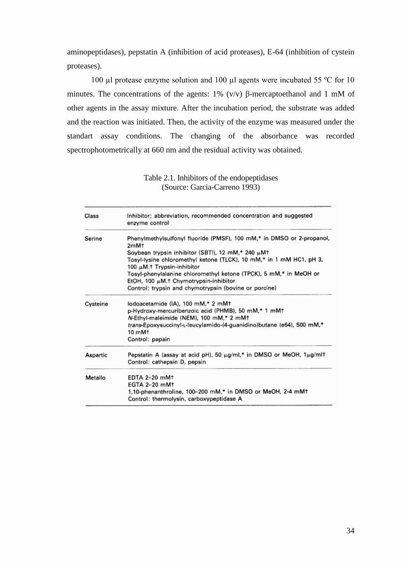
34
aminopeptidases), pepstatin A (inhibition of acid proteases), E-64 (inhibition of cystein
proteases).
100 µl protease enzyme solution and 100 µl agents were incubated 55 ºC for 10
minutes. The concentrations of the agents: 1% (v/v) β-mercaptoethanol and 1 mM of
other agents in the assay mixture. After the incubation period, the substrate was added
and the reaction was initiated. Then, the activity of the enzyme was measured under the
standart assay conditions. The changing of the absorbance was recorded
spectrophotometrically at 660 nm and the residual activity was obtained.
Table 2.1. Inhibitors of the endopeptidases
(Source: Garcia-Carreno 1993)
35
CHAPTER 3
RESULTS AND DISCUSSION
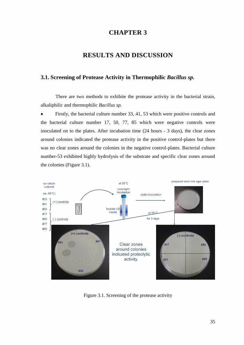
3.1. Screening of Protease Activity in Thermophilic Bacillus sp.
There are two methods to exhibite the protease activity in the bacterial strain,
alkaliphilic and thermophilic Bacillus sp.
Firstly, the bacterial culture number 33, 41, 53 which were positive controls and
the bacterial culture number 17, 50, 77, 85 which were negative controls were
inoculated on to the plates. After incubation time (24 hours - 3 days), the clear zones
around colonies indicated the protease activity in the positive control-plates but there
was no clear zones around the colonies in the negative control-plates. Bacterial culture
number-53 exhibited highly hydrolysis of the substrate and specific clear zones around
the colonies (Figure 3.1).
Figure 3.1. Screening of the protease activity
36
Secondly, the bacterial culture number 33, 41, 53 were cultivated overnight in
LB media. Following the bacterial growth, the activities of the crude enzymes were
determined by recording the change in absorbance at 660 nm with the assay mixture at
55 ºC. When compared the results, there was an increase in absorbance in the activity
test of bacterial culture number-53.
According to these results, the bacterial culture number-53 produced protease
enzyme more than the other bacterial cultures and it was used in the experiments. The
bacterial culter number was chosen and also the specific growth media should be chosen
to obtain high activity and high yield during the experiments. That’s why, a complex
medium including different ions and Luria-Bertani broth medium were prepared.
Following the bacterial growth, the activities of the crude enzymes were determined by
recording the change in absorbance at 660 nm with the assay mixture at 55 ºC. In
comparison to the LB media, there was an increase in absorbance by using the complex
medium. According to these results, the complex media was chosen as a specific growth
media.
3.2. Purification of Protease
In this study, the protease enzyme from alkaliphilic and thermophilic Bacillus
sp. was purified by using the ammonium sulphate precipitation (80%) and ion exchange
chromatography.
Firstly, it should be decided that the protease enzyme was an extracellular or
intracellular enzyme. Thus, the crude extracellular enzyme extract and the crude
intacellular enzyme extract were produced. Following the bacterial growth, the
activities of them were determined by recording the change in absorbance at 660 nm
with the assay mixture at 55 ºC. When compared with the results, the extracellular
enzyme showed high activity. These results suggested that the protease enzyme from
alkaliphilic and thermophilic Bacillus sp. was an extracellular enzyme. That’s why, the
crude extracellular enzyme was used as a starting material in the experiments.
Later, the crude extracellular enzyme was subjected to one of the precipitation
methods to precipitate, purify and concentrate total protein. Ammonium sulphate
precipitation with different saturations from 20% to 90%, ethanol precipitation and
aseton precipitation were used. Following precipitations, the activities of the partial
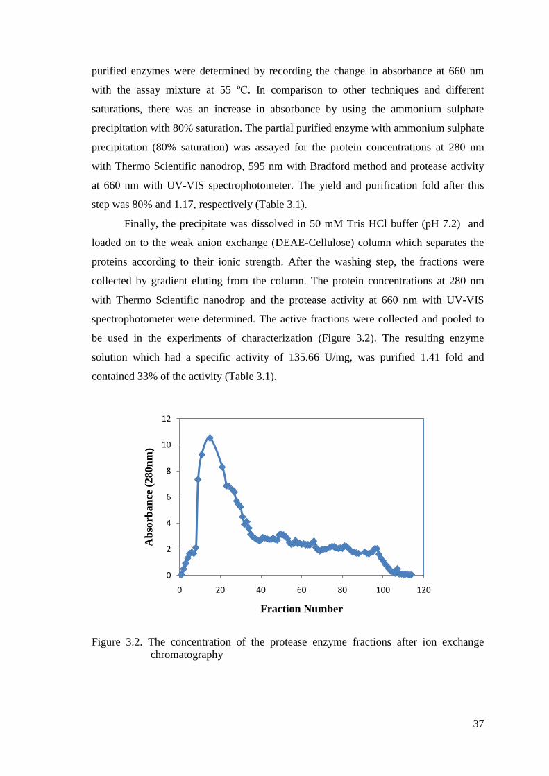
37
purified enzymes were determined by recording the change in absorbance at 660 nm
with the assay mixture at 55 ºC. In comparison to other techniques and different
saturations, there was an increase in absorbance by using the ammonium sulphate
precipitation with 80% saturation. The partial purified enzyme with ammonium sulphate
precipitation (80% saturation) was assayed for the protein concentrations at 280 nm
with Thermo Scientific nanodrop, 595 nm with Bradford method and protease activity
at 660 nm with UV-VIS spectrophotometer. The yield and purification fold after this
step was 80% and 1.17, respectively (Table 3.1).
Finally, the precipitate was dissolved in 50 mM Tris HCl buffer (pH 7.2) and
loaded on to the weak anion exchange (DEAE-Cellulose) column which separates the
proteins according to their ionic strength. After the washing step, the fractions were
collected by gradient eluting from the column. The protein concentrations at 280 nm
with Thermo Scientific nanodrop and the protease activity at 660 nm with UV-VIS
spectrophotometer were determined. The active fractions were collected and pooled to
be used in the experiments of characterization (Figure 3.2). The resulting enzyme
solution which had a specific activity of 135.66 U/mg, was purified 1.41 fold and
contained 33% of the activity (Table 3.1).
Figure 3.2. The concentration of the protease enzyme fractions after ion exchange
chromatography
0
2
4
6
8
10
12
0 20 40 60 80 100 120
Ab
sorb
an
ce (
280n
m)
Fraction Number
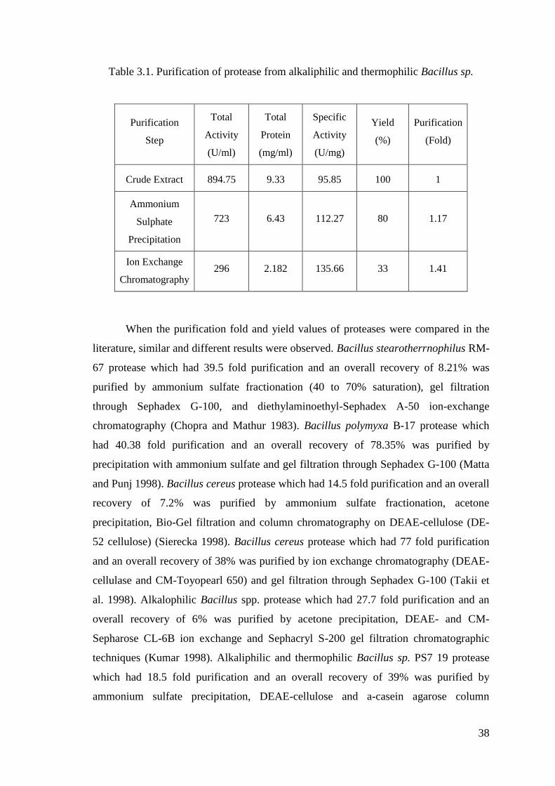
38
Table 3.1. Purification of protease from alkaliphilic and thermophilic Bacillus sp.
Purification
Step
Total
Activity
(U/ml)
Total
Protein
(mg/ml)
Specific
Activity
(U/mg)
Yield
(%)
Purification
(Fold)
Crude Extract 894.75 9.33 95.85 100 1
Ammonium
Sulphate
Precipitation
723 6.43 112.27 80 1.17
Ion Exchange
Chromatography 296 2.182 135.66 33 1.41
When the purification fold and yield values of proteases were compared in the
literature, similar and different results were observed. Bacillus stearotherrnophilus RM-
67 protease which had 39.5 fold purification and an overall recovery of 8.21% was
purified by ammonium sulfate fractionation (40 to 70% saturation), gel filtration
through Sephadex G-100, and diethylaminoethyl-Sephadex A-50 ion-exchange
chromatography (Chopra and Mathur 1983). Bacillus polymyxa B-17 protease which
had 40.38 fold purification and an overall recovery of 78.35% was purified by
precipitation with ammonium sulfate and gel filtration through Sephadex G-100 (Matta
and Punj 1998). Bacillus cereus protease which had 14.5 fold purification and an overall
recovery of 7.2% was purified by ammonium sulfate fractionation, acetone
precipitation, Bio-Gel filtration and column chromatography on DEAE-cellulose (DE-
52 cellulose) (Sierecka 1998). Bacillus cereus protease which had 77 fold purification
and an overall recovery of 38% was purified by ion exchange chromatography (DEAE-
cellulase and CM-Toyopearl 650) and gel filtration through Sephadex G-100 (Takii et
al. 1998). Alkalophilic Bacillus spp. protease which had 27.7 fold purification and an
overall recovery of 6% was purified by acetone precipitation, DEAE- and CM-
Sepharose CL-6B ion exchange and Sephacryl S-200 gel filtration chromatographic
techniques (Kumar 1998). Alkaliphilic and thermophilic Bacillus sp. PS7 19 protease
which had 18.5 fold purification and an overall recovery of 39% was purified by
ammonium sulfate precipitation, DEAE-cellulose and a-casein agarose column
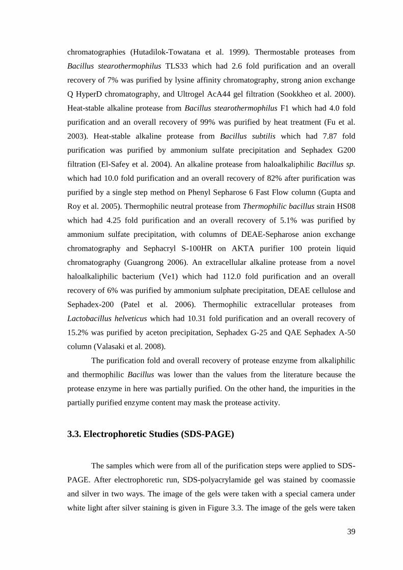
39
chromatographies (Hutadilok-Towatana et al. 1999). Thermostable proteases from
Bacillus stearothermophilus TLS33 which had 2.6 fold purification and an overall
recovery of 7% was purified by lysine affinity chromatography, strong anion exchange
Q HyperD chromatography, and Ultrogel AcA44 gel filtration (Sookkheo et al. 2000).
Heat-stable alkaline protease from Bacillus stearothermophilus F1 which had 4.0 fold
purification and an overall recovery of 99% was purified by heat treatment (Fu et al.
2003). Heat-stable alkaline protease from Bacillus subtilis which had 7.87 fold
purification was purified by ammonium sulfate precipitation and Sephadex G200
filtration (El-Safey et al. 2004). An alkaline protease from haloalkaliphilic Bacillus sp.
which had 10.0 fold purification and an overall recovery of 82% after purification was
purified by a single step method on Phenyl Sepharose 6 Fast Flow column (Gupta and
Roy et al. 2005). Thermophilic neutral protease from Thermophilic bacillus strain HS08
which had 4.25 fold purification and an overall recovery of 5.1% was purified by
ammonium sulfate precipitation, with columns of DEAE-Sepharose anion exchange
chromatography and Sephacryl S-100HR on AKTA purifier 100 protein liquid
chromatography (Guangrong 2006). An extracellular alkaline protease from a novel
haloalkaliphilic bacterium (Ve1) which had 112.0 fold purification and an overall
recovery of 6% was purified by ammonium sulphate precipitation, DEAE cellulose and
Sephadex-200 (Patel et al. 2006). Thermophilic extracellular proteases from
Lactobacillus helveticus which had 10.31 fold purification and an overall recovery of
15.2% was purified by aceton precipitation, Sephadex G-25 and QAE Sephadex A-50
column (Valasaki et al. 2008).
The purification fold and overall recovery of protease enzyme from alkaliphilic
and thermophilic Bacillus was lower than the values from the literature because the
protease enzyme in here was partially purified. On the other hand, the impurities in the
partially purified enzyme content may mask the protease activity.
3.3. Electrophoretic Studies (SDS-PAGE)
The samples which were from all of the purification steps were applied to SDS-
PAGE. After electrophoretic run, SDS-polyacrylamide gel was stained by coomassie
and silver in two ways. The image of the gels were taken with a special camera under
white light after silver staining is given in Figure 3.3. The image of the gels were taken
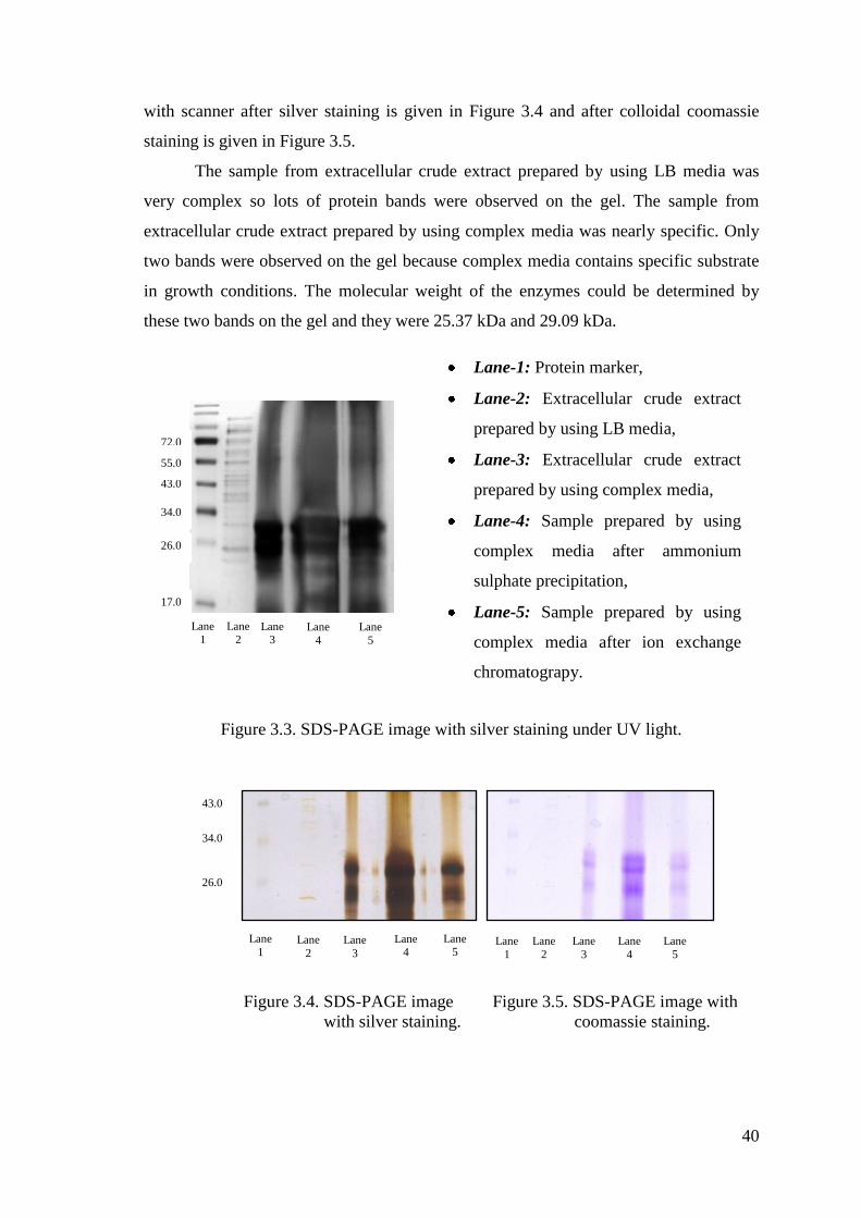
40
with scanner after silver staining is given in Figure 3.4 and after colloidal coomassie
staining is given in Figure 3.5.
The sample from extracellular crude extract prepared by using LB media was
very complex so lots of protein bands were observed on the gel. The sample from
extracellular crude extract prepared by using complex media was nearly specific. Only
two bands were observed on the gel because complex media contains specific substrate
in growth conditions. The molecular weight of the enzymes could be determined by
these two bands on the gel and they were 25.37 kDa and 29.09 kDa.
Figure 3.3. SDS-PAGE image with silver staining under UV light.
Figure 3.4. SDS-PAGE image Figure 3.5. SDS-PAGE image with
with silver staining. coomassie staining.
43.0
34.0
26.0
Lane
1 Lane
2 Lane
3
Lane 4
Lane 5
Lane
1
Lane
2
Lane
3
Lane
4
Lane
5
Lane 2
Lane 3
Lane
4
Lane
5
Lane 1
34.0
26.0
17.0
43.0
55.0
72.0
29.09
25.37
Lane-1: Protein marker,
Lane-2: Extracellular crude extract
prepared by using LB media,
Lane-3: Extracellular crude extract
prepared by using complex media,
Lane-4: Sample prepared by using
complex media after ammonium
sulphate precipitation,
Lane-5: Sample prepared by using
complex media after ion exchange
chromatograpy.
41
When the molecular weights of proteases were compared in the literature,
similar and different results were observed.
The molecular weight of protease from thermophilic Bacillus strain HS08 is 30.9
kDa (Guangrong et al. 2006), molecular weight of thermostable protease from
Geobacillus sp. YMTC 1049 is 59.2 kDa (Zhua et al. 2007), molecular weight of
thermostable protease from Hyperthermophilic Bacillus strain HUTBS71 is 49 kDa
(Akel et al. 2009), molecular weight of an alkaline protease from haloalkaliphilic
Bacillus sp. is 29 kDa (Gupta, Roy et al. 2005), molecular weight of an alkaline
protease from haloalkaliphilic Bacillus sp. is 30 kDa (Patel et al. 2005), molecular
weight of extracellular protease isolated from alkaliphilic and thermophilic Bacillus sp.
PS719 is 42 kDa (Hutadilok-Towatana et al. 1999),
The molecular weights of proteases from Bacillus stearothermophilus RM-67
are 67.610 and 19.950 kDa (Chopra and Mathur 1985), molecular weights of
thermostable proteases from Bacillus stearothermophilus TLS33 are 36 kDa, 53 kDa
and 71 kDa (Sookkheo et al. 2000).
The molecular weight of thermophilic alkaline proteases resistant to SDS and
EDTA from Bacillus sp. GUS1 is 30-47 kDa (Seifzadeh et al. 2008), molecular weight
of protease from Bacillus strain SAL1 is 27 kDa (Almas et al. 2009), molecular weight
of a surfactant-stable high-alkaline protease from Bacillus sp. B001 is 28 kDa (Deng et
al. 2010).
The molecular weight of an extracellular protease from Bacillus subtilis EAG-2
strain is 27 kDa (Ghafoor and Hasnain 2010), molecular weight of an extracellular
alkaline protease produced by Bacillus subtilis (MTTC N0-10110) is 20.5 kDa
(Ramakrishna et al. 2010).
The molecular weight of neutral protease from Bacillus cereus is 29 kDa
(Sierecka 1998), molecular weights of alkaline proteases produced by an alkalophilic
Bacillus are 28 kDa and 29 kDa (Kumar et al. 1999), molecular weights of proteases
from Bacillus cereus MCM B-326 are 36 kDa and 45 kDa (Nilegaonkar et al. 2006),
molecular weight of a solvent and detergent-stable protease from Bacillus cereus is 28
kDa (Doddapaneni et al. 2009), molecular weight of a calciumdependent protease by
Bacillus cereus BG1 is 34 kDa (Ghorbel-Frikha et al. 2005), molecular weight of a
solvent, detergent and oxidizing agent tolerant protease from Bacillus cereus is 38 kDa
(Shah et al. 2010).
42
The molecular weight of an alkaline protease from Bacillus circulans BM15 is
30 kDa (Venugopal and Saramma 2007), molecular weight of thermo- and detergent
stable serine protease from isolated Bacillus circulans is 39.5 kDa (Rao et al. 2009).
The molecular weight of a surfactant-stable alkaline serine-protease from
Bacillus mojavensis A21 is 20 kDa (Haddar et al.2009), molecular weight of an
extracellular, thiol-dependent and oxidation-stable alkaline serine protease from
Bacillus mojavensis is 30 kDa (Beg and Gupta 2003).
The molecular weight of thermostable extracellular protease from Bacillus
polymyxa B-17 is 30 kDa (Matta and Punj 1998), molecular weight of an extracellular
serine alkaline protease of Bacillus clausii GMBAE 42 is 26.5 kDa (Kazan et al. 2005),
molecular weight of protease from Bacillus Firmus Tap5 is 34 kDa (Joshi 2010),
molecular weight of an alkaline-protease from Bacillus licheniformis MP1 is 30 kDa
(Jellouli et al. 2011), molecular weights of proteases from Pseudomonas fluorescens are
between 47 kDa and 51 kDa (Margesin and Schinner 1992).
3.4. Characterization Studies
3.4.1. Effect of pH on Enzyme Activity and Stability
The effect of pH on protease activity was examined by measuring enzyme
activity in the presence of buffers at different pH values (pH 4.0, 5.0, 6.0, 7.0, 8.0, 9.0,
10.0). In the Figure 3.6, the effect of pH showed a bell shaped curve with the highest
activity at pH 8.0. On the other hand, the optimum pH of the protease enzyme isolated
from alkaliphilic and thermophilic Bacillus sp. was pH 8.0. The protease enzyme at
acidic pH values exhibited a little lower activity than the protease enzyme at alkaline
pH values. The enzyme lost 20% of its activity at pH 4.0 and it lost 10% of its activity
at pH 10.0. When the activities at different pH values were compared, it was understood
that this enzyme was nearly stable in these conditions because of having above the 80%
of its activity at all of the pH values.
When the pH values were compared in the literature, similar and different results
were observed in acidic, neutral or alkaline conditions.
The proteases which were secreted in the acidic conditions were isolated from
Geobacillus at pH 6.5 (Hawumba et al. 2002).
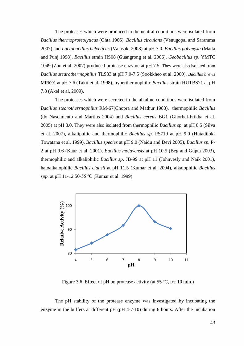
43
The proteases which were produced in the neutral conditions were isolated from
Bacillus thermoproteolyticus (Ohta 1966), Bacillus circulans (Venugopal and Saramma
2007) and Lactobacillus helveticus (Valasaki 2008) at pH 7.0. Bacillus polymyxa (Matta
and Punj 1998), Bacillus strain HS08 (Guangrong et al. 2006), Geobacillus sp. YMTC
1049 (Zhu et al. 2007) produced protease enzyme at pH 7.5. They were also isolated from
Bacillus stearothermophilus TLS33 at pH 7.0-7.5 (Sookkheo et al. 2000), Bacillus brevis
MIB001 at pH 7.6 (Takii et al. 1998), hyperthermophilic Bacillus strain HUTBS71 at pH
7.8 (Akel et al. 2009).
The proteases which were secreted in the alkaline conditions were isolated from
Bacillus stearotherrnophilus RM-67(Chopra and Mathur 1983), thermophilic Bacillus
(do Nascimento and Martins 2004) and Bacillus cereus BG1 (Ghorbel-Frikha et al.
2005) at pH 8.0. They were also isolated from thermophilic Bacillus sp. at pH 8.5 (Silva
et al. 2007), alkaliphilic and thermophilic Bacillus sp. PS719 at pH 9.0 (Hutadilok-
Towatana et al. 1999), Bacillus species at pH 9.0 (Naidu and Devi 2005), Bacillus sp. P-
2 at pH 9.6 (Kaur et al. 2001), Bacillus mojavensis at pH 10.5 (Beg and Gupta 2003),
thermophilic and alkaliphilic Bacillus sp. JB-99 at pH 11 (Johnvesly and Naik 2001),
haloalkalophilic Bacillus clausii at pH 11.5 (Kumar et al. 2004), alkalophilic Bacillus
spp. at pH 11-12 50-55 ºC (Kumar et al. 1999).
Figure 3.6. Effect of pH on protease activity (at 55 ºC, for 10 min.)
The pH stability of the protease enzyme was investigated by incubating the
enzyme in the buffers at different pH (pH 4-7-10) during 6 hours. After the incubation
80
90
100
4 5 6 7 8 9 10 11
Rel
ati
ve
Act
ivit
y (
%)
pH

44
time (1st, 2
nd, 3
rd, 4
th, 5
th and 6
th hour), the residual activities were measured
spectrophotometrically under standart assay conditions. They were calculated and
plotted against pH values as in Figure 3.7. While preparing the control, the pH treatment
was not applied to the enzyme and the activity of the protease enzyme was regarded as
hundred percent.
Three pH values (pH 4-7-10) were examined to compare the differences of the
stabilities. It was understood from the figure that,
The enzyme at pH 4 retained 85% of its activity after an hour and lost 50% of its
activity after six hours incubation time.
The enzyme at pH 7 retained more than 90% of its activity after an hour and lost
40% of its activity after six hours incubation time.
The enzyme at pH 10 retained more than 95% of its activity after an hour and lost
45% of its activity after six hours incubation time.
The results showed that the protease enzyme isolated from alkaliphilic and
thermophilic Bacillus sp. was stable at different pH values (pH 4-7-10) during 3 hours
but after 3 hours, there was a significant loss in the stability.
When the pH stabilities were compared in the literature, similar and different
results were observed. For examples; the purified protease I was stable in the pH range
of 8.8-10.5 and protease II was stable at pH 11.5 when incubated at 37 °C for 30, 60,
and 90 min (Chopra and Mathur 1983). In the acidic conditions, the activity of the
protease enzyme was stable in the range of 4.0-6.0. The protease showed high stability
at pH 9.0. When the pH values were lower or higher than 9.0, the enzyme activity
decreased nearly 70% at pH 7.0 and 50% at pH 11.0 (Hutadilok-Towatana et al. 1999).
The thermostable protease S showed nearly 60% of proteolytic activity in the pH range
of 6-10. Proteases N and B retained little activity above pH 9.0 (Sookkheo 2000). The
protease was stable at pH 9.6 and retained more than 80% of its activity in the pH range
of 7-10 (Kaur et al. 2001). The purified protease enzyme was stable at 70 °C for 24
hours in the pH range from 8.0 to 10.0 (Fu et al. 2003). There was a decrease of nearly
51% of the activity at pH 5.5, 18% of the activity at pH 8.0 and 66% of the activity at
pH 9.0 for 24 hours (do Nascimento and Martins 2004). A decrease of about 15% of the
activity at pH 8.5 and of the activity at pH 10.0 were observed (Silva et al. 2007).

45
Figure 3.7. pH stability of protease (at 55 ºC, for 6 hours)
3.4.2. Effect of Temperature on Enzyme Activity and Stability
The effect of temperature on protease activity was examined by measuring
enzyme activity at different temperatures ranging from 25 ºC to 75 ºC. In the Figure 3.8,
the effect of temperature showed a bell shaped curve with the highest activity at 55 ºC.
On the other hand, the optimum temperature of the protease enzyme isolated from
alkaliphilic and thermophilic Bacillus sp. was 55 ºC. The protease enzyme at
temperatures below 55 ºC lost 15% of its activity and also the protease enzyme at
temperatures above 55 ºC lost 25% of its activity. When the activities at different
temperatures were compared, it was understood that this enzyme was nearly stable in
these conditions because of having above the 75% of its activity at all of the
temperatures.
When the optimum temperature values were compared in the literature, similar
and different results were observed. The protease was highly produced by Bacillus
circulans at 40 ºC (Venugopal and Saramma 2007), Bacillus polymyxa at 50 ºC (Matta
and Punj 1998), alkalophilic Bacillus spp. at 50-55 ºC (Kumar et al. 1999), Bacillus
species 55 ºC (Naidu and Devi 2005), Bacillus mojavensis at 60 ºC (Beg and Gupta
2003), thermophilic Bacillus at 60 ºC (do Nascimento and Martins 2004), Bacillus
40
50
60
70
80
90
100
110
120
0 1 2 3 4 5 6 7
Res
idu
al A
ctiv
ity (
%)
Time (hour)
pH 4
pH 10
pH 7
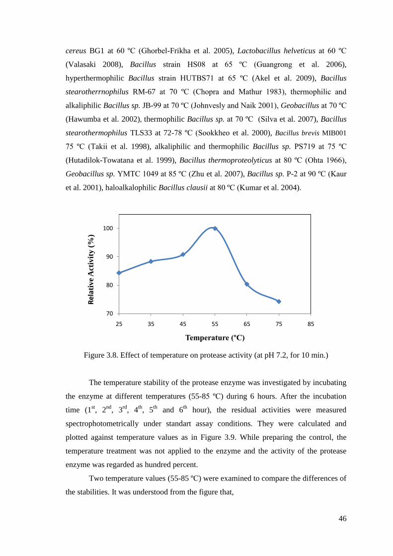
46
cereus BG1 at 60 ºC (Ghorbel-Frikha et al. 2005), Lactobacillus helveticus at 60 ºC
(Valasaki 2008), Bacillus strain HS08 at 65 ºC (Guangrong et al. 2006),
hyperthermophilic Bacillus strain HUTBS71 at 65 ºC (Akel et al. 2009), Bacillus
stearotherrnophilus RM-67 at 70 ºC (Chopra and Mathur 1983), thermophilic and
alkaliphilic Bacillus sp. JB-99 at 70 ºC (Johnvesly and Naik 2001), Geobacillus at 70 ºC
(Hawumba et al. 2002), thermophilic Bacillus sp. at 70 ºC (Silva et al. 2007), Bacillus
stearothermophilus TLS33 at 72-78 ºC (Sookkheo et al. 2000), Bacillus brevis MIB001
75 ºC (Takii et al. 1998), alkaliphilic and thermophilic Bacillus sp. PS719 at 75 ºC
(Hutadilok-Towatana et al. 1999), Bacillus thermoproteolyticus at 80 ºC (Ohta 1966),
Geobacillus sp. YMTC 1049 at 85 ºC (Zhu et al. 2007), Bacillus sp. P-2 at 90 ºC (Kaur
et al. 2001), haloalkalophilic Bacillus clausii at 80 ºC (Kumar et al. 2004).
Figure 3.8. Effect of temperature on protease activity (at pH 7.2, for 10 min.)
The temperature stability of the protease enzyme was investigated by incubating
the enzyme at different temperatures (55-85 ºC) during 6 hours. After the incubation
time (1st, 2
nd, 3
rd, 4
th, 5
th and 6
th hour), the residual activities were measured
spectrophotometrically under standart assay conditions. They were calculated and
plotted against temperature values as in Figure 3.9. While preparing the control, the
temperature treatment was not applied to the enzyme and the activity of the protease
enzyme was regarded as hundred percent.
Two temperature values (55-85 ºC) were examined to compare the differences of
the stabilities. It was understood from the figure that,
70
80
90
100
25 35 45 55 65 75 85
Relat
ive
Act
ivit
y (
%)
Temperature (ºC)
47
The enzyme at 55 ºC retained more than 95% of its activity after an hour and lost
about 3% of its activity after six hours incubation time.
The enzyme at 85 ºC retained more than 95% of its activity after an hour and lost
about 15% of its activity after six hours incubation time.
The results showed that the protease enzyme isolated from alkaliphilic and
thermophilic Bacillus sp. was stable at different temperature values (55-85 ºC) during 6
hours because there was not a significant loss in the stability.
When the temperature stabilities were compared in the literature, similar and
different results were observed. For examples; protease I and II from Bacillus
stearotherrnophilus RM-67 retained 100% activity at 60 °C for 30 min. The purified
protease enzymes were stable at 55 °C for 30 min. Protease I retained 63% of its activity
and protease II retained 20% of its activity after 30 min at 65 °C (Chopra and Mathur
1983). Thermostable neutral protease was stable up to 75 °C and it lost 60% of its
activity at 85 °C (Takii et al. 1998). The protease enzyme retained 35% activity at 70 °C
for 10 min. There was no activity at 85 °C after 10 min (Matta and Punj 1998). The
purified enzyme was stable at temperatures below 40 °C for 15 min. This protease
enzyme was retained 60% of its activity at higher temperatures and lost the activity at
90°C 15 min later (Hutadilok-Towatana et al. 1999). Highly thermostable proteases S
(at 60 °C), N (at 65 °C), and B (at 70 °C) retained more than 90% of the activity. They
lost 50% of the activity at 72, 78, and 90 °C, respectively. After the incubation at 100
°C for 30 min, the protease B, S and N had 31%, 5%, 10% of their initial activities,
respectively (Sookkheo 2000). The enzyme was stable at 90 °C for more than 1 hour. It
also retained 95% of its activity at 99 °C and 37% of its activity at 121 °C after 1 hour.
The half life of protease at 121 °C was 47 minutes (Kaur et al. 2001). The enzyme was
stable for 2 hours at 30 ºC. 14% of the activity was lost at 40 ºC and 84% of the activity
was lost at 80 ºC (do Nascimento and Martins 2004). The protease enzyme retained
80% of the activity at 60 ºC 2 hours later. 70% of the activity was retained at 70 ºC, 15
min later (Silva et al. 2007). The present enzyme was stable at 50 ºC and 60 ºC for 2
hours and it retained 84% of its activity (Akel et al. 2009).

48
Figure 3.9. Thermal stability of protease (at pH 7.2, for 6 hours)
3.4.3. Substrate Specificity of Enzyme
The substrate specificity of the protease enzyme isolated from alkaliphilic and
thermophilic Bacillus sp. was determined by measuring activity towards casein (5%),
azocasein (5%), hammersten casein (5%), skimmilk (5%), BSA (5%) under the assay
conditions. In the Table 3.2, the results showed that the casein was the best substrate
among them and the azocasein decrease the activity of the protease.
Table 3.2. Substrate specificity of protease (at 55 ºC, pH 7.2, for 10 min.)
83
88
93
98
103
1 2 3 4 5 6 7
Res
idu
al
act
ivit
y (
%)
Time (hour)
55 ºC
85 ºC
Substrate Wavelength (nm) Relative Activity (%)
Casein 660 nm 100
Skim milk 660 nm 98.57
Hammersten casein 660 nm 97.5
BSA 660 nm 87.14
Azocasein 440 nm 6.68
49
When the substrate specificities were compared in the literature, similar and
different results were observed. The substrates which were used in the experiments were
haemoglobin, albumin and casein (Sierecka 1998), gelatin (El-Safey and Abdul-Raouf
2004), starch (do Nascimento and Martins 2004), hammarsten casein (Kazan et al.
2005). In the previous studies, alkaline proteases exhibited highest activity towards
casein or modified proteins including haemoglobin, ovalbumin, BSA, fibrin, collagen,
elastin, keratin, azocasein and azocoll. The purified proteases hydrolyzed the protein
substrates such as casein, elastin, keratin, albumin and the synthetic chromogenic
peptide substrates Glu-Gly-Ala-Phe-pNA and Glu-Ala-Ala-Ala-pNA and the results
exhibited thar casein was the best substrate among them (Kumar et al. 1999). The
protease hydrolyzed the substrates, such as gelatin, elastin, albumin, haemoglobin, and
skim milk (Beg and Gupta 2003). Substrates specificity results showed that azocasein
was the best substrate among the azocasein, casein and BSA (Guangrong et al. 2006).
3.4.4. Effect of Metal Ions on Enzyme Activity
The effect of the metal ions on protease activity was measured in the presence of
different metal ions (Ca+2
, Cu+2
, Mg+2
, Na+1
and Zn+2
). The protease enzyme solution
and 10 mM metal ion were incubated for 10 minutes at 55 ºC. After the incubation
period, the substrate was added and the reaction was initiated. Then, the activity of the
enzyme was measured under the standart assay conditions. While preparing the control,
the metal ion was not added into the enzyme solution and its activity was regarded as
hundred percent. In the Figure 3.10, the results showed that Ca+2 and Cu
+2 increase the
activity of the protease, but Mg+2
, Na+1
and Zn+2
did not affect the activity of the
protease so much. According to these results, these metal ions protected the enzyme
against thermal denaturation. It was also known that Ca+2
is the most effective ion on
the activity of the proteases.
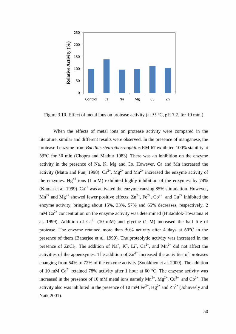
50
Figure 3.10. Effect of metal ions on protease activity (at 55 ºC, pH 7.2, for 10 min.)
When the effects of metal ions on protease activity were compared in the
literature, similar and different results were observed. In the presence of manganese, the
protease I enzyme from Bacillus stearotherrnophilus RM-67 exhibited 100% stability at
65°C for 30 min (Chopra and Mathur 1983). There was an inhibition on the enzyme
activity in the presence of Na, K, Mg and Co. However, Ca and Mn increased the
activity (Matta and Punj 1998). Ca2+
, Mg2+
and Mn2+
increased the enzyme activity of
the enzymes. Hg+2
ions (1 mM) exhibited highly inhibition of the enzymes, by 74%
(Kumar et al. 1999). Ca2+
was activated the enzyme causing 85% stimulation. However,
Mn2+
and Mg2+
showed fewer positive effects. Zn2+
, Fe2+
, Co2+
and Cu2+
inhibited the
enzyme activity, bringing about 15%, 33%, 57% and 65% decreases, respectively. 2
mM Ca2+
concentration on the enzyme activity was determined (Hutadilok-Towatana et
al. 1999). Addition of Ca2+
(10 mM) and glycine (1 M) increased the half life of
protease. The enzyme retained more than 50% activity after 4 days at 60°C in the
presence of them (Banerjee et al. 1999). The proteolytic activity was increased in the
presence of ZnCl2. The addition of Na+, K
+, Li
+, Ca
2+, and Mn
2+ did not affect the
activities of the apoenzymes. The addition of Zn2+
increased the activities of proteases
changing from 54% to 72% of the enzyme activity (Sookkheo et al. 2000). The addition
of 10 mM Ca2+
retained 78% activity after 1 hour at 80 °C. The enzyme activity was
increased in the presence of 10 mM metal ions namely Mn2+
, Mg2+
, Cu2+
and Co2+
. The
activity also was inhibited in the presence of 10 mM Fe3+
, Hg2+
and Zn2+
(Johnvesly and
Naik 2001).
0
50
100
150
200
250
Control Ca Na Mg Cu Zn
Rel
ati
ve
Act
ivit
y (
%)
51
1mM K+, 1mM Hg
2+ and 1mM Cu
2+ were strongly inhibited the enzyme activity. Mn
2+
and Ca2+
were stimulated the activity and they also had a functional role in the
molecular structure of the enzyme (do Nascimento and Martins 2004). There was no
effect in the presence of Mn2+
, Zn2+
and Mg2+
. The enzyme was activated with Ca2+
(1
mM) and Cu2+
(5 mM) (Gupta et al. 2005). The enzyme activity was increased by
Ca2+
and Mg2+
but inhibited by Ba2+
, Zn2+
, Pb2+
, Co2+
, Mn2+
and Cu2+
(Zhu et al. 2007).
The activity of the enzyme was increased in the presence of 5 mM Mn2+
, Ca2+
and Na2+
;
not affected or slightly inhibited by Fe2+
, Co2+
, and Zn2+
, but inhibited remarkably by
EDTA, Cu2+
and Cd2+
(Akel et al. 2009).
3.4.5. Effect of Various Agents on Enzyme Activity
The effect of the organic solvents, surfactans and detergents on the enzyme
activity were investigated to determine whether they acted as inhibitor or activator. The
protease enzyme solution and 10% of the organic solvent or 1% surfactant were
incubated for 10 minutes at 55 ºC. After the incubation period, the substrate was added
and the reaction was initiated. Then, the activity of the enzyme was measured under the
standart assay conditions. While preparing the control, the organic solvents or the
surfactants were not added into the enzyme solution and its activity was regarded as
hundred percent.
3.4.5.1. Effect of Organic Solvents on Enzyme Activity
Benzene did not cause much effect on the activity and the protease enzyme
showed nearly 96% of its activity in the presence of benzene. On the other hand, ethanol
caused much effect on the activity among the other organic solvents and the protease
enzyme showed nearly 84% of its activity in the presence of ethanol. In the Table 3.3,
the results indicated that alkaliphilic and thermophilic protease enzyme was stable in the
presence of 10% of these organic solvents because there was a little loss on the protease
activiy in the experiments.
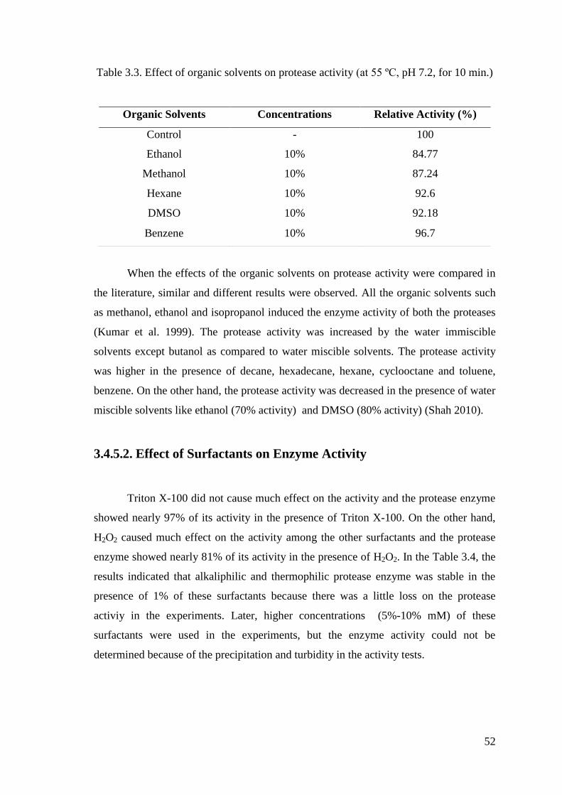
52
Table 3.3. Effect of organic solvents on protease activity (at 55 ºC, pH 7.2, for 10 min.)
When the effects of the organic solvents on protease activity were compared in
the literature, similar and different results were observed. All the organic solvents such
as methanol, ethanol and isopropanol induced the enzyme activity of both the proteases
(Kumar et al. 1999). The protease activity was increased by the water immiscible
solvents except butanol as compared to water miscible solvents. The protease activity
was higher in the presence of decane, hexadecane, hexane, cyclooctane and toluene,
benzene. On the other hand, the protease activity was decreased in the presence of water
miscible solvents like ethanol (70% activity) and DMSO (80% activity) (Shah 2010).
3.4.5.2. Effect of Surfactants on Enzyme Activity
Triton X-100 did not cause much effect on the activity and the protease enzyme
showed nearly 97% of its activity in the presence of Triton X-100. On the other hand,
H2O2 caused much effect on the activity among the other surfactants and the protease
enzyme showed nearly 81% of its activity in the presence of H2O2. In the Table 3.4, the
results indicated that alkaliphilic and thermophilic protease enzyme was stable in the
presence of 1% of these surfactants because there was a little loss on the protease
activiy in the experiments. Later, higher concentrations (5%-10% mM) of these
surfactants were used in the experiments, but the enzyme activity could not be
determined because of the precipitation and turbidity in the activity tests.
Organic Solvents Concentrations Relative Activity (%)
Control - 100
Ethanol
Methanol
10%
10%
84.77
87.24
Hexane 10% 92.6
DMSO 10% 92.18
Benzene 10% 96.7
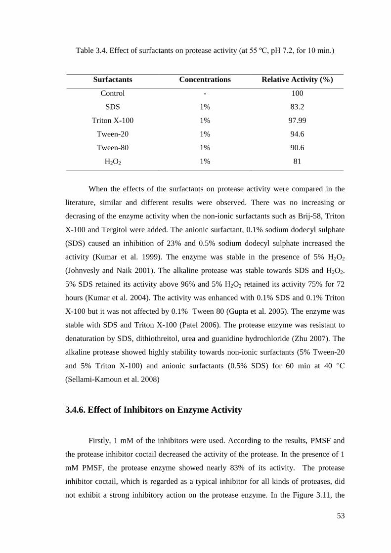
53
Table 3.4. Effect of surfactants on protease activity (at 55 ºC, pH 7.2, for 10 min.)
When the effects of the surfactants on protease activity were compared in the
literature, similar and different results were observed. There was no increasing or
decrasing of the enzyme activity when the non-ionic surfactants such as Brij-58, Triton
X-100 and Tergitol were added. The anionic surfactant, 0.1% sodium dodecyl sulphate
(SDS) caused an inhibition of 23% and 0.5% sodium dodecyl sulphate increased the
activity (Kumar et al. 1999). The enzyme was stable in the presence of 5% H2O2
(Johnvesly and Naik 2001). The alkaline protease was stable towards SDS and H2O2.
5% SDS retained its activity above 96% and 5% H2O2 retained its activity 75% for 72
hours (Kumar et al. 2004). The activity was enhanced with 0.1% SDS and 0.1% Triton
X-100 but it was not affected by 0.1% Tween 80 (Gupta et al. 2005). The enzyme was
stable with SDS and Triton X-100 (Patel 2006). The protease enzyme was resistant to
denaturation by SDS, dithiothreitol, urea and guanidine hydrochloride (Zhu 2007). The
alkaline protease showed highly stability towards non-ionic surfactants (5% Tween-20
and 5% Triton X-100) and anionic surfactants (0.5% SDS) for 60 min at 40 °C
(Sellami-Kamoun et al. 2008)
3.4.6. Effect of Inhibitors on Enzyme Activity
Firstly, 1 mM of the inhibitors were used. According to the results, PMSF and
the protease inhibitor coctail decreased the activity of the protease. In the presence of 1
mM PMSF, the protease enzyme showed nearly 83% of its activity. The protease
inhibitor coctail, which is regarded as a typical inhibitor for all kinds of proteases, did
not exhibit a strong inhibitory action on the protease enzyme. In the Figure 3.11, the
Surfactants Concentrations Relative Activity (%)
Control - 100
SDS
Triton X-100
1%
1%
83.2
97.99
Tween-20 1% 94.6
Tween-80 1% 90.6
H2O2 1% 81

54
results indicated that alkaliphilic and thermophilic protease enzyme was stable in the
presence of 1 mM of these inhibitors because there was a little loss on the protease
activiy in the experiments. Thus, higher concentrations of this agent may be required for
the inhibiton of the enzyme. Later, higher concentraitons (5-10 mM) of these agents
were used in the experiments, but the strongest inhibitor could not be observed because
of the precipitation and turbidity in the activity tests. Thus, the group of enzyme could
not be classified.
Figure 3.11. Effect of inhibitors on protease activity (at 55 ºC, pH 7.2, for 10 min.)
When the effects of the inhibitors on protease activity were compared in the
literature, different results were observed. The B. stearotbermopbilus RM-67 protease I
was inhibited by metal chelating agents 8-hydroxyquinoline-5-sulfonic acid (100%),
ethylenediaminetetraacetate (EDTA) (45%), and 1,10- phenanthroline (29%) but not by
dinitrofluorophosphate (DFP) the inhibitor of alkaline proteases. Thus, it may be
classified as a neutral protease. The B. stearotbermopbilus RM-67 protease II was
inhibited by DFP Thus, the protease should be classified as a serine protease (Chopra
and Mathur 1983). The protease enzyme was inhibited strongly by metal chelating agent
EDTA (Matta and Punj 1998). 1 mM PMSF inhibited the proteases completely (Kumar
et al. 1999). The metal chelators EDTA and 1,10-phenanthroline exhibited strong
inhibition towards the enzyme activity. The protease enzymes were 90% inhibited in the
presence of 10 mM EDTA and 10 mM 1,10-phenanthroline. Thus, they can thus be
classified as metalloproteases (Sookkheo 2000). The proteolytic activity was inhibited
0
50
100
150
Rel
ati
ve
Act
ivit
y (
%)
55
by 1 mM PMSF and TPCK. There was no inhibition in the presence of EDTA and 1,10-
phenanthroline (Johnvesly and Naik 2001). The proteolytic activity was inhibited by
PMSF and it was suggested that the enzyme was serine type protease (Gupta et al.
2005). The protease enzymes were inhibited by phenylmethylsulfonyl fluoride (PMSF).
In the presence of 2-mercaptoethanol and iodoacetate, 80-90% enzyme activities were
retained (Seifzadeh et al. 2008).
56
CHAPTER 4
CONCLUSION
The microorganisms that were previously isolated and characterized as a
Bacillus sp. from Balçova Geotermal region in Ġzmir by Elif Yavuz were used in the
experiments. The aim of this study was to produce the protease enzyme from
alkaliphilic and thermophilic Bacillus sp. by purifing and determine the properties of the
enzyme with the characterization.
Firstly, the bacterial culture number-53 was decided to be used in the
experiments. When the stock cultures were inoculated to the plate including media used
for protease screening, the clear zones around the colonies were observed. This result
indicated the proteolytic activity. After screening the protease activity, complex
medium to grow the bacteria in a specific way was decided to be used. In order to
maintain the purification steps, the enzyme was examined whether it was extracellular
or intracellular enzyme. When compared with the results, it was observed that the
extracellular enzyme showed higher activity than the intracellular enzyme. This
extracellular protease enzyme was purified by ammonium sulphate precipitation (80%
saturation) and ion exchange chromatograpy by using a weak anion exchange (DEAE-
Cellulose) column. The yield and purification fold after purification of the enzyme were
33% and 1.41, respectively.
Later, the characterization studies were examined. The results indicated that the
protease enzyme isolated from alkaliphilic and thermophilic Bacillus sp. had highest
activity at pH 8.0 and 55 ºC. The protease enzyme at acidic pH values exhibited a little
lower activity than the protease enzyme at alkaline pH values. The enzyme lost 20% of
its activity at pH 4.0 and it lost 10% of its activity at pH 10.0. It was understood that
this enzyme is nearly stable in these conditions because of having above the 80% of its
activity at all of the pH values. The results showed that the protease enzyme isolated
from alkaliphilic and thermophilic Bacillus sp. was stable at different pH values (pH 4-
7-10) during 3 hours but after 3 hours, there was a significant loss in the stability. The
protease enzyme at temperatures below 55 ºC lost 15% of its activity and also the
protease enzyme at temperatures above 55 ºC lost 25% of its activity. It was understood
57
that the protease enzyme is also nearly stable at different temperatures because of
having above the 75% of its activity at all of the temperatures. The results showed that
the protease enzyme isolated from alkaliphilic and thermophilic Bacillus sp. was stable
at different pH values (55-85 ºC) during 6 hours because there was not a significant loss
in the stability. When compared the substrates, casein showed higher activity and
azocasein showed lower activity in the experiments. In the effect of metal ions, the
results shows that Ca+2 and Cu
+2 increase the activity of the protease. When the effect of
organic solvents and surfactants on protease activity was investigated, the results
indicated that alkaliphilic and thermophilic protease enzyme was stable in the presence
of 10% of these organic solvents and 1% of these surfactants because there was a little
loss on the protease activiy in the experiments. In order to determine the type of the
protease, the effects of inhibitors on enzyme activity were examined. PMSF and the
protease inhibitor coctail decrease the activity of the protease. In the presence of 1 mM
PMSF, the protease enzyme showed nearly 83% of its activity. The protease inhibitor
coctail, which is regarded as a typical inhibitor for all kinds of proteases, did not exhibit
a strong inhibitory action on protease. The results indicated that alkaliphilic and
thermophilic protease enzyme was stable in the presence of 1 mM of these inhibitors
because there was a little loss on the protease activiy in the experiments. Thus, higher
concentrations of this agent may be required for the inhibiton of the enzyme.
In conclusion, the protease enzyme isolated from alkaliphilic and thermophilic
Bacillus sp. was purified and characterized during the experiments. In the further
studies, it can be immobilized on to organic or inorganic supports to be used in
industrial processes and it is also produced by using recombinant DNA technology to
improve its application in biotechnology. On the other hand, X-ray crystallographic
analyses of the proteases can be defined the features of the catalytic and structural
metal-binding sites.
58
REFERENCES
Akel H., Al-Quadan F., Yousef T.H. 2009. Characterization of a Purified Thermostable
Protease from Hyperthermophilic Bacillus Strain HUTBS71. European Journal of
Scientific Researc. 31(2): 280-288.
Almas S., Hameed A., Shelly D., Mohan P. 2009. Purification and characterization of a
novel protease from Bacillus strain SAL1. African Journal of Biotechnology
8(15): 3603-3609.
Anson M.L. 1938. J. Gen. Physiol. 22: 79-89.
Amara A.A., Salem S.R. and Shabeb M.S.A. 2009. The Possibility to Use Bacterial
Protease and Lipase as Biodetergent. Global Journal of Biotechnology &
Biochemistry 4(2): 104-114.
Ammar M.S., El-Louboudy S.S. and Abdulraouf U.M. 1991. Protease(s) from Bacillus
anthracis S-44 and B. cereus var. mycoids, S-98 isolated from a temple and
slaughter house in Aswan city. Az. J. Microbiol. (13): 12-29.
Anwar A. and Saleemuddin M. 1998. Alkaline proteases: A review. Bioresource
Technology 64(3): 175-183.
Ash C., Priest, F.G. & Collins M.D. 1993. Molecular identification of rRNA group 3
bacilli (Ash, Farrow, Wallbanks and Collins) using a PCR probe test. Proposal for
the creation of a new genus Paenibacillus. Antonie van Leeuwenhoek 64: 253-
260.
Asokan, Jayanthı S., C. 2010. Alkalıne protease production by Bacillus licheniformis
and Bacillus coagulans. Journal of Cell and Tissue Research 10(1): 2119-2123.
Baker J.M., Griffiths M.W. 1993. Predictive modelling of psychrotrophic Bacillus
cereus. J. Food Protect. 56: 684–688.
Baker G., Gaffar S., Cowan D.A., Suharto A.R. 2001. Bacterial Community Analysis
of Indonesian Hotsprings. FEMS Microbiology Letters 200: 103-109.
Banerjee U.C., Sani R.K., Azmi W., Soni R. 1999. Thermostable alkaline protease
from Bacillus brevis and its characterization as a laundry detergent additive.
Process Biochemistry 35(1-2): 213-219.
Banik R.M. and Prakash M. 2004. Laundery detergent compatibility of the alkaline
protease from Bacillus cereus. Microbiol. Res. 159(2): 135-140.
Barett A.J. 1994. Proteolytic enzymes: serine and cysteine peptidases. Methods
Enzymol. 244: 1–15.
59
Beg Q.K., Gupta R. 2003. Purification and characterization of an oxidation-stable, thiol-
dependent serine alkaline protease from Bacillus mojavensis. Enzyme and
Microbial Technology 32: 294–304.
Beynon J. and Bond. J.S. 1989. Proteolytic enzymes, a practical approach. IRL Press,
New York, NY.
Bhosale S.H., Rao M.B., Deshpande V.V., Srinivasan M.C. 1995. Thermostability of
high activity alkaline protease from Conidiobolus coronatus (NCL 86.8.20).
Enzyme Microbiol Technol. 17: 136-139.
Bouzas T.D., Barros-Velazquez J., Villa T.G. 2006. Industrial applications of
hyperthermophilic enzymes: A review. Protein Peptide Lett. 13(7): 645-651.
Bradford M.M. 1976. A rapid and sensitive method for the quantitation of microgram
quantities of protein utilizing the principle of protein-dye binding. Anal Biochem.
72: 248–251.
Brock T.D. 1986. Introduction, an overview of the thermophiles. In Thermophiles:
General, Molecular and Applied Microbiology Edited by: Brock TD. New York:
John Wiley & Sons 1-16.
Bryce W.W. 1966. Manufacturing uses of enzymes. Food Manual 41(1): 18-20.
Campbell, L.L. 1955. Arch. Biochem. Biophys. 64: 154.
Casella M. and Whitaker J. 1990. Enzymatically and chemically modified zein for
improvement of functional properties. J. Fd Biochem. 14: 453-475.
Chopra A. K. and Mathur D. K. 1985. Purification and Characterization of Heat-Stable
Proteases from Bacillus stearothermophilus RM-67. J Dairy Sci. 68: 3202-3211
Cowan D., Danial R., and Morgan H. 1985. Thermophilic protease: Properties and
potential applications. Trends Biotechnol. 3: 68–72.
da Silva C. R., Delatorre A. B., Martins M. L. L. 2007. Effect of the culture conditions
on the production of an extracellular protease by thermophilic Bacillus sp. and
some properties of the enzymatic activity. Brazilian Journal of Microbiology 38:
253-258.
Deng A., Wu J., Zhang Y., Zhang G., Wen T. 2010. Purification and characterization of
a surfactant-stable high-alkaline protease from Bacillus sp. B001. Bioresource
Technology 101: 7100–7106.
do Nascimento, W.C.A. and M.L.L. Martins. 2004. Production and properties of an
extracellular protease from thermophilic Bacillus sp. Brazilian Journal of
Microbiology 35(1-2): 91-96.
Doddapaneni K. K., Tatineni R., Vellanki R. N., Rachcha S., Anabrolu N., Narakuti V.,
Mangamoori L. N. 2007. Purification and characterization of a solvent and
60
detergent-stable novel protease from Bacillus cereus. Microbiological Research
164: 383-390.
Dufresne S., Bousquet J., Boissinot M. & Guay R. 1996. Sulfobacillus disulfidooxidans
sp. nov., a new acidophilic, disulfide-oxidizing, gram-positive, spore-forming
bacterium. Int. J. Syst. Bacteriol. 46: 1056-1064.
El-Safey E. M., Abdul-Raouf U. M. 2004. Production, purification and characterization
of protease enzyme from Bacillus subtilis. Botany and Microbiology Department,
Faculty of Science, Al-Azhar University.
Folin O., and Ciocalteu V. 1929. J. Biol. Chem. 73: 627.
Fogarty W.M., Grilffin P. H. & Joyce A.M. 1974. Enzymes of Bacillus species- Part 2.
Process Biochemistry 9(7): 27-35.
Fu Z, Hamid S. B. A., Razak C. N. A., Basri M., Salleh A. B. and Rahmanb R. N. Z. A.
2003. Secretory expression in Escherichia coli and single-step purification of a
heat-stable alkaline protease. Protein Expression and Purification 28: 63–68.
Fujishige A., Smith K.R., Silen J.L., Agard D.A. 1992. Correct folding of a-lytic
protease is required for its extracellular secretion from Escherichia coli. J Cell
Biol. 118: 33-42.
Garcica-Carreno F. L. 1991. Proteases in food technology. Biotechnology Education
2(4): 150-153.
Garcica-Carreno Fernando Luis. 1992. Protease inhibition in theory an practice.
Biotechnology Education 3(4): 145-150.
Genencor. 2009. Proteases in the Food Industry. USP Enzyme Workshop Rockville, MD.
Gerze A.; Omay D. and Guvenilir Y. 2005. Partial purification and characterization of
protease enzyme from Bacillus subtilis and Bacillus megatherium. Appl Biochem
Biotechnol. 121(124): 335-45.
Ghafoor A., Hasnain S. 2010. Purification and characterization of an extracellular
protease from Bacillus subtilis EAG-2 strain isolated from ornamental plant
nursery. Polish Journal of Microbiology 59(2): 107-112.
Ghorbel-Frikha B., Sellami-Kamoun A., Fakhfakh N., Haddar A., Manni L., Nasri M.
2005. Production and purification of a calcium-dependent protease from Bacillus
cereus BG1. J Ind Microbiol Biotechnol. 32: 186–194.
Godfrey T., and West S. 1996. Industrial enzymology, 2nd ed., p. 3. Macmillan
Publishers Inc., New York, N.Y.
Grimm O. 1958. Advances in the bating field. Leder 9: 308-311.
61
Guangrong H., Tiejing Y. 2006. Purification and characterization of a protease from
Thermophilic Bacillus strain HS08. African Journal of Biotechnology 5(24):
2433-2438.
Gupta A., Roy I., Patel R.K., Singh S.P., Khare S.K., Gupta M.N. 2005. One-step
purification and characterization of an alkaline protease from haloalkaliphilic
Bacillus sp. Journal of Chromatography A. 1075: 103–108.
Gupta R., Beg Q. K. 2002. Bacterial alkaline proteases: molecular approaches and
industrial applications. Applied Microbiology and Biotechnology 59(1): 15-32.
Gupta R., Beg Q. K., Khan S., Chauhan B. 2002. An overview on fermentation,
downstream processing and properties of microbial alkaline proteases. Appl
Microbiol Biotechnol. 60: 381–395.
Gusek T., and Kinsella E. 1988. Properties and potential applications of a unique heat-
stable protease. Food Technol. 49: 102–107.
Haddar A., Bougatef A., Agrebi R., Sellami-Kamoun A., Nasri M. 2009. A novel
surfactant-stable alkaline serine-protease from a newly isolated Bacillus
mojavensis A21. Purification and characterization. Process Biochemistry 44: 29–
35.
Haki G.D.; Rakshit S.K. 2003. Developments in industrially important thermostable
enzymes: a review. Bioresource Technol. 89(1): 17-34.
Harwood C.R. 1989. Introduction to the biotechnology of Bacillus, in: Biotechnology
handbooks 2: Bacillus (Harwood, C.R. ed.) Plenum Press, New York, pp 1-4.
Hase C.C. and Finkelstein R.A. 1993. Bacterial extracellular zinc-containing
metalloproteases. Microbiological Reviews 57(4): 823-837.
Hawumba J.F., Theron J., Brözel V. S. 2002. Thermophilic Protease-Producing
Geobacillus from Buranga Hot Springs in Western Uganda. Current Microbiology
45: 144–150.
Heyndrickx M., Lebbe L., Vancanneyt M. & 7 other authors 1997. A polyphasic
reassessment of the genus Aneurinibacillus, reclassification of Bacillus
thermoaerophilus as Aneurinibacillus thermoaerophilus comb. nov., and emended
descriptions of A. aneurinilyticus corrig., A. migulans, and A. thermoaerophilus.
Int. J. Syst. Bacteriol. 47: 808-817.
Horlkoshi K. 1971. Production of alkaline enzymes by alkalophilic microorganisms. I.
Alkaline protease produced by Bacillus no. 221. Agric. Biol. Chem. 35: 1407-
1414.
Huges R.A.P and Williams R.A.D. 1977. Yelow-pigmented Strains of Thermus ssp.
from Icelandic Hot Springs. Journal of General Microbiology 102: 375-383.
62
Hutadilok-Towatana N., Painupong A. and Suntinanalert P. 1999. Purification and
Characterization of an Extracellular Protease from Alkaliphilic and Thermophilic
Bacillus sp. PS719. Journal Of Bioscience And Bioengineering 87(5): 581-587.
International Union of Biochemistry. 1992. Enzyme nomenclature. Academic Press,
Inc., Orlando, Fla.
Jellouli K., Ghorbel-Bellaaj O., Ayed H.B., Mani L, Agrebi R., Nasri M. 2011.
Alkaline-protease from Bacillus licheniformis MP1: Purification,characterization
and potential application as a detergent additive and for shrimp waste
deproteinization. Process Biochemistry 9.
Johnvesly B., Naik G.R. 2001. Studies on production of thermostable alkaline protease
from thermophilic and alkaliphilic Bacillus sp. JB-99 in a chemically defined
medium. Process Biochemistry 37: 139–144.
Joshi B.H. 2010. Purification and Characterization of a Novel Protease from Bacillus
Firmus Tap5 Isolated from Tannery Waste. Journal of Applied Sciences Research.
6(8): 1068-1076.
Kalisz H.M. 1988 Microbial proteinases. Adv. Biochem. Eng. Biotechnol. 36: 1–65.
Kristjansson J.K. 1989. Thermophilic Organisms as Sources of Thermostable Enzymes.
TIBTECH 7: 349-353.
Matta H., Punj V. 1998. Isolation and partial characterization of a thermostable
extracellular protease of Bacillus polymyxa B-17. International Journal of Food
Microbiology 42: 139-145.
Mustafa Kamal. 1993. Purification and characterization of protease(s) from Bacillus
species. Depertmant of microbiology universitey of Karachi.
Kaur S., Vohra R.M., Kapoor M., Beg Q. K. and Hoondal G.S. 2001. Enhanced
production and characterization of a highly thermostable alkaline protease from
Bacillus sp. P-2. World Journal of Microbiology & Biotechnology 17: 125-129.
Kazan D., Denizci A.A., Kerimak Öner M.N., Erarslan A. 2005. Purification and
characterization of a serine alkaline protease from Bacillus clausii GMBAE 42. J.
Ind. Microbiol. Biotechnol. 32: 335–344.
Kobayashi T., Hakamada Y., Hitomi J., Koike K., and Ito S. 1996. Purification of
alkaline proteases from a Bacillus strain and their possible interrelationship. Appl.
Microbial. Biotechnol. 45: 63-71.
Kohlmann K.L., Nielsen S.S., Steenson L.R. and Ladisch M.R. 1991. Production of
proteases by psychrotrophic microorganisms. Journal of Dairy Science 74(10):
3275-3283.
Kristjansson J.K. 1989. Thermophilic organisms as sources of thermostable enzymes.
Trends in Biotechnology 7(12): 349-353.
63
Krulwich T.A. 1986. Bioenergetics of alkalophilic bacteria. J. Membrane Biol. 89: 113–
25.
Krulwich T.A, Guffanti AA. 1989. Alkalophilic bacteria. Ann. Rev. Microbiol. 43: 435–
63.
Kumar C. G. and Takagi H. 1999. Microbial alkaline proteases: From a bioindustrial
viewpoint. Biotechnology Advances 17(7): 561-594.
Kumar C. G., Joo H.-S., Koo Y.-M., Paik S. R., Chang C.-S. 2004. Thermostable
alkaline protease from a novel marine haloalkalophilic Bacillus clausii isolate.
World Journal of Microbiology & Biotechnology 20: 351–357.
Kumar D., Savitri, Thakur N., Verma R. and Bhalla T.C. 2008. Microbial proteases and
application as Laundry Detergent additive. Research Journal of micribiology.
3(12): 661-672.
Kumar S., Nussinov R.. 2001. How do thermophilic proteins deal with heat? CMLS,
Cell. Mol. Life Sci. 58: 1216–1233.
Laemmli U. K. 1970. Cleavage of structural proteins during the assembly of the head of
bacteriophage T4. Nature 227(5259): 680-5.
Lantz M.S., Ciborowski P. 1994. Zymographic techniques for detection and
characterization of microbial proteases. Methods Enzymol. 235: 563-594.
Liao C.H. and McCallus D.E. 1998. Biochemical and genetic characterization of an
extracellular protease from Pseudomonas fluorescens CY091. Applied and
Environmental Microbiology 64(3): 914-921.
Lovern J.A. 1955. Recent development in the fish by products industry. J. Sci. Food Agric.
6: 233-239.
Mahendran S., Sankaralingam S., Shankar T. and Vifayabaskar P. 2010. Alkalophilic
protease enzyme production from estuarine Bacillus aquimaris. World Journal of
Fish and Marine Sciences 2(5): 436-443.
Mahmoud M.A., Al-Agamy M.H.M., Ashour M.S. and El-Loboudy S.S. Purification
and characterization of neutral protease from soil strain of Bacillus subtilis.
Microbiology and Immunology department, Faculty of Pharmacy, Botany and
Microbiology department, Faculty of Science, Al-Azhar university, Nasr City,
Cairo, Egypt.
Mansfeld J. Metalloproteases. 2007. Department of Biochemistry, Institute of
Biotechnology, Martin-Luther University. Industrial Enzymes 221–242.
Margesin R. and Schinner F. 1992. A comparison of extracellular proteases from three
psychrotrophic strains of pseudomonas fluorescens. J Gen. Appl. Microbiol. 38:
209-225.
64
Matsubara H. & Feder J. 1971. In: The Enzymes (Boyer, P., ed.) Academic Press, New
York 3: 721-792.
Maurer K.-H. 2004. Detergent proteases. Current Opinion in Biotechnology 15: 330–
334.
McMullan G., Christie J.M., Rahman T.J., Banat I.M., Ternan N.G., Marchant R. 2004.
Habitat, applications and genomics of the aerobic, thermophilic genus
Geobacillus. Biochemical Society Transactions 32(2): 214-217.
Meer R.R., Baker J., Bodyfelt F.W., Griffiths M.W. 1991. Psychrotrophic Bacillus
species in fluid milk products a review. J. Food Protect. 54: 969–979.
Meer R.R., Wodburn M.J., Bodyfelt F.W. 1993. Identification and characterization of
heat resistant psychrotrophic bacteria in Oregon grade A raw milk. Dairy Food
Environ. Sanitation 13: 631–637.
Naidu K. S. B., Devi K. L. 2005. Optimization of thermostable alkaline protease
production from species of Bacillus using rice bran. African Journal of
Biotechnology 4(7): 724-726.
Nazina T.N., Tourova T.P., Poltaraus A.B., Novikova E.V., Grigoryan A.A., Ivanova
A.E., Lysenko A.M., Petrunyaka V.V., Osipov G.A., Belyaev S.S., Ivanov M.V.
2001. Taxonomic study of aerobic thermophilic Bacilli: descriptions of
Geobacillus subterraneus gen. nov., sp. nov. and Geobacillus uzenensis sp. nov.
from petroleum reservoirs and transfer of Bacillus stearothermophilus, Bacillus
thermocatenulatus, Bacillus thermoleovorans, Bacillus kaustophilus, Bacillus
thermoglucosidasius and Bacillus thermodenitrificans to Geobacillus as the new
combinations G. stearothermophilus, G. thermocatenulatus, G. thermoleovorans,
G. kaustophilus, G. thermoglucosidasius and G. thermodenitrificans.
International Journal of Systematic and Evolutionary Microbiology 51: 433–446.
Neklyudov A.D., Ivankin A.N., Berdutina A.V. 2000. Properties and uses of protein
hydrolysates (review). Appl. Biochem. Microbiol. 36: 452–459.
Niehaus F., Bertoldo C., Kahler M., Antranikian G. 1999. Exteromophiles as a Source
of Novel Enzymes For Industrial Application. Applied Microbial Biotechnology
51: 711-729.
Nilegaonkar S.S., Zambare V.P., Kanekar P.P., Dhakephalkar P.K., Sarnaik S.S. 2007.
Production and partial characterization of dehairing protease from Bacillus cereus
MCM B-326. Bioresource Technology 98: 1238–1245.
Norris J.R., Berkeley R.C., Logan N.A. & O’Donnel A.G. 1993. The genera Bacillus
and Sporolactobacillus in: The prokaryotes: a handbook on habitants, isolation
and identification. (Starr, M., Stolp, H., Truper, H.G., Balows, A. and Schlegel, H.
eds.) Springer Verlag, Berlin pp. 1711-1742.
Ohta Y. 1966. Thermostable Protease from Thermophilic Bacteria. The Journal Of
Biological Chemistry 242(3): 509-515.
65
Patel R., Dodia M., Singh S. P. 2005. Extracellular alkaline protease from a newly
isolated haloalkaliphilic Bacillus sp.: Production and optimization. Process
Biochemistry 40: 3569–3575.
Patel R. K., Dodia M. S., Joshi R. H., Singh S. P. 2006. Purification and
characterization of alkaline protease from a newly isolated haloalkaliphilic
Bacillus sp. Process Biochemistry 41: 2002–2009.
Priest F.G. 1977. Extracellular enzyme-synthesis in genus Bacillus. Bacteriological
Reviews 41(3): 711-753.
Priest F.G. 1989. Products and applications. In: Bacillus.(Harwood, C.R., ed.),
Biotechnology Handbooks. Vol.2, (Atkinson,T. and Sherwood, R.F., series eds.),
Plenum Press, New York, London, pp. 293-320.
Rahman R.N.Z.A., Razak C.N., Ampon K., Basri M., Yunus W.M.Z.W. & Salleh A.B.
1994. Purification and characterization of heat-stable alkaline protease from
Bacillus stearothermophilus F1. Applied Microbiology and Biotechnology 40:
822-827.
Ramakrishna D.P.N., Gopi Reddy N., Rajagopal S.V. 2010. Purification and properties
of an extracellular alkaline protease produced by Bacillus subtilis. International
Journal of Biotechnology and Biochemistry 6(4): 493–504.
Rao Ch Subba, Sathish T., Ravichandra P., Prakasham R.S. 2009. Characterization of
thermo- and detergent stable serine protease from isolated Bacillus circulans and
evaluation of eco-friendly applications. Process Biochemistry 44: 262–268.
Rao M. B., Tanksale A. M., Ghatge M.S., and Deshpande V.V. 1998. "Molecular and
biotechnological aspects of microbial proteases. Microbiology and Molecular
Biology Reviews 62(3): 597-635.
Seifzadeh S., Sajedi R.H., Sariri R. 2008. Isolation and characterization of thermophilic
alkaline proteases resistant to sodium dodecyl sulfate and ethylene diamine
tetraacetic acid from Bacillus sp. GUS1. Iranian Journal of Biotechnology (6)4:
214-221.
Sellami-Kamoun A., Haddar A., Ali N. E.-H., Ghorbel-Frikha B., Kanoun S., Nasri M.
2008. Stability of thermostable alkaline protease from Bacillus licheniformis RP1
in commercial solid laundry detergent formulations. Microbiological Research
163: 299-306.
Shah K., Mody K., Keshri J., Jha B. 2010. Purification and characterization of a solvent,
detergent and oxidizing agent tolerant protease from Bacillus cereus isolated from
the Gulf of Khambhat. Journal of Molecular Catalysis B: Enzymatic 67: 85–91.
Siddalingeshwara K.G, Uday J., Huchesh C.H., Puttaraju H.P., Karthic J., Sudipta
K.M., Pramod T. and Vishwanatha T. 2010. Screening And Characterization Of
Protease From Bacıllus Sp. International Journal of Applied Biology and
Pharmaceutical Technology 1(2): 575-581.
66
Sierecka J. K. 1998. Purification and partial characterization of a neutral protease from
a virulent strain of Bacillus cereus. The International Journal of Biochemistry &
Cell Biology 30: 579-595.
Sneath P. H. A. 1986. Endospore-forming Gram-positive rods and cocci. In Bergey's
Manual of Systematic Bacteriology, vol. 2, pp. 1104-1207. Edited by P.H.A.
Sneath, N.S. Mair, M.E. Sharpe & J.G. Holt. Baltimore: William & Wilkins.
Soares, V.F.; Castilho, L.R.; Bon, E.P. and Freire, D.M. 2005. High-yield Bacillus
subtilis protease production by solid-state fermentation. Appl. Biochem.
Biotechnol. 121-124: 311-9.
Sookkheo B., Sinchaikul S., Phutrakul S. and Chen S.T. 2000. Purification and
characterization of the highly thermostable proteases from Bacillus
stearothermophilus TLS33. Prot. Exp. Pur. 20: 142-151.
Stadhouders J. 1992. Bacillus cereus in milk and milk products. Int. Dairy Fed. Bull.
275, 3.
Sumantha A., Larroche C. 2006. Microbiology and industrial biotechnology of food-
grade proteases: A perspective. Food Technology and Biotechnology 44(2): 211-
220.
Sung M.-H., Kim H., Bae J.-W., Rhee S.-K., Jeon C. O., Kim K., Kim J.-J., Hong S.-P.,
Lee S.-G., Yoon J.-H., Park Y.-H. and Baek D.-H. 2002. Geobacillus toebii sp.
nov., a novel thermophilic bacterium isolated from hay compost. International
Journal of Systematic and Evolutionary Microbiology 52: 2251–2255
Supuran C. T., A. Scozzafava, et al. 2002. Bacterial protease inhibitors. Medicinal
Research Reviews 22(4): 329-372.
Takii Y., Urata Y., Ueno N. 1998. Thermostable neutral protease resembling
thermolysin derived from Bacillus brevis MIB001. Biosci. Biotechnol. Biochem.
62(5): 1028-1030.
Touzel J.P., O’Donohue M., Debeire P., Samain E. & Breton C. 2000. Thermobacillus
xylanilyticus gen. nov., sp. nov., a new aerobic thermophilic xylan-degrading
bacterium isolated from farm soil. Int. J. Syst. Evol. Microbiol. 50: 315-320.
Turner P., Mamo G. and Karlsson E.N. 2007. Potential and utilization of thermophiles
and thermostable enzymes in biorefining. Microbial Cell Factories 6(9):1-23.
Valasaki K., Staikou A., Theodorou L.G., Charamopoulou V., Zacharaki P.,
Papamichael E.M. 2006. Purification and kinetics of two novel thermophilic
extracellular proteases from Lactobacillus helveticus, from kefir with possible
biotechnological interest. Bioresource Technology 99: 5804–5813.
Vallee B.L. and Auld D.S. 1990. Zinc coordination, function, and structure of zinc
enzymes and other proteins. Biochemistry 29(24): 5647-5659.
67
Varela H., Ferrari M.D., Belobradjic L., Vazquez A., Loperena M.L. 1997. Skin
unhairing proteases of Bacillus subtilis: production and partial characterization.
Biotechnol Lett. 19: 755–758.
Venugopal M., Sarama A.V. 2007. An alkaline protease from Bacillus circulans BM15,
newly isolated from a mangrove station: characterization and application in
laundry detergent formulations. Indian J. Microbiol. 47: 298–303.
Venugopal V., Alur M.D. & Nerkar D.P. 1989. Solublization of fish proteins using
immobilized microbial cells. Biotechnol. Bioeng. 33: 1098-1103.
Wainø M., Tindall B.J., Schumann P. & Ingvorsen K. 1999. Gracilibacillus gen. nov.,
with description of Gracilibacillus halotolerans gen. nov., sp. nov.; transfer of
Bacillus dipsosauri to Gracilibacillus dipsosauri comb. nov., and Bacillus
salexigens to the genus Salibacillus gen. nov., as Salibacillus salexigens comb.
nov. Int. J. Syst. Bacteriol. 49: 821-831.
Ward 0. P. Proteinases, 1983. In Forgarty, W.M. (ed.), Microbial enzymes and
biotechnology. Applied Science Publishers, London p. 251-317.
Wasserman, B. P. 1984. Thermostable enzyme production. Food. Technol. 2: 78–89.
Wasserman B. 1990. Evolution of enzyme technology: progress and prospects. Food
Technol. 118-122.
Wilson S.-A., Young O., Coolbear, T. and Daniel, R.M. 1992. The Use of Proteases
from Extreme Thermophiles for Meat Tenderising. Meat Science 32: 92-103.
Wisotzkey J.D., Jurtshuk P., Fox G.E., Deinhard G. & Poralla K. 1992. Comparative
sequence analyses on the 16S rRNA (rDNA) of Bacillus acidocaldarius, Bacillus
acidoterrestris, and Bacillus cycloheptanicus and proposal for creation of a new
genus, Alicyclobacillus gen. nov. Int. J. Syst. Bacteriol. 42: 263-269.
Yang J.-K., Shih I.-L., Tzeng Y.-M., Wang S.-L. 2000. Production and purification of
protease from a Bacillus subtilis that can deproteinize crustacean wastes. Enzyme
and Microbial Technology 26: 406–413.
Yavuz E., Gunes H., Harsa S. and Yenidunya A.F. 2004. Identification of extracellular
enzyme producing thermophilic bacilli from Balcova (Agamemnon) geothermal
site by ITS rDNA RFLP. Journal of Applied Microbiology 97(4): 810-817.
Zeigler D.R. 2001. The Genus Geobacillus. Bacillus Genetic Stock Center Catalog of
Strains. 3(7): 1-25.
Zhua W., Chab D., Chenga G., Peng Q., Shen P. 2007. Purification and characterization
of a thermostable protease from a newly isolated Geobacillus sp. YMTC 1049.
Enzyme and Microbial Technology 40: 1592–1597.
68
APPENDIX A
PREPARATION OF BRADFORD REAGENT, PROTEIN
STANDARTS AND STANDART CURVE FOR BRADFORD
ASSAY
A.1. Preparation of Bradford Reagent:
10.0 mg Coomassie Brilliant Blue G-250 (CBB G-250), 5 ml 95% ethanol and
10 ml 85% phosphoric acid were used. Firstly, 10.0 mg CBB G-250 is dissolved in 5 ml
95% ethanol and 10 ml phosphoric acid is add. Later, the mixture is diluted to 100 ml
with ultra pure water. The solution is filtered through Whatman No. 1 paper and store at
4 ºC.
A.2. Preparation of Protein Standarts:
Bovine serum albumin (BSA) was used as protein standart. For the stock
solution with a concentration of 0.2 mg/ml; 0.02 g BSA was dissolved in 1 ml dH2O. 10
μl stock solution and 990 μl dH2O were mixed and the final concentration became 0.2
mg/ml. While preparing the standarts, necessary amounts of water, BSA and bradford
reagent were put into cuvettes respectively (Table A.1) and incubated at room
temperature for 5 minutes. The absorbance was measured at 595 nm using a
spectrophotometer.
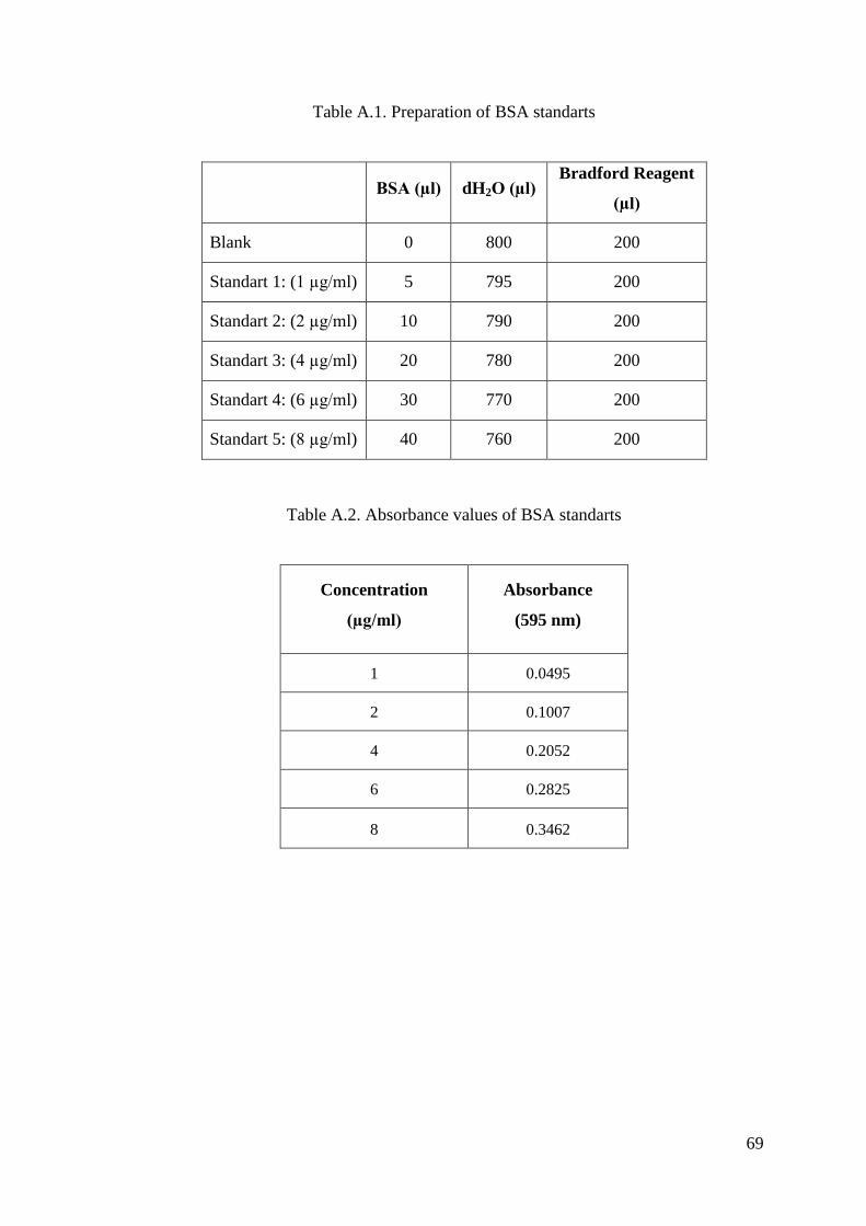
69
Table A.1. Preparation of BSA standarts
BSA (µl) dH2O (µl)
Bradford Reagent
(µl)
Blank 0 800 200
Standart 1: (1 µg/ml) 5 795 200
Standart 2: (2 µg/ml) 10 790 200
Standart 3: (4 µg/ml) 20 780 200
Standart 4: (6 µg/ml) 30 770 200
Standart 5: (8 µg/ml) 40 760 200
Table A.2. Absorbance values of BSA standarts
Concentration
(µg/ml)
Absorbance
(595 nm)
1 0.0495
2 0.1007
4 0.2052
6 0.2825
8 0.3462
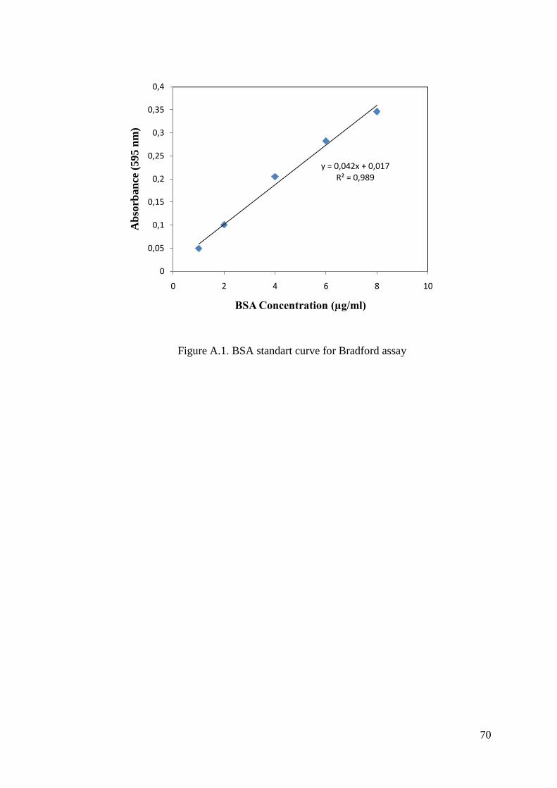
70
Figure A.1. BSA standart curve for Bradford assay
y = 0,042x + 0,017R² = 0,989
0
0,05
0,1
0,15
0,2
0,25
0,3
0,35
0,4
0 2 4 6 8 10
Ab
sorb
an
ce (
595 n
m)
BSA Concentration (µg/ml)

71
APPENDIX B
PROTEOLYTIC ACTIVITY DETERMINATION
STANDARTS AND STANDART CALIBRATION CURVE
FOR L-TYROSINE ASSAY
Table B.1. Preparation of L-Tyrosine standarts
L-Tyrosine
(µl)
dH2O
(µl)
Blank 0 1000
Standart 1: (0.2 mg/ml) 200 800
Standart 2: (0.4 mg/ml) 400 600
Standart 3: (0.6 mg/ml) 600 400
Standart 4: (0.8 mg/ml) 800 200
Standart 5: (1.0 mg/ml) 1000 0
Table B.2. Absorbance values of L-Tyrosine standarts
Concentration (mg/ml) Concentration (µmole) Absorbance (595 nm)
0.2 0.00625 0.054
0.4 0.0125 0.128
0.6 0.01875 0.184
0.8 0.025 0.238
1 0.03125 0.313
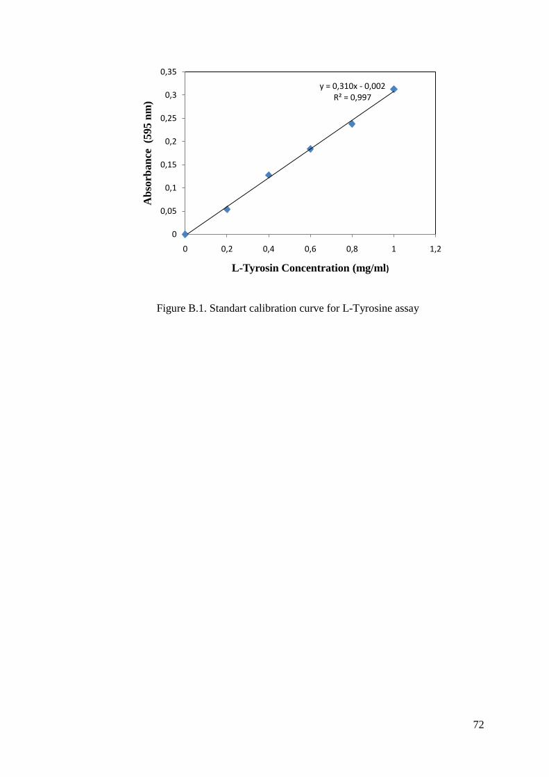
72
Figure B.1. Standart calibration curve for L-Tyrosine assay
y = 0,310x - 0,002R² = 0,997
0
0,05
0,1
0,15
0,2
0,25
0,3
0,35
0 0,2 0,4 0,6 0,8 1 1,2
Ab
sorb
an
ce (5
95 n
m)
L-Tyrosin Concentration (mg/ml)

73
APPENDIX C
MEDIA, BUFFERS, REAGENTS AND SOLUTIONS
C.1. Media and Buffers
Luria Bertani (LB) broth, per liter
10.0 g tryptone, 5.0 g yeast extract, 5.0 g NaCl and dH2O up to 1 liter. The mixture was
autoclaved for sterilization for 15 minutes at 121 °C under high pressure.
Complex medium, per liter
10.0 g glucose, 5.0 g yeast, 2.5 g peptone, 2.5 g casein, 0.3 g MgSO4, 0.02 g FeSO4, 0.2
g ZnSO4, 1.0 g CaSO4, 1.0 g KH2PO4, 1.0 g K2HPO4 and dH2O up to 1 liter. The
mixture was autoclaved for sterilization for 15 minutes at 121 °C under high pressure.
Media Used For Protease Screening, per liter
8 g nutrient broth, 10 g skim milk, 15 g agar agar and dH2O up to 1 liter.
Ingredients except skim milk were dissolved in deionized water. Medium was sterilised
by autoclaving at 121 °C for 15 minutes. Skim-milk is autoclaved separately at 110 °C
for 5 minutes (two times) and added to the medium.
50 mM Tris-HCl pH 7.2
6.055 g tris base was dissolved in 800 ml of deionized water. pH was adjusted to 7.2
with concentrated HCl. Volume is brought to 1 liter with deionized water.
C.2. Reagents Preparation for SDS-PAGE
C.2.1. Stock Solutions in Coomassie Staining
30% Acrylamide Mixture
(29.2 g acrylamide, 0.8 g N’N’-bis-methylene-acrylamide)
Make up to 100 ml with ultrapure water. Filter and store at 4 ºC in the dark for at least
one month.
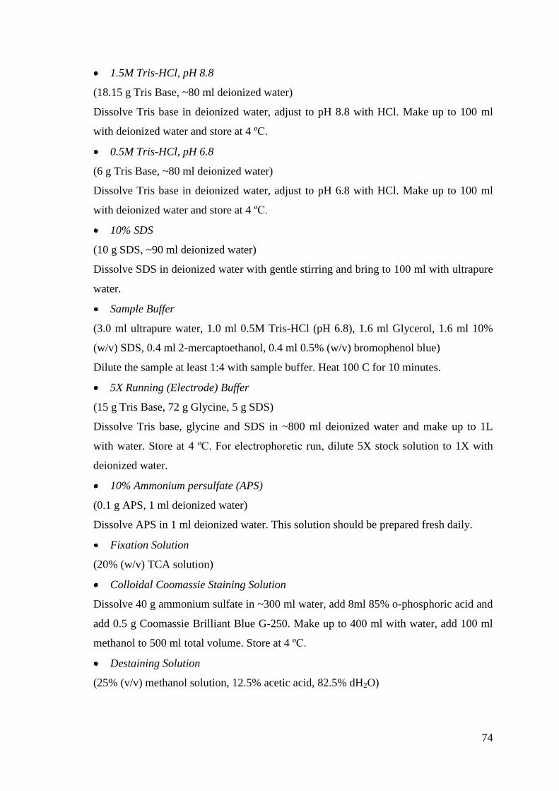
74
1.5M Tris-HCl, pH 8.8
(18.15 g Tris Base, ~80 ml deionized water)
Dissolve Tris base in deionized water, adjust to pH 8.8 with HCl. Make up to 100 ml
with deionized water and store at 4 ºC.
0.5M Tris-HCl, pH 6.8
(6 g Tris Base, ~80 ml deionized water)
Dissolve Tris base in deionized water, adjust to pH 6.8 with HCl. Make up to 100 ml
with deionized water and store at 4 ºC.
10% SDS
(10 g SDS, ~90 ml deionized water)
Dissolve SDS in deionized water with gentle stirring and bring to 100 ml with ultrapure
water.
Sample Buffer
(3.0 ml ultrapure water, 1.0 ml 0.5M Tris-HCl (pH 6.8), 1.6 ml Glycerol, 1.6 ml 10%
(w/v) SDS, 0.4 ml 2-mercaptoethanol, 0.4 ml 0.5% (w/v) bromophenol blue)
Dilute the sample at least 1:4 with sample buffer. Heat 100 C for 10 minutes.
5X Running (Electrode) Buffer
(15 g Tris Base, 72 g Glycine, 5 g SDS)
Dissolve Tris base, glycine and SDS in ~800 ml deionized water and make up to 1L
with water. Store at 4 ºC. For electrophoretic run, dilute 5X stock solution to 1X with
deionized water.
10% Ammonium persulfate (APS)
(0.1 g APS, 1 ml deionized water)
Dissolve APS in 1 ml deionized water. This solution should be prepared fresh daily.
Fixation Solution
(20% (w/v) TCA solution)
Colloidal Coomassie Staining Solution
Dissolve 40 g ammonium sulfate in ~300 ml water, add 8ml 85% o-phosphoric acid and
add 0.5 g Coomassie Brilliant Blue G-250. Make up to 400 ml with water, add 100 ml
methanol to 500 ml total volume. Store at 4 ºC.
Destaining Solution
(25% (v/v) methanol solution, 12.5% acetic acid, 82.5% dH2O)
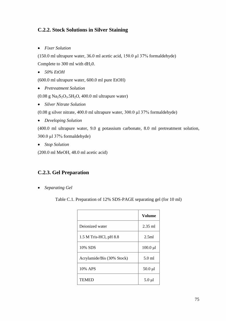
75
C.2.2. Stock Solutions in Silver Staining
Fixer Solution
(150.0 ml ultrapure water, 36.0 ml acetic acid, 150.0 µl 37% formaldehyde)
Complete to 300 ml with dH20.
50% EtOH
(600.0 ml ultrapure water, 600.0 ml pure EtOH)
Pretreatment Solution
(0.08 g Na2S2O3.5H2O, 400.0 ml ultrapure water)
Silver Nitrate Solution
(0.08 g silver nitrate, 400.0 ml ultrapure water, 300.0 µl 37% formaldehyde)
Developing Solution
(400.0 ml ultrapure water, 9.0 g potassium carbonate, 8.0 ml pretreatment solution,
300.0 µl 37% formaldehyde)
Stop Solution
(200.0 ml MeOH, 48.0 ml acetic acid)
C.2.3. Gel Preparation
Separating Gel
Table C.1. Preparation of 12% SDS-PAGE separating gel (for 10 ml)
Volume
Deionized water 2.35 ml
1.5 M Tris-HCl, pH 8.8 2.5ml
10% SDS 100.0 μl
Acrylamide/Bis (30% Stock) 5.0 ml
10% APS 50.0 μl
TEMED 5.0 μl

76
Stacking Gel
Table C.2. Preparation of 4% SDS-PAGE stacking gel (for 5ml)
Volume
Deionized water 3.05 ml
0.5 M Tris-HCl, pH 6.8 1.25ml
10% SDS 50.0 μl
Acrylamide/Bis (30% Stock) 650.0 μl
10% APS 25.0 μl
TEMED 5.0 μl