production of novel bioactive compounds by enzymes,...
TRANSCRIPT

269
Pure Appl. Chem., Vol. 82, No. 1, pp. 269–287, 2010.doi:10.1351/PAC-CON-09-02-05© 2010 IUPAC, Publication date (Web): 16 January 2010
Production of novel bioactive compounds byenzymes, and their application to food*
Takashi Kometani‡
Health Science Laboratory, Ezaki Glico Co., Ltd., Osaka, Japan
Abstract: Recently, people have been paying greater attention to their health and, as a result,the need to use physiologically functional foods was found on the market. For these reasons,the market size of “foods for specified health use” (FOSHU) in Japan has grown and was ap-proximately ¥700 billion in 2008.
Many enzymes such as amylases and proteases have been used in food manufacturingbecause of their diversity, specificity, and mild condition in reaction.
The aim of this investigation was the production of novel bioactive compounds by threekinds of transglycosylation reaction of the amylolytic enzymes, and the research and devel-opment of physiologically functional foods using these compounds.
Phosphoryl oligosaccharides of calcium (POs-Ca) are a complex with Ca andphosphoryl oligosaccharides prepared from potato starch by a hydrolysis (transglycosylationto H2O) of amylolytic enzymes. The chewing gum included POs-Ca prevented dental cariesin humans.
Highly branched cyclic dextrin (HBCD) was produced from amylopectin by branchingenzyme (intramolecular transglycosylation), which had a relatively narrow molecular-weightdistribution compared with commercially available dextrins. The sports drink containingHBCD enhanced swimming endurance in mice and humans.
α-Glycosylhesperidin (G-Hsp) was produced from starch and hesperidin, a flavonoidfound abundantly in citrus fruits, by the intermolecular transglycosylation using cyclodextringlucanotransferase. Oral administration of G-Hsp improved rheumatoid arthritis in mice andhumans, and poor blood circulation in women.
In this study, we looked to prove that this enzymatic modification technique was use-ful in creating unique and effective physiologically functional foods. These functional foodsare expected to improve the health and quality of life of many people.
Keywords: branching enzyme; cyclodextrin glucanotransferase; dental caries; functionalfoods; glycosylhesperidin; highly branched cyclic dextrin; phosphoryl oligosaccharides;rheumatoid arthritis; transglycosylation; sports drinks.
INTRODUCTION
Since ancient times, there has been the concept that food and medicine have a common origin; this con-cept has been especially prominent in China, India, Europe, and Japan. In Japan, studies of the physio-logical function in foods began in 1984 as a national project under the sponsorship of the Ministry of
*Paper based on a presentation at the 13th International Biotechnology Symposium (IBS 2008): “Biotechnology for theSustainability of Human Society”, 12–17 October 2008, Dalian, China. Other presentations are published in this issue, pp. 1–347.‡E-mail: [email protected]

Education, Science, and Culture; in 1991 the Ministry of Health and Welfare initiated a world-first pol-icy by legally approving physiologically functional foods, terming them “foods for specified health use”(FOSHU) [1,2]. The first FOSHU product was hypoallergenic rice, made widely available after multi-site clinical intervention trials [3] were reported in the journal Nature, under the title, “Japan exploresthe boundary between food and medicine” [4]. Japanese food companies have sought to produce phys-iologically functional foods to improve people’s quality of life, and under the FOSHU registration sys-tem, the market size and number of FOSHU in 2008 were approximately ¥700 billion and more than850 products, respectively; the market size increased five-fold in these 10 years.
Among the compounds used in physiologically functional foods, some of them were producedusing enzymes, e.g., peptides produced with protease (which brings about opioid [5], vasorelaxing [6],antihypertensive [7], or immunostimulating effects [8], etc.) and oligosaccharides [9] produced by amy-lolytic enzymes (which promote the proliferation of bifidobacteria, the absorption of minerals, andimmuno potentiating activities, among others).
The aim of this study was to contribute to the maintenance and improvement of human healththrough the provision of physiologically functional foods; we therefore prepared novel bioactive com-pounds originating from food materials with enzymes and applied them to these foods. The material andenzymes that we used were starch and amylolytic enzymes, such as α-amylase, glucoamylase, pullu-lanase, branching enzyme, and cyclodextrin glucanotransferase. As shown in Fig. 1, their reactionstended to follow one of three patterns: (1) hydrolysis (i.e., transglycosylation to H2O, in which glucosemoiety in starch or dextrin is transferred to H2O, to produce smaller molecules), (2) intramoleculartransglycosylation (i.e., in which glucose moiety is transferred to another part of the same molecule, toproduce cyclic compounds such as cyclodextrins), and (3) intermolecular transglycosylation (i.e., inwhich glucose moiety in starch is transferred to another molecule, to produce new glycosides).Transglycosylation is defined as the transfer of a glycosidically bound saccharide to another hydroxylgroup. In this study, we produced: (1) phosphoryl oligosaccharides (POs) from starch, by hydrolysis;(2) highly branched cyclic dextrin (HBCD) from starch, by intramolecular transglycosylation; and (3)α-glycosylhesperidin (G-Hsp) from starch and hesperidin, by intermolecular transglycosylation. Wethen applied these products to chewing gum, to prevent dental caries; sports drinks, to increase athleticendurance; and food, to alleviate rheumatoid arthritis symptoms and improve blood circulation.
T. KOMETANI
© 2010, IUPAC Pure Appl. Chem., Vol. 82, No. 1, pp. 269–287, 2010
270
Fig. 1 Three types of transglycosylation reaction in amylolytic enzymes. �: glucose; �: acceptor compound;–: α-1,4 glycosidic linkage.

PHOSPHORYL OLIGOSACCHARIDES OF CALCIUM AND THEIR APPLICATION(TRANSGLYCOSYLATION TO H2O)
Starches from various sources have been used widely—not only in dairy-based diets, but also in foodmanufacturing, as a nutrient, texture modifier, and a component of such sweeteners as glucose andmalto dextrin. Starch consists of two components, amylose and amylopectin; the former is an essentiallylinear glucan linked with α-1,4 glycosidic linkages, while the latter is highly branched with α-1,6 gly-cosidic linkages.
It was reported that the phosphate (P) groups are located mostly in amylopectin, with very littlepresence in amylose [10]; ester phosphorus is widely distributed in starch from various plants. Amongits components, potato starch is well known to contain esterified phosphoryl groups [10,11], especially,potato amylopectin contains 100–1,000 ppm of ester phosphorus, and about 70 % of the P groups arelinked to C-6 of the glucosyl residues, with the rest being linked to C-3 [12].
In the industrial production of glucose from starch, amylolytic enzymes such as α-amylase,gluco amylase, isoamylase, and pullulanase are hindered by P groups linked to glucosyl residues. Hence,glucose and POs are produced; whereas the former is widely used as a food material and in intravenousfluids, the latter is discarded as an unused material.
As POs have a negative charge based on the P groups within the molecule, cations such as Ca andiron will attach to the molecule. By mixing Ca with POs, phosphoryl oligosaccharides of calcium (POslinked with calcium; POs-Ca) are produced; Ca is then solubilized. Therefore, this study focused on theuse of POs-Ca as solubilized Ca.
Production of POs from potato starch
To produce glucose from potato starch, starch was hydrolyzed into glucose with a combination ofα-amylase (which hydrolyzes an α-1,4 glycosidic linkage at random with endo-wise), glucoamylase(which hydrolyzes an α-1,4 glycosidic linkage from the reducing end of starch), and pullulanase (whichhydrolyzes an α-1,6 glycosidic linkage). The reaction mixture containing glucose and residual oligo -saccharides (POs) was separated with an ion-exchange column; the POs were then fractionated into twofractions, PO-1 and PO-2. The components of the POs were analyzed via high-performance anion-ex-change chromatography and a pulsed amperometric detector system [13]. Fraction PO-1 was the majorcomponent of POs and comprised maltotriose, maltotetraose, and maltopentaose to which one P groupwas attached [14]. Fraction PO-2 predominantly comprised maltopentaose and maltohexaose to whichat least two phosphoryl groups were attached [15]. The structure of fraction PO-1 is shown in Fig. 2.
© 2010, IUPAC Pure Appl. Chem., Vol. 82, No. 1, pp. 269–287, 2010
Enzyme-modified bioactive compounds and their use 271

Prevention of dental caries by POs-Ca
Figure 3 illustrates the cycle of demineralization (i.e., the process of mineral loss from tooth enamelduring the early stages of dental decay) and remineralization (i.e., the reversal of the demineralizationof tooth enamel) that occurs in an oral cavity. In general, after every meal, plaque is always producedat the surface of the tooth enamel by cariogenic bacteria such as Streptococcus mutans. In that plaque,organic acid is produced by bacteria, and the pH of the oral cavity then decreases. This promotes demineralization in the enamel region of the teeth. When oral hygiene and the secretion of saliva aresufficient, remineralization occurs through the buffer action of saliva and by the supply of Ca and P ionsfrom saliva. However, if dental cleaning or the secretion of saliva in the oral cavity is insufficient, thetooth will proceed to decay, creating dental caries. Therefore, it is expected that research into com-pounds that prevent dental caries will be highly advantageous.
The effects of the daily use of a chewing gum containing 2.5 % POs-Ca on the remineralizationof enamel were examined. First of all, an in vitro human saliva immersing (HSI) test was conductedusing POs-Ca [16]. Two types of chewing gum were prepared; one contained 2.5 % POs-Ca and 46 %xylitol as a sweetener (POs-Ca gum), and the other contained 48.5 % xylitol without POs-Ca (xylitolgum). Both gums were sugar-free. While 12 volunteers (6 males and 6 females; mean age, 29.9 year-old) chewed each gum (weighing 3 g) for 20 min, all saliva stimulated was collected for the first 10 min(Fs) and last 10 min (Ls) separately. The volume and mineral contents in the collected saliva were an-alyzed; the results thereof are shown in Table 1. The salivary volume of Fs and Ls secreted on accountof the chewing of gum with or without POs-Ca were nearly equal, and not significantly different. The
T. KOMETANI
© 2010, IUPAC Pure Appl. Chem., Vol. 82, No. 1, pp. 269–287, 2010
272
Fig. 2 Structures of PO-1 fraction of POs.
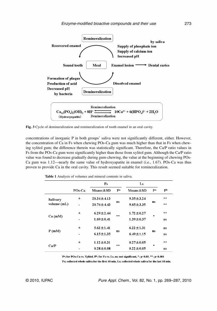
concentrations of inorganic P in both groups’ saliva were not significantly different, either. However,the concentration of Ca in Fs when chewing POs-Ca gum was much higher than that in Fs when chew-ing xylitol gum; the difference therein was statistically significant. Therefore, the Ca/P ratio values inFs from the POs-Ca gum were significantly higher than those from xylitol gum. Although the Ca/P ratiovalue was found to decrease gradually during gum-chewing, the value at the beginning of chewing POs-Ca gum was 1.12—nearly the same value of hydroxyapatite in enamel (i.e., 1.67). POs-Ca was thusproven to provide Ca in the oral cavity. This result seemed suitable for remineralization.
Table 1 Analysis of volumes and mineral contents in saliva.
© 2010, IUPAC Pure Appl. Chem., Vol. 82, No. 1, pp. 269–287, 2010
Enzyme-modified bioactive compounds and their use 273
Fig. 3 Cycle of demineralization and remineralization of tooth enamel in an oral cavity.

To evaluate the effect of POs-Ca on remineralization, we proceeded with an in vitro experiment.Bovine enamel slabs were demineralized with lactate. They were immersed in the mineral solution withor without POs-Ca for 1 week at 37 °C. After this treatment, the level of remineralization in the enamelslabs were analyzed by a previously described method [17,18] and a scanning electron microscopy.Mineral loss in the enamel slabs with POs-Ca was recovered more than that without POs-Ca, as shownin Fig. 4. From these results, it was determined that POs-Ca promoted a greater amount of remineral-ization activity.
Based on the aforementioned results, the effects of chewing gum containing POs-Ca on tooth-enamel remineralization were investigated in situ. Twelve healthy adult volunteers (6 males and 6 fe-males; mean age, 21.0 year-old) were randomly divided into 3 groups, each of which used, in a double-blind manner, chewing gums with or without POs-Ca (based on xylitol as a sweetener), orsucrose-based placebo chewing gum. Each volunteer wore a palatal appliance to which a demineralizedbovine enamel disk was attached, as described above for the in vitro experiment; each volunteer chewedone of the gums 4 times a day (after each meal and before bed time) for 4 weeks. The chewing timewas 20 min, and the palatal appliance was kept in the oral cavity for an additional 20 min.Remineralization of each enamel disk attached to the palatal appliance was evaluated by lesion depth,which was the depth of the demineralized part in the enamel disk from its surface. The lesion depths ofthe enamel disks were shown in Fig. 5 [19]. The remineralization rates (i.e., the percentage of lesiondepth with respect to that after initial demineralization) in the group chewing POs-Ca gum were 67, 54,and 76 % at the first, second, and fourth week, respectively. The corresponding values in the group thatchewed the gum without POs-Ca were 12–23 %—obviously much lower than those in the POs-Ca gumgroup. The values in the placebo gum group were much lower than those of these two groups.
Based on these results, a chewing gum containing POs-Ca obtained FOSHU accreditation fromJapan’s Ministry of Health and Welfare in 2003.
T. KOMETANI
© 2010, IUPAC Pure Appl. Chem., Vol. 82, No. 1, pp. 269–287, 2010
274
Fig. 4 Effects of POs-Ca on remineralization of tooth enamel. Bovine tooth slabs were demineralized in a 0.1 Mlactate gel (pH 5.0) at 37 °C for 3 weeks; they were then immersed in a mineral solution containing 20 mM Hepesbuffer (pH 7.0), 1.5 mM CaCl2, and 0.9 mM KH2PO4, with (B) or without 0.07 % POs-Ca (A). The dental caries-like lesions were scanned with an electron microscope.

HIGHLY BRANCHED CYCLIC DEXTRIN AND ITS APPLICATION (INTRAMOLECULARTRANSGLYCOSYLATION)
Starch and its derivatives—such as oligosaccharides—have been widely used in the food industry, asdescribed above, because various types of amylolytic enzymes that react to starch have been found invarious sources (microorganisms, plants, insects, and animals) and applied in an industrial scale [20].
Whereas branching enzyme (1,4-α-D-glucan: 1,4-α-D-glucan 6-α-D-(1,4-α-D-glucano)-trans-ferase, EC 2.4.1.18; BE) is well known to catalyze the α-1,6 glycosidic linkage of amylopectin andglycogen, it has been reported that the enzyme synthesizes a new dextrin called HBCD. Its structure hasbeen analyzed in great detail [21,22].
Production of HBCD from waxy starch
The actions of branching enzyme and α-amylase with respect to amylopectin are compared in Fig. 6:While the former split the inner chains connecting the cluster units (multi-branched glucan) of amylo -pectin (arrow heads in Fig. 6) and transferred the cluster unit to another site on the same substrate mol-ecule (intramolecular transglycosylation), the latter hydrolyzed the α-1,4 glycosidic linkage in amylo -pectin at random. Therefore, branching enzyme synthesized dextrin with a relatively narrowmolecular-weight distribution, while α-amylase synthesized various dextrins with a wide molecular-weight distribution, according to the reaction condition of the enzyme.
The structure and molecular weight of HBCD have been elucidated to be between those of amylo -pectin and oligosaccharides, and HBCD bears the properties of both amylopectin (i.e., little sweet taste,low osmolality, difficult to brown, etc.) and oligosaccharides (i.e., high water solubility, high stabilityin water, low viscosity, few specific odors that starches have, etc.).
© 2010, IUPAC Pure Appl. Chem., Vol. 82, No. 1, pp. 269–287, 2010
Enzyme-modified bioactive compounds and their use 275
Fig. 5 Comparison of lesion depth by durations of intraoral experiments. �: placebo chewing gum (based onsucrose as a sweetener); : chewing gum without POs-Ca (based on xylitol); �: chewing gum with POs-Ca (basedon xylitol). Dem: initial lesion depth of demineralized enamel disk. Horizontal bars indicate statistically significantdifference at p < 0.05 (Turkey multiple comparison).

Enhancement of endurance performance by HBCD
The main impetuses for ingesting sports drinks are the replacement of body fluids lost to sweat and theprovision of an exogenous form of energy, typically carbohydrates. As the development of fatigue dur-ing prolonged exercise has been associated with the depletion of endogenous carbohydrate stores [23],and given that dehydration results in the loss of more than 2 % of body weight [24], the intake of waterand exogenous energy is very important during exercise.
HBCD was found to bear many unique characteristics not found in most commercially availabledextrins, as described above. Considering these properties, HBCD seemed an appropriate ingredient insports drinks—as an energy source [25].
For this purpose, laboratory mice were exercised and administered HBCD solution. After beingacclimated to the act of swimming, swimming time to exhaustion was measured for all mice; the aver-age time was 64 ± 5 min (means ± SEM) in the adjustable-current pool, with the intensity of the exer-cise estimated at about 50 % VO2max [26]. First, HBCD exhibited a dose-dependent effect on swim-ming endurance: The mean swimming time for the mice that had ingested 500 mg HBCD/kg of bodyweight was 75 ± 4 min, which is significantly different from that for the mice that had ingested the samevolume of water and 166 mg HBCD/kg (65 ± 3 min), when orally administered 30 min after the start-ing of swimming. Therefore, 500 mg HBCD/kg of body weight was orally administered in this study.
To elucidate the effects of HBCD on swimming endurance, 300 μl of a 5 % HBCD solution, a5 % glucose solution (as a control, because glucose is a structural unit of HBCD), or water (as a con-
T. KOMETANI
© 2010, IUPAC Pure Appl. Chem., Vol. 82, No. 1, pp. 269–287, 2010
276
Fig. 6 Reaction pattern of branching enzyme to amylopectin, compared with α-amylase and structure of HBCD.
�: glucose; –: α-1,4 glycosidic linkage; →: α-1,6 glucosidic linkage; �� : reducing end glucose; �: point ofbranching enzyme reaction.

trol) was administered to individual mice 10 min before, 10 min after, or 30 min after the starting ofswimming. As shown in Fig. 7 (A-1, A-2), the mice that had ingested HBCD 10 min before swimmingshowed almost the same level of endurance as those that had ingested water, whereas those that had in-gested glucose showed a 40 % poorer endurance performance than those given HBCD. The mice ad-ministered HBCD 10 min after the starting of swimming were able to swim significantly longer thanthose administered water or glucose (Fig. 7; B-1, B-2). The mice administered HBCD 30 min after thestarting of swimming swam 20 % longer than those administered water, although the endurance per-formance of the former was similar to that of those given glucose (Fig. 7; C-1, C-2). These results in-dicated that, for our purposes, 10 min after the starting of swimming was the best time to administerHBCD to mice. To confirm reproducibility, three cross-experiments were conducted; the swimming
© 2010, IUPAC Pure Appl. Chem., Vol. 82, No. 1, pp. 269–287, 2010
Enzyme-modified bioactive compounds and their use 277
Fig. 7 Relationship between swimming endurance and carbohydrate ingestion. A: 10 min before (n = 9); B: 10 minafter (n = 18); and C: 30 min after the onset of the swimming exercise (n = 14). Values in A-1, B-1, and C-1represent the percentage (means ± SEM) of the swimming time for the mice that had ingested HBCD to that of themice that had ingested water (as 100 %). Values in A-2, B-2, and C-2 represent the percentage (means ± SEM) ofthe swimming time for the mice that had ingested HBCD to that of the mice that had ingested glucose (as 100 %).Comparison vs. water (A-1, B-1, and C-1) and glucose (A-2, B-2, and C-2): *p < 0.05 (Student’s t-test).

time to exhaustion of the mice administered HBCD, glucose, or water 10 min after the starting of swim-ming were 86 ± 6, 68 ± 5, and 65 ± 5 min, respectively. The mice that had ingested HBCD swam morethan 20 % longer than the mice that had ingested glucose or water.
To elucidate the mechanism behind HBCD-based improvements in endurance performance, weexamined the postprandial blood glucose and insulin levels after HBCD ingestion, during exercise. Themice were made to swim in the current pool, as described above, and HBCD, glucose, or water was ad-ministered to the mice 10 min after the starting of swimming. Another 10 min later—that is, 20 minafter the starting of swimming—blood was taken from the neck vein; plasma glucose, insulin, and lac-tate were analyzed; and the results thereof are shown in Table 2. Plasma glucose in the mice adminis-tered HBCD and glucose had increased; apparently, these two substances provided energy to the mice.Additionally, plasma insulin levels had increased according to the concentration of plasma glucose.
Table 2 Changes to metabolic parameters in the serum.
Due to its high molecular weight, HBCD requires more time than glucose for digestion in thesmall intestine during exercise. Therefore, the postprandial glucose and insulin responses in mice thathad ingested HBCD were lower than those that had ingested glucose. On the other hand, as ingestedglucose led to rapidly elevated plasma insulin levels, it is thought to cause hypoglycemia afterward [27].The result, as shown in Fig. 7 (A-1, A-2), is that hypoglycemia was thought to have occurred in micethat had ingested glucose. As a result, the plasma lactate levels of the mice that had ingested glucosewere also higher than those of the mice that had ingested HBCD.
From these results, HBCD is thought not to cause hypoglycemia, and to supply energy longerthan glucose; consequently, it enhances swimming endurance in mice. This finding agrees with that ofa study of human subjects, in which 7 swimmers (20.0 ± 0.3 year-old) who participated in the JapanChampionship in 2003 ingested each 1.5 g/kg HBCD, glucose, or water (as a control), just before swim-ming. After the interval training (at VO2max 75 %; swimming for 5 min and rest for 3 min), swimmingtime to exhaustion (at VO2max 90 %) was measured in a current-flow pool. The percentage of swim-ming time to exhaustion was 163.1 % for HBCD, 91.6 % for glucose comparing with that for water (as100 %), respectively, in which HBCD showed significant difference from water (Student’s t-test, p <0.05).
Gastric emptying time of HBCD-based sports drink
Since the nutrients and water are absorbed mainly from the intestine, not from the stomach, the balancebetween the concentration of carbohydrates and its osmotic pressure to accelerate gastric emptying hasbeen under discussion. A high concentration of carbohydrates in a drink, which is intended to increasethe amount of carbohydrates delivered as an energy source, delays gastric emptying due to high osmoticpressure, and thus decreases the amount of fluid available for absorption. Conversely, a low concentra-tion of carbohydrates accelerates gastric emptying and thus increases the amount of fluid available, but
T. KOMETANI
© 2010, IUPAC Pure Appl. Chem., Vol. 82, No. 1, pp. 269–287, 2010
278

decreases the level of carbohydrates delivered. Therefore, the relationship between carbohydrate con-tent and osmotic pressure in the formulation of sports drinks has been under great scrutiny.
First of all, we measured the rate of gastric emptying of HBCD and other carbohydrate solutions,using ultrasonographic techniques with 7 healthy men and 3 healthy women (26.2 ± 6.7 and 24.3 ± 2.3year-old, respectively) [28]. As the relaxed cross-sectional area of the pylorus antrum has been reportedto be well correlated with gastric volume—which was measured by an extracorporeal ultra sonic echo-image analyzer, after ingesting a drink [29]—the gastric emptying characteristics of the solutions areshown as gastric emptying times derived from gross half-gastric volumes. The gastric emptying timesof the solutions increased as a function of increasing concentrations of HBCD, glucose, and other com-mercially available dextrins based on glucose unit [maltose, dextrin (DE = 40), dextrin (DE = 20), inwhich the dextrose equivalent (DE) is defined as the degree of hydrolysis of starch into glucose], asshown in Fig. 8. A strong correlation was observed between gastric emptying time and the osmotic pres-sure of these solutions, except with water, physiological saline, and HBCD solutions (y = 0.047x +9.856; R2 = 0.875; y: gastric emptying time; x: osmotic pressure). As HBCD has a relatively large mo-lecular weight and contains only a small amount of low-molecular-weight dextrin, solutions thereofhave very low osmotic pressure. For example, a 10 % HBCD solution has an osmotic pressure of9 mOsm/kg; when sports drinks based on 10 % HBCD or 10 % dextrin (DE = 16) contain the same con-centrations of minerals, vitamins, and organic acids, their osmotic pressures are 150 mOsm/kg and269 mOsm/kg, respectively. HBCD-based sports drinks are transferred from the stomach to the smallintestine significantly faster than the dextrin (DE = 16)-based one.
As participants in endurance events frequently suffer from gastrointestinal disorders such asbelching, flatulence, heartburn, nausea, abdominal pain, lateroabdominal stitches, regurgitation, an urgeto defecate, and diarrhea, the effects of ingesting HBCD-based sports drinks on gastrointestinal disor-ders and fatigue during exercise were also investigated [30].
© 2010, IUPAC Pure Appl. Chem., Vol. 82, No. 1, pp. 269–287, 2010
Enzyme-modified bioactive compounds and their use 279
Fig. 8 Relationship between gastric emptying time and osmotic pressure of test solutions. �: HBCD; �: glucose;
�: maltose; �: dextrin (DE = 40); ○: dextrin (DE = 20). Concentrations (%) are represented by the size of thesymbols, from the smallest to the largest: 1.25, 2.5, 5.0, and 10.0 %. �: HBCD-based sports drink; : dextrin(DE = 16)-based sports drink; #: water; : physiological saline.

Seven untrained volunteers (34.3 ± 2.8 year-old) participated in an experiment consisting of a pre-liminary exercise for 9 min (3 min of cycling at each of 3 loads: 56, 71, and 85 W on a cycling ergometer), a 10-min rest, and 30 min of exercise (10 min of cycling at each of 3 loads: 71, 85, and99 W), all at 25 °C. Volunteers were administered sports drinks containing 10 % HBCD, 10 % glucose,10 % dextrin (DE = 16), or water (as a control) just after a 9-min exercise. The mean gastric emptyingtime after the ingestion of an HBCD-based sports drink was significantly faster than that of a glucose-based one. The number of gastrointestinal disorders—i.e., the degree of subjective flatulence and thenumber of belches—was small with HBCD-based drinks during exercise, compared with other drinks.Consequently, volunteers who ingested HBCD-based sports drinks were able to continue to exercisecomfortably, with little fatigue.
These results mentioned above revealed the effects of HBCD on endurance performance, com-pared with other drinks based on glucose or dextrin with glucose units. When HBCD was ingested, itwas transferred from stomach to intestine more quickly to be absorbed as energy, as shown by the ultra -sonograph techniques. Furthermore, not only carbohydrate but also other nutrients such as minerals,amino acids, water, and so on would be utilized quickly during exercise. In addition, the ingestion ofHBCD was found to utilize as a carbohydrate energy, which raised plasma glucose level and did notraise plasma insulin level so much compared with the ingestion of glucose. Therefore, it did not raiseplasma lactate level and, consequently, the endurance performance in mice and humans would be im-proved.
�-GLYCOSYLHESPERIDIN AND ITS APPLICATION (INTERMOLECULARTRANSGLYCOSYLATION)
Hesperidin, discovered in 1936 by Szent-Gyorgi as vitamin P [31], is a flavonoid that is abundant in cit-rus fruits such as Satsuma mandarin oranges (Citrus unshiu Marc.) and Valencia oranges (Citrus sinen-sis Valencia). It is also a major component of the traditional Chinese medicine Chen-pi. Hesperidin iswell known to exert many biological functions; for example, antioxidative, antiinflammatory, anti viral,and anticarcinogenic [32]. However, since hesperidin is not adequately soluble in aqueous solutions andmay not be absorbed well from the intestinal tract, it has not yet been used in the fields of food and med-icine.
Production of G-Hsp from starch and hesperidin
To solubilize hesperidin, we planed to transglycosylate hesperidin into its glycosides with an enzymein an alkaline pH, because it is almost insoluble in water or alcohol but is freely soluble in alkali. Inscreening alkalophilic bacteria from soil, we found cyclodextrin glucanotransferase [1,4-α-D-glucan4-α-D-(1,4-α-D-glucano)-transferase (cyclizing), EC.2.4.1.19; CGTase] from an alkalophilic Bacillussp. A2-5a; it was stable and exhibited relatively strong activity in an alkaline pH [33].
α-Glycosylhesperidin (4G-α-D-glucopyranosylhesperidin (G-Hsp) and series of oligoglucosides;Fig. 9) were synthesized from hesperidin and starch by the enzyme at pH 10; G-Hsp was approximately300 times more soluble than hesperidin in the aqueous phase [34]. G-Hsp was also found to be absorbedfrom the intestine about 3 times more readily than hesperidin [35]. Therefore, it would be interesting todetermine whether G-Hsp exhibits any biological activity in vivo.
T. KOMETANI
© 2010, IUPAC Pure Appl. Chem., Vol. 82, No. 1, pp. 269–287, 2010
280

Improvement of rheumatoid arthritis by G-Hsp
Rheumatoid arthritis is a chronic autoimmune disease of unknown etiology; it causes long-term jointdamage, chronic pain, and loss of function in affected joints [36]. Although a large number of disease-modifying antirheumatic drugs have been developed, such as methotrexate [37,38], biological therapiessuch as anti-TNF-α antibodies [39–41], and other treatments, they are often associated with adverse ef-fects. Therefore, safer approaches for treating rheumatoid arthritis are required.
Recently, it was reported that the oral administration of hesperidin suppressed collagen-inducedarthritis (CIA) in mice [42]. Therefore, the effects of G-Hsp on CIA mice were investigated [35].
To determine the therapeutic effects of G-Hsp on CIA progression, 3 mg G-Hsp was orally ad-ministered to CIA mice (body weight: 30 g) 3 times a week, from day 31 after priming with type II col-lagen emulsified with complete Freund’s adjuvant (10 days after the booster dose). Clinical scores ondays 52, 54, and 56 were significantly improved by treatments with 3 mg G-Hsp, compared with thecontrol (Fig. 10). To assess the preventive effects of G-Hsp in CIA development, in a separate experi-ment, 3 mg of G-Hsp was orally administered to CIA mice 3 times a week, starting from the onset ofdisease (i.e., day 21 after priming). The administration of G-Hsp resulted in significant improvementsin clinical scores at later stages, similar to those undergoing therapeutic treatment.
In histopathological observations, the extent of inflammation among knee joints (increases in syn-ovial and inflammatory cells and pannus formation) in the G-Hsp group improved more than those inthe control group. The synthesis of proteoglycan was reduced and the matrix metalloprotease activatedin the CIA mice, and then each joint was damaged. In the case of G-Hsp group, the former was recov-ered and the latter was inhibited. Such was not the case in the control group. These results indicate thatthe administration of G-Hsp may improve the symptoms of rheumatoid arthritis.
To confirm the effects of G-Hsp on rheumatoid arthritis in humans, 19 volunteers with rheuma-toid arthritis were divided into 2 groups; one (n = 9) was administered 3 g of G-Hsp per day, while theother (n = 10) was administered 3 g of dextrin (DE = 16) as a placebo for 12 weeks, using a double-blind method.
© 2010, IUPAC Pure Appl. Chem., Vol. 82, No. 1, pp. 269–287, 2010
Enzyme-modified bioactive compounds and their use 281
Fig. 9 Structure of G-Hsp. n = 0: monoglycosylhesperidin; n = 1: diglycosylhesperidin; n = 2:triglycosylhesperidin; n = 3: tetraglycosylhesperidin.

As shown in Fig. 11A, the physician’s global assessment of disease activity using a 100-mm vi-sual analog scale (VAS) of the American College of Rheumatology’s (ACR) 20 criteria decreased atweeks 4 and 8 in both the placebo and G-Hsp groups, compared with baseline (in the placebo group,significantly decreased at week 8; in the G-Hsp group, significantly decreased at weeks 4, 8, and 12).Although the VAS evaluation worsened in the placebo group at week 12, the G-Hsp group showed atendency to improve. Although there was no significant difference in the VAS values between the
T. KOMETANI
© 2010, IUPAC Pure Appl. Chem., Vol. 82, No. 1, pp. 269–287, 2010
282
Fig. 10 Therapeutic effects of G-Hsp on CIA in mice. DBA/1J mice with CIA were orally administered water (○;control, n = 18) or G-Hsp (�; n = 17) after the onset of arthritis. Arrow shows the beginning of ingestion of G-Hspin mice. Comparison vs. control: *p < 0.05 (Scheffe’s post-hoc test).
Fig. 11 Assessment of disease activity using a 100-mm VAS. A: Physician’s global assessment of disease activity;B: Patient’s global assessment of disease activity, using a 100-mm VAS in ACR20. Placebo: ○; G-Hsp: �.Comparison vs. baseline. +: p < 0.10; *: p < 0.05 and **p < 0.01 (Student’s t-test). Comparison vs. placebo:+p < 0.10 and *p < 0.05 (Mann–Whitney U-test).

placebo and G-Hsp groups, the G-Hsp group showed a tendency to improve at week 4 (p = 0.078). Inaddition, the global assessment of disease activity by patients themselves had the same tendency as thatmade by the physician; it was shown that the G-Hsp group significantly improved more than the placebogroup at week 12 (Fig. 11B).
As VAS scoring is subjective, we evaluated the concentration of C-reactive protein (CRP) inplasma, as an objective indicator (Fig. 12). CRP in plasma is known to increase with inflammation. Inthe G-Hsp group, CRP decreased significantly compared with the placebo group during the study pe-riod; after the cessation of G-Hsp ingestion, CRP increased gradually. In the placebo group, the CRPvalue did not improve during the study period [35].
In general, therapeutic efficacy was evaluated by a physician using ACR20 criteria for the diag-nosis of rheumatoid arthritis [43]. The administration of G-Hsp was found to be more effective than thatof placebo, with respect to symptom alleviation. Therefore, G-Hsp will be useful as a complemen-tary/alternative medicine in standard antirheumatoid therapy. However, a long-term study with a largersample size (e.g., patients in various stages and classes of rheumatoid arthritis) is necessary.
Improvement in blood circulation by G-Hsp
In Japan, many women feel cold—not only in winter, but also in summer, in air-conditioned environ-ments. One reason for this is poor blood circulation. Therefore, we looked to investigate blood-circula-tion improvements facilitated by the administration of G-Hsp.
Following the ingestion of 50–3600 mg/person G-Hsp and subsequent testing, 250 mg G-Hsp wasfound to be the minimum dose for improvements of blood circulation to be evident. So, G-Hsp (250 mg)or placebo (250 mg sucrose) was administered to 11 women (29.6 ± 3.9 year-old) with coldness of theextremities, using a double-blind, placebo-controlled, cross-over methodology [44]. After ingestion of
© 2010, IUPAC Pure Appl. Chem., Vol. 82, No. 1, pp. 269–287, 2010
Enzyme-modified bioactive compounds and their use 283
Fig. 12 Change in the plasma CRP concentration from baseline in the patients. Patients were administered Placebo(○) or G-Hsp (�), and the plasma CRP concentration was assayed every 4 weeks for 5 months. Comparison vs.baseline: **; p < 0.01, *; p < 0.05, +; p < 0.10 (by Student’s t-test). Comparison vs. placebo group: **; p < 0.01,*; p < 0.05 (by Mann–Whitney U-test).

G-Hsp or placebo, the skin surface temperature after cold-loading at 15 °C for 1 min was recorded, withtime, using thermography (Fig. 13). Whereas the skin surface temperature in the placebo group(<30 °C) was not improved 40 min after cold-loading, that in the G-Hsp group (33–34 °C) was im-proved to preloading levels, just 20 min after cold-loading. Both the width of blood vessels and bloodflow in the finger were reduced after cold-loading, but recovered gradually in the G-Hsp group; recov-ery in the placebo group was slower than that in the G-Hsp group. From these results, it can be con-cluded that the administration of G-Hsp leads to an extension in the width of blood vessels and there-fore an increase in blood flow—both of which consequently lead to a recovery of skin surfacetemperature. Furthermore, the continuous administration of G-Hsp (250 mg/day for 7 days) maintainsthis effect, and may alleviate poor blood circulation.
Figure 14 offers a summary comparison of the bioactivities of G-Hsp and hesperidin. Hesperidin,bearing many bioactivities, was transglycosylated into G-Hsp; its solubility and absorption into thebody increased approximately 300- and 3-fold, compared with those of hesperidin, respectively. G-Hspstill retained the bioactivities of hesperidin, e.g., anticarcinogenic characteristics [45] and improve-ments to bone mass density [46]. Furthermore, newly discovered benefits—namely, blood-circulationimprovements and alleviation of rheumatoid arthritis symptoms—were found during our study.
Because most bioactive compounds exert their activities after being absorbed into the body, im-provements in absorption are very important with regard to the compounds’ use in food and medicine;transglycosylation by enzymes, in particular, will be a very useful tool, as described in this study.
T. KOMETANI
© 2010, IUPAC Pure Appl. Chem., Vol. 82, No. 1, pp. 269–287, 2010
284
Fig. 13 Effect of G-Hsp on the improvement of poor blood circulation in women. A: Placebo (250 mg sucrose) orB: G-Hsp (250 mg) was administered to women with coldness of the extremities; skin surface temperature aftercold-loading at 15 °C for 1 min was recorded, with time, using thermography.

CONCLUSIONS
Recently, consumers paid much greater attention to their health, and in order to respond to variousneeds, many kinds of physiologically functional foods have been needed.
As modifications of compounds can change the structure, characteristics, and bioavailability ofthem, it is possible to create novel functions. This study looked to prove that enzymatic modificationsare useful in creating unique and effective physiologically functional foods.
There are a variety of environmental conditions on the earth—including low or high pressure ortemperature, high salt concentrations, and low or high pH—and a wide variety of organisms live in eachof them, and we can cull a variety of enzymes from them. By using such enzymes, it is possible to pro-duce novel physiologically functional foods that are suitable for people’s needs—which is to say, makethem healthier and improve their quality of life.
REFERENCES
1. S. Arai, T. Osawa, H. Ohigashi, M. Yoshikawa, S. Kaminogawa, M. Watanabe, T. Ogawa,K. Okubo, S. Watanabe, H. Nishino, K. Shinohara, T. Esashi, T. Hirahara. Biosci. Biotechnol.Biochem. 65, 1 (2001).
2. S. Arai, Y. Morinaga, T. Yoshikawa, E. Ichiishi, Y. Kiso, M. Yamazaki, M. Morotomi,M. Shimizu, T. Kuwata, S. Kaminogawa. Biosci. Biotechnol. Biochem. 66, 2017 (2002).
3. M. Watanabe, S. Arai. Food Sci. Technol., Int. 2, 10 (1996).4. D. Swinbacks, J. O’Brien. Nature 364, 180 (1993).5. M. Yoshikawa, M. Takahashi, S. Yang. Curr. Pharm. Des. 9, 1325 (2003).6. H. Fujita, H. Suganuma, H. Usui, K. Kurahashi, R. Nakagiri, R. Sasaki, M. Yoshikawa. Peptides
17, 635 (1996).7. B. A. Murray, R. J. FitzGerald. Curr. Pharm. Des. 13, 773 (2007).8. M. Yoshikawa, H. Fujita, N. Matoba, Y. Takenaka, T. Yamamoto, R. Yamauchi, H. Tsuruki,
K. Takahata. Biofactors 12, 143 (2000).9. T. Nakakuki. J. Appl. Glycosci. 52, 267 (2005).
© 2010, IUPAC Pure Appl. Chem., Vol. 82, No. 1, pp. 269–287, 2010
Enzyme-modified bioactive compounds and their use 285
Fig. 14 Summary of bioactivities of G-Hsp.
10. Y. Takeda, S. Hizukuri. Carbohydr. Res. 102, 321 (1982).11. C. Takeda, Y. Takeda, S. Hizukuri. Carbohydr. Res. 246, 273 (1993).12. S. Hizukuri. Carbohydrates in Food, A. C. Eliasson (Ed.), pp. 375–379, Marcel Dekker, New
York (1996).13. H. Kamasaka, M. Uchida, K. Kusaka, K. Yoshikawa, K. Yamamoto, S. Okada, T. Ichikawa.
Biosci. Biotechnol. Biochem. 59, 1412 (1995).14. H. Kamasaka, K. To-o, K. Kusaka, T. Kuriki, T. Kometani, S. Okada. Biosci. Biotechnol.
Biochem. 61, 1209 (1997).15. H. Kamasaka, K. To-o, K. Kusaka, T. Kuriki, T. Kometani, S. Okada. J. Appl. Glycosci. 44, 285
(1997).16. H. Kamasaka, D. Inaba, K. Minami, T. Nishimura, T. Kuriki, S. Imai, M. Yonemitsu. J. Dent.
Health 52, 105 (2002).17. D. Inaba, O. Takagi, J. Arends. Eur. J. Oral Sci. 105, 74 (1997).18. D. Inaba, R. Tanaka, O. Takagi, M. Yonemitsu, J. Arends. J. Dent. Health 47, 67 (1997).19. D. Inaba, H. Kamasaka, K. Minami, T. Nishimura, T. Kuriki, S. Imai, M. Yonemitsu. J. Dent.
Health 52, 112 (2002).20. Amylase Research Society of Japan (Ed.). Handbook of Amylases and Related Enzymes,
Pergamon Press, Oxford (1988).21. H. Takata, T. Takaha, S. Okada, M. Takagi, T. Imanaka. J. Bacteriol. 178, 1600 (1996).22. H. Takata, T. Takaha, S. Okada, S. Hizukuri, M. Takagi, T. Imanaka. Carbohydr. Res. 295, 91
(1996).23. M. Hargreaves, D. L. Costill, A. Coggan, W. J. Fink, I. Nishibata. Med. Sci. Sports Exerc. 16, 219
(1984).24. D. L. Costill, R. Coté, W. J. Fink, P. Van Handel. Int. J. Sports Med. 2, 130 (1981).25. H. Takii, K. Ishihara, T. Kometani, S. Okada, T. Fushiki. Biosci. Biotechnol. Biochem. 63, 2045
(1999).26. K. Matsumoto, K. Ishihara, K. Tanaka, K. Inoue, T. Fushiki. J. Appl. Physiol. 81, 1843 (1996).27. V. A. Koivisto, S. L. Karonen, E. A. Nikkilä. J. Appl. Physiol. 51, 783 (1981).28. H. Takii, Y. Takii Nagao, T. Kometani, T. Nishimura, T. Nakae, T. Kuriki, T. Fushiki. Int. J. Sports
Med. 26, 314 (2005).29. Y. Nagao, H. Kodama, T. Yonezawa, A. Taguchi, S. Fujino, K. Nakahara, K. Haruma, T. Fushiki.
Biosci. Biotechnol. Biochem. 62, 846 (1998).30. H. Takii, T. Kometani, T. Nishimura, T. Kuriki, T. Fushiki. Food Sci. Technol. Res. 10, 428 (2004).31. St. Rusznyak, A. Szent-Gyorgyi. Nature 138, 27 (1936).32. A. Gang, S. Gang, L. J. D. Zaneveld, A. K. Singla. Phytother. Res. 15, 655 (2001).33. T. Kometani, Y. Terada, T. Nishimura, H. Takii, S. Okada. Biosci. Biotechnol. Biochem. 58, 517
(1994).34. T. Kometani, Y. Terada, T. Nishimura, H. Takii, S. Okada. Biosci. Biotechnol. Biochem. 58, 1990
(1994).35. T. Kometani, T. Fukuda, T. Kakuma, K. Kawaguchi, W. Tamura, Y. Kumazawa, K. Nagata.
Immunopharmacol. Immunotoxicol. 30, 117 (2008).36. E. D. Harris Jr. N. Eng. J. Med. 322, 1277 (1990).37. H. J. Williams, R. F. Willkens, C. O. Samuelson, Jr., G. S. Alarcon, M. Guttadauria, C. Yarboro,
R. P. Polisson, S. R. Weiner, M. E. Luggen, L. M. Billingsley, S. L. Dahl, J. C. Egger, J. C.Reading, J. R. Ward. Arthr. Rheum. 28, 721 (1985).
38. J. M. Kremer Arthr. Rheum. 40, 984 (1997).39. A. den Broeder, L. van de Putte, R. Rau, M. Schattenkirchner, P. Van Riel, O. Sander, C. Binder,
H. Fenner, Y. Bankmann, R. Velagapudi, J. Kempeni, H. Kupper. J. Rheumatol. 29, 2288 (2002).40. D. E. Furst, M. H. Schiff, R. M. Fleischmann, V. Strand, C. A. Birbara, D. Compagnone, S. A.
Fischkoff, E. K. Chartash. J. Rheumatol. 30, 2563 (2003).
T. KOMETANI
© 2010, IUPAC Pure Appl. Chem., Vol. 82, No. 1, pp. 269–287, 2010
286
41. M. J. Elliott, R. N. Maini, M. Feldmann, A. Long-Fox, P. Charles, H. Bijl, JN. Woody. Lancet344, 1105 (1994).
42. K. Kawaguchi, H. Maruyama, T. Kometani, K. Kumazawa. Planta Med. 72, 477 (2006).43. D. T. Felson, J. J. Anderson, M. Boers, C. Bombardier, M. Chernoff, B. Fried, D. Furst,
C. Goldsmith, S. Kieszak, R. Lightfoot, H. Paulus, P. Tugwell, M. Weinblatt, R. Widmark, H. J.Williums, F. Wolfe. Arthr. Rheum. 36, 729 (1993).
44. K. Yoshitani, T. Minami, H. Takumi, Y. Kagami, K. Shiraishi, T. Kometani. J. Jpn. Soc. Nutr.Food Sci. 61, 233 (2008). (in Japanese)
45. T. Tanaka, H. Kohno, R. Suzuki, S. Sugie, T. Kometani. “Modifying effects of hesperidin glyco-side on chemically-induced tongue and colon carcinogenesis in F344 rats”, in Carcinogenesis andModification of Carcinogenesis, T. Tanaka, H. Tsuda (Eds.), pp. 213–227, Research Signpost(2005).
46. H. Chiba, M. Uehara, J. Wu, X. Wang, R. Masuyama, K. Suzuki, K. Kanazawa, Y. Ishimi. J. Nutr.133, 1892 (2003).
© 2010, IUPAC Pure Appl. Chem., Vol. 82, No. 1, pp. 269–287, 2010
Enzyme-modified bioactive compounds and their use 287