production and characteristics of avicel-digesting and non-avicel
TRANSCRIPT

Vol. 54, No. 6APPLIED AND ENVIRONMENTAL MICROBIOLOGY, June 1988, p. 1523-15290099-2240/88/061523-07$02.00/0Copyright ) 1988, American Society for Microbiology
Production and Characteristics of Avicel-Digesting andNon-Avicel-Digesting Cellobiohydrolases from Aspergillus ficum
SHINSAKU HAYASHIDA,* KAIGUO MO, AND AKIRA HOSODA
Department of Agricultural Chemistry, Kyushu University, Fukuoka 812, Japan
Received 3 December 1987/Accepted 23 March 1988
Two immunologically related cellobiohydrolases, cellobiohydrolase I (CBH I) and cellobiohydrolase II (CBHII), were purified from Aspergillusficum. The Avicel-adsorbable CBH I (molecular weight, 128,000) digestedAvicel, cotton, and cellulose powder to cellobiose, but the Avicel-unadsorbable CBH II (molecular weight,50,000) could not digest those substrates. Both enzymes hydrolyzed insoluble cellooligosaccharides (DP 25) tocellobiose. High-pressure liquid chromatographic analysis of soluble cellooligosaccharide hydrolysates revealedthat both enzymes split off strictly cellobiose units from the nonreducing end of the cellulose chain with an
exowise mechanism. CBH I showed glucosyltransferase activity, but CBH II did not. The N-bromosuccinimide-oxidized CBH I was completely inactive but retained the ability to adsorb to Avicel. This suggested that CBHI has separate sites for binding to cellulose and for catalyzing cleavage of glycosidic linkages. Cellobiohydro-lases were of two types, CBH I and CBH II. The former can adsorb to and digest Avicel, while the latter can
do neither.
The mechanism of enzymatic hydrolysis of cellulose hasbeen discussed by many research workers (1, 3, 6, 14).However, the mode of action of cellulolytic enzymes, espe-cially on native cellulose, has not been completely clarified.We had reported in a previous paper (18) that endoglucanasewas divided into two types, endoglucanase I (EG I) andendoglucanase II (EG II), with the ability to adsorb to anddisintegrate cellulosic substrates. EG I could be adsorbedonto cellulosic substrates and rapidly disintegrated the innerpart of cellulose fibrils, and EG II was unable to adsorb toand disintegrate microcrystalline cellulose.The multiplicity of cellobiohydrolases and their properties
were reported by Fagerstam and Pettersson (5), Tilbeurghand Pettersson (15), and Wood et al. (20-22). Cellobiohydro-lase was considered to split off cellobiose units from thenonreducing end of the cellulose chain. However, as a smallamount of glucose and cellotriose were also liberated fromcellotetraose and cellohexaose with cellobiohydrolase, sev-eral studies (11, 17) concluded that this enzyme couldhydrolyze cellulose with a less-random mechanism.Here we tried to isolate two types of cellobiohydrolase
and to elucidate their mode of action on cellulosic sub-strates. This paper describes the purification and propertiesof both Avicel-adsorbable, Avicel-digesting CBH I andAvicel-unadsorbable, non-Avicel-digesting CBH II from astrain of Aspergillus ficum.
MATERIALS AND METHODS
Microorganism. A. ficum IFO 4034 was used throughoutthis study and was obtained from the Institute For Fermen-tation, Osaka, Japan.
Preparation of crude cellulase. A. ficum was cultured in a2-liter Erlenmeyer flask containing solid wheat bran medium(wheat bran, 80 g; pulp floc, 20 g; tap water, 100 ml) at 30°Cfor 4 days. Enzymes were extracted by addition of 600 ml oftap water and left to stand at 4°C for 5 h. The filtrate wascollected and designated as crude cellulase.
Chemicals. Avicel (microcrystalline cellulose), CM (car-
* Corresponding author.
boxymethyl)-cellulose (DS. 0.41), pulp floc, and filter paperwere supplied by the companies described in the previouspaper (8). Soluble cellooligosaccharides were obtained fromSeikagaku Kogyo Co., Tokyo, Japan. Insoluble cellooligo-saccharide was prepared from pulp floc by the method ofMurao et al. (13). N-Bromosuccinimide was purchased fromWako Pure Chemical Industries Co., Osaka, Japan.Enzyme assay. Cellobiohydrolase activity was assayed in a
reaction mixture containing 50 mg of Avicel (or insolublecellooligosaccharide) in 1 ml of 0.1 M acetate buffer (pH 5.0)and 1 ml of enzyme solution after 2 h of incubation at 50°C.Endoglucanase activity was measured by incubating 1 ml of1% CM-cellulose in 0.1 M acetate buffer (pH 5.0) and 1 ml ofenzyme solution at 50°C for 30 min. Reducing sugar formedwas determined by the dinitrosalicylic acid method (10). Oneunit of cellobiohydrolase or endoglucanase activity wasdefined as the amount of enzyme releasing 1 ,umol ofreducing sugar from the substrate per min.
P-Glucosidase activity was measured by incubating 1 ml of1% cellobiose in 0.1 M acetate buffer (pH 5.0) and 1 ml ofenzyme solution at 50°C for 30 min. Glucose formed wasdetermined by glucose oxidase. One unit of ,B-glucosidaseactivity was defined as the amount of enzyme releasing 2,umol of glucose from the substrate per min.
Purification of enzymes. All purification steps were carriedout at 4°C.
(i) Step 1. The culture extract was concentrated by pre-cipitation with ammonium sulfate at a 60% (wt/vol) concen-tration and kept overnight. The resulting precipitate wascollected by filtration and dissolved in a small volume ofdeionized water. The supernatant was dialyzed by PVA-hollow fiber (Kuraray Co., Osaka, Japan). The enzymesolution was then adjusted to pH 3.0 with 1 N hydrochloricacid and allowed to stand for 24 h. The precipitate wasremoved by centrifugation at 13,000 x g for 10 min. Thesupernatant was collected, lyophilized, and applied to aSephadex G-50 column (4.5 by 103 cm). Those fractions thatshowed cellobiohydrolase activity were combined and ly-ophilized.
(ii) Step 2. The lyophilized samples from step 1 were
1523

1524 HAYASHIDA ET AL.
0 "C-0-
oT~~~~~~=H =0~~~ ~ ~~~~~~-I =I-
o 20 40 60 80 100 120 140FRACTION NUMBER (10 ml)
FIG. 1. Sephadex G-50 column chromatography of the cellulasefrom A. ficum. Symbols: , A280; A, cellobiohydrolase activ-ity, with Avicel as the substrate; 0, endoglucanase activity; A,P-glucosidase activity. Experimental details are described in thetext.
applied to a DEAE-Sephadex A-50 column (2.5 by 103 cm)previously buffered (pH 6.8) and eluted with 500 ml of 0.05M phosphate buffer (pH 6.8) and 500 ml of a linear gradientfrom 0.05 to 1 M phosphate buffer (pH 6.8). The flow ratewas 5 ml/20 min. Fractions that contained cellobiohydrolaseactivity were combined, lyophilized, and desalted.
(iii) Step 3. The lyophilized samples from step 2 werefurther applied to a Sephacryl S-300 column (2.5 by 100 cm).Filtration was carried out with 0.1 M acetate buffer (con-taining 0.5 M sodium chloride [pH 6.0]) at a rate of 5 ml/22min.
Hydrolysis of cellulosic substrates. The rate of hydrolysisby enzyme was calculated as a percentage of the total sugarsreleased by acid treatment (12).
Immunological procedure. Antiserum for a purified CBH Ipreparation was prepared by immunizing a rabbit with threeintradermal injections of a mixture consisting of 1 mg ofCBH I and 1 ml of Freund complete adjuvant (Difco Labo-ratories, Detroit, Mich.) at weekly intervals. On week 4, 1 ml
4
31
0 25 50 75 100 125
FRACTION NUMBER 15ml )
of phosphate-buffered saline containing 1 mg of CBH I wasgiven to the animal by intravenous injection. The rabbit wasbled after 1 week, and the serum was collected after centrif-ugation and stored at -20°C. The double immunodiffusiontechnique was carried out according to Williams and Chase(19).HPLC analysis. The products of enzymic hydrolysis of
soluble cellooligosaccharides were determined by high-per-formance liquid chromatographic (HPLC) analysis. Beforeapplying the sugar mixtures to an HPLC column (6 by 250mm, YMC-pack PA-03, Yamamura Chemical LaboratoryCo., Kyoto, Japan), the protein in the mixtures was removedby filtration with collodion bags (Sartorius Co., FederalRepublic of Germany). The mixture (20 ,ul) was injected ontothe column using acetonitrile-water mixtures as eluting sol-vents (60%, vol/vol). A flow rate of 0.5 ml/min was main-tained. The sugars were determined using a differentialrefractometer (Shimadzu Seisakusyo Co., Kyoto, Japan).Chemical modification of CBH I by N-bromosuccinimide.
The purified CBH I was dissolved in 0.02 M acetate buffer(pH 4.0), containing various quantities of N-bromosuccini-mide, and then incubated at 27°C for 5 min. The residualactivity and protein concentration in the supernatant fluidbefore and after adsorption onto cellulosic substrates weredetermined.
General analytical procedures. Protein was determined bythe method of Lowry et al. (9) with crystalline serumalbumin as the standard. Disc gel electrophoresis in a 7.5%polyacrylamide gel at pH 8.3 in Tris-glycine buffer wascarried out by the method of Davis (4). Molecular weightswere estimated by sodium dodecyl sulfate-polyacrylamidegel electrophoresis (18).
RESULTS
Purification of CBH I and CBH II. The Sephadex G-50chromatographic pattern is shown in Fig. 1. Fraction Ashowed the activity toward Avicel, CM-cellulose, and insol-uble cellooligosaccharide, and fraction B showed higheractivity toward insoluble cellooligosaccharide. Fractions Aand B were collected separately, lyophilized, and thenapplied to a DEAE-Sephadex A-50 column. The DEAE-Sephadex A-50 chromatographic pattern of fraction A fromSephadex G-50 is shown in Fig. 2. Fractions 130 through 148
a
I-
-i
FIG. 2. DEAE-Sephadex A-50 column chromatography of fraction A from Sephadex G-50. Symbols: , A180; A, cellobiohydrolaseactivity, with Avicel as the substrate; *, endoglucanase activity; -, phosphate concentration. Experimental details are described in the text.
APPL. ENVIRON. MICROBIOL.
AVICEL- AND NON-AVICEL-DIGESTING CELLOBIOHYDROLASES
2-
3-
! g
FRACTION IBER (5 ml)
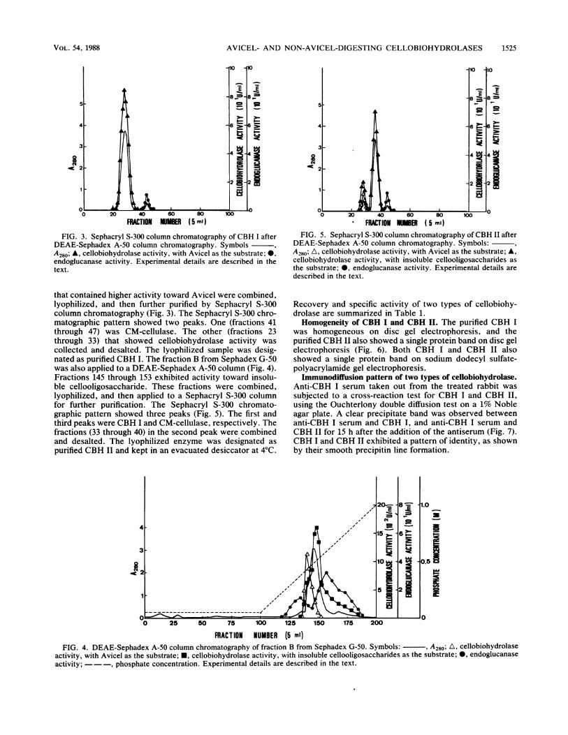
FIG. 3. Sephacryl S-300 column chromatography of CBH I afterDEAE-Sephadex A-50 column chromatography. SymbolsA280; A, cellobiohydrolase activity, with Avicel as the substrate; 0,endoglucanase activity. Experimental details are described in thetext.
that contained higher activity toward Avicel were combined,lyophilized, and then further purified by Sephacryl S-300column chromatography (Fig. 3). The Sephacryl S-300 chro-matographic pattern showed two peaks. One (fractions 41through 47) was CM-cellulase. The other (fractions 23through 33) that showed cellobiohydrolase activity wascollected and desalted. The lyophilized sample was desig-nated as purified CBH I. The fraction B from Sephadex G-50was also applied to a DEAE-Sephadex A-50 column (Fig. 4).Fractions 145 through 153 exhibited activity toward insolu-ble cellooligosaccharide. These fractions were combined,lyophilized, and then applied to a Sephacryl S-300 columnfor further purification. The Sephacryl S-300 chromato-graphic pattern showed three peaks (Fig. 5). The first andthird peaks were CBH I and CM-cellulase, respectively. Thefractions (33 through 40) in the second peak were combinedand desalted. The lyophilized enzyme was designated aspurified CBH II and kept in an evacuated desiccator at 4°C.
FRACTION N1ER ( 5 ml)FIG. 5. Sephacryl S-300 column chromatography ofCBH II after
DEAE-Sephadex A-50 column chromatography. Symbols:A280; A, cellobiohydrolase activity, with Avicel as the substrate; A,cellobiohydrolase activity, with insoluble cellooligosaccharides asthe substrate; 0, endoglucanase activity. Experimental details aredescribed in the text.
Recovery and specific activity of two types of cellobiohy-drolase are summarized in Table 1.Homogeneity of CBH I and CBH II. The purified CBH I
was homogeneous on disc gel electrophoresis, and thepurified CBH II also showed a single protein band on disc gelelectrophoresis (Fig. 6). Both CBH I and CBH II alsoshowed a single protein band on sodium dodecyl sulfate-polyacrylamide gel electrophoresis.
Immunodiffusion pattern of two types of cellobiohydrolase.Anti-CBH I serum taken out from the treated rabbit wassubjected to a cross-reaction test for CBH I and CBH II,using the Ouchterlony double diffusion test on a 1% Nobleagar plate. A clear precipitate band was observed betweenanti-CBH I serum and CBH I, and anti-CBH I serum andCBH II for 15 h after the addition of the antiserum (Fig. 7).CBH I and CBH II exhibited a pattern of identity, as shownby their smooth precipitin line formation.
4--- c
3-
---- ~~~~1O4~0.5~
1 .~~~~~~~~~~~~~~~~~~5 ."0 A 0S0 25 50 75 100 125 150 175 200
FRACT ION NUMBER (5 ml)FIG. 4. DEAE-Sephadex A-50 column chromatography of fraction B from Sephadex G-50. Symbols: , A280; A, cellobiohydrolase
activity, with Avicel as the substrate; *, cellobiohydrolase activity, with insoluble cellooligosaccharides as the substrate; 0, endoglucanaseactivity; ---, phosphate concentration. Experimental details are described in the text.
VOL. 54, 1988 1525

1526 HAYASHIDA ET AL.
TABLE 1. Purification of CBH I and CBH II from A. ficum
Total protein (mg) Total activity (U) Sp act (U/mg) Yield (%)Step
CBH I CBH II CBH I CBH II CBH I CBH II CBH I CBH II
Culture extract 21,550" 237.05 0.011 100Ammonium sulfate precipitation 8,860 221.50 0.025 93.44Sephadex G-50 580 490 17.98 13.23 0.031 0.027 7.58 5.58DEAE-Sephadex A-50 185 149 12.03 7.15 0.065 0.048 5.07 3.02Sephacryl S-300 75 59 11.33 6.73 0.151 0.114 4.78 2.84
a Values between CBH I and CBH II columns represent cellobiohydrolase that has not yet been purified to CBH I or CBH II.
Properties of two types of cellobiohydrolase. (i) Molecularweight. The molecular weight of CBH I was estimated to be128,000 by sodium dodecyl sulfate-polyacrylamide gel elec-trophoresis, and that of CBH II was estimated to be 50,000.
(ii) Thermostability and pH stability. Portions of enzymesolutions, prepared in 0.1 M acetate buffer (pH 5.0), werekept for 10 min at various temperatures. CBH I and CBH IIwere stable at 55 and 60°C. CBH I retained 40% of originalactivity after heating at 65°C, but about 60% residual activityof CBH II was retained at 70°C. CBH I was stable at pH 2.5to 6.0, and CBH II was stable at pH 2.0 to 7.0 after standingat 4°C for 24 h at various pHs.
(iii) Adsorbability of CBH I and CBH II to Avicel. Theprepared CBH I solution (2 mg of protein/ml) in 5 ml of 0.1M acetate buffer (pH 5.0) was applied to 1 g of Avicel,followed by standing at 4°C for 10 min. After centrifugation,cellobiohydrolase activity in the supernatant fluid was as-sayed. Ninety-six percent of CBH I activity was adsorbed,but the prepared CBH II solution (2 mg of protein per ml)could not be adsorbed to Avicel at the same condition.
(iv) Actions on cellulosic substrates. Hydrolysis curves ofCBH I and CBH II on cellulosic substrates are shown in Fig.8. Avicel was hydrolyzed to the extent of 26% by CBH I in168 h of incubation at 50°C, but CBH II showed no activitytoward Avicel. CBH II hydrolyzed insoluble cellooligosac-charide to the extent of 5.6% in 168 h at 50°C. Both CBH Iand CBH II had little capacity to hydrolyze CM-cellulose.
(v) Carbohydrate content. Each enzyme (1 mg of protein)was dissolved in 10 ml of deionized water. To 1 ml of the
FIG. 6. Disc gel electrophoretic patterns of CBH I and CBH 1I.This was done on a polyacrylamide (7.5%) gel column, pH 8.3.About 10 ,ug of enzyme preparations was used, and 2 mA percolumn (0.5 by 8 cm) was applied for 120 min. Staining was donewith 0.005% Coomassie brilliant blue R-250.
enzyme solution was added 1 ml of the phenol reagent and 5ml of concentrated sulfuric acid. The carbohydrate contents,calculated from A490 by using mannose as the standard, weredetermined to be 10.7 and 8.2% in CBH I and CBH II,respectively.
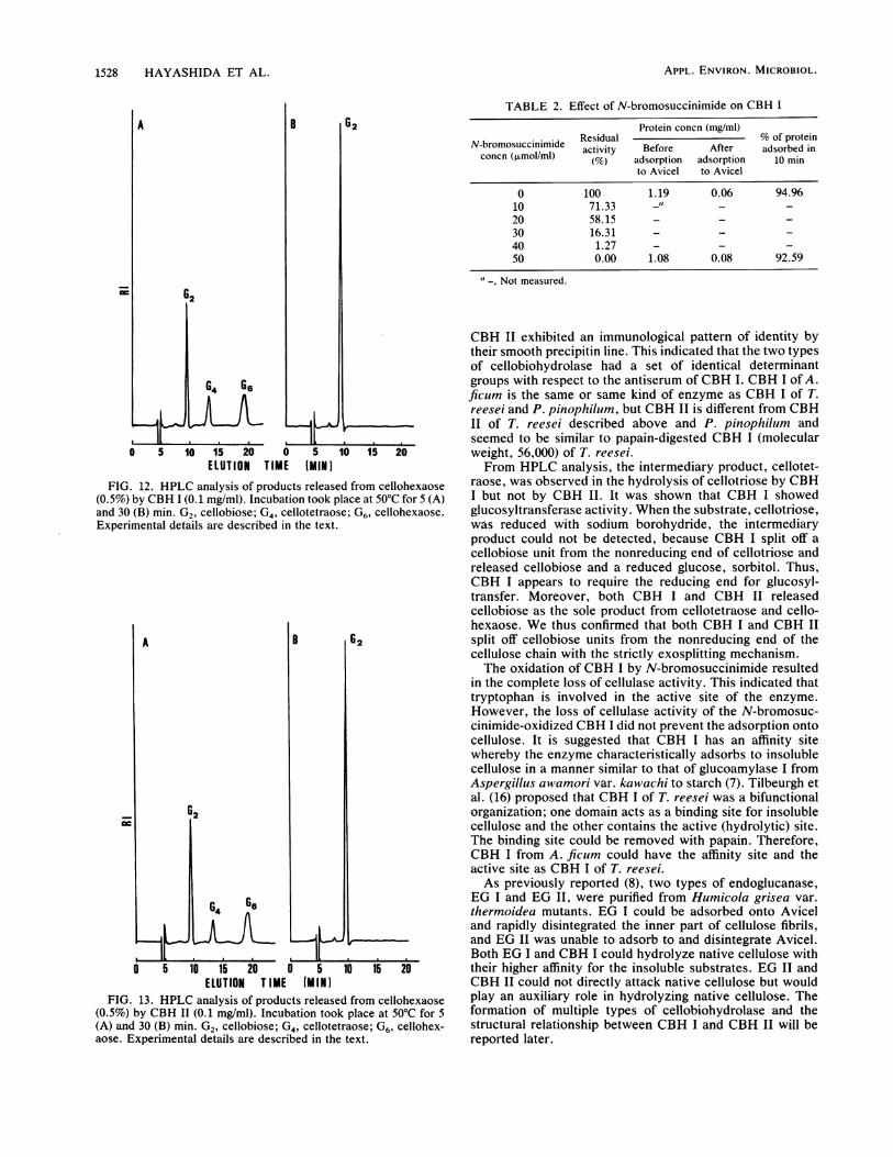
(vi) Hydrolysis products from cellulosic substrates. Thereaction products from Avicel and insoluble cellooligosac-charide by CBH I and CBH II after 24 h of incubation wereidentified by HPLC analysis. The hydrolysis products werecellobiose and traces of glucose. Both CBH I and CBH IIcould attack cellotriose, cellotetraose, cellopentaose, andcellohexaose. The intermediary product, cellotetraose, wasfound in the hydrolysis of cellotriose by CBH I at 2 and 5 minof incubation (Fig. 9) but not by CBH II (Fig. 10). Theintermediary product, cellotetraose, was hydrolyzed to cel-lobiose on prolonged incubation. The molar ratios of cello-triose hydrolysates (glucose-cellobiose) with CBH I andCBH II in 30 min of incubation were 1:2.6 and 1:1 (Fig. 9 and10). When the substrate, cellotriose, was reduced withsodium borohydride, the intermediary, cellotetraose, couldnot be found in the hydrolysates with CBH I at 2 and 5 minof incubation (Fig. 11). The molar ratio of the products(glucose-cellobiose) was 1:1 at 30 min of incubation (Fig. 10).HPLC analyses of products released from cellohexaose withboth enzymes are shown in Fig. 12 and 13. Both enzymesreleased cellobiose as the only product during incubationwith cellohexaose.
(vii) Effect of N-bromosuccinimide on CBH I. N-bromosuc-cinimide at a concentration of 50 ,umol/ml completely de-stroyed the cellulase activity of CBH I, while having noeffect on its ability to adsorb to the substrate (Table 2).
DISCUSSION
Wood and McCrae (20) reported that the highly purified C1component of Trichoderma koningii cellulase was a P-1,4-glucan cellobiohydrolase. This enzyme readily attackedphosphoric acid-swollen cellulose and cotton, producingcellobiose and a little glucose, but had little capacity to
FIG. 7. Double immunodiffusion pattern demonstrating the anti-genic relationships between CBH I and CBH II. Experimentaldetails are described in the text.
APPL. ENVIRON. MICROBIOL.

AVICEL- AND NON-AVICEL-DIGESTING CELLOBIOHYDROLASES
TIME 1 HOURSFIG. 8. Hydrolysis of cellulosic substrates by CBH I and CBH
II. Symbols: 0, CBH I on Avicel; 0, CBH II on insolublecellooligosaccharides; A, CBH II on Avicel. The reaction mixture,containing 50 mg of substrate in 1 ml of buffer and 1 ml of an enzymesolution (0.5 mg/ml), was incubated at 50°C in a stationary state.
hydrolyze CM-cellulose. Cellotetraose and cellohexaosewere similarly hydrolyzed almost exclusively to cellobioseand traces of glucose and cellotriose. Other workers re-ported later (18, 19) that two immunologically unrelatedcellobiohydrolases I and II, isolated from the extracellularcellulase system elaborated by Penicillum pinophilum, actedin a synergistic manner to solubilize the microcrystallinecellulose. They, therefore, suggested that the cellobiohydro-lases may be two stereospecific enzymes concerned with thehydrolysis of two different configurations of nonreducingend groups that would exist in cellulose.According to the studies of Pettersson and his collabora-
tors (1, 2, 5, 15, 16), Trichoderma reesei forms two immu-nologically different cellobiohydrolases, CBH I and CBH II.Both cellobiohydrolases showed activity toward Avicel andsoluble cellodextrins, predominantly yielding cellobiose as
ELUTION TIME MHINIFIG. 10. HPLC analysis of products released from cellotriose
(0.5%) by CBH 11 (0.1 mg/ml). Incubation took place at 50°C for 2(A), 5 (B), and 30 (C) min. Gl, glucose; G2, cellobiose; G3,cellotriose. Experimental details are described in the text.
the reaction product. 4-Methylumbelliferyl P-D-glycosidesderived from cellobiose and 4-methylumbelliferyl-4-o-(P-D-galactopyranosyl)-3-D-glucopyranoside were substrates forCBH I but were not hydrolyzed by CBH II. Limited prote-olysis of CBH I (molecular weight, 65,000) by papain yieldeda core protein (molecular weight, 56,000) that is fully activeagainst small, soluble cellodextrins. Activity against Avicelwas, however, completely lost, and concomitantly de-creased adsorption onto Avicel was observed.The present paper proposes the existence of two types of
cellobiohydrolase, CBH I (molecular weight, 128,000) andCBH II (molecular weight, 50,000) in the culture ofA. ficum.CBH I could be adsorbed onto Avicel and digested Avicel toform cellobiose. CBH II was unable to adsorb to and digestAvicel but hydrolyzed insoluble cellooligosaccharide (DP25) derived from pulp floc to split off cellobiose. CBH I and
;=
ELUTION TIME IMIN)FIG. 9. HPLC analysis of products released from cellotriose
(0.5%) by CBH I (0.1 mg/ml). Incubation took place at 50°C for 2(A), 5 (B), and 30 (C) min. G1, glucose; G2, cellobiose; G3,cellotriose; G4, cellotetraose. Experimental details are described inthe text.
A
626HGA1
B 62
0 5 10 15 20 0 5 10 15 20ELUTION TIME (MIlN
FIG. 11. HPLC analysis of products released from reducedcellotriose (0.5%) by CBH I (0.1 mg/ml). Incubation took place at50°C for 5 (A) and 30 (B) min. G1H, sorbitol; G2, cellobiose; G3H,reduced cellotriose. Experimental details are described in the text.
VOL. 54, 1988 1527
1528 HAYASHIDA ET AL.
ml
A
62
B G2
0 5 10 15 20 0 5 10 15 20ELUTION TIME (MIN I
FIG. 12. HPLC analysis of products released from cellohexaose(0.5%) by CBH I (0.1 mg/ml). Incubation took place at 50°C for 5 (A)and 30 (B) min. G2, cellobiose; G4, cellotetraose; G6, cellohexaose.Experimental details are described in the text.
A
G2
B G2
64 G65
0 5 10 15 20 0 5 10 15 20ELUTION T IME (MINI
FIG. 13. HPLC analysis of products released from cellohexaose(0.5%) by CBH II (0.1 mg/ml). Incubation took place at 50°C for 5(A) and 30 (B) min. G2, cellobiose; G4, cellotetraose; G6, cellohex-aose. Experimental details are described in the text.
TABLE 2. Effect of N-bromosuccinimide on CBH I
Protein concn (mg/ml)N-bromosuccinimide Residual % of proteinconcn (mosuclnmlde activity Before After adsorbed inconcn (,umol/ml) (%fo) adsorption adsorption 10 min
to Avicel to Avicel
0 100 1.19 0.06 94.9610 71.33 -" _ _20 58.15 - - -30 16.31 - - -40 1.27 - - -50 0.00 1.08 0.08 92.59
Not measured.
CBH II exhibited an immunological pattern of identity bytheir smooth precipitin line. This indicated that the two typesof cellobiohydrolase had a set of identical determinantgroups with respect to the antiserum of CBH I. CBH I of A.ficum is the same or same kind of enzyme as CBH I of T.reesei and P. pinophilum, but CBH II is different from CBHII of T. reesei described above and P. pinophilum andseemed to be similar to papain-digested CBH I (molecularweight, 56,000) of T. reesei.From HPLC analysis, the intermediary product, cellotet-
raose, was observed in the hydrolysis of cellotriose by CBHI but not by CBH II. It was shown that CBH I showedglucosyltransferase activity. When the substrate, cellotriose,was reduced with sodium borohydride, the intermediaryproduct could not be detected, because CBH I split off acellobiose unit from the nonreducing end of cellotriose andreleased cellobiose and a reduced glucose, sorbitol. Thus,CBH I appears to require the reducing end for glucosyl-transfer. Moreover, both CBH I and CBH II releasedcellobiose as the sole product from cellotetraose and cello-hexaose. We thus confirmed that both CBH I and CBH IIsplit off cellobiose units from the nonreducing end of thecellulose chain with the strictly exosplitting mechanism.The oxidation of CBH I by N-bromosuccinimide resulted
in the complete loss of cellulase activity. This indicated thattryptophan is involved in the active site of the enzyme.However, the loss of cellulase activity of the N-bromosuc-cinimide-oxidized CBH I did not prevent the adsorption ontocellulose. It is suggested that CBH I has an affinity sitewhereby the enzyme characteristically adsorbs to insolublecellulose in a manner similar to that of glucoamylase I fromAspergillus awamori var. kawachi to starch (7). Tilbeurgh etal. (16) proposed that CBH I of T. reesei was a bifunctionalorganization; one domain acts as a binding site for insolublecellulose and the other contains the active (hydrolytic) site.The binding site could be removed with papain. Therefore,CBH I from A. ficum could have the affinity site and theactive site as CBH I of T. reesei.As previously reported (8), two types of endoglucanase,
EG I and EG II, were purified from Humicola grisea var.thermoidea mutants. EG I could be adsorbed onto Aviceland rapidly disintegrated the inner part of cellulose fibrils,and EG II was unable to adsorb to and disintegrate Avicel.Both EG I and CBH I could hydrolyze native cellulose withtheir higher affinity for the insoluble substrates. EG II andCBH II could not directly attack native cellulose but wouldplay an auxiliary role in hydrolyzing native cellulose. Theformation of multiple types of cellobiohydrolase and thestructural relationship between CBH I and CBH II will bereported later.
APPL. ENVIRON. MICROBIOL.

AVICEL- AND NON-AVICEL-DIGESTING CELLOBIOHYDROLASES
LITERATURE CITED1. Berghem, L. E. R., and L. G. Pettersson. 1973. The mechanism
of enzymatic cellulose degradation. Eur. J. Biochem. 37:21-30.2. Berghem, L. E. R., and L. G. Pettersson. 1975. The mechanism
of enzymatic cellulose degradation. Eur. J. Biochem. 53:55-62.3. Chanzy, H., B. Henrissat, R. Vuong, and M. Schulein. 1983. The
action of 1,4-J3-D-glucan cellobiohydrolase on Valonia cellulosemicrocrystals. FEBS Lett. 153:113-117.
4. Davis, B. J. 1964. Disc electrophoresis. II. Method and applica-tion to human serum proteins. Ann. N.Y. Acad. Sci. 121:404-427.
5. Fagerstam, L. G., and L. G. Pettersson. 1980. The 1,4-p-glucancellobiohydrolases of Trichoderma reesei QM 9414. FEBS Lett.119:97-100.
6. Halliwell, G., and M. Griffin. 1973. The nature and mode ofaction of the cellulolytic component C1 of Trichoderma koningiion native cellulose. Biochem. J. 135:587-594.
7. Hayashida, S., S. Kunisaki, M. Nakao, and P. Q. Flor. 1982.Evidence for raw starch-affinity site on Aspergillus awamoriglucoamylase I. Agric. Biol. Chem. 46:83-89.
8. Hayashida, S., and K. Mo. 1986. Production and characteristicsof Avicel-disintegrating endoglucanase from a protease-negativeHumicola grisea var. thermoidea mutant. Appl. Environ. Mi-crobiol. 51:1041-1046.
9. Lowry, 0. H., N. J. Rosebrough, A. L. Farr, and R. J. Randall.1951. Protein measurement with the Folin phenol reagent. J.Biol. Chem. 193:265-275.
10. Miller, G. L. 1959. Use of dinitrosalicylic acid reagent fordetermination of reducing sugar. Anal. Chem. 31:426-428.
11. Ogawa, K., H. Toyama, and N. Toyama. 1982. Cellulase systemof Trichoderma reesei QM 9414. J. Fac. Agric. Miyazaki Univ.29:191-202.
12. Saeman, J. F. 1945. Kinetics of wood saccharification or hydrol-ysis of cellulose and decomposition of sugars in dilute acid at
high temperature. Ind. Eng. Chem. (Anal. Ed.) 37:43-52.13. Sakamoto, R., M. Arai, and S. Murao. 1984. Enzymatic prop-
erties of hydrocellulase from Aspergillus aculeatus. J. Ferment.Technol. 62:561-567.
14. Takai, M., and J. Hayashi. 1983. Morphologic observation ofcellulose microfibrils degraded by exo- and endocellulases. J.Appl. Polymer Science: Applied Polymer Symposium 37:345-361.
15. Tilbeurgh, H. V., and L. G. Pettersson. 1985. Studies of thecellulolytic system of Trichoderma reesei QM 9414. Eur. J.Biochem. 148:329-334.
16. Tilbeurgh, H. V., P. Tomme, M. Claeyssens, R. Bhikhabhai, andL. G. Pettersson. 1986. Limited proteolysis of the cellobiohy-drolase I from Trichoderma reesei. FEBS Lett. 204:223-227.
17. Tomita, Y., H. Suzuki, and K. Nisizawa. 1974. Further purifica-tion and properties of "Avicelase", a cellulase component ofless-random type from Trichoderma viride. J. Ferment. Tech-nol. 52:233-246.
18. Weber, K., and M. Osborn. 1969. The reliability of molecularweight determination by sodium dodecyl sulfate-polyacrylamidegel electrophoresis. J. Biol. Chem. 244:4406-4412.
19. Williams, C. A., and M. W. Chase (ed.). 1971. Methods inimmunology and immunochemistry, vol. III, p. 515. AcademicPress, Inc., New York.
20. Wood, T. M., and S. I. McCrae. 1972. The purification andproperties of the C, component of Trichoderma koningii cellu-lase. Biochem. J. 128:1183-1192.
21. Wood, T. M., and S. I. McCrae. 1986. The cellulase of Penicil-lium pinophilum. Biochem. J. 234:93-99.
22. Wood, T. M., S. I. McCrae, and C. C. Macfarlane. 1980. Theisolation, purification and properties of the cellobiohydrolasecomponent of Penicillium funiculosum cellulase. Biochem. J.189:51-65.
VOL. 54, 1988 1529