processing ofyellow fever virus polyprotein: role of proteases
TRANSCRIPT

Vol. 63, No. 10
Processing of Yellow Fever Virus Polyprotein: Role of CellularProteases in Maturation of the Structural Proteins
ANDRES RUIZ-LINARES,t ANNIE CAHOUR,t PHILIPPE DESPRkS, MARC GIRARD,AND MICHELE BOULOY*
Unit ofMolecular Virology, Centre National de la Recherche Scientifique UA 545, Institut Pasteur, 75724 Paris Cedex 15, France
Received 10 April 1989/Accepted 27 June 1989
The yellow fever virus (YFV) cDNA segment coding for the part of the precursor polyprotein generating thestructural proteins C (capsid), prM (precursor to the membrane protein M), and E (envelope) was expressedin vitro by using the T7 promoter-polymerase transcription system coupled to translation in rabbit reticulocytelysates. A polypeptide of the expected molecular weight was observed to accumulate in the assay and was
processed into proteins C, prM, and E only when dog pancreas microsomal membranes were added to thetranslation system. Proteins prM and E were translocated inside the endoplasmic reticulum, where prMunderwent glycosylation. Regions essential for translocation of these proteins were localized to the 18- and15-amino-acid C-terminal hydrophobic regions of proteins C and prM, respectively. Translocation of proteinprM appeared to be less efficient than that of protein E. Maturation of these proteins followed different kinetics,indicating that the prM signal is probably cleaved off more slowly. A polypeptide composed of proteins C andprM, similar to the NVx polypeptide described in yellow fever virus-infected cells, was also produced in the invitro system in the presence of membranes. No mature protein M was detected, suggesting that the cleavage ofprM to M is a late processing event mediated by a protease different from endoplasmic reticulum signalases.
Yellow fever virus (YFV) is the prototype of the Flavivir-idae family which includes about 65 viruses, some of whichare of major human health concern, such as yellow fevervirus, dengue virus, and Japanese encephalitis virus (27, 48).The virus is small and enveloped, consisting of an icosahe-dral nucleocapsid containing the single-stranded RNA ge-nome complexed with multiple copies of the basic capsidprotein (C; molecular weight [MW], 13,000 to 16,000) sur-rounded by a host-derived membrane in which two viralproteins, the membrane protein M (MW, 8,000) derived froma glycosylated precursor prM (MW, 27,000) and the enve-
lope protein E (MW, 51,000 to 59,000) (48), are inserted. Thegenomic RNA of positive polarity is the only mRNA pro-duced during infection (14).Sequence analysis of the genomes of several flaviviruses
(6, 15, 16, 33, 34) has shown a similar organization. The viralRNA is approximately 11 kilobases long and has a singleopen reading frame expanding over more than 90% of thegenome, making the flavivirus RNA among the longest of theeucaryotic messengers. By sequencing the N terminus ofpurified viral proteins from several flaviviruses, the geneorder in the open reading frame has been determined to beC-prM-E-NS1-NS2A-NS2B-NS3-NS4a-NS4B-NS5 (1, 2, 4,7, 32, 34, 37). It was concluded that all the viral proteins are
derived from a high-MW precursor which must be cleaved toproduce the individual structural and nonstructural proteins.The proteases responsible for these cleavages are un-
known, but it has been suggested that both cellular and viralproteases could be involved. According to the favoredhypothesis, cellular signalases would be responsible forcleavage of the polyprotein precursor at hydrophobic re-
gions which are located at the N and C termini of each of the
* Corresponding author.t Present address: Department of Genetics, University of Cam-
bridge, Cambridge CB2 3EH, United Kingdom.t Present address: Laboratory of Infectious Diseases, National
Institutes of Health, Bethesda, MD 20892.
structural proteins and of the nonstructural protein NS1.These regions presumably act as signal peptides and stoptransfer sequences and determine the translocation of pro-teins prM, E, and NS1 into the endoplasmic reticulum (ER).The cleavage of protein prM to generate mature protein Mwould be a late event performed by a Golgi protease recog-nizing the consensus sequence Arg-X-Arg/Lys-Arg presentin several viral glycoproteins and in some hormone precur-sors (41). Alternatively, the presence of the tripeptide Cys-Trp-Cys in the prM amino acid sequence suggests that thisprotein might be an autoprotease, as this sequence is con-
served at the active site of thiol proteases (33).The processing of the polyprotein moiety comprising the
nonstructural proteins NS2A to NS5 would require anotherenzyme(s) of cellular or viral origin which recognizes pairs ofbasic amino acids, either Arg-Arg or Lys-Arg, surroundedby short-side-chain amino acids. These cleavages wouldoccur in the cytosolic phase, generating the nonstructuralproteins, some of which could play a role as viral proteases,capping enzymes, or replicases (33, 40, 41).The strategy of flavivirus protein synthesis is still poorly
understood because of a lack of experimental data. Ininfected cells the complete polyprotein precursor has never
been detected, although when infection was carried outunder special conditions, some high-MW polypeptides were
observed (9, 11, 30). Furthermore, in vitro translation of thegenomic RNA in rabbit reticulocyte lysate (RRL) gave a
complex pattern of polypeptides in which no specific viralproducts could be identified (28, 47). However, discretebands corresponding to proteins E and C could be observedwhen tick-borne encephalitis virus RNA was translated inKrebs II cell extracts (22, 42, 43). Interestingly, the produc-tion of these proteins was dependent on the membranefraction of the extract, indicating that cellular proteasesincluded in this fraction probably were responsible for thematuration of the polyprotein.
In the present work, we have used an in vitro translationsystem to study the role of the cellular proteases involved in
4199
JOURNAL OF VIROLOGY, OCt. 1989, p. 4199-42090022-538X/89/104199-11$02.00/0Copyright © 1989, American Society for Microbiology
Dow
nloa
ded
from
http
s://j
ourn
als.
asm
.org
/jour
nal/j
vi o
n 16
Jan
uary
202
2 by
46.
70.1
51.9
8.

4200 RUIZ-LINARES ET AL.
the cleavage of the YFV polyprotein precursor generatingproteins C, prM, and E. In vitro transcripts were synthe-sized by using the T7 promoter-polymerase system andsubsequently translated in RRL in the presence or absenceof dog pancreatic microsomal membranes. The transcriptcoding for the structural proteins C-prM-E was translatedinto a polyprotein precursor, which was efficiently cleavedinto polypeptides corresponding to authentic proteins C,prM, and E in the presence of membranes. We also detectedan incompletely cleaved precursor composed of proteins Cand prM. Progressive deletions of the N-terminal region ofthe polyprotein permitted us to localize the regions respon-sible for the translocation of proteins prM and E intomembrane vesicles. We also showed that the putative N-type glycosylation sites of protein prM were efficientlyrecognized, whereas those of protein E were not. Thisconfirms previous observations indicating that protein Efrom this specific strain of YFV is not glycosylated (17; P.Despres et al., unpublished results).
MATERIALS AND METHODS
Materials. All restriction endonucleases and DNA-modi-fying enzymes, endoglycosidase H, phenylmethylsulfonylfluoride, and ribo- and deoxyribonucleotides were obtainedfrom Boehringer Mannheim Biochemicals. Vector pGEM4,T7 RNA polymerase, RNase inhibitors, RQ1 DNase, RRL,dog pancreatic microsomal membranes, and unlabeledamino acids were purchased from Promega Biotec. ProteinA-Sepharose was purchased from Pharmacia, Inc. Protein-ase K was obtained from Sigma Chemical Co. [35S]methio-nine (800 Ci/mmol) was purchased from Amersham Corp.All oligodeoxyribonucleotides used for sequencing weresynthesized at the Institut Pasteur.
Construction of YFV in vitro expression plasmids. PlasmidpGX.1S, containing the complete region coding for thestructural proteins and the first one-fourth of protein NS1,was obtained by subcloning the AvaI-BamHI fragment de-rived from the YFV cDNA insert of plasmid pAP51 (12) intothe AvaI-BamHI sites of plasmid pGYF5' (Fig. 1A), whichcontains a 1,000-base-pair insert starting with the first nucle-otide of the YFV genomic sequence located 5 nucleotidesdownstream of the T7 promoter transcription start point(35). Plasmid pGX.lS/prM, deleted from part of the prMregion, was obtained by successive treatments of pGX.1Swith NdeI, AvaI, and Klenow fragment as indicated in Fig.1A.Figure 1B shows the construction of plasmid pGX.4,
which is derived from pGYF5' and has conserved the YFV5'noncoding region, including the initiating ATG, followed bythe polylinker region of pGem4.
Several restriction fragments isolated from pGX.1S andrepresenting various parts of the YFV structural proteinswere subcloned into pGX.4 digested with AccI, Sall, orHinclI (depending on the phase of the insert), Klenowtreated, and digested with BamHI in order to orient theinsert (Fig. 1C).General DNA methods. Restriction endonucleases, Kle-
now enzyme, and ligase were used as recommended by themanufacturer; specific deoxynucleotide triphosphates wereomitted when partial Klenow filling in was desired. Treat-ment with S1 exonuclease was performed in a total volumeof 50 ,ul containing up to 10 ig of plasmid DNA, 250 mMNaCl, 1 mM zinc acetate, and 50 mg of bovine serumalbumin per ml in the presence of 5 U of S1 nuclease. Thereaction mixture was incubated for 30 min at 25°C, phenol
extracted, and ethanol precipitated. Samples were treatedwith Klenow before ligation in order to repair overhangs.Restoration of the reading frame was verified in each con-struct by direct sequencing of the plasmid (8) by using anoligodeoxynucleotide complementary to the sequence of T7promoter, except for construct pGX.lS/prM, for which anoligodeoxynucleotide hybridizing at the 3' end of the prMcoding region (position 810 to 794) was used. HB 101bacteria were rendered competent and transformed with theplasmids. Other molecular biological manipulations wereperformed by using standard protocols (25).
In vitro transcription and analysis of transcripts. Linear-ized plasmids (1 Rg) were transcribed in 50 ,ul of a mixturecontaining 20 mM KHPO4 (pH 7.5), 10 mM dithiothreitol, 2mM MgCl2, 4 mM spermidine, 50 ,uM of each ribonucleotidetriphosphate, RNasin (1 U/Ipl), and T7 RNA polymerase (0.5U/pl). The reaction mixture was incubated for 1 h at 37°C,and the DNA template was eliminated by treatment with 2 Uof RQ1 DNase for 15 min at 37°C. The samples werephenol-chloroform extracted and ethanol precipitated twicewith 2 M ammonium acetate. mRNAs thus obtained werefinally taken up in sterile water, and their concentrationswere determined by measuring the optical density at 260 nm.
In vitro translation and analysis of translation products.Translation was performed in a rabbit reticulocyte lysatesystem. The standard reaction mixture (12 ,ul) contained 60%of the commercial preparation of RRL, 20 puM of each aminoacid except methionine, 1 mCi of [35S]methionine per ml (800Ci/mmol), 1 mM magnesium acetate, 170 mM potassiumacetate, and RNA. Incubation was carried out for 60 min at30°C, and the radioactivity incorporated into hot trichloro-acetic acid-precipitable material was estimated for 3-pA sam-ples by using the protocol recommended by the RRL man-ufacturer (Promega Biotec).
Translation in the presence of microsomal membranes wascarried out by the addition of 1 pA of membranes into thereaction mixture described above.
Translation products were analyzed on 14% polyacryl-amide gels after heat denaturation in the presence of 2%sodium dodecyl sulfate and 1% P-mercaptoethanol (21).When immunoprecipitated, the translation products werediluted to 100 plI in a buffer containing 1% Nonidet P-40, 0.15M NaCl, 50 mM Tris hydrochloride, pH 7.5, and 1 mMEDTA and were incubated overnight at 4°C in the presenceof antibodies (up to 5 pA) and protein A-Sepharose. Afterextensive washing, the immunoprecipitates were eluted fromprotein A-Sepharose by boiling them in Laemmli denatur-ation buffer.Enzymatic treatment of translation products. Proteinase K
treatment was performed in a volume of 10 pA containing 6 plIof translation mix and proteinase at a concentration of 0.2mg/ml. After incubation on ice, phenylmethylsulfonyl fluo-ride freshly dissolved in isopropanol was added at a concen-tration of 1 to 2 mg/ml. After a further 5-min incubation onice, 30 plI of sodium dodecyl sulfate-Laemmli buffer wasadded and the samples were boiled immediately for 5 minand loaded onto gels (26).
Endoglycosidase H treatment was carried out with 8-pulsamples of the translation mix, which were diluted into 100pL of 0.1 M sodium citrate (pH 5.5) containing 0.1% sodiumdodecyl sulfate. After denaturation at 100°C for 2 min andcooling, phenylmethylsulfonyl fluoride at a final concentra-tion of 1 to 2 mg/ml and 5 mU of endoglycosidase H wasadded and the samples were incubated overnight at 37°C.
Reactions were stopped by adding 1 ml of 20% trichloro-acetic acid. After 30 min on ice, the precipitates were
J. VIROL.
Dow
nloa
ded
from
http
s://j
ourn
als.
asm
.org
/jour
nal/j
vi o
n 16
Jan
uary
202
2 by
46.
70.1
51.9
8.

EL
Bamill
AvaI
IAvaI
Aval
SamEl BemilHI
'""I N"I"del
Aval
Paircia Ceke"sI
p
S0SphI
NlalIllNiaSIISpb
HI
Xmnl
Hindlt
X-a1 Xmnl
YFV 5'N.C..AT [email protected] PsLI Sail
AcclM-4,,,, HincIlPstISI
p
YFV 5'N.C...ATGCA LGTCG]...M.C.S..SallAcciHinc I
C.
AcciKienowBauilm
SIlKlanuw
(G .4 ) lioci
Acc FL mHt Acci
Hincil Kicauw
armill
BamHI
Avell
p .4 / Bamlnl
FIG. 1. Schematic diagram of the strategy used to construct YFV in vitro expression plasmids. (A) Reconstruction of the completestructural region of YFV cDNA from overlapping plasmids pAP51 and pGYF5'. Plasmid pGX.1S includes the first 2,725 bases of the YFVgenome coding for proteins C, prM, E, and the first 92 amino acids of NS1. An internal deletion of the N-terminal half of protein prM was
introduced in pGX.1S to obtain pGX.lS/prM by using appropriate restriction sites. YFV sequences are shown as bold lines, and vectorsequences are shown as thin lines (PBR327 in plasmid pAP51 and pGEM in pGYF5'). The T7 promoter is indicated by an arrowhead. (B)Construction of plasmids containing the YFV 5' noncoding (YFV 5'NC) region downstream of the T7 promoter. The multiple cloning site(MCS) of vector pGEM4 was placed downstream of the YFV 5'NC region and was subsequently modified with S1 nuclease to eliminateexcess nucleotides between initiating ATG (underlined) and the recognition sites for the enzymes Sall, AccI, and HincII. This site was chosenbecause it permits cloning in all three reading frames. Nucleotide sequences at the YFV junction are given for pGX.1 and pGX.4. Relevantrestriction sites are boxed. Other symbols are as in panel A. (C) Construction of plasmids designed to localize translocation signals of proteinsprM and E. All plasmids were derived from pGX.1S by subcloning of appropriate fragments of the YFV cDNA insert into pGX.4 downstreamof the YFV untranslated leader. Hydrophobic regions in the YFV structural proteins are indicated (O). Other symbols are as in panel A.
4201
A.Be-HI
Dow
nloa
ded
from
http
s://j
ourn
als.
asm
.org
/jour
nal/j
vi o
n 16
Jan
uary
202
2 by
46.
70.1
51.9
8.

4202 RUIZ-LINARES ET AL.
HgaA
5NCR C-
SfaN Pstl EcoRI BamHI
,=-NSI T 3'1f
B EXPRESSIONPLASMII)S
C
I , 122
pGX. I S
TRANSLATION PRODUCTS
prM
285372 1
Arg 270
1E-
778
39aaI
NSI
869
I1Arg 754
prMandE
prMandE
prMandE
pGX./M'2/S
pGX.4EI
PGX.4E/S
104
Az1 .z i77/'Z_AFTrLN VAL ASP Lu Val...
124
I'
~~~~zzz7/zzzzzz7/ZJ~~~~~~-MEr GL' VAtL Vot Vat lie Ala Leu Leu Val Leu Ala Val Gly Pro Ala Tyr Ser..
I.. .
r 7/zY _7Z" NONEMA7CLM Val Gly..
280
FIG. 2. Schematic diagrams of the proteins synthesized by the YFV in vitro expression plasmids. (A) Schematic representation of theYFV cDNA comprising the 5' noncoding region (5'NCR) and the region coding for proteins C, prM, E, and part of NS1. The positions of therestriction sites used to linearize the plasmids are indicated, as are the positions of the hydrophobic regions (E ), delimiting the individualproteins. (B) Representation of the polyproteins encoded by different transcripts synthesized from BamHI-linearized plasmids. Limits of thestructural proteins are indicated, as is the position of the N-terminal residue of each protein. Protein NS1 is represented only by its first 90amino acids, which are present in the precursor synthesized by these transcripts. Hydrophobic regions (_) of the precursor are indicated.The N terminus of each polypeptide encoded by the deleted constructs is indicated. Amino acids in italic type were derived from the constructand do not belong to YFV proteins. The complete sequences of the hydrophobic regions of proteins prM and E are indicated under thepolypeptides encoded by pGX.4M2 and pGX.4E1, respectively. All numbers refer to amino acid positions on the complete YFV polyprotein.The proteins translocated in the presence of membranes are indicated on the right.
centrifuged and the pellets were washed twice with ethanol-ether (1:1), suspended in 40 RI of Laemmli buffer, and loadedonto polyacrylamide gels.
RESULTS
YFV structural protein precursor has no autoproteolyticactivity. Since it was suggested that protein prM might be anautoprotease, we first tested the stability of the polypeptidecomprising the amino acid sequence of proteins C-prM-E.This polypeptide was synthesized in vitro in RRL pro-grammed by synthetic mRNA transcripts. For this purposewe constructed a plasmid, pGX.1S, which contains theregion coding for proteins C, prM, and E and for the first 90amino acids of protein NS1 (Fig. 1 and 2). Plasmid pGX.1Swas derived by recombination from plasmids pAP51 (12) andpGYF5' (35). Runoff transcripts obtained from plasmidpGX.lS linearized at the BamHI site were translated inRRL, and samples were withdrawn after 15, 30, 60, 120, and
180 min of incubation and analyzed on a polyacrylamide gel(Fig. 3A). A polypeptide of 90,000 MW, the MW expectedfor a C-prM-E-NS1 precursor, was synthesized with noevidence for processing to lower-MW products. This indi-cated that under our experimental conditions, no autoprote-olytic activity was detected in the structural protein precur-sor. However, it cannot be excluded that the prM proteinpossesses a protease activity when glycosylated.
Induction of processing of the YFV structural proteinprecursor by microsomal membranes. To test the hypothesisthat the processing leading to the formation of proteins C,prM, and E is due to cellular signalases, we supplementedthe RRL with dog pancreatic microsomal membranes. Sucha system has proved to be extremely useful for studying thetopogenic signals for the transport of a number of cellularand viral proteins into the ER (3, 18, 19).
Transcripts of increasing length were synthesized fromplasmid pGX.1S linearized at the SfaNI, PstI, and EcoRI
MEMBRANETRANSLOCATION
pGX. I S/PRM I/E4 4=ZZ7/ /140 196
pGXAM2 ------- ZZ z72zzzzzz ,.MTrCLN Vt4 Asp Val Leu Tbr Val Gln PheLeu lic Leu Gly MCt Leu Leu Met Tt.r Gly Gly Val Tbr Leu Val...
lx id I-/,Z- -W M--/
-1
J. VIROL.
Dow
nloa
ded
from
http
s://j
ourn
als.
asm
.org
/jour
nal/j
vi o
n 16
Jan
uary
202
2 by
46.
70.1
51.9
8.

YFV PROTEIN PROCESSING 4203
A
I-) 4 5 6
s....- 4_ _ w
B.
pGX.lS
C p ECORI Barn"YC v2 2 3 4 5 6 I89t
-92-92
-69
-46
-46
-30o -.
-30
-14
CpGXAlS CV1-17D
1 2 3 4 5 6 7 8
to a
4
U'
M M M M M-14
MMM
pFIG. 3. Analysis of the translation products synthesized in RRL programmed with pGX.1S transcripts. (A) BamHI runoff transcripts
coding for a polypeptide ending at position 869 of the YFV polyprotein (Fig. 1C and 2) were translated in the absence of microsomes. Sampleswere collected after 0, 15, 30, 60, 120, and 180 min (lanes 1 to 6, respectively). MWs (in thousands) are indicated at the right. (B) pGX.1Stranscripts of increasing size were synthesized from pGX.1S templates linearized at the SfaNI, PstI, EcoRI, and BamHI sites. Thesetranscripts encode polypeptides ending, respectively, at positions 340, 674, 717, and 769 of the YFV polyprotein (Fig. 2). Translation productssynthesized from these transcripts in the absence or presence of membranes were compared with those obtained with the genomic RNAextracted from YFV virions. Products loaded on each lane are indicated. M, translations carried out in the presence of microsomalmembranes. MWs (in thousands) are indicated at the right. (C) Immunoprecipitation of YFV proteins synthesized in vitro or in YFV-infectedcells. Products obtained by in vitro translation of pGX.1S BamHI transcripts were immunoprecipitated with a mouse polyclonal ascitic fluiddirected against infectious viral particles (lanes 1, 2, and 3), a mouse monoclonal antibody (5H3) specific for protein E (lane 4), or a normalmouse serum (lane 8). Extracts of monkey kidney CV-1 cells infected with YFV were immunoprecipitated with YFV polyclonal antibodies(lane 5), anti-E monoclonal antibody (lane 6), or a normal mouse serum (lane 7). Where indicated, in vitro translations were performed in thepresence of membrane (M [lanes 2, 3, and 4]) and submitted to proteinase K treatment (P) prior to immunoprecipitation. Lane M, Molecularsize markers.
restriction sites within the region coding for protein E or atthe BamHI site within the region coding for protein NS1(Fig. 2A). In the absence of membranes, the SfaNI, PstI,EcoRI, or BamHI transcripts induced the synthesis of a
polypeptide of the MW predicted from the coding capacity ofthe corresponding mRNAs (Fig. 3B, lanes 1, 3, 5, and 7).With regard to the YFV genomic RNA translation products(lane 9), the pattern of bands was extremely complex, thusmaking difficult the identification of any viral protein andconfirming previous reports on the in vitro translation ofother flavivirus genomic RNAs (28, 47).
(i) Description of the products. When membranes were
included in the translation reaction (Fig. 3B, lanes 2, 4, 6, 8,and 10), the polypeptide precursor was clearly processed.Every transcript led to the synthesis of at least four poly-peptides of 15, 27, 34, and 37 kilodaltons (kDa), respectively.On the basis of their molecular masses, the 15- and 27-kDapolypeptides are likely to be proteins C and prM, respec-tively. In many experiments the protein C band was difficultto detect unless the gel was exposed for a very long time. Aswill be shown below, the 34- to 37-kDa doublet correspondsto a polypeptide comprising proteins C and prM. In addition,a fifth band was observed when the PstI, EcoRI, and BamHItranscripts were translated. This polypeptide varied in sizefrom approximately 40 kDa in the PstI transcript-derivedproducts (lane 4) to 54 kDa in the translation products fromthe BamHI transcript (lane 8) or from genomic YFV RNA
(lane 10). As the N terminus of protein E corresponds toamino acid 285 in the polyprotein (32, 33), it could bededuced that the region of the mature protein E expressedfrom the SfaNI, PstI, and EcoRI runoff transcripts repre-sents 55, 389, and 482 amino acids, respectively, while theBamHI transcript contains the entire sequence of the Eprotein (Fig. 2A). Therefore, on the basis of the apparentMWs of the products, these results strongly suggest that inthe presence of membranes, the precursor was processed togenerate protein E. However, definite identification of the Eprotein was performed by immunoprecipitations.
(ii) Identification of the envelope protein. Two envelope-specific antibodies were used: a polyclonal ascitic fluidwhich reacted with most of the structural proteins andseveral nonstructural proteins and monoclonal antibody5H3, which reacted with protein E (36). All the polypeptidessynthesized from the BamHI runoff transcripts were precip-itated by the polyclonal antibodies (Fig. 3C; compare lanes 1and 2 with lanes 7 and 8), but only the 54-kDa protein and theunprocessed precursor were recognized by monoclonal an-
tibody 5H3 (Fig. 3C, lane 4). Other monoclonal antibodiesspecific for the envelope protein (kindly provided by A.Barrett and J. Schlesinger) were also reactive. As a control,[35S]methionine-labeled proteins from YFV-infected CV1cells were immunoprecipitated with either the polyclonalserum (lane 5) or the monoclonal antibody 5H3 (lane 6). Asexpected, the E proteins synthesized in vitro and in infected
M
a
VOL. 63, 1989
Dow
nloa
ded
from
http
s://j
ourn
als.
asm
.org
/jour
nal/j
vi o
n 16
Jan
uary
202
2 by
46.
70.1
51.9
8.
4204 RUIZ-LINARES ET AL.
pG X -1 S aY FV pGX -lS"iprMaYFV Gx.tSaC2 5 6 7 8 9 to0 12)
Y....':
Al:lM M
pM'M
pM M
p
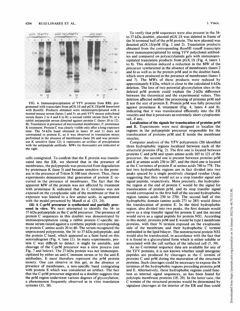
FIG. 4. Immunoprecipitation of YFV proteins from RRL pro-grammed with transcripts from pGX.1S and pGX.lS/prM linearizedwith BamHI. Products obtained were immunoprecipitated with anormal mouse serum (lanes 1 and 5); an anti-YFV mouse polyclonalserum (lanes 2 to 4 and 6 to 8); a normal rabbit serum (lane 9); or a
rabbit antipeptide serum directed against protein C (lanes 10 to 12).M, Translation in presence of microsomal membranes; P, proteinaseK treatment. Protein C was clearly visible only after a long exposuretime. The 54-kDa band obtained in lanes 10 and 11 does notcorrespond to protein E, as it was observed in translation mixesperformed in the absence of membranes (lane 10) and was protein-ase K sensitive (lane 12); it represents an artifact of precipitationwith the antipeptide antibody. MWs (in thousands) are indicated atthe right.
cells comigrated. To confirm that the E protein was translo-cated into the ER, we showed that in the presence ofmembranes, the polypeptide was protected from degradationby proteinase K (lane 3) and became sensitive to the prote-ase in the presence of Triton X-100 (not shown). Thus, theseexperiments demonstrate that generation of protein E oc-curred in the presence of membranes. The fact that theapparent MW of the protein was not affected by treatmentwith proteinase K indicated that its C terminus was notexposed on the cytoplasmic side of the membrane or that itsexposure was limited to a few amino acids, in agreementwith the model presented by Mandl et al. (23, 24).
(iii) A C-prM precursor is synthesized and partially proc-
essed in vitro. We next attempted to identify the 34- to37-kDa polypeptide as the C-prM precursor. The presence ofprotein C sequences in this doublet was demonstrated byimmunoprecipitation using a rabbit protein C-specific im-mune serum raised against a synthetic peptide correspondingto protein C amino acids 20 to 40. The serum recognized theunprocessed polyprotein, the 34- to 37-kDa polypeptide, andthe protein C band, which appeared as a faint band on thisautoradiogram (Fig. 4, lane 11). In many experiments, pro-
tein C was difficult to detect; it might be unstable, andcleavage of the C-prM precursor was a slow process (seeFig. 7 and below). The 27-kDa protein was not immunopre-cipitated by either an anti-C immune serum or by the anti-Eantibodies. It must therefore represent the prM proteinmoiety. One can observe in this gel, in the absence or
presence of membranes, a non-specific band comigratingwith protein E which was considered an artifact. The factthat the C-prM precursor migrated as a doublet suggests thatthe prM region underwent various degrees of glycosylation,a phenomenon frequently observed in in vitro translationsystems (31, 38).
To verify that prM sequences were also present in the 34-to 37-kDa doublet, plasmid pGX.1S was deleted in frame ofthe N-terminal half of the prM protein. The new plasmid wasdenoted pGX.1S/prM (Fig. 1 and 2). Translation productsobtained from the corresponding BamHI runoff transcriptswere immunoprecipitated by using YFV polyclonal antibod-ies and compared on polyacrylamide gels with immunopre-cipitated translation products from pGX.1S (Fig. 4, lanes 1to 8). This deletion induced a reduction in the MW of theprecursor synthesized in the absence of membranes (lanes 2and 6) as well as in the protein prM and in the doublet band,which were produced in the presence of membranes (lanes 3and 7). The MWs of these products were reduced byapproximately 8 kDa, which is close to the calculated 6-kDadeletion. The loss of two potential glycosylation sites in thedeleted prM protein could explain the 2-kDa differencebetween the theoretical and the experimental values. Thisdeletion affected neither the processing of proteins prM andE nor the size of protein E. Protein prM was fully protectedagainst proteinase K treatment (Fig. 4, lanes 4 and 8),indicating that it was translocated efficiently into the ERvesicles and that it possesses an extremely short cytoplasmictail, if any.
Localization of the signals for translocation of proteins prMand E. Experiments were next designed to localize theregions in the polypeptide precursor responsible for thetranslocation of proteins prM and E inside the membranevesicles.Computer analysis of the YFV polyprotein (20) identified
three hydrophobic regions localized between each of thestructural proteins (Fig. 2). The first one is located betweenproteins C and prM and spans amino acids 105 to 121 of theprecursor, the second one is present between proteins prMand E at amino acids 250 to 287, and the third one is locatedat the C terminus of protein E at amino acids 740 to 778. Thelast two hydrophobic regions are in fact divided into twopeaks spaced by a single positively charged residue (Arg),suggesting that they would act as a stop transfer signal andsignal peptide, respectively. More precisely, the hydropho-bic region at the end of protein C would be the signal fortranslocation of protein prM, and its stop transfer signalwould correspond to the first half of the second hydrophobicregion (amino acids 250 to 270). The second half of thishydrophobic domain (amino acids 271 to 285) would directthe translocation of protein E. In the third hydrophobicregion, also divided into two peaks, the first domain wouldserve as a stop transfer signal for protein E and the secondwould serve as a signal peptide for protein NS1. Accordingto this model, proteins prM and E would be type I membraneproteins, with their N termini exposed on the exoplasmicside of the membrane and their hydrophobic C terminiembedded in the lipid bilayer. The nonstructural protein NS1would also be translocated, in accordance with the fact thatit is found in a glycosylated form which is either soluble orassociated with the cell surface of the infected cell (5, 39).As no C-terminal sequence data are available for any of
the YFV proteins, it is not known whether small intergenicpeptides are produced by cleavages at the C termini ofproteins C and prM during the maturation of the structuralproteins. Such cleavages could be necessary to expose the Nterminus of the hydrophobic regions preceding proteins prMand E. Alternatively, these hydrophobic regions could func-tion as internal signal sequences, as has been found forpolytopic membrane proteins (18, 29). In the latter case, theC termini of the structural proteins would be determined bysignalase cleavages at the interior of the ER and thus could
J. VIROL.
Dow
nloa
ded
from
http
s://j
ourn
als.
asm
.org
/jour
nal/j
vi o
n 16
Jan
uary
202
2 by
46.
70.1
51.9
8.

YFV PROTEIN PROCESSING 4205
c ;X-M2 G.X 4M2_S XGX.4X4E 'S
:123 4 5 t 8 3 1C li. 1s' 1 14115 1M
12 4.35 !
AnGX 4M2
1 2 3 4 5 -67.-. l 2 3 4 5 6 7
46
* -92
_ - 69-46
.W. _-30-w...'..v.
* _ .O~~~~~~~1
30
B
< pGX.4El
z 1 2 3 4 5-69
46
1 m _
- 30
M M M MEH P P
T
MM9TP P P P
T
FIG. 5. Localization of regions essential for the translocation ofproteins prM and E. Transcripts obtained from templates linearizedat the BamHI site were translated in the presence or absence ofmembranes (M) and submitted to a proteinase K treatment (P) in thepresence or absence of Triton X-100 (T). Details of the polypeptidesencoded by each transcript are given in Fig. 2. MWs (in thousands)are indicated at the right.
coincide with the N termini of proteins prM and E. How-ever, this also implies that the C protein would be associatedwith membranes.To define the role played by the hydrophobic regions of
the precursor as signals for translocation of proteins prM andE, several plasmids were constructed.Plasmid pGX4 was derived from plasmid pGYF5' by
deletion of the YFV coding region and insertion downstreamof the initiating ATG codon of the multiple cloning site fromplasmid pGem4 (Fig. 1B). This plasmid produces in vitro T7transcripts containing the 5' noncoding region of YFV. Theinitiation codon was conserved and followed by the multiplecloning site, which allowed the cloning in phase and efficientin vitro expression of different open reading frames (35).Different fragments of cDNA coding for various YFV struc-tural proteins were then subcloned into plasmid pGX4 inorder to produce plasmids with N-terminal deletions in thepolyprotein precursor (Fig. 1C and 2). Four plasmids werethus constructed. pGX.4M2 coded for a polypeptide begin-ning with the hydrophobic region from the C terminus ofprotein C, and plasmid pGX.4M2/S coded for a polypeptidelacking this hydrophobic region but beginning at the thirdamino acid of protein prM. Plasmid pGX4.E1 expressed theregion coding for protein E preceded by the second hydro-phobic peak located after arginine 270 and present at the Cterminus of protein prM. The last construct, pGX.4E/S,coded for a polypeptide beginning 5 amino acids upstreamfrom the N terminus of the mature E protein.The polyprotein expressed from pGX4M2 transcripts was
processed, and proteins prM and E were efficiently translo-cated (Fig. 5, lanes 1 to 4), indicating that the first 103 aminoacids of the capsid protein are unnecessary for translocation.Furthermore, the 37- to 34-kDa doublet disappeared, aswould be expected for the C-prM precursor.The sequence coding for the hydrophobic region at the C
terminus of protein C was deleted in plasmid pGX.4M2/S.Thus the resulting transcript expressed a polyprotein, the Nterminus of which corresponded to the third amino acid of
M M M M M MEH P P P
T
FIG. 6. In vitro glycosylation of proteins prM and E. Translationproducts were submitted to proteinase K (P) or endoglycosidase H(EH) treatment as indicated. M, Microsomal membranes; T, TritonX-100. (A) Transcripts obtained from pGX.4M2 linearized at theHgaI or BamHI site (position 270 or 869 of the YFV polyprotein[Fig. 2]). (B) Transcripts obtained from pGX.4E1 linearized at thePstI site (position 674 of the YFV polyprotein [Fig. 2]). MWs (inthousands) are indicated at the right.
protein prM. In this case, protein E was translocated butprotein prM was not. Indeed, protein E was resistant toprotease treatment, but the 16-kDa polypeptide which mustrepresent the unglycosylated form of protein prM was not(Fig. 5, lanes 5 to 8). This result indicates that the regionessential for protein prM translocation is located within thelast 18 amino acids of protein C and that proteins prM and Ebear independent translocation signals.
Deletion of most of the protein prM, with the exception ofits C-terminal hydrophobic region (plasmid pGX.4E), did notalter the translocation of protein E (Fig. 5, lanes 9 to 12), butthe absence of this hydrophobic zone in plasmid pGX.4E/Sprevented the translocation of the protein (Fig. 5, lanes 13 to16). This suggests that the last 15 amino acids in protein prMare necessary for the translocation of protein E, whichconfirms the prediction based on the rule of von Heijne (44,45) that this region exhibits all the characteristics of a signalpeptide. This also confirms the results of our recent in vivostudies using a simian virus 40 expression vector whichindicate that the region within amino acids 271 to 285 of theYFV polyprotein acts as a signal peptide for the transloca-tion of the envelope protein as well as for the heterologouspoliovirus VPO protein (13; P. Despres et al., manuscript inpreparation).From the experiments carried out with transcripts from
pGX.4E/S, it is not clear whether NS1 was translocated ornot (Fig. 6, lanes 13 to 16). However, the polypeptidecontains the putative signal sequence for its translocation,and if processing of protein NS1 had occurred in the pres-ence of membranes, the apparent MW of the precursorwould have been reduced by approximately 10 kDa, gener-ating two polypeptides of 10 and 60 kDa, respectively. The10-kDa polypeptide was not detected, possibly because of itssmall size. In addition, analysis of the processed polypep-tides (lane 14) did reveal the presence of a band about 10 kDasmaller than the precursor. However, since a similar poly-peptide was synthesized in the absence of membranes, anunambiguous conclusion could not be drawn. These exper-iments indicate merely that the translocation of protein E did
M M NI M M MP P P P
T T
VOL. 63, 1989
Dow
nloa
ded
from
http
s://j
ourn
als.
asm
.org
/jour
nal/j
vi o
n 16
Jan
uary
202
2 by
46.
70.1
51.9
8.
4206 RUIZ-LINARES ET AL.
not occur, since no polypeptide was protected from protein-ase K digestion (lane 15).
Protein prM is efficiently glycosylated in vitro, whereasprotein E is not. The precursor to protein M, prM, possessesfour potential N glycosylation sites located at amino acids134, 150, 172, and 266 of the YFV polyprotein. The site atposition 266 is probably not used, since it is located in thetransmembrane hydrophobic region of the protein. The threeremaining sites are located in the N-terminal half of proteinprM, a region not conserved in protein M. As the predictedMW for the prM backbone is 21 kDa, the observed MW of 27kDa in our in vitro experiments suggests that this protein isefficiently glycosylated.
In order to demonstrate the in vitro glycosylation ofprotein prM, we synthesized transcripts from pGX.4M2linearized at the HgaI site (Fig. 1C and 2). These transcriptsshould code for a polypeptide which contains at its Nterminus the signal for translocation of protein prM but hasdeleted at its C terminus the 15 amino acids which precedethe sequence of the envelope protein. The encoded polypep-tide should have a MW close to that of the native prMprotein. The pGX.4M2/HgaI transcripts code for a polypep-tide of 18 kDa that is partially transformed into a 27-kDapolypeptide in the presence of membranes (Fig. 6A).
This polypeptide is glycosylated, as evidenced by itssensitivity to endoglycosidase H treatment (lane 3). Asexpected, the glycosylated form of prM is protected fromproteinase K digestion (lane 4) but is digested in the presenceof detergent (lane 5). A similar 27-kDa polypeptide is alsosynthesized and processed in translation reactions carriedout with longer BamHI transcripts (lanes 6 and 7), indicatingthat the C terminus of prM is in close proximity to the HgaIsite.
Protein E possesses two potential N-linked glycosylationsites at asparagine residues 594 and 755 of the YFV poly-protein. As in the case of protein prM, the second site isthought to be nonfunctional because it is located in theC-terminal hydrophobic region at the C terminus of theprotein. To investigate whether asparagine 594 could beeffectively recognized, in vitro transcripts were synthesizedfrom plasmid pGX4.E1 linearized with PstI. These tran-scripts should code for the N-terminal region of protein Econtaining asparagine 594 followed by 20 amino acids. Anexpected polypeptide of 40 kDa was synthesized upontranslation (Fig. 6B, lane 1). In this size range it should beeasy to detect small variations in MW due to the addition ofpolysaccharide residues. Translocation products obtained inthe presence of membranes (Fig. 6B, lane 2) were treatedwith proteinase K (lanes 4 and 5) and endoglycosidase H(lane 3). The observed product did not change in MW afterthe addition of membranes or after endoglycosidase Htreatment, indicating that no glycosylation had occurred.However, the polypeptide was efficiently translocated, asdemonstrated by its resistance to proteinase K in the ab-sence of Triton X-100. The lack of glycosylation of protein Eis in agreement with previous observations (17) and with thefact that the only accessible glycosylation site in the proteinis found in a weak context for glycosylation.
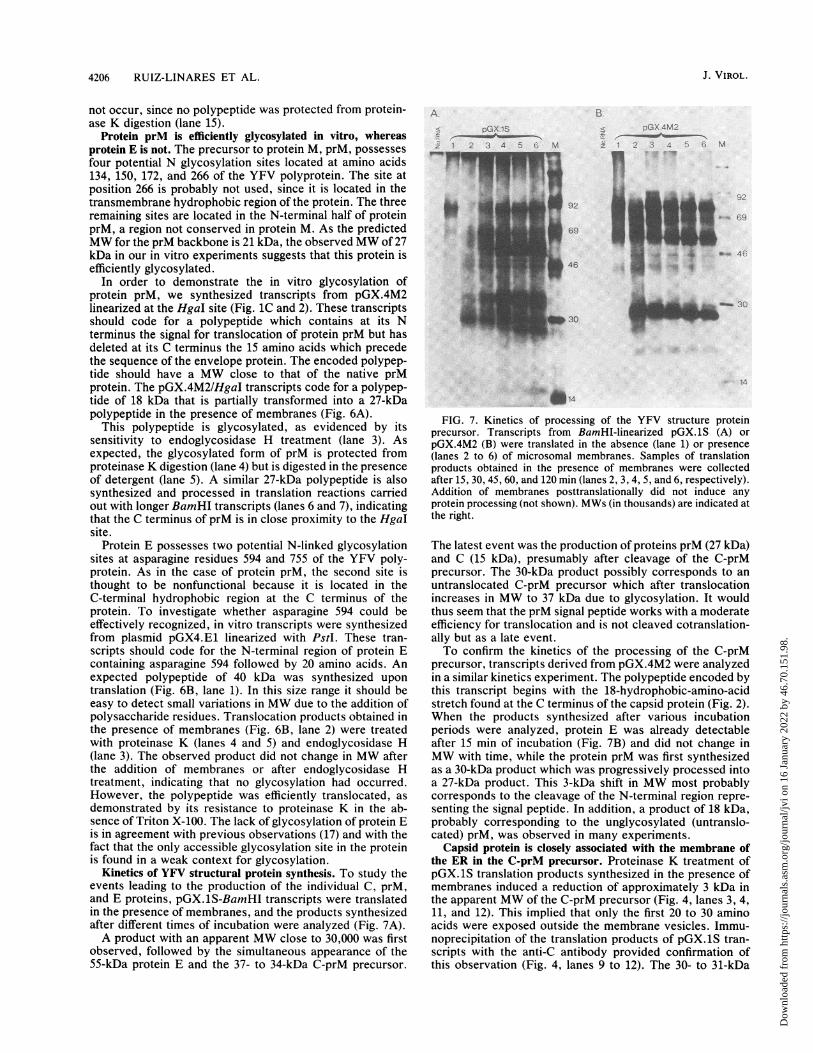
Kinetics of YFV structural protein synthesis. To study theevents leading to the production of the individual C, prM,and E proteins, pGX.lS-BamHI transcripts were translatedin the presence of membranes, and the products synthesizedafter different times of incubation were analyzed (Fig. 7A).A product with an apparent MW close to 30,000 was first
observed, followed by the simultaneous appearance of the55-kDa protein E and the 37- to 34-kDa C-prM precursor.
ApGX IS
_ 2 3 4 5 6 M
.i
pI.-
B.
- 45:- i , D -_ .X ..) .
}
c92
69
.. wo-
M
L46
,303o~~~~~~~~~~~~~~~~~~~~~~~~~~~~~~~~~~~~~~~~~~~~~~~.3.
14~~~~~~~~~~~~~1
FIG. 7. Kinetics of processing of the YFV structure proteinprecursor. Transcripts from BamHI-linearized pGX. 1S (A) orpGX.4M2 (B) were translated in the absence (lane 1) or presence(lanes 2 to 6) of microsomal membranes. Samples of translationproducts obtained in the presence of membranes were collectedafter 15, 30, 45, 60, and 120 min (lanes 2, 3, 4, 5, and 6, respectively).Addition of membranes posttranslationally did not induce anyprotein processing (not shown). MWs (in thousands) are indicated atthe right.
The latest event was the production of proteins prM (27 kDa)and C (15 kDa), presumably after cleavage of the C-prMprecursor. The 30-kDa product possibly corresponds to anuntranslocated C-prM precursor which after translocationincreases in MW to 37 kDa due to glycosylation. It wouldthus seem that the prM signal peptide works with a moderateefficiency for translocation and is not cleaved cotranslation-ally but as a late event.To confirm the kinetics of the processing of the C-prM
precursor, transcripts derived from pGX.4M2 were analyzedin a similar kinetics experiment. The polypeptide encoded bythis transcript begins with the 18-hydrophobic-amino-acidstretch found at the C terminus of the capsid protein (Fig. 2).When the products synthesized after various incubationperiods were analyzed, protein E was already detectableafter 15 min of incubation (Fig. 7B) and did not change inMW with time, while the protein prM was first synthesizedas a 30-kDa product which was progressively processed intoa 27-kDa product. This 3-kDa shift in MW most probablycorresponds to the cleavage of the N-terminal region repre-senting the signal peptide. In addition, a product of 18 kDa,probably corresponding to the unglycosylated (untranslo-cated) prM, was observed in many experiments.
Capsid protein is closely associated with the membrane ofthe ER in the C-prM precursor. Proteinase K treatment ofpGX.iS translation products synthesized in the presence ofmembranes induced a reduction of approximately 3 kDa inthe apparent MW of the C-prM precursor (Fig. 4, lanes 3, 4,11, and 12). This implied that only the first 20 to 30 aminoacids were exposed outside the membrane vesicles. Immu-noprecipitation of the translation products of pGX.1S tran-scripts with the anti-C antibody provided confirmation ofthis observation (Fig. 4, lanes 9 to 12). The 30- to 31-kDa
J. VIROL.
:.ml T - ---
A. :. -,la, "'..
Tf
Dow
nloa
ded
from
http
s://j
ourn
als.
asm
.org
/jour
nal/j
vi o
n 16
Jan
uary
202
2 by
46.
70.1
51.9
8.

YFV PROTEIN PROCESSING 4207
doublet observed after proteinase K digestion was stillrecognized by the C-specific antibodies (Fig. 4, lanes 11 and12). Since this antibody is directed against capsid proteinamino acids 20 to 40, the region digested by proteinase Kmust not extend very much further than the first 20 aminoacids. As the capsid protein is fairly hydrophobic fromresidue 42 onward, it is possible that the rest of the proteinis closely associated with the membrane and thus is pro-tected from proteinase K digestion.
Unfortunately, it was not possible to verify whether thispartial resistance of protein C to proteinase K digestion alsooccurred when it was cleaved from the C-prM precursor,because protein C could only be detected clearly on gelsafter long exposure times. This problem was worse whenimmunoprecipitations were performed. For this reason andbecause of the poor resolving power of these gels forlow-MW polypeptides, we could not determine whether thecapsid protein was partially or totally degraded after protein-ase K treatment.
DISCUSSION
We have established an in vitro transcription-translationsystem to study the processing of the YFV polyprotein andto define the role played by cellular proteases in the matu-ration of the viral structural proteins.By using this approach, it was found that (i) production of
proteins C, prM, and E was dependent on the addition ofmicrosomal membranes to the translation system. ProteinsprM and E were translocated inside the ER membrane,where protein prM, but not protein E, was glycosylated. Thetranslocated proteins are totally resistant to proteinase Kdigestion, indicating that they do not have a domain locatedin the cytoplasmic side of the membrane but most likely areanchored in the membrane by the hydrophobic regionspresent at their C termini. Proteins prM and E are thusmembrane proteins of the type I, with their N terminiexposed on the exoplasmic side of the membrane and their Ctermini anchored in it.
(ii) Translocation of proteins prM and E inside the ERvesicles is dependent on the last 18 or 15 amino acids presentat the carboxylic ends of proteins C and prM, respectively.These two signals function independently of each other; theyare active when located in an N-terminal position from theprotein they translocate, and the prM signal, at least, canalso function as an internal translocation sequence.
(iii) The prM signal seems to be less efficient for translo-cation than the E signal, since a significant amount of prM isnot translocated inside the ER membrane, whereas nearly allof the E protein is translocated. Furthermore, cleavage ofthe prM signal is not complete and takes place after trans-location of the protein, as evidenced by the constant pres-ence of a translocated C-prM precursor.
(iv) Protein C is not translocated inside the ER vesicles butremains very closely associated with the ER membrane byits C terminus, at least in the form of the translocated C-prMprecursor. The existence of the translocated C-prM precur-sor suggests that previous exposure of the N terminus of thehydrophobic region preceding protein prM is not essentialfor its translocation. Thus, it would function as an internalsignal sequence.These results confirmed the schematic representation pro-
posed by Coia et al. (10), which demonstrate the dependenceof the processing of the YFV polyprotein on cellular prote-ases. The cellular protease involved must be of the signalasetype, recognizing cleavage sites located after signals for
translocation at the N-terminal parts of prM and E proteins.Cleavage of prM to produce M was not effected in oursystem, suggesting that it is mediated by a protease presentin the export vesicles of the cell. Cleavage of the C terminusof the capsid protein seems to be initially performed by thesignalase that liberates the N terminus of prM. This wouldleave a capsid protein associated with membranes, the Cterminus of which has to be cleaved a second time in orderto make the capsid protein available for RNA encapsidation.It is possible that the second cleavage is effected by the viralprotease responsible for the cleavages of the polyproteinprecursor yielding the nonstructural proteins. This enzymerecognizes pairs of basic residues followed by a small-side-chain amino acid. Such a sequence is found just Nterminal to the membrane-spanning domain of the capsidprotein and is conserved in most flaviviruses (23). Matura-tion of the virus could thus be regulated by complex kineticsof cleavage, leading to isolated capsid, membrane, andenvelope proteins.
Processing of the YFV polyprotein seems to be an inter-esting model of polytopic protein processing. Indeed, aseries of translocation and stop transfer signals are foundseparating each structural protein and protein NS1. Thestructural proteins of the Flaviviridae seem to be unique inthat the stop transfer and signal for translocation are locatedin a single hydrophobic region, separated only by a basicresidue. In the present report we have delimited the regionsessential for translocation of the prM and E proteins. Fur-ther studies are needed to show whether these regions aresufficient for translocation and whether they are signalrecognition particle dependent, which would confirm theirrole as signal peptides (46). Further characterization of theregions acting as stop transfer signals is needed in order torelate the internal translocation signal to those found in typeII membrane proteins. This seems particularly relevant inthe case of the hydrophobic region separating proteins C andprM, as the C-prM precursor has an orientation relative tothe ER membrane which is typical of type II proteins with acytoplasmic N terminus and a luminal C terminus. Interest-ingly, the two potential signal peptides differ in their com-petence for translocation as well as in the kinetics withwhich they are cleaved off. Such a difference in signalefficiency could be important for the regulation of the virallife cycle, as it would regulate the rate of maturation of virusparticles.
ACKNOWLEDGMENTS
We thank R. Landenheim for stimulating discussions and J.Schlesinger, A. Barrett, and F. Rodhain for providing monoclonalantibodies and hyperimmune sera.
This work was supported in part by contract 85/120 from DRET.A.R.L. was a fellow of the International Network of Biotechnology.
LITERATURE CITED1. Bell, J. R., R. M. Kinney, D. W. Trent, E. M. Lenches, L.
Dalgarno, and J. M. Strauss. 1985. N-terminal amino acidsequences of structural proteins of three flaviviruses. Virology143:224-229.
2. Biedrizycka, A., M. R. Cauchi, A. Darthomoeusz, J. J. Gorman,and P. Wright. 1987. Characterization of protease cleavage sitesinvolved in the formation of the envelope glycoprotein and threenonstructural proteins of dengue virus type 2, New Guinea Cstrain. J. Gen. Virol. 68:1317-1326.
3. Blobel, G., and B. Dobbertstein. 1975. Transfer of protein across
membranes. I. Presence of proteolytic processed and unproc-essed nascent immunoglobulin light chains on membrane-boundribosomes of murine myeloma. J. Cell. Biol. 67:835-851.
VOL. 63, 1989
Dow
nloa
ded
from
http
s://j
ourn
als.
asm
.org
/jour
nal/j
vi o
n 16
Jan
uary
202
2 by
46.
70.1
51.9
8.

4208 RUIZ-LINARES ET AL.
4. Boege, U., P. X. Heinz, G. Wengler, and C. Kunz. 1983. Aminoacid composition and amino terminal sequences of the structuralproteins of a flavivirus, European tick-borne encephalitis virus.Virology 126:651-657.
5. Cardiff, R. D., and J. K. Lund. 1976. Distribution of dengue-2antigens by electron immunocytochemistry. Infect. Immun.13:1699-1709.
6. Castle, E., U. Leidner, T. Nowak, G. Wengler, and G. Wengler.1986. Primary structure of the West Nile flavivirus genomeregion coding for all nonstructural proteins. Virology 149:10-26.
7. Castle, E., T. Nowak, U. Leidner, G. Wengler, and G. Wengler.1985. Sequence analysis of the viral core protein and themembrane-associated proteins Vl and NV2 of the flavivirusWest Nile virus and of the genome sequence for these proteins.Virology 145:227-236.
8. Chen, E. G., and P. H. Seeburg. 1985. DNA Lab. Methods4:165-170.
9. Cleaves, G. R. 1985. Identification of dengue type 2 virus-specific high molecular weight proteins in virus-infected BHKcells. J. Gen. Virol. 66:2767-2771.
10. Coia, G., M. D. Parker, G. Speight, M. E. Byrne, and E. G.Westaway. 1988. Nucleotide and complete amino acid sequenceof Kunjin virus. Definitive gene order and characteristics of thevirus-specified proteins. J. Gen. Virol. 69:1-21.
11. Crawford, G. R., and P. J. Wright. 1987. Characterization ofnovel viral polyproteins detected in cells infected by the flavi-virus Kunjin and radiolabelled in the presence of the leucineanalogue hydroxyleucine. J. Gen. Virol. 68:365-376.
12. Despres, P., A. Cahour, A. Dupuy, V. Deubel, M. Bouloy, J. P.Digoutte, and M. Girard. 1987. High genetic stability of theregion coding for the structural proteins of yellow fever virusstrain 17D. J. Gen. Virol. 68:2245-2247.
13. Despres, P., A. Cahour, C. Wychowski, M. Girard, and M.Bouloy. 1988. Expression of the yellow fever virus envelopeprotein using hybrid SV40/yellow fever viruses. Ann. Inst.Pasteur Virol. 139:59-67.
14. Despres, P., V. Deubel, M. Bouloy, and M. Girard. 1986.Identification and characterization of intracellular yellow fevervirus-specific RNA: absence of subgenomic RNA. Ann. Inst.Pasteur Virol. 137:193-204.
15. Deubel, V., R. M. Kinney, and D. W. Trent. 1986. Nucleotidesequence and deduced amino acid sequence of the structuralproteins of dengue type 2 virus, Jamaican genotype. Virology155:365-377.
16. Deubel, V., R. M. Kinney, and D. W. Trent. 1988. Nucleotidesequence and deduced amino acid sequence of the nonstructuralproteins of Dengue type 2 virus, Jamaica genotype: comparativeanalysis of the full length genome. Virology 165:234-244.
17. Deubel, V., J. J. Schlesinger, J. P. Digoutte, and M. Girard.1987. Comparative immunochemical and biological analysis ofAfrican and South American yellow fever viruses. Arch. Virol.94:331-339.
18. Friedlander, M., and B. Blobel. 1985. Bovine opsin has morethan one signal sequence. Nature (London) 318:338-443.
19. Garoff, H. 1985. Using recombinant DNA techniques to studyprotein targeting in the eukaryotic cell. Annu. Rev. Cell Biol.1:403-445.
20. Kyte, J., and R. F. Doolittle. 1982. A simple method fordisplaying the hydropathic character of a protein. J. Mol. Biol.157:105-132.
21. Laemmli, U. K. 1970. Cleavage of the structural proteins duringthe assembly of the head of bacteriophage T4. Nature (London)227:680-685.
22. Lyapustin, V. N., Y. V. Svitkin, T. Y. Ugarova, V. A. Lash-kevich, and V. I. Agol. 1986. A tentative model of formation ofstructural proteins of tick-borne encephalitis virus (flavivirus).FEBS Lett. 200:314-316.
23. Mandl, C. W., F. Guirakhoo, H. Holzmann, F. Heinz, and C.Kunz. 1989. Antigenic structure of the flavivirus envelopeprotein E at the molecular level, using tick-borne encephalitisvirus as a model. J. Virol. 63:564-571.
24. Mandl, C. W., F. X. Heinz, and C. Kunz. 1988. Sequence of thestructural proteins of the tick-borne encephalitis virus (Western
subtype) and comparative analysis with other flavivirus. Virol-ogy 166:197-205.
25. Maniatis, T., E. F. Fritsch, and J. Sambrook. 1982. Molecularcloning: a laboratory manual. Cold Spring Harbor Laboratory,Cold Spring Harbor, N.Y.
26. Melancon, P., and H. Garoff. 1986. Reinitiation of translocationin the Semliki forest virus structural polyprotein: identificationof the signal for the El glycoprotein. EMBO J. 5:1551-1560.
27. Monath, T. P. 1986. Pathology of the flaviviruses, p. 375-440. InS. Schlesinger and M. J. Schlesinger (ed.), The Togaviridae andFlaviviridae. Academic Press, Inc., New York.
28. Monckton, R. P., and E. S. Westaway. 1982. Restricted transla-tion of the genome of the flavivirus Kunjin in vitro. J. Gen.Virol. 63:227-232.
29. Mueckler, M., and H. F. Lodish. 1986. The human glucosetransporter can insert posttranslationally into microsomes. Cell44:629-637.
30. Ozden, S., and B. Poirier. 1985. Dengue virus induced polypep-tide synthesis. Arch. Virol. 85:129-137.
31. Perara, E., and V. R. Lingappa. 1985. A former amino terminalsignal sequence engineered to an internal location directs trans-location of both flanking protein domains. J. Cell. Biol. 101:2292-2301.
32. Rice, C. M., R. Aebersold, D. B. Teplow, J. Pata, J. R. Bell,A. V. Varndam, D. W. Trent, M. W. Brandiss, J. J. Schlesinger,and J. H. Strauss. 1986. Partial N termini amino acid sequencesof three nonstructural proteins of two flaviviruses. Virology151:1-9.
33. Rice, C. M., E. M. Lenches, S. R. Eddy, S. J. Shin, R. L. Sheets,and J. H. Strauss. 1985. Nucleotide sequence of yellow fevervirus: implications for flavivirus gene expression and evolution.Science 229:726-733.
34. Rice, C. M., and J. H. Strauss. 1986. Structure of the flavivirusgenome, p. 279-326. In S. Schlesinger and M. J. Schlesinger(ed.), The Togaviridae and Flaviviridae. Academic Press, Inc.,New York.
35. Ruiz-Linares, A., M. Bouloy, M. Girard, and A. Cahour. 1989.Modulations of the in vitro translational efficiencies of yellowfever virus in RNAs: interactions between coding and noncod-ing regions. Nucleic Acids Res. 17:2463-2476.
36. Schlesinger, J. J., M. W. Brandiss, and T. P. Monath. 1983.Monoclonal antibodies distinguish between wild and vaccinestrains of yellow fever virus by neutralisation, hemagglutina-tion, inhibition and immune precipitation of the virus envelope.Virology 125:8-17.
37. Speight, S., G. Coia, M. D. Parker, and E. G. Westaway. 1988.Gene mapping and positive identification of the nonstructuralproteins NS2A, NS2B, NS3 and NS5 of the flavivirus Kunjinand their cleavage sites. J. Gen. Virol. 69:23-34.
38. Spiess, M., and H. F. Lodish. 1986. An internal signal sequence:the asialoglycoprotein receptor membrane anchor. Cell 44:177-185.
39. Stohlman, S. A., C. L. Wisseman, 0. R. Eylar, and D. J.Silverman. 1975. Dengue virus-induced modifications of hostcell membranes. J. Virol. 16:1017-1026.
40. Strauss, J. H., E. G. Strauss, C. S. Hahn, and C. M. Rice. 1987.The genomes of alphaviruses and flaviviruses: organization andtranslation, p. 75-104. In D. J. Rowlands, M. A. Mahy, andB. W. J. Mahy (ed.), The molecular biology of the positivestrand RNA viruses. Academic Press, Inc. (London), Ltd.,London.
41. Strauss, J. H., E. G. Strauss, C. S. Hahn, Y. S. Hahn, R. Galler,W. R. Hardy, and C. M. Rice. 1987. Replication of alphavirusesand flaviviruses: proteolytic processing of polyproteins. UCLASymp. Mol. Cell. Biol. 49:209-225.
42. Svitkin, Y. V., T. Y. Ugarova, T. V. Chernovskaya, V. N.Lyapustin, V. A. Lashkevich, and V. I. Agol. 1981. Translationof tick-borne encephalitis virus (Flavivirus) genome in vitro:synthesis of two structural polypeptides. Virology 110:26-34.
43. Svitkin, Y. V., V. N. Lyapustin, V. A. Lashkevich, and V. I.Agol. 1984. Differences between translation products of tick-borne encephalitis virus RNA in cell-free systems from Krebs-2cells and rabbit reticulocytes: involvement of membranes in the
J. VIROL.
Dow
nloa
ded
from
http
s://j
ourn
als.
asm
.org
/jour
nal/j
vi o
n 16
Jan
uary
202
2 by
46.
70.1
51.9
8.

YFV PROTEIN PROCESSING
processing of nascent precursors of flavivirus structural pro-
teins. Virology 135:536-541.44. von Heijne, G. 1985. Signal sequences. The limits of variation. J.
Mol. Biol. 184:99-105.45. von Heijne, G. 1986. A new method for predicting signal
sequence cleavage site. Nucleic Acids Res. 14:4683-4690.46. Walter, P., and V. R. Lingappa. 1986. Mechanism of protein
translocation across the endoplasmic reticulum membrane.
Annu. Rev. Cell Biol. 2:499-516.47. Wengler, G., M. Beato, and G. Wengler. 1979. In vitro transla-
tion of 42S virus specific RNA from cells infected with theflavivirus West Nile virus. Virology 96:516-529.
48. Westaway, E. G., S. Y. Gaidamovitch, M. S. Horzinek, A.Igarashi, L. Kaariainen, D. K. Lvov, J. S. Porterfield, P. K. P.Russel, and D. W. Trent. 1985. Flaviviridae. Intervirology24:183-192.
VOL. 63, 1989 4209
Dow
nloa
ded
from
http
s://j
ourn
als.
asm
.org
/jour
nal/j
vi o
n 16
Jan
uary
202
2 by
46.
70.1
51.9
8.