probiotics and their efficacy in improving oral …probiotics and their efficacy in improving oral...
TRANSCRIPT
Journal of Applied Pharmaceutical Science Vol. 2 (11), pp. 151-163, November, 2012 Available online at http://www.japsonline.com DOI: 10.7324/JAPS.2012.21128 ISSN 2231-3354
Probiotics and Their Efficacy in Improving Oral Health: A Review Pranay Jain* and Priyanka Sharma Deptt.of Biotechnology, University Institute of Engineering & Technology Kurukshetra University, Kurukshetra, Haryana, India.
ARTICLE INFO
ABSTRACT
Article history: Received on: 13/10/2012 Revised on: 05/11/2012 Accepted on: 15/11/2012 Available online: 30/11/2012
The changing food habits and lifestyle has resulted in deterioration of oral health in the people of all ages. The increasing global problems with the traditional disease management strategies have prompted the investigators to hunt for the new and better alternatives to deal with health issues. The global demand for chemical free, less harmful and easier solutions to health problems has increased in past few years. Probiotics or the foods with ‘live cultures’ have come up as one of the most promising alternate to traditional disease management. Probiotics are those viable microorganisms which are constituents of natural microflora of human body. Probiotic therapy decreases the risk of colonization by oral pathogens without depleting the friendly microflora. Probiotics resembles the human body microbiota and are readily incorporated in the natural microflora of human body. They are harmless and easy to consume in many edible forms (such as cheese, yoghurt, etc.). The inability of the antibiotics to discriminate good bacteria from the disease causing bacteria, the development of antimicrobial resistant mutants and the increasing rate of antibiotic associated side effects and complications suggests an urgent need to switch our therapeutic approach from traditional antibiotics to the probiotic therapy for oral care. The use of probiotics in routine life is likely to improve the oral health. This review demonstrates the action of Probiotics on oral health and disease.
Key words: Probiotics, Live cultures, Antimicrobial Resistance, Probiotic Therapy, Microbiota.
INTRODUCTION
The term ‘Probiotic’ meaning “for life” was coined by Lilley and Stillwell (1965). Probiotics have amazingly come up with the potential for not only preventing the attack of oral pathogens but also the ability to treat various oral diseases. Thus, asssuring healthy living and increased longevity (Meurman, 2005). Probiotics reminds of the very old and forgotten concept of ‘Bacteriotherapy’ which stated that beneficial bacteria occurring naturally in the human body can be administered in the patient’s body to restore patient’s health and wellbeing (Meurman, 2005). Bacteriotherapy gave rise to the concept of modern day probiotics. Probiotics have been extensively studied for their intestinal benefits.
The human intestine has a reservoir of microorganisms naturally inhabiting the intestine as symbiont. They are referred to as ‘gut or the intestinal flora’. In lieu of the shelter that the human body provides, the intestinal flora performs several important functions in the human body such as fermenting
undigested energy substrate, strengthening the immune system, protection against the growth of the pathogenic bacteria, promoting gut development, production of vitamins (such as Vitamin K and Biotin) and production of hormones for fat storage. The process whereby probiotics are used to restore the normal intestinal microflora to provide resistance against antibiotics is termed ‘Microbial interference therapy’. Probiotics being safe for human consumption and resistant to bile and acidic environment survives in the intestine, colonize the human gut and show bacteriocin production to block the invasion of intestine cells by enteroinvasive bacteria(Parvez et al., 2006). On the other hand, Broad spectrum antibiotics, being unable to distinguish between beneficial and harmful bacteria, kill both and alter the number of natural microbiota. This results in a downfall in host’s health. Earlier, Probiotics were associated with only gut health but recently several investigators have suggested their potential applicability in the improvement of oral health. The organism capable of adhering to and colonize the surface of the oral cavity constitute ‘Oral Probiotics’. (Table-1) There is an urgent need to switch our therapeutic approach from traditional antibiotics to the probiotic therapy for oral care.
* Corresponding Author Dr. Pranay Jain, Assistant Professor, Deptt. Of Biotechnology, UIET, KU, Kurukshetra, Haryana, India.
152 Jain and Sharma / Journal of Applied Pharmaceutical Science 2 (11); 2012: 151-163
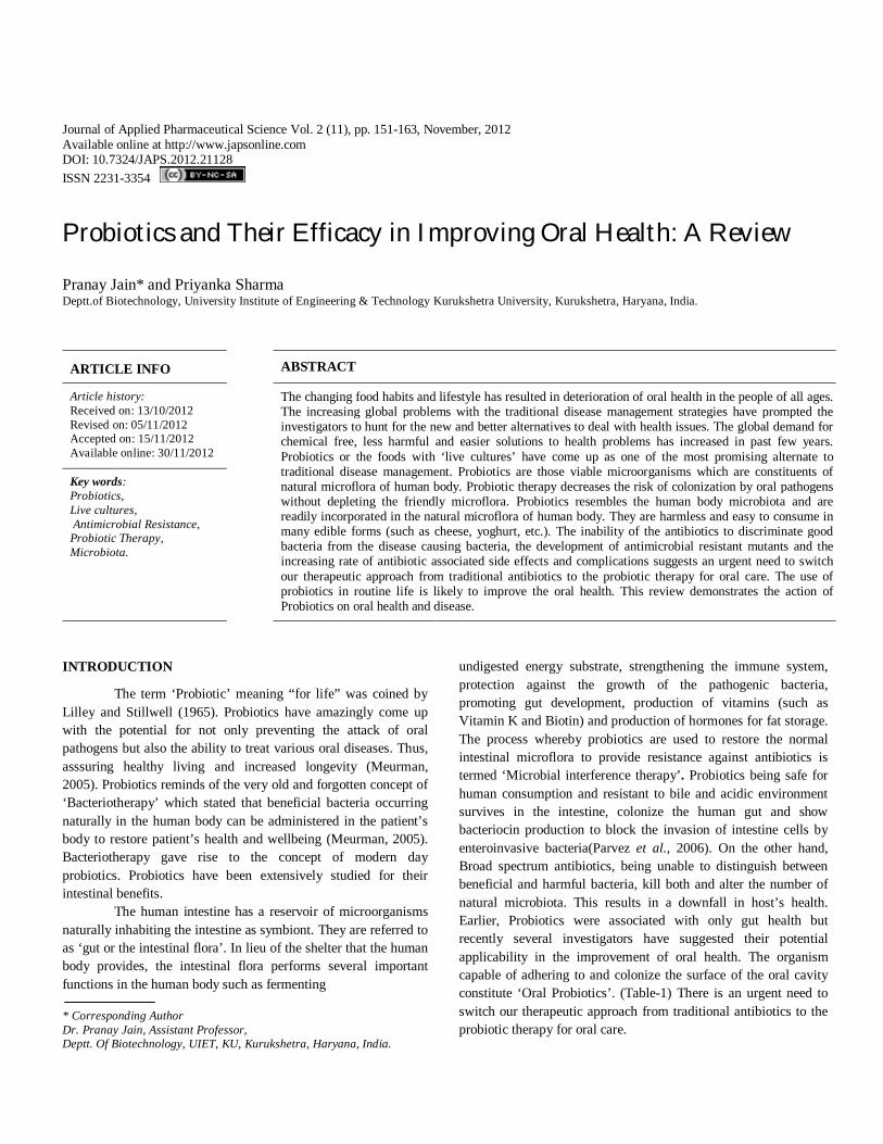
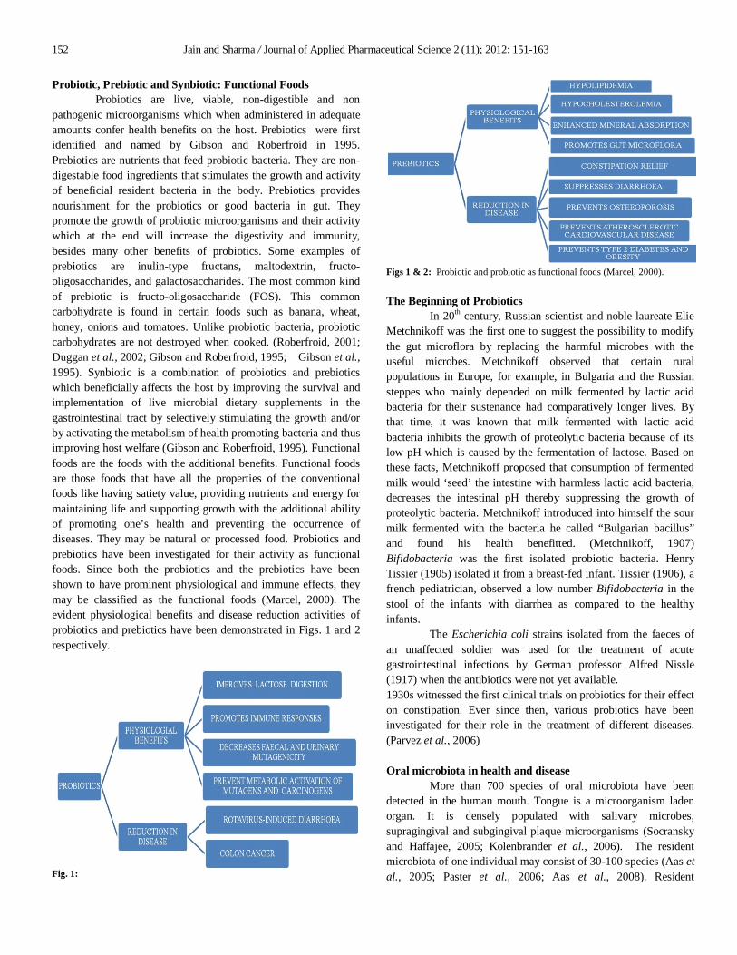
Probiotic, Prebiotic and Synbiotic: Functional Foods Probiotics are live, viable, non-digestible and non pathogenic microorganisms which when administered in adequate amounts confer health benefits on the host. Prebiotics were first identified and named by Gibson and Roberfroid in 1995. Prebiotics are nutrients that feed probiotic bacteria. They are non-digestable food ingredients that stimulates the growth and activity of beneficial resident bacteria in the body. Prebiotics provides nourishment for the probiotics or good bacteria in gut. They promote the growth of probiotic microorganisms and their activity which at the end will increase the digestivity and immunity, besides many other benefits of probiotics. Some examples of prebiotics are inulin-type fructans, maltodextrin, fructo-oligosaccharides, and galactosaccharides. The most common kind of prebiotic is fructo-oligosaccharide (FOS). This common carbohydrate is found in certain foods such as banana, wheat, honey, onions and tomatoes. Unlike probiotic bacteria, probiotic carbohydrates are not destroyed when cooked. (Roberfroid, 2001; Duggan et al., 2002; Gibson and Roberfroid, 1995; Gibson et al., 1995). Synbiotic is a combination of probiotics and prebiotics which beneficially affects the host by improving the survival and implementation of live microbial dietary supplements in the gastrointestinal tract by selectively stimulating the growth and/or by activating the metabolism of health promoting bacteria and thus improving host welfare (Gibson and Roberfroid, 1995). Functional foods are the foods with the additional benefits. Functional foods are those foods that have all the properties of the conventional foods like having satiety value, providing nutrients and energy for maintaining life and supporting growth with the additional ability of promoting one’s health and preventing the occurrence of diseases. They may be natural or processed food. Probiotics and prebiotics have been investigated for their activity as functional foods. Since both the probiotics and the prebiotics have been shown to have prominent physiological and immune effects, they may be classified as the functional foods (Marcel, 2000). The evident physiological benefits and disease reduction activities of probiotics and prebiotics have been demonstrated in Figs. 1 and 2 respectively.
Fig. 1:
Figs 1 & 2: Probiotic and probiotic as functional foods (Marcel, 2000). The Beginning of Probiotics In 20th century, Russian scientist and noble laureate Elie Metchnikoff was the first one to suggest the possibility to modify the gut microflora by replacing the harmful microbes with the useful microbes. Metchnikoff observed that certain rural populations in Europe, for example, in Bulgaria and the Russian steppes who mainly depended on milk fermented by lactic acid bacteria for their sustenance had comparatively longer lives. By that time, it was known that milk fermented with lactic acid bacteria inhibits the growth of proteolytic bacteria because of its low pH which is caused by the fermentation of lactose. Based on these facts, Metchnikoff proposed that consumption of fermented milk would ‘seed’ the intestine with harmless lactic acid bacteria, decreases the intestinal pH thereby suppressing the growth of proteolytic bacteria. Metchnikoff introduced into himself the sour milk fermented with the bacteria he called “Bulgarian bacillus” and found his health benefitted. (Metchnikoff, 1907) Bifidobacteria was the first isolated probiotic bacteria. Henry Tissier (1905) isolated it from a breast-fed infant. Tissier (1906), a french pediatrician, observed a low number Bifidobacteria in the stool of the infants with diarrhea as compared to the healthy infants. The Escherichia coli strains isolated from the faeces of an unaffected soldier was used for the treatment of acute gastrointestinal infections by German professor Alfred Nissle (1917) when the antibiotics were not yet available. 1930s witnessed the first clinical trials on probiotics for their effect on constipation. Ever since then, various probiotics have been investigated for their role in the treatment of different diseases. (Parvez et al., 2006) Oral microbiota in health and disease More than 700 species of oral microbiota have been detected in the human mouth. Tongue is a microorganism laden organ. It is densely populated with salivary microbes, supragingival and subgingival plaque microorganisms (Socransky and Haffajee, 2005; Kolenbrander et al., 2006). The resident microbiota of one individual may consist of 30-100 species (Aas et al., 2005; Paster et al., 2006; Aas et al., 2008). Resident
Jain and Sharma / Journal of Applied Pharmaceutical Science 2 (11); 2012: 151-163 153
microbiota plays many important functions such as reducing the susceptibility to pathogen attack, prevention of pathogen colonization and developing immune response against pathogens. Figure 3 demonstrates the various functions of the resident microbiota.
Fig. 3: Functions of the Resident Microbiota.
In the oral cavity, bacteria resides either integrated into the oral biofilm or in planktonic state. The integration of planktonic bacteria into biofilm results into the activation of certain distinct genes which differentiates them from the planktonic counterparts. Now they tend to be much more resistant to the environmental factors and the antimicrobial agents. Oral biofilm is dynamic and hence its composition keeps changing. The complexity of oral biofilm increases as they mature. (Burme et al., 1999; Rudney, 2000). Saliva is a complex medium in mouth which contains different bactericidal, bacteriostatic and inhibitory proteins that collectively may damage a variety of species in planktonic state. Ingested Probiotics are first exposed to the salivary proteins such as lysozyme, lactoferrin, histatin, salivary peroxidase, cystatins, and secretory IgA which affects the adhesion, morphology, metabolic activity and viability of the probiotic microorganism. On the other hand, Saliva tends to propagate oral biofilm and contributes to the microbial diversity in mouth. The continuous salivary flow in the oral cavity can lead to detachment of some microbes from the biofilm surfaces, modulating microbial colonization. Different strains show specific response to proteolytic enzymes and this strain-specific response need to be considered when selecting probiotics for the oral cavity (Germaine and Tellefson, 1986; Rudney et al.,1991; Bosch et al., 2003; Hahnel et al., 2008; Groschl et al., 2009;). Many bacterial species have been found to survive within buccal epithelial cells (Rudney et al., 2005). Lactobacilli from saliva samples include Lactobacillus paracasei, L. plantarum, L. rhamnosus, and L.
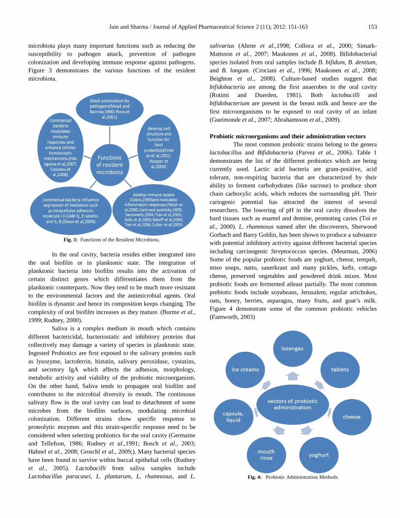
salivarius (Ahrne et al.,1998; Colloca et al., 2000; Simark-Mattsson et al., 2007; Maukonen et al., 2008). Bifidobacterial species isolated from oral samples include B. bifidum, B. dentium, and B. longum. (Crociani et al., 1996; Maukonen et al., 2008; Beighton et al., 2008). Culture-based studies suggest that bifidobacteria are among the first anaerobes in the oral cavity (Rotimi and Duerden, 1981). Both lactobacilli and bifidobacterium are present in the breast milk and hence are the first microorganisms to be exposed to oral cavity of an infant (Gueimonde et al., 2007; Abrahamsson et al., 2009). Probiotic microorganisms and their administration vectors The most common probiotic strains belong to the genera lactobacillus and Bifidobacteria (Parvez et al., 2006). Table 1 demonstrates the list of the different probiotics which are being currently used. Lactic acid bacteria are gram-positive, acid tolerant, non-respiring bacteria that are characterized by their ability to ferment carbohydrates (like sucrose) to produce short chain carboxylic acids, which reduces the surrounding pH. Their cariogenic potential has attracted the interest of several researchers. The lowering of pH in the oral cavity dissolves the hard tissues such as enamel and dentine, promoting caries (Toi et al., 2000). L. rhamnosus named after the discoverers, Sherwood Gorbach and Barry Goldin, has been shown to produce a substance with potential inhibitory activity against different bacterial species including carcinogenic Streptococcus species. (Meurman, 2006) Some of the popular probiotic foods are yoghurt, cheese, tempeh, miso soups, natto, sauerkraut and many pickles, kefir, cottage cheese, preserved vegetables and powdered drink mixes. Most probiotic foods are fermented atleast partially. The most common prebiotic foods include soyabeans, Jerusalem, regular artichokes, oats, honey, berries, asparagus, many fruits, and goat’s milk. Figure 4 demonstrate some of the common probiotic vehicles (Famworth, 2003)
Fig. 4: Probiotic Administration Methods.
154 Jain and Sharma / Journal of Applied Pharmaceutical Science 2 (11); 2012: 151-163
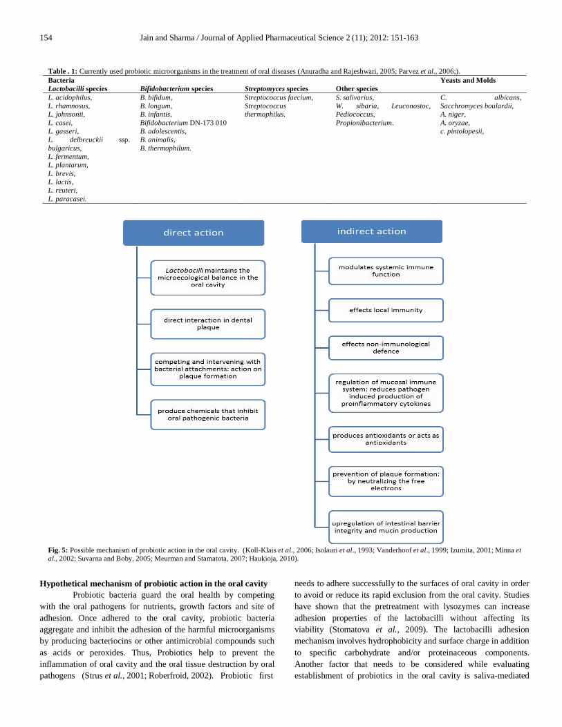
Hypothetical mechanism of probiotic action in the oral cavity Probiotic bacteria guard the oral health by competing with the oral pathogens for nutrients, growth factors and site of adhesion. Once adhered to the oral cavity, probiotic bacteria aggregate and inhibit the adhesion of the harmful microorganisms by producing bacteriocins or other antimicrobial compounds such as acids or peroxides. Thus, Probiotics help to prevent the inflammation of oral cavity and the oral tissue destruction by oral pathogens (Strus et al., 2001; Roberfroid, 2002). Probiotic first
needs to adhere successfully to the surfaces of oral cavity in order to avoid or reduce its rapid exclusion from the oral cavity. Studies have shown that the pretreatment with lysozymes can increase adhesion properties of the lactobacilli without affecting its viability (Stomatova et al., 2009). The lactobacilli adhesion mechanism involves hydrophobicity and surface charge in addition to specific carbohydrate and/or proteinaceous components. Another factor that needs to be considered while evaluating establishment of probiotics in the oral cavity is saliva-mediated
Table . 1: Currently used probiotic microorganisms in the treatment of oral diseases (Anuradha and Rajeshwari, 2005; Parvez et al., 2006;). Bacteria Yeasts and Molds Lactobacilli species Bifidobacterium species Streptomyces species Other species L. acidophilus, L. rhamnosus, L. johnsonii, L. casei, L. gasseri, L. delbreuckii ssp. bulgaricus, L. fermentum, L. plantarum, L. brevis, L. lactis, L. reuteri, L. paracasei.
B. bifidum, B. longum, B. infantis, Bifidobacterium DN-173 010 B. adolescentis, B. animalis, B. thermophilum.
Streptococcus faecium, Streptococcus thermophilus.
S. salivarius, W. sibaria, Leuconostoc, Pediococcus, Propionibacterium.
C. albicans, Sacchromyces boulardii, A. niger, A. oryzae, c. pintolopesii,
Fig. 5: Possible mechanism of probiotic action in the oral cavity. (Koll-Klais et al., 2006; Isolauri et al., 1993; Vanderhoof et al., 1999; Izumita, 2001; Minna et al., 2002; Suvarna and Boby, 2005; Meurman and Stamatota, 2007; Haukioja, 2010).
Jain and Sharma / Journal of Applied Pharmaceutical Science 2 (11); 2012: 151-163 155
aggregation. Those microorganisms that have the ability to co-aggregate may have greater advantage over non co-aggregating organisms which are easily removed from the mouth (He et al., 2001; Lorca et al., 2002; Carlen et al., 2003; Nikawa et al., 2004). The Probiotics have a three step action mechanism -
i. Stimulates and modulates immune response, ii. Normalize intestinal microflora
Ensures colonization resistance Controls irritable bowel syndrome and other
inflammatory bowel diseases. iii. And also have the metabolic effects like-
Bile salt deconjugation and secretion, Lactose hydrolysis, Reduction in toxigenic and mutagenic reactions
in gut. Supply of nutrients to colon epithelium.
Probiotics first competes with the oral pathogens for adhesion site and then colonizes the oral surface. After the probiotics aggregate the oral surface, they compete with oral pathogens for nutrients, growth factors and also produce antimicrobial compounds, including organic acids, hydrogen peroxide, carbon peroxide, diacetyl, low molecular weight antimicrobial substances, bacteriocins, and adhesion inhibitors (Silva et al., 1987; Ouwehand, 1998). Probiotics can also activate and modulate the immune system (Kato et al., 1983), and they have been shown to reinforce gut defence by immune exclusion, immune elimination, and immune regulation (Isolauri et al., 2002). Fig. 5 demonstrates the hypothetical mechanism of probiotic action. Probiotics have been investigated for their role in the activation of oral immune inductive sites. The diffuse lymphoid aggregates of the waldeyer’s ring contain the immune inductive sites in the oral cavity. Lingual and pharyngeal tonsils and adenoids contain most of the lymphatic tissue. Dendritic cells in the mucosal surfaces play vital role in antigen presentation and in activating T-cell responses. Depending on the signals from dendritic cells either immune tolerance or active immune response toward a specific antigen may occur (Meurman and Stamatova, 2007). Acute otitis media and probiotic therapy Acute otitis media is a common viral infection which becomes infected by bacteria in young children and is characterized by acute ear pain . Children with acute otitis media have been observed to harbour fewer α-hemolytic Streptococci in the nasopharynx than those resistant to acute otitis media (Bernstein et al., 1993; Brook and Yocum, 1999; Fujimori et al., 1996). The α-hemolytic stretococci interferes with the growth of pathogens causing acute otitis media (Tano et al., 1999). After spraying α -hemolytic Stretococci into the nose of 108 otitis-prone children regularly for 10 days and the final administration of the ‘booster dose’ after 2 months, 42% (22 of 53) of the children in the placebo group remained healthy during follow-up period and had a normal tympanic membrane as compared with 22% (12 of 55) of
the children in the placebo group. The spray was administered immediately after the antibiotic therapy and consisted of two Streptococcus sanguinis strains, two Streptococcus mitis strains and one Streptococcus oralis strain, in equal proportions (Roos et al., 2001). Hatakka and coworkers examined the effect of probiotic capsules containing two L. rhamnosus strains, one Bifidobacterium breve strain, one Propionibacterium freudenreichii strains in otitis-prone children (Hatakka et al., 2007). The probiotic treatment showed the tendency to decrease but not significantly reduce the occurrence of acute otitis media. Probiotic milk containing L. rhamnosus GG promoted the nasal colonization of Staphylococcus aureus, Streptococcus pneumoniae and β-hemolytic Streptococci (Gluck and Gebbers, 2003) and had initial effects on respiratory tract infections in children attending day care centers (Hatakka et al., 2001). Voice prostheses and probiotic therapy A voice prosthesis is an artificial device, usually made up of silicone that is used to help the laryngectomized patients to speak. This device has a very short life time because of the excessive growth of the microorganisms, especially Candida species on its surface. As a result, there is improper closure of the valve of prostheses leading to leakage of food into the wind pipe, causing breathing troubles. Since yeast and bacterial colonization of esophageal side of prosthesis impedes fluent speech, respiration and swallowing (Izdebski et al., 1987; Mahieu et al., 1986; Neu et al., 1993) because of either leakage or increased airflow resistance. Therefore, it is needed to replace the voice prostheses regularly, every 1-2 weeks to 3-4 months. In a study, the buttermilk containing Lactobacillus lactis and Lactococcus lactis ssp. cremoris and a fermented milk drink containing L. casei Shirota were examined for their ability to decrease the amount of bacteria and yeast on voice prostheses in both in vitro and in vivo studies. The results showed that the consumption of fermented milk containing L. casei Shirota increased the lifetime of voice prostheses by four times (Schwandt et al., 2005) Residence time of Probiotics in oral cavity Resident microbiota performs several functions and benefits health and shields the body from various pathogenic microorganisms (Fig.2). Caglar et al. (2006) studied the residence time of probiotics in oral cavity after withdrawal of probiotic treatment. Two-week use of a L. reuteri enriched yogurt showed a reduced S. mutans level in oral cavity. The effects were observed during use and for a few days after discontinuation. Wolf et al. (1995) observed the loss of L. reuteri colonization; two months after having probiotic use discontinued.
Yli-Knuuttila et al. (2006) studied L. rhamnosus administration and oral cavity colonization and came to the conclusion that permanent colonization in oral cavity was unlikely in most cases. And therefore, regular use of probiotics was suggested. According to a study conducted by Haukioja et al (2006), binding strength of 17 Lactobacillus strains and 7
156 Jain and Sharma / Journal of Applied Pharmaceutical Science 2 (11); 2012: 151-163
Bifidobacteria strains to saliva and oral mucous membrane was variable in different strains which caused an increased residence time of probiotic in oral cavity. Horz et al. (2007) assessed the Latency period of probiotic S. salivarius K12, 4 tablets/day for 3 days in several oral cavity areas in a 35-day follow-up. The results showed gradual reduction in S. salivarius level beginning 8 days after treatment withdrawal. However, probiotic may be found on oral mucous membrane, tongue and in stimulated saliva for more than 3 weeks.
Most of the studies on probiotics have been conducted in adults and none suggested permanent installation of probiotics in oral cavity. One of the chief reasons might be that adults already have an established microflora. It is, therefore, necessary to carry out further research on infants for it may increase the chances of permanent colonization of probiotic in oral cavity when a regular exposure of probiotics from early childhood is given. Antagonistic activity of Probiotics against Dental Caries Dental caries is a localized, progressive demineralization of the hard tissues of the crown and root surfaces of teeth. This occurs within a bacteria-laden gelatinous material called Dental plaque that adheres to tooth surfaces and becomes colonized by bacteria. Streptococcus mutans is the most destructive gram-positive bacterial strain in the mouth which ferments the sugar (carbohydrates) in the diet. This bacterial digestion of sugar produces lactic acid which destroys the enamel of teeth by creating an acidic environment around it. The initial microscopic damage gradually penetrates deeper through the layers of the tooth causing a cavity to form which leads to decay. Streptococcus mutans widely known as the main etiological agent of dental caries is a gram-positive bacteria which forms an insoluble glucan for adhesion, aggregation and biofilm formation. This glucan is synthesized from the glucose moiety of sucrose and plays an important role in the ability of S. mutans to potentiate the formation of dental caries. Lactobacilli and Bifidobacteria have been reported as promising bacteria for prevention of dental caries. Nase et al. inspired by the gastrointestinal benefits of lactobacilli and its in vitro inhibitory activity on a caries pathogen Streptococcus sobrinus (Meurman et al., 1995), became the first researcher to investigate the inhibition of caries pathogens using lactobacillus strain, L. rhamnosus CG. Another reason for choosing L. rhamnonus was that they are not cariogenic as they cannot ferment sucrose or lactose (Homofermentive lactobacilli). The study examined the effects of long-term L. rhamnonus CG consumption on children’s health (Hatakka et al., 2001). A significantly reduced risk of dental caries was observed in the children of age 1-6 yrs on oral administration of L. rhamnosus for seven consecutive months. Ahola et al. (2002) compared the effects of cheese containing L. rhamnosus GG and L.rhamnosus LC 705 with the regular cheese. His hypothesis was based on the above study and on the positive effects of cheese on dental health. They found that Lactobacillus gasseri when ingested in the form of probiotic dairy product reduced the Streptococcal mutans count in saliva of adults and showed prevention of caries in children
(Ahola et al, 2002). One of the obligate heterofermentative residents of human gastrointestinal tract, L. reuteri has also been investigated for its caries preventing effects. It was reported that eating L. reuteri containing yoghurt daily for 2 continuous weeks reduced the S.mutans levels in saliva by 0.5 log10 colony-forming units. The reduced S. mutans levels were maintained for atleast upto 2 weeks after discontinuing the consumption (Nikawa et al., 2004). L.reuteri has been reported to produce water-soluble, broad-spectrum antimicrobial compounds that exhibit antagonistic activity such as reuterin (Talarico et al., 1988) and reutericyclin (Ganzle et al., 2000). These compounds are resistant to proteolytic and lipolytic enzymes (El and Debevere, 1998) and are active over a wide range of pH. Caglar et al. (2005) examined the effect of L.reuteri ATCC 55730 on the level of S.mutans and lactobacilli in saliva of adults (21-24 years of age). One hundred and twenty healthy adults were randomly divided into four groups. Group A drank 200ml of water through a L.reuteri ATCC 55730 containing straw (once daily for 3 weeks), group B drank 200 ml of water through a placebo straw (once daily for 3 weeks), Group C consumed L.reuteri ATCC 55730 tablets (once daily for 3 weeks), While group D was given placebo tablets without bacteria (once daily for 3 weeks). Probiotic consumption in straw or tablet form recorded a significant reduction in S.mutans levels (Caglar et al., 2005). Caglar and his co-workers also studied the effects of bifidobacteria on oral health of 21 healthy individuals (21-24 years of age) consuming bifidobacterium-containing yoghurt for four consecutive time periods (period 1,2,3 and 4). The subjects were given a daily dose of 200gms of yoghurt containing either bifidobacterium DN-173010 (7x10⁷ Colony forming units/gram) or no bifidobacteria (control) for 2 weeks during the period 2 & 4. Period 1 and 3 were run-ins and washout periods of 1 and 4 weeks, respectively (Caglar et al., 2006). A semi-quantitative diagnostic kit determining the salivary count of S. mutans and lactobacilli showed a decrease in salivary S. mutans count in the bifidobacterium containing yoghurt with no effect on lactobacillus count. Although lactic acid bacteria have cariogenic potential but there are evidences that lactobacilli are much more related to caries progression than to the initiation of a caries lesion (Edwardsson, 1974; Maltz et al., 2002). An artificial caries model showed that caries lesions formed by S. mutans and Actinomyces isrealii are much deeper than those produced by lactobacilli although in the presence of L. acidophilus, the growth of S.mutans and A. israelli showed synergistic effect (Shen et al., 2004). Lactobacilli strains are naturally found in the oral microbiota of healthy individuals. Sookkhee and coworkers screened 3790 lactic acid bacteria for their ability to inhibit the in vitro growth of various oral pathogens. L. paracasei and L. rhamnosus were found to have potent antimicrobial activity against a number of oral pathogens (Sookkhee et al., 2001). Milk and milk products are the most popular carriers of probiotics. Milk contains calcium, calcium lactate and other
Jain and Sharma / Journal of Applied Pharmaceutical Science 2 (11); 2012: 151-163 157
organic and inorganic compounds with known anti-cariogenic properties (Gedalia et al.,1991; Kashket and Yaskell, 1997). Thus, they prevent the colonization of oral pathogens in oral cavity (Schupbach et al., 1996). After 1 week of daily consumption of 250 grams of yoghurt, containing L. rhamnosus CG, lactobacilli was found to harbour the saliva for upto 2 weeks after discontinuing the consumption of yoghurt (Nase et al., 2001). A similar experiment using fruit juice containing L. rhamnosus GG for 2 weeks, detected L. rhamnosus GG in the saliva for upto one week after discontinuation of the fruit juice. Hillman and co-workers have isolated a S. mutans strains from the oral cavity of a healthy adult which is capable of inhibiting many of the other S. mutans strains naturally found in the oral cavity by producing a bacteriocin called mutacin 1140 (Hillman et al., 1987). Mutants capable of producing threefold elevated amounts of mutacin 1140 were found to displace the resident microbiota and colonize the oral cavity (Hillman et al., 1985; Hillman et al., 1987). A study on 23 dairy bacterial strains reported two strains, namely, Streptococcus thermophilus and Lactococcus lactis with the ability to incorporate into the biofilm similar to the dental plaque and grow along with five other strains of oral bacterial species associated with supragingival plaque. Lactobacillus lactis was reported to modulate the growth of oral microflora, eliminating the colonization of Streptococcus oralis, Veillonela dispar, Actinomyces and carcinogenic Strep. sobrinus (Comelli et al., 2002). Till now the probiotics were consumed in liquid form. The difference between probiotics intake in liquid and capsule forms on S. mutans count was observed by a study using probiotic bacteria Lactobacillus sporogens, Lactobacillus bifidum, L. casei, L. termophilus, L. acidophilus, L. bulgarius, L. rhamnosus. The S.mutans count increased irrespective of the fact whether the probiotics were ingested in liquid or in capsular form (Montalto et al., 2004). Koll-Klais et al. (2005) studied various strains of lactobacilli and found inhibitory action of 69% strains on the growth of S. mutans and 82% strains inhibited the growth of P. gingivalis. Strahnic et al. (2007) conducted a study on antagonistic activity of probiotic strains L.salivarius and L. fermentum and found both the strains to be antagonistic to the growth of S.mutan and Streptococcus pneumonia. L.salivarius survived the low pH produced by the increased count of S. mutans. Stamatova et al. (2007) stated that L. rhamnosus and Lactobacillus bulgarius produced inhibitory effects against P. gingivali, Fusobacterium nucleatum and Streptococcal species.Bifidobacterium compete with some black pigmented anaerobes for vitamin k, an essential growth factor. (Hojo et al., 2007). Daily ingestion of L. salivarius in tablet form displayed the inhibition of Porphyromonas gingivalis, Prevotella intermedium, and Prevotella nigrescens. (Ishikawa et al., 2003). Van et al. (2009) reported the inhibitory action of Bdellovibrio bacteriovorus on A. actinomycetemcommitans and suggested the scope of using B. bacteriovorus in prevention and treatment of periodontitis. The oral administration of Lactobacillus salivarius, in tablet form resulted in a reduced Plaque index and probing pocket depth in
patients who were smokers as compared to a placebo group. (Shimauchi et al., 2008). The LGG bacteria showed inability to ferment lactose or sucrose. Hence, the probiotic LGG bacteria are expected to be beneficial for oral health. (Stamatova et al., 2009) Antagonistic activity of Probiotics against Periodontal diseases Periodontal disease is inflammation of dental support tissues which comprises of our gums, outer layer of the roots of our teeth, the bony socket that anchors our teeth, and the associated connective tissue. Periodontal disease is initiated by plaque formation. Probiotics have proved to inhibit plaque formation by lowering the salivary pH and producing antioxidants which utilize the free electrons required for mineralization of plaque. Plaque associated bacteria is unable to form the plaque in the above conditions. Therefore, Probiotics, indirectly, helps to prevent periodontal diseases. Various studies investigating the use of Probiotics for the treatment of gingivitis, plaque level, periodontitis have shown very encouraging results. A significant reduction in the number of periodontopathogens in plaque has been reported. The present knowledge of plaque-related periodontontitis considers three factors to be responsible for the occurrence of disease. These factors are- presence of a susceptible host, presence of a pathogen and low proportion or absence of beneficial microbiota (Socransky and Haffajee, 1992; Slots and Rams, 1991; Wolff et al., 1994). Probiotics increase the number of beneficial microflora, competes with the pathogenic species to inhibit its growth and to prevent the occurrence of a disease (Roberts and Darveau, 2002). Periodontal surgery (Haffajee et al., 2003) to reduce the depth of the periodontal pockets alters the subgingival flora. The microflora is now characterized by high proportion of gram- positive aerobic species. Recolonization by less pathogenic bacteria occurred within 1-2 weeks (Goodson et al., 1991; Harper and Robinson, 1987; Magnusson et al., 1984). However, subgingival flora grows rapidly to resume its form and number within weeks to months (Magnusson et al., 1984; Mousques et al., 1980; Quirynen et al., 2005; Van et al., 1988; Wade et al., 1992). The re-establishment of subgingival microbiota depended on many factors viz. the level of oral hygiene, the efficacy of the subgingival debridement and the residual probing depth (Magnusson et al., 1984; Pedrazzoli et al., 1991; Petersilka et al., 2002; Sbordone, 1969; Van et al., 1988). Subgingival plaque samples of patients with localized juvenile periodontitis and patients with refractory periodontitis were found to lack some of the bacterial species otherwise found in the subgingival plaque samples of the healthy patients. These bacterial species were identified as Streptococcus sanguinis (Truper and De Clari, 1997) and streptococcus uberis. These bacterial species were found to inhibit the growth of Aggregatibacter actinomycetemcomitans (Norskov-Lauritsen and Kilian, 2006) and other periodontal pathogens (Hillman and Socransky, 1982; Hillman et al., 1985) by producing hydrogen peroxide (Hillman and Shivers, 1988). In vitro study of L. salivarius TI 2711 behaviour isolated from a healthy human
158 Jain and Sharma / Journal of Applied Pharmaceutical Science 2 (11); 2012: 151-163
volunteer showed inhibitory action on P. gingivalis, Prevotella intermedia and Prevotella nigrescens after 6-12 hrs coculturing (Ishikawa et al., 2003). Krasse and coworkers (2005) performed a double blind, placebo- controlled study on randomly selected 59 patients with modern-to-severe gingivitis to evaluate the oral benefits of two different strains of L. reuteri. Experiment began with the plaque removal pretreatment of all the patients. He divided the patients into three groups viz. placebo group (with 18 patients) who consumed a chewing gum twice daily for 2 weeks, probiotic A group (with 20 patients) were instructed to consume a chewing gum with L. reuteri strain A twice daily for 2 weeks and probiotic B group (with 21 patients) consumed a chewing gum with strain B of L. reuteri twice daily for 2 weeks. The results showed a significant reduction in plaque scores for both the probiotic groups with no reduction in the plaque scores for placebo group (Krasse et al., 2005). Probiotic mouth wash has been demonstrated to reduce the incidence of plaque formation and gingivitis in 6-8 year old children (Harini and Anegundi, 2010). The presence of two species, Streptococcus oralis and S. uberis has proved to inhibit both in vitro and in vivo growth of periodontopathogens. Their presence has been demonstrated as an indicator of good periodontal health (Hillman et al., 1985). Riccia and coworkers (2007) studied a group of patients with chronic periodontitis and found the anti-inflammatory effects of Lactobacillus brevis. He also showed the possibility of L. brevis being antagonistic, leading to reduced plaque formation and gingival index. The influence of Lactobacilli in the oral cavity is evident from the research findings that demonstrate the inhibition of the growth of periodontopathogens in the presence of Lactobacilli (Sookkhee et al., 2001; Ishikawa et al., 2003; Koll-Klais et al., 2005). Periodontal destruction and inflammation are closely associated with decreased level of certain lactic acid bacteria (Koll-Klais et al., 2005). The presence of homofermentative lactobacilli, especially Lactobacillus gasseri results in lower risk of dental plaque and inflammation. Lactobacillus gasseri and L. fermentum were found more prevalent in the oral cavity of healthy individuals as compared to those with chronic periodontosis (Koll-Klais et al., 2005). Kang and coworkers isolated Weissella cibaria from children’s saliva and tested its ability to reduce dental plaque. W. cibaria is a gram-positive, non-spore forming, non-motile, heterofermentative lactobacilli which can be easily isolated from the fermented foods. The results proved the ability of W. cibaria to inhibit the biofilm formation, both in vitro and in vivo (Kang et al., 2006). Teughal and coworkers examined the effect of seven different bacterial strains on the colonization of hard surfaces and epithelial cells by A. actinomycetemcomitans, P. gingivalis, P. intermedia and Tannerella forsythia (Teughal et al., 2007; Van et al., 2007).
The in vitro analysis concluded that S. sanguinis KTH- 4, S. salivarius TOVE and S. mitis BMS are the most potent inhibitors of periodontal pathogens. Hence, probiotics are capable of inducing physical and chemical changes in the microbial flora
of oral cavity (Teughal et al., 2007). Probiotics can compete with the resident microbiota for essential nutrients (Sanders, 1969), inhibit the growth of pathogens (Wilson, 2005) and they can generate immune response against the virulence factors of pathogens (Yasul et al., 1999). Antagonistic activity of Probiotics against Halitosis Halitosis or Bad Breath is the condition when the breath has unpleasant odor. Halitosis is not only a dental problem but also an embarrassing social problem. It is a bacterial disorder of mouth. Food debris adhere to the posterior portion of the tongue which is the most common site for bacteria to colonize and produce malodorous substances such as volatile sulphur compounds (VSCs). VSCs are the by-products of microbial degradation of proteins, blood, mucins found in saliva, and the traces of food retained in the oral cavity. Although there are various reasons for halitosis like respiratory tract infections, metabolic disorders and consumption of some particular foods but the most common reason for halitosis is the imbalance of the normal microflora of the oral cavity (Scrully and Greenman, 2008). A study on the bacterial species isolated from the tongue of a halitosis sufferer and then compared with the subjects that are considered healthy found Atopobium parvulum, Eubacterium sulci, Fusobacterium periodonticum to be mostly associated with halitosis and Streptococcus salivarius to be most prevalent in the healthy subjects. S. salivarius being capable of producing bacteriocins contributes to the reduction of bacterial species producing VSCs that produces foul smell. It is therefore, known as the commensal probiotic of oral cavity (Kazor et al., 2003). Probiotics are marketed for the treatment of both mouth and gut associated halitosis. The administration of probiotic bacteria have been found to suppress the odor producing bacteria, resulting in a decrease in the foul smelling gases arising in the mouth. A study on the patients of halitosis reported reduced levels of volatile sulphur compounds after consumption of gum or lozenges containing S. salivarius K12 (Burton et al, 2006). Kang and colleagues (2006) studied the ability of various strains of Weilonella cibaria to inhibit the production of volatile sulphur compounds by F. nucleatum. Results showed that W. cibaria produces hydrogen peroxide which inhibited the growth of F. nucleatum. When a probiotic solution containing W. cibaria was used for gargling, there was a marked reduction in the production of hydrogen sulphide and methanethiol and hence, reduction in foul smell (Kang et al., 2006). Probiotics and Candida species Candida species specially, C. albicans is a leading cause of fungal infection in oral cavity; it is particularly common in the elderly and in immunocompromised patients. The intake of probiotics in cheese containing L. rhamnosus GG and Propionibacterium freudenreichii resulted in reduced risk of C. albicans infections (Hatakka et al., 2007). On assessing the effects of various Lactobacillus strains in oral cavity Koll et al. (2008) found that most strains suppressed
Jain and Sharma / Journal of Applied Pharmaceutical Science 2 (11); 2012: 151-163 159
the growth of periodontal pathogens, including Actinomycetemcomitans (60 out of 67 tested strains); Porphyromona gingivalis (35 out of 42 strains), P. intermedia (26 out of 42 strains), and cariogenic S. mutans (37 out of 67 strains). No inhibition was found, however, for C. albicans growth. Hasslof et al. (2010) investigated the inhibitory action of those Lactobacilli strains which are used in commercially available probiotic products on growth of oral Streptococci mutans and C. albicans by agar overlay method. The results obtained showed that at concentrations ranging from 109 to 105 CFU/ml, all lactobacilli strains inhibited the growth of the Streptococci mutans completely with the exception of L. acidophilus La5 that executed only a slight inhibition of some strains at concentrations corresponding to 107 and 105 CFU/ml. Although Candida growth was reduced by all the lactobacilli strains tested but the effect was weaker than for Streptococci mutans. The strongest inhibition on Candida albicans was displayed by L. reuteri and two L. plantarum strains. Elahi and coworkers (2005) compared the C. candida count in mice fed with L. acidophilus to the control group and discovered the ability of L. acidophilus to control oral candidiasis. However, no observable delay in C. albicans colonization was noted when heat killed L. casei and L. acidophilus were given to the immunocompromised mice (Wagner et al., 2000). Probiotics and HIV Recently the role of probiotics to slow down the progression of AIDS (Acquired immunodeficiency syndrome) has been postulated by Lin Tao and colleagues (2008). A screening of saliva taken from hundreds of volunteers showed that some Lactobacillus strains produced proteins capable of binding a particular type of sugar, called mannose, found on HIV envelope. The binding of the sugar enables the bacteria to stick to the mucosal lining of the mouth and digestive tract and colonize them. One of the strain showed abundant mannose-binding protein particles into its surroundings which binded to the sugar coating and hence neutralized HIV. They also observed that immune cells trapped by lactobacilli formed a clump. This configuration would immobilize any immune cells harboring HIV and prevent them from infecting other cells. Future Prospectives Many researchers have reported significant benefits in oral health on administration of Probiotics. Genetic engineering and the recombinant DNA technology can further improve the probiotic characteristics. Lactic acid production by acidogenic bacteria has been considered to be the major cause for the production of caries lesion. The probiotic acidogenic bacteria can be engineered genetically to prevent dental caries. Mutations can be induced to create the mutants with increased bacteriocin production. Such mutant strains displace the indigenous strains and colonize the oral cavity. Still many in vitro and in vivo tests for the presence of the desirable characteristics must be carried out and various random trials need to be performed to find out the
most potent probiotics organisms for oral health and the most effective ways of their administration. CONCLUSION
Probiotics are an emerging field of microbiology with immense potential. Probiotic organisms being identical to the natural microflora of human body are safe, easily acceptable by the body, devoid of side-effects. They are of great interest to the researchers and its application as therapeutic agent is a topic of extensive research to the scientists of the world. Probiotics have been analyzed for treatment and prevention of various diseases and disorders of human body and the results obtained are very encouraging. Probiotics have turned out to be very promising in ensuring oral health and wellbeing. ACKNOWLEDGEMENT
The authors are grateful to the Director, University Institute of Engineering & Technology, Kurukshetra University, Kurukshetra for providing infrastructural facilities to carry out the research work. REFERENCES
Aas JA, Griffen AL, Dardis SR, Lee Am, Olsen I, Dewhirst FE, Leys EJ, Paster BJ. Bacteria of dental caries in temporary and permanent teeth in children and young adult. J Clin Microbial 2008; 46:1407-14017
Aas JA, Paster BJ, Strokes LN, Olsen I, Dewhirst FE. Defining the normal bacteria flora of the oral cavity, J Clin Microbol 2005; 43: 5721-5732
Ahola AJ, Yli-Knuuttila H, Suomalainen T, Poussa T, Ahlstrom A, Meurman JH, Korpela R. Short term consumption of probiotic-containing cheese and its effect on dental caries risk factors. Arch Oral Biol 2002; 47: 799-804.
Ahrne S, Nobaek S, Jeppsson B, Adlerberth I, Wold AE, Molin G. The normal Lactobacillus flora of healthy human rectal and oral mucosa. J Appl Microbiol 1998; 85: 88-94.
Anuradha S, Rajeshwari K. Probiotics in health and disease. J Ind Acad Clin Med 2005; 6(1): 67-72.
Bernstein JM, Faden HF, Dryja DM, Wactawski-Wende J. Micro-ecology of the nasopharyngeal bacterial flora in otitis-prone and non-otitis-prone children. Acta Otolaryngol 1993; 113: 88–92.
Bosch JA, Turkenburg M, Nazmi K, Veerman CI, de Geus JC, Nieuw Amerongen AV. Stress as a determinant of saliva-mediated adherence and coadherence of oral and nonoral microorganisms. Psychosomat Med 2003; 65: 604-612.
Brook I, Yocum P. Bacterial interference in the adenoids of otitis media-prone children. Pediatr Infect Dis J 1999; 18: 835–837.
Burne RA, Quivey RG, Marquis RE. Physiologic homeostasis and stress responses in oral biofilms. Meth Enzymol 1999; 310: 441-460.
Caglar E, Cildir SK, Ergeneli S, Sandalli N, Twetman S. Salivary mutans streptococci and lactobacilli levels after ingestion of the probiotic bacterium Lactobacillus reuteri ATCC 55730 by straws or tablets. Acta Odontol Scand 2006; 64: 314–318.
Caglar E, Cildir SK, Ergeneli S, Sandalli N, Twetman S. Salivary mutans streptococci and lactobacilli levels after ingestion of the probiotic bacterium Lactobacillus reuteri ATCC 55730 by straws or tablets. Acta Odontol Scand 2006; 64:314-318.
Caglar E, Sandalli N, Twetman S, Kavaloglu S, Ergeneli S, Selvi S. Effect of yogurt with Bifidobacterium DN-173 010 on salivary mutans streptococci and lactobacilli in young adults. Acta Odontol Scand 2005; 63: 317–320.
160 Jain and Sharma / Journal of Applied Pharmaceutical Science 2 (11); 2012: 151-163
Cario E, Podolsky DK. Intestinal epithelial tolerance versus intolerance of commensals. Molec Immunol 2005; 42:887-893.
Carlen A, Rudiger SG, Loggner I, Olsson J. Bacteria-binding plasma proteins in pellicles formed on hydroxyapatite in vitro and on teeth in vivo. Oral Microbiol Immunol 2003;18: 203-207.
Cebra JJ. Influences of microbiota on intestinal immune system development. Am J Clin Nutr 1999; 69:1046S-51S.
Chung J, Ha ES, Park HR, Kim S. Isolation and characterization of Lactobacillus species inhibiting the formation of Streptococcus mutans biofilm. Oral Microbiol Immunol 2004; 19(3): 214- 216.
Collier-Hyams LS, Sloane V, Batten BC, Neish AS. Cutting edge: bacterial modulation of epithelial signalling via changes in neddylation of cullin-1. J Immunol 2005;175:4194-4198.
Colloca ME, Ahumada MC, Lopez ME, Nader-Macias ME. Surface properties of lactobacilli isolated from healthy subjects. Oral Dis 2000; 6:227-233.
Comelli EM, Guggenheim B, Stingele F, Neeser JR. Selection of dairy bacterial strains as probiotics for oral health. Eur J Oral Sci 2002; 110(3):218-24.
Cosseau C, Devine DA, Dullaghan E, Gardy JL, Chikatmaria A, Gellatly S, Yu LL, Pistolic J, Falsafi R, Tagg J, Hancock RE. The commensal Streptococcus salivarius down-regulates immune responses of human epithelial cells and promotes host-microbe homeostasis. Infect Immun 2008; 76:4163-4175.
Dixon DR, Bainbridge BW, Darveau RP. Modulation of the innate immune response within the periodontium. Periodontol 2000 2004; 35:53-74.
Duggan C, Gannon J, Walker WA: Protective nutrients and functional foods for the gastrointestinal tract, Am J Clin Nutr 2002; 75:789.
Edwardsson S. Bacteriological studies on deep areas of carious dentine. Odontol Revy Suppl 1974; 32: 1–143.
El Ziney MG, Debevere JM. The effect of reuterin on Listeria monocytogenes and Escherichia coli O157:H7 in milk and cottage cheese. J Food Prot 1998; 61: 1275– 1280.
Elahi S, Pang G, Ashman R, Clancy R. Enhanced clearance of Candida albicans from the oral cavities of mice following oral administration of Lactobacillus acidophilus. Clin Exp Immunol 2005; 141: 29-36.
Farnworth ER, ed. Handbook of Fermented Functional Foods. Boca Raton, FL: CRC Press; 2003.
Freitas M, Axelsson LG, Cayuela C, Midtvedt T, Trugnan G. Microbialhost interactions specifically control the glocosylation pattern of intestinal mouse mocosa. Histochem Cell Biol 2002; 118:149-161.
Fujimori I, Kikushima K, Goto R, Hisamatsu K, Murakami Y, Yamada T. Investigation of the nasopharyngeal bacterial flora in children with otitis media with effusion. ORL J Otorhinolaryngol Relat Spec 1996; 58: 147–150.
Ganzle MG, Holtzel A, Walter J, Jung G, Hammes WP. Characterization of reutericyclin produced by Lactobacillus reuteri LTH2584. Appl Environ Microbiol 2000; 66: 4325–4333.
Gedalia I, Dakuar A, Shapira L, Lewinstein I, Goultschin J, Rahamim E. Enamel softening with Coca-Cola and rehardening with milk or saliva. Am J Dent 1991; 4: 120–122.
Germaine GR, Tellefson LM. Potential role of lysozyme in bactericidal activity of in vitro-acquired salivary pellicle against Streptococcus faecium 9790. Infect Immun 1986; 54:846-854.
Gibson GR, Beatty EB, Wang X, et al: Selective stimulation of Bifidobacteria in the human colon by oligofructose and inulin, Gastroenterol 1995; 108: 975.50.
Gibson GR, Roberfroid MB: Dietary modulation of the human colonic microbiota: Introducing the concept of prebiotics, J Nutr 1995; 125: 1401.49. Gluck U, Gebbers JO. Ingested probiotics reduce nasal colonization with pathogenic bacteria (Staphylococcus aureus, Streptococcus pneumoniae, and beta-hemolytic streptococci). Am J Clin Nutr 2003; 77: 517–520.
Goodson JM, Tanner A, McArdle S, Dix K, Watanabe SM. Multicenter evaluation of tetracycline fiber therapy. III. Microbiological response. J Periodontal Res 1991; 26: 440– 451.
Gröschl M, Wendler O, Topf HG, Bohlender J, Köhler H. Significance of salivary adrenomedullin in the maintenance of oral health: Stimulation of oral cell proliferation and antibacterial properties. Regul Pept 2009; 154:16-22.
Haffajee AD, Arguello EI, Ximenez-Fyvie LA, Socransky SS. Controlling the plaque biofilm. Int Dent J 2003; 53 (Suppl. 3): 191–199.
Hahnel S, Rosentritt M, Handel G, Bürgers R. Influence of saliva substitute films on initial Streptococcus mutans adhesion to enamel and dental substrata. J Dent 2008; 36: 977-983.
Harini PM & Anegundi RT. Efficacy of a probiotic and chlorhexidine mouth rinses: A short term clinical study. JISPPD 2010; 28(3):179-182.
Harper DS, Robinson PJ. Correlation of histometric, microbial, and clinical indicators of periodontal disease status before and after root planing. J Clin Periodontol 1987; 14: 190–196.
Hasegawa Y, Mans JJ, Mao S, Lopez MC, Baker HV,
Handfield M, Lamont RJ. Gingival and epithelial cell transcriptional responses to commensal and oppurtunistic oral microbial species. Infect Immun 2007; 75:2540-2547.
Hasslöf P, Hedberg M, Twetman S, Stecksén-Blicks C. Growth inhibition of oral mutans streptococci and candida by commercial probiotic lactobacilli - an in vitro study. BMC Oral Health 2010; 10:18.
Hatakka K, Blomgren K, Pohjavuori S, Kaijalainen T, Poussa T, Leinonen M, Korpela R, Pitkaranta A. Treatment of acute otitis media with probiotics in otitis-prone children: a double-blind, placebo-controlled randomized study. Clin Nutr 2007; 26: 314–321.
Hatakka K, Ahola AJ, Yli-Knuuttila H, Richardson M, Poussa T, Meurman JH, Korpela R. Probiotics reduces the prevalence of oral candida in the elderly randomized controlled trial. J Dent Res 2007; 86:125-30.
Hatakka K, Savilahti E, Ponka A, Meurman JH, Poussa T, Nase L, Saxelin M, Korpela R. Effect of long term consumption of probiotic milk on infections in children attending day care centres: double blind, randomized trial. Br Med J 2001; 322: 1327.
Haukioja A, Yli-Knuuttila H, Loimaranta V, Kari K, Ouwehand AC, Meurman JH, Tenovuo J. Oral adhesion and survival of probiotic and other lactobacilli and bifidobacteria in vitro. Oral Microbiol Immunol 2006; 21:326-32.
Haukioja A. Probiotics and Oral Health. Eurp J of Dent 2010; 4: 348-355.
He F, Ouwehand A, Isolauri E, Hosoda M, Benno Y, Salminen S. Differences in composition and mucosal adhesion of bifidobacteria isolated from healthy adults and healthy seniors. Curr Microbiol 2001; 43:351-354.
Hillman JD, Chen A, Duncan M, Lee SW. Evidence that L-(+)-lactate dehydrogenase deficiency is lethal in Streptococcus mutans. Infect Immun 1994; 62: 60–64.
Hillman JD, Dzuback AL, Andrews SW. Colonization of the human oral cavity by a Streptococcus mutans mutant producing increased bacteriocin. J Dent Res 1987; 66: 1092–1094.
Hillman JD, Shivers M. Interaction between wild-type, mutant and revertant forms of the bacterium Streptococcus sanguis and the bacterium Actinobacillus actinomycetemcomitans in vitro and in the gnotobiotic rat. Arch Oral Biol 1988; 33: 395–401.
Hillman JD, Socransky SS, Shivers M. The relationships between streptococcal species and periodontopathic bacteria in human dental plaque. Arch Oral Biol 1985; 30: 791–795.
Hillman JD, Socransky SS. Bacterial interference in the oral ecology of Actinobacillus actinomycetemcomitans and its relationship to human periodontosis. Arch Oral Biol 1982; 27: 75–77. Hillman JD, Yaphe BI, Johnson KP. Colonization of the human oral cavity by a strain of Streptococcus mutans. J Dent Res 1985; 64: 1272–1274.
Hoju K, Mizoguchi C, Takemoto N, Ohshima T, Gomi K, Arai T, Maeda N. Distribution of salivary Lactobacillus and Bifidobacterium species in periodontal health and disease. Biosci Biotechnol Biochem 2007; 71:152-157.
Jain and Sharma / Journal of Applied Pharmaceutical Science 2 (11); 2012: 151-163 161
Hooper LV, Falk PG, Gordan JI, Analyzing the molecular foundations of commensalism in the mouse intestine. Curr Opin Microbiol 2000; 3:79-85.
Horz HP, Meinelt A, Houben B, Conrads G. Distribution and persistence of probiotic Streptococcus salivarius K12 in the human oral cavity as determined by real-time quantitative polymerase chain reaction. Oral Microbiol Immunol 2007; 22:126-30.
Ishikawa H, Aiba Y, Nakanishi M, Oh-Hashi Y, Koga Y. Suppression of periodontal pathogenic bacteria by the administration of Lactobacillus salivarius T12711. J Jap Soc Periodontol 2003; 45:105-12.
Ishikawa H, Aiba Y, Nakanishi M, Oh-hashi Y, Koga Y. Suppression of periodontal pathogenic bacteria in the saliva of humans by the administration of Lactobacillus salivarius TI 2711. J Jpn Soc Periodontol 2003; 45: 105–112.
Isolauri E, Majamaa H, Arvola T, Rantala I, Virtanen E, Arvilommi H. Lactobacillus casei strain GG reverses increased intestinal permeability induced by cow milk in suckling rats. Gastroenterology 1993; 105: 1643-1650.
Isolauri E, Kirjavainen PV, Salminen S. Probiotics-a role in the treatment of intestinal infection and inflammation? Gut 2002; 50(Suppl III): 54–59.
Izdebski K, Ross JC, Lee S. Fungal colonization of tracheoesophageal voice prosthesis. Laryngoscope 1987; 97: 594–597.
Izumita D. A new approach in dentistry. Clinical and Basic Medical Research on EM-X--A Collection of Research Papers 2001; 2:77-81.
Kang MS, Chung J, Kim SM, Yang KH, Oh JS. Effect of Weissella cibaria isolates on the formation of Streptococcus mutans biofilm. Caries Res 2006; 40: 418–425.
Kashket S, Yaskell T. Effectiveness of calcium lactate added to food in reducing intraoral demineralization of enamel. Caries Res 1997; 31: 429–433.
Kato I, Yokura T, Mutai M. Macrophage activation by Lactobacillus casei in mice. Micr Immunol 1983; 27: 611–618.
Kelly D, Campbell JI, King TP, Grant G, Jansson EA, Coutts AGP, Pettersson S, Conway S. Commensal anaerobic gut bacteria attenuate inflammation by regulating nuclear-cytoplasmic shuttling of PPAR-gamma and RelA. Nature Immunol 2004; 5:104-112.
Kolenbrander PE, Palmer RJ, Rickard AH, Jakubovics NS, Chalmer NI, Diaz PI. Bacterial interation and succession during plaque development. Periodontal 2000 2006; 42:47-79
Kõll P, Mändar R, Marcotte H, Leibur E, Mikelsaar M, Hammarström L. Characterization of oral lactobacilli as potential probiotics for oral health. Oral Microbiol Immunol 2008; 23:139-47.
Koll-Klais P et al. Oral lactobacilli in chronic periodontitis: species composition and antimicrobial activity 2006. IADR Congress, Dublin, 13-16 Sept (Abstract 0081)
Koll-Klais P, Mandar R, Leibur E, Marcotte H, Hammarstrom L, Mikelsaar M. Oral lactobacilli in chronic periodontitis and periodontal health: species composition and antimicrobial activity. Oral Microbiol Immunol 2005; 20: 354–361.
Koll-Klais P, Mandar R, Leibur E, Mikelsaar M. Oral microbial ecology in chronic periodontitis and periodontal health. Microb Ecol Health Dis 2005; 17: 146–155.
Krasse P, Carlsson B, Dahl C, Paulsson A, Nilsson A, Sinkiewicz G. Decreased gum bleeding and reduced gingivitis by the probiotic Lactobacillus reuteri. Swed Dent J 2005; 30: 55–60.
Lilly DM & Stillwell RH. Probiotic growth promoting substances produced by microorganisms. Science 1965; 147: 747-748.
Lin T. Current opinion in HIV and AIDS 2008; 3:599-602. Lorca G, Torino MI, de Valdez GF, Ljungh A. Lactobacilli express cell surface proteins which mediate binding of immobilized collagen and fibrinogen. FEMS Microbiol Lett 2002; 26:31-37. Magnusson I, Lindhe J, Yoneyama T, Liljenberg B. Recolonization of a subgingival microbiota following scaling in deep pockets. J Clin Periodontol 1984; 11: 193–207.
Mahieu HF, Van Saene HK, Rosingh HJ, Schutte HK. Candida vegetations on silicone voice prostheses. Arch Otolaryngol Head Neck Surg 1986; 112: 321–325.
Maltz M, de Oliveira EF, Fontanella V, Bianchi R. A clinical, microbiologic, and radiographic study of deep caries lesions after incomplete caries removal. Quintessence Int 2002; 33: 151–159.
Maukonen J, Mätto J, Suihko ML, Saarela M. Intra-individual diversity and similarity of salivary and faecal microbiota. J Med Microbiol 2008; 57(Pt 12):1560-1568.
Mead GC, Barrow PA, Salmonella control in poultry by competitive-exclusion or immunization, Lett Appl Microbial 1990; 10:221-227
Metchnikoff E. Lactic acid as inhibitory intestinal putrefaction. In Chalmers Mitchell P, ed. The prolongation of life: optimistic studies. London: Heinemann 1907; 161-183.
Meurman JH, Antila H, Salminen S. Recovery of Lactobacillus strain GG (ATCC 53103) from saliva of healthy volunteers after consumption of yoghurt prepared with the bacterium. Microb Ecol Health Dis 1994; 7: 295–298.
Meurman JH. Probiotics: do they have a role in oral medicine and dentistry? Eur J Oral Sci 2005; 113:188-195.
Minna KS, Tynkkynen S, Rautelin H, Saxelin M, Vaara M, Ruutu P, Sarna S, Valtonen V, Järvinen A. Lactobacillus bacterium during a rapid increase in probiotic use of L. Rhamnosus GG in Finland. CID 2002, 35:1155-60.
Mousques T, Listgarten MA, Phillips RW. Effect of scaling and root planing on the composition of the human subgingival microbial flora. J Periodontal Res 1980; 15: 144– 151.
Nase L, Hatakka K, Savilahti E, Saxelin M, Ponka A, Poussa T, Korpela R, Meurman JH. Effect of long-term consumption of a probiotic bacterium, Lactobacillus rhamnosus GG, in milk on dental caries and caries risk in children. Caries Res 2001; 35: 412–420.
Neish AS, Gewirtz AT, Zeng H, Young AN, Hobert ME, Karmali V, Rao AS, Madara JL. Prokaryotic regulation of epithelial responses by inhibition of I kappa B-alpha ubiquitination. Science 2000; 289:1560-1563.
Neu TR, Van der Mei HC, Busscher HJ, Dijk F, Verkerke GJ. Biodeterioration of medical-grade silicone rubber used for voice prostheses: a SEM study. Biomaterials 1993; 14: 459–464.
Nikawa H, Makihira S, Fukushima H, Nishimura H, Ozaki Y, Ishida K, Darmawan S, Hamada T, Hara Matsumoto A, Takemoto T, Aimi R. Lactobacillus reuteri in bovine milk fermented decreases the oral carriage of mutans streptococci. Int J Food Microbiol 2004;95:219-223.
Norskov-Lauritsen N, Kilian M. Reclassification of Actinobacillus actinomycetemcomitans, Haemophilus aphrophilus, Haemophilus paraphrophilus and Haemophilus segnis as Aggregatibacter actinomycetemcomitans gen. nov., comb. nov., Aggregatibacter aphrophilus comb. nov. and Aggregatibacter segnis comb. nov., and emended description of Aggregatibacter aphrophilus to include V factor-dependent and V factor-independent isolates. Int J Syst Evol Microbiol 2006: 56: 2135–2146.
Ouwehand AC. Antimicrobial components from LAB. In: Salminen S, Wright A, eds. Lactic acid bacteria. New York: Marcel Dekker Inc., 1998; 139–159.
Parvez S, Malik KA, Ah Kang S, Kim HY. Probiotics and their fermented food products are beneficial for health. J of Appl Microbiol 2006, 100: 1171-1185.
Paster BJ, Olsen I,Aas JA, Dewhirst FE. The breadth of bacterial diversity in the human periodontal pocket and the other oral site. Periodontal 2000 2006; 42:80-7
Pedrazzoli V, Kilian M, Karring T, Kirkegaard E. Effect of surgical and non-surgical periodontal treatment on periodontal status and subgingival microbiota. J Clin Periodontol 1991: 18: 598–604.
Petersilka GJ, Ehmke B, Flemmig TF. Antimicrobial effects of mechanical debridement. Periodontol 2000 2002: 28: 56–71. Quirynen M, Vogels R, Pauwels M, Haffajee AD, Socransky SS, Uzel NG, van Steenberghe D. Initial subgingival colonization of pristine pockets. J Dent Res 2005: 84: 340–344.
Rakoff-Nahoum S, Paglino J, Eslami-Varzaneh F, Edberg S, Medzhitov R. Recognition of commensal microflora by toll-like receptors is required for intestinal homeostasis. Cell 2004; 118:229-241.
162 Jain and Sharma / Journal of Applied Pharmaceutical Science 2 (11); 2012: 151-163
Riccia DN, Bizzini F, Perilli MG, Polimeni A, Trinchieri V, Amicosante G, Cifone MG. Anti-inflammatory effects of Lactobacillus brevis (CD2) on periodontal disease. Oral Dis 2007; 13(4):376-85.
Roberfroid MB Prebiotics and probiotics: are they functional foods? Am J Clin Nutr 2002; 71(6 Suppl): 1692S-7S; discussion 1688S-90S.
Roberfroid MB: Prebiotics: Preferential substrates for specifi c germs? Am J Clin Nutr 2001; 73 (suppl): 406S.51.
Roberts FA, Darveau RP. Beneficial bacteria of the periodontium. Periodontol 2000 2002: 30: 40–50.
Roos K, Hakansson EG, Holm S. Effect of recolonisation with interfering alpha streptococci on recurrences of acute and secretory otitis media in children: randomized placebo controlled trial. Br Med J 2001; 322: 210–212.
Rotimi VO, Duerden BI. The development of the bacterial flora in normal neonates. J Med Microbiol 1981; 14:51-62.
Rudney JD, Chen R, Sedgewick GJ, Actinobacillus actinomycetem-commitans, Porphyromonas gingivalis and tannerella forsynthesis are components of a polymicrobial intracellular flora within human buccal cells. J Dent Res 2005; 84:59-63
Rudney JD, Chen R, Zhang G, Streptococci Dominate the diverse flora within buccal cells. J Dent Res 2005; 84:59-63
Rudney JD, Krig MA, Neuvar EK, Soberay AH, Iverson L. Antimicrobial proteins in human unstimulated whole saliva in relation to each other, and to measures of health status, dental plaque accumulation and composition. Arch Oral Biol 1991; 36:497-506.
Rudney JD. Saliva and dental plaque. Adv Dent Res 2000; 14:29-39.
Sanders E. Bacterial interference. I. Its occurrence among the respiratory tract flora and characterization of inhibition of group A streptococci by viridans streptococci. J Infect Dis 1969; 120: 698–707.
Sansonetti PJ. War and peace at mucosal surfaces. Nature Rev Immunol 2004;4:953-64.
Sbordone L, Ramaglia L, Gulletta E, Iacono V. Recolonization of the subgingival microflora after scaling and root planing in human periodontitis. J Periodontol 1990; 61: 579–584.
Schupbach P, Neeser JR, Golliard M, Rouvet M, Guggenheim B. Incorporation of caseinoglycomacropeptide and caseinophosphopeptide into the salivary pellicle inhibits adherence of mutans streptococci. J Dent Res 1996; 75: 1779–1788.
Shen S, Samaranayake LP, Yip HK. In vitro growth, acidogenicity and cariogenicity of predominant human root caries flora. J Dent 2004; 32: 667–678.
Shimauchi H, Mayanagi G, Nakaya S, Minamibuchi M, Ito Y, Yamaki K, Hirata H. Improvement of periodontal condition by probiotics with lactobacillus salivarius WB 21: A randomized, double- blind, placebocontrolled study. J Clin Periodontol 2008;35:897-905.
Shimazaki Y, Shirota T, Uchida K, Yonemoto K, Kiyohara Y, Iida M, Saito T, Yamashita Y. Intake of dairy products and periodontal disease: the Hisayama Study. J Periodontol 2008, 79(1): 131-7.
Silva M, Jacobus NV, Deneke C, Gorbach SL. Antimicrobial substance from a human Lactobacillus strain. Antimicrob Agents Chemother 1987; 31: 1231–1233.
Simark-Mattsson C, Emilson CG, Håkansson EG, Jacobsson C, Roos K, Holm S. Lactobacillus-mediated interference of mutans streptococci in caries-free vs. caries-active subjects. Eur J Oral Sci 2007;115:308-314.
Slots J, Rams TE. New views on periodontal microbiota in special patient categories. J Clin Periodontol 1991; 18: 411–420.
Socransky SS, Haffajee AD. Periodontal microbial ecology. Periodontal 2000 2005; 38:135-87 Socransky SS, Haffajee AD. The bacterial etiology of destructive periodontal disease: current concepts. J Periodontol 1992; 63: 322–331.
Sookkhee S, Chulasiri M, Prachyabrued W. Lactic acid bacteria from healthy oral cavity of Thai volunteers: inhibition of oral pathogens. J Appl Microbiol 2001; 90: 172–179.
Stamatova I, Kari K, Vladimirov S, Meurman JH. In vitro evaluation of yoghurt starter lactobacilli and Lactobacillus rhamnosus GG
adhesion to saliva-coated surfaces. Oral Microbiol Immunol 2009; 24:218–223.
Strahinic I, Busarcevic M, Pavlica D, Milasin J, Golic N, Topisirovic L. Molecular and biochemical characterizations of human oral lactobacilli as putative probiotic candidates. Oral Microbiol Immunol 2007; 22(2): 111-117.
Strus M, Pakosz K, Gosciniak H, Przondo- Mordarska A, Rozynek E, Pituch H, Meisel-Mikołajczyk F, Heczko PB. Anatgonistic activity of Lactobacillus bacteria strains against anaerobic gastrointestinal tract pathogens (Helicobacter pylori, Compylobacter coli, Campylobacter jejuni, Clostridium difficile). Med Doew Mikrobiol 2001; 53(2): 133-42
Suvarna VC and Boby VG. Probiotics in human health - A current assessment. Current science 2005; 88: 1744-48
Talarico TL, Casas IA, Chung TC, Dobrogosz WJ. Production and isolation of reuterin, a growth inhibitor produced by Lactobacillus reuteri. Antimicrob Agents Chemother 1988; 32: 1854–1858.
Tano K, Olofsson C, Grahn-Hakansson E, Holm SE. In vitro inhibition of S. pneumoniae, nontypable H. influenza and M. catharralis by alpha-hemolytic streptococci from healthy children. Int J Pediatr Otorhinolaryngol 1999; 47: 49–56.
Teughels W, Kinder Haake SA, Sliepen I, Pauwels M, Van Eldere J, Cassiman JJ, Quirynen M. Bacteria interfere with A. actinomycetemcomitans colonization. J Dent Res 2007; 87: 611–617.
Tien MT, Girardin SE, Regnault B, Le Bourhis L, Dilles MA, Coppee JY, et al. Anti-inflammatory effect of Lactobacillus casei on Shigella-infected human intestinal epithelial cells. J Immunol 2006; 176:1228-37.
Tissier H., Taxonomy and ecology of bifidobacteria, Bifidobacteria Microflora 1905; 3: 11–28.
Tissier H., Traitement des infections intestinales par la methode de la flore bacterienne de l’intestin 1906, CR.Soc Biol, 60:359-361
Toi CS, Mogodiri R, Cleaton-Jones PE. Mutans streptococci and lactobacilli on healthy and carious teeth in the same mouth of children with and without dental carie¨s. Microb Ecol Health Dis 2000;12:227–233.
Tran AX, Lester ME, Stead CM, Raetz CRH, Maskell DJ, McGrath SC, et al. Resistance to the antimicrobial peptide polymyxin requires myristoylation of Escherichia coli and Salmonella typhimurium lipid A. J Biol Chem 2005; 280:28186-94.
Truper HG, De Clari L. Taxonomic note: necessary correction of specific epithets formed as substantives (nouns) in apposition. Int J Syst Bacteriol 1997; 47: 908–909.
Twetman S and Stecksen-Blicks C. Probiotics and oral health effects in children. Int J Pead Dent 2008; 18: 3-10.
Van Essche M, Quirynen M, Sliepen I, Van Eldere J, Teughels W. Bdellovibrio bacteriovorus attacks Aggregatibacter actinomycetemcommitans. J Dent Res 2009; 88:182-186.
Van Hoogmoed CG, Geertsema-Doornbusch G, Teughels W, Quirynen M, Busscher HJ, Van der Mei HC. Reduction of periodontal pathogens adhesion by antagonistic strains. Oral Microbiol Immunol 2007.
Van Winkelhoff AJ, van d V, de Graaff J. Microbial succession in recolonizing deep periodontal pockets after a single course of supra- and subgingival debridement. J Clin Periodontol 1988; 15: 116–122.
Vanderhoof JA, Whitney DB, Antonson DL, Hanner TL, Lupo JV, Young RJ. Lactobacillus GG in the prevention of antibiotic associated diarrhea in children. J Ped 1999; 135: 564-568.
Wade WG, Moran J, Morgan JR, Newcombe R, Addy M. The effects of antimicrobial acrylic strips on the subgingival microflora in chronic periodontitis. J Clin Periodontol 1992; 19: 127–134.
Wagner RD, Pierson C, Warner T, Dohnalek M, Hilty M, Balish E. Probiotic effects of feeding heat-killed Lactobacillus acidophilus and Lactobacillus casei to Candida albicans-colonized immunodeficient mice. J Food Prot 2000; 63:638-644.
Wilson M. Manipulation of the indigenous microbiota. In: Wilson M, editors. Microbial inhabitants of humans. New York: Cambridge University Press, 2005; 395–416.
Wolf BW, K. A. Garleb KA, Ataya DG and Casas IA. Safety and tolerance of Lactobacillus reuteri in healthy adult male subjects. Microb Ecol Health Dis 1995; 8:41-50.
Jain and Sharma / Journal of Applied Pharmaceutical Science 2 (11); 2012: 151-163 163
Wolff L, Dahlen GG, Aeppli DM. Bacteria as risk markers for periodontitis. J Periodontol 1994; 65: 498–510.
Yasui H, Shida K, Matsuzaki T, Yokokura T. Immunomodulatory function of lactic acid bacteria. Antonie Van Leeuwenhoek 1999: 76: 383–389.
Yli-Knuuttila H, Snäll J, Kari K, Meurman JH et al. Colonization of Lactobacillus rhamnosus GG in the oral cavity. Oral Microbiol Immunol 2006; 21:129-31.
How to cite this article:
Pranay Jain and Priyanka Sharm. Probiotics and Their Efficacy in Improving Oral Health: A Review J App Pharm Sci. 2012; 2 (11): 151-163.