probing isoform-specific functions of polypeptide galnac … · probing isoform-specific...
TRANSCRIPT

Probing isoform-specific functions of polypeptideGalNAc-transferases using zinc finger nucleaseglycoengineered SimpleCellsKatrine T.-B. G. Schjoldager, Sergey Y. Vakhrushev, Yun Kong, Catharina Steentoft, Aaron S. Nudelman,Nis B. Pedersen, Hans H. Wandall, Ulla Mandel, Eric P. Bennett, Steven B. Levery, and Henrik Clausen1
Copenhagen Center for Glycomics, Departments of Cellular and Molecular Medicine and School of Dentistry, Faculty of Health Sciences, University ofCopenhagen, DK-2200 Copenhagen N, Denmark
Edited by Stuart A. Kornfeld, Washington University School of Medicine, St. Louis, MO, and approved April 10, 2012 (received for review March 1, 2012)
Our knowledge of the O-glycoproteome [N-acetylgalactosamine(GalNAc) type] is highly limited. The O-glycoproteome is differentiallyregulated in cells by dynamic expression of a subset of 20 polypeptideGalNAc-transferases (GalNAc-Ts), and methods to identify importantfunctions of individual GalNAc-Ts are largely unavailable. We recentlyintroduced SimpleCells, i.e., human cell lines made deficient in O-gly-can extension by zinc finger nuclease targeting of a key gene in O-glycan elongation (Cosmc),which allows for proteome-widediscoveryof O-glycoproteins. Herewe have extended the SimpleCell concept toinclude proteome-wide discovery of unique functions of individualGalNAc-Ts.We used the GalNAc-T2 isoform implicated in dyslipidemiaand the human HepG2 liver cell line to demonstrate unique functionsof this isoform. We confirm that GalNAc-T2–directed site-specific O-glycosylation inhibits proprotein activation of the lipase inhibitorANGPTL3 in HepG2 cells and further identify eight O-glycoproteinsexclusively glycosylated by T2 of which one, ApoC-III, is implicatedin dyslipidemia. Our study supports an essential role for GalNAc-T2in lipidmetabolism, provides serumbiomarkers forGalNAc-T2enzymefunction, and validates the use ofGALNT gene targetingwith Simple-Cells for broad discovery of disease-causing deficiencies in O-glycosyl-ation. The presented glycoengineering strategy opens the way forproteome-wide discovery of functions of GalNAc-T isoforms and theirrole in congenital diseases and disorders.
apolipoproteins | angiopoietin-like proteins | genetic engineering |glycoproteins
The GALNT2 gene involved in O-glycosylation has been associ-ated with aberrant serum levels of triglyceride (TG) and high-
density lipoprotein cholesterol (HDL-C) by several genome-wideassociation studies (GWAS) (1, 2). A direct functional role of thisgene was obtained by transient knockdown and overexpression ofgalnt2 inmouse liver, which resulted in increased and lowered plasmaHDL-C, respectively (3).GALNT2 is a member of the largest familyof homologous glycosyltransferase isoforms (up to 20) catalyzingthe same glycosidic linkage (GalNAcα1-O-Ser/Thr) and initiatingprotein O-glycosylation (4, 5). These isoforms have overlappingbut distinct peptide substrate specificities and identifying essentialunique functions for individual GalNAc-T isoforms has been noto-riously difficult. In search of putative functions of GALNT2 in lipidmetabolism, we previously screened a number of known and pre-dicted O-glycoproteins with roles in lipid metabolism for O-glyco-sylation by the encoded GalNAc-T2 enzyme and identifiedANGPTL3 (6). We found that site-specific O-glycosylation of a Thrresidue adjacent to the proprotein convertase (PC) processing site inANGPTL3 that activates this lipase inhibitor was performed only bythe GalNAc-T2 isoform. More recently, a heterozygous mutation inGALNT2 resulting in slightly reduced kinetic properties of theencoded GalNAc-T2 enzyme was found in two probands with ele-vatedHDLand reducedTG(7). SerumApoC-III in these individualsalso showeda slight increase in thenonglycosylatedApoC-III variant.GalNAc-type (mucin-type)O-glycosylation is arguably themost
abundant and complex form of protein glycosylation, and yet theleast understood with respect to site of attachments in proteins (5,8). Site-specific O-glycosylation is an important regulator of
protein function, with the most recently discovered function beingcoregulation of proprotein convertase processing of proteins (6,9). Compared with other types of protein glycosylation, GalNAc-type O-glycosylation is unique with its 20 distinct isoenzymes (5,8). This allows for an incomparable level of cell- and protein-specific, as well as dynamic, regulation of site-directed glycosyla-tion, which can be essential for development and health (8–10).However, technical limitations hamper our understanding of theO-glycoproteome and functions of site-specific O-glycosylation.We recently developed an O-glycoproteomic strategy to identify
O-glycosylation sites using zinc finger nuclease (ZFN) glyco-engineered human cell lines (11). ZFN targeting of the O-glycanelongation pathway results in stable “SimpleCell” lines with ho-mogenous truncated O-glycosylation with GalNAcα (Tn) or Neu-Acα2–6GalNAcα (STn) O-glycans. This method allows forstraightforward isolation and identification of GalNAc O-glycopep-tides from total cell lysates or secretions using lectin chromatographyand nanoflow liquid chromatography tandem mass spectrometry(nLC/MS/MS) with electron transfer dissociation (ETD) for glycansite specification (Fig. 1A). Using this strategy, we have identifiedhundreds of unique O-glycoproteins and O-glycan sites.Here, we have extended the strategy to include differential
analysis of the function of a single GalNAc-T isoform, GalNAc-T2, in the human HepG2 liver cell line. We generated isogeniccell models with and without GalNAc-T2 and identified non-redundant functions of this isoform in HepG2, including glyco-sylation of ApoC-III and ANGPTL3. The presented strategynow opens the way for proteome-wide discovery of site-specificO-glycosylation events controlled by distinct GalNAc-T isoforms.
ResultsGeneration of a HepG2 SimpleCell Line Deficient in GALNT2.We usedthe epithelial hepatocellular carcinoma cell line HepG2 to se-quentially knock out COSMC and GALNT2. We first targetedCOSMC [HepG2-SC (clones 2G4 and 3B5)] followed byGALNT2[HepG2-SC/T2−/− (clones 2G4-1F3 and 2G4-3G11)]. We alsotargetedGALNT2 in wild-typeHepG2 cells [HepG2-T2−/− (clones2C4 and 2G2)] (Fig. 1A). As controls, we generated HepG2 celllines with knockout of the other common GalNAc-T expressed inthe liver, GALNT1 [HepG2-SC/T1−/− (clones 2G4-2G9 and 2G4-2E11) and HepG2-T1−/− (clones 1F5 and 1F3)]. Furthermore, toconfirm that effects observed in GALNT2 knockout clones werespecific, we reintroduced GALNT2 under control of the AAVS1promoter at the AAV1 safe harbor site using ZFN targeting inHepG2-T2−/− [HepG2-T2−/−/T2+/+ (clone 2C4-4B10)]. The O-
Author contributions: K.T.-B.G.S., S.Y.V., C.S., H.H.W., U.M., E.P.B., S.B.L., and H.C. de-signed research; K.T.-B.G.S., S.Y.V., Y.K., C.S., A.S.N., N.B.P., U.M., and E.P.B. performedresearch; K.T.-B.G.S., S.Y.V., Y.K., and S.B.L. analyzed data; and K.T.-B.G.S. and H.C. wrotethe paper.
The authors declare no conflict of interest.
This article is a PNAS Direct Submission.
See Commentary on page 9672.1To whom correspondence should be addressed. E-mail: [email protected].
This article contains supporting information online at www.pnas.org/lookup/suppl/doi:10.1073/pnas.1203563109/-/DCSupplemental.
www.pnas.org/cgi/doi/10.1073/pnas.1203563109 PNAS | June 19, 2012 | vol. 109 | no. 25 | 9893–9898
CELL
BIOLO
GY
SEECO
MMEN
TARY
Dow
nloa
ded
by g
uest
on
Feb
ruar
y 10
, 202
0

glycan structures produced in HepG2 are mainly ST (Neu5Acα2–6Galβ1–3GalNAcα1-O-Ser/Thr) and very little if any STn (Neu5-Acα2–6GalNAcα1-O-Ser/Thr), so COSMC knockout cells wereselected by anti-Tn reactivity. DNA from selected clones was se-quenced to confirm mutations (Fig. 1B). We also monitored gly-cosyltransferase expression in HepG2 wild-type and knockoutclones with our panel of monoclonal antibodies to GalNAc-Ts (T1,T2, T3, T4, T6, T11, T12, and T14), sialyltransferases (ST3Gal-Iand ST6GalNAc-I), and a galactosyltransferase (β4Gal-T1), andobserved no changes in expression except for loss of GalNAc-T2(Fig. 1C). Thus, no evidence of compensatory regulation was found.
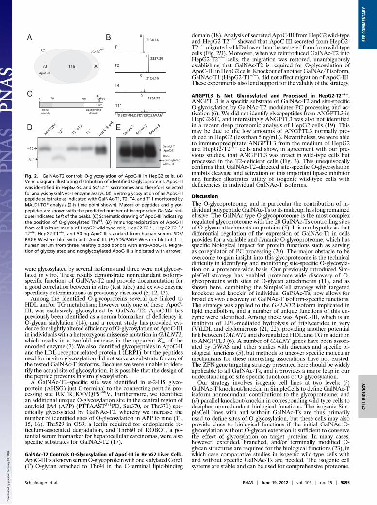
Using Differential O-Glycoproteomics to Define Isoform-SpecificFunctions of GalNAc-Ts. We used our previously reported Simple-Cell glycoproteomics strategy to isolate and sequence GalNAcglycopeptides from tryptic digests by lectin weak-affinity chroma-tography (LWAC) on immobilized Vicia villosa agglutinin (VVA)lectin followed by nLC/MS/MS analysis (11). The protocol foranalysis of secreted glycoproteins was modified to include an initialenrichment step by chromatography on a short VVA column toreduce volume and irrelevant protein content before trypsin di-gestion. We identified 219 O-glycosylation sites in total, of which73 were found only in HepG2-SC, whereas 30 were identified onlyin HepG2-SC/T2−/− (Fig. 2A and Table S1). Compared with ourprevious study (11), which included in total three SimpleCell and
two wild-type cell lines, the HepG2 SimpleCells yielded an addi-tional 81 O-glycopeptides, identifying 45 additional O-glycopro-teins. The secretome contained more glycopeptides derived frompredicted secreted (∼75%) compared with predicted intracellularor membrane-bound proteins (∼25%), whereas the opposite wasfound for the total lysate (∼35% compared with ∼65%), asassessed by ontology terms of annotated proteins in UniProt.The present proteomic analysis, although not comprehensive,
was more than sufficient to quickly identify a significant number ofcandidate GalNAc-T isoform-specific O-glycan sites. Thus, the 73sites identified only in HepG2-SC and not HepG2-SC/T2−/− areclearly potential isoform-specific substrates for GalNAc-T2 inHepG2. To further explore these sites, we synthesized represen-tative peptide substrates for these sites and tested in vitro glyco-sylation using the most common GalNAc-Ts expressed in HepG2(Table 1 and Fig. S1).Among the peptide substrates identified only in HepG2-SC and
not in HepG2-SC/T2−/− (16 in total), 8 peptides were glycosylatedexclusively by GalNAc-T2, 5 were glycosylated by T2 and severalother isoforms, and 3 were not glycosylated by any isoform tested assummarized in Table 1. Among peptide substrates identified inboth SC and SC/T2−/− (10 in total), 8 were glycosylated by severalisoforms, and 2 were not glycosylated by any isoform tested.We also included a selection of glycopeptides identified only in SC/T2−/− (5 in total). Among the corresponding synthetic peptides, two
A
C
B
Fig. 1. Probing isoform-specific functions of GalNAc-Ts by ZFN gene targeting glycosyltransferases. (A) Depiction of the strategy for identification of differentialGalNAc-T isoform-specific O-glycoproteomes. (Section 1) ZFN targeting of COSMC in HepG2 (SC) generates cell lineswith homogenous and truncatedO-glycosylation(GalNAc), and subsequent targeting of GALNT2 (SC/T2−/−) eliminates GalNAc-T2 nonredundant O-glycosylation allowing for comparative analysis of O-glyco-proteomes of isogenic cell lines with and without GALNT2. (Section 2) Proteins from total cell lysates or cell culture supernatant from HepG2-SC or SC/T2−/− aredigested by trypsin. (Section 3) GalNAc-glycopeptides are isolated and separated by lectin weak affinity chromatography. (Section 4) O-glycosylation sites areidentified by nLC/MS/MS and comparison between the two cell lines produce candidates for GalNAc-T2–specific contribution. (B) Depiction of Cosmc and GalNAc-T2protein domains and targeted DNA sequences where introduced mutations are shown. (C) Immunofluorescence staining of isogenic HepG2 cell lines as indicated.
9894 | www.pnas.org/cgi/doi/10.1073/pnas.1203563109 Schjoldager et al.
Dow
nloa
ded
by g
uest
on
Feb
ruar
y 10
, 202
0
were glycosylated by several isoforms and three were not glycosy-lated in vitro. These results demonstrate nonredundant isoform-specific functions of GalNAc-T2 and provide documentation fora good correlation between in vitro (test tube) and ex vivo enzymespecificity determinations as previously discussed (5, 12, 13).Among the identified O-glycoproteins several are linked to
HDL and/or TG metabolism; however only one of these, ApoC-III, was exclusively glycosylated by GalNAc-T2. ApoC-III haspreviously been identified as a serum biomarker of deficiency inO-glycan sialylation (14), and a recent study has provided evi-dence for slightly altered efficiency of O-glycosylation of ApoC-IIIin individuals with a heterozygousmissense mutation inGALNT2,which results in a twofold increase in the apparent Km of theencoded enzyme (7). We also identified glycopeptides in ApoC-IIand the LDL-receptor related protein-1 (LRP1), but the peptidesused for in vitro glycosylation did not serve as substrate for any ofthe tested GalNAc-T isoforms. Because we were unable to iden-tify the actual site of glycosylation, it is possible that the design ofthe peptide prevents in vitro glycosylation.A GalNAc-T2–specific site was identified in α-2-HS glyco-
protein (AHSG) just C-terminal to the connecting peptide pro-cessing site RKTR↓KVVQPS346V. Furthermore, we identifiedan additional unique O-glycosylation site in the central region ofamyloid βA4 (APP) (PTTAAST371PD, Ser370, or Thr371) spe-cifically glycosylated by GalNAc-T2, whereby we increase thenumber of identified sites of O-glycosylation in APP to nine (11,15, 16). Thr529 in OS9, a lectin required for endoplasmic re-ticulum-associated degradation, and Thr660 of ROBO1, a po-tential serum biomarker for hepatocellular carcinomas, were alsospecific substrates for GalNAc-T2 (17).
GalNAc-T2 Controls O-Glycosylation of ApoC-III in HepG2 Liver Cells.ApoC-III is aknownserumO-glycoproteinwithone sialylatedCore1(T) O-glycan attached to Thr94 in the C-terminal lipid-binding
domain (18). Analysis of secreted ApoC-III fromHepG2 wild-typeand HepG2-T2−/− showed that ApoC-III secreted from HepG2-T2−/−migrated∼1 kDa lower than the secreted form fromwild-typecells (Fig. 2D). Moreover, when we reintroduced GalNAc-T2 intoHepG2-T2−/− cells, the migration was restored, unambiguouslyestablishing that GalNAc-T2 is required for O-glycosylation ofApoC-III inHepG2 cells. Knockout of anotherGalNAc-T isoform,GalNAc-T1 (HepG2-T1−/−), did not affect migration of ApoC-III.These experiments also lend support for the validity of the strategy.
ANGPTL3 Is Not Glycosylated and Processed in HepG2-T2−/−.ANGPTL3 is a specific substrate of GalNAc-T2 and site-specificO-glycosylation by GalNAc-T2 modulates PC processing and ac-tivation (6). We did not identify glycopeptides from ANGPTL3 inHepG2-SC, and interestingly ANGPTL3 was also not identifiedin a recent deep proteomic analysis of HepG2 cells (19). Thismay be due to the low amounts of ANGPTL3 normally pro-duced in HepG2 (less than 5 ng/mL). Nevertheless, we were ableto immunoprecipitate ANGPTL3 from the medium of HepG2and HepG2-T2−/− cells and show, in agreement with our pre-vious studies, that ANGPTL3 was intact in wild-type cells butprocessed in the T2-deficient cells (Fig. 3). This unequivocallyconfirms that GalNAc-T2–directed site-specific O-glycosylationinhibits cleavage and activation of this important lipase inhibitorand further illustrates utility of isogenic wild-type cells withdeficiencies in individual GalNAc-T isoforms.
DiscussionThe O-glycoproteome, and in particular the contribution of in-dividual polypeptideGalNAc-Ts to its makeup, has long remainedelusive. The GalNAc-type O-glycoproteome is the most complexregulated glycoproteome with the 20 GalNAc-Ts controlling sitesof O-glycan attachments on proteins (5). It is our hypothesis thatdifferential regulation of the expression of GalNAc-Ts in cellsprovides for a variable and dynamic O-glycoproteome, which hasspecific biological impact for protein functions such as servingas coregulator of PC processing (20). The major obstacle to beovercome to gain insight into this glycoproteome is the technicaldifficulty in identifying and monitoring site-specific O-glycosyla-tion on a proteome-wide basis. Our previously introduced Sim-pleCell strategy has enabled proteome-wide discovery of O-glycoproteins with sites of O-glycan attachments (11), and asshown here, combining the SimpleCell strategy with targetedknockout and knockin of individual GalNAc-Ts, now allows forbroad ex vivo discovery of GalNAc-T isoform-specific functions.The strategy was applied to the GALNT2 isoform implicated inlipid metabolism, and a number of unique functions of this en-zyme were identified. Among these was ApoC-III, which is aninhibitor of LPL-mediated hydrolysis of triglycerides in very(V)LDL and chylomicrons (21, 22), providing another potentiallink betweenGALNT2 and dysregulatedHDL and TG in additionto ANGPTL3 (6). A number of GALNT genes have been associ-ated by GWAS and other studies with diseases and specific bi-ological functions (5), but methods to uncover specific molecularmechanisms for these interesting associations have not existed.The ZFN gene targeting strategy presented here should be widelyapplicable to all GalNAc-Ts, and it provides a major leap in ourunderstanding of site-specific functions of O-glycosylation.Our strategy involves isogenic cell lines at two levels: (i)
GalNAc-T knockout/knockin in SimpleCells to define GalNAc-Tisoform nonredundant contributions to the glycoproteome; and(ii) parallel knockout/knockin in corresponding wild-type cells todecipher nonredundant biological functions. The isogenic Sim-pleCell lines with and without GalNAc-Ts are thus primarilyused to define sites of O-glycosylation, but these cells may alsoprovide clues to biological functions if the initial GalNAc O-glycosylation without O-glycan extension is sufficient to conservethe effect of glycosylation on target proteins. In many cases,however, extended, branched, and/or terminally modified O-glycan structures are required for the biological functions (23), inwhich case comparative studies in isogenic wild-type cells withand without specific GalNAc-Ts are needed. The isogenic cellsystems are stable and can be used for comprehensive proteome,
Disialyl-T
ApoC-III
Non-
glycosylated
ApoC-III
1 20 68 99
Lipid binding
domainSignal
peptide
Thr94
A B
D
8.7
~10
wild
type
T2-/-
T2-/- +T2
T1-/-
ApoC-III s
td.
73 116 30
SC/T2-/-SC
ApoC-III
C
000
0
00
0
2134.32
2134.19
2337.39
2134.14
T1
T2
T4
T11
E
0
0
0
1
Serum
1
Serum
2
Serum
3
Fig. 2. GalNAc-T2 controls O-glycosylation of ApoC-III in HepG2 cells. (A)Venn diagram illustrating distribution of identified O-glycoproteins. ApoC-IIIwas identified in HepG2-SC and SC/T2−/− secretomes and therefore selectedfor analysis by GalNAc-T enzyme assays. (B) In vitro glycosylation of anApoC-IIIpeptide substrate as indicated with GalNAc-T1, T2, T4, and T11 monitored byMALDI-TOF analysis (2-h time point shown). Masses of peptides and glyco-peptides are shown with the predicted number of incorporated GalNAc resi-dues indicated Left of the peaks. (C) Schematic drawing of ApoC-III indicatingthe position of O-glycosylated Thr94. (D) Immunoprecipitation of ApoC-IIIfrom cell culture media of HepG2 wild-type cells, HepG2-T2−/−, HepG2-T2−/−/T2+/+, HepG2-T1−/−, and 50 ng ApoC-III standard from human serum. SDS/PAGE Western blot with anti–ApoC-III. (E) SDS/PAGE Western blot of 1 μLhuman serum from three healthy blood donors with anti–ApoC-III. Migra-tion of glycosylated and nonglycosylated ApoC-III is indicated with arrows.
Schjoldager et al. PNAS | June 19, 2012 | vol. 109 | no. 25 | 9895
CELL
BIOLO
GY
SEECO
MMEN
TARY
Dow
nloa
ded
by g
uest
on
Feb
ruar
y 10
, 202
0

transcriptome, and more in-depth cell biology analyses to iden-tify biological pathways affected by loss of a single GalNAc-T.This is an important feature considering a number of exciting,albeit often conflicting, proposed roles of GalNAc-Ts.Thus, specific GALNTs have been implicated in TGFβ sig-
naling (24), regulation of sensitivity to TNF-related apoptosis-inducing ligand (TRAIL)-induced apoptosis in colonic cancercells lines (25), regulation of cell surface expression of MUC1 inbreast cancer (26), and function of fibronectin in the epithelial–mesenchymal transition (EMT) process (27, 28). Although thesestudies are intriguing, they mainly rely on RNA silencing strat-egies, which have inherent problems with glycosyltransferases,where complete and specific knockout is required. Furthermore,the specific glycosylation events underlying the biological func-tions were not elucidated. Introducing ZFN engineered isogeniccell models and SimpleCells now allows for analysis of pheno-typic and functional consequences of single GalNAc-T isoforms.GALNTs appear to play driver roles in malignancies, and stable
isogenic cancer cell models with deficiencies in GALNTs have thepotential to uncover functions of site-specific O-glycosylation incarcinogenesis. The repertoire of enzyme isoforms expressed incancer cells is markedly altered from the normal counterparts (29–34); however, the consequences of these changes for the O-glyco-proteome and carcinogenesis remain unknown. The locus 9q22including theGALNT12 gene is a susceptibility locus for colorectalcancer (35) and heterozygous germ line as well as somatic
inactivating mutations have been identified in cancer patients (36).GALNT12 is thus a prime candidate for the proposed strategy.We previously developed LWAC for isolation of Tn glycopep-
tides released from total cell lysates. Here we further developed
Table 1. Summary of O-glycopeptides identified in HepG2-SC cells and in vitro GalNAc-T enzyme analysis
Protein name Sequence† T1‡ T2 T4 T11 Sites in T2+§ Sites in T2−
16 AFP 122 HNCFLAHKKPTPASIPLFQV 141 −{ 1* − − 1 0AHSG 337 RKTRTVVQPSVGAAAGPVVP 356 − 1* NA NA 1 0APP 361 PVKLPTTAASTPDAVDK 377 − 1 1 − 1 0APOC2 36 FLTQVKESLSSYWESAKTAA 55 − − − − 1 0APOC3 81 FSEFWDLDPEVRPTSAVAA 99 − 1* − − 1 0CRLF1 324 AGIWSEWSHPTAASTPRSER 343 2–3 1–2 − − 1 0DKK1 50 GGAAGHPGSAVSAAPGILYP 69 1 1 1 1 1 0IGFBP6 117 PKESKPQAGTARPQDVNRRD 136 1 1 − − 1 0ITIH2 591 QLLAERSLAPTAAAKRRITR 610 − 1* − − 1 0LRP1 I 3723 GDGTDEEDCEPPTAHTTHCK 3742 − − − − 1 0MATN3 43 GPGGSPGRRPSPAAPDGAPA 62 − 1* − − 2 0NPTXR 72 PALPGAPAASAHPLPPGPLF 91 − 1* − − 2 0OS9 521 VVPKKPPPSPQPTEEDPEHR 540 − 1–2* − − 1 0RELN 174 AQQLCEQGAPTDVTVHPHLA 193 1 1 1 1 1 0ROBO1 650 DPVKTQDVLPTSQGVDHKQV 669 − 1* − − 1 0STC2 241 EAGHHLPEPSSRETGRGAKG 260 − − − − 1 0
10 APOE 301 VQAAVGTSAAPVPSDNH 317 − 1–2 1–2 − 2 2DLK1 251 SPQQVTRLPSGYGLAYRLTP 270 1 − NA 1 1 1GPC6 365 RPYNPEERPTTAAGTSLDRL 384 1 1 NA − 1 1LRP1 II 3527 PKEECDERTCEPYQFRCKNN 3546 − − − − 1 1NID1 304 GLEDVGTTPFSYKALRRGGA 323 1 1 NA − 2 2NUCB1 32 GAPNKEETPATESPDTGLYY 51 1 − 1 − 1 1PLTP 476 RPADVRASTAPTPSTAAV 493 2–3 1–2 NA NA 3 3SDC2 91 LNIQNKIPAQTKSPEETDKE 110 1 − NA − 1 1SDC4 90 PERAGSGSQVPTEPKKLEEN 109 − − − − 1 1ST6GalI 66 SSTQDPHRGRQTLGSLRGLA 85 1–3 − 1 − 3 3
5 APOA2 45 GKDLMEKVKSPELQAEAKSY 64 − − − − 0 1APOF 262 DQKDANISQPETTKEGLRAI 281 1 1 − − 0 1CX3CL1 188 VPPVSTAATWQSSAPHQPGP 207 1–2 1–2 1–2 − 0 4IGFBP1 159 DGSKALHVTNIKKWKEPCRI 178 − − − − 0 1PRAP1 64 PVQKPKLLTTEEKPRGQGRG 83 − − − − 0 2
NA, not analyzed.*T2-specific sites.†Sequence of synthetic peptide designed to cover identified O-glycosylation site(s) (underlined).‡In vitro glycosylation of synthetic peptides using GalNAc-T1, -T2, -T4, and -T11.§Number of sites identified in glycopeptides from HepG2 SC (T2+) or HepG2 SC/GALNT2−/− (T2−). 0 (glycopeptide not identified), 1, 2, or3 [1, 2, or 3 site(s) identified].{Number of GalNAc residues added to the peptide substrate. (-) no GalNAc added.
Full length
ANGPTL3
Cleaved
ANGPTL3
62
49
38
28
wild
type
T2-/-
Rec.ANGPTL3
Fig. 3. Increased PC processing of ANGPTL3 in HepG2-T2−/− cells. (A) Immu-noprecipitation of ANGPTL3 from HepG2 wild-type and T2−/− cells inducedwith 5 μM T0901317 for 48 h. SDS/PAGE Western blot with anti-ANGPTL3.Control lane includes 20 ng recombinant nonglycosylated ANGPTL3. Full-length ANGPTL3 migrates to ∼62 kDa and cleaved N-terminal ANGPTL3migrates to ∼38 kDa as indicated by arrows.
9896 | www.pnas.org/cgi/doi/10.1073/pnas.1203563109 Schjoldager et al.
Dow
nloa
ded
by g
uest
on
Feb
ruar
y 10
, 202
0

a two-step VVA lectin strategy to isolate secreted GalNAc gly-coproteins in culture medium with serum followed by digestionand isolation of GalNAc glycopeptides. This strategy providesa unique opportunity to identify secreted O-glycoproteins fromdifferent human cells that may have potential for serving as serumbiomarkers such as ApoC-III. We did not identify ANGPTL3 inthe proteomic strategy used, but the isogenic wild-type cell systemsstill allowed us to confirm that activation of this inhibitor of lipasesthat also affects HDL and TG is inhibited by GalNAc-T2 O-gly-cosylation (Fig. 3). We are currently developing strategies to im-prove the coverage of the GalNAc glycoproteome using differentendoproteases and isoelectric focusing.An important finding was a high degree of correlation between
ex vivo substrate specificity of GalNAc-Ts and the substratespecificity determined by in vitro assays (Table 1). Althoughexpected on the basis of past studies with a few select sequences (9,37, 38), this hypothesis has been validated on a proteome-widescale. GalNAc-Ts function in Golgi where their protein substratesare folded. It is thus not empirically logical that the substratespecificities of GalNAc-Ts would be characterized by a short lin-ear sequence motif (5, 39). The two POFUTs initiating protein O-fucosylation have strict substrate specificities for the correctlyfolded EGF repeat domain and do not function with short syn-thetic peptides (40). This restricted specificity may be because O-fucosylation occurs in ER at the same site as folding, allowing fordifferential glycosylation of Notch and Thrombospondin EGFrepeats by the two isoenzymes (41). The 20 GalNAc-Ts offera much more complex scenario, and this may be one reason fortopological separation fromER in contrast to the initiation step ofmost other types of protein glycosylation (5). Interestingly, how-ever, Bard and colleagues have found that the GalNAc-Ts and theinitiation step of O-glycosylation can be selectively relocated toER by activation of Src (42). It is conceivable that this inducessubstantial changes in the O-glycoproteome and perhaps alter-ations in other O-glycoproteomes by competition in ER (8).In summary, we present a unique strategy to identify non-
redundant O-glycosylation controlled by individual polypeptideGalNAc-T isoforms and to uncover site-specific biological func-tions hereof. Given that there is increasingly more evidence, fromGWAS and other studies, demonstrating important biologicalfunctions of the large GalNAc-T gene family, it is becoming evenmore urgent to uncover the underlying molecular mechanisms aswell as identify biological pathways affected. The isogenic cellmodels presented opens the way for such studies and the strategiesapplied are generally applicable to other biological pathways.
Materials and MethodsZFN Gene Targeting. ZFN targeting constructs for COSMC and GALNT2 werecustomproduced (Sigma-Aldrich) with the following binding sites. Cutting sitesare indicated in parentheses: COSMC 5′-CCCAACCAGGTAGT(AGAAGGCT)GTT-GTTCAGATATGGCTGTT-3′; and GALNT2 5′-GTCGGCCCTACTCAGGAC(CGTGGT)CAGGTGAGGCCAGGAGAT-3′. For stable integration of GALNT2, a ZFN in-tegration vector was designed for insertion into the adeno-associated virusintegration site 1 (AAVS1) locus. Expression was driven by the endogenousAAVS1 promoter as previously described (43), by including a splice acceptor anda 2A “self-cleaving” peptide directly 5′ to the full GALNT2 ORF. A polyA signalsequencewas appended 3′ to theORF sequence, and all of the aforementionedsequences wereflanked by left and right arms of homology from the pZDonor-AAVS1 puromycin vector (Sigma-Aldrich) at the 5′ and 3′ ends, respectively.Finally, the above synthetic gene was inserted into the pZDonor vector (Sigma-Aldrich) via Blpl–Pfol restriction enzyme digestion, thus generating thepAAVS1-Sig2a-GALNT2-ZFN integration (pAAVS1-T2) vector.
HepG2 cells (kind gift from Novo Nordisk) were transfected with 1 vial ofmRNA (Sigma-Aldrich) or 5 μg of endotoxin free pAAVS1-T2 plasmid DNA forknockin experiments (mixed with 5 μL of pAAVS1 ZFN mRNA (Sigma-Aldrich)using nucleofection on an Amaxa Nucleofector (Lonza). Cells were cloned bylimited dilution, trypsinized andfixed in ice-cold acetone on teflon-coated slidesand stained with monoclonal antibodies (MAbs) to Tn (5F4), GalNAc-T2 (UH4and 2E10), GalNAc-T1 (UH3), or C1GalT (5B6) (11). COSMC knockout clones(SimpleCells) were selected by reactivity with MAbs to Tn and loss of reactivitywith MAb to the C1GalT1 enzyme, and clones were confirmed to have COSMCmutations using PCR and sequencing using primers 5′-AGGGAGGGATGATTTG-GAAG-3′ and 5′-TTGTCAGAACCATTTGGAGGT-3′. GALNT2 and GALNT1 knock-out clones were selected for loss of reactivity with MAbs to GalNAc-T2 and -T1,
and mutations confirmed by PCR using primers GALNT2PZFN F/R (5′-CCATCC-CAGTTGGTCAGTCT-3′/5′-CTGTGCTGAGCAGTCAGGAG-3′) or GALNT1PZFN F/R(5′-GAATAGTGCCAGGCCACACT-3′/5′-AAAGCAAACTTGGGAGGAAAT-3′) andsequencing. GALNT2 knockin clones were screened for reactivity with MAbs toGalNAc-T2. For immunocytochemistry, HepG2 cells were grown on sterile Di-agnostic Imaging printed slides (Clearcell, Histolab) andfixedwith acetone. Cellswere then sequentially incubated with MAbs overnight and FITC-conjugatedrabbit antibody tomouse Ig (Dako) for 45min andmountedwith ProLong Goldantifade reagent (DAPI) (Invitrogen). Fluorescence microscopy was performedusing a Zeiss Axioskop 2 plus with an AxioCam MR3. Bit depth and pixeldimensions were 36 bit and 1,388 × 1,040 pixels, respectively.
LWAC Isolation of Tn O-Glycopeptides. A total of 100 mL of cell culture su-pernatant (secretome) or 0.5 mL packed cells (total cell lysate) was harvestedfrom HepG2 COSMC knockout and HepG2 COSMC/GALNT2 double knockout.Secretome: Cell culture supernatants were cleared by centrifugation (2,500 gfor 10 min), dialyzed (molecular weight cutoff, 3,500 Da) twice against 5 L 1mM Tris-HCl, pH 7.4, filtered (0.45 μm), diluted in 100 mL buffer A (10 mMTris, pH 8, 150 mM NaCl, 1 mM CaCl2/MgCl2/MnCl2/ZnCl2, 1 M urea), andsubjected to the first Vicia villosa agglutinin (VVA) lectin chromatography forenrichment of glycoproteins. The VVA agarose (Vector Laboratories) (0.8 mLin 2-mL syringe) was equilibrated in buffer A, the sample loaded twice fol-lowed by 10–20 column volumes (CV) wash in buffer A, and enriched Tnglycoproteins eluted with 2 × 1 mL 0.2 M GalNAc. The eluate was dialyzedagainst 2 × 5 L 50 mM ammonium bicarbonate, lyophilized, dissolved in 0.5mL 50 mM ammonium bicarbonate, and further processed as cell lysates fordigestion and isolation of Tn glycopeptides. Total cell lysates: Packed cellswere lysed in 0.1% RapiGest (Waters) in 50 mM ammonium bicarbonate witha sonic probe and the solution cleared by centrifugation (1,000 g for 10 min).The cleared lysate and secretome samples were heated for 10 min at 80 °C,followed by reduction (5 mM DTT, 60 °C, 0.5 h) and alkylation (10 mMiodoacetamide, RT, 30 min), and digestion with trypsin (25 μg) (Roche)[37 °C, overnight (ON)]. Digests were treated with TFA (6 μL, 37 °C, 20 min),cleared by centrifugation, purified on C18 Sep-Pak (Waters), lyophilized,resuspended in 1 mL buffer A, and injected to a preequilibrated 2.6 m longVVA column (packed in PFA tubing 1/16 inch × 50 feet, flow 100 μL min−1)for isolation of glycopeptides (11). The column was washed with 0.4 Mglucose in buffer A, eluted with 0.2 M GalNAc (2 CV) and 0.4 M GalNAc(1 CV), and glycopeptide fractions purified by Stage Tips (Thermo Scientific)for analysis. A detailed step-by-step protocol is published elsewhere (44).
nLC/MS/MS Analysis. Liquid chromatography–tandem mass spectrometry wasperformed on a system composed of an EASY-nLC II (Thermo Fisher Scien-tific) interfaced via a nanoSpray Flex ion source to an LTQ-Orbitrap XL hybridspectrometer (Thermo Fisher Scientific), equipped for both higher-energy C-trap dissociation (HCD)- and ETD-MS2 modes, enabling peptide sequenceanalysis without and with retention of glycan site-specific fragments, re-spectively. The conditions of LC analysis were essentially as described pre-viously (11), except that the nLC was operated using a single analyticalcolumn set up (polar end-capped C18-silica; 10 cm length, 75 μm inner di-ameter and 3-μm particle size).
A data-dependent mass spectral acquisition routine, HCD triggering ofsubsequent ETD scan, was used for all runs. Briefly, a precursor MS1 scan (m/z350–1,700) of intact peptides was acquired in the Orbitrap at a resolution set-ting of 30,000, followed by Orbitrap HCD-MS2 (m/z of 100–2,000) of the threemost abundant multiply charged precursors above 5,000 counts in the MS1spectrum; the appearance of a HexNAc fragment atm/z 204.086 (in practice a±m/z 0.15 window was used) in the HCD-MS2 spectrum triggered a subsequentETD-MS2 from the same precursor with a resolution setting of 15,000 (11).
Data Analysis. Data processing was carried out using Proteome Discoverer 1.2software (Thermo Fisher Scientific) as previously described (11), with onlysmall changes in preprocessing and processing procedures (e.g., in theHexNAc subtraction routine for correctly interpreting HCD spectra, exactmasses of 1×, 2×, 3×, and 4× HexNAc units were subtracted from the cor-responding precursor ion mass, generating four distinct files, instead of onlyup to 3 HexNAc units). All candidate-matched glycopeptides associated witheach protein were validated by inspection. The results obtained were ac-cumulated in a single list (Table S1; raw data related to this paper can bedownloaded from ProteomeCommons.org using the following tranche hash:JQoOtUklwXlG7eEiT9B9Avxb8yloxZj7dqeq62goVARNygxG4Pp8jnudiFZavgFT6-OMpK6JkAqY4BlcsDKNhAJE1mGIAAAAAAABFyQ==).
Glycosyltransferase Assays.All recombinant glycosyltransferaseswere expressedas soluble secreted truncated proteins in insect cells (45). Screening assays for
Schjoldager et al. PNAS | June 19, 2012 | vol. 109 | no. 25 | 9897
CELL
BIOLO
GY
SEECO
MMEN
TARY
Dow
nloa
ded
by g
uest
on
Feb
ruar
y 10
, 202
0

GalNAc-T glycosylation of peptides (Schafer-N; NeoBioSci) were performed asproduct development assays in 25 μL of 25 mM cacodylic acid sodium, pH 7.4,10 mMMnCl2, 0.25% Triton X-100, 1.5 mMUDP-GalNAc (Sigma-Aldrich), 10 μgof acceptor peptides, and 0.2–0.5 μg of purified enzyme.
Immunoprecipitation. ProteinAbeads (cat. no. sc-2001, Santa Cruz)werewashedinPBSandIPbuffer (20mMTris-base,pH7.4,150mMNaCl,1%TritonX-100,1mMEDTA, protease inhibitor mixture; Roche). Beads were preincubated with poly-clonal rabbit antihuman ApoC-III (4 μg) (Abcam; ab21032) or rabbit antihumanANGPTL3 1:2,000 (Alexis Biochemical; 210448) for 1.5 h at room temperature. Thebeads were washed and incubated with conditioned media harvested fromHepG2 wild-type and mutant cells overnight at 4 °C. ANGPTL3 expression wasinducedwith5μMT0901317 (LXRagonist) (Sigma-Aldrich) for 48hbeforeharvestof conditionedmedia. After a thoroughwash, ApoC-III or ANGPTL3was eluted inLDS loading buffer (NuPAGE; Invitrogen) and 5mMDTT and beadswere pelleted
by centrifugation. Sample proteins were separated in 10 or 12% NuPAGE NovexBis-Tris in MES running buffer and blotted onto nitrocellulose membranes.Membranes were blocked in 5% (wt/vol) dry milk for 40 min and incubated inprimary antibodies diluted in Tris-buffered saline (TBS) plus 0.05% Tween 20(polyclonal rabbit antihuman ApoC-III 1:3,000 or biotinylated sheep antihumanANGPTL3 1:2,000; R&D Systems; BAF 3829) overnight at 4 °C. After wash, mem-branes were incubated in secondary antibodies (antirabbit-HRP 1:3,000 (Dako)and streptavidin-HRP (P0397) 1:3,000 and developed using ECL (Pierce).
ACKNOWLEDGMENTS. This work was supported by Kirsten and FreddyJohansens Fond, A. P. Møller og Hustru Chastine Mc-Kinney Møllers Fond tilAlmene Formaal, the Carlsberg Foundation, the Novo Nordisk Foundation,the Danish Research Councils, a program of excellence from the University ofCopenhagen, National Institutes of Health Grant (U01CA128437), and theDanish National Research Foundation.
1. Willer CJ, et al. (2008) Newly identified loci that influence lipid concentrations andrisk of coronary artery disease. Nat Genet 40:161–169.
2. Kathiresan S, et al. (2008) Six new loci associated with blood low-density lipoproteincholesterol, high-density lipoprotein cholesterol or triglycerides in humans. Nat Genet40:189–197.
3. Teslovich TM, et al. (2010) Biological, clinical and population relevance of 95 loci forblood lipids. Nature 466:707–713.
4. White T, et al. (1995) Purification and cDNA cloning of a human UDP-N-acetyl-α-D-galactosamine:polypeptide N-acetylgalactosaminyltransferase. J Biol Chem 270:24156–24165.
5. Bennett EP, et al. (2011) Control of mucin-type O-glycosylation: A classification of thepolypeptide GalNAc-transferase gene family. Glycobiology, 10.1093/glycob/cwr182.
6. Schjoldager KT, et al. (2010) O-glycosylation modulates proprotein convertase activationof angiopoietin-like protein 3: Possible role of polypeptide GalNAc-transferase-2 inregulation of concentrations of plasma lipids. J Biol Chem 285:36293–36303.
7. Holleboom AG, et al. (2011) Heterozygosity for a loss-of-function mutation in GALNT2improves plasma triglyceride clearance in man. Cell Metab 14:811–818.
8. Gill DJ, Clausen H, Bard F (2011) Location, location, location: New insights into O-GalNAc protein glycosylation. Trends Cell Biol 21:149–158.
9. Kato K, et al. (2006) Polypeptide GalNAc-transferase T3 and familial tumoral calci-nosis. Secretion of fibroblast growth factor 23 requires O-glycosylation. J Biol Chem281:18370–18377.
10. Schwientek T, et al. (2002) Functional conservation of subfamilies of putative UDP-N-acetylgalactosamine:polypeptide N-acetylgalactosaminyltransferases in Drosophila,Caenorhabditis elegans, and mammals. One subfamily composed of l(2)35Aa is es-sential in Drosophila. J Biol Chem 277:22623–22638.
11. Steentoft C, et al. (2011) Mining the O-glycoproteome using zinc-finger nuclease-glycoengineered SimpleCell lines. Nat Methods 8:977–982.
12. Hassan H, Bennett EP, Mandel U, Hollingsworth MA, Clausen H (2000) in Carbohy-drates in Chemistry and Biology: A Comprehension Handbook, eds Ernst B, Hart GW,and Sinay P (Wiley, New York), pp 273–292.
13. DeFrees S, et al. (2006) GlycoPEGylation of recombinant therapeutic proteins pro-duced in Escherichia coli. Glycobiology 16:833–843.
14. Wopereis S, et al. (2003) Apolipoprotein C-III isofocusing in the diagnosis of geneticdefects in O-glycan biosynthesis. Clin Chem 49:1839–1845.
15. Perdivara I, et al. (2009) Elucidation of O-glycosylation structures of the beta-amyloidprecursor protein by liquid chromatography-mass spectrometry using electrontransfer dissociation and collision induced dissociation. J Proteome Res 8:631–642.
16. Halim A, et al. (2011) Site-specific characterization of threonine, serine, and tyrosineglycosylations of amyloid precursor protein/amyloid β-peptides in human cerebro-spinal fluid. Proc Natl Acad Sci USA 108:11848–11853.
17. Ito H, et al. (2006) Identification of ROBO1 as a novel hepatocellular carcinoma antigenand a potential therapeutic and diagnostic target. Clin Cancer Res 12:3257–3264.
18. Brewer HB, Jr., Shulman R, Herbert P, Ronan R, Wehrly K (1974) The complete aminoacid sequence of alanine apolipoprotein (apoC-3), and apolipoprotein from humanplasma very low density lipoproteins. J Biol Chem 249:4975–4984.
19. Geiger T, Wehner A, Schaab C, Cox J, Mann M (2012) Comparative proteomic analysisof eleven common cell lines reveals ubiquitous but varying expression of most pro-teins. Mol Cell Proteomics, 11: M111.014050.
20. Gram Schjoldager KT, et al. (2011) A systematic study of site-specific GalNAc-type O-gly-cosylation modulating proprotein convertase processing. J Biol Chem 286:40122–40132.
21. Jong MC, et al. (2001) Apolipoprotein C-III deficiency accelerates triglyceride hydro-lysis by lipoprotein lipase in wild-type and apoE knockout mice. J Lipid Res 42:1578–1585.
22. Jong MC, Hofker MH, Havekes LM (1999) Role of ApoCs in lipoprotein metabolism:Functional differences between ApoC1, ApoC2, and ApoC3. Arterioscler Thromb VascBiol 19:472–484.
23. Liu W, et al. (1998) Identification of N-terminal residues on P-selectin glycoproteinligand-1 required for binding to P-selectin. J Biol Chem 273:7078–7087.
24. Herr P, Korniychuk G, Yamamoto Y, Grubisic K, Oelgeschläger M (2008) Regulation ofTGF-(beta) signalling by N-acetylgalactosaminyltransferase-like 1. Development 135:1813–1822.
25. Wagner KW, et al. (2007) Death-receptor O-glycosylation controls tumor-cell sensi-tivity to the proapoptotic ligand Apo2L/TRAIL. Nat Med 13:1070–1077.
26. Park JH, et al. (2010) Critical roles ofmucin 1glycosylationby transactivatedpolypeptideN-acetylgalactosaminyltransferase 6 in mammary carcinogenesis. Cancer Res 70:2759–2769.
27. Park JH, Katagiri T, Chung S, Kijima K, Nakamura Y (2011) Polypeptide N-acetylga-lactosaminyltransferase 6 disrupts mammary acinar morphogenesis through O-gly-cosylation of fibronectin. Neoplasia 13:320–326.
28. Freire-de-Lima L, et al. (2011) Involvement of O-glycosylation defining oncofetal fi-bronectin in epithelial-mesenchymal transition process. Proc Natl Acad Sci USA 108:17690–17695.
29. Brooks SA, Carter TM, Bennett EP, Clausen H, Mandel U (2007) Immunolocalisation ofmembers of the polypeptide N-acetylgalactosaminyl transferase (ppGalNAc-T) familyis consistent with biologically relevant altered cell surface glycosylation in breastcancer. Acta Histochem 109:273–284.
30. Rajpert-De Meyts E, et al. (2007) Changes in the profile of simple mucin-type O-gly-cans and polypeptide GalNAc-transferases in human testis and testicular neoplasmsare associated with germ cell maturation and tumour differentiation. Virchows Arch451:805–814.
31. Mandel U, et al. (1999) Expression of polypeptide GalNAc-transferases in stratified ep-ithelia and squamous cell carcinomas: Immunohistological evaluation using monoclonalantibodies to three members of the GalNAc-transferase family. Glycobiology 9:43–52.
32. Gomes J, et al. (2009) Expression of UDP-N-acetyl-D-galactosamine: Polypeptide N-acetylgalactosaminyltransferase-6 in gastric mucosa, intestinal metaplasia, and gastriccarcinoma. J Histochem Cytochem 57:79–86.
33. Berois N, et al. (2006) UDP-N-acetyl-D-galactosamine: polypeptide N-acetylgalacto-saminyltransferase-6 as a new immunohistochemical breast cancer marker. J Histo-chem Cytochem 54:317–328.
34. Sutherlin ME, et al. (1997) Expression of three UDP-N-acetyl-α-D-galactosamine:polypeptide GalNAc N-acetylgalactosaminyltransferases in adenocarcinoma cell lines.Cancer Res 57:4744–4748.
35. Wiesner GL, et al. (2003) A subset of familial colorectal neoplasia kindreds linked tochromosome 9q22.2-31.2. Proc Natl Acad Sci USA 100:12961–12965.
36. Guda K, et al. (2009) Inactivating germ-line and somatic mutations in polypeptide N-acetylgalactosaminyltransferase 12 in human colon cancers. Proc Natl Acad Sci USA106:12921–12925.
37. Bennett EP, Hassan H, Clausen H (1996) cDNA cloning and expression of a novel hu-man UDP-N-acetyl-α-D-galactosamine. Polypeptide N-acetylgalactosaminyltransferase,GalNAc-t3. J Biol Chem 271:17006–17012.
38. Nehrke K, Hagen FK, Tabak LA (1998) Isoform-specific O-glycosylation by murine UDP-GalNAc:polypeptide N-acetylgalactosaminyltransferase-T3, in vivo. Glycobiology 8:367–371.
39. Gerken TA, Raman J, Fritz TA, Jamison O (2006) Identification of common and uniquepeptide substrate preferences for the UDP-GalNAc:polypeptide α-N-acetylgalactosa-minyltransferases T1 and T2 derived from oriented random peptide substrates. J BiolChem 281:32403–32416.
40. Wang Y, et al. (2001) Modification of epidermal growth factor-like repeats with O-fucose. Molecular cloning and expression of a novel GDP-fucose protein O-fucosyl-transferase. J Biol Chem 276:40338–40345.
41. Shao L, Haltiwanger RS (2003) O-fucose modifications of epidermal growth factor-likerepeats and thrombospondin type 1 repeats: Unusual modifications in unusual places.Cell Mol Life Sci 60:241–250.
42. Gill DJ, Chia J, Senewiratne J, Bard F (2010) Regulation of O-glycosylation throughGolgi-to-ER relocation of initiation enzymes. J Cell Biol 189:843–858.
43. DeKelver RC, et al. (2010) Functional genomics, proteomics, and regulatory DNAanalysis in isogenic settings using zinc finger nuclease-driven transgenesis into a safeharbor locus in the human genome. Genome Res 20:1133–1142.
44. Steentoft C, Bennett EP, Clausen H (2012) Glycoengineering of human cell lines usingZinc Finger nuclease gene targeting: SimpleCells with homogenous GalNAc O-glyco-sylation allow isolation of the O-glycoproteome by one-step lectin affinity chroma-tography. Methods Mol Biol, in press.
45. Wandall HH, et al. (1997) Substrate specificities of three members of the human UDP-N-acetyl-α-D-galactosamine:Polypeptide N-acetylgalactosaminyltransferase family,GalNAc-T1, -T2, and -T3. J Biol Chem 272:23503–23514.
9898 | www.pnas.org/cgi/doi/10.1073/pnas.1203563109 Schjoldager et al.
Dow
nloa
ded
by g
uest
on
Feb
ruar
y 10
, 202
0