probing guest geometry and dynamics through host–guest interactions
TRANSCRIPT

COMMUNICATIONS
Angew. Chem. Int. Ed. 2001, 40, No. 10 � WILEY-VCH Verlag GmbH, D-69451 Weinheim, 2001 1433-7851/01/4010-1879 $ 17.50+.50/0 1879
Probing Guest Geometry and Dynamicsthrough Host ± Guest Interactions**Takahiro Kusukawa, Michito Yoshizawa, andMakoto Fujita*
Conformational freedom of molecules is restricted whenthey are enclathrated within the limited interior space ofhollow compounds.[1] Analysis of the inclusion geometry ofguest molecules is important because molecules in a specific
geometry or aggregation are expected to show uniqueproperties and reactivities which do not appear when theyare in solution. The restricted conformations are in generalanalyzed by observing guest molecules themselves. On theother hand, information on the geometry of guests can be readout by the spectroscopic analysis of host frameworks providedthe host and the guest communicate with each other throughsubtle host ± guest interactions. Several reports have dealtwith the conformational analysis of guests by the observationof host frameworks.[2, 3] However, the observed spectra of thehosts are often not sufficiently simple to analyze details.
Coordination nanocage 1 has recently been shown tostrongly bind a variety of neutral substrates within itsnanosized cavity.[4] As a result of the high symmetry of cage
1 (Td) the twelve pyridine rings in 1 are all equivalent, and theNMR spectrum of the empty cage displays only a set ofpyridine protons at d� 9.47 (PyHa) and 8.92 (PyHb) (Fig-ure 1 a). When the cage accommodates guest molecule(s) withlower symmetry the Td symmetry of the cage is desymme-trized and the symmetry of the entity follows that of theguest(s) if the cage and the guest(s) strongly interact. Forexample, if the guest has C1 symmetry then 48 pyridineprotons of 1 become inequivalent and, in principle, 48 in-dependent pyridine protons should be observed in the1H NMR spectrum. Scheme 1 summarizes the relationshipbetween the guest symmetry and the maximum number ofpyridine protons which should appear in the NMR spectra.Thus, an analysis of the symmetry of the cage probes howguests are accommodated in the cavity. We show in thefollowing discussions some examples of probing the guestgeometry by NMR analysis of the host symmetry, where allpredicted structures have been confirmed by X-ray analyses.The analysis of the symmetry by NMR spectroscopic analysisalso elucidates the dynamic motion of included guests.
The 1:2 complexation of cage 1 with 4,4'-dimethoxydiben-zoyl (2 a) provides a fine example. The 1:2 inclusion complex(2 a)2� 1 was easily prepared by mixing a solution of 2 a inhexane (saturated, 1 mL) and a solution of 1 in D2O (5.3 mm,2 mL), and stirring the mixture at 80 8C for 0.5 h. After
[1] M. A. Gallop, R. W. Barrett, W. J. Dower, S. P. A. Fodor, E. M.Gordon, J. Med. Chem. 1994, 37, 1233 ± 1251; b) M. A. Gallop, R. W.Barrett, W. J. Dower, S. P. A. Fodor, E. M. Gordon, J. Med. Chem.1994, 37, 1385 ± 1401.
[2] a) A. Studer, S. Hadida, R. Ferritto, S.-Y. Kim, P. Jeger, P. Wipf, D. P.Curran, Science 1997, 275, 823 ± 826; b) D. P. Curran, Angew. Chem.1998, 110, 1230 ± 1255; D. P. Curran, Angew. Chem. Int. Ed. 1998, 37,1174 ± 1196.
[3] a) D. J. Gravert, K. D. Janda, Chem. Rev. 1997, 97, 489 ± 509; b) P. H.Toy, K. D. Janda, Acc. Chem. Res. 2000, 33, 546 ± 554.
[4] a) R. M. Kim, M. Manna, S. M. Hutchins, P. R. Griffin, N. A. Yates,A. M. Bernick, K. T. Chapman, Proc. Natl. Acad. Sci. USA 1996, 93,10012 ± 10 017; b) N. J. Hovestd, A. Ford, J. T. B. H. Jastrzebski, G.van Koten, J. Org. Chem. 2000, 65, 5338 ± 6344.
[5] H. Perrier, M. Labelle, J. Org. Chem. 1999, 64, 2110 ± 2113.[6] Merck Index, 12th ed. (Eds.: S. Budaveri, P. E. Heckelman, J. F.
Kinneary, M. J. O�Neil, A. Smith), Merck, Whitehouse Station, NJ,1996, p. 1505.
[7] K. A. Fletcher, M. E. R. McHale, K. S. Coym, W. E. Acree, Jr., Can. J.Chem. 1997, 75, 258 ± 261.
[8] Saturated solutions were prepared by stirring excess E isomers in eachgiven solvent for 72 h at 23 8C. Undissolved solid was removed fromthese suspensions by microfiltration. The filtrate was diluted in THFand the absorbance of the resulting solution was used to determine theamount of the solute present.
[9] a) G. S. Hammond, J. Saltiel, A. A. Lamola, N. J. Turro, J. S. Bradshaw,D. O. Cowan, R. C. Counsell, V. Vogt, C. Dalton, J. Am. Chem. Soc.1964, 86, 3197 ± 3217; b) W. G. Herkstroeter, G. S. Hammond, J. Am.Chem. Soc. 1966, 88, 4769 ± 4777.
[10] M. A. Ali, Y. Tsuda, Chem. Pharm. Bull. 1992, 40, 2842.[11] D. F. Deter, Y. W. Chu, J. Am. Chem. Soc. 1955, 77, 4410 ± 4411.[12] P. Caramella, P. Grunager, 1,3-Dipolar Cycloaddition Chemistry,
Vol. 1, Wiley-Interscience, New York, 1984, p. 291.[13] T. Mukaiyama, T. Hoshino, J. Am. Chem. Soc. 1960, 82, 5339 ± 5342.[14] M. Christl, R. Huisgen, Chem. Ber. 1973, 106, 3345 ± 3367.[15] a) J.-F. Cheng, A. M. M. Mjalli, Tetrahedron Lett. 1998, 39, 939 ± 942;
b) N. Zou, B. Jiang, J. Comb. Chem. 2000, 2, 6 ± 7.[16] A. Studer, D. P. Curran, Tetrahedron 1997, 19, 6681 ± 6696.[17] K.-C. Liu, B. R. Shelton, R. K. Howe, J. Org. Chem. 1980, 45, 3916 ±
3918.
[*] Prof. Dr. M. FujitaCREST, Japan Science and Technology Corporation (JST)Department of Applied ChemistryGraduate School of Engineering, Nagoya UniversityChikusa-ku, Nagoya 464-8603 (Japan)Fax:(�81) 52-789-3199E-mail : [email protected]
Dr. T. Kusukawa, M. YoshizawaDepartment of Applied ChemistryGraduate School of Engineering, Nagoya University
[**] This work was supported by the CREST (Core Research for Evolu-tional Science and Technology) project of Japan Science andTechnology Corporation.
Supporting information for this article is available on the WWW underhttp://www.angewandte.com or from the author.

COMMUNICATIONS
1880 � WILEY-VCH Verlag GmbH, D-69451 Weinheim, 2001 1433-7851/01/4010-1880 $ 17.50+.50/0 Angew. Chem. Int. Ed. 2001, 40, No. 10
Figure 1. 1H NMR observations of the enclathration of2a in nanocage 1. a) Empty 1. b) and c) (2 a)2� 1 ´ .d) (2 b)2� 1.
Scheme 1. The relationships between the guest symmetry and the maximum number of pyridine protons that should appear in the NMR spectra.

COMMUNICATIONS
Angew. Chem. Int. Ed. 2001, 40, No. 10 � WILEY-VCH Verlag GmbH, D-69451 Weinheim, 2001 1433-7851/01/4010-1881 $ 17.50+.50/0 1881
complexation was complete, the pyridine protons of the hostwere split into six pairs: six PyHa protons at d� 9.35 ± 9.8 andsix PyHb protons at d� 8.35 ± 9.2 (Figure 1 b). This observa-tion suggested the desymmetrization of the host into anS4 symmetric entity with 12 inequvalent protons on eachligand (see case e of Scheme 1 where ligands A ± D are allequivalent but each ligand possesses no symmetry element).The S4 symmetry can originate from a specific orientation oftwo guest molecules in the cavity and will be generated only ifthey aggregate orthogonally and adopt a chiral, twistedconformation with opposite screw senses [Eq. (1)].[5]
The S4 orientation of the guests was also supported by theguest signals in the 1H NMR spectrum. A tight contact of thetwo guest molecules was shown by NOEs (1.2 ± 18.4 %)between the methoxy protons and other aromatic protons.These interactions must be intermolecular because thecorresponding NOEs were not observed in the NMR spec-trum of free 2 in CDCl3. The twisted conformation of 2 wasconsistent with the observation of four different chemicalshifts for the four aromatic protons on the phenylene groups(Figure 1 c). The observation of the chirality of each guestdemonstrated that two conformations of enclathrated 2 a werecompletely frozen at room temperature, while those of free 2 aare in rapid equilibrium in solution. With the asymmetricsubstituted analogue 2 b, a diastereomeric pair (see below)was observed in a 1:1 ratio (Figure 1 d).
The proposed orientation of 2 a was confirmed by an X-rayanalysis (Figure 2). As expected, two molecules were packedorthogonally and each of them adopted a twisted conforma-tion with a dihedral angle (y) of 80.6 ± 80.88 between the twocarbonyl groups. This angle is considerably smaller than thatof dibenzoyl in its crystal structure (y� 124.68),[6] but veryclose to that of an optimized conformation (y�ÿ83.278).[7]
Thus, each guest adopts its optimal geometry withoutinfluence from crystal-packing effects.
The dynamics of the guest were studied using variabletemperature NMR spectroscopy. Upon heating the sample,
Figure 2. Crystal structure of (2 a)2� 1.
the signals for the PyHa and PyHb protons coalesced at 343 Kinto a broad pair of signals around d� 9.6 and 9.0, respec-tively. Since the average chemical shifts of the pyridineprotons did not change significantly, the coalescence of thesignals can be explained by the rapid P/M interconversion ofthe guest to give the apparent Td-symmetric environment ofthe host.
The enclathration of a cyclic siloxane trimer (3) withC3v symmetry was also studied. This guest molecule wassynthesized in situ within the cavity of 1 by a hydrolysis oftrimethoxy(m-tolyl)silane followed by condensation[Eq. (2)].[8] Eight pyridine signals were observed in the
aromatic region of the 1H NMR spectrum for cage 1, which isin good agreement with C3v symmetry (see case f inScheme 1). Thus, the observed 1H NMR spectrum suggestsone C3v-symmetrized ligand (A) and three C2v-symmetrizedligands (B). Ligand A gave a pair of doublets (Ha and Hb; 6 Hintegral for each) while ligand B gave three pairs of doublets(Hc and Hd, He and Hf, and Hg and Hh; 2 H integral for each,Figure 3). Indeed, the spectrum of guest 3 was consistent with

COMMUNICATIONS
1882 � WILEY-VCH Verlag GmbH, D-69451 Weinheim, 2001 1433-7851/01/4010-1882 $ 17.50+.50/0 Angew. Chem. Int. Ed. 2001, 40, No. 10
the strong binding of 3 in the cavity: highly upfield-shiftedmethyl protons at d�ÿ0.37 and aromatic protons at d� 6.88,6.63, 5.21, and 4.45 were observed.
The C3v inclusion geometry was also evident from an X-rayanalysis (Figure 4). A C3-axis passes through both the host andthe guest frameworks as expected. The all-cis configuration of3 with respect to an almost planer six-membered siloxane ringwas confirmed. The guest conformation in the solid structureis in fact C3 (not C3v) as a consequence of the presence ofasymmetric m-tolyl groups. Thus, the observation of C3v
symmetry for the host framework probes the rapid flippingof m-tolyl groups in solution which gives an apparent C3v
structure. While guest 3 is frozen or allowed to only spinaround the C3v axis at room temperature, its motion becomesrandom at elevated temperatures. The host signals coalescedat 313 K in variable-temperature NMR measurements (Fig-ure 5), which showed that the guest turned around rapidlywithin the cage above this temperature.
The present study has shown that the specific conformationand orientation of guest molecules in cagelike host 1 can bepredicted in a dynamic fashion by analyzing not the guest butthe host framework as a result of the simple, highly symmetricstructure of 1. The facile probe of guest information by thismethod will make possible the design of new properties andreactivities of molecules.[9]
Figure 4. The top and side views of the 3� 1.
Experimental Section
Typical procedure: A suspension of 2a (61.1 mg, 0.226 mmol) in hexane(1 mL) was stirred with a solution of 1 in D2O (7.5 mm, 1.5 mL) for 0.5 h.The two phases were then separated. Analysis of the aqueous phase byNMR spectroscopy showed the stoichiometric formation of (2a)2� 1. Theaqueous solution was condensed to give (2 a)2� 1 as a pale yellow powder(44.5 mg) in 97 % yield.
(2a)2� 1: 1H NMR (500 MHz, D2O, 300 K, TMS as external standard): d�9.78 (d, J� 6.0 Hz, 4H, PyHa), 9.74 (d, J� 5.9 Hz, 4 H, PyHa), 9.74 (d, J�5.9 Hz, 4H, PyHa), 9.61 (d, J� 5.4 Hz, 4H, PyHa), 9.36 (d, J� 6.0 Hz, 4H,PyHa), 9.18 (d, J� 6.0 Hz, 4 H, PyHb), 9.08 (d, J� 5.9 Hz, 4 H, PyHb), 9.05(d, J� 5.9 Hz, 4 H, PyHb), 8.93 (d, J� 5.9 Hz, 4 H, PyHb), 8.61 (d, J�5.4 Hz, 4 H, PyHb), 8.50 (t, J� 8.6 Hz, 12H, bpy), 8.40 (t, J� 8.4 Hz,12H, bpy), 8.37 (d, J� 6.0 Hz, 4H, PyHb), 7.7 ± 7.5 (m, 28H, 24H for bpy,4H for diketone ArH), 5.66 (d, J� 8.9 Hz, 4H, diketone ArH), 4.05 (d, J�8.9 Hz, 4H, diketone ArH), 3.52 (d, J� 8.8 Hz, 4H, diketone ArH), 2.80 (s,12H, diketone CH3O-); 13C NMR (125 MHz, D2O, 300 K): d� 193.0 (s,diketone CO), 169.6 (s, triazine), 169.5 (s, triazine), 169.3 (s, triazine), 163.0(s, diketone), 157.14 (s, bpy), 157.08 (s, bpy), 153.5 (d, py), 153.3 (d, py),153.2 (d, py), 153.0 (d, py), 152.9 (d, py), 152.8 (d, py), 150.6 (d, bpy), 150.4
Figure 3. 1H NMR spectra of 3� 1 at 278 K.

COMMUNICATIONS
Angew. Chem. Int. Ed. 2001, 40, No. 10 � WILEY-VCH Verlag GmbH, D-69451 Weinheim, 2001 1433-7851/01/4010-1883 $ 17.50+.50/0 1883
(d, py), 150.3 (d, py), 146.3 (s, py), 145.3 (s, py), 145.0 (s, py), 143.3 (d, bpy),143.2 (d, bpy), 132.3 (d, diketone Ar), 132.3 (d, diketone Ar), 129.7 (d,diketone Ar), 128.5 (d, py, bpy), 127.8 (d, py), 127.6 (d, py), 126.5 (d, py),126.2 (d, py), 124.9 (d, bpy), 124.8 (d, bpy), 124.1 (s, diketone Ar), 120.4 (s,diketone Ar), 107.4 (d, diketone Ar), 56.5 (q, diketone CH3O-).
An aqueous solution of (2a)2� 1 (7.5 mm, 0.5 mL) was allowed to stand atambient temperature for a few days to give single crystals of (2a)2� 1. Apale yellow crystal was selected under ambient conditions, attached to thetip of a glass fiber, and transferred to a Bruker SMART CCD diffrac-tometer. Crystal data for (2a)2� 1: C164H124N48O44Pd6, Mr� 4109.51, crystaldimensions 0.50� 0.50� 0.50 mm3, tetragonal space group P43212 (no. 96),a� b� 29.535(3), c� 31.351(4) �, V� 27 349(5) �3, Z� 4, 1calcd�0.998 gcmÿ3, F(000)� 8288, radiation, l(MoKa)� 0.71073 �, T� 298 K,reflections collected/unique 88 739/12 358 (Rint� 0.0615). The structure wassolved by direct methods (SHELXL-97) and refined by full-matrix least-squares methods on F 2 with 865 parameters. R1� 0.1080 (I> 2s(I)), wR2�0.2809, GOF 2.151; max./min. residual density 1.000/ÿ 0.655 e �ÿ3. Furtherrefinement was unsuccessful because of the high degree of disorder of thecounterions and water molecules.
Preparation of 3� 1: m-Tolyltrimethoxylsilane (17.9 mg; 8.45� 10ÿ2 mmol,5 equiv) was suspended in a solution of 1 (60.3 mg; 16.9� 10ÿ3 mmol,6.5 mm) in H2O (2.6 mL) at 100 8C and the mixture was stirred at the sametemperature for 1 h. After the mixture was cooled down to roomtemperature, the solution was filtered, and evaporated to dryness. Thecrude product was purified by crystallization from H2O to give 3� 1 as apale yellow powder (61.5 mg; 15.3� 10ÿ3 mmol) in 90 % yield. Physicaldata of 3� 1: m.p. > 300 8C; 1H NMR (500 MHz, D2O, 278 K, TMS as
external standard): d� 9.77 (d, 6H, PyHa),9.63 (d, 6H, PyHe), 9.55 (d, 12 H, PyHc,h),9.27 (d, 6 H, PyHb), 9.02 (d, 6H, PyHg), 8.75(d, 6H, PyHf), 8.70 (br s, 6H, PyHd), 8.61 (d,12 H, bpy), 8.48 (t, 12 H, bpy), 7.9 ± 7.8 (m,3 H, bpy), 7.87 (m, 6H, bpy), 7.68 ± 7.72 (m,9 H, bpy), 7.61 (m, 6 H, bpy), 6.88 (t, J�7.0 Hz, 3H, ArH), 6.63 (d, J� 7.0 Hz, 3H,ArH), 5.21 (d, J� 7.0 Hz, 3 H, ArH), 4.45 (s,3 H, ArH), ÿ0.37 (s, 18H, CH3); 13C NMR(125 MHz, D2O, 278 K, TMS as externalstandard): d� 169.8 (s), 169.4 (s), 168.3 (s),156.7 (s), 152.9 (d), 152.4 (d), 152.2 (d), 151.7(d), 150.3 (d), 150.1 (d), 146.1 (s), 145.9 (s),142.8 (d), 134.4 (s), 132.9 (d), 130.4 (d), 130.2(d), 129.3 (s), 128.0 (d), 127.7 (d), 127.1 (d),126.9 (d), 126.5 (d), 126.2 (d), 124.6 (d), 18.5(q, CH3); 29Si NMR (99 MHz, D2O, 278 K,TMS as external standard): d�ÿ63.9; IR(KBr, cmÿ1): 3400 (br s), 3030 (br s), 1603,1576, 1520, 1375, 1313, 1249, 1032 (O-Si-O),958, 869, 826, 802, 771, 744, 722, 701, 641, 520,458; elemental analysis (%) calcd forC153H120O42N48Pd6Si3 ´ 16H2O: C 42.60, H3.55, N 15.58; found: C 42.77, H 3.44, N 15.35.
An aqueous solution of 3� 1 (16.9�10ÿ2 mmol, 2.6 mL) was allowed to stand atambient temperature for a few days to givesingle crystal of 3� 1. A colorless crystal wasselected under ambient conditions, attachedto the tip of a glass fiber, and transferred to aBruker SMART CCD diffractometer. Crys-tal data for 3� 1: C153H120N48O42Pd6Si3, Mr�4025.64, crystal dimensions 0.40� 0.20�0.20 mm3, trigonal space group P3Åc1(no. 165), a� b� 27.6231(9), c�34.1541(17) �, V� 22569.3(15) �3, Z� 4,1calcd� 1.185 gcmÿ3, F(000)� 8112, radiation,l(MoKa)� 0.71073 �, T� 105 K, reflectionscollected/unique 70124/6814 (Rint� 0.0568).The structure was solved by direct methods(SHELXL-97) and refined by full-matrixleast-squares methods on F 2 with 667 pa-rameters. R1� 0.1307 (I> 2s(I)), wR2�
0.3578, GOF 3.012; max./min. residual density 2.250/ÿ 0.874 e�ÿ3. Owingto the high degree of disorder of the counterions and water molecules,further refinement was unsuccessful. Crystallographic data (excludingstructure factors) for the structures reported in this paper have beendeposited with the Cambridge Crystallographic Data Centre as supple-mentary publication nos. CCDC-155770 ((2a)2� 1) and CCDC-155771(3� 1). Copies of the data can be obtained free of charge on application toCCDC, 12 Union Road, Cambridge CB2 1EZ, UK (fax: (�44) 1223-336-033; e-mail : [email protected]).
Received: January 8, 2001 [Z 16383]
[1] D. J. Cram, J. M. Cram, Container Molecules and Their Guests, RoyalSociety of Chemistry, Cambridge, 1994.
[2] a) P. Timmerman, W. Verboom, F. C. J. M. van Veggel, J. P. M.van Duynhoven, D. N. Reinhoudt, Angew. Chem. 1994, 106, 2437 ±2440; Angew. Chem. Int. Ed. Engl. 1994, 33, 2345 ± 2348; b) A. M. A.van Wageningen, P. Timmerman, J. P. M. van Duynhoven, W. Ver-boom, F. C. M. van Veggel, D. N. Reinhoudt, Chem. Eur. J. 1997, 3,639 ± 654; c) J. R. Fraser, B. Borecka, J. Trotter, J. C. Sherman, J. Org.Chem. 1995, 60, 1207 ± 1213; d) J. C. Sherman, C. B. Knobler, D. J.Cram, J. Am. Chem. Soc. 1991, 113, 2194 ± 2204; e) R. G. Chapman,J. C. Sherman, J. Org. Chem. 2000, 65, 513 ± 516.
[3] a) T. Heinz, D. M. Rudkevich, J. Rebek, Jr., Nature 1998, 394, 764 ±766; b) T. Heinz, D. M. Rudkevich, J. Rebek, Jr., Angew. Chem. 1999,
Figure 5. Variable-temperature 1H NMR spectra of 3� 1 in D2O. PyHa and PyHb protons are marked witha circle (*) and a square (&). The coalescence at 313 K indicates the random movement of the guest abovethis temperature, which results in the loss of the apparent C3v symmetry of the cage.
COMMUNICATIONS
1884 � WILEY-VCH Verlag GmbH, D-69451 Weinheim, 2001 1433-7851/01/4010-1884 $ 17.50+.50/0 Angew. Chem. Int. Ed. 2001, 40, No. 10
Chiral Salen ± Aluminum Complexes asCatalysts for Enantioselective Aldol Reactionsof Aldehydes and 5-Alkoxyoxazoles:An Efficient Approach to the AsymmetricSynthesis of syn and anti b-Hydroxy-a-aminoAcid Derivatives**David A. Evans,* Jacob M. Janey, Nabi Magomedov,and Jason S. Tedrow
The stereoselective synthesis of a-amino-b-hydroxy acids isa topic of ongoing interest as these structures are found innumerous peptide-based natural products including thevancomycin antibiotics.[1] The stereoselective synthesis oftrans-5-substituted 2-oxazoline-4-carboxylates has been re-ported using an aldol reaction between aldehydes and methylisocyanoacetate,[2] and the enantioselective version of thisreaction has subsequently been developed by Ito and Hayashiet al.[3] A related aldol addition/acyl transfer process wherein5-methoxyoxazole 1 functions as the glycine enolate synthonhas been described by Suga and Ibata et al. [Eq. (1)].[4] In
N
OAr OMe O
NAr
CO2Me
Ph
CHO
N
OAr OMe
MO
H
PhH
+
1 Ar = p-MeOC6H4 2
(1)
Lewis acid catalysis
+
contrast to the Hayashi ± Ito reaction, this addition affords themore versatile cis-2-oxazoline-4-carboxylates 2. These sub-strates may be cleanly isomerized to their trans counterparts,thus providing access to both a-amino-b-hydroxy acid dia-stereomers from the same reaction sequence.
In the current rendition, the Suga ± Ibata reaction [Eq. (1)]is mediated by an uncharacterized metal complex derivedfrom trimethylaluminum and (R)- or (S)-BINOL. This chiralcomplex affords cis-2 with good diastereoselection (4:1 ± 20:1)but variable enantioselectivities. As described, this reactionsuffers from high catalyst loading (30%), long reaction times,and the requirement of excess aldehyde to drive the reactionsto completion.[4] Here we describe the synthesis and charac-terization of the chiral aluminum complexes (R)-4 and theiruse in the catalysis of this cis-diastereoselective aldol reactionwith enantioselectivities in excess of 90 % for a range ofaromatic aldehydes.
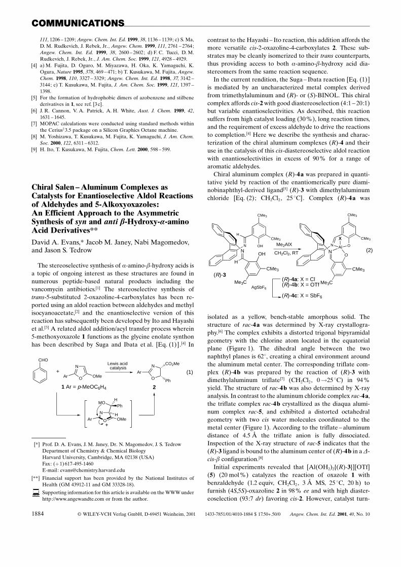
Chiral aluminum complex (R)-4 a was prepared in quanti-tative yield by reaction of the enantiomerically pure diami-nobinaphthyl-derived ligand[5] (R)-3 with dimethylaluminumchloride [Eq. (2); CH2Cl2, 25 8C]. Complex (R)-4 a was
AgSbF6
X
Al O
O
N
N
CMe3
Me3C
CMe3
CMe3
OH
OH
N
N
CMe3
Me3C
CMe3
CMe3
H
H
Me2AlX
CH2Cl2, RT
(R)-3
(2)
(R)-4a: X = Cl(R)-4b: X = OTf
(R)-4c: X = SbF6
isolated as a yellow, bench-stable amorphous solid. Thestructure of rac-4 a was determined by X-ray crystallogra-phy.[6] The complex exhibits a distorted trigonal bipyramidalgeometry with the chlorine atom located in the equatorialplane (Figure 1). The dihedral angle between the twonaphthyl planes is 628, creating a chiral environment aroundthe aluminum metal center. The corresponding triflate com-plex (R)-4 b was prepared by the reaction of (R)-3 withdimethylaluminum triflate[7] (CH2Cl2, 0!25 8C) in 94 %yield. The structure of rac-4 b was also determined by X-rayanalysis. In contrast to the aluminum chloride complex rac-4 a,the triflate complex rac-4 b crystallized as the diaqua alumi-num complex rac-5, and exhibited a distorted octahedralgeometry with two cis water molecules coordinated to themetal center (Figure 1). According to the triflate ± aluminumdistance of 4.5 � the triflate anion is fully dissociated.Inspection of the X-ray structure of rac-5 indicates that the(R)-3 ligand is bound to the aluminum center of (R)-4 b in a D-cis-b configuration.[8]
Initial experiments revealed that [Al(OH2)2{(R)-3}][OTf](5) (20 mol %) catalyzes the reaction of oxazole 1 withbenzaldehyde (1.2 equiv, CH2Cl2, 3 � MS, 25 8C, 20 h) tofurnish (4S,5S)-oxazoline 2 in 98 % ee and with high diaster-eoselection (93:7 dr) favoring cis-2. However, catalyst turn-
111, 1206 ± 1209; Angew. Chem. Int. Ed. 1999, 38, 1136 ± 1139; c) S. Ma,D. M. Rudkevich, J. Rebek, Jr., Angew. Chem. 1999, 111, 2761 ± 2764;Angew. Chem. Int. Ed. 1999, 38, 2600 ± 2602; d) F. C. Tucci, D. M.Rudkevich, J. Rebek, Jr., J. Am. Chem. Soc. 1999, 121, 4928 ± 4929.
[4] a) M. Fujita, D. Oguro, M. Miyazawa, H. Oka, K. Yamaguchi, K.Ogura, Nature 1995, 378, 469 ± 471; b) T. Kusukawa, M. Fujita, Angew.Chem. 1998, 110, 3327 ± 3329; Angew. Chem. Int. Ed. 1998, 37, 3142 ±3144; c) T. Kusukawa, M. Fujita, J. Am. Chem. Soc. 1999, 121, 1397 ±1398.
[5] For the formation of hydrophobic dimers of azobenzene and stilbenederivatives in 1, see ref. [3c].
[6] J. R. Cannon, V. A. Patrick, A. H. White, Aust. J. Chem. 1989, 42,1631 ± 1645.
[7] MOPAC calculations were conducted using standard methods withinthe Cerius2 3.5 package on a Silicon Graphics Octane machine.
[8] M. Yoshizawa, T. Kusukawa, M. Fujita, K. Yamaguchi, J. Am. Chem.Soc. 2000, 122, 6311 ± 6312.
[9] H. Ito, T. Kusukawa, M. Fujita, Chem. Lett. 2000, 598 ± 599.
[*] Prof. D. A. Evans, J. M. Janey, Dr. N. Magomedov, J. S. TedrowDepartment of Chemistry & Chemical BiologyHarvard University, Cambridge, MA 02138 (USA)Fax: (�1) 617-495-1460E-mail : [email protected]
[**] Financial support has been provided by the National Institutes ofHealth (GM 43912-11 and GM 33328-18).
Supporting information for this article is available on the WWW underhttp://www.angewandte.com or from the author.