primer on medical genomics part ii: background principles ... · 786 primer on medical genomics...
TRANSCRIPT

Mayo Clin Proc, August 2002, Vol 77 Primer on Medical Genomics Part II 785
Mayo Clin Proc. 2002;77:785-808 785 © 2002 Mayo Foundation for Medical Education and Research
Medical Genomics
Primer on Medical GenomicsPart II: Background Principles and Methods in Molecular Genetics
AYALEW TEFFERI, MD; ERIC D. WIEBEN, PHD; GORDON W. DEWALD , PHD; DAVID A. H. WHITEMAN , MD;MATTHEW E. BERNARD, MD; AND THOMAS C. SPELSBERG, PHD
From the Division of Hematology and Internal Medicine (A.T.), De-partment of Biochemistry and Molecular Biology (E.D.W., T.C.S.),Division of Laboratory Genetics (G.W.D.), Department of MedicalGenetics (D.A.H.W.), and Department of Family Medicine (M.E.B.),Mayo Clinic, Rochester, Minn. The authors are members of theMayo Clinic Genomics Education Steering Committee.
A glossary of terms frequently used in medical genomics appears onpages 805 through 808.
Individual reprints of this article are not available. The entire Primeron Medical Genomics will be available for purchase from the Pro-ceedings Editorial Office at a later date.
The nucleus of every human cell contains the full comple-ment of the human genome, which consists of approxi-mately 30,000 to 70,000 named and unnamed genes andmany intergenic DNA sequences. The double-helical DNAmolecule in a human cell, associated with special proteins,is highly compacted into 22 pairs of autosomal chromo-somes and an additional pair of sex chromosomes. Theentire cellular DNA consists of approximately 3 billionbase pairs, of which only 1% is thought to encode a func-tional protein or a polypeptide. Genetic information isexpressed and regulated through a complex system ofDNA transcription, RNA processing, RNA translation,and posttranslational and cotranslational modification ofproteins. Advances in molecular biology techniques haveallowed accurate and rapid characterization of DNAsequences as well as identification and quantification ofcellular RNA and protein. Global analytic methods andhuman genetic mapping are expected to accelerate theprocess of identification and localization of disease genes.
In this second part of an educational series in medicalgenomics, selected principles and methods in molecularbiology are recapped, with the intent to prepare the readerfor forthcoming articles with a more direct focus on as-pects of the subject matter.
Mayo Clin Proc. 2002;77:785-808
BAC = bacterial artificial chromosome; bp = base pairs;cDNA = complementary DNA; cM = centiMorgan; EST =expressed sequence tag; FISH = fluorescence in situ hybridiza-tion; FRET = fluorescence resonance energy transfer; kb =kilobase; Mb = million base; mRNA = messenger RNA;mtDNA = mitochondrial DNA; ORF = open reading frame;PCR = polymerase chain reaction; RFLP = restriction frag-ment length polymorphism; rRNA = ribosomal RNA; RT-PCR = reverse transcription-PCR; snRNP = small nuclearribonucleoprotein; tRNA = transfer RNA; UTR = untrans-lated region; VNTR = variable numbers of tandem repeats;YAC = yeast artificial chromosome
The Human Genome Project has provided near-total se-quence information about our genes and their sur-
roundings.1,2 This monumental accomplishment has beenaccompanied by equally remarkable progress in biomedicalresearch and computer science.3-5 Biologists and clinical in-vestigators are now well positioned to perform global analy-sis of genetic polymorphisms-mutations,6,7 gene expression,8
protein identity and function (proteomics),9 and the 3-dimen-sional structure of proteins (structural genomics).10,11 Theresulting information should accelerate the development ofnovel and targeted therapeutics that are tailored to individualgenomic profiles (pharmacogenomics).12,13 The massiveamount of data generated by genome-wide experiments
requires the development of comprehensive genomic-pro-teomic databases and software that enable megadata access,transfer, and manipulation (bioinformatics).14,15
For editorial comment, see page 745.
The development of a structural and functional genomicinfrastructure will undoubtedly accelerate our understand-ing of diseases and help refine methods of disease pre-diction, diagnosis, staging, and treatment.16,17 Clinicians,clinician investigators, and students of medicine must beprepared to participate effectively in the “new” genomicsrevolution and tackle the economical and ethical issues thatare integral to all aspects of medical genomics.18-21 Theintent of the current communication is to begin the processof education in this regard, with a clinician-oriented reviewof basic principles and methods in molecular biology thatare relevant to genomics. In subsequent articles, the afore-mentioned components of genomics will be discussed andupdated periodically.
GENES AND GENE EXPRESSIONGenes and DNA
A gene, the original Mendel “factor,”22 is the basic unitof heredity.23,24 A gene is defined as a contiguous region of
For personal use. Mass reproduce only with permission from Mayo Clinic Proceedings.

Primer on Medical Genomics Part II Mayo Clin Proc, August 2002, Vol 77786
DNA25,26 that includes a defined set of exons (protein-coding regions of DNA)27 and introns (DNA regions inter-spersed between exons).28 The basic unit of DNA is called adeoxyribonucleotide (nucleotide for short) and is made upof 1 of 4 nitrogenous bases (adenine [A], guanine [G],thymine [T], cytosine [C]) that is attached to the 1′ carbonof a deoxyribose sugar, which in turn is attached to aphosphate group through its 3′ and 5′ carbons29 (Figure 1).Each DNA strand is a polymer of many nucleotides that areattached to each other through their phosphate and sugargroups (forming a sugar-phosphate backbone of DNA).31
There are 2 orientations of sugar-phosphate connections: 5′to 3′ (carbon 5′ of the sugar moiety of a particular nucleotidein a strand is attached to a phosphate group that is in turnattached to carbon 3′ of the next nucleotide) and 3′ to 5′.
A DNA molecule is made up of 2 antiparallel (1 strandis oriented 5′ to 3′ and the other 3′ to 5′) polymers (strands)of nucleotides (side-by-side chains) in a helical configura-tion that forms a double helix (Figure 1).32 The 2 strands ofDNA are noncovalently (loosely) connected to each otherthrough their nitrogenous bases by hydrogen bonds (A
always bonds with T and G with C). Both strands of DNAare used as templates for new complementary DNA(cDNA) synthesis during DNA replication that precedescell division. As such, each of the daughter cells contains 1parental and 1 newly synthesized strand of DNA (ie, DNAreplication is semiconservative).33
Although the accurate number of genes in the humangenome remains to be clarified,34 the initial drafts from theHuman Genome Project suggest the presence of approxi-mately 30,000 named and unnamed genes and manyintergenic DNA sequences.1,2 In other words, the humangenome represents the entire DNA sequence (approxi-mately 3 billion base pairs [bp] per haploid set of chromo-somes) in a human cell and is made up of approximately1% exons (protein-coding regions of DNA), 24% introns(DNA regions interspersed between exons), and 75%intergenic DNA (DNA regions that flank genes).35 Theintergenic DNA consists largely (55% of the total genome)of repetitive sequences, including inverted repeats.36-38
In this regard, it is helpful to recall the different catego-ries of DNA that make up the human genome. The mostcommon class of DNA, also the functionally relevant one,is called single-copy DNA and appears only once or a fewtimes across the entire genome. In contrast, repetitive DNAsequences appear multiple times and may be found eitherin a series of adjacent clusters (tandem repeats)39 or scat-tered at different regions of the genome (interspersed re-peats).40,41 Tandem repeats make up “satellite” DNA, aband of DNA that separates from other DNA on a cesiumchloride density gradient centrifugation.42,43 Satellite DNAis usually localized near the centromere.44 The termsminisatellites (20-70 bp repeats) and microsatellites (2-4bp repeats) are used to refer to tandem repeats of smallsize.45 The total length of such tandem repeats is highlyvariable among individuals and between cells of differentclonal origin. Accordingly, this variability has been used indifferent investigations, including person identificationand cell chimerism.46
Scattered DNA sequence repeats can also be short (shortinterspersed nuclear elements [SINEs], eg, Alu repeats of100-300 bp long) or long (long interspersed nuclear ele-ments [LINEs], eg, L1 [line-1; long inserted element-1]sequences of 1-5 kilobase [kb] long).41 An Alu element is aspecific DNA sequence that derives its name from the Alu1 endonuclease because of a single recognition site for theparticular enzyme located near the center of the Alu se-quence. Approximately one half million Alu repeats arescattered throughout human genome and serve no apparentfunction. Some DNA sequences can self-replicate, and theadditional copies might migrate to different locationswithin the genome (mobile elements or transposons).47
Some mobile elements may have originated from viral
Figure 1. Components and structure of DNA. A = adenine; G =guanine; T = thymine; C = cytosine. See text for further details.Copyright 1998 from Alberts et al.30(p186) Reproduced by permis-sion of Garland Publishing, Inc, part of The Taylor & FrancisGroup.
For personal use. Mass reproduce only with permission from Mayo Clinic Proceedings.

Mayo Clin Proc, August 2002, Vol 77 Primer on Medical Genomics Part II 787
RNA and propagate via an RNA intermediate by usingreverse transcriptase (retrotransposons).48,49
Most human DNA is organized into 46 chromosomesthat reside in the nucleus of every nucleated cell. However,some DNA resides in mitochondria that are located in thecytoplasm and number in the hundreds per individual cell.50
Each mitochondrion has many copies of a circular DNAmolecule (the mitochondrial chromosome), which is ap-proximately 16,500 bp long, and is replicated and ex-pressed within the organelle.51 The mitochondrial DNA(mtDNA) may have coevolved with nuclear DNA andencodes for 2 ribosomal RNAs (rRNAs), 22 transfer RNAs(tRNAs), and 13 functional polypeptides that are involvedin oxidative phosphorylation.52 Mitochondrial DNA differsfrom nuclear DNA in terms of not having introns anddisplaying high mutation rates.53,54 Because mitochondriaare located in the cytoplasm, inheritance of mtDNA isexclusively maternal.55 The combination of uniparental in-heritance and high mutation rates has made mtDNA impor-tant in the study of human evolutionary genetics.56-58
Each chromosome represents 1 double-stranded DNAmolecule that is closely packed with special proteins calledhistones (DNA + histones + regulatory proteins = chroma-tin) (Figure 2).59,60 Chromatin condenses substantially dur-ing the metaphase stage of mitosis and becomes visible bylight microscopy. Chromatin that remains condensed dur-ing interphase is called heterochromatin.61-63 Chromatinthat condenses only during metaphase is called euchroma-tin. In general, euchromatin contains more functionalgenes, and heterochromatin contains more nonfunctionalrepetitive DNA elements.
In women, the 46 chromosomes in each cell are found in23 homologous pairs (2 haploid sets of chromosomes): 1set inherited from the father and the other from the mother.The same is true for men except the twenty-third chromo-some pair is only partially homologous, at the point whereit pairs an X to a Y chromosome.64 The smallest and largesthuman chromosomes contain about 50 and 250 million bp,respectively. Each human gene is usually more than 10,000bp long.1 However, only a tenth of this actually codes foramino acids of the mature protein product.
Gene ExpressionThe information contained in a given gene is expressed
if the DNA sequence of the gene is first transcribed into anRNA molecule that, after being processed in the nucleus,will carry the information into the cytoplasm for translationinto a protein.65 Unless indicated otherwise, the mecha-nisms described subsequently pertain primarily to eukary-otic cells.
Transcription .—Transcription starts with formation ofan RNA sequence from a DNA template representing a
gene. The protein-coding gene has both transcribed andnontranscribed regions of DNA.66 The transcribed regionis first copied onto a nuclear RNA called the primarytranscript (pre-messenger RNA [mRNA] or heteroge-neous nuclear RNA) in a process catalyzed by the enzymeRNA polymerase II.67 Part of the nontranscribed region(the promoter sequence) of the gene is the site of theinitial contact with RNA polymerase II and controlstranscription of the primary transcript; it is usually lo-cated upstream (20-100 bp) of the start of the transcribedregion.68,69 Other regulatory elements of transcription(enhancers-silencers) can be on either side of the tran-scription start site and be several thousand nucleotidesaway from the place where transcription actually ini-tiates.70 However, both promoters and enhancers-silenc-ers usually reside on the same stretch of DNA (ie, are cis-acting).
Figure 2. Packing of the DNA molecule with histone proteins toform chromatin packages that are condensed into chromosomes.Copyright 1998 from Alberts et al.30(p253) Reproduced by permis-sion of Garland Publishing, Inc, part of The Taylor & FrancisGroup.
For personal use. Mass reproduce only with permission from Mayo Clinic Proceedings.

Primer on Medical Genomics Part II Mayo Clin Proc, August 2002, Vol 77788
Of note, neither the cis-acting sequences of DNA northe RNA polymerase II alone could initiate transcriptionwithout the help of proteins called transcription factors.71-73
These “trans-acting” proteins recognize and bind to the cis-acting DNA sequences and together with RNA polymeraseII and other coactivators of transcription factors form thetranscription complex that ultimately regulates transcrip-tion either in a positive or a negative manner (Figure 3).74,75
In this regard, promoters often contain characteristic se-quences such as TATA and CATT that are recognizedby transcription factors.76-79 Transcription factors may begrouped into different classes based on motifs present intheir 3-dimensional protein structure (eg, helix-turn-helix,helix-loop-helix, Leucine zipper, zinc finger).80,81 Theseunique motifs allow recognition of the aforementionedspecific DNA sequences that are cis-acting regulators oftranscription.82
During transcription and after completion, the primarytranscript (always synthesized from 5′ to 3′ orientation)undergoes several modifications before the mature mRNAis released into the cytoplasm for protein translation.83
These include “capping,” addition of a “poly-A tail,” and“splicing” of the primary transcript.84 First, an extra nucle-otide (the cap), a 7-methylguanosine (m7G-P-P-P) is placedat the 5′ end soon after the initiation of transcription.85 Thismodification is believed to protect RNA degradation andmark the starting point for protein translation by binding to
ribosomes. Subsequently, the end of transcription is her-alded when RNA polymerase II reaches a “terminationsequence” (TTATT on the DNA template and AAUAAAon the primary transcript), which, together with other pro-teins, signals cleavage at the polyadenylation site (10-35nucleotides downstream of the termination sequence).86,87
Finally, poly(A) polymerase adds a stretch of approxi-mately 200 adenine nucleotides (poly-A tail) at the 3′ endof the primary transcript.88
Once synthesis of the primary transcript is completed,the introns of pre-mRNA are spliced out and the remainingsegments rejoined to form the mature mRNA.89,90 The latterincludes both translated and untranslated regions (UTRs)of exons, the cap, and the poly-A tail (Figure 4).86 Splicingoccurs in a large complex called a spliceosome, whichcontains the primary transcript and its associated proteins,several protein splicing factors, and a class of small nuclearRNAs that are present in the cell as small nuclear ribo-nucleoproteins (snRNPs).91-95 There is increasing evidencethat both snRNPs and intronic RNA may possess catalyticactivity (ie, are ribozymes) and contribute to RNA splic-ing.96-98 The spliceosome recognizes the 5′ and 3′ splicesites on the exon-intron boundaries via conserved consen-sus sequences within the intron.99 The actual process in-volves both lariat (loop and tail) formation of the excisedintron and transesterification of the 5′ and 3′ splice sites ofthe exons.100-102 Alternative splicing occurs not infre-
Figure 3. Transcription initiation complex in a eukaryocyte. See text for details. Copyright1998 from Alberts et al.30(p266) Reproduced by permission of Garland Publishing, Inc, part ofThe Taylor & Francis Group.
For personal use. Mass reproduce only with permission from Mayo Clinic Proceedings.

Mayo Clin Proc, August 2002, Vol 77 Primer on Medical Genomics Part II 789
quently, resulting in different mRNA species and subse-quently different proteins from the same primary tran-script.103-106 Alternative splicing may occur in as many asone half or more of all human genes and sometimes resultsin altered translation efficiency rather than synthesis of adifferent polypeptide. The widespread occurrence of thisphenomenon increases the coding capacity of genes andcontributes to proteomic diversity. The regulation of alter-native splicing is an active area of current research, andsplicing regulators may include snRNPs.104,107-109
In conventional terms, the final mRNA represents thesense template, whereas the cDNA strand that was used asa template for its synthesis is referred to as the antisensestrand. Regardless, not all the nucleotide sequences ofmRNA are ultimately translated into proteins.110 The veryfirst and last transcribed regions of the primary transcriptcontain sequences (UTRs) that do not code for proteindespite being part of the first and last exons.111-113 None-theless, these sequences are important for the function ofthe mRNA and often control the stability, subcellular lo-calization, and translational efficiency of an individualmRNA.114-117 The 5′ UTR, which might be hundreds ofbases long, is followed by the protein coding sequence ofthe mRNA (open reading frame [ORF]) that is in turnfollowed by the 3′ UTR.118
Translation.—The mRNA is translated into protein inthe cytoplasm of the cell with the help of ribosomes. Ribo-somes are made up of 2 subunits (60S and 40S), eachconsisting of proteins and rRNA.119 The mature mRNA that
is released into the cytoplasm docks onto rRNA, and itsORF is recognized by another form of cytoplasmic RNAcalled transfer RNA120 (Figure 5). A cloverleaf-shapedadaptor molecule, tRNA carries a specific amino acid at its3′ end.122 At the opposite end of the amino acid–binding
Figure 4. Steps leading from gene to protein for eucaryotes (A) and procaryotes (B). mRNA =messenger RNA. See text for further details. Copyright 1998 from Alberts et al.30(p223) Reproducedby permission of Garland Publishing, Inc, part of The Taylor & Francis Group.
Figure 5. Translation of messenger RNA (mRNA) into aminoacids in the cytoplasm of a cell. Process requires the concertedeffort of mRNA, transfer RNA (tRNA), and ribosomal RNA(rRNA). See text for further details. Reproduced with permissionfrom Lodish et al.121(p117)
For personal use. Mass reproduce only with permission from Mayo Clinic Proceedings.

Primer on Medical Genomics Part II Mayo Clin Proc, August 2002, Vol 77790
site, the tRNA is equipped with a 3 nucleotide sequence (ie,the “anticodon”) that undergoes complementary base pair-ing with the mRNA “codon.”123,124 This codon-anticodoninteraction, which is dictated by the specific nucleotidesequence on the mRNA (the genetic code), aligns anti-codon-specific amino acids that subsequently form a grow-ing polypeptide chain through covalent peptide bonding.125
Polypeptide synthesis on the ribosome may be subdi-vided into initiation, elongation, and termination phases.First, elongation factors (eg, eIF4F) recognize the 5′ cap ofmRNA, recruit other components of the initiation complex,and subsequently bind to the rRNA on the small (40S)ribosomal subunit.126 The initiation complex (40S ribo-some, elongation factors such as eIF4, initiator tRNA, etc)rolls along the mRNA sequence until it reaches the “startcodon” (AUG). Subsequently, polypeptide synthesis startswith disintegration of the initiation complex and recruit-ment of the 60S ribosome along with other elongationfactors.127 As the ribosome continues moving along themRNA genetic code, successive amino acid–attachedtRNAs are recruited, and the polypeptide chain grows untila “stop codon” (UAA, UAG, or UGA) signals release ofthe finished product.128,129 Current evidence suggests thatthe enzymatic activity required to form peptide bonds be-tween adjacent incoming amino acids may reside in therRNA, which functions as a ribozyme.130
Many newly synthesized proteins are further modifiedafter or during polypeptide synthesis.131 Posttranslationalor cotranslational modifications of proteins ensure properprotein folding, targeting, activation, and stability.132 Thespecific processes include proteolysis, glycosylation, phos-phorylation, acylation, metal binding, and assumption ofsecondary and tertiary structures.133 Chaperones are en-zymes that guide proper protein folding by binding andreleasing misfolded proteins.134 Damaged or surplus pro-teins are removed through various mechanisms, includinglysosomal and nonlysosomal protein degradation. Non-lysosomal protein degradation is facilitated by ubiquitina-tion (a process of tagging proteins with ubiquitin to removethem by proteolysis). Ubiquitin is a small protein (76amino acids long) that binds and marks damaged or excessproteins for subsequent degradation by the proteasomecomplex.135-137 The proteasome is a multiunit protease com-plex that degrades ubiquitin-tagged proteins.138 Ubiquitina-tion also influences signal transduction and apoptosis byfacilitating controlled degradation of pertinent regulatorymolecules.139,140
GENOMICS-RELATED LABORATORY METHODSPolymerase Chain Reaction
Molecular cloning or DNA amplification often involvessynthesis of multiple copies (clones) of a DNA sequence of
interest (the target DNA). This may be accomplished byeither inserting the target DNA into a bacterium that iscapable of extended cell division (recombinant DNA tech-nology)141-143 or using the polymerase chain reaction (PCR)for in vitro cloning of DNA (Figure 6).144-148 Polymerasechain reaction is an in vitro method of replicating relativelysmall DNA sequences into millions of copies over a shortperiod.
A typical PCR reaction requires (1) two oligonucleotideprimers (PCR primers), (2) a thermally stable DNA poly-merase, (3) an ample amount of free deoxynucleotides, and(4) a small amount of DNA (the sample) that contains thesequence of interest (ie, the region to be amplified).149 ThePCR primers (short DNA sequences of approximately 20bp in length) are synthesized in the laboratory, and theirnucleotide sequences are designed to be complementary toDNA sequences that flank (lie on either side of) the DNAfragment of interest (the target).150-152 Therefore, one has toknow the nucleotide sequence of the target DNA fragmentto use PCR technology. Because DNA synthesis can pro-ceed only in the 5′ to 3′ direction, a PCR primer allowsextension of DNA in only 1 direction (ie, the other side is adead end). Therefore, to replicate both strands of the DNAmolecule (the complementary strands), 2 PCR primers(forward and reverse primers) are needed, and they areplaced on either side of the DNA sequence of interest oncomplementary strands (ie, the forward primer on 1 strandof DNA and the reverse primer on the second strand ofDNA that is complementary to the first strand). This ap-proach allows amplification of the double-stranded targetsequence that is located between the 2 primers.145,148
At the beginning of PCR, the sample DNA is first heatedto a temperature greater than 90°C to separate the 2 pairedDNA strands (DNA denaturation). The reaction is thencooled to approximately 40°C, in the presence of excessprimers, to allow annealing (hybridization) of the specificprimers to their complementary sequences on the targetDNA (which is now in single strands). After the primers arebound to the DNA sequences that are immediately adjacentto the target sequence, new complementary strands ofDNA, which are extensions of the bound PCR primers, areformed by recruiting the free nucleotides in the reaction.This step of primer extension occurs in an intermediatetemperature of approximately 70°C with the help of athermally stable DNA polymerase (an enzyme originallyderived from the bacterium Thermus aquaticus).153-155 Thenewly synthesized DNA is double stranded, starting at the 5′end with 1 of the 2 primers (Figure 6). After this first cycle ofPCR, the same process is repeated 20 to 30 times (20-30heating and cooling cycles), and each cycle results in thedoubling of the target DNA region, with a final theoreticaltarget amplification of approximately 1 million-fold or more
For personal use. Mass reproduce only with permission from Mayo Clinic Proceedings.

Mayo Clin Proc, August 2002, Vol 77 Primer on Medical Genomics Part II 791
(Figure 6). Of note, the final amplified product consistsmainly of fragments that comprise the region of DNA be-tween the 2 primers (ie, the sequence of interest) (Figure 6).
Several modifications of PCR allow enhanced specific-ity and broader applicability. For example, the specificityof PCR may be enhanced by subjecting the product of thefirst PCR to another round of PCR amplification by using asecond pair of primers that are placed internal of the firstpair. The second pair of primers are called nested primersand the reaction nested PCR.156 In another modification,more than 1 DNA sequence of interest may be interrogatedin a single PCR by using multiple primers (multiplexPCR).157,158 Multiplex PCR promises to be an importantdiagnostic tool in infectious diseases.159,160
In general, PCR technology works best when the DNAfragment of interest is relatively small (<1000 bp).145 Thetechnique is easily automated to allow detection and ampli-fication of nanomolar quantities of DNA with a rapid turn-around time (a few hours). Because of the rapidity of the
assay and the small amount of test sample needed (nano-gram quantities), PCR is an extremely useful and efficientmethod of genetic testing in disease diagnosis, monitoringof treatment response, detection of minimal residual dis-ease, and tissue typing.156,161-164 Furthermore, the ability toextract DNA from various biological samples combinedwith PCR technology has substantially affected both foren-sic medicine and archeological studies.165-168 However, theextreme sensitivity of PCR makes it vulnerable to the pos-sibility of false-positive test results due to trace amounts ofcontaminants.169
Reverse Transcription-PCR.—There are severalmethods of detecting and quantifying specific mRNA frombiological samples, including Northern blotting, nucleaseprotection assays, and reverse transcription (RT)-PCR. All3 methods start with acquisition of fresh biological samples(blood, bone marrow, biopsy material, cell line, etc) thatallows extraction of either total RNA or purified mRNA(polyadenylated RNA).170,171 In Northern blotting a specific
Figure 6. Amplification of DNA using polymerase chain reaction technique. See text for further details. Copyright 1998 fromAlberts et al.30(p533) Reproduced by permission of Garland Publishing, Inc, part of The Taylor & Francis Group.
For personal use. Mass reproduce only with permission from Mayo Clinic Proceedings.

Primer on Medical Genomics Part II Mayo Clin Proc, August 2002, Vol 77792
RNA species, among a mixture of RNAs, is first size-separated by gel electrophoresis. The RNA is subsequent-ly transferred to a membrane where hybridization occurswith either radioactively or nonisotopically labeled DNAprobes.172,173 A nuclease protection assay uses single-strand–specific endonucleases to remove by digestionsingle- but not double-stranded DNA.174 In the particularassay, the sample RNA is incubated with excess labeled,single-stranded DNA molecules that are complementary tothe specific mRNA. The subsequent hybridization be-tween the mRNA and the labeled DNA strands protectsthe specific mRNA from being digested with the single-strand–specific endonucleases. In both Northern blottingand nuclease protection assays, the amount of steady-statemRNA level is estimated from the density of the labelingmolecule.
In RT-PCR, traditional PCR is combined with reversetranscription to amplify mRNA.175-178 In reverse transcrip-tion, mRNA (sense) in the test sample is converted to asingle-stranded cDNA (antisense) in a reaction mediatedby reverse transcriptase. Reverse transcriptase (a retroviralenzyme) is an RNA-dependent DNA polymerase that cansynthesize a DNA sequence that is complementary to aspecific mRNA template.179 The synthesized cDNA repre-sents only the exons of the corresponding genomic DNA.The subsequent PCR amplification method is called re-verse transcription-polymerase chain reaction to distin-guish it from the standard PCR used to amplify genomicDNA. Reverse transcription-PCR is an extremely powerfuland sensitive tool to detect and quantify mRNA (ie, geneexpression) from minute amounts of test samples.176,180
In RT-PCR, mRNA quantification may be performedboth in relative (relative RT-PCR) and in absolute (com-petitive RT-PCR) terms. In relative RT-PCR, the amountof the target mRNA is estimated in comparison with acoamplified internal control. The resultant ratio is thencompared across different samples. In competitive RT-PCR,181 a known quantity of a synthetic RNA molecule,which is similar in sequence but shorter compared with thetarget mRNA, is added to the reaction. This results in thesynthesis of 2 cDNA templates (target and synthetic) thatwill compete for the same primers during PCR amplifica-tion. As a result, the abundance of the target PCR product isinversely proportional to the amount of the synthetic RNAthat is added to the reaction. Therefore, by performing aseries of PCRs with addition of serial dilutions of thesynthetic RNA, a concentration curve can be constructedthat is used to estimate the quantity of the endogenoustarget product.182
Recent medical uses of RT-PCR include detection oflymph node micrometastasis in prostate cancer,183 breastcancer tumor contamination of bone marrow,184 viral con-
tamination of vaccines,185 and monitoring of minimal re-sidual disease in acute leukemia.186,187
Real-Time PCR.—In RT-PCR, the steady-state levelof a specific mRNA is indirectly estimated from the levelof the corresponding amplified product. The amplifiedproduct is usually quantified after the reaction is completed(ie, “end point” quantification).180 Alternatively, the pres-ence and quantity of PCR products may be monitored in“real time” as the reaction progresses.188 Monitoring theabundance of PCR products in multiple cycles of PCRprovides dynamic range values that are more informativethan end point measurements.189 Of note, target amplifica-tion during the initial cycles of PCR is exponential becausethe presence of excess primers in the reaction minimizesrenaturation of the complementary strands of the targetDNA. As the concentration of an amplified product in-creases, the possibility of renaturation of its complemen-tary strands increases. As a result, the amplification ratedecreases and plateaus subsequently. The transition pointfrom the exponential to the plateau phase of PCR may varymarkedly for different samples. Therefore, it is more accu-rate to quantify the specific mRNA products of PCR duringthe exponential phase of target amplification. Real-timePCR accomplishes this task by quantifying reaction prod-ucts in every cycle of PCR.178,188,190,191
Real-time PCR depends on the detection and quantifica-tion of fluorescent molecules that are bound to the PCRproduct. Specific binding of fluorescent molecules to thePCR product is achieved by several methods, includingTaqMan192 and SYBR green.193 The TaqMan probe is adual-labeled oligonucleotide probe that contains a fluores-cent dye at its 5′ base and another molecule (a quencher) atits 3′ base. The quencher absorbs (quenches) fluorescencewhen it is situated close to the fluorescent molecule. Thisphenomenon is called fluorescence resonance energytransfer (FRET).194 Therefore, the probe itself does notfluoresce. However, the fluorescent molecule is separatedfrom the quencher molecule during primer extensionthrough the exonuclease activity of Taq polymerase (thespecific polymerase used in this particular PCR), wherebyfluorescence is unmasked.195,196 Accordingly, the degree offluorescence increases with an increase in the level of PCRproduct. Other FRET-dependent methods include molecu-lar beacons197 and scorpions.198 In contrast, the SYBR greenmethod is not FRET dependent. SYBR green is a dye thatemits fluorescence only when it binds to double-strandedDNA.193 Therefore, the fluorescence signal is enhancedwith continued synthesis of the PCR product.
A discussion on the quantification of the signals ob-tained from real-time PCR is beyond the scope of thisreview, and the reader is referred to recent overviews onthis subject.180,189 Potential applications of real-time PCR
For personal use. Mass reproduce only with permission from Mayo Clinic Proceedings.

Mayo Clin Proc, August 2002, Vol 77 Primer on Medical Genomics Part II 793
include disease diagnosis and monitoring,198-201 biosafetyand genetic stability testing,202 radiation exposure assess-ment,203 mutation detection,204 quantification of cytokineprofiles in immune cells or inflamed tissue,189 and in vivoimaging of cellular processes.205
Electrophoresis and BlottingDNA, RNA, or proteins may be separated by size with
use of agarose or polyacrylamide gels through elec-trophoresis. DNA and RNA are uniformly, negativelycharged, and under most circumstances their gel separationis a function of their size and topology (linear fragmentsmove in a different manner from supercoiled fragments ofthe same length). In contrast, proteins must first be dena-tured (unfolded) and negatively coated with an ionic deter-gent (sodium dodecyl sulfate) to allow gel separation solelyby size, avoiding disturbances from their inherent positiveand negative charge. Isoelectric focusing separates mol-ecules by their net (positive or negative) charge.206
In Southern blotting (named after Dr Edward Southern,who described the technique), DNA is first cut into piecesusing restriction endonucleases (enzymes that digest DNAat specific sites that are marked by a 4- to 8-memberspecific nucleotide sequence), and then the restriction frag-ments are subjected to agarose gel electrophoresis (Figure7).207 The double-stranded DNA fragments on the gel arethen turned into single strands by denaturation and trans-ferred to a DNA-binding membrane, such as nitrocelluloseor nylon, to make a permanent copy of single-strandedDNA. A DNA sequence of interest is then visualized bya radiolabeled reporter probe after a hybridization stepfollowed by autoradiography. In addition to target iden-tification and quantification, Southern blotting may al-low detection of mutations that result in alterations of
restriction fragment lengths (ie, detect restriction frag-ment length polymorphisms [RFLPs]). Larger DNA mol-ecules are separated by pulsed-field gel electrophoresis(intermittent application of voltage gradients to disorientlarge DNA molecules and optimize separation by sizeonly).208,209
Northern blotting (named to reflect similarity to South-ern blotting) is used to identify specific RNAs.173 Westernblotting is used to identify specific proteins that have beenseparated by size with use of polyacrylamide gel electro-phoresis.210,211 The size-fractionated proteins are transferredto a nitrocellulose membrane, and proteins of interest areidentified and quantified by visualization with reporter-linked antibodies. In 2-dimensional gel electrophoresis,polyacrylamide gel electrophoresis is combined with sepa-ration of proteins by their isoelectric point (the pH wheretheir overall charge is neutral).212
Cutting, Pasting, and Altering the Topology of DNADifferent enzymes specialize in digesting and twisting
(and untwisting) the DNA molecule. Endonucleases cleaveDNA (break the sugar-phosphate ester bond between 2deoxynucleotides) from the middle, while exonucleasessequentially remove nucleotides from the ends of DNAstrands.213,214 DNA polymerases often possess exonucleaseactivity as part of their proofreading capability in additionto their synthetic function.215 For many laboratory experi-ments, it is useful to remove the exonuclease activity ofEscherichia coli DNA polymerase I by brief proteolyticdigestion, leading to a synthetically active DNA poly-merase called the Klenow fragment.216,217 Restriction endo-nucleases act at specific DNA sites, which are marked byprecise 4- to 8-nucleotide sequences. Such restriction di-gestion of DNA is the key to recombinant technology and
Figure 7. Southern blotting. See text for further details. Reproduced with permission from Lodish etal.121(p240)
For personal use. Mass reproduce only with permission from Mayo Clinic Proceedings.

Primer on Medical Genomics Part II Mayo Clin Proc, August 2002, Vol 77794
chromosome mapping. Ligases attach 2 separate DNAfragments (reestablish the sugar-phosphate bond) and areimportant in recombinant technology.218
DNA is naturally highly compacted but may take arelaxed (α-helical) or even a supercoiled state (a coilingand double coiling of the α-helical thread). A positivesupercoil tends to tighten further and supercoil the neigh-
boring double helix, and a negative supercoil tends to un-wind the neighboring double helix. Supercoiling and relax-ing of DNA are often mediated by topoisomerases.219,220
Topoisomerases alter the topology (degree of coiling) ofDNA by creating transient, either single (topoisomerase I) ordouble (topoisomerase II) strand breaks and allowing DNAto wind or unwind through those breaks. DNA gyrase is atype of topoisomerase that relieves supercoiling.221
Clones, Libraries, and PolymorphismsClones.—Any DNA can be amplified (mass and strand
number increased) in the laboratory by using either recom-binant technology or PCR. Recombinant cloning startswith preparation of restriction fragments of DNA (eg, fromhuman cells) that are then fused (using DNA ligase) tovector DNA (a carrier DNA molecule capable of self-replication) that has been digested with the same restrictionenzymes so that they have similar split ends that allowDNA ligation.222 This “recombinant” DNA molecule istransported and amplified by the vector (a carrier moleculecapable of self-replication) (Figure 8). Vectors in currentuse include plasmids (circular double-stranded DNA mol-ecules that replicate extrachromosomally in bacteria),bacteriophages (bacterial virus with a larger than plasmiddouble-stranded DNA molecule), cosmids (large plasmidscapable of carrying large DNA inserts), and bacterial artifi-cial chromosomes (BACs) or yeast artificial chromosomes(YACs) (artificially constructed chromosome-like struc-tures that carry very large DNA fragments to be amplified[cloned]).29,223-226 Specifically, plasmids and bacteriophagesare used for the insertion of small foreign DNA (<20 kb);BACs and YACs are used for larger DNA molecules (300-1000 kb). Because YACs can carry large DNA fragments,they have been particularly important in the mapping of thehuman genome.227
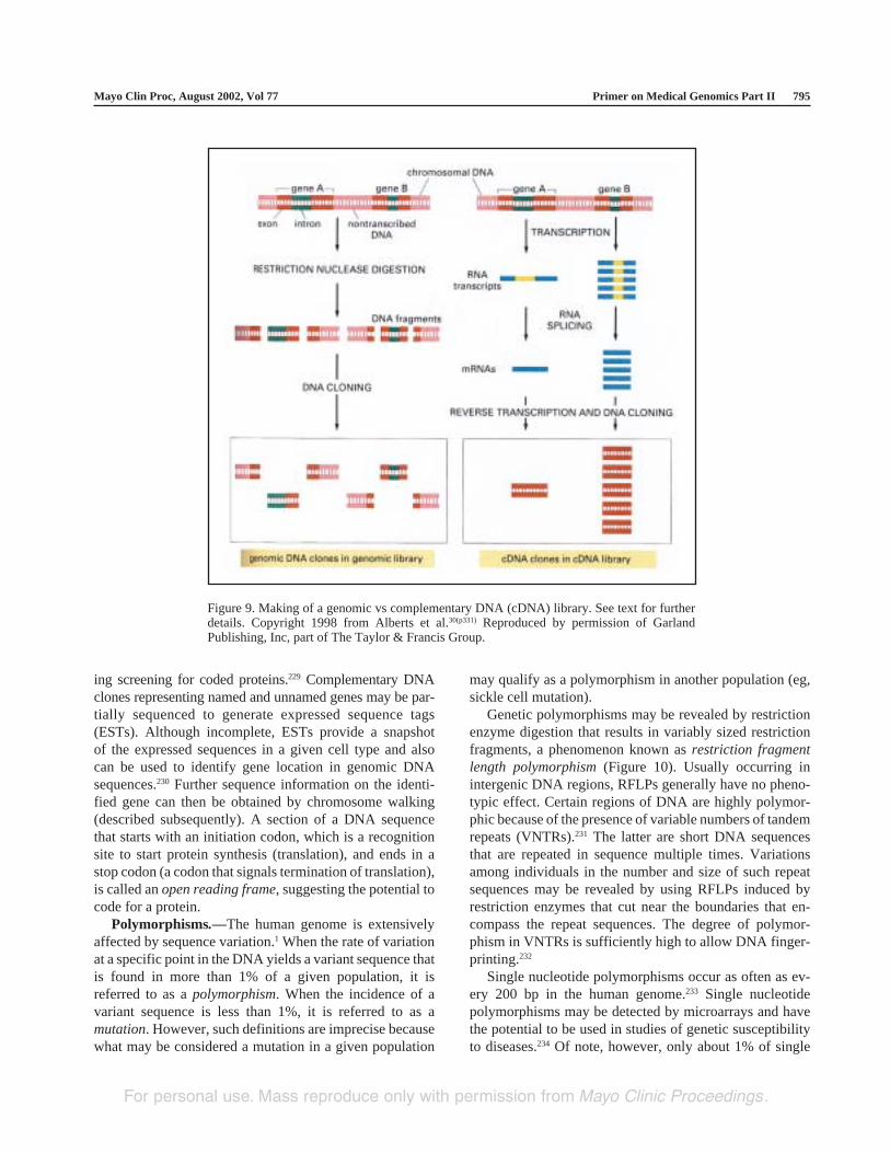
Libraries .—Chromosomal DNA isolated directly froma cell is called genomic DNA. DNA made from reversetranscription of mRNA is called complementary DNA. Aset of DNA clones from a particular source is called a DNAlibrary (genomic or cDNA library depending on the sourceof the clones) (Figure 9). To prepare a human DNA library,genomic DNA or cDNA is first digested with restrictionenzymes, and the restriction fragments (inserts) are recom-bined with a similarly digested vector DNA. A populationof recombinant DNA molecules with different humanDNA inserts is then cloned in a bacterial host. A collectionof thousands of vector-linked human DNA inserts repre-sents the human DNA library.228
Complementary DNA libraries have the advantage ofcarrying only exons (protein-encoding regions of genes)and may be modified by flanking the inserts with transcrip-tion and translation signals (expression vectors), thus allow-
Figure 8. General procedure for cloning a DNA fragment in aplasmid vector. Reproduced with permission from Lodish etal.121(p210)
For personal use. Mass reproduce only with permission from Mayo Clinic Proceedings.
Mayo Clin Proc, August 2002, Vol 77 Primer on Medical Genomics Part II 795
ing screening for coded proteins.229 Complementary DNAclones representing named and unnamed genes may be par-tially sequenced to generate expressed sequence tags(ESTs). Although incomplete, ESTs provide a snapshotof the expressed sequences in a given cell type and alsocan be used to identify gene location in genomic DNAsequences.230 Further sequence information on the identi-fied gene can then be obtained by chromosome walking(described subsequently). A section of a DNA sequencethat starts with an initiation codon, which is a recognitionsite to start protein synthesis (translation), and ends in astop codon (a codon that signals termination of translation),is called an open reading frame, suggesting the potential tocode for a protein.
Polymorphisms.—The human genome is extensivelyaffected by sequence variation.1 When the rate of variationat a specific point in the DNA yields a variant sequence thatis found in more than 1% of a given population, it isreferred to as a polymorphism. When the incidence of avariant sequence is less than 1%, it is referred to as amutation. However, such definitions are imprecise becausewhat may be considered a mutation in a given population
may qualify as a polymorphism in another population (eg,sickle cell mutation).
Genetic polymorphisms may be revealed by restrictionenzyme digestion that results in variably sized restrictionfragments, a phenomenon known as restriction fragmentlength polymorphism (Figure 10). Usually occurring inintergenic DNA regions, RFLPs generally have no pheno-typic effect. Certain regions of DNA are highly polymor-phic because of the presence of variable numbers of tandemrepeats (VNTRs).231 The latter are short DNA sequencesthat are repeated in sequence multiple times. Variationsamong individuals in the number and size of such repeatsequences may be revealed by using RFLPs induced byrestriction enzymes that cut near the boundaries that en-compass the repeat sequences. The degree of polymor-phism in VNTRs is sufficiently high to allow DNA finger-printing.232
Single nucleotide polymorphisms occur as often as ev-ery 200 bp in the human genome.233 Single nucleotidepolymorphisms may be detected by microarrays and havethe potential to be used in studies of genetic susceptibilityto diseases.234 Of note, however, only about 1% of single
Figure 9. Making of a genomic vs complementary DNA (cDNA) library. See text for furtherdetails. Copyright 1998 from Alberts et al.30(p331) Reproduced by permission of GarlandPublishing, Inc, part of The Taylor & Francis Group.
For personal use. Mass reproduce only with permission from Mayo Clinic Proceedings.

Primer on Medical Genomics Part II Mayo Clin Proc, August 2002, Vol 77796
nucleotide polymorphisms result in an alteration in proteincoding at the amino acid level.
DNA SequencingDNA sequencing entails spelling out the nucleotide
chain of a DNA region of interest.235-237 Sufficient quanti-ties of the target DNA region of interest are obtained fromeither a bacterial-cloned fragment or a PCR-amplified se-quence. A short DNA primer is used to start the synthesisof a complementary strand to the original template to besequenced. With the use of specific chemical analogues ofnucleotides (dideoxynucleotides), which are capable of ter-minating chain extension at specific nucleotide bases, thesynthesis of the complementary strand is halted (nested) atspecific nucleotide bases, resulting in the synthesis of DNA
fragments with varying lengths that differ by 1 nucleotide(Figure 11). The set of nested fragments that are radiola-beled or tagged with fluorescent dyes can then be separatedby gel electrophoresis, resulting in a pattern that allows thedetermination of the specific nucleotide at the end of eachsuccessive fragment. Currently, an automated computer-ized laser detection system is used for rapid accrual ofsequence information.
Gene MappingHuman genetic mapping entails (1) the localization or
mapping of genes to each of the 23 chromosome pairs and(2) the determination of the order and spacing of the geneon the particular chromosome.238 This is done in 1 of 2ways: physical mapping (based on estimates of physicaldistance measured in bp) or genetic linkage mapping(based on the frequency of meiotic crossing over that isobserved between 2 loci of a chromosome).239 Linkagedistance is measured in centiMorgans (cM), named forThomas H. Morgan, the father of Drosophila (fruit fly)genetics, who defined the concept of genetic linkage.240
Locating a disease gene is the first step toward cloning thegene itself.
Physical Mapping.—Physical mapping of genes maybe achieved by different methods with differing levels ofresolution that range from an entire chromosome to a singlebase pair (the base sequence). Methods of low-resolutionphysical mapping include karyotyping, fluorescence in situhybridization (FISH), and somatic cell hybridization.
Human chromosomes may be identified by size and bydistinctive banding patterns seen under light microscopyafter staining with special DNA-binding dyes (Giemsa).241
This method of chromosome visualization is called karyo-typing and provides a metaphase picture of the 22 homolo-gous pairs of autosomal chromosomes and the 1 additionalpair of sex chromosomes lined up by size. Such a chromo-some picture allows identification of gross abnormalities(chromosomal deletions, duplications, translocations) thatmay be associated with a particular disease phenotype (eg,chronic myeloid leukemia is associated with a gross trans-location between chromosomes 9 and 22).242 As a result,the disease gene is physically mapped to the vicinity of theabnormal karyotypic lesion.
In FISH, a priori knowledge of a DNA sequence of thespecific gene or of a marker near the gene is required.Fluorescent-tagged probes, by cloning of the specific se-quence, are then prepared and used to localize the gene ofinterest to a particular chromosome by slide exposure ofmetaphase or interphase chromosomes with the labeledprobe.243
In somatic cell hybridization, human and nonhumancells are fused under specific experimental conditions.244
Figure 10. Analysis of restriction fragment length polymor-phisms. See text for further details. Reproduced with permissionfrom Lodish et al.121(p271)
For personal use. Mass reproduce only with permission from Mayo Clinic Proceedings.

Mayo Clin Proc, August 2002, Vol 77 Primer on Medical Genomics Part II 797
Subsequently, the hybrid cells are allowed to replicateunder specific conditions whereby they start losing a vari-able number of the human chromosomes.245,246 Eventually,hybrid cells that have retained 1 or 2 specific human chro-mosomes are selected for further testing. Karyotyping al-lows distinction of human from nonhuman (usually mouse)chromosomes and identification of the specific humanchromosome that is retained in a hybrid cell. Additionalexperiments are then performed to look for the presence orabsence of a specific DNA sequence or protein product in agiven hybrid cell, and this would allow the assignment ofthe gene for a particular protein product to a specific chro-
mosome. Somatic cell hybrids may provide additionalresolution when the hybrid is constructed from cells con-taining either structurally abnormal or radiation-damagedhuman chromosomes (so-called radiation hybrids).247 Simi-larly, somatic cell hybridization and FISH techniques maybe combined for improved resolution.248
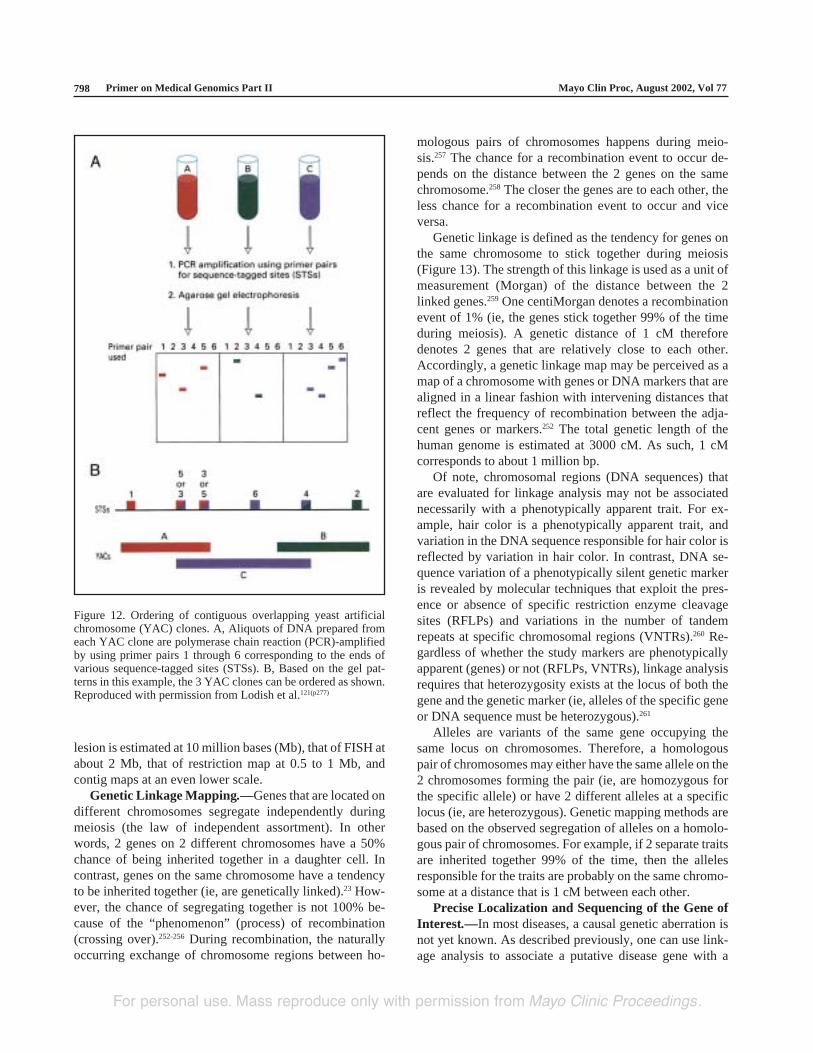
Other physical maps include cDNA maps that localizethe chromosomal positions of expressed genes,249,250 RFLPmaps of restriction cleavage sites,251 and contig maps ofoverlapping sequences of YACs, BACs, or cosmids (Fig-ure 12) (described in more detail subsequently).227 Theresolution grade of karyotypic identification of a genetic
Figure 11. Sanger (dideoxy) method for sequencing DNA fragments. See text for furtherdetails. Reproduced with permission from Lodish et al.121(p234)
For personal use. Mass reproduce only with permission from Mayo Clinic Proceedings.
Primer on Medical Genomics Part II Mayo Clin Proc, August 2002, Vol 77798
lesion is estimated at 10 million bases (Mb), that of FISH atabout 2 Mb, that of restriction map at 0.5 to 1 Mb, andcontig maps at an even lower scale.
Genetic Linkage Mapping.—Genes that are located ondifferent chromosomes segregate independently duringmeiosis (the law of independent assortment). In otherwords, 2 genes on 2 different chromosomes have a 50%chance of being inherited together in a daughter cell. Incontrast, genes on the same chromosome have a tendencyto be inherited together (ie, are genetically linked).23 How-ever, the chance of segregating together is not 100% be-cause of the “phenomenon” (process) of recombination(crossing over).252-256 During recombination, the naturallyoccurring exchange of chromosome regions between ho-
mologous pairs of chromosomes happens during meio-sis.257 The chance for a recombination event to occur de-pends on the distance between the 2 genes on the samechromosome.258 The closer the genes are to each other, theless chance for a recombination event to occur and viceversa.
Genetic linkage is defined as the tendency for genes onthe same chromosome to stick together during meiosis(Figure 13). The strength of this linkage is used as a unit ofmeasurement (Morgan) of the distance between the 2linked genes.259 One centiMorgan denotes a recombinationevent of 1% (ie, the genes stick together 99% of the timeduring meiosis). A genetic distance of 1 cM thereforedenotes 2 genes that are relatively close to each other.Accordingly, a genetic linkage map may be perceived as amap of a chromosome with genes or DNA markers that arealigned in a linear fashion with intervening distances thatreflect the frequency of recombination between the adja-cent genes or markers.252 The total genetic length of thehuman genome is estimated at 3000 cM. As such, 1 cMcorresponds to about 1 million bp.
Of note, chromosomal regions (DNA sequences) thatare evaluated for linkage analysis may not be associatednecessarily with a phenotypically apparent trait. For ex-ample, hair color is a phenotypically apparent trait, andvariation in the DNA sequence responsible for hair color isreflected by variation in hair color. In contrast, DNA se-quence variation of a phenotypically silent genetic markeris revealed by molecular techniques that exploit the pres-ence or absence of specific restriction enzyme cleavagesites (RFLPs) and variations in the number of tandemrepeats at specific chromosomal regions (VNTRs).260 Re-gardless of whether the study markers are phenotypicallyapparent (genes) or not (RFLPs, VNTRs), linkage analysisrequires that heterozygosity exists at the locus of both thegene and the genetic marker (ie, alleles of the specific geneor DNA sequence must be heterozygous).261
Alleles are variants of the same gene occupying thesame locus on chromosomes. Therefore, a homologouspair of chromosomes may either have the same allele on the2 chromosomes forming the pair (ie, are homozygous forthe specific allele) or have 2 different alleles at a specificlocus (ie, are heterozygous). Genetic mapping methods arebased on the observed segregation of alleles on a homolo-gous pair of chromosomes. For example, if 2 separate traitsare inherited together 99% of the time, then the allelesresponsible for the traits are probably on the same chromo-some at a distance that is 1 cM between each other.
Precise Localization and Sequencing of the Gene ofInterest.—In most diseases, a causal genetic aberration isnot yet known. As described previously, one can use link-age analysis to associate a putative disease gene with a
Figure 12. Ordering of contiguous overlapping yeast artificialchromosome (YAC) clones. A, Aliquots of DNA prepared fromeach YAC clone are polymerase chain reaction (PCR)-amplifiedby using primer pairs 1 through 6 corresponding to the ends ofvarious sequence-tagged sites (STSs). B, Based on the gel pat-terns in this example, the 3 YAC clones can be ordered as shown.Reproduced with permission from Lodish et al.121(p277)
For personal use. Mass reproduce only with permission from Mayo Clinic Proceedings.

Mayo Clin Proc, August 2002, Vol 77 Primer on Medical Genomics Part II 799
chromosomal locale that is detectable either molecularly orphenotypically.260 Once the gross location of the putativedisease gene is determined by linkage analysis, one canapply positional cloning for more precise identification andsequencing of the gene of interest.262,263
Positional cloning starts with the selection, by using theDNA sequence of the linked marker as a probe, of vectorclones from a genomic library that carry DNA sequencesthat overlap with those from the linked genetic marker. Asmentioned previously, a genomic library is constructed bypartially digesting the entire genome with restriction en-zymes, followed by cloning of the resultant fragments intoappropriate vectors.264 Because the DNA products of re-striction digestion are relatively large, the vectors usuallyused are YAC and BAC vectors. Using the DNA sequenceof the linked marker, one can pick YAC or BAC cloneswith sequences that overlap with those of the linkedmarker.265 Alternatively, sequence tagged sites, which areunique, relatively short DNA sequences with known chro-mosomal location, may be used to identify clones withoverlapping sequences. Subsequently, the overlapping se-quences themselves may be used as probes to select addi-tional YAC or BAC clones with sequences that are par-tially overlapping with the probes used.226,266
The stepwise process of identifying YAC or BACclones based on overlapping DNA sequences is calledchromosome walking.267 This process of chromosomewalking continues until the disease gene is identified. Thelinear alignment of YACs or BACs with overlapping se-quences results in a continuous DNA segment called acontig map. Such contig maps have been essential for the
Figure 13. Recombination during meiosis. See text for furtherdetails. Reproduced with permission from Lodish et al.121(p270)
analysis of human and nonhuman genomes as well as forspecific chromosomal regions.225.227,268,269
Probe-identified clones can also be individually mappedfor sites complementary to ESTs. An EST that coseg-regates with the gene of interest is determined and thensequenced. Alternatively, the clone containing the linkagemarkers may have already been sequenced, and residentgenes are sought by looking for ORFs. Finally, the candi-date gene is identified by mutational analysis that comparesORFs from a patient-derived DNA library to those ob-tained from nonaffected individuals.
CONCLUSIONThis review on the basic principles of molecular biology isnot intended to be comprehensive but to serve as an intro-duction to this subject. Additional information is easilyaccessible from numerous outstanding textbooks of mo-lecular biology.30,121,270
REFERENCES1. Venter JC, Adams MD, Myers EW, et al. The sequence of the
human genome. Science. 2001;291:1304-1351.2. Lander ES, Linton LM, Birren B, et al. Initial sequencing and
analysis of the human genome. Nature. 2001;409:860-921.3. Meldrum D. Automation for genomics, part two: sequencers,
microarrays, and future trends. Genome Res. 2000;10:1288-1303.
4. Gershon D. Microarray technology: an array of opportunities.Nature. 2002;416:885-891.
5. Gulino A. Biotechnology and molecular diagnostics. Forum(Genova). 1999;9(3, suppl 3):37-46.
6. Jordan B, Charest A, Dowd JF, et al. Genome complexity reduc-tion for SNP genotyping analysis. Proc Natl Acad Sci U S A. 2002;99:2942-2947.
7. Topol EJ, McCarthy J, Gabriel S, et al. Single nucleotide poly-morphisms in multiple novel thrombospondin genes may be asso-ciated with familial premature myocardial infarction. Circulation.2001;104:2641-2644.
8. Alizadeh AA, Eisen MB, Davis RE, et al. Distinct types of diffuselarge B-cell lymphoma identified by gene expression profiling.Nature. 2000;403:503-511.
9. Marshall T, Williams KM. Proteomics and its impact upon bio-medical science. Br J Biomed Sci. 2002;59:47-64.
10. Service RF. Structural genomics offers high-speed look at pro-teins. Science. 2000;287:1954-1956.
11. Chance MR, Bresnick AR, Burley SK, et al. Structural genomics:a pipeline for providing structures for the biologist. Protein Sci.2002;11:723-738.
12. Altman RB, Klein TE. Challenges for biomedical informatics andpharmacogenomics. Annu Rev Pharmacol Toxicol. 2002;42:113-133.
13. Weng Z, DeLisi C. Protein therapeutics: promises and challengesfor the 21st century. Trends Biotechnol. 2002;20:29-35.
14. Bayat A. Science, medicine, and the future: bioinformatics. BMJ.2002;324:1018-1022.
15. Roos DS. Computational biology: bioinformatics—trying toswim in a sea of data. Science. 2001;291:1260-1261.
16. Collins FS, McKusick VA. Implications of the Human GenomeProject for medical science. JAMA. 2001;285:540-544.
17. Subramanian G, Adams MD, Venter JC, Broder S. Implications ofthe human genome for understanding human biology and medi-cine. JAMA. 2001;286:2296-2307.
For personal use. Mass reproduce only with permission from Mayo Clinic Proceedings.

Primer on Medical Genomics Part II Mayo Clin Proc, August 2002, Vol 77800
18. Butler D. Genomics: are you ready for the revolution? Nature.2001;409:758-760.
19. Magee J, Gordon JI, Whelan A. Bringing the human genome andthe revolution in bioinformatics to the medical school classroom:a case report from Washington University School of Medicine.Acad Med. 2001;76:852-855.
20. Fears R, Weatherall D, Poste G. The impact of genetics on medi-cal education and training. Br Med Bull. 1999;55:460-470.
21. Rothstein MA, Epps PG. Ethical and legal implications of phar-macogenomics. Nat Rev Genet. 2001;2:228-231.
22. Mendel G. Experiments in plant hybridization. Verh NaturforschVereines Brunn. 1865;4:3-47.
23. Morgan TH. Sex-limited inheritance in Drosophila. Science.1910;32:120-122.
24. Demerec M. What is a gene? J Hered. 1933;24:368-378.25. Avery O, MacLeod C, McCarty M. Studies on the chemical nature
of the substance inducing transformation of pneumococcal type. JExp Med. 1944;79:137-158.
26. Hershey AD, Chase M. Independent functions of viral protein andnucleic acid in growth of bacteriophage. J Gen Physiol. 1952;36:39-56.
27. Beadle GW, Tatum E. Genetic control of biochemical reactions inneurospora. Proc Natl Acad Sci U S A. 1941;27:499-506.
28. Jhiang SM, Garey JR, Riggs AF. Exon-intron organization ingenes of earthworm and vertebrate globins. Science. 1988;240:334-336.
29. Cohen JA. Chemistry and structure of nucleic acids of bacterio-phages: many forms of nucleic acids of bacteriophages show theways that information is stored and reproduced. Science. 1967;158:343-351.
30. Alberts B, Bray D, Johnson A, et al. Essential Cell Biology: AnIntroduction to the Molecular Biology of the Cell. New York, NY:Garland Publishing; 1998.
31. Dhingra MM, Sarma RH. Why do nucleic acids have 3′5′phosphodiester bonds? Nature. 1978;272:798-801.
32. Watson JD, Crick FHC. Molecular structure of nucleic acids: astructure for deoxyribose nucleic acid. Nature. 1953;171:737-738.
33. Meselson M, Stahl FW. The replication of DNA in E. coli. ProcNatl Acad Sci U S A. 1958;44:671-682.
34. Saha S, Sparks AB, Rago C, et al. Using the transcriptome toannotate the genome. Nat Biotechnol. 2002;20:508-512.
35. Avise JC. Evolving genomic metaphors: a new look at the lan-guage of DNA. Science. 2001;294:86-87.
36. Schmid CW, Deininger PL. Sequence organization of the humangenome. Cell. 1975;6:345-358.
37. Gu Z, Wang H, Nekrutenko A, Li WH. Densities, length propor-tions, and other distributional features of repetitive sequences inthe human genome estimated from 430 megabases of genomicsequence. Gene. 2000;259:81-88.
38. Dott PJ, Chuang CR, Saunders GF. Inverted repetitive sequencesin the human genome. Biochemistry. 1976;15:4120-4125.
39. Willard HF. The genomics of long tandem arrays of satellite DNAin the human genome. Genome. 1989;31:737-744.
40. Moyzis RK, Torney DC, Meyne J, et al. The distribution ofinterspersed repetitive DNA sequences in the human genome.Genomics. 1989;4:273-289.
41. Smit AF. The origin of interspersed repeats in the human genome.Curr Opin Genet Dev. 1996;6:743-748.
42. Moustacchi E, Williamson DH. Physiological variations in sat-ellite components of yeast DNA detected by density gradientcentrifugation. Biochem Biophys Res Commun. 1966;23:56-61.
43. Moore DE, Abuelo JG. Human repetitious DNA, satellite DNAand chromosome interarm fibres. Nature. 1971;234:467-468.
44. Yunis JJ, Roldan L, Yasmineh WG, Lee JC. Staining of satelliteDNA in metaphase chromosomes. Nature. 1971;231:532-533.
45. Moxon ER, Wills C. DNA microsatellites: agents of evolution?Sci Am. 1999;280:94-99.
46. Wong Z, Wilson V, Patel I, Povey S, Jeffreys AJ. Characterizationof a panel of highly variable minisatellites cloned from humanDNA. Ann Hum Genet. 1987;51(pt 4):269-288.
47. Prak ET, Kazazian HH Jr. Mobile elements and the human ge-nome [published correction appears in Nat Rev Genet. 2001;2:78].Nat Rev Genet. 2000;1:134-144.
48. Jagadeeswaran P, Forget BG, Weissman SM. Short interspersedrepetitive DNA elements in eucaryotes: transposable DNA ele-ments generated by reverse transcription of RNA pol III tran-scripts? Cell. 1981;26(2, pt 2):141-142.
49. Patience C, Wilkinson DA, Weiss RA. Our retroviral heritage.Trends Genet. 1997;13:116-120.
50. Johns DR. The other human genome: mitochondrial DNA anddisease. Nat Med. 1996;2:1065-1068.
51. Nass MM. Mitochondrial DNA: advances, problems, and goals.Science. 1968;165:25-35.
52. Cann RL, Brown WM, Wilson AC. Polymorphic sites and themechanism of evolution in human mitochondrial DNA. Genetics.1984;106:479-499.
53. Brown WM, George M Jr, Wilson AC. Rapid evolution of animalmitochondrial DNA. Proc Natl Acad Sci U S A. 1979;76:1967-1971.
54. Cann RL, Wilson AC. Length mutations in human mitochondrialDNA. Genetics. 1983;104:699-711.
55. Mattson MP. Mother’s legacy: mitochondrial DNA mutations andAlzheimer’s disease. Trends Neurosci. 1997;20:373-375.
56. Avise JC. Mitochondrial DNA and the evolutionary genetics ofhigher animals. Philos Trans R Soc Lond B Biol Sci. 1986;312:325-342.
57. Vigilant L, Stoneking M, Harpending H, Hawkes K, Wilson AC.African populations and the evolution of human mitochondrialDNA. Science. 1991;253:1503-1507.
58. Ferris SD, Wilson AC, Brown WM. Evolutionary tree for apesand humans based on cleavage maps of mitochondrial DNA. ProcNatl Acad Sci U S A. 1981;78:2432-2436.
59. Kornberg RD. Chromatin structure: a repeating unit of histonesand DNA. Science. 1974;184:868-871.
60. Elgin SC. Chromatin structure, DNA structure. Nature. 1982;300:402-403.
61. Brown SW. Heterochromatin. Science. 1966;151:417-425.62. Yasmineh WG, Yunis JJ. Isolation of mammalian heterochroma-
tin and euchromatin. Methods Cell Biol. 1974;8:151-177.63. Wallrath LL. Unfolding the mysteries of heterochromatin. Curr
Opin Genet Dev. 1998;8:147-153.64. Quintana-Murci L, Jamain S, Fellous M. Origin and evolution of
mammalian sex chromosomes [in French]. C R Acad Sci III. 2001;324:1-11.
65. Nirenberg MW, Leder P. RNA codewors and protein synthesis.Science. 1964;145:1399-1407.
66. Efstratiadis A, Kafatos FC, Maniatis T. The primary structure ofrabbit beta-globin mRNA as determined from cloned DNA. Cell.1977;10:571-585.
67. Lewin B. Units of transcription and translation: the relationshipbetween heterogeneous nuclear RNA and messenger RNA. Cell.1975;4:11-20.
68. Busby S, Ebright RH. Promoter structure, promoter recognition,and transcription activation in prokaryotes. Cell. 1994;79:743-746.
69. Saltzman AG, Weinmann R. Promoter specificity and modulationof RNA polymerase II transcription. FASEB J. 1989;3:1723-1733.
70. Jeang KT, Khoury G. The mechanistic role of enhancer elementsin eukaryotic transcription. Bioessays. 1988;8:104-107.
71. Reinberg D, Orphanides G, Ebright R, et al. The RNA polymeraseII general transcription factors: past, present, and future. ColdSpring Harb Symp Quant Biol. 1998;63:83-103.
72. Muller CW. Transcription factors: global and detailed views. CurrOpin Struct Biol. 2001;11:26-32.
73. Lopez-Rodriguez C, Aramburu J, Rakeman AS, et al. NF-AT5:the NF-AT family of transcription factors expands in a new direc-tion. Cold Spring Harb Symp Quant Biol. 1999;64:517-526.
For personal use. Mass reproduce only with permission from Mayo Clinic Proceedings.

Mayo Clin Proc, August 2002, Vol 77 Primer on Medical Genomics Part II 801
74. Jones NC, Rigby PW, Ziff EB. Trans-acting protein factors andthe regulation of eukaryotic transcription: lessons from studies onDNA tumor viruses. Genes Dev. 1988;2:267-281.
75. Sassone-Corsi P, Verma IM. Modulation of c-fos gene transcrip-tion by negative and positive cellular factors. Nature. 1987;326:507-510.
76. Mathis DJ, Chambon P. The SV40 early region TATA box isrequired for accurate in vitro initiation of transcription. Nature.1981;290:310-315.
77. Rowlands T, Baumann P, Jackson SP. The TATA-binding pro-tein: a general transcription factor in eukaryotes and archaebacte-ria. Science. 1994;264:1326-1329.
78. Nimer S, Fraser J, Richards J, Lynch M, Gasson J. The repeatedsequence CATT(A/T) is required for granulocyte-macrophagecolony-stimulating factor promoter activity. Mol Cell Biol. 1990;10:6084-6088.
79. White RJ, Jackson SP. The TATA-binding protein: a central rolein transcription by RNA polymerases I, II and III. Trends Genet.1992;8:284-288.
80. Pabo CO, Sauer RT. Transcription factors: structural families andprinciples of DNA recognition. Annu Rev Biochem. 1992;61:1053-1095.
81. Orphanides G, Lagrange T, Reinberg D. The general transcrip-tion factors of RNA polymerase II. Genes Dev. 1996;10:2657-2683.
82. Khorasanizadeh S, Rastinejad F. Transcription factors: the rightcombination for the DNA lock. Curr Biol. 1999;9:R456-R458.
83. Proudfoot N. Connecting transcription to messenger RNA pro-cessing. Trends Biochem Sci. 2000;25:290-293.
84. Fong N, Bentley DL. Capping, splicing, and 3′ processing areindependently stimulated by RNA polymerase II: different func-tions for different segments of the CTD. Genes Dev. 2001;15:1783-1795.
85. Winicov I, Perry RP. Synthesis methylation, and capping ofnuclear RNA by a subcellular system. Biochemistry. 1976;15:5039-5046.
86. Minvielle-Sebastia L, Keller W. mRNA polyadenylation and itscoupling to other RNA processing reactions and to transcription.Curr Opin Cell Biol. 1999;11:352-357.
87. Reeder RH, Lang WH. Terminating transcription in eukaryotes:lessons learned from RNA polymerase I. Trends Biochem Sci.1997;22:473-477.
88. Darnell JE, Philipson L, Wall R, Adesnik M. Polyadenylic acidsequences: role in conversion of nuclear RNA into messengerRNA. Science. 1971;174:507-510.
89. Crick F. Split genes and RNA splicing. Science. 1979;204:264-271.
90. Palacios IM. RNA processing: splicing and the cytoplasmiclocalisation of mRNA. Curr Biol. 2002;12:R50-R52.
91. Yang VW, Lerner MR, Steitz JA, Flint SJ. A small nuclear ribo-nucleoprotein is required for splicing of adenoviral early RNAsequences. Proc Natl Acad Sci U S A. 1981;78:1371-1375.
92. Ohshima Y, Itoh M, Okada N, Miyata T. Novel models for RNAsplicing that involve a small nuclear RNA. Proc Natl Acad SciU S A. 1981;78:4471-4474.
93. Choi YD, Grabowski PJ, Sharp PA, Dreyfuss G. Heterogeneousnuclear ribonucleoproteins: role in RNA splicing. Science. 1986;231:1534-1539.
94. Kiss T. Small nucleolar RNA-guided post-transcriptional modifi-cation of cellular RNAs. EMBO J. 2001;20:3617-3622.
95. Hernandez N. Small nuclear RNA genes: a model system to studyfundamental mechanisms of transcription. J Biol Chem. 2001;276:26733-26736.
96. Kruger K, Grabowski PJ, Zaug AJ, Sands J, Gottschling DE, CechTR. Self-splicing RNA: autoexcision and autocyclization of theribosomal RNA intervening sequence of Tetrahymena. Cell.1982;31:147-157.
97. Valadkhan S, Manley JL. Splicing-related catalysis by protein-free snRNAs. Nature. 2001;413:701-707.
98. Newman A. Molecular biology: RNA enzymes for RNA splicing.Nature. 2001;413:695-696.
99. Rogers J, Wall R. A mechanism for RNA splicing. Proc Natl AcadSci U S A. 1980;77:1877-1979.
100. Padgett RA, Konarska MM, Grabowski PJ, Hardy SF, Sharp PA.Lariat RNA’s as intermediates and products in the splicing ofmessenger RNA precursors. Science. 1984;225:898-903.
101. Keller W. The RNA lariat: a new ring to the splicing of mRNAprecursors. Cell. 1984;39(3, pt 2):423-425.
102. Chetverin AB. The puzzle of RNA recombination. FEBS Lett.1999;460:1-5.
103. Choi E, Kuehl M, Wall R. RNA splicing generates a variant lightchain from an aberrantly rearranged kappa gene. Nature. 1980;286:776-779.
104. Rueter SM, Dawson TR, Emeson RB. Regulation of alternativesplicing by RNA editing. Nature. 1999;399:75-80.
105. Lamond AI. RNA splicing: running rings around RNA. Nature.1999;397:655-656.
106. Graveley BR, Hertel KJ, Maniatis T. SR proteins are ‘locators’ ofthe RNA splicing machinery [letter]. Curr Biol. 1999;9:R6-R7.
107. Mayeda A, Krainer AR. Regulation of alternative pre-mRNA splic-ing by hnRNP A1 and splicing factor SF2. Cell. 1992;68:365-375.
108. Wu JI, Reed RB, Grabowski PJ, Artzt K. Function of quaking inmyelination: regulation of alternative splicing. Proc Natl Acad SciU S A. 2002;99:4233-4238.
109. Cooper TA. Highlights of alternative splicing regulation session:yes, no, maybe—a history of paradigm shifts. Sci STKE. 2001;2001:PE35.
110. Pesole G, Mignone F, Gissi C, Grillo G, Licciulli F, Liuni S.Structural and functional features of eukaryotic mRNA untrans-lated regions. Gene. 2001;276:73-81.
111. Adams JM, Cory S. Untranslated nucleotide sequence at the 5′-end of R17 bacteriophage RNA. Nature. 1970;227:570-574.
112. Forget BG, Marotta CA, Weissman SM, Cohen-Solal M. Nucle-otide sequences of the 3′-terminal untranslated region of messen-ger RNA for human beta globin chain. Proc Natl Acad Sci U S A.1975;72:3614-3618.
113. Chang JC, Temple GF, Poon R, Neumann KH, Kan YW. Thenucleotide sequences of the untranslated 5′ regions of humanalpha- and beta-globin mRNAs. Proc Natl Acad Sci U S A. 1977;74:5145-5149.
114. Veyrune JL, Hesketh J, Blanchard JM. 3′ Untranslated regions ofc-myc and c-fos mRNAs: multifunctional elements regulatingmRNA translation, degradation and subcellular localization. ProgMol Subcell Biol. 1997;18:35-63.
115. Wormington M. Unmasking the role of the 3′ UTR in the cyto-plasmic polyadenylation and translational regulation of maternalmRNAs. Bioessays. 1994;16:533-535.
116. Sonenberg N. mRNA translation: influence of the 5′ and 3′ un-translated regions. Curr Opin Genet Dev. 1994;4:310-315.
117. Dickey LF, Wang YH, Shull GE, Wortman IA III, Theil EC. Theimportance of the 3′-untranslated region in the translational con-trol of ferritin mRNA. J Biol Chem. 1988;263:3071-3074.
118. Mignone F, Gissi C, Liuni S, Pesole G. Untranslated regions ofmRNAs. Genome Biol. 2002;3:0004.1-0004.10.
119. Lafontaine DL, Tollervey D. The function and synthesis of ribo-somes. Nat Rev Mol Cell Biol. 2001;2:514-520.
120. Schimmel P, Ribas de Pouplana L. Transfer RNA: from minihelixto genetic code. Cell. 1995;81:983-986.
121. Lodish H, Berk A, Zipursky SL, Matsudaira P, Baltimore D,Darnell J. Molecular Cell Biology. 4th ed. New York, NY: WHFreeman and Co; 2000.
122. Brown GL, Lee S. Amino-acid-transfer ribonucleic acid: structureand function. Br Med Bull. 1965;21:236-243.
123. Saks ME, Sampson JR, Abelson JN. The transfer RNA identityproblem: a search for rules. Science. 1994;263:191-197.
124. Noller HF, Yusupov MM, Yusupova GZ, Baucom A, Cate JH.Translocation of tRNA during protein synthesis. FEBS Lett. 2002;514:11-16.
For personal use. Mass reproduce only with permission from Mayo Clinic Proceedings.

Primer on Medical Genomics Part II Mayo Clin Proc, August 2002, Vol 77802
125. Moore PB. Ribosomes: protein synthesis in slow motion. CurrBiol. 1997;7:R179-R181.
126. Pestova TV, Hellen CU. The structure and function of initiationfactors in eukaryotic protein synthesis. Cell Mol Life Sci. 2000;57:651-674.
127. Czworkowski J, Moore PB. The elongation phase of protein syn-thesis. Prog Nucleic Acid Res Mol Biol. 1996;54:293-332.
128. Tate WP, Brown CM. Translational termination: “stop” for pro-tein synthesis or “pause” for regulation of gene expression. Bio-chemistry. 1992;31:2443-2450.
129. Tuite MF, Stansfield I. Termination of protein synthesis. Mol BiolRep. 1994;19:171-181.
130. Doherty EA, Doudna JA. Ribozyme structures and mechanisms.Annu Rev Biophys Biomol Struct. 2001;30:457-475.
131. Wold F. In vivo chemical modification of proteins (post-transla-tional modification). Annu Rev Biochem. 1981;50:783-814.
132. Yewdell JW. Not such a dismal science: the economics of proteinsynthesis, folding, degradation and antigen processing. TrendsCell Biol. 2001;11:294-297.
133. Han KK, Martinage A. Post-translational chemical modifica-tion(s) of proteins. Int J Biochem. 1992;24:19-28.
134. Saibil H. Molecular chaperones: containers and surfaces for fold-ing, stabilising or unfolding proteins. Curr Opin Struct Biol. 2000;10:251-258.
135. Johnson ES, Bartel B, Seufert W, Varshavsky A. Ubiquitin as adegradation signal. EMBO J. 1992;11:497-505.
136. Hochstrasser M. Ubiquitin-dependent protein degradation. AnnuRev Genet. 1996;30:405-439.
137. Laney JD, Hochstrasser M. Substrate targeting in the ubiquitinsystem. Cell. 1999;97:427-430.
138. Ciechanover A. The ubiquitin-proteasome proteolytic pathway.Cell. 1994;79:13-21.
139. Lo RS, Massague J. Ubiquitin-dependent degradation of TGF-beta-activated smad2. Nat Cell Biol. 1999;1:472-478.
140. Jesenberger V, Jentsch S. Deadly encounter: ubiquitin meetsapoptosis. Nat Rev Mol Cell Biol. 2002;3:112-121.
141. Rosenthal N. Tools of the trade—recombinant DNA. N Engl JMed. 1994;331:315-317.
142. Morrow JF. Recombinant DNA techniques. Methods Enzymol.1979;68:3-24.
143. Emery AE. Recombinant DNA technology. Lancet. 1981;2:1406-1409.
144. Mullis K, Faloona F, Scharf S, Saiki R, Horn G, Erlich H. Specificenzymatic amplification of DNA in vitro: the polymerase chainreaction. Cold Spring Harb Symp Quant Biol. 1986;51(pt 1):263-273.
145. Eisenstein BI. The polymerase chain reaction: a new method ofusing molecular genetics for medical diagnosis. N Engl J Med.1990;322:178-183.
146. Saiki RK, Scharf S, Faloona F, et al. Enzymatic amplification ofbeta-globin genomic sequences and restriction site analysis fordiagnosis of sickle cell anemia. Science. 1985;230:1350-1354.
147. Erlich HA. Principles and applications of the polymerase chainreaction. Rev Immunogenet. 1999;1:127-134.
148. Erlich HA, Gelfand D, Sninsky JJ. Recent advances in the poly-merase chain reaction. Science. 1991;252:1643-1651.
149. Baumforth KR, Nelson PN, Digby JE, O’Neil JD, Murray PG.Demystified...the polymerase chain reaction. Mol Pathol. 1999;52:1-10.
150. Dieffenbach CW, Lowe TM, Dveksler GS. General concepts forPCR primer design. PCR Methods Appl. 1993;3:S30-S37.
151. Lowe T, Sharefkin J, Yang SQ, Dieffenbach CW. A computerprogram for selection of oligonucleotide primers for polymerasechain reactions. Nucleic Acids Res. 1990;18:1757-1761.
152. Rychlik W. Selection of primers for polymerase chain reaction.Mol Biotechnol. 1995;3:129-134.
153. Brock TD, Freeze H. Thermus aquaticus gen. n. and sp. n., anonsporulating extreme thermophile. J Bacteriol. 1969;98:289-297.
154. Chien A, Edgar DB, Trela JM. Deoxyribonucleic acid polymerasefrom the extreme thermophile Thermus aquaticus. J Bacteriol.1976;127:1550-1557.
155. Sato S, Hutchinson CA III, Harris JI. A thermostable sequence-specific endonuclease from Thermus aquaticus. Proc Natl AcadSci U S A. 1977;74:542-546.
156. Aurelius E, Johansson B, Skoldenberg B, Staland A, Forsgren M.Rapid diagnosis of herpes simplex encephalitis by nested poly-merase chain reaction assay of cerebrospinal fluid. Lancet. 1991;337:189-192.
157. Burgart LJ, Robinson RA, Heller MJ, Wilke WW, Iakoubova OK,Cheville JC. Multiplex polymerase chain reaction. Mod Pathol.1992;5:320-323.
158. Markoulatos P, Siafakas N, Moncany M. Multiplex polymerasechain reaction: a practical approach. J Clin Lab Anal. 2002;16:47-51.
159. Mahony JB. Multiplex polymerase chain reaction for the diagno-sis of sexually transmitted diseases. Clin Lab Med. 1996;16:61-71.
160. Na BK, Kim JH, Shin GC, et al. Detection and typing of respira-tory adenoviruses in a single-tube multiplex polymerase chain re-action. J Med Virol. 2002;66:512-517.
161. Bustin SA, Dorudi S. Molecular assessment of tumour stage anddisease recurrence using PCR-based assays. Mol Med Today.1998;4:389-396.
162. Biondi A, Rambaldi A. Polymerase chain reaction (PCR) ap-proach for the evaluation of minimal residual disease in acute leu-kemia. Stem Cells. 1994;12:394-401.
163. Raj GV, Moreno JG, Gomella LG. Utilization of polymerasechain reaction technology in the detection of solid tumors. Can-cer. 1998;82:1419-1442.
164. Jackson JB. The polymerase chain reaction in transfusion medi-cine. Transfusion. 1990;30:51-57.
165. Westwood SA, Werrett DJ. An evaluation of the polymerase chainreaction method for forensic applications. Forensic Sci Int. 1990;45:201-215.
166. Decorte R, Cassiman JJ. Forensic medicine and the polymerasechain reaction technique. J Med Genet. 1993;30:625-633.
167. Hagelberg E, Bell LS, Allen T, Boyde A, Jones SJ, Clegg JB.Analysis of ancient bone DNA: techniques and applications.Philos Trans R Soc Lond B Biol Sci. 1991;333:399-407.
168. Krings M, Stone A, Schmitz RW, Krainitzki H, Stoneking M,Paabo S. Neandertal DNA sequences and the origin of modernhumans. Cell. 1997;90:19-30.
169. Lo YM, Mehal WZ, Fleming KA. False-positive results and thepolymerase chain reaction [letter]. Lancet. 1988;2:679.
170. Chomczynski P, Sacchi N. Single-step method of RNA isolationby acid guanidinium thiocyanate-phenol-chloroform extraction.Anal Biochem. 1987;162:156-159.
171. Mies C. A simple, rapid method for isolating RNA from paraffin-embedded tissues for reverse transcription-polymerase chain reac-tion (RT-PCR). J Histochem Cytochem. 1994;42:811-813.
172. Alwine JC, Kemp DJ, Stark GR. Method for detection of specificRNAs in agarose gels by transfer to diazobenzyloxymethyl-paperand hybridization with DNA probes. Proc Natl Acad Sci U S A.1977;74:5350-5354.
173. Fernyhough P. Quantification of mRNA levels using Northernblotting. Methods Mol Biol. 2001;169:53-63.
174. Tanay VA, Tancowny BP, Glencorse TA, Bateson AN. The quan-titative analysis of multiple mRNA species using oligonucleotideprobes in an S1 nuclease protection assay. Mol Biotechnol. 1997;7:217-229.
175. Rappolee DA, Wang A, Mark D, Werb Z. Novel method forstudying mRNA phenotypes in single or small numbers of cells. JCell Biochem. 1989;39:1-11.
176. Becker-Andre M, Hahlbrock K. Absolute mRNA quantificationusing the polymerase chain reaction (PCR): a novel approach by aPCR aided transcript titration assay (PATTY). Nucleic Acids Res.1989;17:9437-9446.
For personal use. Mass reproduce only with permission from Mayo Clinic Proceedings.

Mayo Clin Proc, August 2002, Vol 77 Primer on Medical Genomics Part II 803
177. Hamoui S, Benedetto JP, Garret M, Bonnet J. Quantitation ofmRNA species by RT-PCR on total mRNA population. PCRMethods Appl. 1994;4:160-166.
178. Bustin SA. Absolute quantification of mRNA using real-timereverse transcription polymerase chain reaction assays. J MolEndocrinol. 2000;25:169-193.
179. Brooks EM, Sheflin LG, Spaulding SW. Secondary structure inthe 3′ UTR of EGF and the choice of reverse transcriptases affectthe detection of message diversity by RT-PCR. Biotechniques.1995;19:806-812, 814-815.
180. Freeman WM, Walker SJ, Vrana KE. Quantitative RT-PCR: pit-falls and potential. Biotechniques. 1999;26:112-122, 124-125.
181. Gattei V, Degan M, De Iuliis A, Rossi FM, Aldinucci D, Pinto A.Competitive reverse-transcriptase PCR: a useful alternative toNorthern blotting for quantitative estimation of relative abun-dances of specific mRNAs in precious samples. Biochem J.1997;325:565-567.
182. Gilliland G, Perrin S, Blanchard K, Bunn HF. Analysis ofcytokine mRNA and DNA: detection and quantitation by com-petitive polymerase chain reaction. Proc Natl Acad Sci U S A.1990;87:2725-2729.
183. Deguchi T, Yang M, Kawada Y. Micrometastasis of prostatecancer to lymph nodes: detection by means of reverse transcrip-tion-polymerase chain reaction. J Natl Cancer Inst. 1997;89:1471-1473.
184. Ballestrero A, Coviello DA, Garuti A, et al. Reverse-transcriptasepolymerase chain reaction of the maspin gene in the detection ofbone marrow breast carcinoma cell contamination. Cancer. 2001;92:2030-2035.
185. Hauptli D, Bruckner L, Ottiger HP. Use of reverse transcriptasepolymerase chain reaction for detection of vaccine contaminationby avian leukosis virus. J Virol Methods. 1997;66:71-81.
186. Diverio D, Pandolfi PP, Rossi V, Biondi A, Pelicci PG, Lo CocoF. Monitoring of treatment outcome in acute promyelocytic leuke-mia by RT- PCR. Leukemia. 1994;8(suppl 2):S63-S65.
187. Muto A, Mori S, Matsushita H, et al. Serial quantification ofminimal residual disease of t(8;21) acute myelogenous leukaemiawith RT-competitive PCR assay. Br J Haematol. 1996;95:85-94.
188. Gibson UE, Heid CA, Williams PM. A novel method for real timequantitative RT-PCR. Genome Res. 1996;6:995-1001.
189. Giulietti A, Overbergh L, Valckx D, Decallonne B, Bouillon R,Mathieu C. An overview of real-time quantitative PCR: applica-tions to quantify cytokine gene expression. Methods. 2001;25:386-401.
190. Higuchi R, Fockler C, Dollinger G, Watson R. Kinetic PCRanalysis: real-time monitoring of DNA amplification reactions.Biotechnology (N Y). 1993;11:1026-1030.
191. Oldach D. “Real-time” polymerase chain reaction. Gastroenterol-ogy. 1999;116:763-764.
192. Heid CA, Stevens J, Livak KJ, Williams PM. Real time quantita-tive PCR. Genome Res. 1996;6:986-994.
193. Simpson DA, Feeney S, Boyle C, Stitt AW. Retinal VEGF mRNAmeasured by SYBR green I fluorescence: a versatile approach toquantitative PCR. Mol Vis. 2000;6:178-183.
194. Cardullo RA, Agrawal S, Flores C, Zamecnik PC, Wolf DE.Detection of nucleic acid hybridization by nonradiative fluores-cence resonance energy transfer. Proc Natl Acad Sci U S A. 1988;85:8790-8794.
195. Holland PM, Abramson RD, Watson R, Gelfand DH. Detection ofspecific polymerase chain reaction product by utilizing the 5′----3′exonuclease activity of Thermus aquaticus DNA polymerase.Proc Natl Acad Sci U S A. 1991;88:7276-7280.
196. King HC, Sinha AA. Gene expression profile analysis by DNAmicroarrays: promise and pitfalls. JAMA. 2001;286:2280-2288.
197. Tyagi S, Kramer FR. Molecular beacons: probes that fluoresceupon hybridization. Nat Biotechnol. 1996;14:303-308.
198. Whitcombe D, Theaker J, Guy SP, Brown T, Little S. Detection ofPCR products using self-probing amplicons and fluorescence. NatBiotechnol. 1999;17:804-807.
199. Aldape K, Ginzinger DG, Godfrey TE. Real-time quantitativepolymerase chain reaction: a potential tool for genetic analysis inneuropathology. Brain Pathol. 2002;12:54-66.
200. Satou J, Funato T, Satoh N, et al. Quantitative PCR determinationof human cytomegalovirus in blood cells. J Clin Lab Anal. 2001;15:122-126.
201. Kaiser K, Rabodonirina M, Picot S. Real time quantitative PCRand RT—PCR for analysis of Pneumocystis carinii hominis. JMicrobiol Methods. 2001;45:113-118.
202. Lovatt A. Applications of quantitative PCR in the biosafety andgenetic stability assessment of biotechnology products. J Bio-technol. 2002;82:279-300.
203. Blakely WF, Prasanna PG, Grace MB, Miller AC. Radiationexposure assessment using cytological and molecular biomarkers.Radiat Prot Dosimetry. 2001;97:17-23.
204. Lyon E. Mutation detection using fluorescent hybridization probesand melting curve analysis. Expert Rev Mol Diagn. 2001;1:92-101.
205. Tung CH, Mahmood U, Bredow S, Weissleder R. In vivo imagingof proteolytic enzyme activity using a novel molecular reporter.Cancer Res. 2000;60:4953-4958.
206. Latner AL. Isoelectric focusing in liquid and gels as applied toclinical chemistry. Adv Clin Chem. 1975;17:193-250.
207. Southern EM. Detection of specific sequences among DNA frag-ments separated by gel electrophoresis. J Mol Biol. 1975;98:503-517.
208. Cantor CR, Smith CL, Mathew MK. Pulsed-field gel electro-phoresis of very large DNA molecules. Annu Rev Biophys Bio-phys Chem. 1988;17:287-304.
209. Maule J. Pulsed-field gel electrophoresis. Mol Biotechnol. 1998;9:107-126.
210. Egger D, Bienz K. Protein (Western) blotting. Mol Biotechnol.1994;1:289-305.
211. Fido RJ, Tatham AS, Shewry PR. Western blotting analysis.Methods Mol Biol. 1995;49:423-437.
212. Lilley KS, Razzaq A, Dupree P. Two-dimensional gel electro-phoresis: recent advances in sample preparation, detection andquantitation. Curr Opin Chem Biol. 2002;6:46-50.
213. Heitman J. How the EcoRI endonuclease recognizes and cleavesDNA. Bioessays. 1992;14:445-454.
214. Clark D, Henikoff S. Ordered deletions using exonuclease III.Methods Mol Biol. 1996;57:139-147.
215. Cai H, Yu H, McEntee K, Kunkel TA, Goodman MF. Purificationand properties of wild-type and exonuclease-deficient DNA poly-merase II from Escherichia coli. J Biol Chem. 1995;270:15327-15335.
216. Joyce CM, Grindley ND. Construction of a plasmid that overpro-duces the large proteolytic fragment (Klenow fragment) of DNApolymerase I of Escherichia coli. Proc Natl Acad Sci U S A. 1983;80:1830-1834.
217. Steitz TA, Beese L, Freemont PS, Friedman JM, Sanderson MR.Structural studies of Klenow fragment: an enzyme with two activesites. Cold Spring Harb Symp Quant Biol. 1987;52:465-471.
218. Lehman IR. DNA ligase: structure, mechanism, and function.Science. 1974;186:790-797.
219. Giaccone G. DNA topoisomerases and topoisomerase inhibitors.Pathol Biol (Paris). 1994;42:346-352.
220. Osheroff N, Zechiedrich EL, Gale KC. Catalytic function of DNAtopoisomerase II. Bioessays. 1991;13:269-273.
221. Cozzarelli NR. DNA gyrase and the supercoiling of DNA. Sci-ence. 1980;207:953-960.
222. Sinsheimer RL. Recombinant DNA. Annu Rev Biochem. 1977;46:415-438.
223. Jakobovits A. YAC vectors: humanizing the mouse genome. CurrBiol. 1994;4:761-763.
224. Minton NP, Oultram JD. Host: vector systems for gene cloning inClostridium. Microbiol Sci. 1988;5:310-315.
225. Kim UJ, Shizuya H, Kang HL, et al. A bacterial artificial chromo-some-based framework contig map of human chromosome 22q.Proc Natl Acad Sci U S A. 1996;93:6297-6301.
For personal use. Mass reproduce only with permission from Mayo Clinic Proceedings.
Primer on Medical Genomics Part II Mayo Clin Proc, August 2002, Vol 77804
226. Moir DT, Dorman TE, Smyth AP, Smith DR. A human genomeYAC library in a selectable high-copy-number vector. Gene.1993;125:229-232.
227. Chumakov IM, Rigault P, Le Gall I, et al. A YAC contig map ofthe human genome. Nature. 1995;377(6547 suppl):175-297.
228. Kao FT, Hartz JA, Law ML, Davidson JN. Isolation and chromo-somal localization of unique DNA sequences from a human ge-nomic library. Proc Natl Acad Sci U S A. 1982;79:865-869.
229. Christensen AC. Bacteriophage lambda-based expression vectors.Mol Biotechnol. 2001;17:219-224.
230. Wolfsberg TG, Landsman D. Expressed sequence tags (ESTs).Methods Biochem Anal. 2001;43:283-301.
231. Nakamura Y, Leppert M, O’Connell P, et al. Variable number oftandem repeat (VNTR) markers for human gene mapping. Sci-ence. 1987;235:1616-1622.
232. Jeffreys AJ, Wilson V, Wong Z, et al. Highly variable mini-satellites and DNA fingerprints. Biochem Soc Symp. 1987;53:165-180.
233. Wang Z, Moult J. SNPs, protein structure, and disease. HumMutat. 2001;17:263-270.
234. Gura T. Genetics: can SNPs deliver on susceptibility genes? Sci-ence. 2001;293:593-595.
235. Graham CA, Hill AJ. Introduction to DNA sequencing. MethodsMol Biol. 2001;167:1-12.
236. Griffin HG, Griffin AM. DNA sequencing. Methods Mol Biol.1993;23:1-8.
237. Marziali A, Akeson M. New DNA sequencing methods. Annu RevBiomed Eng. 2001;3:195-223.
238. Smith MJ, Goodfellow PN. Gene mapping and genetic diseases.Curr Opin Cell Biol. 1989;1:460-465.
239. Human gene mapping, genetic linkage, and clinical applications.Ann Intern Med. 1980;93:469-479.
240. Morgan TH. Chromosomes and heredity. Am Nat. 1910;44:449-496.
241. Korenberg JR, Rykowski MC. Human genome organization: Alu,lines, and the molecular structure of metaphase chromosomebands. Cell. 1988;53:391-400.
242. Nowell PC, Hungerford DA. A minute chromosome in humanchronic granulocytic leukemia. J Natl Cancer Inst. 1960;25:85.
243. Dewald GW, Wyatt WA, Juneau AL, et al. Highly sensitivefluorescence in situ hybridization method to detect double BCR/ABL fusion and monitor response to therapy in chronic myeloidleukemia. Blood. 1998;91:3357-3365.
244. Ruddle FH, Bootsma D, Stefani M, et al. Workshop on mappingby somatic cell hybridization. Prog Clin Biol Res. 1982;103(ptA):145-153.
245. Tischfield JA, Ruddle FH. Assignment of the gene for adeninephosphoribosyltransferase to human chromosome 16 by mouse-human somatic cell hybridization. Proc Natl Acad Sci U S A.1974;71:45-49.
246. Deisseroth A, Nienhuis A, Turner P, et al. Localization of thehuman alpha-globin structural gene to chromosome 16 in somaticcell hybrids by molecular hybridization assay. Cell. 1977;12:205-218.
247. Cox DR, Burmeister M, Price ER, Kim S, Myers RM. Radiationhybrid mapping: a somatic cell genetic method for constructinghigh-resolution maps of mammalian chromosomes. Science.1990;250:245-250.
248. Watson G, See CG, Woo P. Use of somatic cell hybrids andfluorescence in situ hybridization to localize the functional serumamyloid A (SAA) genes to chromosome 11p15.4-p15.1 and the
entire SAA superfamily to chromosome 11p15. Genomics. 1994;23:694-696.
249. Korenberg JR, Chen XN, Adams MD, Venter JC. Toward acDNA map of the human genome. Genomics. 1995;29:364-370.
250. Reymond A, Friedli M, Henrichsen CN, et al. From PREDs andopen reading frames to cDNA isolation: revisiting the humanchromosome 21 transcription map. Genomics. 2001;78:46-54.
251. Vissing H, Grosveld F, Solomon E, et al. Progress towards con-struction of a total restriction fragment map of a human chromo-some. Nucleic Acids Res. 1987;15:1363-1375.
252. Sturtevant AH. The linear arrangement of six sex-linked factors inDrosophila, as shown by their mode of association. J Exp Biol.1913;14:43-59.
253. Putrament A. Recombination and chromosome structure in eu-karyotes. Genet Res. 1971;18:85-95.
254. Bowcock AM, Hebert JM, Wijsman E, Gadi I, Cavalli-Sforza LL,Boyd CD. High recombination between two physically close hu-man basement membrane collagen genes at the distal end ofchromosome 13q. Proc Natl Acad Sci U S A. 1988;85:2701-2705.
255. Smith KN, Nicolas A. Recombination at work for meiosis. CurrOpin Genet Dev. 1998;8:200-211.
256. Lynn A, Kashuk C, Petersen MB, et al. Patterns of meiotic recom-bination on the long arm of human chromosome 21. Genome Res.2000;10:1319-1332.
257. Tease C. Cytological detection of crossing-over in BUdR substi-tuted meiotic chromosomes using the fluorescent plus Giemsatechnique. Nature. 1978;272:823-824.
258. Morton NE. Sequential tests for the detection of linkage. HumGenet. 1955;7:277-318.
259. Robson EB. The human gene map. Philos Trans R Soc Lond BBiol Sci. 1988;319:229-237.
260. Pulst SM. Genetic linkage analysis. Arch Neurol. 1999;56:667-672.
261. White R, Lalouel JM, Leppert M, Lathrop M, Nakamura Y,O’Connell P. Linkage maps of human chromosomes. Genome.1989;31:1066-1072.
262. Collins FS. Of needles and haystacks: finding human diseasegenes by positional cloning. Clin Res. 1991;39:615-623.
263. Xu L, Li Y. Positional cloning. Methods Mol Biol. 2000;136:285-296.
264. Ruddle FH. A new era in mammalian gene mapping: somatic cellgenetics and recombinant DNA methodologies. Nature. 1981;294:115-120.
265. Parimoo S, Patanjali SR, Kolluri R, Xu H, Wei H, Weissman SM.cDNA selection and other approaches in positional cloning. AnalBiochem. 1995;228:1-17.
266. Korenberg JR, Chen XN, Sun Z, et al. Human genome anatomy:BACs integrating the genetic and cytogenetic maps for bridginggenome and biomedicine. Genome Res. 1999;9:994-1001.
267. Spoerel NA, Kafatos FC. Isolation of full-length genes: walkingthe chromosome. Methods Enzymol. 1987;152:598-603.
268. Rubio JP, Triglia T, Kemp DJ, de Bruin D, Ravetch JV, CowmanAF. A YAC contig map of Plasmodium falciparum chromosome4: characterization of a DNA amplification between two recentlyseparated isolates. Genomics. 1995;26:192-198.
269. Lu-Kuo JM, Le Paslier D, Weissenbach J, Chumakov I, Cohen D,Ward DC. Construction of a YAC contig and a STS map spanningat least seven megabasepairs in chromosome 5q34-35. Hum MolGenet. 1994;3:99-106.
270. Lewin B. Genes VII. Oxford, England: Oxford University Press;2000.
Primer on Medical Genomics Part III will appear in the September issue.
For personal use. Mass reproduce only with permission from Mayo Clinic Proceedings.

Mayo Clin Proc, August 2002, Vol 77 Primer on Medical Genomics Part II 805
Glossary of Terms Frequently Used in Medical Genomics
Allele—One of 2 or more alternative forms of a DNA sequence;eg, most persons have 2 functional (normal) alleles of the phenyl-alanine hydroxylase gene, whereas carriers of phenylketonuriahave 1 functional allele and 1 mutant allele.Alternative splicing—Process by which different messengerRNAs (mRNAs) are produced from the same primary transcript,through variations in the splicing pattern of the transcript.Alu element—Short repetitive DNA sequence that is scatteredthroughout the genome and is identified by a restriction enzyme(Alu 1 endonuclease) recognition site near the middle of thesequence.Autosomes—Chromosomes not involved with sex differentiation(1-22 in humans).Bacteriophage—Virus that infects bacteria, commonly known asa phage.Gene bank—Collection of recombinant DNA molecules con-taining inserts that together comprise the entire genome of anorganism.CentiMorgan (cM)—Distance between DNA loci as determinedon a genetic map. One centiMorgan distance indicates 2 markersare inherited separately 1% of the time. For the physical map, 1cM is approximately equal to 1 million bases.Centromere—Heterochromatic-constricted portion of a chromo-some where the chromatids are joined.Chromosome—Self-replicating nucleic acid molecule contain-ing a number of genes. In bacteria, the entire genome is containedwithin 1 double-stranded (ds), circular DNA chromosome. In eu-karyotes, chromosomes are linear DNA duplexes, and most organ-isms have genomes divided between a number of such chromo-somes, that number being characteristic for a particular species.Chromosome walking—Method used to identify which clone ina gene bank contains a desired gene or sequence that cannot beselected for easily. The gene bank must contain the entire DNAsequence of the chromosome as a series of overlapping fragments.These fragments can be generated either by random shear or bypartial digestion with a 4-base pair [bp] cutter such as Sau 3A. Aseries of colony hybridizations is then carried out, starting withsame cloned gene that has already been identified and that isknown to be on the same chromosome as the desired gene. Thisidentified gene is used as a probe to pick out clones containingadjacent sequences; these are then used as probes themselves toidentify clones carrying sequences adjacent to them, etc. At eachround of hybridization one “walks” further along the chromosomefrom the identified gene.Clone—Colony of cells, all descended from a single ancestralcell, therefore genetically homogeneous; also the act of preparingand propagating a genetically defined cell. This term is used in anumber of senses. As a noun it may mean (1) a population ofrecombinant DNA molecules all carrying the same inserted se-quence or (2) a population of cells or organisms of identicalgenotype. It is most frequently used to describe a colony ofmicroorganisms that harbors a specific DNA fragment insertedinto a vector molecule. As a verb, “to clone” means to use in vitrorecombination techniques to insert a particular gene or other DNAsequence into a vector molecule.Coding sequence—Portion of a gene that directly specifies theamino acid sequence of its protein product. Noncoding sequencesof genes include control regions, such as promoters, operators,and terminators, as well as the intron sequences of certain eukary-otic genes.
Complementary—Nucleic acid strand is said to be com-plementary to another strand if it is able to form a perfectlyhydrogen-bonded duplex with it, according to the Watson-Crick rules of base pairing, which include the antiparallel align-ment of the strands 3′ to 5′ at each end of the duplex. Example:An mRNA molecule is complementary to one of the DNAstrands of the gene that encodes it. The RNA sequence 5′-AUGGCAUUUCGGCCCCACUGA-3′ is complementary to theDNA sequence 3′-TACCGTAAAGCCGGGGTGACT-5′.Complementary DNA (cDNA)—DNA complement of an RNAsequence; it is synthesized by the enzyme RNA-primed DNApolymerase of reverse transcriptase. The single-stranded DNAproduct of this enzyme (the reverse transcript) may be convertedinto the double-stranded form by DNA-primed DNA polymerase(DNA polymerase) and inserted into a suitable vector to make acDNA clone; cDNA cloning is commonly used to achieve expres-sion of mammalian genes in bacteria or yeast. Since the cDNAclone is a copy of the mature mRNA molecule, it contains nointrons to act as a barrier to expression.Digestion—Treatment of a substrate molecule with an enzymepreparation in which covalent bonds are hydrolyzed.DNA fingerprinting— Method to generate a pattern of DNArestriction fragments that are unique to an individual, especiallyfor forensic purposes. DNA from blood, semen, or other tissuesample is isolated and cleaved with a restriction endonuclease, theproducts being separated by polyacrylamide gel electrophoresis,and fragments containing a specific nucleotide sequence are de-tected by hybridization with an appropriate DNA probe to pro-duce the unique pattern.DNA gyrase—A type II topoisomerase of Escherichia coli,which is essential for DNA replication. This enzyme can induceor relax supercoiling.DNA polymerase—Any enzyme that synthesizes DNA by copy-ing a template strand. DNA polymerases synthesize DNA in the 5′to 3′ direction by successively adding nucleotides to the free 3′hydroxyl group of the growing strand. The template strand deter-mines the order of addition of nucleotides via Watson-Crick basepairing. E coli has 3 DNA polymerases: polymerase I is involvedin repair synthesis, polymerase III is responsible for DNA replica-tion, and the function of polymerase II is unknown at present (seeDNA polymerase I, nick translation, Okazaki fragment). Animalcells have α, β, γ, and δ polymerases, with α and δ apparentlyresponsible for replication of nuclear DNA and γ for replication ofmitochondria. All these function with a DNA strand as a template.Retroviruses possess a unique DNA polymerase (reverse tran-scriptase) that uses an RNA template.DNA topoisomerase—Enzyme capable of altering the linkingnumber of DNA, usually measured by following a change in thedegree of supercoiling of dsDNA molecules. Various topoisom-erases can increase or relax supercoiling, convert single-strandedrings to intertwined double-stranded rings, tie and untie knots insingle-stranded and duplex rings, and catenate and decatenateduplex rings. Topoisomerase II of E coli = gyrase.Endonuclease—One of a large group of enzymes that cleavenucleic acids at positions within the chain. Some act on both RNAand DNA (eg, S1 nuclease, EC 3.1.30.1, which is specific forsingle-stranded molecules). Ribonucleases such as pancreatic, T1,etc, are specific for RNA, deoxyribonucleases for DNA. Bacterialrestriction endonucleases are crucial in recombinant DNA technol-ogy for their ability to cleave dsDNA at highly specific sites.
For personal use. Mass reproduce only with permission from Mayo Clinic Proceedings.

Primer on Medical Genomics Part II Mayo Clin Proc, August 2002, Vol 77806
Enhancer—Sequence of DNA that increases the rate of tran-scription of adjacent sequences; it may be located at varyingdistances and oriented upstream from, downstream from, orwithin a structural gene (see promoter).Euchromatin—In cytology, the lightly staining regions of chro-mosomes that contain less-condensed chromatin and that are theregions transcribed as RNA (see heterochromatin).Exon—From “expressed,” a region of a eukaryotic gene thatencodes protein, as opposed to an intron, ie, a region of the genethat does not.Expressed sequence tags (ESTs)—Known DNA sequencesfrom gene segments expressed as RNA that can be used to cloneand characterize these genes.Fluorescence in situ hybridization (FISH)—Nonisotopic meth-od to label DNA probes for in situ hybridization. The ability to usemultiple fluorochromes in the same reaction increases the utilityof this procedure. The resolving power of FISH is further en-hanced if interphase chromosomes are studied.Gene bank—Collection of cloned DNA fragments from a singlegenome. Ideally, the bank should contain cloned representativesof all the DNA sequences in the genome. For large genomes, suchbanks are made by shotgun cloning.Genetic map—Indirect measure of distance, constructed by de-termining how frequently 2 markers (DNA polymorphisms,physical traits, or syndromes) are inherited together. Distances ingenetic maps are measured in cM (see physical map).Genomic library—Collection of transformed cells, each ofwhich contains DNA fragments, the entire population represent-ing the total genome of an organism, eg, a rat library containingfragments that together comprise the entire rat genome. Appropri-ate screening methods select a single transformed cell containinga specific gene.Heterochromatin—Condensed chromatin in certain chromo-some regions that often exhibits individual variability and is notthought to contain actively transcribed genes.Heterozygote—Individual with 2 different alleles (eg, gene,polymorphic marker) at a single locus.Histones—Proteins found in the nuclei of all eukaryotic cellswhere they are complexed to DNA in chromatin and chromo-somes. They are of relatively low molecular weight and are basic,having a very high arginine-lysine content; highly conserved andcan be grouped into 5 major classes. Two copies of H2A, H2B,H3, and H4 bind to about 200 bps of DNA to form the repeatingstructure of chromatin, the nucleosome, with H1 binding to thelinker sequence. Histones may act as nonspecific repressors ofgene transcription.Homologous chromosomes—Chromosomes that pair with eachother at meiosis or chromosomes in different species that haveretained most of the same genes during their evolution from acommon ancestor.Homozygote—Individual with 2 identical alleles (eg, gene, poly-morphism) at a single locus.Intron— Sequence within a eukaryotic gene that is not repre-sented in the protein product sequence of that gene. Intron se-quences are transcribed into RNA and must be excised, and theRNA molecule relegated before it can be translated, a processknown as intron splicing. Some genes of higher eukaryotes con-tain a large number of introns that make up the bulk of the DNAsequence of the gene. Introns are also found in genes whose RNAtranscripts are not translated, ie, eukaryotic ribosomal RNA(rRNA) and transfer RNA (tRNA) genes. In these cases the intronsequence does not appear in the functional RNA molecule.
Inverted repeat—Two regions of a nucleic acid molecule thathave the same nucleotide sequence but in opposite orientation toone another (compare, direct repeat), eg, GCACTTG........GTTCACG CGTGAAC........CAAGTGC.Karyotype—Individual’s or cell’s chromosomal constitution(number, size, and morphology), determined by examination ofchromosomes with light microscopy and use of stains.Klenow fragment—Product of limited proteolysis of E coli DNApolymerase I that retains the polymerase and 3′ to 5′ exonuclease(proofreading) activity but is missing the 5′ to 3′ nuclease activity.Ligase—One of a class of enzymes that joins 2 substrate mol-ecules in energy-dependent, usually adenosine triphosphate(ATP)-dependent reactions, eg, an amino acid–activating en-zyme, a carboxylase (see synthetase). It seals the nicks in DNAmolecules by forming a phosphodiester bond between adjacentnucleotides, which have a free 5′-phosphate and 3′-hydroxylgroup. The ligase enzyme encoded by phage T4 is usually used ingene cloning experiments; it requires ATP as a cofactor. T4 ligaseis used in vitro to join vector and insert DNA.Linkage—Tendency to inherit together 2 or more nonalleleicgenes or DNA markers than is to be expected by independentassortment. Genes-DNA markers are linked because they aresufficiently close to each other on the same chromosome.Messenger RNA (mRNA)—Single-stranded RNA molecule thatspecifies the amino acid sequence of 1 or more polypeptidechains; information is translated during protein synthesis whenribosomes bind to the mRNA. In prokaryotes mRNA is the pri-mary transcript from DNA sequence, and protein synthesis startswhile the mRNA is still being synthesized. Prokaryote mRNAsare usually very short lived (average half-life, 5 minutes). Incontrast, in eukaryotes the primary transcripts (heterogeneousnuclear RNA [hnRNA]) are synthesized in the nucleus, and theyare extensively processed to give the mRNA which is exported tothe cytoplasm where protein synthesis takes place. This process-ing includes the addition of a 5′-5′-linked 7-methylguanylate“cap” at the 5′ end and a sequence of adenylate groups at the 3′end (the poly-A tail), as well as the removal of any introns and thesplicing together of exons. Only 10% of hnRNA leaves thenucleus. Eukaryote mRNAs are comparatively long lived, withhalf-life ranging from 30 minutes to 24 hours.Microsatellites—Same as for minisatellites except that the poly-morphism allele size is smaller, eg, <1 kilobase, and the basic corerepeat unit involves a 2- to 4-nucleotide bp repeat motif; alsoknown as SSRs (simple sequence repeats). One example is re-peats of the motif CA, eg, CACACACACA, which is also writtenas AC (ACACACACAC, etc). Terminology is confusing becausethese repeats are identical.Minisatellites—Repetitive DNA consisting of small numbers ofrepeating nucleotides in a tandem configuration, used for geneticlinkage and DNA fingerprinting studies.Northern blotting— Procedure analogous to Southern blotting inwhich RNA, not DNA, is transferred or blotted from a gel to asuitable binding matrix, such as a nitrocellulose sheet.Nucleic acid—DNA or RNA molecule that can be singlestranded or double stranded. When very small, it is an oligo-nucleotide.Nucleotide—Repeating structural unit of chromatin that consistsof a complex of 8 molecules of histones and a DNA double helixwrapped twice around them.Okazaki fragment—Short fragments of newly synthesized DNAstrands produced during DNA replication. All the known DNApolymerases can only synthesize DNA in 1 direction, the 5′ to 3′
For personal use. Mass reproduce only with permission from Mayo Clinic Proceedings.

Mayo Clin Proc, August 2002, Vol 77 Primer on Medical Genomics Part II 807
direction. However, as the strands separate, replication forks willbe moving along 1 parental strand in the 3′ to 5′ direction and 5′ to3′ on the other parental strand. On the former, the leading strand,DNA can be synthesized continuously in the 5′ to 3′ direction. Onthe other, the lagging strand, the DNA synthesis can occur onlywhen a stretch of single-stranded DNA has been exposed andproceeds in the direction opposite to the movement of the replica-tion fork (still 5′ to 3′). Thus, it is discontinuous, and the series offragments are then covalently linked by ligases to give a continu-ous strand. Such fragments were first observed by Okazaki usingpulse-chase labeling with radioactive thymidine. In eukaryotes,Okazaki fragments are typically a few hundred nucleotides long,whereas in prokaryotes they may contain several thousands ofnucleotides.Oligonucleotides—Small single-stranded segments of DNA,typically 20 to 30 nucleotide bases, that are synthesized in vitro.Uses include DNA sequencing, DNA amplification, and DNAprobes.Open reading frame (ORF)—A reading frame uninterrupted bystop codons. The existence of ORFs is usually inferred from theDNA (rather than the RNA) sequence.Polyacrylamide gel electrophoresis (PAGE)—Method forseparating nucleic acid or protein molecules based on their mo-lecular size. Molecules migrate through the inert gel matrix underthe influence of an electric field. For protein PAGE, detergentssuch as sodium dodecyl sulfate are often added to ensure that allmolecules have a uniform charge. Secondary structure often leadsto anomalous migration of molecules. Thus, it is common todenature protein samples by boiling them before PAGE. Fornucleic acids, denaturing agents such as formamide, urea, ormethyl mercuric hydroxide are often incorporated into the gelitself, which may also be run at high temperature. PAGE is used toseparate the products of DNA-sequencing reactions, and the gelsused are highly denaturing because molecules differing in size bya single nucleotide must be resolved.Polymerase—Enzyme that will form oligomeric molecules frommonomers. A DNA polymerase will synthesize DNA fromdeoxynucleoside triphosphates by using a cDNA strand and aprimer. An RNA polymerase will synthesize RNA frommonoribonucleoside triphosphates and a cDNA strand.Polymerase chain reaction (PCR)—Technique to amplify aspecific region of dsDNA. An excess of 2 oligonucleotide primerscomplementary to 2 sequences that flank the region to be ampli-fied are annealed to denatured DNA and subsequently elongated,usually by a heat-stable DNA polymerase from Thermusaquaticus (Taq polymerase). Each cycle involves heating to dena-ture dsDNA and cooling to allow annealing of excess primer totemplate and elongation of the primers by the Taq polymerase; thenumber of target sequence fragments between flanking primersdoubles with each cycle.Primary structure— Amino acid sequence of a protein or thenucleotide sequence of a polynucleotide.Primer—DNA polymerase, unlike RNA polymerase, is unable toinitiate the de novo synthesis of a polynucleotide chain; it canonly add nucleotides to a free 3′-hydroxyl group at the end of apreexisting chain. A short oligonucleotide, known as a primer, isneeded to supply such a hydroxyl group for initiation of DNAsynthesis; RNA primer is used for initiation of DNA replication invivo.Probe—As a noun, a probe is a specific DNA or RNA sequencethat has been radioactively labeled to a high specific activity.Probes are used to detect complementary sequences by hybridiza-
tion techniques such as Southern or Northern blotting or colonyhybridization. As a verb, “to probe” is the act of hybridization todetect a specific gene or transcript, eg, “We probed our bank withlabeled rRNA to detect clones containing rDNA sequences.”Processing—Posttranscriptional modification of RNA or post-translational modification of a protein.Promoter—DNA region, usually upstream to the coding se-quence of a gene or operon, which binds RNA polymerase anddirects the enzyme to the correct transcriptional start site.Quaternary structure—Arrangement in space of polypeptidesubunits that make up a multimeric protein.Recombination—Crossing over (breakage and rejoining) be-tween 2 loci, resulting in new combinations of genetic markers-traits at those loci, eg, imagine that 1 locus has 4 genetic markerslinearly arranged, a-b-c-d, and the second locus is b-b-c-a. Re-combination involving these 2 regions between the b-c markerswould give new genetic combinations, ie, a-b-c-a and b-b-c-d.Recombination frequency—Number of recombinants dividedby the total number of progeny. This frequency is used as a guidein assessing the relative distances between loci on a geneticmap.Restriction endonucleases—Class of bacterial enzymes that cutDNA at specific sites. In bacteria their function is to destroyforeign DNA, such as that of bacteriophages (host DNA is spe-cifically modified by methylation at these sites). They have veryspecific recognition and cutting sites. The recognition sites areshort, 4 to 8 nucleotides, and are usually palindromic sequences.Because both strands have the same sequence running in oppositedirections, the enzymes make double-stranded breaks, which, ifthe site of cleavage is off-center, generate fragments with shortsingle-stranded tails; these can hybridize to the tails of otherfragments and are called “sticky ends.” They are generally namedaccording to the bacterium from which they were isolated (thefirst letter of genus name and the first 2 letters of the specificname). The bacterial strain is identified, and multiple enzymesfrom the same strain are given Roman numerals. For example, the2 enzymes isolated from the R strain of E coli are designated EcoRI and Eco RII.Restriction fragment length polymorphism (RFLP)—DNApolymorphisms deriving from changes in nucleotides at restric-tion sites, yielding DNA segments of different lengths after re-striction and gel electrophoresis.Reverse transcriptase—RNA-directed DNA polymerase; en-zyme first discovered in Retroviridae, which can constructdsDNA molecules from the single-stranded RNA templates oftheir genomes. Can be inhibited by the drug zidovudine. Reversetranscription now appears also to be involved in movement ofcertain mobile genetic elements such as the Ty plasmid in yeast,in the replication of other viruses such as hepatitis B, and possiblyin the generation of mammalian pseudogenes.Ribonucleases—Phosphodiesterases that degrade RNA, verywidely distributed.Ribonucleoprotein (RNP)—Complexes of RNA and proteininvolved in a wide range of cellular processes. Besides ribo-somes (with which RNP was originally almost synonymous), ineukaryotic cells both initial RNA transcripts in the nuclease(hnRNA) and cytoplasmic mRNAs exist as complexes withspecific sets of proteins. Processing (splicing) of the former iscarried out by small nuclear RNPs. Other examples are thesignal recognitions particle responsible for targeting proteins toendoplasmic reticulum and a complex involved in terminationof transcription.
For personal use. Mass reproduce only with permission from Mayo Clinic Proceedings.

Primer on Medical Genomics Part II Mayo Clin Proc, August 2002, Vol 77808
Ribosomal protein—Protein present within the ribosomal sub-units. In prokaryotes there are 31 to 34 proteins in the largesubunit and 21 in the small subunit. Eukaryotic subunits have 45to 50 (large subunit) and 33 (small subunits) proteins.Ribosome—Subcellular complex responsible for protein syn-thesis. It consists of 2 subunits that both contain 1 or more RNAmolecules (rRNAs) and a large number of proteins. The smallsubunit is responsible for binding mRNA and is subsequent-ly joined by the large subunit, which accepts the aminoacyltRNA molecule and carries out the process of peptide bondformation.RNA polymerase—Enzyme that transcribes DNA into RNA; it isable to initiate RNA synthesis on a DNA template in the absenceof any primer molecule. Prokaryotes have a single RNA poly-merase that synthesizes all classes of RNA molecules. Eukaryoteshave 3 RNA polymerases with different transcriptional specifici-ties: RNA polymerase A (or I) synthesizes the large rRNA precur-sor, B (or II) synthesizes mRNA, C (or III) synthesizes tRNA and5s rRNA species.Satellite chromosome—Chromosome that seems to be an addi-tion to the normal genome.Satellite DNA—Polynucleotides that are separable (based ontheir characteristic density) from the bulk of nuclear DNA andthat have repetitive sequences.Sodium dodecyl sulfate (SDS) gel electrophoresis—Techniqueto dissociate multimeric proteins and to separate all proteinsaccording to their apparent molecular weights; proteins aretreated with SDS, an anionic detergent, and are separated byelectrophoresis on polyacrylamide gels that incorporate SDS, thedetergent adsorbing to the protein in proportion to its mass anddominating its electric charge.Secondary structure—Regular folding of a protein in repeatedpatterns, eg, α-helix, β-pleated sheet, β-turns; also the doublehelix structure of a polynucleotide.Sequence tagged site (STS)—Method to provide unambiguousidentification of DNA markers generated by the Human GenomeProject. STSs comprise short single-copy DNA sequences thatcharacterize mapping landmarks on the genome.Single nucleotide polymorphisms—Single nucleotide substitu-tions detected by DNA sequencing.Somatic cell hybrid—Hybrid formed from the fusion of differentcells. These usually come from different species, eg, human androdent hybrids are frequently used for human gene mapping.Southern blotting—Technique that combines the resolvingpower of agarose gel electrophoresis with the sensitivity ofnucleic acid hybridization. DNA fragments separated in agarosegel are denatured in situ and then blotted or transferred, usually bycapillary action, from the gel to a nitrocellulose sheet, or otherbinding matrix, placed directly on top of the gel. Single-strandedDNA binds to the nitrocellulose and is then available for hybrid-ization with labeled, 32P or biotinylated, single-stranded DNA or
RNA. Labeled nucleic acid is known as the probe and, in the caseof DNA, is often prepared by nick translation. The hybrids aredetected by autoradiography, in the case of 32P, or a color change,in the case of a biotinylated probe. This extremely sensitive andpowerful technique, often described as “blotting,” is named afterits inventor, E. M. Southern.Tandem repeats—Small sections of repetitive DNA in the ge-nome, arranged in head-to-tail formation.Telomeres—Two ends of a chromosome.Termination codon—Three codons, UAA known as ochre,UAG as amber, and UGA as opal, that do not code for an aminoacid but act as signals for the termination of protein synthesis.They are not represented by any tRNA, and termination iscatalyzed by protein release factors. There are 2 release fac-tors in E coli: RF1 recognizes UAA and UAG, and RF2 recog-nizes UAA and UGA. Eukaryotes have a single GTP-requiringfactor, eRF.Topoisomerases—Enzymes that change the degree of supercoil-ing in DNA by cutting 1 or both strands. Type I topoisomerasescut only 1 strand of DNA; type I topoisomerase of E coli (omegaprotein) relaxes negatively supercoiled DNA and does not act onpositively supercoiled DNA. Type II topoisomerases cut bothstrands of DNA; type II topoisomerase of E coli (DNA gyrase)increases the degree of negative supercoiling in DNA and re-quires ATP. It is inhibited by several antibiotics, including nali-dixic acid and ovobiocin.Transfer RNA (tRNA)— Low-molecular-weight RNAs that spe-cifically bind amino acids by aminoacylation and possess a spe-cial nucleotide triplet, the anticodon, sometimes containing thebase inosine, by which they recognize codons on mRNA. By thisrecognition the appropriate tRNAs are brought into alignment.See text for further classification.Variable numbers of tandem repeats (VNTRs)—DNA poly-morphisms deriving from genomics regions with stretches ofrepeating nucleotides, yielding differently sized DNA segmentsafter restriction and gel electrophoresis. Largest category ofVNTRs are minisatellites including CA repeats.Vector—General term applied to a DNA molecule, derived froma plasmid or bacteriophage, into which fragments of DNA may beinserted or cloned. The vector should contain 1 or more uniquerestriction sites for this purpose and be capable of autonomousreplication in a defined host or vehicle organism, such that theclone sequence is reproduced. The vector molecule should confersome well-defined phenotype on the host organism, which iseither selectable, eg, drug resistance, or readily detected, eg,plaque formation.Western blotting—Technique used to detect specific proteins, amixture of proteins being separated by PAGE, blotted onto nitro-cellulose, and then exposed to a radiolabeled immunoglobulindirected to the desired protein, which is revealed by autoradiogra-phy (see Northern blotting, Southern blotting).
For personal use. Mass reproduce only with permission from Mayo Clinic Proceedings.