prf as a barrier membrane in guided bone regeneration · fiflˆfifl 2 prf as a barrier membrane in...
TRANSCRIPT
Seediscussions,stats,andauthorprofilesforthispublicationat:https://www.researchgate.net/publication/321626157
PRFasaBarrierMembraneinGuidedBoneRegeneration
ArticleinDentistrytoday·December2017
CITATIONS
0
READS
744
2authors:
Someoftheauthorsofthispublicationarealsoworkingontheserelatedprojects:
InvestigatetheeffectoflowercentrifugationspeedandtimeonPlateletRichFibrin(PRF)formulations
leadingtotheiradvancedprotocols:A-PRF+andi-PRFViewproject
ScientificChairInternationalacademyofCeramicImplantology6thWorldCongressViewproject
RichardJMiron
NovaSoutheasternUniversity
144PUBLICATIONS1,553CITATIONS
SEEPROFILE
MichaelPikos
TheOhioStateUniversity
17PUBLICATIONS471CITATIONS
SEEPROFILE
AllcontentfollowingthispagewasuploadedbyRichardJMironon07December2017.
Theuserhasrequestedenhancementofthedownloadedfile.

A Peer-Reviewed CE Activity by
Approved PACE Program Provider FAGD/MAGD Credit Approval does not imply acceptance by a state or provincial board of dentistry or AGD endorsement. June 1, 2015 to May 31, 2018 AGD PACE approval number: 309062
CONTINUING EDUCATION
PRF as a Barrier Membrane in Guided Bone Regeneration
Course Number: 216
Richard J. Miron, DDS, MSc, PhD, and Michael A. Pikos, DDS
Upon successful completion of this CE activity, 2 CE credit hours may be awarded.
Dentistry Today, Inc, is an ADA CERP Recognized Provider. ADA CERP is a service of the American Dental Association to assist dental professionals in indentifying quality providers of continuing dental education. ADA CERP does not approve or endorse individual courses or instructors, nor does it imply acceptance of credit hours by boards of dentistry. Concerns or complaints about a CE provider may be directed to the provider or to ADA CERP at ada.org/goto/cerp.
Opinions expressed by CE authors are their own and may not reflect those of Dentistry Today. Mention of specific product names does
not infer endorsement by Dentistry Today. Information contained in CE articles and courses is not a substitute for sound clinical judgment
and accepted standards of care. Participants are urged to contact their state dental boards for continuing education requirements.
CONTINUING EDUCATION
1
Platelet-rich fibrin (PRF), which was initially developed in the early 2000s,1 has seen a wide and steady increase in pop-ularity as a regenerative agent capable of being utilized as
a natural barrier membrane in guided bone regeneration (GBR). While much focus to date has been placed on the biological prop-erties of PRF, this article introduces how to utilize this 100% nat-ural fibrin clot as a barrier membrane for GBR procedures. The authors discuss the clinical relevance of utilizing PRF as a sole bar-rier membrane, and examine biologically, as well as clinically, the pros and cons of combining PRF over/under a standard collagen barrier membrane for routine GBR procedures in everyday clin-ical practice.
Biological Advantages of Platelet-Rich FibrinThe use of platelet concentrates as a regenerative agent in dental medicine has been utilized for more than 2 decades. In the late 1990s, Marx et al2,3 pioneered the novel discovery that platelet concentrates harvested using a centrifuge (a procedure termed platelet-rich plasma [PRP]) could be utilized to reach supra-phys-iological doses of growth factors, further improving the regen-erative outcomes of various clinical applications in oral and maxillofacial surgery. Two of the main reported drawbacks from the procedures were (1) the supplemental use of anti-coagulants, which prevented optimal wound healing, and (2) the procedure was deemed lengthy (2 centrifugation cycles) for routine appli-cations in everyday dental practice. For these reasons, a second generation platelet concentrate, termed platelet-rich fibrin (PRF), was developed by Choukroun et al1 a few years later with anti-co-agulant removal. This platelet concentrate utilized only one cen-trifugation cycle (typically 8 to 12 minutes) and has been shown to vastly increase the regenerative potential of tissues by favoring the slow and gradual release of growth factors trapped within its fibrin matrix.4
PRF clots are formed at the end of the centrifugation cycle in the upper layer of centrifugation tubes (Figure 1). Following their removal, they may be utilized for different modalities in dentistry by either being compressed and utilized for extraction sockets, cut into small fragments and mixed with bone grafting particles, or alternatively flattened to produce barrier membranes for GBR pro-cedures (Figure 2). The following is a discussion of the 3 principal advantages of utilizing fibrin scaffolds as an adjunct to collagen barrier membranes during standard GBR procedures.
Lack of a Foreign Body ReactionOne of the main advantages of utilizing PRF as a barrier mem-brane for GBR procedures is that PRF is entirely derived from autologous blood without any additives and therefore does not cause a foreign body reaction.5 Since blood is collected without the use of anti-coagulants, the fibrin clot entraps a variety of cell-types
PRF as a Barrier Membrane in Guided Bone RegenerationEffective Date: 12/1/2017 Expiration Date: 12/1/2020
Learning Objectives: After reading this article, the individual will learn: (1) the clinical relevance of using platelet-rich fibrin (PRF) as a barrier membrane, and (2) the advantages and disadvantages of combining PRF over or under a standard collagen barrier membrane for routine guided bone regeneration procedures.
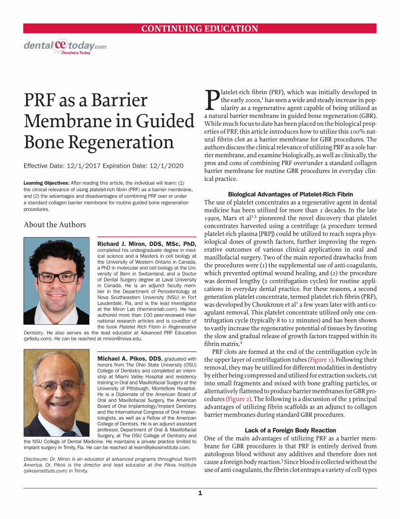
Richard J. Miron, DDS, MSc, PhD, completed his undergraduate degree in med-ical science and a Masters in cell biology at the University of Western Ontario in Canada, a PhD in molecular and cell biology at the Uni-versity of Bern in Switzerland, and a Doctor of Dental Surgery degree at Laval University in Canada. He is an adjunct faculty mem-ber in the Department of Periodontology at Nova Southeastern University (NSU) in Fort Lauderdale, Fla, and is the lead investigator at the Miron Lab (themironlab.com). He has authored more than 100 peer-reviewed inter-national research articles and is co-editor of the book Platelet Rich Fibrin in Regenerative
Dentistry. He also serves as the lead educator at Advanced PRF Education (prfedu.com). He can be reached at [email protected].
Michael A. Pikos, DDS, graduated with honors from The Ohio State University (OSU) College of Dentistry and completed an intern-ship at Miami Valley Hospital and residency training in Oral and Maxillofacial Surgery at the University of Pittsburgh, Montefiore Hospital. He is a Diplomate of the American Board of Oral and Maxillofacial Surgery, the American Board of Oral Implantology/Implant Dentistry, and the International Congress of Oral Implan-tologists, as well as a Fellow of the American College of Dentists. He is an adjunct assistant professor, Department of Oral & Maxillofacial Surgery, at The OSU College of Dentistry and
the NSU College of Dental Medicine. He maintains a private practice limited to implant surgery in Trinity, Fla. He can be reached at [email protected].
Disclosure: Dr. Miron is an educator at advanced programs throughout North America. Dr. Pikos is the director and lead educator at the Pikos Institute (pikosinstitute.com) in Trinity.
About the Authors

CONTINUING EDUCATION
2
PRF as a Barrier Membrane in Guided Bone Regenerationthat simultaneously act as a “defense system” toward incoming pathogens. For-eign body giant cells are not formed at the material surface as a result of its 100% autologous source, in contrast to many col-lagen barrier membranes fabricated from porcine or bovine origins.6,7 PRF mem-branes, however, typically have very short resorption times ranging from a 10- to a 28-day period.8 During their resorption, a slow and gradual release of growth factors is observed from within the PRF matrix.9
Release of Growth Factors From Within the ScaffoldThe main advantage of PRF versus other resorbable barrier membranes is that PRF contains living cells (namely platelets and leukocytes) capable of further secreting growth factors. As a result, the release of active proteins found in blood, including vascular endothelial growth factor (VEGF), platelet-derived growth factor (PDGF), and transforming growth factor-beta (TGF-beta), are all found within the PRF matrix and slowly released over time.9 Recent research from within the authors’ laboratories has shown that while PRP releases growth factors in higher levels at very early time points due to its gel- or liq-uid-like consistency, PRF scaffolds release growth factors and cytokines more slowly and gradually over time (Figure 3).9 PRF membranes are gradually resorbed within 14 days. During this time, a slow and continuous secretion of growth factors from within the scaffolds leads to an overall higher total release of growth factors when compared to PRP after 10 days (Figure 3). For cellular activity, the slow and gradual release of bioactive molecules, as opposed to their “shotgun” delivery as found in PRP, is favored for long-term tissue stimulation, and, for this reason, PRF has gradually seen a steady and more widespread use in regenerative dentistry.
Incorporation of Leukocytes Within Platelet-Rich Fibrin The inclusion of leukocytes within PRF scaffolds has been deemed one of the main advantages and reasons for the high regenerative capacity of PRF.4 In fact, from a terminology point of view, PRF was later termed L-PRF in the late 2000s in response to the growing evidence that leukocytes were shown
Figure 1. Follow-ing centrifuga-tion of blood in platelet-rich fibrin (PRF) tubes, the blood is sepa-rated into a lower layer containing red blood cells and an upper, plasma-rich layer. The upper layer contains PRF, which may be removed and utilized for regenerative procedures. Figure 2. The upper PRF may be removed from the PRF tubes and
placed in a PRF box and flattened to produce PRF membranes.
Figure 3. Growth factor release comparison between platelet-rich plasma (PRP) and PRF. (a) Growth factor release at earlier time points favors PRP, whereas at later time points, growth factor release is higher in the PRF groups. (b) PRF demonstrates a higher total accumulated growth factor release after 10 days compared to PRP.
a
b

CONTINUING EDUCATION
3
PRF as a Barrier Membrane in Guided Bone Regeneration
to be one of the key players within PRF scaffolds.10-12 As a result, further research within the past 3 to 5 years has focused on increas-ing their incorporation within the PRF matrix. Leukocytes are not only shown to secrete a wide array of growth factors responsible for tissue wound healing and regeneration, but also act to fight incoming pathogens from infection.13 This finding was best exemplified in a series of key studies that added leukocytes to PRP (no leukocytes) and demonstrated the marked increase in gener-ative potential of PRP when white blood cells were added.14-16 Interestingly, in a controlled study utilizing PRF for third molar extractions where 200 bi-lateral mandibular third molars were extracted and filled with either (1) PRF alone or (2) standard empty controls,17 the control group displayed an infection and/or dry socket rate of 9.5%. Alternatively, by simply placing PRF into the extraction socket, infection rates and dry sockets were drastically reduced to 1%, representing a near tenfold decrease in complication rates.17 Furthermore, the presence of leukocytes has also been reported to decrease perceived pain from patients and significantly reduce the number of analgesics taken postsurgery.18 Therefore, leukocyte incorporation and centrifugation protocols aimed at further enhancing their concentrations have added much value during the regeneration process with PRF.
Use of PRF as a Sole Barrier Membrane During GBR Procedures
Since PRF has a short resorption time ranging from 10 to 15 days, one of the questions commonly arising is “Can we use PRF alone as a replacement to collagen barrier membranes?” In general, the use of barrier membranes was developed to prevent fast-growing soft tissues from entering the slowly growing compartment con-taining bone.19 While theoretically their role has been debated over the past decade, the question commonly arising is “When and if can PRF be utilized alone or should it be routinely combined with a collagen barrier membrane?” The consensus agreed upon by a group of experts in the field promoted the use of PRF as a sole barrier membrane only when re-entry (a second surgical flap)
was not required. A common example of this is during window closure of a lateral window filled with a bone grafting material (Figure 4) when re-entry is not expected. Furthermore, many
Figure 4. (a) A lateral window sinus elevation procedure filled with a bone grafting material. (b) PRF membrane alone may be utilized to close the lateral window and favor soft-tissue healing over such regenerative approaches (case performed by Dr. Michael A. Pikos).
Figure 5. (a) to (d) An immediate implant placement with PRF being utilized to fill the gap and improve soft-tissue healing around the coronal portion of the implant (case performed by Dr. Michael A. Pikos).
Figure 6. (a) and (b) PRF can also be additionally utilized around the implant healing cap to further improve soft-tissue healing during immediate implant placement. (c) Notice the healing of soft tissues around this imme-diate implant after a 3-week healing period (case performed by Dr. Michael A. Pikos).
a b a b
a b
c d
c

CONTINUING EDUCATION
4
PRF as a Barrier Membrane in Guided Bone Regeneration
clinicians have begun to experiment with the use of PRF during immediate implant placement without flap elevation (Figure 5). PRF membranes can be placed around the implant collars to facil-itate more rapid soft-tissue healing without having to utilize a col-lagen barrier membrane. Similarly, PRF can also be utilized in a
“poncho” technique and wrapped around the healing cap to favor soft-tissue attachment and prevent infection (Figure 6). More frequently during GBR procedures, however (and most notably during extensive GBR cases), PRF membranes are combined with either a collagen barrier membrane or titanium/titanium-rein-forced membranes. The following is a description of how to pro-ceed in such cases.
Use of PRF Membranes With Collagen Barrier Membranes
One of the questions that arises most frequently during the authors’ continuing education courses is whether to use the PRF membranes over or under standard collagen barrier membranes. To address this question, it is best that the treating clinician understand the biological advantages and disadvantages of each scenario. One of the main advantages of PRF scaffolds is their incorporation of defense-fighting leukocytes. During large GBR procedures, one of the concerns with utilizing avital barrier membranes is their risk of exposure and subsequent infection. From this point of view, if a PRF membrane is placed underneath a collagen barrier membrane, the avascular and avital collagen membrane runs the risk of being left exposed to the oral cavity; a potential risk of exposure/infection may occur. In the event that a suture is accidentally dislodged or tension within the flap causes slight exposure to the underlying membrane, infection of the collagen scaffold is a possibility. In the reverse order, if
Figure 7. (a) to (d) A large guided bone regeneration (GBR) procedure utilizing both autogenous bone blocks and particulate grafting material. Following bone augmentation, a collagen barrier membrane was soaked in a fibrin clot and thereafter placed over the GBR procedure simultaneously in combination with a fibrin clot (case performed by Dr. Michael A. Pikos).
Figure 8. (a) to (f) Bone augmentation procedures performed with non-porous PTFE membranes may benefit from the added advantage of combining PRF underneath the barrier membranes, since blood supply from the periosteum is limited when utilizing low-porosity PTFE-titanium reinforced mem-branes (case performed by Dr. Michael A. Pikos).
a b
a b c
d e f
c d
CONTINUING EDUCATION
5
PRF as a Barrier Membrane in Guided Bone Regeneration
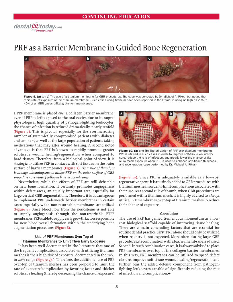
a PRF membrane is placed over a collagen barrier membrane, even if PRF is left exposed to the oral cavity, due to its supra-physiological high quantity of pathogen-fighting leukocytes, the chance of infection is reduced dramatically, nearly tenfold (Figure 7). This is pivotal, especially for the ever-increasing number of systemically compromised patients with diabetes and smokers, as well as the large population of patients taking medications that may alter wound healing. A second noted advantage is that PRF is known to rapidly promote greater soft-tissue wound healing/regeneration when compared to hard tissues. Therefore, from a biological point of view, it is strategic to utilize PRF in contact with soft tissues on the outer surface of barrier membranes (Figure 7). As a rule of thumb, it is always advantageous to utilize PRF on the outer surface of GBR procedures over-top of collagen barrier membranes.
Nevertheless, while the effects of PRF are still debatable on new bone formation, it certainly promotes angiogenesis within defect areas, an equally important area, especially for large vertical GBR augmentations. Therefore, it is advantageous to implement PRF underneath barrier membranes in certain cases, especially when non-resorbable membranes are utilized (Figure 8). Since blood flow from the periosteum is not able to supply angiogenesis through the non-resorbable PTFE membranes, PRF is able to supply early growth factors responsible for new blood vessel formation within the underlying bone augmentation procedures (Figure 8).
Use of PRF Membranes Over-Top of Titanium Membranes to Limit Their Early Exposure
It has been well documented in the literature that one of the frequent complications associated with utilizing titanium meshes is their high risk of exposure, documented in the 20% to 40% range (Figure 9).20 Therefore, the additional use of PRF over-top of titanium meshes has been proposed to limit the rate of exposure/complication by favoring faster and thicker soft-tissue healing (thereby decreasing the chance of exposure)
(Figure 10). Since PRF is adequately available as a low-cost regenerative agent, it is routinely added to GBR procedures with titanium meshes in order to limit complications associated with their use. As a second rule of thumb, when GBR procedures are performed with a titanium mesh, it is highly advised to always utilize PRF membranes over-top of titanium meshes to reduce their chance of exposure.
ConclusionThe use of PRF has gained tremendous momentum as a low-cost biological scaffold capable of improving tissue healing. There are 2 main concluding factors that are essential for routine dental practice. First, PRF alone should only be utilized when re-entry is not expected. More often during large GBR procedures, its combination with a barrier membrane is advised. Second, in such combination cases, it is always advised to place PRF membranes over-top of the collagen barrier membranes. In this way, PRF membranes can be utilized to speed defect closure, improve soft-tissue wound healing/regeneration, and further bear the added defense component from pathogen-fighting leukocytes capable of significantly reducing the rate of infection and complication.F
Figure 9. (a) to (c) The use of a titanium membrane for GBR procedures. The case was corrected by Dr. Michael A. Pikos, but notice the rapid rate of exposure of the titanium membrane. Such cases using titanium have been reported in the literature rising as high as 20% to 40% of all GBR cases utilizing titanium membranes.
Figure 10. (a) and (b) The utilization of PRF over titanium membranes. PRF is utilized in such cases in order to improve soft-tissue wound clo-sure, reduce the rate of infection, and greatly lower the chance of tita-nium mesh exposure when PRF is used to enhance soft-tissue thickness and regeneration (case performed by Dr. Michael A. Pikos).
a b
a b
c

CONTINUING EDUCATION
6
References1. Choukroun J, Adda F, Schoeffler C, et al. An opportunity in perio-im-
plantology: the PRF [in French]. Implantodontie. 2001;42:55-62. 2. Marx RE. Platelet-rich plasma: evidence to support its use. J Oral
Maxillofac Surg. 2004;62:489-496.3. Marx RE, Carlson ER, Eichstaedt RM, et al. Platelet-rich plasma:
growth factor enhancement for bone grafts. Oral Surg Oral Med Oral Pathol Oral Radiol Endod. 1998;85:638-646.
4. Miron RJ, Fujioka-Kobayashi M, Bishara M, et al. Platelet-rich fibrin and soft tissue wound healing: a systematic review. Tissue Eng Part B Rev. 2017;23:83-99.
5. Miron RJ, Bosshardt DD. OsteoMacs: key players around bone biomaterials. Biomaterials. 2016;82:1-19.
6. Barbeck M, Lorenz J, Holthaus MG, et al. Porcine dermis and pericardium-based, non-cross-linked materials induce multinucle-ated giant cells after their in vivo implantation: a physiological reaction? J Oral Implantol. 2015;41:e267-e281.
7. Moses O, Vitrial D, Aboodi G, et al. Biodegradation of three dif-ferent collagen membranes in the rat calvarium: a comparative study. J Periodontol. 2008;79:905-911.
8. Yamashita Y, Chen K, Kuroda S, et al. Stability of platelet-rich fibrin in vivo: histological study in rats. Journal of Oral Tissue Engineering. 2016;14:83-90.
9. Kobayashi E, Flückiger L, Fujioka-Kobayashi M, et al. Comparative release of growth factors from PRP, PRF, and advanced-PRF. Clin Oral Investig. 2016;20:2353-2360.
10. Mazor Z, Horowitz RA, Del Corso M, et al. Sinus floor augmentation with simultaneous implant placement using Choukroun’s plate-let-rich fibrin as the sole grafting material: a radiologic and histo-logic study at 6 months. J Periodontol. 2009;80:2056-2064.
11. Dohan Ehrenfest DM, Diss A, Odin G, et al. In vitro effects of Choukroun’s PRF (platelet-rich fibrin) on human gingival fibro-blasts, dermal prekeratinocytes, preadipocytes, and maxillofacial osteoblasts in primary cultures. Oral Surg Oral Med Oral Pathol Oral Radiol Endod. 2009;108:341-352.
12. Dohan Ehrenfest DM, Del Corso M, Diss A, et al. Three-dimensional architecture and cell composition of a Choukroun’s platelet-rich fibrin clot and membrane. J Periodontol. 2010;81:546-555.
13. Everts PA, Overdevest EP, Jakimowicz JJ, et al. The use of autol-ogous platelet-leukocyte gels to enhance the healing process in surgery, a review. Surg Endosc. 2007;21:2063-2068.
14. Kawazoe T, Kim HH. Tissue augmentation by white blood cell-con-taining platelet-rich plasma. Cell Transplant. 2012;21:601-607.
15. Perut F, Filardo G, Mariani E, et al. Preparation method and growth factor content of platelet concentrate influence the osteo-genic differentiation of bone marrow stromal cells. Cytotherapy. 2013;15:830-839.
16. Pirraco RP, Reis RL, Marques AP. Effect of monocytes/macro-phages on the early osteogenic differentiation of hBMSCs. J Tis-sue Eng Regen Med. 2013;7:392-400.
17. Hoaglin DR, Lines GK. Prevention of localized osteitis in man-dibular third-molar sites using platelet-rich fibrin. Int J Dent. 2013;2013:875380.
18. Bilginaylar K, Uyanik LO. Evaluation of the effects of platelet-rich fibrin and piezosurgery on outcomes after removal of impacted mandib-ular third molars. Br J Oral Maxillofac Surg. 2016;54:629-633.
19. Dahlin C, Linde A, Gottlow J, et al. Healing of bone defects by guided tissue regeneration. Plast Reconstr Surg. 1988;81:672-676.
20. Louis PJ, Gutta R, Said-Al-Naief N, et al. Reconstruction of the max-illa and mandible with particulate bone graft and titanium mesh for implant placement. J Oral Maxillofac Surg. 2008;66:235-245.
PRF as a Barrier Membrane in Guided Bone Regeneration
CONTINUING EDUCATION
7
PRF as a Barrier Membrane in Guided Bone Regeneration
1. Platelet-rich plasma, developed in the late 1990s, involved two centrifugation cycles. Platelet-rich fibrin (PRF), a second-gen-eration platelet concentrate, involves only one centrifugation cycle.
a. The first statement is true, the second is false.b. The first statement is false, the second is true.c. Both statements are true.d. Both statements are false.
2. PRF is entirely derived from autologous blood without additives and does not cause a foreign body reaction.
a. True.b. False.
3. PRF membranes typically have resorption times ranging from:a. 3 to 7 days.b. 7 to 10 days.c. 10 to 28 days.d. 28 to 42 days.
4. The following is/are found within the PRF matrix and slowly released over time:
a. Vascular endothelial growth factor.b. Platelet-derived growth factor.c. Transforming growth factor-beta.d. All of the above.
5. The inclusion of _________ within PRF scaffolds is one of the main reasons for the high regenerative capacity of PRF.
a. Neutrophils.b. Leukocytes.c. Monocytes.d. Granulocytes.
6. One study found that by placing PRF into an extraction socket, infection rates and dry sockets were reduced to _________.
a. 9.5%.b. 5%.c. 3%.d. 1%.
7. The consensus is that PRF should be used as a sole barrier membrane only when re-entry (a second surgical flap) is not required.
a. True.b. False.
8. If a PRF membrane is placed over a collagen barrier membrane, even if PRF is left exposed to the oral cavity, the chance of infection is reduced nearly _________.
a. Twofold.b. Fivefold.c. Sevenfold.d. Tenfold.
POST EXAMINATION INFORMATION
To receive continuing education credit for participation in this educational activity, you must complete the program post examination and receive a score of 70% or better.
Traditional Completion Option:You may fax or mail your answers with payment to Dentistry Today (see Traditional Completion Information on following page). All information requested must be provided in order to process the program for credit. Be sure to complete your “Payment,” “Personal Certification Information,” “Answers,” and “Evaluation” forms. Your exam will be graded within 72 hours of receipt. Upon successful completion of the post-exam (70% or higher), a letter of completion will be mailed to the address provided.
Online Completion Option:Use this page to review the questions and mark your answers. Return to dentalcetoday.com and sign in. If you have not pre-viously purchased the program, select it from the “Online Courses” listing and complete the online purchase process. Once purchased, the program will be added to your User History page where a Take Exam link will be provided directly across from the program title. Select the Take Exam link, complete all the program questions and Submit your answers. An immediate grade report will be provided. Upon receiving a passing grade, complete the online evaluation form. Upon submitting the form, your Letter of Completion will be provided immediately for printing.
General Program Information:Online users may log in to dentalcetoday.com any time in the future to access previously purchased programs and view or print letters of completion and results.
POST EXAMINATION QUESTIONS

CONTINUING EDUCATION
8
PRF as a Barrier Membrane in Guided Bone Regeneration9. One frequent complication associated with titanium meshes
is their high risk of exposure, documented in the range of _________.
a. 5% to 10%.b. 10% to 20%.c. 20% to 40%.d. 40% to 50%.
10. PRF alone should only be utilized when re-entry is not expected. More often during large guided bone regenera-tion procedures, its combination with a barrier membrane is advised.
a. The first sentence is true, the second is false.b. The first sentence is false, the second is true.c. Both sentences are true.d. Both sentences are false.

CONTINUING EDUCATION
9
PRF as a Barrier Membrane in Guided Bone Regeneration
PROGRAM EVAUATION FORM
Please complete the following activity evaluation questions.
Rating Scale: Excellent = 5 and Poor = 0
Course objectives were achieved.
Content was useful and benefited your clinical practice.
Review questions were clear and relevant to the editorial.
Illustrations and photographs were clear and relevant.
Written presentation was informative and concise.
How much time did you spend reading the activity and
completing the test?
What aspect of this course was most helpful and why?
What topics interest you for future Dentistry Today CE courses?
Dentistry Today, Inc, is an ADA CERP Recognized Provider. ADA CERP is a service of the American Dental Association to assist dental professionals in indentifying quality providers of continuing dental education. ADA CERP does not approve or endorse individual courses or instructors, nor does it imply acceptance of credit hours by boards of dentistry. Concerns or complaints about a CE provider may be directed to the provider or to ADA CERP at ada.org/goto/cerp.
Approved PACE Program Provider FAGD/MAGD Credit Approval does not imply acceptance by a state or provincial board of dentistry or AGD endorsement. June 1, 2015 to May 31, 2018 AGD PACE approval number: 309062
PROGRAM COMPLETION INFORMATIONIf you wish to purchase and complete this activity traditionally (mail or fax) rather than online, you must provide the information requested below. Please be sure to select your answers carefully and complete the evaluation information. To receive credit you must answer at least 7 of the 10 questions correctly.Complete online at: dentalcetoday.com
TRADITIONAL COMPLETION INFORMATION:
Mail or fax this completed form with payment to:Dentistry TodayDepartment of Continuing Education100 Passaic AvenueFairfield, NJ 07004Fax: 973-882-3622
PAYMENT & CREDIT INFORMATION:Examination Fee: $40.00 Credit Hours: 2.0Note: There is a $10 surcharge to process a check drawn on any bank other than a US bank. Should you have addi-tional questions, please contact us at (973) 882-4700.
o I have enclosed a check or money order.
o I am using a credit card.
My credit card information is provided below.
o American Express o Visa o MC o Discover
Please provide the following (please print clearly):
Exact Name on Credit Card
Credit Card # Expiration Date
Signature
PERSONAL CERTIFICATION INFORMATION:
Last Name (PLEASE PRINT CLEARLY OR TYPE)
First Name
Profession / Credentials License Number
Street Address
Suite or Apartment Number
City State Zip Code
Daytime Telephone Number With Area Code
Fax Number With Area Code
E-mail Address
ANSWER FORM: COURSE #: 216Please check the correct box for each question below.
1. o a o b o c o d
2. o True o False
3. o a o b o c o d
4. o a o b o c o d
5. o a o b o c o d
6. o a o b o c o d
7. o True o False
8. o a o b o c o d
9. o a o b o c o d
10. o a o b o c o d
View publication statsView publication stats