prevention of vascular inflammation by prevention of ... · coated nanoparticles were characterized...
TRANSCRIPT
1
Prevention of Vascular Inflammation by Nanoparticle Targeting
of Adherent Neutrophils
Zhenjia Wang, Jing Li , Jaehyung Cho, and Asrar B. Malik
Mice: Adult (6-8 weeks) male wild-type (C57BL/6), FcγRIII-/- (B6.129-Fcgr3<tm1Sjv>/j),
Mac-1-/- (CD11b/CD18), and LFA1-/- (CD11a/CD18) mice were purchased from Jackson
Laboratory (Bar Harbor, Maine). CD1 mice (6-8 weeks) were purchased from Charles
River Laboratories (Wilmington, MA). The University of Illinois Institutional Animal Care
and Use Committee approved all animal care and experimental protocols used in these
studies. All experiments were made under anesthesia using intraperitoneal injection of
the mixture of ketamine (80 mg/kg), xylazine (2 mg/kg), and acepromazine (2 mg/kg) in
saline. CD1 male mice were used in the LPS-induced acute lung injury studies.
Reagents and Antibodies: Lipopolysaccharide (LPS) and bovine serum albumin (BSA)
were purchased from Sigma (St. Louis, MO). Glutaraldehyde was obtained from
Electron Microscopy Sciences (Hatfield, PA). Piceatannol, the water-insoluble
Syk/Zap70 kinase inhibitor1,2, was purchased from Tokyo Chemical Industry (TCI)
(Pittsburgh, PA). Cy5 (HIDCI, 2-[5-(1,3-dihydro-1, 3, 3-trimethyl-2H-indol-2-ylidene)-1,
3-pentadienyl]-1, 3, 3-trimethyl-3H-indolium iodide, 635nm (excitation)/660nm
(emission)) was purchased from Exciton (Dayton, OH). Alexa Fluor-488 and Alex Fluor-
647-labeled anti-mouse Gr-1(Ly-6G/Ly-C6) and anti-mouse F4/80 antibodies, Alexa
Prevention of vascular inflammation by nanoparticle targeting of adherent neutrophils
SUPPLEMENTARY INFORMATIONDOI: 10.1038/NNANO.2014.17
NATURE NANOTECHNOLOGY | www.nature.com/naturenanotechnology 1
© 2014 Macmillan Publishers Limited. All rights reserved.
2
Fluor-488-labeled IgG 2b κ as isotype control for anti-mouse Gr-1 and F4/80, and
recombinant mouse TNF-α were purchased from Biolegend (San Diego, CA).
Carboxylated polystyrene fluorescent yellow-green nanoparticles (100 nm in a diameter,
excitation/emission: 505nm/515nm) and Alex-Fluor 647-NHS for bio-conjugation were
purchased from Invitrogen (Grand Island, NY). EDC (1-Ethyl-3-(3-
dimethylaminopropyl)carbodiimide) and Sulfo-NHS (N-hydroxysulfo succinimide) were
purchased from Pierce (Rockford, IL). Diff-Quick staining kit was purchased from Fisher
Scientific. N-formyl-methionyl-leucyl-phenylalanine (fMLF) and other chemicals were
from Sigma (St. Louis, MO). Rabbit anti-phospho Syk (Tyr525/526) and anti-mouse β-
actin antibodies were purchased from Cell Signaling (Danvers, MA). CellTiter 96®
AQueous one solution cell proliferation assay was purchased from Promega (Madison,
WI). Isolated mouse lung endothelial cells (MLECs) were cultured as described3.
Purified human fibrinogen was kindly provided by Deane F. Mosher (University of
Wisconsin, Madison, WI).
Measuring size of albumin nanoparticles: The size of albumin nanoparticles was
measured using dynamics light scattering (Dyatt Inc.) at 633 nm. Diluted albumin
nanoparticle solution was spread on a copper grid (300 mesh) and the size of
nanoparticles was determined by transmission electron microscope (Jeol JEM-1220).
Synthesis of BSA-conjugated polystyrene nanoparticles: BSA molecules were
covalently conjugated with yellow-green polystyrene nanoparticles (excitation/emission:
505nm/515nm) with a diameter of 100 nm according to manufacturer’s instruction. To
© 2014 Macmillan Publishers Limited. All rights reserved.

3
activate COOH groups on fluorescent polystyrene nanoparticles with EDC and Sulfo-
NHS for reaction of 15 min. The mixture was centrifuged using Nanosep-50K
(OD50C33) (Pall Inc.) and was dissolved in PBS buffer (pH 7.4). After activation of
nanoparticles, a defined amount of BSA solution was added to the activated particle
solution, and incubated for 2 hours at RT. Free BSA molecules were separated from
BSA-coated nanoparticles using Nanosep-300K centrifuge device (OD300C33). BSA-
coated nanoparticles were characterized by absorption spectra using Carey 300 Bio4. In
the study of uptake of BSA-coated nanoparticles in neutrophils (Figure 2e), we
intravenously infused 5 µl of 6.5 nM nanoparticles in each mouse.
In vitro cytotoxicity assay. Mouse neutrophils were isolated as described5.
Neutrophils (1 x 10⁵ cells) were stimulated with or without 10 µM fMLF for 10 minutes at
37ºC. After washing, cells were resuspended in RPMI1640 medium and treated with
albumin nanoparticles or piceatannol-loaded nanoparticles, 800 µg/ml (200 µM as
piceatannol), at 37ºC for 1 hour. Cells were then incubated with MTT solution at 37ºC
for 4 hours in the dark. Absorbance was recorded using a plate reader (PHERAstar,
BMG biotech) at a wavelength of 490 nm. Incubation of MTT solution with each reagent
without cells was counted as the blank for each sample. Cell viability after treatment
with 150 µM H2O2 was used as a positive control.
Syk phosphorylation. Isolated mouse neutrophils (2 x 10⁶ cells) were plated onto a
fibrinogen-coated wells of a 6 well plate and incubated with albumin nanoparticles or
piceatannol-loaded nanoparticles, 800 µg/ml (200 µM as piceatannol), at 37ºC in the
presence of 50 ng/ml TNF-α for 30 minutes. Non-adherent neutrophils were collected
© 2014 Macmillan Publishers Limited. All rights reserved.

4
and lysed with RIPA buffer (Tris-HCl, pH 7.4 containing 1% Triton-X100, 0.05% SDS,
proteinase inhibitor cocktail, 1 mM PMSF, 1 mM Na3VO4, and 1 mM NaF). Also,
adherent neutrophils were lysed and combined with the lysate of non-adherent cells.
The final lysate was electrophoresed under reduced conditions and immunoblotted with
an anti-phosphoSyk-Tyr525/526 antibody. The band density was measured by
densitometry using Scion Image (v4.0).
Flow cytometric analysis. Mouse neutrophils treated with or without 10 µM fMLF were
incubated with 100 µg/ml of Cy5-labeled albumin nanoparticles, followed by incubation
with Alexa Fluor 488-conjugated anti-mouse Gr-1 antibodies. Cells were fixed and
analyzed by flow cytometry (Cyan ADP, Beckman Coulter). Mouse neutrophils were
identified by forward and side scatter as well as Gr-1 expression.
Flow chamber assay. A flow chamber assay was performed as described with
modifications5. Confluent mouse lung endothelial cells (MLECs) on fibrinogen-coated
glass coverslips were stimulated by murine TNF-α (20 ng/ml) for 6 hours and placed
into a parallel plate flow chamber (Bioptech). Mouse neutrophils, 2 x 10⁶, were treated
with albumin nanoparticles or piceatannol-loaded nanoparticles, 800 µg/ml (200 µM as
piceatannol), for 20 minutes at 37ºC and then incubated with fMLF for 10 minutes. After
being washed and resuspended in RPMI1640 medium containing 0.1% BSA,
neutrophils were perfused for 10 minutes over activated MLECs under venous shear (1
dyne/cm2). The flow chamber and microscope system were kept at 37ºC in a plexiglass
chamber. Real-time images were acquired using a Nikon microscope (ECLIPSE Ti,
Melville, NY) equipped with 10 x/0.25 NA objective lens and recorded with a digital
camera (CoolSNAP ES2). The data were analyzed through NIS Elements (AR 3.2).
© 2014 Macmillan Publishers Limited. All rights reserved.

5
Adherent (determined either as round and spread) and migrating neutrophils were
counted in a field of 0.15 mm2. The number of firmly adherent neutrophils was counted
in 5 separate fields.
Lung tissue myeloperoxidase (MPO) activity: Acute lung injury was induced by
intraperitoneal injection of LPS (10 mg/kg body weight) in male CD1 mice (6-8 weeks
old). At 2 hour after LPS injection, piceatannol-loaded albumin nanoparticles in saline
were injected into mice via tail vein. At 8 hours post-LPS injection, lungs were isolated,
and the half were used for MPO measurement and the others were fixed with 4%
paraformaldehyde for histological analysis. MPO activity in the lung tissues was
measured as described 6. Briefly, lungs were isolated and homogenized in 1 ml of PBS
(pH 6.0, 50 mM) containing 0.5% hexadecyltrmethylammonium bromide for 20 seconds.
The homogenates were centrifuged at 14,000 rpm for 20 minutes at 4°C, and the pellet
was collected and resuspended with the same buffer (0.5 ml). After homogenization and
centrifugation, the supernatant was collected and mixed with PBS containing 0.2 mg/ml
o-dianisidine hydrochloride and 0.05% H2O2 at a final concentration of 1/30 (v/v).
Absorbance change was measured at 460 nm for 3 minutes using DU-530/UV-Vis
spectrometer (Beckman Coulter). MPO activity was calculated as change in absorbance
over time and normalized with dry lung weight. To microscopically image infiltration of
neutrophils in lungs, chemo-staining was done as described7. Briefly, formalin-fixed,
paraffin-embedded tissue samples were sectioned at 5µm thickness and mounted on
Starfrost/Plus slides. Slides were incubated in Naphthol AS-D chloroacetate (Sigma)
according to manufacturer’s instructions. Slides were rinsed in distilled water, stained
© 2014 Macmillan Publishers Limited. All rights reserved.

6
with hematoxylin, and mounted with aqueous solution. The slides were imaged using a
microscope and stained neutrophils were quantified per field (0.6 mm2).
Neutrophil and monocyte counts in mouse bronchoalveolar lavage (BAL). At 2
hour after intraperitoneal injection of LPS (10 mg/kg mouse weight), PBS, BSA
nanoparticles, or piceatannol-loaded BSA nanoparticles (piceatannol concentration at
4.3 mg/kg mouse weight) were intravenously infused into CD1 mice. At 6 hours later,
mouse BAL fluid was collected by inserting a needle into the upper trachea. Lavage was
performed by introducing 3 sequential 1 ml of PBS into the lungs and carefully
withdrawing BAL fluid. The BAL fluid was centrifuged at 1600 rpm for 5 min. Cells were
resuspended in 0.5 ml PBS and counted in a hemocytometer. Slides were also
prepared using an aliquot of each sample and stained with the mixture of xanthene dye
and thiazine dye using Diff-Quick staining protocol8. Neutrophils and monocytes were
quantified under a microscope.
© 2014 Macmillan Publishers Limited. All rights reserved.
7
Movie 1. The merged intravital microscopic images show that Cy5-loaded albumin
nanoparticles (red) are internalized by leukocytes adherent to venular endothelial cells
following TNF-α-induced cremaster venular inflammation in WT mice.
Movie 2. Cy5-loaded albumin nanoparticles are internalized by adherent and the very
slowly moving Gr-1 positive neutrophils post-TNF-α-induced venular inflammation in WT
mice. Neutrophils were visualized by infusion of Alexa Fluor 488-conjugated anti-Gr-1
antibodies.
Movie 3. Cy5-loaded albumin nanoparticles are not internalized by circulating
neutrophils in non-TNF-α-treated cremaster muscle venules in WT mice. Neutrophils
were visualized by infusion of Alexa Fluor 488-conjugated anti-Gr-1 antibodies.
Movie 4. Cy5-loaded albumin nanoparticles are not internalized by monocytes on TNF-
α-stimulated venular endothelial cells in WT mice. Monocytes were visualized by
infusion of Alexa Fluor-488-labeled anti-mouse F4/80 antibodies.
Movie 5. Alexa Fluor-647-conjugated albumin nanoparticles (red) show puncta after
internalization by adherent neutrophils (green) on the TNF-α-stimulated venular
endothelial cells in WT mice. Puncta (red) in neutrophils represent single albumin
© 2014 Macmillan Publishers Limited. All rights reserved.

8
nanoparticles and their aggregates. Neutrophils were visualized by infusion of Alexa
Fluor-488-labeled anti-mouse Gr-1 antibodies.
Movie 6. BSA-conjugated polystyrene nanoparticles (100 nm in diameter) bind to the
surface of adherent neutrophils without internalization post-TNF-α-induced venular
inflammation in WT mice. Neutrophils were visualized by infusion of Alexa Fluor-647-
labeled anti-mouse Gr-1 antibodies.
Movie 7-8. Neutrophil adhesion to TNF-α-stimulated venular endothelial cells in WT
mice before (movie 7) and 1 hour after piceatannol-loaded albumin nanoparticles
(movie 8). Neutrophils were visualized by infusion of Alexa Fluor-488-labeled anti-
mouse Gr-1 antibodies.
© 2014 Macmillan Publishers Limited. All rights reserved.

9
Supplementary Figures
Figure S1. Scheme for preparing albumin nanoparticles using BSA (bovine serum
albumin) incorporated with fluorescent molecules or a drug.
BSAEthanol
Crosslinking
Nanoparticles
Drug or imaging agents
© 2014 Macmillan Publishers Limited. All rights reserved.

10
Figure S2. Transmission electron microscopy (TEM) shows size of albumin
nanoparticles averaging 100 nm in a diameter. Scale bar: 500 nm.
© 2014 Macmillan Publishers Limited. All rights reserved.
11
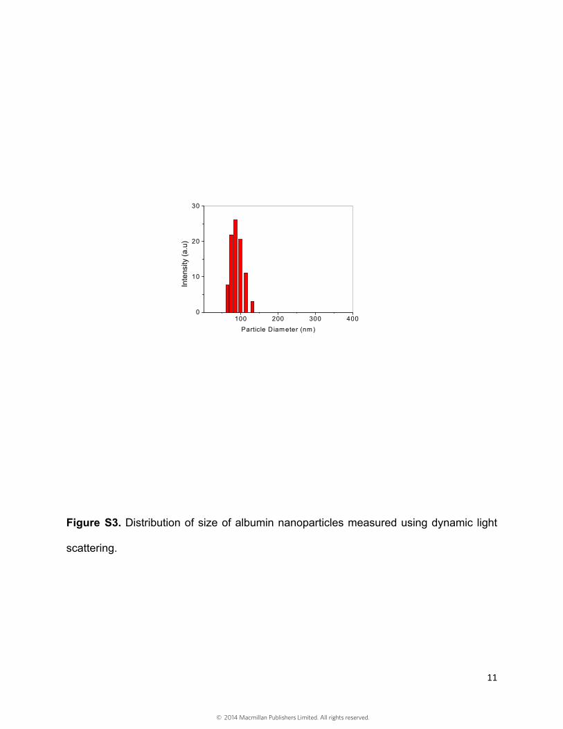
Figure S3. Distribution of size of albumin nanoparticles measured using dynamic light
scattering.
100 200 300 4000
10
20
30
Inte
nsity
(a.
u)
Particle Diameter (nm)
© 2014 Macmillan Publishers Limited. All rights reserved.

12
Figure S4. Specificity of anti-Gr-1 antibody in mouse neutrophils. Alexa-Fluor-488
isotype matched IgG 2b, κ (1.5 µg/mouse) is the control. Alexa-Fluor-488 anti-mouse
Gr-1 (1.5 µg/mouse) is infused i.v. in 3 hr TNF-α-treated cremaster muscle venulee.
Cremaster muscle venules are imaged using intravtial microscope.
Control
Anti-Gr-1
Tissue IsotypeIgG
Merge
Tissue Anti-Gr-1 Merge
© 2014 Macmillan Publishers Limited. All rights reserved.

13
Figure S5. Loading of piceatannol in albumin nanoparticles is dependent of the amount
of initially added piceatannol. Amount of picetannol loading in albumin nanoparticles
was determined using the absorption spectrum of piceatannol. Results represent mean
± SEM.
0.0 0.5 1.00.0
0.3
0.6
0.9
Pic
ea
tan
no
l-lo
ad
ed in
P
art
icle
s (m
g)
Added piceatannol (mg)
© 2014 Macmillan Publishers Limited. All rights reserved.
14
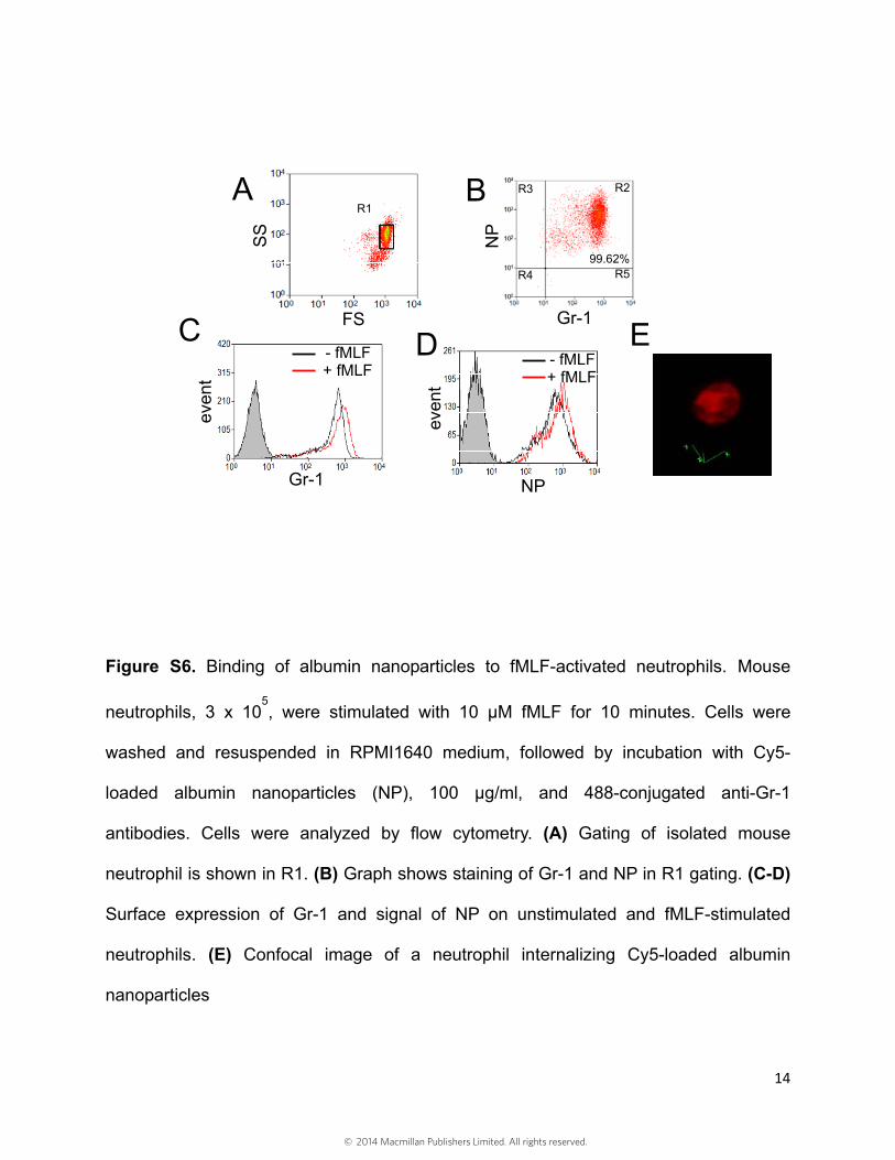
Figure S6. Binding of albumin nanoparticles to fMLF-activated neutrophils. Mouse
neutrophils, 3 x 10⁵, were stimulated with 10 µM fMLF for 10 minutes. Cells were
washed and resuspended in RPMI1640 medium, followed by incubation with Cy5-
loaded albumin nanoparticles (NP), 100 µg/ml, and 488-conjugated anti-Gr-1
antibodies. Cells were analyzed by flow cytometry. (A) Gating of isolated mouse
neutrophil is shown in R1. (B) Graph shows staining of Gr-1 and NP in R1 gating. (C-D)
Surface expression of Gr-1 and signal of NP on unstimulated and fMLF-stimulated
neutrophils. (E) Confocal image of a neutrophil internalizing Cy5-loaded albumin
nanoparticles
NP
even
t
- fMLF+ fMLF
Gr-1
even
t
- fMLF+ fMLF
R1
FS
SS
NP
Gr-1
R2R3
R5R499.62%
A B
C D E
© 2014 Macmillan Publishers Limited. All rights reserved.

15
Figure S7. Lung tissue section stained with neutrophil-specific esterase for evaluating
effects of piecatannol-loaded albumin nanoparticles. Pink objects are neutrophils and
blue objects for cell nuclei. We quantified the numbers of neutrophils per field and
averaged over 20 fields (0.6 mm2) in 3 mice per group (results shown in Figure 4I)
PBS Pic-loaded Alb Nanoat 0.86 mg (pic)/kg
Pic-loaded Alb Nanoat 4.3 mg (pic)/kg
© 2014 Macmillan Publishers Limited. All rights reserved.

16
REFERENCES
1 Zarbock, A., Lowell, C. A. & Ley, K. Spleen tyrosine kinase Syk is necessary for E-selectin-induced alpha(L)beta(2) integrin-mediated rolling on intercellular adhesion molecule-1. Immunity 26, 773-783 (2007).
2 Evans, R., Lellouch, A. C., Svensson, L., McDowall, A. & Hogg, N. The integrin LFA-1 signals through ZAP-70 to regulate expression of high-affinity LFA-1 on T lymphocytes. Blood 117, 3331-3342 (2011).
3 Tiruppathi, C. et al. Impairment of store-operated Ca2+ entry in TRPC4(-/-) mice interferes with increase in lung microvascular permeability. Circ. Res. 91, 70-76 (2002).
4 Wang, Z., Tiruppathi, C., Minshall, R. D. & Malik, A. B. Size and dynamics of caveolae studied using nanoparticles in living endothelial cells. ACS Nano 3, 4110-4116 (2009).
5 Hahm, E. et al. Extracellular protein disulfide isomerase regulates ligand-binding activity of alphaMbeta2 integrin and neutrophil recruitment during vascular inflammation. Blood 121, 3789-3800 (2013).
6 Garrean, S. et al. Caveolin-1 regulates NF-kappaB activation and lung inflammatory response to sepsis induced by lipopolysaccharide. J. Immunol.177, 4853-4860 (2006).
7 Lomas-Neira, J. L., Chung, C. S., Wesche, D. E., Perl, M. & Ayala, A. In vivo gene silencing (with siRNA) of pulmonary expression of MIP-2 versus KC results in divergent effects on hemorrhage-induced, neutrophil-mediated septic acute lung injury. J. Leukoc. Biol. 77, 846-853 (2005).
8 Huang, X., Wu, J., Spong, S. & Sheppard, D. The integrin alphavbeta6 is critical for keratinocyte migration on both its known ligand, fibronectin, and on vitronectin. J. Cell Sci. 111, 2189-2195 (1998).
© 2014 Macmillan Publishers Limited. All rights reserved.