prevention and control of healthcare- associated …
TRANSCRIPT
PREVENTION AND CONTROL OF HEALTHCARE- ASSOCIATED INFECTIONSBasic Recommendations

This publication was made possible thanks to funding and cooperation from the Infection Control Center, U.S. Centers for Disease Control and Prevention, under Cooperative Agreement CDC-RFA-CK13-1301, “Building Capacity and Networks to Address Emerging Infectious Diseases in the Americas.”Our special thanks go to the Infection Control Program, Department of Patient Quality and Safety, Di-vision of Human Stewardship and Development, Undersecretariat of Health Care Networks, Ministry of Health of Chile, for its technical support in the preparation of this document.
PREVENTION AND CONTROL OF HEALTHCARE- ASSOCIATED INFECTIONSBasic Recommendations

Original version in Spanish
Prevención y control de infecciones asociadas a la atención de la salud. Recomendaciones Básicas
ISBN: 978-92-75-31954-3
Prevention and control of healthcare–associated infections. Basic Recommendations.
ISBN: 978-92-75-11954-9
© Pan American Health Organization 2018
All rights reserved. Publications of the Pan American Health Organization are available on the PAHO website (www.paho.org). Requests for permission to reproduce or translate PAHO Publications should be addressed to the Communications Department through the PAHO website (www.paho.org/permissions).
Suggested citation. Pan American Health Organization. Prevention and control of healthcare–associated infections. Basic Recommendations. Washington, D.C.: PAHO; 2018.
Cataloguing-in-Publication (CIP) data. CIP data are available at http://iris.paho.org.
Publications of the Pan American Health Organization enjoy copyright protection in accor-dance with the provisions of Protocol 2 of the Universal Copyright Convention.
The designations employed and the presentation of the material in this publication do not imply the expression of any opinion whatsoever on the part of the Secretariat of the Pan Amer-ican Health Organization concerning the status of any country, territory, city or area or of its authorities, or concerning the delimitation of its frontiers or boundaries.
The mention of specific companies or of certain manufacturers’ products does not imply that they are endorsed or recommended by the Pan American Health Organization in preference to others of a similar nature that are not mentioned. Errors and omissions excepted, the names of proprietary products are distinguished by initial capital letters.
All reasonable precautions have been taken by the Pan American Health Organization to verify the information contained in this publication. However, the published material is being dis-tributed without warranty of any kind, either expressed or implied. The responsibility for the interpretation and use of the material lies with the reader. In no event shall the Pan American Health Organization be liable for damages arising from its use.

Index
9 / Abbreviations
11 / I. Essential elements for implementing infection control measures in health facilities
18 / References
19 / II. Chain of microorganism transmission in healthcare
28 / References
29 / III. Standard precautions
29 / Hand hygiene29 / Why are hands a source of transmission of microorganisms that can
cause HAIs?30 / What should we know about the transmission of pathogens by the
hands?31 / Is hand hygiene sufficient to prevent HAIs?31 / How is hand hygiene achieved?32 / Hand washing32 / What are the properties of the necessary elements in hand washing?35 / Hand drying38 / What hand-washing technique should be used?40 / Use of alcohol-based solutions40 / What properties should alcohol-based solutions have?40 / What characteristics should alcohol-based solutions have?42 / What factors should be considered with regard to availability of and
access to these solutions?43 / What scrubbing/rubbing technique should be used with alcohol-based
solutions?46 / When should hand hygiene be done?47 / What is known about conditions that affect adherence to hand hygiene?

47 / What strategies can help to encourage hand hygiene?54 / Summary55 / References60 / Personal protective equipment (PPE)60 / What is PPE?61 / What general points should be considered in selecting PPE for an
institution?61 / What are the different PPE items/components?63 / What criteria need to be considered in selecting gloves?64 / How does the risk for HAI transmission affect the composition of the
gloves?65 / What precautions should be taken when putting on and removing
gloves?67 / How does risk for HAI transmission affect the composition of gowns?69 / What is a type N95 or FFP2 respirator?70 / Are respirators as easy to use as masks?73 / How long can respirators be used without interruption? Can they be
reused?74 / What characteristics should eye protection (safety glasses, goggles, face
shields) have beyond serving as a barrier?75 / Other PPE: boots, jumpsuits, hoods75 / What type of PPE should be used?76 / How is PPE used in an institution?78 / What conditions can affect adherence to PPE use?79 / What principles apply to the PPE donning and doffing sequence?81 / Summary81 / References86 / Prevention of sharps accidents86 / Why is prevention of accidents involving sharp instruments part of
standard precautions?86 / What are the factors that place health workers at risk for infection from
punctures and cuts?88 / What types of objects or materials pose a risk of exposure?88 / Who is exposed?89 / How can sharps accidents be avoided?91 / What does safe handling of sharps entail?

92 / How should sharps be discarded?93 / What measures other than the standard precautions can be used?93 / References95 / Management of the environment95 / Why is management of the environment included in the standard
precautions?95 / Does the environment contribute to all HAIs?98 / What does “environment” comprise in the context of HAIs?98 / How should the environment be managed?99 / Aren’t cleaning and disinfection the same thing?100 / Cleaning and disinfection of low-risk surfaces103 / What is the best way to supervise the cleaning process?104 / Is handling of patient clothing often associated with HAIs?105 / What steps should be considered in the management of laundry to
prevent HAIs?107 / Waste107 / Does the waste in health facilities carry a greater risk for HAIs?108 / How can hospital waste be managed to prevent HAIs?111 / References
115 / IV. Additional precautions based on mode of transmission
115 / What is the difference between the standard precautions and the additional precautions based on mode of transmission?
116 / What is involved in applying the additional precautions based on mode of transmission?
118 / What needs to be known in order to decide if additional precautions are required?
118 / Contact transmission119 / Where should a patient requiring contact precautions be located?120 / What are the requirements for the room or hospital area to be occupied
by a patient needing additional precautions?121 / What measures should be taken during care for patients requiring con-
tact precautions?122 / Droplet transmission122 / What is the objective of precautions against droplet transmission?122 / Where should a patient requiring droplet precautions be located?

123 / What are the requirements for the room or hospital area to be occupied by a patient needing droplet precautions?
124 / What measures should be taken during care for patients requiring drop-let precautions?
124 / Airborne transmission (via droplet nuclei)125 / Where should a patient requiring airborne transmission precautions be
located, and what are the pertinent requirements for the room or hospi-tal area?
127 / What measures should be taken during care for patients who require airborne transmission precautions (infections transmitted via aerosols)?
117 / Patients with infections that have more than one mode of transmission128 / Cohort isolation128 / When is cohort isolation indicated?128 / How is cohort isolation applied?129 / When can the indication for additional precautions based on modes of
transmission be suspended?130 / References
133 / V. Precautions for preventing infections of public health importance due to resistant and multiresistant agents
133 / Standard precautions and multiresistant agents134 / What are the main recommended measures?141 / Is active supervision of those who prescribe antimicrobial drugs (guid-
ance on the use of antibiotics) an effective measure?141 / References
145 / Editors/Technical Advisers/Collaborators

BASIC RECOMMENDATIONS 9
Abbreviations
CFU colony-forming unit
ESBL extended-spectrum β-lactamase-producing Enterobacteriaceae
HBV hepatitis B virus
HAI healthcare-associated infection
HCV hepatitis C virus
HEPA high-efficiency particulate air
MERS Middle East respiratory syndrome
MRSA methicillin-resistant Staphylococcus aureus
PAHO Pan American Health Organization
PPE personal protective equipment
ppm parts per million
SARS severe acute respiratory syndrome
VRE vancomycin-resistant Enterococcus
WHO World Health Organization


BASIC RECOMMENDATIONS 11
Essential elements for implementing infection control measures in health facilities
IAccording to available scientific evidence, interventions that yield the best results are those that are allowed to be used only if they are performed correctly, which often require structural and cultural changes on the part of health teams. When interventions of this kind are not feasible, it is neces-sary to introduce measures that will modify a health team’s behavior, can be easily followed and adhered to over time, and will demonstrate long-term effectiveness.
So far, no single method has been discovered that meets all of these requirements with respect to healthcare-associated infections (HAIs). However, there is consensus on some of the basic elements that will help ensure sustained appli-cation of standard precautions, as well as other measures designed to reduce the incidence of HAIs. Some of these measures are specific to a particular type of intervention, as discussed later, but there are also a number of general

12 PREVENTION AND CONTROL OF INFECTIONS
strategies for implementing interventions to prevent HAIs. The basic elements of the strategies recommended in this document are: (1) availability of a directive or description of what should be done, (2) training, (3) evaluation, and (4) establishment of a culture and changes in behavior.
1. Directives: These are documents that include instruc-tions on how to perform a procedure and under what conditions. They lay the groundwork for conducting an activity or a project, especially when the procedure must always be performed in the same way, system-atically, by anyone who carries it out. The directives should be based, whether explicitly or tacitly, on available scientific information and on best practic-es for dealing with the issue in question. They can be in the form of instructions, protocols, standards, procedure manuals, or clinical guidelines. Their pur-pose is to direct and support healthcare personnel by describing effective practices for preventing infections and reducing their variability. Below is a list of the desirable characteristics of these directives:
• They should be prepared locally by each institution. While they should be backed by international scien-tific publications or national legislation, it is better if they are prepared by staff formally trained in HAI control in the health establishment itself in collabo-ration with the personnel who will be administering, complying with, or enforcing directives.
• They should have official status, that is, they should be signed by the highest official in the institution.
• They should be based on the best available evidence but summarized in easily understood terms for those who will be following them.
BASIC RECOMMENDATIONS 13
• They should be consistent with national legislation.
• They should contain brief, precise instructions and their application should be as uncomplicated as possible.
• They should contain instructions or a list of compul-sory, clearly defined steps to be performed.
• They should be made public and widely dissemi-nated, especially among the people who will be per-forming the procedures or supervising them (health workers and other collaborators in the institution).
• They should be easy to access at all times.
• They should be updated regularly.
• They should be backed by financial support and on-going resources for implementing them and ensur-ing that they are maintained.
2. Training: The aim is to impart information about the contents of institutional directives. Activities should be conducted to promote effective commu-nication (decisive but showing empathy and active listening) for personnel in the health facility where the directives will be applied. The training should be geared toward the development of competency, including knowledge (knowing), skills (doing), and attitudes (recognizing the problem and having the ability to get personnel involved in following the directives). There should be clarity on the following aspects:
2.1 Objectives
• Specificity. Choose specific personnel to be trained according to the content of the institutional

14 PREVENTION AND CONTROL OF INFECTIONS
directive. Preference should be given to activities with specific content and a clear time frame. It is better to hold several training sessions with differ-ent content, targeted toward the particular type of personnel to be trained, than a long training program that covers all areas. Decisions on who should be trained (desired training coverage) should be based on the content of the directive and who has already had training in the particular area (actual coverage), which makes it possible to focus available resources on coverage gaps.
• Knowledge. Limit the content to be covered.
2.2 Time frame. The proposed objectives should be taken into account when estimating the time re-quired for the training process.
2.3 Cost and available budget.
2.4 Competencies of the professionals facilitating the training, to include at minimum:
• An attitude of belief and confidence in the materi-al being presented.
• An ability to share/repeat and disseminate practic-es and behaviors without supervision.
• A focus on the content of the directives and not on beliefs or opinions.
• Respect for the institutional culture without alter-ing the directives.
2.5 Methods for presenting the content.
• Information
w Use active oral and body-based means of com-munication (for example, face-to-face), as

BASIC RECOMMENDATIONS 15
opposed to passive modalities such as screen-savers, posters, or pamphlets, which can be used to support the training but should not be the only means of conveying information.
w Give preference to interpersonal communi-cation in small groups when explaining what should be done and why.
• Demonstrations and practice of skills:
w Select and transmit the knowledge, practices, and behaviors that are expected to result from the training.
w Offer sufficient and satisfactory explanations to justify the basis for the practices with a view to achieving the expected skills or results.
w Adjust content to take into account the habit-ual activities of the personnel.
w If possible, conduct demonstrations and prac-tices under typical working conditions at the facilities and with the supplies that are normal-ly used.
w Allow and encourage adaptations of the pro-cesses followed in individuals’ normal work-ing environment while always adhering to the principles that have to be respected.
3. Evaluation: A system should be in place to measure compliance with the directives based on two comple-mentary types of evaluation, both of which should always be used.
§ Performance audit. Try to measure either the behav-ior change desired or the number of infections or

16 PREVENTION AND CONTROL OF INFECTIONS
colonizations observed in a unit as a result of the behavior.
§ Process evaluation. Determine whether or not the planned processes were actually carried out, regard-less of the results.
3.1. Elements or strategies that should be evaluated among others:
• Supervision or assessment of individual compli-ance through observation or some other method, with immediate, non-punitive, instructive feed-back to an individual when a flaw in a process is noted.
• Assessment of collective compliance through ob-servation or some other method, with feedback and learning for the entire group.
• Surveys, interviews, or group discussions about the products, supplies, or technologies currently in use (soaps, alcohol solutions, hand-drying sys-tems, types of personal protective equipment) in order to identify problems related to acceptance, application, and use of resources.
4. Development of a culture of safety.
The culture of an organization is the set of referenc-es shared by all members, developed over the course of the organization’s history and based on the signif-icance attributed to situations and relationships in everyday exchanges. It represents the sum of practic-es, beliefs, symbols, rituals, values, and expectations about what is considered appropriate that largely pre-vails in an institution and results in concrete everyday practices that may be negative or positive. The process
BASIC RECOMMENDATIONS 17
of change in a health institution can be seen only as the evolution of its own culture. The critical elements in this process are trust and a feeling by all personnel that they belong to the institution. Understanding the culture, as well as its processes of change, depends on the capacity or willingness of people to cultivate an organizational climate of ongoing learning [1].
The culture of safety in health facilities should be the product of interactions between individual and group values, attitudes, perceptions, competencies, and be-havior patterns that determine people’s actions and attitudes and the organization’s style and efficiency in managing the safety of patients [1].
In a setting where people are aware of the risks asso-ciated with clinical activities, some of the following strategies, though not systematically demonstrated to be effective, can be expected to yield good results [2]:
• Identify people who are in a position to assume formal or informal leadership in the institution and get them involved.
• Maintain a climate of non-punitive cooperation and encourage teamwork.
• Set group goals, with or without incentives.
• Incorporate and show appreciation for initiatives to point out solutions by the personnel participat-ing in the clinical exercises.

References
1. Mintzberg H. Criando organizações eficazes: estruturas em cinco configurações. 2nd ed. São Paulo: McGill University; 2012.
2. De Bono S, Heling G, Borg M. Organizational culture and its implications for infection prevention and control in healthcare institutions. J Hosp Infect. 2014;86(1):1-6. Avail-able from: http://dx.doi.org/10.1016/j.jhin.2013.10.007.
18 PREVENTION AND CONTROL OF INFECTIONS

BASIC RECOMMENDATIONS 19
Chain of microorganism transmission in healthcare
IIIn order for an infection or colonization to occur, there must be a sequence of events that come together to transmit an infectious microorganism to a susceptible host. Infections or colonizations can arise in the community or in health institutions.
Healthcare-associated infections (HAIs) originate in health institutions and can affect both patients and personnel during the course of providing care.
An HAI is the result of a sequence of interactions and spe-cial conditions that make it possible for an infectious agent to enter and affect a susceptible host. Specifically, the micro- organism must leave the place where it usually lives and re-produces (the reservoir) via a portal of exit; then, through a transmission mechanism, it must find a portal of entry on a person who is likely to acquire the infection (the suscep-tible host). Then the host has to develop the disease. This sequence of specific interactions is known as the chain of transmission (Figures 1 and 2).

20 PREVENTION AND CONTROL OF INFECTIONS
Figure 1. Chain of transmission of HAIs
Source: Chile, Ministerio de Salud. Programa de Control de Infecciones Asociadas a la Atención de Salud. 1989.
Figure 2. Chain of transmission

BASIC RECOMMENDATIONS 21
Below are definitions of the main terms related to the chain of transmission:
§ Microorganism: Biological agent capable of coloniz-ing or creating an infection in a host.
• Infection: Presence of a microorganism in the host tissue, where it lives, grows, multiplies, and induces an immune response in the host that generates signs and symptoms.
• Colonization: Presence of a microorganism in the host tissue, where it lives, grows, and multiplies but does not show signs or symptoms. It may or may not induce an immune response.
Microorganisms may be bacteria, viruses, fungi, parasites, or prions. An agent that produces an infection has the fol-lowing characteristics:
• Infective dose.
• Virulence: capacity of the agent to cause severe dis-ease or death.
• Invasiveness: capacity of the agent to penetrate host tissues and multiply.
• Pathogenicity: capacity of the agent to cause disease through a variety of mechanisms.
§ Reservoir: Habitat in which microorganisms live, grow, and multiply. A reservoir may be an inanimate object, the environment, or an animate being, either animal or human. The main reservoir of agents re-sponsible for HAIs is a patient infected or colonized by a microorganism, regardless of whether the agent is sensitive or resistant to antimicrobial drugs. Often the host is a healthy carrier of the microorganism

22 PREVENTION AND CONTROL OF INFECTIONS
who does not present symptoms of infectious disease, which makes it more difficult to identify the reservoir.
§ Portal of exit: Point where the microorganism leaves the host, which tends to be the site where the agent is usually located. The main portals of exit are the upper respiratory tract, the lower digestive tract, and areas near breaks in the skin that are colonized or infected.
§ Mechanism or mode of transmission: Manner or component by means of which the microorganism travels from the reservoir’s portal of exit to the portal of entry on the susceptible host (see examples of in-fections based on the mechanism of transmission in Chapter IV, Additional precautions based on mode of transmission). In the case of HAIs, the main mecha-nisms of transmission are as follows:
Contact
• Direct: The microorganism travels from the reser-voir’s portal of exit to the susceptible host without the mediation of other elements to accomplish the transmission. For example, this can occur through (1) direct contact between the blood or body fluids containing the infective microorganism in a patient with Ebola virus disease and the mucous membranes or skin lesions of a caregiver or nearby patient who was not using protective barriers or performing hand hygiene; (2) direct contact without gloves between a health worker or another patient and a nearby pa-tient who has scabies; (3) direct hand contact, with-out gloves, between a health worker and a patient with oral herpes simplex 1 lesions, leading to subse-quent appearance of a herpes whitlow on the finger that was in contact with the patient’s mouth; or (4)

BASIC RECOMMENDATIONS 23
a microorganism-contaminated drug that is injected into the bloodstream of a host.
• Indirect: The infective microorganism ends up reaching the susceptible host through an inanimate intermediary (clothes, surfaces in the room, other fomites) or an animate one (for example, the con-taminated hands of a health worker touching an-other patient). Transmission cannot occur unless the agent has the capacity to survive in the environment; its mere presence in the environment is not suffi-cient for transmission to take place. Thus, identify-ing a microorganism in the environment does not necessarily mean that it will have retained its infec-tive capacity or that it will participate in the chain of transmission.
Examples of transmission by indirect contact are (1) transmission of Clostridium difficile spores on the hands of a health worker from a symptomatic in-fected patient to a susceptible host (for example, by handling feces without using gloves), (2) transmis-sion of syncytial respiratory virus particulates on a toy that was in contact with a symptomatic patient and passed on to a susceptible host who touched the toy and then touched his or her facial mucous membranes, and (3) transmission of hepatitis C vi-rus from an infected dialysis patient to other suscep-tible dialysis patients by administering a drug from a multidose syringe shared by health workers with more than one patient.
§ Aerosols: These clusters of particles are produced when an air current passes across the surface of a liq-uid, creating small particles at the interface between the air and the liquid. Their size is inversely related to

24 PREVENTION AND CONTROL OF INFECTIONS
the speed of the air: events that cause the air to travel across the respiratory mucous membrane and the ep-ithelium at high speeds are likely to produce smaller particles [1]. Depending on their size, the microor-ganism-bearing particles are classified as follows:
• Droplets: These are created when an infective patient transmits microorganisms within particles (droplets) ranging from 5 µm (microns) to 100 µm in diam-eter. They usually come from the respiratory tract (mouth or nose) in the course of coughing, sneezing, or speaking and measure 20 µm in diameter, which means that they can remain in suspension for only a few seconds (smaller droplets can stay in suspension for up to a few minutes). They do not have the ca-pacity to travel farther than 1 meter from the person who emits them [2]. Droplet transmission, as with contact transmission, can be indirect (through an intermediary) or direct (without one).
• Droplet nuclei (airborne transmission): Alterna-tively, when microorganisms are transmitted via particles smaller than 5 µm in diameter, they can stay airborne for prolonged periods and are capa-ble of traveling longer distances than droplets when moved by air currents [3]. Once they are airborne, they can be inhaled and enter the alveoli of individ-uals sharing the same room, even if these individ-uals have not had direct contact with the infected patient. Droplet nuclei can be generated directly by patients through a cough or sneeze (as in the case of tuberculosis) or during procedures on patients car-rying microorganisms not normally transmitted via these mechanisms. These procedures might include tracheal intubation, noninvasive positive-pressure

BASIC RECOMMENDATIONS 25
ventilation, invasive high-frequency ventilation, pre- and post-intubation airway aspiration, trache-otomy, respiratory kinesiotherapy, nebulization, fi-brobronchoscopy, sputum induction, or centrifuga-tion of samples and instruments used to cut tissues. While there is strong evidence from epidemiological studies on transmission of the SARS (severe acute respiratory syndrome) coronavirus, the procedures that entail the greatest risk are tracheal intubation, noninvasive ventilation, tracheotomy, and manual preintubation ventilation [4]. In addition, studies of tuberculosis cite the risk of procedures in which saws are used to cut tissues (surgeries, autopsies) [1].
§ Portal of entry: This is the point at which the microor-ganism enters the susceptible host, which must then provide conditions under which the microorganism can survive and reproduce while also setting the stage for its toxins and other pathogenic factors to act. The main portals of entry are the upper respiratory tract, the digestive system, and breaks in the skin. Agents can also be transported to normally healthy cavities or sterile tissues via invasive instruments.
§ Susceptible host: This is the final link in the chain. Whether or not the microorganism causes infection and disease in its host will depend on a number of constitutional, genetic, immune-related, and other nonspecific factors. In the event of an invasion of microorganisms, all of these factors come together to determine whether the host will perish or find the ca-pacity to fight off the infection.
Prevention and treatment interventions seek to interrupt the chain of transmission at one or more of its links. There
26 PREVENTION AND CONTROL OF INFECTIONS
are two main types of interventions: standard precautions and those that are based on the transmission mechanisms of specific microorganisms:
§ Standard precautions: These measures apply to all pa-tients regardless of diagnosis or whether or not they are known to have an infection or are colonized by an agent. Their purpose is to reduce the transmission of pathogenic microorganisms by preventing exposure to body fluids. The standard precautions are hand hygiene, use of personal protective equipment (PPE), prevention of exposure due to accidents with sharp instruments, and care in managing the environment and handling apparel, waste, solutions, and equip-ment.
§ Precautions based on the mechanism of transmission: These measures are used when patients have a known diagnosis of infection or colonization by an epidemi-ologically important infectious microorganism or its presence is suspected.
In order for an infection or colonization to occur, each link in the chain of transmission must be present; if one is miss-ing, the chain will be interrupted and there will be no trans-mission (Table 1).

BASIC RECOMMENDATIONS 27
Table 1. Components in the chain of transmission and interventions to prevent transmission
Component in the chain
Possible interventions
Microorganism • If an infection has developed, specific treatment of the disease to shorten the infectious period.
• Elimination of the microorganism from surfaces in the environment through cleaning, use of disinfectants, and steril-ization when the microorganism is part of the chain of transmission.
Reservoir • Animate (patients, health workers): im-munization, training in eradication.
• Antisepsis.• Environment, surfaces: cleaning, disinfec-
tion, sterilization.
Portal of exit • Aseptic technique, standard precautions, additional precautions.
Transmission mechanism
• Standard precautions, additional precau-tions depending on mode of transmis-sion.
Portal of entry • Aseptic technique, standard precautions, additional precautions depending on mode of transmission.
Host • Immunization, specific prophylaxis, prop-er treatment of any underlying disease or other condition that affects immunity.

28 PREVENTION AND CONTROL OF INFECTIONS
References
1. World Health Organization. Infection prevention and con-trol of epidemic- and pandemic-prone acute respiratory dis-eases in health care. Geneva: WHO; 2014. Available from: http://www.who.int/csr/bioriskreduction/infection_control/publication/en/.
2. Tang JW, Settles GS. Images in clinical medicine: coughing and aerosols. N Engl J Med. 2008;359(15):e19. Available from: http://www.ncbi.nlm.nih.gov/pubmed/18843121.
3. Canadian Agency for Drugs and Technologies in Health. Wear compliance and donning/doffing of respiratory protec-tion for bioaerosols or infectious agents. Ottawa: CADTH; 2014. Available from: https://www.ncbi.nlm.nih.gov/ pubmedhealth/PMH0070174/.
4. Tran K, et al. Aerosol generating procedures and risk of trans-mission of acute respiratory infections to healthcare workers: a systematic review. PLoS ONE. 2012;7(4):e35797. Available from: http://dx.plos.org/10.1371/journal.pone.0035797.

BASIC RECOMMENDATIONS 29
Standard precautions
III A. Hand hygiene
Why are hands a source of transmission of microorgan-isms that can cause HAIs?
Two types of microbial flora or microbiota can be found on the hands, those that reside there and transitory ones. Both participate in the transmission of HAIs when the hands of health personnel touch patients or objects of their environment.
§ Resident flora or microbiota. The skin of the hands is normally colonized with microorganisms, most often bacteria such as Propionibacterium spp., Corynebac-terium spp., coagulase-negative Staphylococcus, and Acinetobacter spp. Yeasts such as Candida parapsilosis can also be found under the nails [1]. The resident flora consists of microorganisms that usually live on the superficial levels of the horny layer of the skin and cannot be totally removed. They can cause HAIs upon contact with normally sterile cavities, mucous membranes, conjunctiva, or breaks in the skin of the susceptible host (2).

30 PREVENTION AND CONTROL OF INFECTIONS
§ Transitory flora or microbiota. These microorganisms do not typically reside on the skin. They are acquired through contact with animate or inanimate surfaces contaminated with microorganisms. They do not re-main permanently, and they can be removed via hand hygiene. They may be of various types, including the bacterium Staphylococcus aureus, Gram-negative bacil-li, fungi, viruses, and others. Many of these agents are pathogenic and can subsist on the skin of the hands. They are the microorganisms most often associated with HAIs (2).
What should we know about the transmission of patho-gens by the hands?
1. In order to cause infection or colonization, the micro-organisms have to be present on the skin of the health worker’s hands at the time care is being administered.
2. The main source of contamination of a health work-er’s hands is an infected patient. Another possible source is a patient colonized with pathogenic micro-organisms, as with a newborn in a neonatal intensive care unit [3, 4, 5, 6].
3. These microorganisms can be found on objects, equipment, instruments, or environmental surfaces, most often in the environment surrounding patients or on articles used in their care. When touched by health workers, they become part of the transitory flo-ra on the hand.
4. In order to cause an infection or colonization, the mi-croorganisms present on the hands must be able to survive through the time during which care is being provided.

BASIC RECOMMENDATIONS 31
5. The microorganisms can be removed from the hands through hand hygiene.
Is hand hygiene sufficient to prevent HAIs?
§ Hand hygiene reduces the number of microorgan-isms present on the hands (mainly transitory flora), it is one of the most effective strategies for preventing cross-transmission of the microorganisms that cause HAIs. However, while it is the most important com-ponent of the standard precautions, hand hygiene alone is not always sufficient to prevent HAIs.
§ Hand hygiene is a necessary component in the pre-vention of HAIs, but there are many others—for example, precautions taken during access to cavities or normally sterile tissues during invasive procedures or when touching parts of the urinary system or the blood vessels.
§ While the HAI prevention strategy requires a multi-faceted approach, hand hygiene is fundamental.
How is hand hygiene achieved?
In the case of the standard precautions, two types of hand hygiene methods have been describeda: (1) washing the hands with water and detergent or soap, with or without an antiseptic, and (2) rubbing the hands with an alcohol-based solution. Both of these methods are designed to remove dirt, organic matter, and transitory flora or microbiota (Table 2).
a. Hand hygiene in specific contexts such as surgery is not covered here.

32 PREVENTION AND CONTROL OF INFECTIONS
Table 2. Characteristics of the two main hand hygiene methods used as a standard precaution
Hand washing Application of alcohol-based solution
Scrubbing of the hands with soap and water and then rinsing, usually under a stream of water, to remove microorgan-isms by wiping them away and removing the chemical product.
Scrubbing or rubbing of the hands with an alcohol-based solution to remove microorgan-isms through the microbicidal effect of the alcohol.
Between 0.6 and 1.1 log10 CFUs are removed in 15 seconds and between 1.8 and 2.8 log10 CFUs are removed in 30 seconds.
Between 3.2 and 5.8 log10 CFUs are removed in 10 seconds.
Note. CFUs = colony-forming units. Adapted from: Widmer AF. Replace hand washing with use of a waterless alcohol hand rub? Clin Infect Dis. 2000;31(1):136-43.
Hand washing
What are the properties of the necessary elements in hand washing?
Hand washing requires the following specific elements: wa-ter, soap, a washing installation, and a drying method.
1. Water. Although water is essential for hand washing, it cannot by itself remove grease or dirt, which means that soap is needed.
• Microorganism load. According to national and in-ternational standards, the water should be potable [7]. Hence, it should not have been used for other purposes, as there is no assurance that it still meets the standard.

BASIC RECOMMENDATIONS 33
• Temperature. It should be possible to regulate the temperature for the comfort of the user. Very cold or very hot water will discourage its use.
• Flow. The pressure of the water coming from the pipe should be low enough that the water does not spatter the user. Conversely, if the flow is very weak or clean water from containers is being used, it will take longer to remove soap from the hands, which may discourage hand washing and reduce adher-ence. Also, if running water is not available, the use of dispensers (Victoria Bucket type) is recommend-ed to create a unidirectional flow and avoid stagna-tion (Figure 3).
Figure 3. Example of a unidirectional-flow water dispenser
2. Soap. The two main soap types are solid and liquid. Attributes of the soap such as color, emollient, and scent can make a difference in acceptance or rejection by personnel and therefore affect hand-washing ad-herence. The preferences of personnel should be con-sidered when selecting the soap to be used. There are soap formulations with and without antiseptic con-tent for use in hand washing.

34 PREVENTION AND CONTROL OF INFECTIONS
• Bar or liquid soap. There is no difference in effective-ness between liquid and bar soaps in terms of elimi-nating dirt. Some studies have found that solid soaps have a higher bacterial count, suggesting that they could potentially contribute to infection outbreaks [8, 9]; however, liquid soaps and their dispensers, and not bar soaps, have been implicated in epidem-ic outbreaks [10, 11]. It is recommended that solid soaps be kept clean and dry—for example, in a wire basket-type soap dish. With liquid soaps, several au-thors suggest avoiding refillable containers. If that is not possible, there should be a protocol in place for cleaning and disinfecting the container before refill-ing it [12, 13, 14, 15, 16].
• Soap with or without an antiseptic. From a micro-biological standpoint, soaps without antiseptic con-tent do not have a microbicidal effect; they remove microorganisms by dragging them away. It takes 15 seconds of washing to remove between 0.6 and 1.1 log10 colony-forming units (CFUs) and 30 seconds to remove between 1.8 and 2.8 log10 CFUs [17]. Soaps with antiseptic content (chlorhexidine, io-dized povidone, etc.) are designed to clean organic matter and remove microorganisms, especially res-ident and transitory flora. Depending on the par-ticular antiseptic, they have a dragging as well as a microbicidal effect, in addition to a residual effect (for example, soaps that contain chlorhexidine). It is important to keep in mind that some personnel may have skin reactions and others may reject soaps with antiseptics that stain the skin.
• The use of soap for hand washing depends to a large ex-tent on user acceptance. Therefore, it is recommended

BASIC RECOMMENDATIONS 35
that different types of soap be evaluated before and during their incorporation into the healthcare setting.
• When soaps are used continuously, their residue may irritate the skin, which can diminish adherence to any method. Therefore, hands should always be rinsed until all soap residue is removed.
3. Hand-washing installations should encourage, not hinder, the practice. Failure to meet any of the fol-lowing conditions will affect adherence to hand wash-ing: (1) close proximity to the healthcare site, (2) easy accessibility and adequate space for performing the movements related to the procedure, (3) ability to regulate water temperature and pressure, (4) close proximity of soap or detergent, (5) good lighting, (6) site cleanliness, and (7) nearby access to supplies for drying the hands. If disposable paper towels are used, there should be a waste bin sufficiently large that the used towels do not overflow and fall on the floor.
Hand drying
There are several methods for drying hands. The most com-mon include [18]:
§ Disposable paper towels: Dispensers either provide individual sheets or allow the user to cut them off at the desired size. Health workers usually prefer paper towels because they are efficient and drying is fast (10 seconds). The dispensers must be kept filled because if towels are not available to health workers, they may not wash their hands.
36 PREVENTION AND CONTROL OF INFECTIONS
§ Cloth towels. Two types of systems for cloth towels are used:
• A dispenser that automatically places used towels in a reserve compartment inside the dispenser itself to prevent them from being used again. The contents of the reserve compartment need to be removed on a regular basis and taken to the laundry, where the used towels are washed and then reloaded in the dispenser.
• An endless roll of cloth several meters long. A sin-gle dry section of the roll is exposed to the user, and, after it is used, it is passed automatically to a compartment inside the dispenser. The cloth is then washed and dried at high temperatures before being returned to the operator for reuse.
• Regular household towels that different people use multiple times get wet and remain damp. They are rejected by health workers and should not be used.
§ Air dryers. These wall-mounted electrical devic-es work by generating a current of lukewarm air that dries the hands via evaporation or via jets of high-pressure air that create a drag effect on the mois-ture. Both forms are slow, taking up to 45 seconds to dry the hands, which reduces their effectiveness. They can produce air currents that scatter dust or other sus-pended particulates up to a radius of 1 meter. For this reason, some authors do not recommend their use in units with high-risk patients, although it has not been demonstrated that this effect is associated with the in-cidence of HAIs or epidemic outbreaks.

BASIC RECOMMENDATIONS 37
Studies that have evaluated the capacity to remove residual bacteria after hand washing have not found any significant difference between the various drying methods, although dispersion of particulates has been observed in the case of air-drying systems. None of the methods have been direct-ly associated with epidemic outbreaks or cross-infections. What is important is that the drying method be immediate-ly next to the hand-washing site. Table 3 outlines some of the attributes of the most frequently used drying methods. If it is possible to install more than one acceptable method, preference should be given to the one most widely accepted by users.
Table 3. Drying methods and their main attributes
Drying method Drying time (seconds)
Acceptance by users
Cost Noise
Paper towels 10-20 High Higha None
Hot air dryers 40-45 Medium Low High
Air jet dryers 40-45 Medium Lowb High
Cloth towels in au-tomatic dispensers
10-20 Low Not evalu-atedc
None
Adapted from: Huang C, Ma W, Stack S. The hygienic efficacy of different hand-drying methods: a review of the evidence. Mayo Clin Proc. 2012;87(8):791-8.
a. Paper towel dispensers should be regularly stocked to ensure ongoing access. Lack of towels discourages hand washing and reduces adherence. b. Air jet dryers are costly to procure and install but inexpensive to support. c. No evaluations have compared this method with others.
38 PREVENTION AND CONTROL OF INFECTIONS
What hand-washing technique should be used?
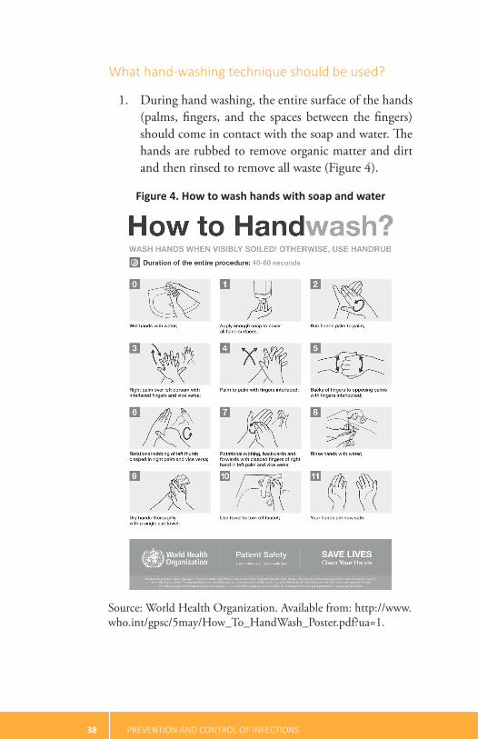
1. During hand washing, the entire surface of the hands (palms, fingers, and the spaces between the fingers) should come in contact with the soap and water. The hands are rubbed to remove organic matter and dirt and then rinsed to remove all waste (Figure 4).
Figure 4. How to wash hands with soap and water
Source: World Health Organization. Available from: http://www.who.int/gpsc/5may/How_To_HandWash_Poster.pdf?ua=1.

BASIC RECOMMENDATIONS 39
2. Jewelry (hoop earrings, rings, watches). Although wearing jewelry is not a good clinical practice, since it makes correct hand washing more difficult, studies that have evaluated its association with higher rates of HAIs and disease outbreaks have had inconsistent results. While some studies have associated the use of jewelry with increased colonization by infectious agents such as Enterobacteriaceae and reduced hand hygiene effectiveness [19, 20, 21], others have not found such an association or a higher risk of microor-ganism transmission via the hands. In fact, no studies have linked wearing jewelry to an increased incidence of HAIs or epidemic outbreaks [22, 23, 24, 25]. Therefore, local decisions on wearing or not wearing jewelry should take into account the perception of risk by users, the cultural acceptance of the practice, and the potential risk of perforating gloves, especially in the case of rings that are not flat.
3. Fingernails. No association has been demonstrat-ed between HAIs and the use of nail polish or arti-ficial nails. Although some studies have linked nail polish and artificial nails to increased colonization by Gram-negative bacteria and fungi and to reduced hand hygiene effectiveness [26, 27], other investiga-tions have not corroborated these results [22]. Other studies have examined the link between the use of nail polish or artificial nails and outbreaks of HAIs in high-risk units (intensive care units, neonatology units, and spinal and heart surgery wings), but no clear mecha-nisms of transmission could be found. Nor could it be determined whether any apparent risk was directly associated with the practice or, rather, with a failure to follow other standard precautions, difficulty in

40 PREVENTION AND CONTROL OF INFECTIONS
verifying hand hygiene, variations in the permeability of gloves, inapparent lesions or infections of the nails, and so forth [1, 6, 28, 29, 30]. It makes sense to avoid wearing artificial nails, especially when health workers are treating patients in high-risk units (intensive care, neonatology, surgery wing) or in a clinical unit that is dealing with an epidemic outbreak. In any case, as with the use of rings, it is important to consider the perception of risk by users and cultural acceptance of the practice.
Use of alcohol-based solutions
Hand hygiene with alcohol-based solutions is subject to certain special conditions depending on the products used, their availability, and access to them when they are needed. If alcohol-based solutions are used, there is no need for oth-er installations.
What properties should alcohol-based solutions have?
These solutions should have a broad-spectrum effect; should be fast acting, non-toxic, unaltered by environmental fac-tors, odorless, economical, user-friendly, and fast drying; and should have appropriate viscosity.
What characteristics should alcohol-based solutions have?
§ Alcohol-based solutions for topical use to disinfect the hands (with alcohol concentrations of 60% to 95%) have an immediate antimicrobial effect when

BASIC RECOMMENDATIONS 41
they come in contact with a bacterium because they denature the bacterial proteins. Their bactericidal ef-fect is 3.2 to 5.8 log10 CFUs in 10 seconds (Table 4).
Table 4. Characteristics of the main hand hygiene products
Characteristic Soap without antiseptic
Antiseptic soap
Alcohol solutions
Eliminates or-ganic matter
Yes Yes No
Eliminates bac-teria (in vivo)
Good Good Very good
Estimated length of the procedure
1 to 2 minutes 1 to 2 minutes 30 seconds
Cost Very low Low Very low
Site where performed
Sink Sink Anywhere
Requires towel to dry hands
Yes Yes No
Requires an installation
Yes Yes No
Effects on the skin
Very rare Rare Very rare
Flammable No No Yes
Meets at least 40% of hand hygiene requirements
Rarely Rarely Possibly
Adapted from: Widmer AF. Replace hand washing with use of a waterless alcohol hand rub? Clin Infect Dis. 2000;31(1):136-43.

42 PREVENTION AND CONTROL OF INFECTIONS
§ Alcohol-based solutions do not clean. Thus, the hands should be free of visible dirt. When they are visibly soiled, they should be washed and dried before applying the alcohol solution. For this reason, the two procedures, hand washing and application of an alco-hol-based solution, are complementary; one does not take the place of the other.
§ There are many available products, usually in the form of a gel or hydroalcoholic foaming solution, and their alcohol concentration ranges from 60% to 95%. The gel or foam solutions can be flammable, so it is important not to expose the hands to fire or sparks until they are fully dry. The characteristics that affect acceptance by users are scent, drying time, residual viscosity, and drying effect on the skin. The last-men-tioned effect is not very common because emollients are added in the commercially available solutions.
What factors should be considered with regard to availability of and access to these solutions?
§ Alcohol-based solutions should be placed near where a patient is being treated, preferably beside the pa-tient’s bed or where medication is being prepared. Installing wall-mounted dispensers far from the treat-ment area, such as near a sink, in a hall, or at the entrance to a room, makes them less likely to be used, since personnel have to go out of their way to access them. Thus, one of the main advantages of using the method—namely, the short procedure time—is lost. Placing them far away also sends unclear messages that could lead to incorrect procedures. For example,
BASIC RECOMMENDATIONS 43
placing the alcohol-based solution near a washstand might suggest the need to disinfect the hands with the solution immediately after washing with soap and water.
§ Alcohol-based solutions come in small sizes for indi-vidual use and larger containers with a pump. If pos-sible, they should not be refilled. If they are going to be refilled, the container will have to be cleaned to eliminate any residue and then dried. A container that still has solution in it should never be refilled.
§ A solution’s validity period will vary depending on the manufacturer and the product. Therefore, the expira-tion date should be checked in each case.
What scrubbing/rubbing technique should be used with alcohol-based solutions?
§ If the hands have visible dirt or have been in direct contact with body fluids, it will first be necessary to wash them with soap and water and dry them.
§ Once the hands are dry and have no visible dirt, a squirt of the solution approximately 1 milliliter thick (a sufficient amount to spread over the entire hand) is placed in the palm and then rubbed until all of the surfaces of the hands (palms, fingers, backs) are in contact with the solution. Continue to rub until it dries (Figure 5).
44 PREVENTION AND CONTROL OF INFECTIONS
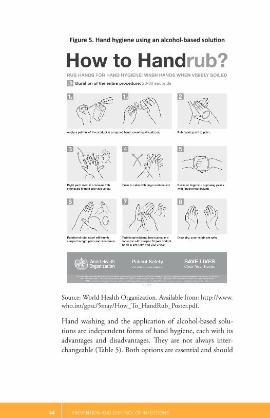
Figure 5. Hand hygiene using an alcohol-based solution
Source: World Health Organization. Available from: http://www.who.int/gpsc/5may/How_To_HandRub_Poster.pdf.
Hand washing and the application of alcohol-based solu-tions are independent forms of hand hygiene, each with its advantages and disadvantages. They are not always inter-changeable (Table 5). Both options are essential and should
BASIC RECOMMENDATIONS 45
always be available to improve adherence to an institution’s practices.
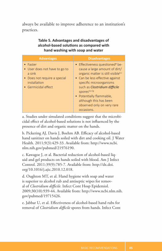
Table 5. Advantages and disadvantages of alcohol-based solutions as compared with
hand washing with soap and water
Advantages Disadvantages
• Faster • User does not have to go to
a sink • Does not require a special
installation • Germicidal effect
• Effectiveness questioneda be-cause a large amount of dirt/organic matter is still visibleb,c
• Can be less effective against specific microorganisms such as Clostridium difficile sporesd,e,f,g
• Potentially flammable, although this has been observed only on very rare occasions.
a. Studies under simulated conditions suggest that the microbi-cidal effect of alcohol-based solutions is not influenced by the presence of dirt and organic matter on the hands.
b. Pickering AJ, Davis J, Boehm AB. Efficacy of alcohol-based hand sanitizer on hands soiled with dirt and cooking oil. J Water Health. 2011;9(3):429-33. Available from: http://www.ncbi.nlm.nih.gov/pubmed/21976190.
c. Kawagoe J, et al. Bacterial reduction of alcohol-based liq-uid and gel products on hands soiled with blood. Am J Infect Control. 2011:39(9):785-7. Available from: http://dx.doi.org/10.1016/j.ajic.2010.12.018.
d. Oughton MT, et al. Hand hygiene with soap and water is superior to alcohol rub and antiseptic wipes for remov-al of Clostridium difficile. Infect Cont Hosp Epidemiol. 2009;30(10):939-44. Available from: http://www.ncbi.nlm.nih.gov/pubmed/19715426.
e. Jabbar U, et al. Effectiveness of alcohol-based hand rubs for removal of Clostridium difficile spores from hands. Infect Cont

46 PREVENTION AND CONTROL OF INFECTIONS
Hosp Epidemiol. 2010;31(6):565-70. Available from: http://www.ncbi.nlm.nih.gov/pubmed/20429659.
f. Boyce JM, et al. Lack of association between the increased incidence of Clostridium difficile-associated disease and the in-creasing use of alcohol-based hand rubs. Infect Cont Hosp Epidemiol. 2006;27(5):479-83. Available from: http://www.ncbi.nlm.nih.gov/pubmed/16671029.
g. No association has been found between increased use of alcohol-based solutions for hand hygiene and increased incidence of diarrheal disease from Clostridium difficile. If avoiding the use of alcohol-based solutions reduces overall adherence to hand hygiene, this negative impact should be taken into account in the final decision.
When should hand hygiene be done?
Just as important as good technique is performing hand hy-giene at the proper time—namely, when the likelihood of contamination and carrying infectious agents to a portal of entry on a susceptible host is highest. Several situations, or “moments,” have been identified as important times to dis-infect the hands. The World Health Organization (WHO) promotes five widely recognized key moments for hand hy-giene (Figure 6).
Figure 6. Five moments for hand hygiene recommended by the World Health Organization

BASIC RECOMMENDATIONS 47
What is known about conditions that affect adherence to hand hygiene?
§ Studies conducted in intensive care units have shown that, in general, the level of hand-washing adherence is about 40%. Compliance varies with the individu-al health worker and the workload. Nursing person-nel show greater compliance than other health team members. Higher workloads reduce compliance [2].
§ Incorporation of alcohol-based solutions has been shown to increase hand hygiene adherence by nearly 55%. According to one study, the use of alcohol-based solutions increased over time from 5% to 22% with-out reductions in hand washing [31].
§ Hugonnet et al. [31] observed a preference for alco-hol-based solutions over hand washing with soap and water. The preference was related to the following factors: being a physician, performing high-risk pro-cedures (e.g., touching blood vessels, changing from one site of activity to another on the same patient), and having to perform many procedures per hour.
What strategies can help to encourage hand hygiene?
A number of different strategies have been investigated to improve compliance with and adherence to hand hygiene, as well as sustainability over time. Two important strategies are (1) education of personnel and (2) education of patients, family members, and visitors.
1. education of personnel One successful experience [32] started with a brief training session that included

48 PREVENTION AND CONTROL OF INFECTIONS
handing out alcohol-based solutions for individual use. Following the training, one of two additional in-terventions that had yielded good results in the past was added to the program. The two interventions will be called the “Geneva intervention” and the “Wash-ington intervention,” after the locations where they were conducted the first time. The interventions are described in Table 6.
Table 6. Training strategies to improve hand hygiene adherence
Geneva Interventiona Washington Interventionb
Components: 1. Use of color posters (30 × 42
cm) on hand hygiene in treat-ment rooms, as well as in places where personnel pass when they are in transit, with messages on (1) the purpose of hand hygiene, (2) when it should be done, and (3) the recommended technique.
2. A brief staff training session on the use of alcohol-based solutions.
3. Distribution of individual bot-tles of alcohol-based solution.
4. Supervision of compliance via external observation, with immediate individual feed-back on the findings without punitive consequences.
Components: 1. Participation of personnel in
the facility where improved compliance is desired
2. Also, in the same workplace, participation of the clinical team in looking for and proposing ways to improve and organize hand hygiene, depending on the duties of the personnel and the circumstances in the facility; based on local experience.
(cont. next page)

BASIC RECOMMENDATIONS 49
Geneva Interventiona Washington Interventionb
This intervention was associat-ed with a 56% increase in the use of alcohol-based solutions, particularly in infectious patient rooms. However, the same improvement was not observed in internal medicine areas.
This intervention was associated with a 48% increase in the use of alcohol-based solutions that was sustained until a subse-quent evaluation two years later.
a. Larson EL, et al. An organizational climate intervention as-sociated with increased handwashing and decreased nosocomial infections. Behav Med. 2000;26(1):14-22.
b. Pittet D, et al. Effectiveness of a hospital-wide programme to improve compliance with hand hygiene: Infection Control Programme. Lancet. 2000;356(9238):1307-12.
A systematic review of the literature found greater adher-ence to hand hygiene programs that included multimode interventions along at least five simultaneous intervention components (Table 7).
Table 7. Components of multimode interventions and description
Intervention component
Description
Structure Ensuring and supervising adequate access to water with adjustable temperature, soap (with or without antiseptic), and alcohol-based solutions for hand hygiene. “Adequate access” refers to availability of the requirements described in the previous para-graphs, proximity to the area where patients are being treated, and sufficient resources to ensure continuity.It will be necessary to remove physical and pro-cess-related obstacles that might hinder access to installations.
(cont. next page)

50 PREVENTION AND CONTROL OF INFECTIONS
Intervention component
Description
Education and training
Actively and regularly disseminating information to health workers on the importance of hand hygiene and proper technique. The health team’s hand hygiene practice should be strengthened by presenting the evidence for endorsing it and by scheduling repeated individu-al or group training programs. This activity can be held either at the workplace or elsewhere (face-to-face or through distance learning). Conducting it in the workplace provides the opportunity to assess the presence of barriers on site; prefer-ences for certain soaps, drying methods, and alcohol-based solutions; and whether there is convenient access to installations for performing hand hygiene. It can facilitate adaptation to the real situation and the preferences of personnel. On-site training also encourages personnel to develop the necessary skills.
Feedback Monitoring adherence to the practice and providing regular feedback to the people on the health team. Feedback can be provided through field and supervisory visits to directly observe compliance with hand hygiene (when to perform it and the technique) as part of a program that encom-passes all areas of patient care. Emphasis should be placed on high-risk areas (emergency room, intensive care unit, neonatology unit), direct or indirect supervision of indicators of compliance and correct times for hand hygiene (use of soap or alcohol-based solutions in the different areas), and the application of technology.a,b Various sys-tems can be used, such as counting the number of times the alcohol or soap was dispensed, elec-tronic monitoring with a camera, or immediate feedback.
(cont. next page)

BASIC RECOMMENDATIONS 51
Intervention component
Description
Reminders in the workplace
Disseminating messages in the workplace that emphasize the importance of hand hygiene and correct practice, using graphics, posters, auditory material, e-mail, and other methods. In one study, an attempt was made to involve patients in the methods mentioned through reminders to health workers of when they should use hand hygienec; but however, there is no evi-dence on the impact of this intervention.
Climate of institutional safety
Enlisting management teams in promoting institutional engagement in the prioritization of hand hygiene as a strategy for the prevention and control of HAIs.
Adapted from: Luangasanatip N, et al. Comparative efficacy of interventions to promote hand hygiene in hospital: systemat-ic review and network meta-analysis. BMJ. 2015;351:h3728. Available from: http://www.bmj.com/lookup/doi/10.1136/bmj.h3728.
a. Srigley JA, et al. Hand hygiene monitoring technology: a systematic review of efficacy. J Hosp Infect. 2015;89(1):51-60. Available from: http://linkinghub.elsevier.com/retrieve/pii/S019567011400320X.
b. Ward M, et al. Automated and electronically assisted hand hygiene monitoring systems: a systematic review. Am J Infect Control. 2014;42(5):472-8. Available from: http://dx.doi.org/10.1016/j.ajic.2014.01.002.
c. Davis R, et al. Systematic review of the effectiveness of strat-egies to encourage patients to remind healthcare professionals about their hand hygiene. J Hosp Infect. 2015;89(3):141-2. Available from: http://linkinghub.elsevier.com/retrieve/pii/S0195670114003752.
The five intervention components described above had bet-ter results when three additional components were included:

52 PREVENTION AND CONTROL OF INFECTIONS
§ Targets. Progressive hand hygiene adherence targets were established for both the health team as a whole and the individual health workers.
§ Accountability. Strategies were included for sharing the individual and group results and their conse-quences with the health workers.
§ Incentives and rewards. These were granted based on the achievement of expected results. They were eco-nomic or in some other form.
Table 8. Assessment of results using multimodal strategies
Strategy Odds ratio (confidence interval = 95%) for
achievement of hand hygiene
No strategy or the customary practice
1.0
One intervention component 4.30 (0.43 to 46.57)
Five intervention components 6.51 (1.58 to 31.91)
Five intervention components + one supplementary component
11.83 (2.67 to 53.79)
Adapted from: Luangasanatip N, et al. Comparative efficacy of interventions to promote hand hygiene in hospital: systemat-ic review and network meta-analysis. BMJ. 2015;351:h3728. Available from: http://www.bmj.com/lookup/doi/10.1136/bmj.h3728.
2. education of patients, family members, and visitors. Three different perspectives have been used in assessing strategies for educating and engaging pa-tients, family members, and visitors in hand hygiene:
§ Participants are made responsible for remind-ing members of the health team when their hands should be disinfected in the workplace, either as the

BASIC RECOMMENDATIONS 53
only strategy or as part of a multimodal strategy. A systematic review of the literature [34] found that this approach had favorable results when measured according to patients’ intention to remind health workers that it was time to use hand hygiene. This was especially true when patients were involved in designing the strategy, as well as when health work-ers actively promoted the practice among patients. However, the impact of this intervention on hand hygiene adherence is unknown.
§ As part of the strategy for assessing the timeliness of health workers’ hand hygiene, rather than imme-diate direct feedback being provided, special forms are designed for patients to report the information to the officials responsible for controlling HAIs in hospitals. Although the impact of this intervention is unknown, it has been seen to facilitate patient and visitor involvement, since it avoids the discom-fort and perception of insecurity that some patients might feel if they have to face health workers direct-ly, especially physicians [35].
§ Patients and their companions are seen not only as reservoirs in the chain of transmission but also as ve-hicles of transmission of infectious agents. Although transmission of HAIs through the hands of patients and their companions tends to be considered less important than transmission through the hands of health workers, some studies have shown low levels of adherence to hand hygiene by patients and their family members and visitors, which represents a po-tential risk for transmission [36, 37, 38]. Studies on strategies for improving hand hygiene in patients and their companions are still inconclusive, as is

54 PREVENTION AND CONTROL OF INFECTIONS
evidence of the impact of these practices on HAIs, but there is agreement on the need for both health workers and health institutions to provide the means for hand hygiene to patients and their companions [39, 40, 41, 42].
Summary
Compliance with and adherence to hand hygiene practices are affected by a number of factors. The following condi-tions contribute to success:
1. Supplies are well stocked and installations are in working order, easily accessible, and located near the sites where patients receive care.
2. Training has been provided and personnel know how to disinfect their hands and how to use the products and installations. They also know the moments when this practice is required (see the WHO five moments described above).
3. Compliance is assessed regularly to identify the fac-tors that may hinder or favor it.
4. Successful experiences have been adopted and adapt-ed, and their impact has been assessed in the local setting.
5. The characteristics of the supplies and installations have been examined with a view to improving accep-tance by personnel.
6. Multimodal strategies have been incorporated to promote hand washing and the use of alcohol-based solutions as supplementary measures in addition to

BASIC RECOMMENDATIONS 55
the standard precautions for infection control, not as isolated measures.
7. Personnel participate in decisions to improve com-pliance with hand hygiene insofar as possible, tak-ing into account their duties, their adaptation to the working environment, and the available products and installations.
References
1. McNeil SA, Foster CL, et al. Effect of hand cleansing with antimicrobial soap or alcohol-based gel on microbial colo-nization of artificial fingernails worn by health care workers. Clin Infect Dis. 2001;32(3):36-72.
2. World Health Organization. WHO guidelines on hand hy-giene in health care: first global patient safety challenge—clean care is safer care. Geneva: WHO; 2009. Available from: http://whqlibdoc.who.int/publications/2009/9789241597906_eng.pdf
3. Cassettari VC, et al. Risk factors for colonization of newborn infants during an outbreak of extended-spectrum β-lact-amase-producing Klebsiella pneumoniae in an intermedi-ate-risk neonatal unit. J Hosp Infect. 2009;71(4):340-7.
4. Moolenaar RL, et al. A prolonged outbreak of Pseudomonas aeruginosa in a neonatal intensive care unit: did staff finger-nails play a role in disease transmission? Infect Control Hosp Epidemiol. 2000 Feb;21(2):80-5. Available from: http://www.ncbi.nlm.nih.gov/pubmed/10697282.
5. Cantey JB, et al. Prompt control of an outbreak caused by extended-spectrum β-lactamase-producing Klebsiel-la pneumoniae in a neonatal intensive care unit. J Pediatr. 2013;163(3):672-9.e1-3.
6. Gupta A, et al. Outbreak of extended-spectrum beta-lac-tamase-producing Klebsiella pneumoniae in a neonatal
56 PREVENTION AND CONTROL OF INFECTIONS
intensive care unit linked to artificial nails. Infect Cont Hosp Epidemiol. 2004;25(3):210-5. Available from: http://www.ncbi.nlm.nih.gov/pubmed/15061412.
7. World Health Organization. WHO guidelines for drinking- water quality. Geneva: WHO; 2008. Available from: http://apps.who.int/iris/bitstream/10665/44584/1/ 9789241548151_eng.pdf.
8. Kabara JJ, Brady MB. Contamination of bar soaps under “in-use” conditions. J Environ Pathol Toxicol Oncol. 1984;5(4-5):1-14. Available from: http://www.ncbi.nlm.nih.gov/pubmed/6394740.
9. McBride ME. Microbial flora of in-use soap products. Appl Environ Microb. 1984;48(2):338-41.
10. Heinze JE, Yackovich F. Washing with contaminated bar soap is unlikely to transfer bacteria. Epidemiol Infect. 1988;101(1):135-42. Available from: http://www.ncbi.nlm.nih.gov/pubmed/3402545.
11. Zapka CA, et al. Bacterial hand contamination and trans-fer after use of contaminated bulk-soap-refillable dispens-ers. Appl Environ Microb. 2011;77(9):2898–904. Available from: http://aem.asm.org/content/77/9/2898.full.
12. Rabier V, et al. Hand washing soap as a source of neonatal Ser-ratia marcescens outbreak. Acta Paediatr. 2008;97(10):1381-5.
13. Buffet-Bataillon S, et al. Outbreak of Serratia marcescens in a neonatal intensive care unit: contaminated unmedicated liq-uid soap and risk factors. J Hosp Infect. 2009;72(1):17-22.
14. Lanini S, et al. Molecular epidemiology of a Pseudomonas aeruginosa hospital outbreak driven by a contaminated disin-fectant-soap dispenser. PLoS ONE. 2011;6(2):e17064.
15. Sartor C, et al. Nosocomial Serratia marcescens infections associated with extrinsic contamination of a liquid nonmed-icated soap. Infect Cont Hosp Epidemiol. 2000;21(3):196-9. Available from: http://www.ncbi.nlm.nih.gov/pubmed/ 10738989.

BASIC RECOMMENDATIONS 57
16. Archibald LK, et al. Serratia marcescens outbreak associated with extrinsic contamination of 1% chlorxylenol soap. Infect Cont Hosp Epidemiol. 1997;18(10):704-9. Available from: http://www.ncbi.nlm.nih.gov/pubmed/9350463.
17. Doebbeling BN, et al. Removal of nosocomial pathogens from the contaminated glove: implications for glove reuse and handwashing. Ann Intern Med. 1988;109(5):394-8. Avail-able from: http://www.ncbi.nlm.nih.gov/pubmed/3136685.
18. Huang C, Ma W, Stack S. The hygienic efficacy of different hand-drying methods: a review of the evidence. Mayo Clin Proc. 2012:87(8):791-8.
19. Kelsall NKR, et al. Should finger rings be removed prior to scrubbing for theatre? J Hosp Infect. 2006;62(4):450-2.
20. Yildirim I, et al. A prospective comparative study of the rela-tionship between different types of ring and microbial hand colonization among pediatric intensive care unit nurses. Int J Nurs Stud. 2008;45(11): 1572-6. Available from: http://www.ncbi.nlm.nih.gov/pubmed/18479684.
21. Fagernes M, Lingaas E, Bjark P. Impact of a single plain finger ring on the bacterial load on the hands of healthcare workers. Infect Control Hosp Epidemiol. 2007;28(10):1191-5. Avail-able from: http://www.ncbi.nlm.nih.gov/pubmed/17828698.
22. Fagernes M, Lingaas E. Impact of finger rings on transmis-sion of bacteria during hand contact. Infect Control Hosp Epidemiol. 2009;30(5):427-32. Available from: http://www.ncbi.nlm.nih.gov/pubmed/19344265
23. Salisbury DM, et al. The effect of rings on microbial load of health care workers’ hands. Am J Infect Control. 1997;25(1):24-7.
24. Waterman TR, et al. Comparison of bacterial counts in glove juice of surgeons wearing smooth band rings versus those without rings. Am J infect Control. 2006;34(7):421–5. Available from: http://www.sciencedirect.com/science/ article/pii/S0196655306000800.

58 PREVENTION AND CONTROL OF INFECTIONS
25. Trick WE, et al. Impact of ring wearing on hand contami-nation and comparison of hand hygiene agents in a hospi-tal. Clin Infect Dis. 2003;36(11):1383-90. Available from: http://www.ncbi.nlm.nih.gov/pubmed/12766832.
26. McNeil SA, Nordstrom-Lerner L, et al. Outbreak of ster-nal surgical site infections due to Pseudomonas aeruginosa traced to a scrub nurse with onychomycosis. Clin Infect Dis. 2001;33(3):317-23.
27. Hedderwick SA, et al. Pathogenic organisms associated with artificial fingernails worn by healthcare workers. Infect Con-trol Hosp Epidemiol. 2000;21(8):505-9. Available from: http://www.ncbi.nlm.nih.gov/pubmed/10968715.
28. Parry MF, et al. Candida osteomyelitis and diskitis after spi-nal surgery: an outbreak that implicates artificial nail use. Clin Infect Dis. 2001;32(3):352-7. Available from: http://www.ncbi.nlm.nih.gov/pubmed/11170941.
29. Passaro DJ, et al. Postoperative Serratia marcescens wound infections traced to an out-of-hospital source. J Infect Dis. 1997;175(4):992-5.
30. Gordin FM, et al. A cluster of hemodialysis-related bacte-remia linked to artificial fingernails. Infect Cont Hosp Epi-demiol. 2007;28(6):743-4. Available from: http://www.ncbi.nlm.nih.gov/pubmed/17520554.
31. Hugonnet S, Perneger TV, Pittet D. Alcohol-based handrub improves compliance with hand hygiene in intensive care units. Arch Intern Med. 2002;162(9):1037-43.
32. Whitby M, et al. Three successful interventions in health care workers that improve compliance with hand hygiene: is sustained replication possible? Am J Infect Control. 2008;36(5):349-55.
33. Luangasanatip N, et al. Comparative efficacy of interven-tions to promote hand hygiene in hospital: systematic review and network meta-analysis. BMJ. 2015;351:h3728. Avail-able from: http://www.bmj.com/lookup/doi/10.1136/bmj.h3728.
BASIC RECOMMENDATIONS 59
34. Davis R, et al. Systematic review of the effectiveness of strate-gies to encourage patients to remind healthcare professionals about their hand hygiene. J Hosp Infect. 2015;89(3):141-62. Available from: http://linkinghub.elsevier.com/retrieve/pii/S0195670114003752.
35. Bittle MJ, LaMarche S. Engaging the patient as observer to promote hand hygiene compliance in ambulatory care. Jt Comm J Qual Patient Saf. 2009;35(10):519-25. Available from: http://www.ncbi.nlm.nih.gov/pubmed/19886091.
36. Banfield KR, Kerr KG. Could hospital patients’ hands con-stitute a missing link? J Hosp Infect. 2005;61(3):183-8.
37. Istenes N, et al. Patients’ potential role in the transmission of health care-associated infections: prevalence of contamination with bacterial pathogens and patient attitudes toward hand hygiene. Am J Infect Control. 2013;41(9):793-798. Avail-able from: http://dx.doi.org/10.1016/j.ajic.2012.11.012.
38. Randle J, Arthur A, Vaughan N. Twenty-four-hour observa-tional study of hospital hand hygiene compliance. J Hosp Infect. 2010;76(3):252-5. Available from: http://dx.doi.org/10.1016/j.jhin.2010.06.027.
39. Chen YC, Chiang LC. Effectiveness of hand-washing teach-ing programs for families of children in paediatric intensive care units. J Clin Nurs. 2007;16(6):1173-9.
40. Ardizzone LL, et al. Patient hand hygiene practices in surgical patients. Am J Infect Control. 2013;41(6):487-91. Available from: http://dx.doi.org/10.1016/j.ajic.2012.05.029.
41. Burnett E, Lee K, Kydd P. Hand hygiene: what about our pa-tients? J Infect Prev. 2008;9(1):19-24. Available from: http://bji.sagepub.com/cgi/doi/10.1177/1469044607085549.
42. Whiller J, Cooper T. Clean hands: how to encourage good hygiene by patients. Nurs Times. 2000:96(46):37. Available from: https://www.nursingtimes.net/clean-hands-how-to-encourage-good-hygiene-by-patients/206059.article.

60 PREVENTION AND CONTROL OF INFECTIONS
B. Personal protective equipment (PPE)
What is personal protective equipment?
A group of items that can be used separately or in combi-nation, personal protective equipment is intended to form a barrier that prevents contact between health workers and a patient, object, or environment in order to prevent the transmission of infectious agents while providing care. It is called personal protective equipment because it can keep health workers from becoming infected or passing on mi-croorganisms from infected patients by protecting their various portals of entry (mucous membranes, airway, skin) from direct contact. It is important to distinguish the items used to protect personnel from the practices followed to pre-vent infections in patients or transmission between patients.
PPE should be used in combination with other control and prevention strategies. The decision to use it depends on the mode of transmission of the infection, which will determine, for example, whether only standard precautions apply or if isolation from contact with droplets or aerosols is required.
The recommendations on using this equipment are based on the opinions of experts, transmission mechanisms, the known portals of entry, perception of risk, severity of the disease, and other considerations. So far, no experimental studies have provided concrete information on its impact in specific circumstances [1, 2]. Assessing its effectiveness has been difficult because most of the studies have looked at combinations of different types of PPE or different types of PPE in combination with other standard precautions. These
BASIC RECOMMENDATIONS 61
combinations make it impossible to interpret the individual effect of each item, particularly with regard to the use of masks, eyeglasses, or face shields [3, 4, 5].
What general points should be considered in selecting PPE for an institution?
Each PPE item may come in a variety of designs, be made of different materials, or have different properties. In select-ing PPE items, the following two points should be consid-ered: (1) previous knowledge about the item among per-sonnel and their familiarity with its use and (2) compliance with national regulations or codes that impose minimum requirements in the technical specifications of the equip-ment—for example, certification of the manufacturing process, impermeability, or impenetrability with respect to certain pathogens [6, 7, 8].
What are the different PPE items/components?
The main components of PPE—in other words, the items most frequently used—are gloves, gowns, and waterproof aprons; eye protection (glasses, goggles, face shields); and devices to protect the mucous membranes of the mouth (masks, face shields) (Table 9).

62 PREVENTION AND CONTROL OF INFECTIONS
Table 9. Types of personal protective equipment
Item Generic item illustration
Gowns
Safety eyewear: glasses, wrap-arounds, goggles
Face protectors, face shields
Respirators
Masks
Gloves
(cont. next page)

BASIC RECOMMENDATIONS 63
Item Generic item illustration
Waterproof aprons
§ gloves. One of the main PPE components, gloves are intended to prevent contact of the skin on the hands with contaminated sources such as the skin of patients colonized or infected with multiresistant microorganisms, blood, or body fluids and to avoid colonization of the hands by microbial flora from pa-tients [9]. They may differ in the following respects: sterility (sterile or non-sterile), the material they are made of (latex, nitrile, vinyl), size, and length (only to the wrist or covering the entire forearm).
What criteria need to be considered in selecting gloves?
§ Sterility: The decision will depend on the type of procedure for which the gloves are to be used. If the procedure involves an aseptic technique, they should always be sterile. Only non-sterile or clean gloves are needed for routine patient care procedures.
§ Impermeability
§ Type of material
§ Flexibility or rigidity of the material
§ Risk of triggering allergies (hypoallergenic material)
§ Adjustability of length to cover wrist or forearm
§ Size

64 PREVENTION AND CONTROL OF INFECTIONS
How does the risk for HAI transmission affect the com-position of the gloves?
There is no available evidence linking the risk for HAIs with any specific material (latex versus other materials). Studies conducted under laboratory conditions have found that vi-nyl has less of a barrier effect than nitrile or latex, which means that it has a smaller protective effect in the event of percutaneous exposure [9, 10]. At the same time, a clinical study that compared different types of gloves used in operat-ing rooms/suites and examined the risk of exposure to sharp instruments in surgical procedures lasting longer than six hours found that gloves made of nitrile and neoprene were more likely to develop defects or visible breaks than those made of latex [11]. However, this clinical study did not find that any of the gloves had a lesser protective effect against percutaneous exposure [12].
The use of double-layer gloves is evaluated in terms of pre-venting infection of the surgical wound and use in the oper-ating room/suite, but that topic is beyond the scope of the present document.
recommendations
§ When use of long-sleeve gowns is indicated, place gloves on top of the cuff of the gown.
§ Always change gloves between patients.
§ Use gloves only when necessary; excessive use can cause certain types of dermatitis and increase sensitiv-ity to latex.
§ Perform hand hygiene immediately before and, even more important, after using gloves. Under no circum-stance should glove use replace hand hygiene.

BASIC RECOMMENDATIONS 65
§ It may be necessary to change gloves while caring for the same patient when different activities are required and the gloves become contaminated.
§ Change gloves whenever they develop breaks or tears.
What precautions should be taken when putting on and removing gloves?
Ongoing training and supervision are necessary to ensure that the correct procedure is followed in putting on and, es-pecially, taking off gloves. Studies have shown that improper technique in removing gloves can lead to contamination of the hands and spread of fluids and microorganisms to the health worker’s clothes, the environment, and people near-by, thus creating a potential mechanism for transmitting microorganisms capable of surviving in the environment [13]. The hands become contaminated when the gloves are removed, regardless of whether or not the gloves have any visible perforations [14]. Since it is impossible to ensure that gloves will remain uncontaminated while they are be-ing used, and since there is a high likelihood that the health worker will not notice all perforations, breaks, or tears, hand hygiene after removing gloves must always be performed, even if the hands do not appear to be soiled; this is especially true if gloves are going to be used with the next patient [9, 15].
It is not recommended that gloves be sterilized and reused because it is difficult to ensure that they are not contaminat-ed and the cost-benefit ratio is insignificant.
§ gowns and aprons. A gown is an item of apparel, usually made of textile (disposable or reusable), plas-tic, or paper, that covers the body from the neck to the knees and the arms down to the wrists. It has an

66 PREVENTION AND CONTROL OF INFECTIONS
opening and closing mechanism, usually located in the back. Some gowns have short sleeves and some open and close in the front. Their purpose is to keep the health worker’s regular clothes from becoming contaminated or soiled during procedures that might create blood spatters, secretions, or excretions. Their use is also indicated when the procedure to be per-formed could place most of the health worker’s cloth-ing in contact with patients carrying specific microor-ganisms or with surfaces near patients.
On the other hand, an apron is made of waterproof material and covers the front of the body from the neck to the knees but not the arms. It should be used only as a second, waterproof barrier on top of the gown during procedures that generate a large volume of blood or body fluids.
Types of gowns
§ Gowns made of cotton or linen. Since they are permeable, they should be used only when a low vol-ume of patient secretions, blood, or body fluids is expected.
§ Gowns made of plastic. They are usually waterproof, although some do not meet this requirement. If per-sonnel expect to be exposed to a high volume of flu-ids, waterproof aprons should be used.
§ Sterile gowns. These are intended for use when aseptic techniques are followed during invasive procedures. For other procedures, non-sterile gowns should be used.
BASIC RECOMMENDATIONS 67
How does risk for HAI transmission affect the composi-tion of gowns?
In studies that have assessed the risk of operative wound infections, no differences have been found between reusable sterile gowns made of washable cotton or linen and sterile gowns made of plastic [16, 17]. To this point, when gowns are used to protect personnel from exposure to pathogens in blood and body fluids or to prevent cross-transmission between patients via health workers, the effect of the gown-ing material has not been determined. Most of the available research is in the form of laboratory studies, and the results vary depending on the material, the length of time it was used, and the pressure applied to test its permeability. Fur-thermore, a number of authors consider literature reviews conducted before 2000 [18] as no longer being valid be-cause there have been so many recent changes in technology and manufacturing standards and regulations.
The following factors should be considered in decisions re-garding whether to select disposable or waterproof apparel: (1) cost (reusable gowns for use by each individual patient and the cost of laundering them), (2) demonstrated im-permeability of the specific material used, (3) user comfort (disposable materials tend to be less comfortable because of body temperature), (4) design, and (5) risk and impact of exposure to microorganisms [19, 20].
With regard to design, the choice of sleeve length—whether short sleeves or long sleeves with elastic cuffs—will depend on the risk of exposure. Usually, if the possibility of body fluid spattering or spilling is high, long sleeves will need to be used to keep fluids from landing on uncovered skin.
68 PREVENTION AND CONTROL OF INFECTIONS
Types of waterproof aprons
§ Disposable versus reusable
§ Rigid or flexible material
Special attention should be paid to how the garment fastens. It should be easy to put on and, more importantly, to take off so that the user does not come in contact with fluids when it is being removed. In the case of reusable aprons, consideration should be given to how they are cleaned and disinfected.
§ protection of the facial mucous membranes (mouth, nose, and conjunctiva). The conjunctiva and the mucous membranes of the nose and mouth are portals of entry for infectious agents. In certain circumstances, it is essential to protect them. In these cases, the barriers are mainly used to protect health workers during dental care or to provide isolation from contagion by aerosols, droplets, or any other risk of contact with body fluids coming directly or indirectly from a patient. The mucous membranes in the nose and mouth can be effectively protected from contact with body fluids or droplets and aero-sols by using a variety of protective elements alone or in combination. Specific indications are given in Chapter IV (additional precautions based on mode of transmission).
Types of mouth and nose protectors
§ Masks. These are non-occlusive devices that cover the health worker’s nose and mouth with the aim of re-ducing the likelihood of contact between the mucous membranes in these areas and the potentially infec-tious body fluids of another person. Masks come in a

BASIC RECOMMENDATIONS 69
variety of designs: for example, some are pleated and unfold over the mouth, while others are preformed. The most appropriate type should be selected based on the purpose and the comfort of the user. Masks that are not preformed moisten more easily and lend themselves to contact with the user’s mucous mem-branes. Although its impact has not been evaluated, this effect should be considered.
§ Respirators with particle filters. These devices also cover the mouth and the nose, but unlike a mask, they filter the air, thus reducing inhalation of particles and protecting personnel from airborne pathogens. In order to achieve their purpose, they have to create an occlusive seal around the nose and the mouth. They can act as filters for air that is breathed in (with or without valves to facilitate breathing in and out) or as artificial providers of clean air for a person who is isolated from the outside, as in autonomous systems [21].
What is a type N95 or FFP2 respirator?
Respirators with filters come in different types (R95, N99, N95), based on a combination of two characteristics [21, 22]:
§ Efficiency in filtering particles measuring as small as 0.1 to 0.3 µm (designated “95” if they remove 95% of such particles, “99” if they remove 99%, or “100” if they remove all particles).
§ Degree of oil resistance (series N is nonresistant, series R is resistant, and series P is oil-proof ).

70 PREVENTION AND CONTROL OF INFECTIONS
The initials FFP in the designation of some respirators stand for filtering facepiece and refer to their ability to filter parti-cles. In the health field, the most commonly used filter is the FFP2, which can filter 94% of particles measuring as small as 0.4 µm in diameter and is resistant to both oil-based and dry aerosols.
Are respirators as easy to use as masks?
All personnel should be familiar with and trained in the cor-rect placement and removal of both masks and respirators and should be able to distinguish between the two devices. In order for the filter on a respirator to function properly, air has to pass through it. This is important, because if oc-clusion around the face is not complete, the filter will not be effective.
Training in the use of respirators should include ensuring that personnel are able to perform the following:
§ Adjustment test. The purpose is to find out if air is leaking or the respirator is not filtering properly. This test must be performed in order to select the most appropriate type and size of respirator for the individ-ual being fitted. A person should use only the specif-ic respirator that successfully passed the adjustment test. International organizations suggest that the test be performed at least once a year, whenever a new type of respirator is introduced in a health facility, and whenever a physical change in the user might alter the balance between his or her face and the type and size of the respirator. There are several types of adjustment tests, usually divided into two broad categories [23]:
• Qualitative tests (Figure 7). Following one of several standardized protocols, the tester exposes the user

BASIC RECOMMENDATIONS 71
wearing the respirator to aerosols with characteris-tic scents to determine whether he or she can smell them. If the scent can be detected, it means that the respirator is not properly adjusted on the user’s face and needs to be switched to a different size or more appropriate model.
• Quantitative tests. The device’s effectiveness is eval-uated with special equipment that measures the number of particles inside and outside the respirator when it is in use.
Figure 7. Qualitative adjustment test
Source: OSHA et al. Hospital respiratory protection program toolkit: resources for respirator program administrators. May 2015.
• Seal check. The seal should be checked before each use of the respirator to ensure that it is working correctly before it touches the patient. If this is not done, there is no assurance that the respirator is properly filtering the inhaled air and therefore has the protective effect equivalent to that of a mask (Figure 8).

72 PREVENTION AND CONTROL OF INFECTIONS
Figure 8. Sequence of the seal check before each use
Source: WHO. Available from : http://www.who.int/csr/ resources/publications/SEALCHECK_EN_A2s.pdf?ua=1.
Masks and respirators are often used to prevent and con-trol infections due to respiratory viruses and other drop-let-transmitted agents that favor the nose or mouth as the portal of entry. There are no differences between masks and respirators in degree of protection against these agents. Res-pirators are associated with higher costs, user inconvenience,
1. Cup the respirator in your hand with the nosepiece at your fingertips allowing the headbands to hand freely below your hand.
2. Position the respirator under your chin with the nosepiece up.3. Pull the top strap over your head resting it high at the back of
your head. Pull the bottom strap over your head and position it around the neck below the ears.
4. Place fingertips of both hands at the top of the metal nose-piece. Mould the nosepiece (USING TWO FINGERS OF EACH HAND) to the shape of your nose. Pinching the nosepiece using one hand may result in less effective respirator perfor-mance.
5. Cover the front of the respirator with both hands, being care-ful not to disturb the position of respirator.a) Positive seal check: Exhale sharply. A positive pressure in-
side the respirator = no leakage. If leakage, adjust position and/or tension straps. Retest the seal. Repeat the steps until respirator is sealed properly.
b) Negative seal check: Inhale deeply. If no leakage, negative pressure will make respirator cling to your face. Leakage will result in loss of negative pressure in the respirator due to air entering through gaps in the seal.

BASIC RECOMMENDATIONS 73
and irritation of the skin on the face and neck [3,4]. Their use is indicated to prevent the transmission of infections caused by airborne microorganisms such as Mycobacterium tuberculosis.
how long can respirators be used without interruption? Can they be reused?
§ Long-term use of respirators. A respirator can be used continuously for up to about eight hours (the length of time may vary depending on the manufacturer’s specifications), as long as users do not touch its sur-face with their hands and seal checks are performed repeatedly to make sure it is working properly. Addi-tional protective measures (face shields) should also be used if it is expected that exposure to droplets will be high. Respirators should be discarded after use in the following circumstances: when they have been worn during a procedure highly likely to generate aerosols, when they are visibly contaminated with any body fluid, when the seal check is unsatisfactory, or when the user experiences a significant increase in re-sistance to respiratory effort.
§ Reuse of respirators. There is no consensus on the effectiveness of respirators in real conditions, nor are there any studies on the subject in the literature. Some authors, based on the results of a study con-ducted under controlled laboratory conditions [24],b suggest that if the same person reuses a respirator, it should not be used more than five times. After that, its filtration efficiency will decline significantly [25].
b. A total of 17 subjects participated in 20 sequences of placing and removing the respirator with seal checks each time. The study at-tempted to simulate a 10-hour shift with intermittent use of the res-pirator for one hour and pauses without the respirator between each one-hour period.

74 PREVENTION AND CONTROL OF INFECTIONS
Types of eye protection
§ Safety glasses or goggles. Unlike optical glasses (regu-lar eyeglasses), safety glasses prevent aerosols, spatters, and droplets from coming in contact with the con-junctival mucous membranes. Regular glasses cannot be used for this purpose because they do not have bar-riers to protect the conjunctival mucous membranes. If a health worker needs to use medically prescribed glasses, there are goggles that can be adapted to ac-commodate them. It is important for them to be ad-justed on the sides, under the eyes, and in the front. Also, the work area should have an indirect ventila-tion system to prevent the safety glasses from cloud-ing up and hindering the user’s vision.
§ Face screens or shields. Unlike glasses, these types of equipment cover the entire face, from the forehead to the chin, and the frontal and parietal area of the skull. They do not require additional eye protection or a mask to guard against droplet-transmissible agents. They have an adjustable fastening arrangement in the front that also blocks spatters. When they are used to prevent the transmission of infectious agents via droplets, they allow the user to speak to other health workers more clearly than with the usual mask.
what characteristics should eye protection (safety glasses, goggles, face shields) have beyond serving as a barrier?
§ It should not tarnish.
§ It should be easy to adjust around the nose and over the forehead.
§ It should be made of hypoallergenic material.

BASIC RECOMMENDATIONS 75
§ It should have a fastening system or an adjustable means of holding it in place.
§ It should be washable.
§ It should allow for use over optical glasses.
Other PPE: boots, jumpsuits, hoods
Other items that may be regarded as components of PPE are headgear, jumpsuits, coveralls, arrangements for covering the neck, hoods, and shoe covers. There are no studies show-ing that these products provide effective protection against infectious agents during the provision of healthcare. Rather, they are used to avoid soiling regular clothes or shoes.
what type of ppe should be used?
The type of equipment to be used and its specifications will mainly depend on the risk of exposure and user convenience.
1. Criteria for estimating the risk of exposure:
• The type of procedure to be performed or care to be administered. It is important to foresee the duration of contact with the patient and the degree of expo-sure to specific situations that involve a high risk of contamination, generation of aerosols, or contact with or handling of sharps.
• Any suspected presence of etiologic agents or infec-tions to be avoided. Knowledge of the natural histo-ry of the disease, as well as the mode of transmission of the agent, will dictate the combination of PPE elements to be used and for how long.
2. User convenience. Health facilities should keep on hand the types and sizes of PPE that may be required
76 PREVENTION AND CONTROL OF INFECTIONS
by the individual health workers, taking into account their physical frame and any special conditions (for example, personnel or patient allergy to latex, cli-mate). While using more than the minimum required PPE elements may give the impression of greater safety, it can hinder health workers’ performance and even expose them to greater risks during patient care while not providing additional protection [26]. There should be a balance between the desired protective ef-fect and the risks and drawbacks of incorporating too many elements.
In the regular care of any patient, the use of gloves should be considered when touching body parts that may be contami-nated. If spatters of secretions, stool, or blood are expected, facial protection and use of a gown or apron should be add-ed. When precautions are being taken with a patient based on mode of transmission, the personal protective equip-ment should be selected according to the type of isolation indicated.
how is ppe used in an institution?
1. The PPE elements should be available whenever they are needed. To maintain an adequate supply, it will be necessary to calculate the required stock levels in the sizes used by the health team that will be providing the care. For this purpose, the following steps are rec-ommended: (1) determine how many care procedures the patient will require over a given period of time (in the case of disposable PPE, allow for one garment or device per patient procedure for each health work-er; in the case of reusable gowns, estimate one gown per patient of the type regularly used by the health worker, or one gown per patient and one per type of
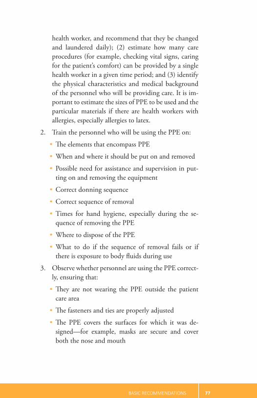
BASIC RECOMMENDATIONS 77
health worker, and recommend that they be changed and laundered daily); (2) estimate how many care procedures (for example, checking vital signs, caring for the patient’s comfort) can be provided by a single health worker in a given time period; and (3) identify the physical characteristics and medical background of the personnel who will be providing care. It is im-portant to estimate the sizes of PPE to be used and the particular materials if there are health workers with allergies, especially allergies to latex.
2. Train the personnel who will be using the PPE on:
• The elements that encompass PPE
• When and where it should be put on and removed
• Possible need for assistance and supervision in put-ting on and removing the equipment
• Correct donning sequence
• Correct sequence of removal
• Times for hand hygiene, especially during the se-quence of removing the PPE
• Where to dispose of the PPE
• What to do if the sequence of removal fails or if there is exposure to body fluids during use
3. Observe whether personnel are using the PPE correct-ly, ensuring that:
• They are not wearing the PPE outside the patient care area
• The fasteners and ties are properly adjusted
• The PPE covers the surfaces for which it was de-signed—for example, masks are secure and cover both the nose and mouth

78 PREVENTION AND CONTROL OF INFECTIONS
• The PPE is the correct size and appropriate for the risk in question
• Health workers do not touch their face or eyes with gloved hands while they are providing care
what conditions can affect adherence to ppe use?
No single strategy has proven to be effective in achieving sustained adherence and correct PPE use over time. The var-ious studies that have evaluated interventions were based on structured face-to-face training or distance learning us-ing computer-based media or the Internet, either alone or coupled with simulations [27], skill assessments by trained peers, practical training, or video support [28]. These strat-egies have demonstrated only short-term results, followed by a reduction in adherence down to levels similar to those before the intervention. Although one reason for noncom-pliance is not knowing what should be done or how to do it, other reasons for gradually reduced adherence include excessive workload, insufficient time, low perception of risk, and location of the PPE more than 3 meters from the entrance to the patient’s room [29, 30]. All of these factors should be considered when planning an intervention.
The use of respirators should be evaluated frequently be-cause misuse is common. The reasons for misuse cited most often are the inconvenience of adjusting the straps, pressure on the face, difficulty breathing, and itching in the area un-der pressure [25].
Studies of perceived risk have found differences in percep-tions and willingness to use PPE depending on the type of health professional [31]. The differences are most pro-nounced when it comes to the specific interventions that
BASIC RECOMMENDATIONS 79
the professionals perform and their functions may be bet-ter than training activities or mass education. Given that these activities have only a brief effect on adherence and proper PPE use over time, it is essential to have regular ongoing training programs, especially on the complex se-quence of removing the apparel and the risks of exposure to especially serious diseases [32]. These situations are more apparent when precautions are based on modes of transmis-sion, which requires knowing or suspecting that the disease is present. Some authors have suggested that it would be useful to have reminders posted in places where PPE is re-moved, with trained professionals supervising the donning and doffing processes [33], while others suggest focusing on the supervision of PPE handling only for specific high-risk stages, such as removal of the gown or apron [34]. With any of these strategies, they are likely to be more successful if they have support from the institution and the management team, as well as the officials directly in charge of the person-nel involved [32].
what principles apply to the ppe donning and doffing sequence?
Using PPE protects the health team, but missteps during its use and, especially, its removal can lead to the transmission of microorganisms.
There are several different sequences for donning and doff-ing PPE (see http://www.who.int/csr/resources/publica-tions/PPE_EN_A1sl.pdf?ua=1), but once one of them has been adopted, the following general principles should al-ways be observed:
§ The steps involved in donning PPE should follow a given order. This sequence ensures that the item will

80 PREVENTION AND CONTROL OF INFECTIONS
be properly used and remain in place during perfor-mance of clinical activities. It will also facilitate later removal under safe, controlled conditions.
§ Since the front of the PPE garment and the arms and hands are the parts that will have the most contact with patients, they are the parts most likely to be con-taminated when the garment is removed.
§ Because the user’s face has the most portals of entry (conjunctival, nasal, and oral mucous membranes), it is considered the area at greatest risk. Therefore, when the PPE is being removed, care should be taken to en-sure that the face is protected at all times and has no contact with contaminated items. The last step in re-moving the PPE will be the facial components, which should be taken off after all of the other parts have been removed and the hands have been disinfected.
§ Practice in donning and doffing PPE should be an ongoing activity for personnel who are expected to use it. The personnel should know the sequence of steps, which should be planned and supervised. Train-ing should be done using models of the locally avail-able equipment so that personnel will be familiar with specific characteristics such as fastening systems, ad-justment procedures, and the resistance and flexibility of the material. Personnel should be trained on the basis of the role they play, either individually or in small groups. Feedback based on observations during regular in service use can be more useful than mass activities. While no one method is infallible, system-atically practicing and executing the steps under su-pervision has been associated with the least likelihood of self-contamination and contamination of the envi-ronment [13, 35].

BASIC RECOMMENDATIONS 81
Summary
§ Make sure an adequate stock of PPE is on hand.
§ Involve personnel in the selection of PPE.
§ Avoid variability in the types of materials and models to be used in the institution so that training can be more standardized.
§ Use the items available in the institution to train per-sonnel in the correct placement, use, and removal of PPE.
§ Enlist leaders to set an example with respect to PPE use.
§ Check for compliance with or inappropriate use of PPE.
§ Dispose of items safely.
§ Train personnel in risk assessment and indications for the use of PPE.
References1. Loveday HP, et al. A systematic review of the evidence for
interventions for the prevention and control of meticil-lin-resistant Staphylococcus aureus (1996-2004): report to the Joint MRSA Working Party (Subgroup A). J Hosp Infect. 2006;63(suppl 1):45-70.
2. Pratt RJ, et al. National evidence-based guidelines for pre-venting healthcare-associated infections in NHS hospitals in England. J Hosp Infect. 2007;44:32-38.
3. Jefferson T, et al. Physical interventions to interrupt or reduce the spread of respiratory viruses. Cochrane Database Syst Rev. 2011 Jul 6;(7):p.CD006207. Available from: http://www.ncbi.nlm.nih.gov/pubmed/21735402.

82 PREVENTION AND CONTROL OF INFECTIONS
4. Lee K, et al. Physical interventions to interrupt or reduce the spread of respiratory viruses—resource use implications: a systematic review. Ottawa: Canadian Agency for Drugs and Technologies in Health; 2011.
5. Bin-Reza F, et al. The use of masks and respirators to pre-vent transmission of influenza: a systematic review of the scientific evidence. Influenza Other Respir Viruses. 2012;6(4):257-67. Available from: http://doi.wiley.com/10.1111/j.1750-2659.2011.00307.
6. European Committee for Standardization. Protective cloth-ing: performance requirements and tests methods for protec-tive clothing against infective agents. 2003.
7. ASTM International. Standard test method for resistance of materials used in protective clothing to penetration by blood-borne pathogens using Phi-X174 bacteriophage pen-etration as a test system. Active Standard ASTM F1671/F1671M developed by Subcommittee F23.40; Book of Stan-dards Volume 11.03. Available from: https://www.astm.org/ Standards/F1671.htm.
8. International Organization for Standardization. Textiles—physiological effects—measurement of thermal and water-va-por resistance under steady-state conditions (sweating guard-ed-hotplate test). 2013. Available from: http://www.iso.org/iso/catalogue_detail.htm?csnumber=65962.
9. Olsen RJ, et al. Examination gloves as barriers to hand con-tamination in clinical practice. JAMA. 1993;270(3):350-3.
10. Rego A, Roley L. In-use barrier integrity of gloves: la-tex and nitrile superior to vinyl. Am J Infect Control. 1999;27(5):405-10. Available from: http://www.ncbi.nlm.nih.gov/pubmed/10511487.
11. Korniewicz DM, et al. Failure rates in nonlatex surgical gloves. Am J Infect Control. 2004;32(5):268-73. Avail-able from: http://linkinghub.elsevier.com/retrieve/pii/S019665530400358X.
12. Mischke C, et al. Gloves, extra gloves, or special types of gloves for preventing percutaneous exposure injuries in healthcare personnel. Cochrane Database Syst Rev. 2014;3(2):CD009573. Available from: http://www.ncbi.nlm.nih.gov/pubmed/24610769.
BASIC RECOMMENDATIONS 83
13. Lai JYF, et al. Comparison of hand contamination rates and environmental contamination levels between two dif-ferent glove removal methods and distances. Am J Infect Control. 2011;39(2):104-11. Available from: http://dx.doi.org/10.1016/j.ajic.2010.06.007.
14. Doebbeling BN, et al. Removal of nosocomial pathogens from the contaminated glove: implications for glove reuse and handwashing. Ann Intern Med. 1988;109(5):394-8. Avail-able from: http://www.ncbi.nlm.nih.gov/pubmed/3136685.
15. Hübner NO, et al. The durability of examination gloves used on intensive care units. BMC Infect Dis. 2013;13:226. Available from: http://www.biomedcentral.com/1471-2334/13/226.
16. Burgatti JC, Lacerda RA. Systematic review of surgical gowns in the control of contamination/surgical site infection. Rev Esc Enferm USP. 2009;43(1):229-36.
17. Garibaldi RA, et al. Comparison of nonwoven and woven gown and drape fabric to prevent intraoperative wound contamination and postoperative infection [Abstract]. Am J Surg. 1986;152(5):505-9. Available from: https://www.ncbi.nlm.nih.gov/pubmed/3535551.
18. Rutala WA, Weber DJ. A review of single-use and reusable gowns and drapes in health care. Infect Control Hosp Epide-miol. 2001;22(4):248-57.
19. Kilinc FS. A review of isolation gowns in healthcare: fabric and gown properties. J Eng Fiber Fabr. 2015;10(3):180-90.
20. Kilinc FS. Isolation gowns in health care settings: labo-ratory studies, regulations and standards, and potential barriers of gown selection and use. Am J Infect Control. 2016;44(1):104-11. Available from: http://www.ncbi.nlm.nih.gov/pubmed/26391468.
21. Canadian Agency for Drugs and Technologies in Health. Respiratory precautions for protection from bioaerosols or infectious agents: a review of the clinical effectiveness and guidelines, Ottawa: CADTH; 2014. Available from: http://www.ncbi.nlm.nih.gov/books/PMH0070162/.
22. World Health Organization. WHO Policy on TB infec-tion control in health-care facilities, congregate settings and households. Geneva: WHO; 2009. Available from: http://www.who.int/tb/publications/tb-facilities-policy/en/.

84 PREVENTION AND CONTROL OF INFECTIONS
23. National Institute for Occupational Safety and Health. Hos-pital respiratory protection program toolkit: resources for res-pirator program administrators. 2015. Available from: http://www.cdc.gov/niosh/docs/2015-117/pdfs/2015-117.pdf.
24. Bergman MS, et al. Impact of multiple consecutive donnings on filtering facepiece respirator fit. Am J Infect Control. 2012;40(4):375-80.
25. Canadian Agency for Drugs and Technologies in Health. Wear compliance and donning/doffing of respiratory protec-tion for bioaerosols or infectious agents. Ottawa: CADTH; 2014. Available from: https://www.ncbi.nlm.nih.gov/ pubmedhealth/PMH0070174/.
26. Sprecher AG et al., Personal protective equipment for fi-lovirus epidemics: a call for better evidence. J Infect Dis. 2015;212(suppl 2):S98-100. Available from: http://jid. oxfordjournals.org/lookup/doi/10.1093/infdis/jiv153.
27. Ho C-Y, et al. Personal protective equipment in health care: can online infection control courses transfer knowledge and improve proper selection and use? Am J Infect Con-trol. 2008;36(10):e33-7. Available from: http://linkinghub. elsevier.com/retrieve/pii/S0196655308007487.
28. Beam EL, et al. A method for evaluating health care workers’ personal protective equipment technique. Am J Infect Con-trol. 2011;39(5):415-20. Available from: http://linkinghub.elsevier.com/retrieve/pii/S019665531000893X.
29. Gammon J, Morgan-Samuel H, Gould D. A review of the evidence for suboptimal compliance of healthcare practi-tioners to standard/universal infection control precautions. J Clin Nurs. 2008;17(2):157-67.
30. Kinlay J, et al. Barriers to the use of face protection for standard precautions by health care providers. Am J Infect Control. 2015;43(2):169-70. Available from: http://www.sciencedirect.com/science/article/pii/S0196655314013005.
31. Gralton J, Rawlinson WD, McLaws M. Health care work-ers’ perceptions predicts uptake of personal protective equipment. Am J Infect Control. 2013;41(1):2-7. Avail-able from: http://www.sciencedirect.com/science/article/pii/S0196655312001538.
32. Nichol K, et al. Behind the mask: determinants of nurses’ ad-herence to facial protective equipment. Am J Infect Control.

BASIC RECOMMENDATIONS 85
2013;41(1):8-13. Available from: http://linkinghub.elsevier.com/retrieve/pii/S0196655312001046.
33. Northington WE, et al. Training retention of level C person-al protective equipment use by emergency medical services personnel. Acad Emerg Med. 2007;14(10):846-9. Avail-able from: https://www.ncbi.nlm.nih.gov/pubmed/?term= Training+retention+of+Level+C+personal+protective+ equipment+use+by+emergency+medical+services+personnel.
34. Casanova LM, et al. Effect of single- versus double-gloving on virus transfer to health care workers’ skin and clothing during removal of personal protective equipment. Am J In-fect Control. 2012;40(4):369-74. Available from: http://linkinghub.elsevier.com/retrieve/pii/S0196655311006882.
35. Guo YP, Li Y, Wong PLH. Environment and body contam-ination: a comparison of two different removal methods in three types of personal protective clothing. Am J Infect Con-trol. 2014;42(4):e39-45. Available from: http://www.ncbi.nlm.nih.gov/pubmed/24679582.
86 PREVENTION AND CONTROL OF INFECTIONS
C. Prevention of sharps accidents
Why is prevention of accidents involving sharp instruments part of standard precaution?
Clinical practice involves risks for health workers, includ-ing exposure to blood and other body fluids from patients through spatters on mucous membranes or breaks in the skin or through percutaneous injuries such as pricks or cuts from needles or other sharps. The risk of infection occurs be-cause some patients are asymptomatic carriers of infectious agents that can be transmitted by blood, such as HIV, hepa-titis B virus (HBV), and hepatitis C virus (HCV). Given the possible asymptomatic nature of these infections, protective measures need to be taken in the presence of all patients, regardless of whether or not they are known carriers. For this reason, the measures are called standard precautions. Exposure to spatters is prevented by the use of barriers or personal protective equipment. This chapter will deal with the subject of punctures and cuts.
What are the factors that place health workers at risk for infection from punctures and cuts?
The risk of health workers becoming infected depends on the prevalence of infections in their patients, the nature and frequency of exposure, and their own immune status. Fur-thermore, both the nature and frequency of exposure are strongly associated with the type of care being provided (Table 10).
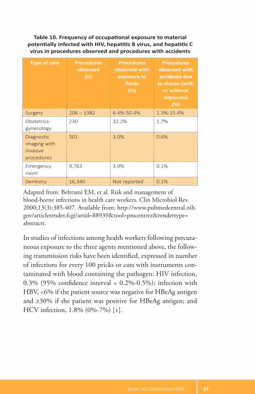
BASIC RECOMMENDATIONS 87
Table 10. Frequency of occupational exposure to material potentially infected with HIV, hepatitis B virus, and hepatitis C virus in procedures observed and procedures with accidents
Type of care Procedures observed
(n)
Procedures observed with
exposure to fluids
(%)
Procedures observed with accidents due
to sharps (with or without exposure)
(%)
Surgery 206 – 1382 6.4%-50.4% 1.3%-15.4%
Obstetrics- gynecology
230 32.2% 1.7%
Diagnostic imaging with invasive procedures
501 3.0% 0.6%
Emergency room
9,763 3.9% 0.1%
Dentistry 16,340 Not reported 0.1%
Adapted from: Beltrami EM, et al. Risk and management of blood-borne infections in health care workers. Clin Microbiol Rev. 2000;13(3):385-407. Available from: http://www.pubmedcentral.nih.gov/articlerender.fcgi?artid=88939&tool=pmcentrez&rendertype= abstractt.
In studies of infections among health workers following percuta-neous exposure to the three agents mentioned above, the follow-ing transmission risks have been identified, expressed in number of infections for every 100 pricks or cuts with instruments con-taminated with blood containing the pathogen: HIV infection, 0.3% (95% confidence interval = 0.2%-0.5%); infection with HBV, <6% if the patient source was negative for HBeAg antigen and ≥30% if the patient was positive for HBeAg antigen; and HCV infection, 1.8% (0%-7%) [1].
88 PREVENTION AND CONTROL OF INFECTIONS
What types of objects or materials pose a risk of exposure?
Sharps include any of the various objects with a sharp edge or pointed tip used in clinical care that can cut or penetrate the skin or the mucous membranes. Examples are solid nee-dles such as those used in suturing, hollow injection nee-dles, scalp vein-type intravascular cannulas, scalpel blades, scissors, biopsy forceps, glass ampoules containing infective material (blood, fluid from a sterile cavity, or any fluid with visible blood), and dental instruments, including high-speed hand instruments and drills.
Who is exposed?
Any person who has been in contact with a sharp instru-ment contaminated with blood or other fluid from a nor-mally sterile cavity of the body is considered to have been exposed. Several factors affect the risk for a sharps accident, including the type of procedure and instruments employed, use of gloves, skill and training in the practice, knowledge of occupational and infection control measures, procedure followed in handling needles and syringes, health worker fatigue and workload, illumination, pressure of working conditions, and perception of risk.
The epidemiology of percutaneous exposures depends on a variety of factors, including the type of procedure per-formed and the objects used in the facility, the frequency of the procedures, and application of the established pre-vention measures. The practices that are most often associ-ated with pricks and cuts are recapping of previously used needles and unsafe collection and disposal of sharps. Thus,
BASIC RECOMMENDATIONS 89
the health workers who tend to be most exposed are those working in surgical areas, emergency rooms, sample collec-tion sites, and laboratories, as well as any personnel who handle objects contaminated with blood during tasks other than healthcare [2].
All personnel working in a health facility are exposed to ac-cidents with sharps, but there are situations in which the risk is greater:
§ Before a procedure, when laying out the scalpel blade or loading a syringe, although this type of exposure does not involve a risk of infection since the material is not contaminated with blood or fluids
§ During a surgical procedure, when transferring a blood-contaminated sharp (scalpel, trephine, guide, etc.) from one hand to the other
§ Immediately after performing a procedure, during re-capping of needles
§ During and after disposal of sharps, by:
• Perforating the waste receptacle designated for dis-posal of sharps when the container is not mois-ture-proof or resistant to punctures or perforations
• Allowing a receptacle to overflow so that sharps are exposed
• Using receptacles without a protective cover
How can sharps accidents be avoided?
Many measures to prevent sharps accidents are based on before-and-after studies, opinions of specialists, statistical models, and perceptions of risk. There are few well-designed

90 PREVENTION AND CONTROL OF INFECTIONS
controlled clinical trials with an adequate sample size be-cause these matters are regulated by national standards in a number of countries. Also, it is difficult to measure the impact if the interventions are being practiced by an entire group. Furthermore, various methods are used to assess the results, which are usually based on incident reports filed by the affected health workers.
The best way to prevent injection-related sharps accidents is to avoid any unnecessary injections. The clinical team should look into the availability of effective oral or non-in-jectable parenteral therapies and standardize their admin-istration locally. Still, it is not enough to standardize the criteria and institutionalize them in writing; they will need to be disseminated and integrated into the practice of the clinical personnel responsible for applying them. Compli-ance should be supervised by trained staff. Finally, the re-sults from compliance assessments should be shared regu-larly and systematically with the pertinent units and clinical professionals [3].
Once the indications for use of sharps have been evaluated and accepted, the following measures should be taken to reduce the risk of accidents of this kind:c
§ Use of gloves
§ Safe handling of instruments
§ Separation and safe disposal of used sharps
§ Use of devices with an active and passive safety mechanism
c. When sharps are used in invasive procedures, attention should be given to preventing infections. The measures should include, at the very least, hand hygiene before and after the procedure, use of sterile gloves, application of an antiseptic to any puncture site, and appli-cation of a sterile covering on the site where a permanent catheter is inserted.

BASIC RECOMMENDATIONS 91
Whatever measures are adopted, they should be accompa-nied by a strategy of regular staff training that includes be-havior modification. It is recommended that facilities adopt modalities that can be replicated through practice in the field, use locally available supplies and equipment, and re-hearse the procedures that involve sharps in areas where they are usually performed [4, 5].
What does safe handling of sharps entail?
The main strategies are designed to avoid the practices that entail the greatest risk. Best practices that should be pro-moted with the health team include:
§ Using assistants for procedures that involve changing syringes or performing certain maneuvers (e.g., draw-ing a sample to test for arterial blood gases), when patients are agitated, or when patients are young children
§ Not recapping previously used needles
§ Not handling or disassembling a sharp directly with the fingers; if necessary, use tweezers
§ Ensuring at all times that the tip or edge of the sharp is pointing away from the body of the user and the assistant
§ Avoiding the transfer of unprotected sharps from the place where they were used to the disposal site (dis-posal receptacles should be located immediately next to the site where the procedure was performed)
§ Using a tray to receive and deliver sharps, such as scalpels, and avoiding hand-to-hand transfer between health workers
92 PREVENTION AND CONTROL OF INFECTIONS
§ Communicating verbally when a sharp object is handed from one person to another.
Also, technologies have been evaluated to prevent or min-imize human error in clinical practice. These approaches involve behavioral changes—for example, the use of retract-able needles to prevent accidental punctures. Opinions dif-fer on the impact of incorporating those technologies and their cost-effectiveness [6]. However, in specific practices, such as surgical care, studies have consistently demonstrat-ed the effectiveness of blunt-tip suture needles (as opposed to sharp-tip needles) and double-layer gloves in preventing percutaneous exposures [7, 8]. Regardless of these results, before any of these devices are introduced into clinical prac-tice, health workers should be trained in their use and be-come familiar with them.
How should sharps be discarded?
Once the procedure involving the use of a sharp has been performed, the sharp should be disposed of immediate-ly and safely in a special container for sharps waste. These containers should be waterproof and resistant to punctures and cuts. They should also be properly labeled (for example, with the biohazard symbol) and used only for sharps. Final-ly, they should be sealed with a cover.
The following conditions for managing containers facilitate adherence and increase safety in handling them:
§ They should be placed near the site where the pro-cedure is carried out and at a height that makes it possible to see the opening when a sharp is inserted. They should never be left on the floor or within reach of children.

BASIC RECOMMENDATIONS 93
§ They should not be filled above three-fourths of their capacity; when they reach that level, they should be replaced with an empty container [World Health Organization, 2010].
§ No disinfectant (chlorine or other) should be used on the receptacle; it will have no effect because chlorine is inactive on organic matter. Furthermore, if hospital waste is burned, exposure of the hypochlorite to heat can generate toxic gases.
What measures other than the standard precautions can be used?
There are other measures for preventing infections, not list-ed in the standard precautions, that will help to improve the safety of health workers. They include the introduction of protocols for vaccinating personnel against HBV and strat-egies to ensure compliance; management of postexposure to HBV and HIV; and the use of notification systems and exposure analysis to determine what caused the accidents and how they can be prevented with institutional measures.
References
1. Beltrami EM, et al. Risk and management of blood-borne infections in health care workers. Clin Microbiol Rev. 2000;13(3):385-407. Available from: http://www. pubmedcentral.nih.gov/articlerender.fcgi?artid=88939& tool=pmcentrez&rendertype=abstract.
2. Lavoie MC, Verbeek JH, Pahwa M. Devices for preventing percutaneous exposure injuries caused by needles in health-care personnel. Cochrane Database Syst Rev. 2014 Mar 9;(3):CD009740.

94 PREVENTION AND CONTROL OF INFECTIONS
3. World Health Organization. Injection safety and related in-fection control. 2010. [Incomplete reference; could not be verified.]
4. Bryce E, et al. Sharps injuries: defining prevention priorities. Am J Infect Control. 1999;27(5):447-52. Available from: http://www.ncbi.nlm.nih.gov/pubmed/10511494
5. Castella A. Preventability of percutaneous injuries in health-care workers: a year-long survey in Italy. J Hosp Infect. 2003;55(4):290-4. Available from: http://linkinghub.elsevier. com/retrieve/pii/S0195670103003323.
6. Brachman GO, et al. WHO guideline on the use of safety-en-gineered syringes for intramuscular, intradermal, and subcu-taneous injections in health care settings. Geneva: WHO; 2015. Available from: http://apps.who.int/iris/bitstream/ 10665/250144/1/9789241549820-eng.pdf?ua=1.
7. Parantainen A, et al. Blunt versus sharp suture needles for pre-venting percutaneous exposure incidents in surgical staff. Co-chrane Database Syst Rev. 2011;(11):CD009170. Available from: http://www.ncbi.nlm.nih.gov/pubmed/22071864.
8. Watt AM, et al. Scalpel safety in the operative setting: a systematic review. Surgery. 2010;147(1):98-106. Avail-able from: http://www.sciencedirect.com/science/article/pii/S0039606009004978.

BASIC RECOMMENDATIONS 95
D. Management of the environment
Why is management of the environment included in the standard precautions?
Patients are the main reservoir of the microorganisms that cause HAIs. Infection can be transmitted from patients through various mechanisms, including autoinfection, car-rying an infectious agent from one part of the body to an-other (for example, from the intestine to the urinary tract), and cross-transmission between patients via the hands of health workers following contact with these agents. It is es-timated that 20% to 40% of infections are attributable to cross-transmission.
Does the environment contribute to all HAIs?
HAIs tend to be contracted through direct contact with an infected patient; in the case of some microorganisms, how-ever, it has been found that the environment plays a role in the chain of transmission. The following characteristics of infectious agents can contribute to environmental trans-mission:
§ Ability to survive on environmental surfaces for pro-longed periods, either in a vegetative state or as spores
§ Ability to maintain virulence following exposure to the environment
§ Ability to colonize patients without symptoms
§ Ability to temporarily contaminate the hands of health workers
§ Ability to cause an infection with a low dose.

96 PREVENTION AND CONTROL OF INFECTIONS
Although several microorganisms have all of these charac-teristics, the fact that one is found in the environment does not necessarily mean it can transmit an infection. For an infection to be transmitted, all components in the chain of transmission have to be present. Thus, merely identifying an agent within a patient’s environment is not sufficient to as-sume that it was a cause of disease [1]. The theory that con-taminated surfaces contribute to the chain of transmission has been corroborated only for a small number of infectious agents (Table 11):
Table 11. Studies on the role of the environment in transmission of healthcare-associated infectious agents
Type of study
Infectious agent
Mathe-matical
transmis-siona
Micro- biological studiesb
Epidemi-ological studiesc
Inter-vention studiesd
Outbreak studiese
Clostridium difficile
× × × × ×
Acinetobacter spp. × × × ×
Vancomycin-resis-tant Enterococcus spp.
× × × ×
Pseudomonas aeruginosa
× × ×
Methicillin-resis-tant Staphylococ-cus aureus
× × × ×
Norovirus × ×
Multiresistant Gram-negative bacilli
× ×
Adapted from: Otter JA, et al. Evidence that contaminated surfaces contribute to the transmission of hospital pathogens and
BASIC RECOMMENDATIONS 97
an overview of strategies to address contaminated surfaces in hos-pital settings. Am J Infect Control. 2013;41(suppl):S6-11.
a. Direct evidence studies in which the number of patients expected to be infected by the particular microorganism is esti-mated using mathematical models based on the agent’s known mechanism of transmission and then compared with the number of cases actually observed. If there are more cases in reality, con-sideration is given to possible factors or conditions that might alter the agent’s known mechanism of transmission—in other words, the presence of other mechanisms of transmission.
b. Studies using various microbiological or biochemical tech-niques in which a microorganism has been consistently identified in the environment.
c. Case-control studies and cohort studies.
d. Studies that look at the incidence of an infection over a given period, after which an intervention is introduced to control or reduce the incidence of the infection; the incidence is then measured in the intervention group (also known as quasi-experi-mental studies).
e. Studies of outbreaks and other epidemiological investigations, which contribute better evidence on the role of the environment in the transmission of infectious agents.
When infections are endemic, few HAIs are associated with contamination of equipment, surfaces, waste products, or patient clothing. The association has been observed more often in outbreaks. In the following list of proposed in-terventions and recommendations, some are based not on epidemiological research or clinical trials of acceptable size and design, but rather, on basic hygiene measures, common sense, cultural considerations, and the consensus of experts.

98 PREVENTION AND CONTROL OF INFECTIONS
What does “environment” comprise in the context of HAIs?
§ fomites and instruments. Inanimate items that should be cleaned and disinfected or sterilized, de-pending on what they are going to be used for. This topic has been addressed in other manuals that specif-ically cover cleaning, disinfection, and sterilization of medical articles [2].
§ surfaces and equipment. All surfaces and accesso-ries in the patient’s environment that are used tem-porarily or on an ongoing basis during care, includ-ing fixed or mobile non-disposable items (furniture, equipment, etc.).
§ waste. All discarded materials or objects used in pa-tient care or in the patient environment that are to be removed from the health institution, usually classified as solid or liquid waste or biological or medical waste.
§ clothing and bedding. All textile items used by the patient, including bed linens, towels, hospital gowns, and pajamas.
How should the environment be managed?
Some studies have shown a correlation between proper management of contaminated surfaces, based on cleaning or disinfection, and reductions in the risk of HAI cross-trans-mission and outbreaks associated with certain infectious agents, including methicillin-resistant Staphylococcus au-reus, vancomycin-resistant Enterococcus, extended-spectrum β-lactamase-producing Enterobacteriaceae (ESBL), Klebsi-ella pneumoniae, Pseudomonas aeruginosa and multiresistant Acinetobacter baumannii, norovirus, and Clostridium difficile
BASIC RECOMMENDATIONS 99
[1, 3, 4, 5, 6]. However, not all microorganisms respond similarly to all methods and types of cleaning and disin-fection. For example, isolates of norovirus and Clostridium difficile have shown greater resistance to common cleaning and disinfection processes. Therefore, the cleaning and dis-infection program should be adapted to the properties of the microorganisms in question and to the risks associated with HAI-related equipment, surfaces, and clothing [3].
Environmental elements should be classified based on an HAI risk analysis that takes the following criteria into ac-count [7]:
§ Degree of patient exposure. A distinction should be made between equipment or surfaces in direct con-tact with the patient and those in which there is only minimal contact.
§ Properties of the microorganisms involved or poten-tially involved in the infection, based on the patient’s risk profile. These properties should include survival time on surfaces, resistance to disinfectants, infective dose, and pathogenicity.
This characterization should be different for each unit and for each type of patient. For example, because of their risk profile, patients in intensive care units, on dialysis, or who have received transplants are at greater risk for multiresis-tant bacterial infections than those in less complex medi-cal-surgical units or situations [8, 9, 10, 11].
Aren’t cleaning and disinfection the same thing?
Although cleaning and disinfection are often confused, they are not the same thing. Cleaning removes dirt deposited on

100 PREVENTION AND CONTROL OF INFECTIONS
inanimate surfaces by mechanical (friction), physical (tem-perature), or chemical means for a certain length of time [12]. Disinfection, on the other hand, is a physical or chem-ical process in which vegetative microorganisms are removed from inanimate objects but without the assurance that bac-terial spores have been removed. In this document, disin-fection refers to the use of chemical agents known as inter-mediate-level disinfectants (phenols, sodium hypochlorite) that remove vegetative bacteria and some bacterial spores, as well as the use of chemical agents known as low-level disin-fectants (quaternary ammonium compounds) that remove vegetative bacteria, fungi, and some viruses for a short time (less than 10 minutes) [2].
Cleaning and disinfection of low-risk surfaces
In cleaning and disinfection, low-risk surfaces and equip-ment are those that will not come in contact with mucous membranes or breaks in the skin.
Multidisciplinary working teams composed of personnel from the departments involved in cleaning and selecting the disinfectant products for purchase should participate in formulating the programs for cleaning and disinfecting low-risk clinical surfaces and equipment. These teams should in-clude administrative, nursing, and pharmacy personnel and those responsible for HAI control, supplies, and cleaning. The programs should have:
1. Clearly defined duties and responsibilities for each of the different types of personnel involved in the clean-ing and disinfection activities.
2. Criteria for classifying the surfaces that will be subject to intervention.

BASIC RECOMMENDATIONS 101
3. A list of the steps that need to be taken and their fre-quency. This may vary depending on the type of ac-tivity. For example:
§ A surface that has only been in brief contact with a patient or personnel (low-touch surface) requires daily cleaning (wiped with a moist rag or towel with or without detergent); additional cleaning and subsequent disinfection would be done only in the presence of body fluids, organic matter or when the patient is discharged.
§ A surface in an environment that has been involved in the chain of transmission of microorganisms from a patient (high-touch surface) requires cleaning and subsequent disinfection.
§ It is always necessary to remove organic matter me-chanically with water and detergent before applying any type of disinfectant. When the use of disinfec-tants is being considered after the use of detergents, it is necessary to be aware of combinations of prod-ucts that can generate chemical reactions or gases that are toxic for humans (for example, an ammo-nium combined with hypochlorite). In such cases, the detergent should not be used and time should be allowed for the surface to dry before applying a disinfectant product.
§ The process of selecting the products and supplies needed for cleaning and disinfection should include a review of those approved under national regula-tions and by the health facility. The manufacturer’s directions should be observed when preparing and using the product. The following characteristics are usually evaluated when selecting a disinfectant:
102 PREVENTION AND CONTROL OF INFECTIONS
broad spectrum activity; brief latency time between application and effect; compatibility with the sur-face on which it will be applied, other chemicals that might be used, and the environment; low toxicity for humans; minimal allergenic effect; low volatility; tolerable scent and acceptability by users and others in the environment; ease of preparation, handling, using, and storage; and low cost. Examples of prod-ucts for surface disinfection are as follows:
• Chlorine solutions (1,000-5,000 parts per mil-lion). See the chlorine dilution formula at the end of this chapter.
• Alcohol (70%).
• Quaternary ammonium solutions. Different solu-tions have different degrees of activity (see specific manual on the subject).
• If contamination with Clostridium difficile spores is suspected, 0.05% chlorine solutions are preferred. Hydrogen peroxide-based disinfectants have also been shown to be effective against C. difficile.
• For information on other disinfectants for use against HAIs, a good resource in Spanish is the document Limpieza y desinfección de superficies hospitalarias [Cleaning and Disinfection of Hos-pital Surfaces], prepared by the National Health Surveillance Agency of Brazil with support from the Pan American Health Organization (PAHO) [12].
4. Methods of applying detergents and disinfectants. Consideration should always be given to the safety of the person doing the cleaning, especially with regard

BASIC RECOMMENDATIONS 103
to exposure to chemical agents that can have a nega-tive effect on health. Cleaning should always precede disinfection.
5. Education and training of the personnel who will be performing each of the cleaning activities, whether followed by disinfection or not. These workers may be in contact with isolation rooms, which means that they should be familiar with pertinent measures, ap-ply these and receive support when they are perform-ing their duties.
6. Regular supervision of cleaning activities and periodic feedback on the results.
What is the best way to supervise the cleaning process?
There is no evidence that one method is superior to others, which means that it will be necessary to assess the advantag-es and disadvantages of each method and choose the ones that are most sustainable over time (Table 12).
Table 12. Methods of supervising the cleaning and disinfection processes: advantages and disadvantages
Method Advantages Disadvantages
Visual inspection SimpleLow costImmediate results
Not very objective (depends on the observer)
(cont. next page)
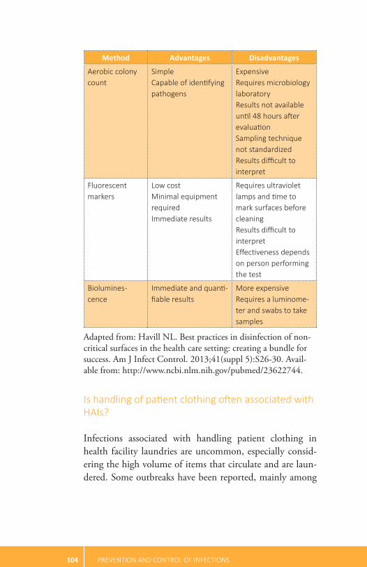
104 PREVENTION AND CONTROL OF INFECTIONS
Method Advantages Disadvantages
Aerobic colony count
SimpleCapable of identifying pathogens
Expensive Requires microbiology laboratory Results not available until 48 hours after evaluation Sampling technique not standardized Results difficult to interpret
Fluorescent markers
Low costMinimal equipment requiredImmediate results
Requires ultraviolet lamps and time to mark surfaces before cleaning Results difficult to interpret Effectiveness depends on person performing the test
Biolumines-cence
Immediate and quanti-fiable results
More expensive Requires a luminome-ter and swabs to take samples
Adapted from: Havill NL. Best practices in disinfection of non-critical surfaces in the health care setting: creating a bundle for success. Am J Infect Control. 2013;41(suppl 5):S26-30. Avail-able from: http://www.ncbi.nlm.nih.gov/pubmed/23622744.
Is handling of patient clothing often associated with HAIs?
Infections associated with handling patient clothing in health facility laundries are uncommon, especially consid-ering the high volume of items that circulate and are laun-dered. Some outbreaks have been reported, mainly among

BASIC RECOMMENDATIONS 105
laundry personnel. The species identified were Sarcoptes sca-biei, Salmonella typhimurium, Salmonella hadar, Microspo-rum canis, Rhizopus delemar, hepatitis A virus, and Q fever. Apparel contaminated with body fluids may contain bacte-rial loads of up to 108 CFU per 1 cm2 of fabric. However, the control measures that have been adopted for handling and processing them have succeeded in reducing the risk of transmitting infection [13, 14, 15]. That said, there is still potential for health workers to be cut and punctured by sharp objects inside their garments, which is another reason for emphasizing the importance of safe handling and dis-posal of sharps.
What steps should be considered in the management of laundry to prevent HAIs?
The recommendations here are similar to those for cleaning and disinfection, including the need to prepare and dissem-inate institutional protocols that cover transportation and laundering, among other processes. In addition, there are certain specific recommendations for this phase of the pro-cess [16, 17]:
§ After wearing clinical garments:
• Do not shake them.
• Put them immediately in closed, clearly marked containers that will be used to remove them from the patient area. These garments should never be temporarily laid on furniture or other surfaces in the patient area.
• Place clothing with blood or other body fluids in waterproof and leak-proof containers (for example, closed bags).

106 PREVENTION AND CONTROL OF INFECTIONS
§ When taking soiled clothing or linen to the laundry after it has been removed from the clinical service:
• Ensure that there is no direct contact between pa-tient clothing or linen and the body of the health worker who is transporting it. If possible, transport it in closed containers.
• Do not shake it.
• Remove any solid organic matter (for example, stool) before placing items in containers for soiled clothes.
• There is no need to separate clothing and linen from infected and uninfected patients because it has not been shown that the former contain more microor-ganisms than the latter [18]. This practice may vary depending on the institution.
§ When items are being separated in the laundry [19].
• The institution should determine the point in the laundry process at which the clothing and linen will be separated and classified by type of item or fabric. This classification is intended to reduce the risk of workers being exposed to contaminated clothes or linen or any sharps that may be present. It is also important to protect the laundry and washing ma-chines from exposure to potentially harmful materi-als (needles, scalpels, solid objects with sharp surfac-es, etc).
• Personnel should wear gloves that are thick enough to reduce the risk of percutaneous exposure.
§ When the clothing and linen are being washed [20, 21, 22, 23]:
• Wash by machine, not by hand.

BASIC RECOMMENDATIONS 107
• Use hot water (≥70° C). Do not dry clean.
• Remove dirt and organic matter mechanically.
• Use detergent for clothes and linen.
• Wash for at least 20 minutes.
• If any of the above requirements are not met, it is recommended that the process be repeated.
• It is usually recommended that all items be dried and pressed at >150° C, but it may be necessary to alter the temperature based on the manufacturer’s instructions for the particular fabric.
§ During transportation and storage:
• Transport clean clothes and linen from outside laun-dries in packages well protected by fabric covers to prevent contamination with dirt or dust in the course of loading and unloading.
• Store gowns and linen for clinical use in a dry and dirt- and dust-free area (for example, in closed cabinets).
Personnel should wear PPE at all times, depending on the assessment of risk of exposure. Usually, a gown, apron, and thick gloves are required when handling soiled clothing. Masks and eyeglasses can be used if the items are to be shak-en or handled in large quantities.
Waste
does the waste in health facilities carry a greater risk for HAIs?
It has been estimated that between 20% and 40% of the waste generated by health facilities poses a risk similar to

108 PREVENTION AND CONTROL OF INFECTIONS
that of domestic waste, with similar characteristics in terms of types of microorganisms. Some studies have found that waste from isolation units has a lower microbial burden than that from ordinary healthcare units [24]. Some authors suggest that no more than 3% of the total volume of clinical waste has infectious potential [25] and that the perception of risk is greater than the real risk in the case of HBV, HCV, and HIV infections [26]. Other studies have failed to find a higher prevalence of HBV infection among hospital waste management personnel than among those who handle com-munity waste [27, 28]. Thus, there is no evidence that there is a greater infectious risk in handling hospital waste than there is in handling domestic waste.
how can hospital waste be managed to prevent HAIs?
With the exception of sharps waste, which requires special handling (and has already been discussed), there is no need to take additional measures to manage waste other than those for handling waste from infected patients. Body fluids (urine, blood, and stool) can be disposed of through the sewerage system, where they are quickly diluted, without having to apply an additional disinfectant.
When it is not possible to dispose of waste through the sew-erage system, it should be handled only after assessing the risk of exposure for workers. All waste should be transport-ed using protective measures for workers—that is, in bags made of strong waterproof material.
All countries have regulations on the management of hospi-tal waste outside the health facility. These regulations should be followed, especially the ones on handling containers of

BASIC RECOMMENDATIONS 109
contaminated sharps and waste from pathology units and microbiology and clinical laboratories.
FORMULA FOR DILUTING SODIUM HYPOCHLORITE USING COMMERCIAL PRODUCTS
Some health establishments use commercial solutions to prepare chlorine dilutions at 1,000 parts per million (ppm) or 5,000 ppm. These dilutions can be challenging because the commercial solutions come in different concentrations. The following table shows examples of how to prepare the dilution based on different original concentra-tions. It is important to know that:
0.5% solution = 5,000 ppm0.1% solution = 1,000 ppm
Formula for diluting sodium hypochlorite:Total parts of water added = [% of concentrated original ÷ % of desired concentration] – 1
Examples
Commercial solution
Desired solution
Formula Result Preparation
5.0% con-centrated chlorine solution
Chlorine solution diluted to 0.5% (5,000 ppm)
[5.0% ÷
0.5%] – 1 9 Add 9 parts
water to 1 part commercial chlorine solution at 5.0%
5.0% con-centrated chlorine solution
Chlorine solution diluted to 0.1% (1,000 ppm)
[5.0% ÷
0.1%] – 1 49 Add 49
parts water to 1 part commercial chlorine solution at 5.0%
(cont. next page)

110 PREVENTION AND CONTROL OF INFECTIONS
Formula for diluting sodium hypochlorite:Total parts of water added = [% of concentrated original ÷ % of desired concentration] – 1
Examples
Commercial solution
Desired solution
Formula Result Preparation
Chlorine solution diluted to 0.1% (1,000 ppm)
[5.5% ÷
0.1%] – 1 54 Add 54
parts water to 1 part commercial chlorine solution at 5.5%
6.0% con-centrated chlorine solution
Chlorine solution diluted to 0.1% (1,000 ppm)
[6% ÷ 0.1%]
– 1 59 Add 59
parts water to 1 part commercial chlorine solution at 6.0%
6.0% con-centrated chlorine solution
Chlorine solution diluted to 0.1% (1,000 ppm)
[6% ÷ 0.5%]
– 1 11 Add 11
parts water to 1 part commercial chlorine solution at 6.0%
It should be noted that the concentrations of some commercial chlorine solutions are different from the figures indicated on the label. For this reason, solutions with known concentrations are preferred.

BASIC RECOMMENDATIONS 111
References
1. Weber DJ, et al. Role of the environment in the transmission of Clostridium difficile in health care facilities. Am J Infect Control. 2013;41(5):S105-10. Available from: http://dx.doi.org/10.1016/j.ajic.2012.12.009.
2. Acosta-Gnass SI, De Andrade Stempliuk V. Sterilization manual for health centers. Washington, DC: Pan American Health Organization; 2009.
3. Dancer SJ. The role of environmental cleaning in the control of hospital-acquired infection. J Hosp In-fect. 2009;73(4):378-85. Available from: http://dx.doi.org/10.1016/j.jhin.2009.03.030.
4. Sattar SA, Maillard JY. The crucial role of wiping in decon-tamination of high-touch environmental surfaces: review of current status and directions for the future. Am J Infect Control. 2013;41(5):S97-104. Available from: http://dx.doi.org/10.1016/j.ajic.2012.10.032.
5. Weber DJ, et al. Role of hospital surfaces in the transmis-sion of emerging health care-associated pathogens: norovirus, Clostridium difficile, and Acinetobacter species. Am J Infect Control. 2010;38(suppl 1):S25-33.
6. Otter JA, et al. Evidence that contaminated surfaces contrib-ute to the transmission of hospital pathogens and an over-view of strategies to address contaminated surfaces in hospital settings. Am J Infect Control. 2013;41(suppl):S6-11.
7. Carling PC, et al. Identifying opportunities to enhance en-vironmental cleaning in 23 acute care hospitals. Infect Con-trol Hosp Epidemiol. 2008;29(1):1-7. Available from: http://www.ncbi.nlm.nih.gov/pubmed/18171180.
8. Ziakas PD, et al. Trends and significance of VRE coloniza-tion in the ICU: a meta-analysis of published studies. PloS ONE. 2013;8(9):e75658.
9. Ziakas PD, Anagnostou T, Mylonakis E. The prevalence and significance of methicillin-resistant Staphylococcus au-reus colonization at admission in the general ICU setting:

112 PREVENTION AND CONTROL OF INFECTIONS
a meta-analysis of published studies. Crit Care Med. 2014;42(2):433-44. Available from: http://www.ncbi.nlm.nih.gov/pubmed/24145849.
10. Ziakas PD, et al. MRSA and VRE colonization in solid organ transplantation: a meta-analysis of published studies. Am J Transplant. 2014;14(8):1887-94. Available from: http://www.ncbi.nlm.nih.gov/pubmed/25040438.
11. Zacharioudakis IM, et al. Meta-analysis of methicillin-resis-tant Staphylococcus aureus colonization and risk of infection in dialysis patients: a systematic review and meta-analysis. J Am Soc Nephrol. 2014;25(9):2131-41. Available from: http://www.ncbi.nlm.nih.gov/pubmed/24652802.
12. Brasil, Agencia Nacional de Vigilancia Sanitaria. Limpieza y desinfección de superficies hospitalarias. Brasília; 2010 [trans-lation into Spanish]. Available from: http://www.cocemi. com.uy/docs/limpiezahosp_dic2010.pdf.
13. Fijan S, Turk SŠ. Hospital textiles: are they a possible vehi-cle for healthcare-associated infections? Int J Environ Res Public Health. 2012;9(9):3330-43. Available from: http://www.pubmedcentral.nih.gov/articlerender.fcgi?artid= 3499872&tool=pmcentrez&rendertype=abstract.
14. Mitchell A, Spencer M, Edmiston C. Role of health-care apparel and other healthcare textiles in the transmis-sion of pathogens: a review of the literature. J Hosp In-fect. 2015;90(4):285-92. Available from: http://dx.doi.org/10.1016/j.jhin.2015.02.017.
15. Duffy J, et al. Mucormycosis outbreak associated with hos-pital linens. Pediatr Infect Dis J. 2014;33(5):472-6. Avail-able from: https://www.ncbi.nlm.nih.gov/pubmed/?term= Mucormycosis+outbreak+associated+with+hospital+linens.
16. Fijan S, et al. Antimicrobial disinfection effect of a laun-dering procedure for hospital textiles against various indi-cator bacteria and fungi using different substrates for sim-ulating human excrements. Diagn Microbiol Infect Dis. 2007;57(3):251-7. Available from: http://www.ncbi.nlm.nih.gov/pubmed/17046191.

BASIC RECOMMENDATIONS 113
17. United States Centers for Disease Control and Prevention. Laundry: washing infected material. Healthcare-associated infections. 2011. Available from: https://www.cdc.gov/HAI/prevent/laundry.html.
18. Weinstein SA, et al. Bacterial surface contamination of pa-tients’ linen: isolation precautions versus standard care. Am J Infect Control. 1989;17(5):264-7. Available from: http://www.ncbi.nlm.nih.gov/pubmed/2817514.
19. Sehulster L, et al. Guidelines for environmental infection control in health-care facilities: recommendations from CDC and the Healthcare Infection Control Practices Ad-visory Committee (HICPAC). MMWR Recomm Rep. 2003;52(RR-10):1-42.
20. Tano E, Melhus A. Level of decontamination after washing textiles at 60°C or 70°C followed by tumble drying. Infect Ecol Epidemiol. 2014;4:24314. Available from: http://www.ncbi.nlm.nih.gov/pubmed/25413829.
21. Cottenden AM, et al. Is there a risk of cross-infection from laundered reusable bedpads? Br J Nurs. 1999;8(17):1161-3. Available from: http://www.ncbi.nlm.nih.gov/pubmed/ 10897698.
22. Lakdawala N, et al. Effectiveness of low-temperature domes-tic laundry on the decontamination of healthcare workers’ uniforms. Infect Cont Hosp Epidemol. 2011;32(11):1103-8. Available from: http://www.ncbi.nlm.nih.gov/pubmed/ 22011538.
23. Nurs Manag (Harrow). Washing uniforms below 60°C may increase risk of bacterial infection. 2015;22(1):6. Available from: http://www.ncbi.nlm.nih.gov/pubmed/25806441.
24. Weinstein R. Microbiologic contamination of hospital trash from patients on isolation precautions versus standard care. Am J Infect Control. 1988;16:76.
25. Daschner FD, Dettenkofer M. Protecting the patient and the environment: new aspects and challenges in hospital in-fection control. J Hosp Infect. 1997;36(1):7-15. Available from: http://www.sciencedirect.com/science/article/B6WJP- 4CDJ2GD-8K/2/e706fa2c5b87b255507c5629490b38bd.
114 PREVENTION AND CONTROL OF INFECTIONS
26. Rutala WA, Mayhall CG. Medical waste. Infect Cont Hosp Epidemiol. 1992;13(1):38-48. Available from: http://www.ncbi.nlm.nih.gov/pubmed/1545111.
27. Cimino JA. Health and safety in the solid waste industry. Am J Public Health. 1975;65(1):38-46. Available from: http://www.ncbi.nlm.nih.gov/pubmed/45848.
28. Ferreira JA, et al. Hepatitis B morbidity in municipal and hospital waste collection workers in the city of Rio de Janeiro. Infect Cont Hosp Epidemiol. 1999;20(9):591-2. Available from: http://www.ncbi.nlm.nih.gov/pubmed/10501252.

BASIC RECOMMENDATIONS 115
Additional precautions based on mode of transmission
IVThe standard precautions are not always sufficient to con-trol the transmission of certain infectious agents; additional precautions may need to be taken to address specific cases. These additional precautions are described below, based on the agents’ mode of transmission: by contact, droplets, air (droplet nuclei), and other means. This section also covers the subject of cohort isolation.
What is the difference between the standard precautions and the additional precautions based on mode of transmission?
Generally speaking, the additional precautions are comple-mentary measures. Unlike the standard precautions, which are applied without needing to know the patient’s infection or colonization status, they are applied only when the pa-tient is known to have or suspected of having an infectious disease, especially if the disease would still be in the infec-tious period. They are also applied in some cases in which the patient is known to be colonized with an agent that is

116 PREVENTION AND CONTROL OF INFECTIONS
resistant to the antimicrobial drugs of public health impor-tance (see chapter on this subject).
Many of the additional precautions are based on opinions of experts, the transmission mechanism, the known portal of entry, the perception of risk, and the severity of the dis-ease, among other considerations. Most controlled clinical trials and other evidence-based data have been concerned with specific pathologies and microorganisms, and the pri-mary focus has been on infections with methicillin-resistant Staphylococcus aureus (MRSA) and vancomycin-resistant Enterococcus (VRE) [1]. In fact, the effectiveness of the ad-ditional precautions has been observed in the control of ep-idemic outbreaks due to a number of different infectious agents.
What is involved in applying the additional precautions based on mode of transmission?
In order to implement the additional precautions, it is nec-essary to:
1. Have definitions and protocols in place that are eas-ily accessible to personnel in the health facility. They should indicate the type of additional precaution that will be needed for each known or suspected infectious disease or infectious agent of public health impor-tance. This information will facilitate implementa-tion of the measures when they are appropriate and promote a dialogue between health workers and the professionals who direct the clinical services.
2. Provide ongoing education and training for the per-sonnel in charge of deciding on the application of additional precautions and those responsible for
BASIC RECOMMENDATIONS 117
implementing them. Local resources and other planned activities should be taken into account. Some authors recommend using summarized guidelines to facilitate the expeditious application of local protocols. Health workers should be familiar with these summa-ries and be able to access them easily. They should be available in several places—for example, on the back of ID cards or employee credential cards, in clinical files, on computer screen protectors, and so on [2].
3. Ensure that professionals who are in training and those who have not had specific training on the subject do not participate in the care of patients for whom precautions based on the mode of transmission are indicated.
4. Have programs for evaluating compliance with the measures and providing feedback to the personnel in-volved in applying them to avoid or correct possible missteps.
5. Establish strategies for avoiding anxiety, confusion, or rejection of the measure by patients or their family members, regardless of the precaution selected. For example, they should be informed of the reasons for the procedure, the time it may take, and any specif-ic precautions that should be followed during family member visits [3].
6. Ensure that the standard precautions continue to be applied and remain unchanged when the addition-al measures are applied. The standard precautions should always be applied, including in areas where pa-tients are undergoing triage to set priorities for their care and places where patients enter the facility before it is known whether or not they have an infectious clinical condition.
118 PREVENTION AND CONTROL OF INFECTIONS
What needs to be known in order to decide if additional precautions are required?
§ What is the patient’s confirmed or suspected diagnosis?
§ What is the infectious agent and its mode of transmission?
§ What is the natural history of the disease? For what period of time is the agent infectious?
§ What type of procedure will the patient be undergoing?
§ What measures need to be taken to avoid transmis-sion of the infectious agent (cross-transmission)? Is there any risk of contamination? Where should the patient be located? What type of PPE will need to be used?
contact transmission
This mode of transmission is the most common. It can oc-cur in two forms:
§ Direct contact, when microorganisms travel from the reservoir directly to the susceptible individual with-out any other elements involved in the transmission.
§ Indirect contact, when the susceptible host comes into contact with the infective microorganism through an inanimate intermediary (clothing, fomites, surfaces in the room) or an animate one (via the hands of a health worker when in contact with another patient). The infectious agent must have the capacity to sur-vive in the environment. Therefore, identifying an agent in the environment is not sufficient evidence

BASIC RECOMMENDATIONS 119
to account for transmission, since it does not neces-sarily mean that the microorganism has retained its infective capacity or that it has been involved in the chain of transmission. Some of the microorganisms that are transmitted this way include Clostridium dif-ficile, Acinetobacter spp., Enterococcus spp. (including vancomycin-resistant strains), Pseudomonas aerugi-nosa, Klebsiella spp., Staphylococcus aureus (including methicillin-resistant strains), norovirus, respirato-ry syncytial virus, rotavirus, Gram-negative bacilli, and enterobacteria that are sensitive or resistant to antimicrobial drugs—for example, extended-spec-trum β-lactamase- or carbapenemase-producing Enterobacteriaceae.
Where should a patient requiring contact precautions be located?
Given the potential for indirect transmission of infectious agents, the decision regarding where to locate the patient should always be based on an analysis of the risk of trans-mitting HAIs to other patients:
1. Place the patient in a separate room or a room with other patients who have the same diagnosis (the same agent and the same genotype). The latter option is especially useful when the facility has a large number of cases caused by the same infectious agent (for ex-ample, during outbreaks).
2. When no separate room is available, an exception can be made to place the patient in a room with other patients as long as the following conditions are met:
• The patient can be placed in a shared room if the sur-rounding area is large enough to allow for the patient’s
120 PREVENTION AND CONTROL OF INFECTIONS
comfort and also for the space and supplies required by other patients (intravenous medication stand, footstool, etc.).
• Do not place a patient indicated for isolation in the same room with patients undergoing invasive proce-dures or immunocompromised patients who could suffer serious consequences.
What are the requirements for the room or hospital area to be occupied by a patient needing additional precau-tions?
The room should meet the same conditions as for the stan-dard precautions. In other words, it should have the nec-essary arrangements for the use of PPE and hand hygiene, especially a sink with running water that can be adjusted for temperature, soap, disposable paper towels or a system for drying hands (requirements for hand hygiene), and an alco-hol-based solution for hand disinfection at the point of care.
In addition,
§ If precautions are being taken for enteric diseases and the patient is ambulatory, the patient should have a separate bathroom for his or her exclusive use or, if this is not possible, some other means of minimizing the risk of transmitting microorganisms through con-tact with stools (an individual or disposable bedpan, basin, or potty). If the patient has to share a bath-room, it should be cleaned and disinfected under su-pervision after every use.
§ If PPE is to be used, there should be a place to keep it before entering the room or the patient’s area and a receptacle where it can be discarded after caring for the patient and before leaving the room or unit.

BASIC RECOMMENDATIONS 121
§ There should be a clear, visible sign at the entrance to the room or patient area warning that contact pre-cautions are being taken and including the applicable instructions.
What measures should be taken during care for patients requiring contact precautions?
The use of barrier PPE elements should be considered if direct physical contact with the patient or indirect contact with potentially contaminated surfaces is expected. The PPE should be single-use equipment or only for use with the par-ticular patient. It should be put on before entering the room or patient area and removed before leaving. At the very least, use of the following barrier elements should be considered:
§ Single-use gloves. Waterproof, disposable, non-reus-able gloves should be used, and hand hygiene should be performed both before putting them on and after removing them.
§ Dedicated gown. It does not have to be disposable, but it must be used only to care for one specific patient. The same gown may be used by different members of the health team caring for the same patient. Experts suggest that it should be changed or laundered daily.
If there is a chance of spattering or contact with body fluids, the standard precautions should be applied: protect the fa-cial mucous membranes and wear a waterproof apron over the gown.
When transferring a patient who requires these measures, any personnel who come in contact with the patient should follow the indications above and make certain that the sup-plies are disposable or properly cleaned and disinfected be-fore reusing them.

122 PREVENTION AND CONTROL OF INFECTIONS
droplet transmission
In this form of transmission, particles measuring 5 to 100 µm (typically ≥20 µm) in diameter (droplets) are emitted from the respiratory tract of the infectious patient when he or she coughs, sneezes, or talks. When particles are very small, they remain airborne only for a few seconds (except for droplets <20 µm, which can stay in suspension for sev-eral minutes) and travel less than 1 meter. Examples of mi-croorganisms or diseases that are transmitted in this way are diphtheria, whooping cough, meningococcal meningitis, influenza, adenoviruses, and coronaviruses (such as SARS coronavirus and MERS [Middle East respiratory syndrome] coronavirus).
What is the objective of precautions against droplet transmission?
The precautions are intended to prevent droplets transmit-ted by a patient from coming into contact with the con-junctival, nasal, or oral mucous membranes of a susceptible host (health worker or another patient) located less than 1 meter away. It should be remembered that the conjuncti-val membranes drain into the nasal cavity through the tear ducts, which means that they are a portal of entry for certain infectious agents, especially viruses.
Where should a patient requiring droplet precautions be located?
The decision regarding where to locate the patient should always be based on an analysis of the risk of transmitting HAIs to other patients. The options are:
1. Place the patient in a separate room or a room with other patients who have the same diagnosis, the same agent, and the same phenotype.

BASIC RECOMMENDATIONS 123
2. On an exceptional basis, when the facility has a large number of cases caused by the same agent (for exam-ple, during hospital outbreaks due to outbreaks in the community), the patient may share a room with other patients as long as the following conditions are met:
• The patient can be placed in a shared room if there is at least 1 meter of surrounding area to allow for the patient’s comfort and also for space and supplies required by other patients (intravenous medication stand, footstool, etc.).
• Do not place a patient indicated for isolation in the same room with patients undergoing invasive proce-dures or immunocompromised patients who could suffer serious consequences.
3. This option is especially useful when the facility has a large number of cases caused by the same infectious agent (for example, during outbreaks).
What are the requirements for the room or hospital area to be occupied by a patient needing droplet precautions?
The room should meet the same conditions as for the stan-dard precautions—in other words, it should have the nec-essary arrangements for the use of PPE and hand hygiene, especially a sink with running water that can be adjusted for temperature, soap, disposable paper towels or a system for drying hands (requirements for hand hygiene), and an alcohol-based solution for hand disinfection at the point of care. In addition:
§ If PPE is to be used, there should be a place to keep it no farther than 1 meter from the patient’s area and a receptacle where it can be left after caring for the patient and before leaving the room or unit.

124 PREVENTION AND CONTROL OF INFECTIONS
§ The room should have a door that closes, as well as good ventilation.
§ There should be a clear, visible sign in the room or pa-tient area warning that droplet precautions are being taken and including the applicable instructions.
What measures should be taken during care for patients requiring droplet precautions?
The decision to use PPE will depend on the expected dis-tance from the patient. If it is less than 1 meter, guidelines are as follows [4]:
§ Protect facial mucosa with a preformed surgical-type mask, preferably not pleated, in addition to (1) eye-wear or (2) a face shield. If the face shield is long and extends below the chin, there is no need to use a mask. It is not necessary, or even advisable, to use both eyewear and a face shield at the same time be-cause it would restrict the user’s vision.
§ Apply standard precautionary measures if there is risk of spattering or contact with body fluids, including:
• A gown and, if necessary, an apron
• Single-use gloves (disposable, non-reusable) to pro-tect against exposure to droplets deposited on sur-faces in the environment near the patient
• Hand hygiene, performed both before putting on gloves and after removing them
During transfer, patients should wear a conventional or non-folding surgical mask to cover exhaled droplets.
airborne transmission (via droplet nuclei)
This type of transmission takes place in the air through the dissemination of particulates measuring less than 5 µm in
BASIC RECOMMENDATIONS 125
diameter. When pushed by air currents, these particulates can remain airborne for prolonged periods and travel greater distances than droplets [5]. Therefore, they can be inhaled and enter the alveoli of individuals in the same room, even if they have not had direct contact with the infected patient.
Droplet nuclei may be generated directly by the patient when he or she coughs or sneezes (e.g., in the case of pa-tients with tuberculosis) or by clinical procedures, including tracheal intubation, noninvasive positive-pressure ventila-tion, invasive high-frequency ventilation, airway aspiration, tracheotomy, respiratory kinesiotherapy, fogging, fiber-op-tic bronchoscopy, sputum induction, centrifugation of sam-ples, and procedures using saws to cut tissues. Among these procedures, according to studies on transmission of the SARS coronavirus, the ones that pose the greatest risk are tracheal intubation, noninvasive ventilation, tracheotomy, and manual ventilation prior to intubation [6]. Examples of microorganisms transmitted by this means are the Myco-bacterium tuberculosis bacillus (from bacilliferous patients) and the measles, chickenpox, and disseminated shingles viruses.
Where should a patient requiring airborne transmission precautions be located, and what are the pertinent re-quirements for the room or hospital area?
This decision should always be based on an analysis of the risk of transmitting HAIs to other patients. Options are as follows:
1. A separate room that allows for:
• Restricted access.
• Ventilation to the outside of the building, never toward areas where other patients are located. The

126 PREVENTION AND CONTROL OF INFECTIONS
door should always be closed. The use of negative pressure has been recommended, if it is available, al-though it has not been shown to be more effective than a window that opens to the outside [7, 8].
• When the room does not have exterior ventilation or the climate does not make it possible to open the windows, air extraction systems that allow for at least 6 to 12 air changes per hour can be used. If the air is not vented to the outside and ends up reach-ing other patient areas or closed spaces, it should be treated with a system that uses HEPA (high-efficien-cy particulate air) filters [9].
2. A shared room. If necessary, the patient can be placed in a room with other patients who have the same di-agnosis, the same agent, and the same genotype. If in doubt, especially in cases where the infectious agent can develop antimicrobial resistance (for example, Mycobacterium tuberculosis), always place the patient in a separate room.
3. The room should meet the same conditions as for the standard precautions—in other words, it should have the necessary arrangements for the use of PPE and hand hygiene, including a sink with running water that can be adjusted for temperature, soap, dispos-able paper towels or a system for drying hands (re-quirements for hand hygiene), and an alcohol-based solution for hand disinfection at the point of care. In addition:
• There should be a place to keep the PPE outside the patient’s area and a receptacle where it can be dis-carded after caring for the patient and before leaving the room or unit.

BASIC RECOMMENDATIONS 127
• There should be a clear, visible sign in the room or patient area warning that droplet nuclei precau-tions are being taken and including the applicable instructions.
What measures should be taken during care for patients who require airborne transmission precautions (infec-tions transmitted via aerosols)?
Given the transmission mechanism, a health worker ap-proaching a patient with this type of infection should use a type N95 or FFP2 high-efficiency respirator or equivalent before entering the room, touching the patient, or initiating a procedure that might generate aerosols [10].
The clinical personnel who provide care should be immu-nized before treating patients with vaccine-preventable dis-eases. The vaccination record should be documented in an official registry or on a personal vaccination card.
Moving the patient to other units should be restricted. If it is necessary, the accompanying personnel should follow the relevant indications. In addition, a surgical mask may be placed on the patient to cover exhaled droplets, subject to the patient’s general status and ability to breathe with the mask in place.
Patients with infections that have more than one mode of transmission
It has been documented that some diseases can be transmit-ted by more than one route. For example, chickenpox can be transmitted through both direct and indirect contact, drop-lets, and airborne nuclei. In such cases, all of the measures

128 PREVENTION AND CONTROL OF INFECTIONS
described for each type of isolation should be instituted, giving preference to the strictest measure when there is more than one possibility.
Cohort isolation
The purpose of this type of isolation is to use resources cost-effectively, applying the same measures to a group of patients with the same infection caused by the same agent. Cohort isolation is often used as a control measure when the number of infected patients surpasses the institution’s normal capacity (for example, during outbreaks or periods of hyperendemic disease), both to make optimal use of re-sources and to facilitate supervision of care. A multicenter study on this subject found more than a 40% reduction in compliance with standard precautions when a large number of patients required precautions against contact. Grouping them in a cohort made it easier to supervise care and provide feedback to the clinical team [11].
When is cohort isolation indicated?
It is used when a significant number of patients need to be isolated and they all have the same disease, caused by the same agent, and require the same types of precautions (contact, droplets, or airborne). Thus, cohort isolation may be used for contact, droplet, or airborne precautions. It can also be helpful in controlling outbreaks of highly com-municable diseases or outbreaks that have been difficult to handle.
How is cohort isolation applied?
§ Limit the cohort to confirmed cases of infection by the same infectious agent (agent, strain, or clone)

BASIC RECOMMENDATIONS 129
based on the best available information at the time the decision is made.
§ Assign personnel to exclusive care of the cohort in order to avoid contact with other patients. No other patients should be seen by the personnel attending the cohort.
§ Set aside a physical space for exclusive use by patients infected with the agent. It should include a nursing station, a storage area for supplies, and bathrooms.
§ Close down the cohort isolation area when the last case has been discharged.
When can the indication for additional precautions based on modes of transmission be suspended?
The decision to suspend additional precautions based on the mode of transmission for one or more patients is complicat-ed, and the supporting evidence is limited. Various criteria have been proposed depending on the epidemiological situ-ation. They can be summarized as follows:
§ If the infective period is known, the additional mea-sures should be applied until the patient is no lon-ger in a position to transmit the agent. Depending on the disease, this may be the number of days since the onset of symptoms (with influenza, for example, the measures should be applied for five days starting from the first appearance of symptoms), the num-ber of days without symptoms (e.g., 48 hours with-out diarrhea in patients treated for diarrhea due to Clostridium difficile), or the number of days since the start of an effective treatment (for example, in pa-tients with impetigo, treatment should be given for

130 PREVENTION AND CONTROL OF INFECTIONS
24 hours before lifting the measures). In some cases, it may be based on laboratory results (e.g., negative spu-tum-smear microscopy in patients with tuberculosis).
§ If the infective period of the disease being treated is unknown (for example, a pulmonary infection due to Burkholderia cepacia complex or another bacterium resistant to antimicrobial drugs), there is no estab-lished recommendation. Some authors suggest main-taining isolation until the patient is discharged [12], while others recommend isolating the patient until at least two carrier studies (detection tests), taken at different times, are negative [13]. There are no con-clusive studies indicating the best way to proceed in these cases.
The decision to discontinue the additional precautions should be made locally, based on the best available informa-tion. The criterion used should be noted for the record so that the information is available for subsequent evaluations. When the additional precautions are suspended for a given patient, the standard precautions should still be followed.
References
1. Tacconelli E. Screening and isolation for infection control. J Hosp Infect. 2009;73(4):371-7. Available from: http://dx.doi.org/10.1016/j.jhin.2009.05.002.
2. Russell CD, et al. Healthcare workers’ decision-making about transmission-based infection control precautions is improved by a guidance summary card. J Hosp Infect. 2015;90(3):235-9.
3. Abad C, Fearday A, Safdar N. Adverse effects of isolation in hospitalized patients: a systematic review. J Hosp Infect. 2010;76(2):97-102. Available from: http://linkinghub. elsevier.com/retrieve/pii/S0195670110002446.

BASIC RECOMMENDATIONS 131
4. Jefferson T, et al. Physical interventions to interrupt or re-duce the spread of respiratory viruses. Cochrane Database Syst Rev. 2011 Jul 6;(7):CD006207. Available from: http://www.ncbi.nlm.nih.gov/pubmed/21735402.
5. Canadian Agency for Drugs and Technologies in Health. Wear compliance and donning/doffing of respiratory protec-tion for bioaerosols or infectious agents. Ottawa: CADTH; 2014. Available from: https://www.ncbi.nlm.nih.gov/ pubmedhealth/PMH0070174/.
6. Tran K, et al. Aerosol generating procedures and risk of trans-mission of acute respiratory infections to healthcare workers: a systematic review. PLoS ONE. 2012;7(4):e35797. Available from: http://dx.plos.org/10.1371/journal.pone.0035797.
7. World Health Organization. WHO policy on TB infec-tion control in health-care facilities, congregate settings and households. Geneva: WHO; 2009. Available from: http://www.who.int/tb/publications/tb-facilities-policy/en/.
8. Atkinson J, et al., eds. Natural ventilation for infection con-trol in health-care settings. Geneva: World Health Organi-zation; 2009. Available from: https://www.ncbi.nlm.nih.gov/books/NBK143284/.
9. Jensen P, et al. Guidelines for preventing the transmission of Mycobacterium tuberculosis in health-care settings, 2005. MMWR Morb Mortal Wkly Rep. 2005;54(RR-17):1-141.
10. Canadian Agency for Drugs and Technologies in Health. Respiratory precautions for protection from bioaerosols or infectious agents: a review of the clinical effectiveness and guidelines. Ottawa: CADTH; 2014. Available from: http://www.ncbi.nlm.nih.gov/books/PMH0070162/.
11. Dhar S, et al. Contact precautions: more is not necessari-ly better. Infect Control Hosp Epidemiol. 2014;35(3):213-9. Available from: http://www.journals.cambridge.org/ abstract_S0195941700035189.
12. Siegel JD, et al. 2007 guideline for isolation precautions: preventing transmission of infectious agents in health care settings. Am J Infect Control. 2007 ;35(suppl 2):S65-164.

132 PREVENTION AND CONTROL OF INFECTIONS
13. Tacconelli E, et al. ESCMID guidelines for the management of the infection control measures to reduce transmission of multidrug-resistant Gram-negative bacteria in hospitalized patients. Clin Microbiol Infect. 2014;20(suppl 1):1-55.

BASIC RECOMMENDATIONS 133
Precautions for preventing infections of public health importance due to resistant and multiresistant agents
VStandard precautions and multiresistant agents
Although antimicrobial resistance poses a threat to public health, its health impact on HAIs and the effectiveness of the measures aimed at controlling their dissemination have been difficult to assess [1, 2, 3, 4, 5, 6, 7, 8, 9]. This is true especially because:
§ There is no good definition of multiresistance (the best known and most commonly used definition was published in 2011) [10]. Information prior to that year does not include the same definition.
§ The variables measured are not the same. For exam-ple, some studies measure infections only, others in-clude colonizations, and still others add temporary transmission of infectious agents.

134 PREVENTION AND CONTROL OF INFECTIONS
§ It is difficult to identify the cause of harm in patients who are seriously ill and have been subjected to a number of risky procedures.
What are the main recommended measures?
While some of the measures for preventing and controlling HAIs are valid for any microorganism, others should be assessed in terms of the epidemic situation in the particu-lar institution (ongoing state of endemic disease, epidem-ic outbreak, or endemic disease that has been difficult to combat) and the infectious agent involved. The following recommendations are based on expert opinions, evidence obtained from a few controlled clinical trials, quasi-experi-mental before-and-after studies, and nonexperimental stud-ies (controlled case and cohort studies). They mainly refer to containment of the various types of HAIs due to meth-icillin-resistant Staphylococcus aureus, vancomycin-resistant Enterococcus, extended-spectrum β-lactamase-producing Enterobacteriaceae, Klebsiella pneumoniae, Pseudomonas aeruginosa, and Acinetobacter baumannii that are resistant to more than one agent in three or more categories of antimi-crobial drugs [11, 12, 13, 14, 15, 16]. It has been observed that some interventions shown to be effective for certain types of HAIs do not necessarily yield the same result for others.
The scarcity of available evidence, coupled with the recent spike in research on effective interventions in light of the im-portance of the problem, has resulted in highly dynamic in-formation. As a result, some of the recommendations in Ta-bles 13 and 14 may change in the near future. The measures

BASIC RECOMMENDATIONS 135
have been organized according to the epidemiological sit-uation in which they are applied and whether the problem is periods of endemic disease, outbreaks, or hyperendemic disease.
Table 13. Measures for containing healthcare-associated infections during periods of endemic disease
Measure Microorganisms against which the intervention has proven to
be effective
Improved adherence to hand hygiene measures (structure and inputs, education, supervi-sion and feedback).
MRSA, VRE, ESBL, and multire-sistant Klebsiella pneumoniae, Pseudomonas aeruginosa, and Acinetobacter baumannii
Isolation from contact with pa-tients colonized or infected with the microorganism according to local protocols. In Klebsiella pneumoniae, Acinetobacter baumannii, and MRSA infections and colonizations, a separate room is preferred. There is no consensus in the literature on when to recommend suspend-ing the measures and discharg-ing the patient or on follow-up strategies such as periodic cul-turing of colonized and infected patients. The measures may be suspended when the patient produces two or more negative cultures for the agent previously detected.
MRSA, VRE, ESBL (except for Escherichia coli), and multire-sistant Klebsiella pneumoniae, Pseudomonas aeruginosa, and Acinetobacter baumannii
(cont. next page)
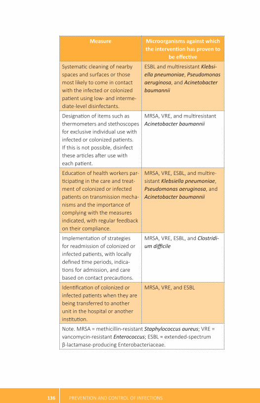
136 PREVENTION AND CONTROL OF INFECTIONS
Measure Microorganisms against which the intervention has proven to
be effective
Systematic cleaning of nearby spaces and surfaces or those most likely to come in contact with the infected or colonized patient using low- and interme-diate-level disinfectants.
ESBL and multiresistant Klebsi-ella pneumoniae, Pseudomonas aeruginosa, and Acinetobacter baumannii
Designation of items such as thermometers and stethoscopes for exclusive individual use with infected or colonized patients. If this is not possible, disinfect these articles after use with each patient.
MRSA, VRE, and multiresistant Acinetobacter baumannii
Education of health workers par-ticipating in the care and treat-ment of colonized or infected patients on transmission mecha-nisms and the importance of complying with the measures indicated, with regular feedback on their compliance.
MRSA, VRE, ESBL, and multire-sistant Klebsiella pneumoniae, Pseudomonas aeruginosa, and Acinetobacter baumannii
Implementation of strategies for readmission of colonized or infected patients, with locally defined time periods, indica-tions for admission, and care based on contact precautions.
MRSA, VRE, ESBL, and Clostridi-um difficile
Identification of colonized or infected patients when they are being transferred to another unit in the hospital or another institution.
MRSA, VRE, and ESBL
Note. MRSA = methicillin-resistant Staphylococcus aureus; VRE = vancomycin-resistant Enterococcus; ESBL = extended-spectrum β-lactamase-producing Enterobacteriaceae.
BASIC RECOMMENDATIONS 137
Sources: Calfee DP, et al. Strategies to prevent methicillin-resis-tant Staphylococcus aureus transmission and infection in acute care hospitals: 2014 update. Infect Control Hosp Epidemiol. 2014;35(7):772-96. Available from: http://www.ncbi.nlm.nih.gov/pubmed/24915205.
European Centre for Disease Prevention and Control. System-atic review of the effectiveness of infection control measures to prevent the transmission of carbapenemase-producing Entero-bacteriaceae through cross-border transfer of patients. ECDC Technical Report. Stockholm: ECDC; 2014.
Muto C, et al. SHEA guideline for preventing nosocomial transmission of multidrug-resistant strains of Staphylococcus aureus and Enterococcus spp. Infect Control Hosp Epidemiol. 2003;24(5):362-86.
Siegel JD, et al. Management of multidrug-resistant organisms in health care settings, 2006. Am J Infect Control. 2007;35(sup-pl 2):S165-93. Available from: https://www.ncbi.nlm.nih.gov/pubmed/18068814
Tacconelli E, et al. ESCMID guidelines for the management of the infection control measures to reduce transmission of multi-drug-resistant Gram-negative bacteria in hospitalized patients. Clin Microbiol Infect. 2014;20(suppl 1):1-55.
Wilson APR, et al. Prevention and control of multidrug-re-sistant Gram-negative bacteria: recommendations from a joint working party. J Hosp Infect. 2015;92(suppl 1):S1-44. Available from: http://linkinghub.elsevier.com/retrieve/pii/S019567011500314X.
Table 14 lists additional measures that can be implemented when the ones above are considered insufficient (after hav-ing confirmed compliance)—for example, when there is a steady increase in endemic infections, hyperendemic situa-tions, or outbreaks of HAIs or in the case of establishments that have adopted policies for the eradication of certain in-fectious agents.

138 PREVENTION AND CONTROL OF INFECTIONS
Tabl
e 14
. Add
ition
al m
easu
res f
or c
onta
inin
g he
alth
care
-ass
ocia
ted
infe
ction
s dur
ing
perio
ds o
f end
emic
dis
ease
Mea
sure
M
icro
orga
nism
s aga
inst
whi
ch
the
inte
rven
tion
has p
rove
n to
be
effec
tive
Impl
emen
tatio
n of
acti
ve c
ase-
findi
ng s
trat
egie
s to
iden
tify
infe
cted
and
col
oniz
ed p
atien
ts th
roug
h de
tec-
tion
cultu
res
or o
ther
test
s fo
r the
pur
pose
with
a v
iew
to is
olati
ng g
roup
s at
risk
, suc
h as
pati
ents
who
are
:•
hosp
italiz
ed in
inte
nsiv
e ca
re u
nits
•
unde
rgoi
ng p
rolo
nged
anti
mic
robi
al tr
eatm
ent
• un
derg
oing
imm
unos
uppr
essi
ve tr
eatm
ent
• di
agno
sed
with
hem
atol
ogic
can
cers
• be
ing
trea
ted
for e
xten
sive
bur
ns•
usin
g in
vasi
ve d
evic
es (i
nvas
ive
mec
hani
cal v
entil
ation
, hem
odia
lysi
s, c
entr
al v
ascu
lar c
athe
ters
)•
from
est
ablis
hmen
ts o
r uni
ts w
ith h
yper
ende
mic
dis
ease
s or
out
brea
ks c
ause
d by
mul
tires
ista
nt in
fec-
tious
age
nts
• sh
arin
g a
room
with
infe
cted
or c
olon
ized
pati
ents
Ther
e ar
e no
con
trol
led
stud
ies
on th
is s
trat
egy.
The
sch
edul
e re
com
men
ded
by e
xper
ts fo
r con
ducti
ng
activ
e ca
se-fi
ndin
g sh
ould
be
cons
iste
nt w
ith th
e ep
idem
iolo
gica
l con
text
. For
exa
mpl
e, s
tart
det
ectin
g in
fecti
ons
upon
adm
issi
on a
nd m
easu
re th
e w
eekl
y pr
eval
ence
to d
isco
ver t
he p
erio
d w
ith th
e la
rges
t num
-be
r of c
ases
. The
n, a
s th
e nu
mbe
r of c
ases
sta
rts
to fa
ll, b
egin
mea
surin
g th
e pr
eval
ence
eve
ry tw
o w
eeks
an
d th
en m
onth
ly, b
imon
thly
, and
, fina
lly, s
emia
nnua
lly.
The
sam
plin
g si
tes
that
hav
e be
en re
com
men
ded
are
as fo
llow
s:•
MRS
A: c
ultu
res
of s
ites
whe
re th
ere
are
brea
ks in
the
skin
(wou
nds,
cut
s, p
unct
ures
), cu
lture
s of
nas
al
and
thro
at s
wab
s (a
lso
endo
trac
heal
, per
igas
tros
tom
y, p
erin
eal,
and
perir
ecta
l sw
abs)
•
VRE:
cul
ture
s or
sw
abs
of th
e re
ctal
or p
erire
ctal
are
a (o
r, in
som
e ca
ses,
cul
ture
s of
sto
ol s
ampl
es)
• M
ultir
esis
tant
Gra
m-n
egati
ve b
acill
i: cu
lture
s of
rect
al o
r per
irect
al s
wab
s, s
ometi
mes
in c
ombi
natio
n w
ith s
wab
s fr
om e
ndot
rach
eal a
spira
te, e
xpec
tora
tion,
spu
tum
, the
nas
al o
r pha
ryng
eal a
reas
, ski
n fr
om
the
axill
ary
or in
guin
al a
reas
, or d
isch
arge
from
wou
nds
MRS
A, V
RE, E
SBL,
and
mul
tires
ista
nt K
lebs
iella
pne
umon
i-ae
, Pse
udom
onas
aer
ugin
osa,
and
Ac
inet
obac
ter b
aum
anni
i (con
t. ne
xt p
age)

BASIC RECOMMENDATIONS 139
Mea
sure
M
icro
orga
nism
s aga
inst
whi
ch
the
inte
rven
tion
has p
rove
n to
be
effec
tive
Stre
ngth
enin
g of
sup
ervi
sion
of t
he d
isin
fecti
on p
roce
ss a
t the
hig
hest
leve
l if t
here
is s
uspi
cion
that
end
o-sc
opic
pro
cedu
res
mig
ht b
e in
volv
ed a
s a
mic
roor
gani
sm tr
ansm
issi
on m
echa
nism
.ES
BL a
nd m
ultir
esis
tant
Kle
bsie
lla
pneu
mon
iae,
Pse
udom
onas
aer
u-gi
nosa
, and
Aci
neto
bact
er b
aum
anni
i
Testi
ng o
f hea
lth w
orke
rs w
ith d
etec
tion
cultu
res
if th
ere
is s
uspi
cion
that
they
mig
ht b
e in
volv
ed in
a m
i-cr
oorg
anis
m tr
ansm
issi
on m
echa
nism
, eith
er to
inte
rrup
t the
col
oniz
ation
pro
cess
or t
o ex
clud
e th
em fr
om
prov
idin
g pa
tient
car
e.
MRS
A an
d ES
BL
Impl
emen
tatio
n of
mec
hani
sms
to a
lert
clin
ical
per
sonn
el w
hen
patie
nts
are
foun
d to
hav
e be
en c
arrie
rs o
r w
ere
hosp
italiz
ed in
uni
ts w
ith c
olon
ized
/inf
ecte
d pa
tient
s, in
ord
er to
initi
ate
isol
ation
mea
sure
s.ES
BL a
nd m
ultir
esis
tant
Kle
bsie
lla
pneu
mon
iae
isol
ates
Isol
ation
of c
ohor
ts.
MRS
A, V
RE, E
SBL,
and
mul
tires
ista
nt
Kleb
siella
pne
umon
iae
isol
ates
Isol
ation
in a
sep
arat
e ro
om.
VRE,
ESB
L, a
nd m
ultir
esis
tant
Kl
ebsie
lla p
neum
onia
e, P
seud
omo-
nas a
erug
inos
a, a
nd A
cine
toba
cter
ba
uman
nii
Dai
ly b
athi
ng o
f pati
ents
in in
tens
ive
care
uni
ts w
ith c
hlor
hexi
dine
soa
p or
chl
orhe
xidi
ne w
ipes
/tow
elett
es.
MRS
A an
d VR
E
In h
ospi
tals
with
cul
ture
det
ectio
n pr
ogra
ms,
era
dica
tion
of M
RSA
nasa
l tra
nsm
issi
on in
nas
al c
arrie
r pa
tient
s w
ith m
upiro
cin
and
bath
ing
of p
atien
ts w
ith c
hlor
hexi
dine
soa
p or
chl
orhe
xidi
ne w
ipes
. The
use
of
mup
iroci
n to
era
dica
te M
RSA
in p
atien
ts w
ithou
t pre
viou
s sc
reen
ing
has
not b
een
show
n to
hav
e an
im
pact
.
MRS
A
Impl
emen
tatio
n of
inte
nsifi
ed p
rogr
ams
to s
uper
vise
cle
anin
g an
d di
sinf
ectio
n, in
clud
ing
supe
rvis
ion
of th
e pr
epar
ation
of d
isin
fect
ants
, with
feed
back
for p
erso
nnel
who
per
form
the
proc
edur
e. N
on-c
ritica
l arti
cles
sh
ould
not
be
shar
ed b
etw
een
patie
nts
unle
ss th
ey a
re in
the
sam
e co
hort
.
ESBL
and
mul
tires
ista
nt K
lebs
iella
pn
eum
onia
e, P
seud
omon
as a
eru-
gino
sa, a
nd A
cine
toba
cter
bau
man
nii
Not
e. M
RSA
= m
ethi
cilli
n-re
sist
ant S
taph
yloc
occu
s aur
eus;
VRE
= v
anco
myc
in-r
esis
tant
Ent
eroc
occu
s; E
SBL
= ex
tend
ed-s
pect
rum
β-la
ctam
ase-
prod
uc-
ing
Ente
roba
cter
iace
ae.

140 PREVENTION AND CONTROL OF INFECTIONS
Sources: Calfee DP, et al. Strategies to prevent methicillin-resis-tant Staphylococcus aureus transmission and infection in acute care hospitals: 2014 update. Infect Control Hosp Epidemiol. 2014;35(7):772-96. Available from: http://www.ncbi.nlm.nih.gov/pubmed/24915205.
European Centre for Disease Prevention and Control. System-atic review of the effectiveness of infection control measures to prevent the transmission of carbapenemase-producing Entero-bacteriaceae through cross-border transfer of patients. ECDC Technical Report. Stockholm: ECDC; 2014.
Muto C, et al. SHEA guideline for preventing nosocomial transmission of multidrug-resistant strains of Staphylococcus aureus and Enterococcus spp. Infect Control Hosp Epidemiol. 2003;24(5):362-86.
Siegel JD, et al. Management of multidrug-resistant organisms in health care settings, 2006. Am J Infect Control. 2007;35(sup-pl 2):S165-93. Available from: https://www.ncbi.nlm.nih.gov/pubmed/18068814.
Tacconelli E, et al. ESCMID guidelines for the management of the infection control measures to reduce transmission of multi-drug-resistant Gram-negative bacteria in hospitalized patients. Clin Microbiol Infect.2014;20(suppl 1):1-55.
Wilson APR, et al. Prevention and control of multidrug-re-sistant Gram-negative bacteria: recommendations from a joint working party. J Hosp Infect. 2015;92(suppl 1):S1-44. Available from: http://linkinghub.elsevier.com/retrieve/pii/S019567011500314X.
The success of measures for dealing with an outbreak or hy-perendemic disease depends on the level of compliance with measures applicable to any endemic disease. If reliable in-formation on the effectiveness of a given measure is lacking, it is important to evaluate its success or failure whenever it is applied.

BASIC RECOMMENDATIONS 141
Is active supervision of those who prescribe antimicrobial drugs (guidance on the use of antibiotics) an effective measure?
Although guidance strategies have been considered effective in the prevention and containment of antimicrobial resis-tance, the studies that have evaluated them have been in-consistent in their results, and these strategies have always been included with other measures for controlling infec-tions (as noted in Tables 13 and 14). Thus, when providing guidance, it should always be offered along with other mea-sures and never as an isolated strategy [17, 18, 19, 20, 21, 22, 23, 24, 25, 26].
References
1. Diaz Granados CA, et al. Comparison of mortality associ-ated with vancomycin-resistant and vancomycin-susceptible enterococcal bloodstream infections: a meta-analysis. Clin Infect Dis. 2005;41(3):327-33.
2. Cosgrove SE. The relationship between antimicrobial resis-tance and patient outcomes: mortality, length of hospital stay, and health care costs. Clin Infect Dis. 2006;42(suppl 2):82-9.
3. Ziakas PD, et al. MRSA and VRE colonization in solid organ transplantation: a meta-analysis of published studies. Am J Transplant. 2014;14(8):1887-94. Available from: http://www.ncbi.nlm.nih.gov/pubmed/25040438.
4. Ziakas PD, et al. Trends and significance of VRE coloniza-tion in the ICU: a meta-analysis of published studies. PloS ONE. 2013;8:9. Available from: http://dx.doi.org/10.1371/journal.pone.0075658.
5. Ziakas PD, Anagnostou T, Mylonakis E. The prevalence and significance of methicillin-resistant Staphylococcus
142 PREVENTION AND CONTROL OF INFECTIONS
aureus colonization at admission in the general ICU set-ting: a meta-analysis of published studies. Crit Care Med. 2014;42(2):433-44. Available from: http://www.ncbi.nlm.nih.gov/pubmed/24145849.
6. Zaky A. Interventions to prevent the acquisition of resistant Gram-negative bacteria in critically ill patients: a systemat-ic review and meta-analysis [master’s thesis]. Available from: https://digital.lib.washington.edu/researchworks/bitstream/handle/1773/21810/ZAKY_washington_0250O_10922.pdf?sequence=1.
7. Zacharioudakis IM, et al. Meta-analysis of methicillin-resis-tant Staphylococcus aureus colonization and risk of infection in dialysis patients. J Am Soc Nephrol. 2014;25(9):2131-41. Available from: http://www.ncbi.nlm.nih.gov/pubmed/ 24652802.
8. Shenoy ES, et al. Natural history of colonization with meth-icillin-resistant Staphylococcus aureus (MRSA) and vancomy-cin-resistant Enterococcus (VRE): a systematic review. BMC Infect Dis. 2014;14(1):177. Available from: http://www.biomedcentral.com/1471-2334/14/177.
9. Nathwani D, et al. Clinical and economic consequenc-es of hospital-acquired resistant and multidrug-resistant Pseudomonas aeruginosa infections: a systematic review and meta-analysis. Antimicrob Resist Infect Control. 2014;3: 32. Available from: http://www.pubmedcentral.nih.gov/articlerender.fcgi?artid=4219028&tool=pmcentrez& rendertype=abstract.
10. Magiorakos A, et al. Bacteria: an international expert pro-posal for interim standard definitions for acquired resis-tance. Clin Microbiol Infect. 2012;18:268-81. Available from: http://onlinelibrary.wiley.com/doi/10.1111/j.1469-0691.2011.03570.x/full.
11. Wilson APR, et al. Prevention and control of multidrug-re-sistant Gram-negative bacteria: recommendations from a joint working party. J Hosp Infect. 2015;92(suppl 1):S1-44. Available from: http://linkinghub.elsevier.com/retrieve/pii/S019567011500314X.

BASIC RECOMMENDATIONS 143
12. European Centre for Disease Prevention and Control. Sys-tematic review of the effectiveness of infection control mea-sures to prevent the transmission of carbapenemase-pro-ducing Enterobacteriaceae through cross-border transfer of patients. ECDC Technical Report. Stockholm: ECDC; 2014. Available from: http://ecdc.europa.eu/en/publications/ Publications/CPE-systematic-review-effectiveness-infection- control-measures-to-prevent-transmission-2014.pdf.
13. Calfee DP, et al. Strategies to prevent methicillin-resistant Staphylococcus aureus transmission and infection in acute care hospitals: 2014 update. Infect Control Hosp Epidemiol. 2014;35(7):772-96. Available from: http://www.ncbi.nlm.nih.gov/pubmed/24915205.
14. Tacconelli E, et al. ESCMID guidelines for the management of the infection control measures to reduce transmission of multidrug-resistant Gram-negative bacteria in hospitalized patients. Clin Microbiol Infect. 2014;20(suppl 1):1-55.
15. Siegel JD, et al. Management of multidrug-resistant organ-isms in health care settings, 2006. Am J Infect Control. 2007;35(suppl 2):S165-93. Available from: https://www.ncbi. nlm.nih.gov/pubmed/18068814.
16. Muto C, et al. SHEA guideline for preventing nosocomial transmission of multidrug-resistant strains of Staphylococcus aureus and Enterococcus spp. Infect Control Hosp Epidemiol. 2003;24(5):362-86.
17. Loveday HP, et al. A systematic review of the evidence for interventions for the prevention and control of meticil-lin-resistant Staphylococcus aureus (1996-2004): report to the Joint MRSA Working Party (Subgroup A). J Hosp Infect. 2006;63(suppl 1):S45-70.
18. Davey P, et al. Interventions to improve antibiotic prescrib-ing practices for hospital inpatients. Cochrane Database Syst Rev. 2017 Feb 9;2:CD003543. Available from: https://www.ncbi.nlm.nih.gov/pubmed/28178770.
19. de Bruin MA, Riley LW. Does vancomycin prescribing in-tervention affect vancomycin-resistant enterococcus infection

144 PREVENTION AND CONTROL OF INFECTIONS
and colonization in hospitals? A systematic review. BMC In-fect Dis. 2007;7:24. Available from: http://www.scopus.com/inward/record.url?eid=2-s2.0-34248204669&partnerID= tZOtx3Any1.
20. Filice G, et al. Antimicrobial stewardship programs in inpa-tient settings: a systematic review. Washington, DC: US De-partment of Veterans Affairs; 2013.
21. Drekonja DM, et al. Antimicrobial stewardship in outpatient settings: a systematic review. Infect Control Hosp Epidemiol. 2015;36(2):142-52. Available from: http://www.ncbi.nlm.nih.gov/pubmed/25632996.
22. Karanika S, et al. Clinical and economic outcomes from the implementation of hospital-based antimicrobial stewardship programs: a systematic review and meta-analysis. Antimicrob Agents Chemother. 2016;60(8):4840-52. Available from: http://aac.asm.org/lookup/doi/10.1128/AAC.00825-16.
23. Kaki R, et al. Impact of antimicrobial stewardship in crit-ical care: a systematic review. J Antimicrob Chemoth. 2011;66(6):1223-30. Available from: http://www.jac.oxford-journals.org/cgi/doi/10.1093/jac/dkr137.
24. Schuts EC, et al. Current evidence on hospital antimicrobial stewardship objectives: a systematic review and meta-analy-sis. Lancet Infect Dis. 2016;16(7):847-56.
25. Smith MJ, Gerber JS, Hersh AL. Inpatient antimicrobi-al stewardship in pediatrics: a systematic review. J Ped In-fect Dis. 2015;4(4):e127-35. Available from: http://jpids. oxfordjournals.org/cgi/doi/10.1093/jpids/piu141.
26. Zhang YZ, Singh S. Antibiotic stewardship programmes in intensive care units: why, how, and where are they lead-ing us? World J Crit Care Med. 2015;4(1):13-28. Available from: http://www.pubmedcentral.nih.gov/articlerender.fcgi? artid=4326760&tool=pmcentrez&rendertype=abstract.

BASIC RECOMMENDATIONS 145
Authors: Fernando Otaiza, Mauro Orsini, Monica Pohlenz (Chile Ministry of Health)
Editors: Valeska de Andrade Stempliuk, Ricardo Bustamante
Technical review: Maria Roxane Salvatierra
Collaborators: Aguayo, Belisario (Santiago, Chile); Albornoz, Henry (Montevideo, Uruguay); Arce, Marlen (San José, Costa Rica); Bernal, Cornelia (Itaugua, Paraguay); Clara, Liliana (Buenos Aires, Argentina); Corredor, Sandra (Bogotá, Colombia); Cruz, Carolina (Valdivia, Chile); Cuéllar, Luis (Lima, Peru); Féliz Féliz, Luis Elpidio (Santo Domingo, Dominican Republic); Garro, Gladys (Lima, Peru; Panama City, Panama); Giuffré, Carolina (Buenos Aires, Argentina); Holt, Nancy (Asunción, Paraguay); Hoyos, Claudia (Quito, Ecuador); Leal, Aura Lucia (Bogotá, Colombia); Lopez, Patricia (Santiago, Chile); Maimone, Stella (Buenos Aires, Argentina); Mejia, Carlos (Guatemala City, Guatemala); Padoveze, Maria Clara (São Paulo, Brazil); Silvestre, Monica (Guatemala City, Guatemala); Vanegas, Blanca Stella (Bogotá, Colombia); Vega, Maria (Asunción, Para-guay); Volkow, Patricia (Mexico City, Mexico); Zubieta, Miriam (La Paz, Bolivia); Zurita, Jeannete (Quito, Ecuador).
Design: Rosario Muñoz


525 Twenty-third Street, N.W., Washington, D.C. 20037, United States of America