prevention and control of coxiella burnetii infection among humans
TRANSCRIPT

1
Prevention and Control of Coxiella burnetii Infection among Humans and Animals: Guidance for a Coordinated Public
Health and Animal Health Response, 2013
National Association of State Public Health Veterinarians National Assembly of State Animal Health Officials
Q fever, an acute or chronic zoonotic illness caused by the bacterium Coxiella burnetii, has received international attention in recent years, primarily due to a large‐scale outbreak in the Netherlands from 2007 to 2010 involving more than 4,000 human cases and the euthanasia of 50,000 goats, one of the primary reservoirs for the bacterium (94). In 2011, a Q fever outbreak in the northwestern U.S. implicated 21 goat farms in three states and resulted in 20 human infections (21). The Netherlands outbreak, the largest reported in history, and the recent U.S. outbreak illustrate the importance of a coordinated animal and human health response to such outbreaks and the need for comprehensive response guidance for public health and animal health officials. In January 2012, the NASPHV Q Fever Committee was formed under joint leadership of the National Association of State Public Health Veterinarians (NASPHV) and the National Assembly of State Animal Health Officials (NASAHO) to formulate recommendations for a coordinated response to Q fever outbreaks. This document provides a brief background on the epidemiology, diagnosis and management of C. burnetii infections in humans and animals, as well as guidance for conducting an integrated investigation and response to C. burnetii infection among humans and animals. The recommendations are intended to guide public health officials, animal health officials, physicians, veterinarians, and others concerned with control of C. burnetii infection and protection of public health. Committee Members: Alicia Anderson, Centers for Disease Control and Prevention Tom Boyer, American Goat Federation Ann Garvey, Co‐chair, National Association of State Public Health Veterinarians Katherine Marshall, USDA Centers for Epidemiology and Animal Health Paula Menzies, University of Guelph Julia Murphy, National Association of State Public Health Veterinarians Paul Plummer, Iowa State University Gatz Riddel, American Association of Bovine Practitioners Paul Rodgers, American Sheep Industry Joni Scheftel, National Association of State Public Health Veterinarians Tahnee Szymanski, Co‐Chair, National Assembly of State Animal Health Officials

2
Introduction
Coxiella burnetii is an intracellular, gram‐negative bacterium that is the causative agent of Q fever in humans and coxiellosis in animals. The organism is ubiquitous in the environment where it can persist in a spore‐like form for years (97). Goats, sheep and cattle are the species most clinically affected by infection and are most often implicated as the source of human infection (14,73). Q fever has long been considered an occupational disease associated with exposure to livestock by farmers, veterinarians, slaughter facility workers, and animal researchers, although it is not restricted to these groups. The primary mode of transmission to humans is inhalation of pathogen‐contaminated dust or aerosols. Coxiella burnetii is extremely hardy and may become airborne, travelling on wind currents for over a mile (66,93). Thus, exposure to C. burnetii can occur without direct contact with infected animals. In fact, 63% of U.S. cases reported to CDC from 2000 to 2010 did not report contact with livestock (3). Upon notification of a human case of Q fever, a public health agency must attempt to determine the likely source of infection. In the course of investigating a single Q fever case, public health officials may seek assistance from their jurisdiction's animal health agency. However, for larger case clusters or outbreak investigations, a well‐coordinated, public health‐animal health response is necessary. Q fever outbreak investigations involving humans and animals are complex due to the primarily aerosol transmission of the organism, the ubiquitous and environmentally persistent nature of the organism, the low incidence of clinical disease in most infected herds, chronic shedding in animals, difficulties with test result interpretation in all species, and the possibility of chronic infection of humans. Managing these complexities successfully requires close collaboration between human health and animal health professionals. This document provides background information on C. burnetii infection in humans and animals and details measures for public health and animal health officials to consider when conducting a joint investigation. In addition, Appendices 1‐8 contain a series of template documents—fact sheets, health alerts for the medical and veterinary communities, etc.—to assist with response. The recommendations herein are intended to provide a framework for response in the United States. They are not intended to serve as a comprehensive resource for human or animal case management or to supersede state and local regulations. This guidance was formulated based on the best current evidence and the collective experience and knowledge of the National Q Fever Committee members. The Committee expects to update the document periodically as more information becomes available.

3
I. Infection in Humans (Q fever) Epidemiology Q fever is a zoonotic disease with global distribution that was first recognized in the U.S. in the early 1940’s and made a nationally notifiable disease in 1999. Since then, the number of human Q fever cases reported annually to the Centers for Disease Control and Prevention (CDC) has increased from 17 cases with onset in 2000 to 167 cases with onset in 2008 (24). Approximately 3% of the U.S. adult general population is seropositive for C. burnetii (2).
Goats, sheep and cattle are the principle sources of human infection. The primary mode of transmission is inhalation of pathogen‐contaminated aerosols from excreta, especially birth products. Parturient cats, kittens, dogs and other animals have also been associated with human cases (17,53,54,58,59). C. burnetii is easily dispersed into the air and airborne transmission of the disease to people living over a mile from the animal sources has been reported (64). As a result, Q fever can occur in persons without occupational risk or other contact with livestock or their environments (36,75,89). Transmission may also occur from tick bites, and a number of tick species have been implicated (44,89). Foodborne transmission from drinking contaminated raw milk (86), sexual transmission, and indirect transmission after exposure to wool, straw, clothing, linens, and other contaminated materials have been documented (63). Humans are highly susceptible to C. burnetii infection, with an infectious dose of one to ten organisms (48,90). Person‐to‐person transmission occurs rarely. These factors make it difficult to define what constitutes an exposure to Q fever and to identify potential human cases during an investigation. Clinical Presentation in Humans Acute Q Fever The incubation period of Q fever is typically around 20 days, with a range of 3 to 30 days (45). As many as 50% of human C. burnetii infections are asymptomatic; symptomatic infections most commonly present as a non‐specific febrile illness that may occur in conjunction with pneumonia or hepatitis (22,23,63). Untreated, acute Q fever has a low case fatality rate (<2%) and, when treated, the case‐fatality rate is negligible (63). Acute Q fever is characterized by sudden onset of fever to 104º‐105º F, chills, profuse sweating, severe headache with retro orbital pain, weakness, nausea, vomiting, diarrhea, non‐productive cough, and abdominal or chest pain. Untreated, the fever can persist for up to 9 to14 days (45). Approximately 30% to 50% of patients develop pneumonia. Acute hepatitis, meningoencephalitis, and myocarditis can also occur. Abnormal liver function tests and thrombocytopenia are common. Pregnant women are considered to be at increased risk for pre‐term delivery or miscarriage although a recently published prospective study of pregnant women from the Netherlands outbreak did not identify an increased risk associated with seropositive status (4,19,74,93). Although the majority of people with acute Q fever recover completely, a post‐Q fever fatigue syndrome has been reported in 20% to 42% of acute cases (12,11,38,56,65,94). This syndrome

4
is characterized by constant or recurring fatigue, night sweats, severe headaches, photophobia, myalgia, mood changes, and difficulty sleeping, which persist for more than one year following acute infection (57). Chronic Q fever Chronic Q fever most commonly presents as endocarditis occurring weeks to years after an acute infection (28). Other manifestations include vascular infections and infections of the bone, liver or reproductive organs (63). Chronic Q fever is rare, occurring in fewer than 5% of acute cases. Persons with valvular surgery, valvular prosthesis or aneurisms are at highest risk for development of chronic Q fever. Other risk factors include renal insufficiency, age > 60 years, pregnancy, and immunosuppression (50).
Testing Methods & Interpretation Diagnosis of acute Q fever in people exhibiting clinical signs requires demonstration of a four‐fold rise in antibody titer or detection of C. burnetti organisms or DNA in blood or tissue (63).
The indirect fluorescent antibody test (IFA) is the most commonly used serological test for diagnosis of acute and chronic Q fever and for follow‐up of patients at risk for chronic Q fever (27,43,72,96). There are two distinct antigenic phases of C. burnetii infection detected by serology: Phase I and Phase II. In acute Q fever, the Phase II IgG titer is elevated and is higher than the Phase I IgG titer. In chronic Q fever, the Phase I IgG titer is also elevated and is typically higher than the Phase II IgG titer (2). Testing of paired serum samples to demonstrate a four‐fold rise in Phase II IgG antibody titer is required to confirm a diagnosis of acute Q fever (23,96). The first sample should be taken as soon after onset of illness as possible, preferably during the first week, and the second sample should be taken two to four weeks later. Detectable antibody titers are not typically present until seven to ten days after illness onset; therefore negative or very low titers are expected during the first week of illness and do not rule out Q fever (23,63). A single positive convalescent serum IgG phase II>1:128 in a patient with Q fever symptoms of over 1 week’s duration indicates a probable acute infection (3). IFA serology for Phase I and Phase II IgM antibodies may provide ancillary information for diagnosis of both acute and chronic C. burnetii infection. However IgM antibodies may persist for months or longer and IgM has a much lower specificity for C. burnetii than IgG (27,75). Cross reactions with Legionella and Bartonella species have been reported (30,55,69). During the first week of acute illness and prior to antibiotic administration, whole blood or serum can be tested by PCR for C. burnetii (23,32,52,98). Although a positive PCR result is diagnostic, a negative result does not rule out Q fever, and treatment should not be withheld due to a negative result. Routine blood cultures cannot detect the organism.

5
Treatment and Monitoring in Humans Doxycycline has the highest therapeutic efficacy against C. burnetii and is the treatment of choice for acute Q fever in adults, children over age 8, and in children of all ages with severe illness (3,23). Oral doxycycline should be given for 14 to 21 days at a dosage of 100 mg twice daily to adults and 2.2 mg/kg body weight twice daily to children under 45 kg (100 lbs). When tetracyclines are contraindicated (e.g. for children under 8 years of age, pregnant women, and those allergic to tetracyclines), other antibiotics may be used, such as trimethoprim/sulfamethoxazole, fluoroquinolones or macrolides (3,63). The benefits of using doxycycline outweigh the potential risk of staining of permanent teeth in severely ill or hospitalized children under age 8 with acute Q fever (23).Treatment should be given within the first 3 days of illness for maximum efficacy and should not be delayed pending the results of laboratory tests or withheld because of the initial negative laboratory result in the first week of illness (3). Prophylactic treatment after a suspected exposure to C. burnetti is not recommended as this may extend the incubation period but is not likely to prevent infection from occurring (23). Treatment of asymptomatic or resolved infections is not routinely recommended either, although it may be considered in patients with risk factors for development of chronic Q fever infections (3). Healthy patients with no identified risk factor for development of Chronic Q fever should be clinically evaluated and serologically tested by IFA 6 months following acute infection (3). Patients with cardiac or other risk factors for development of chronic disease should be clinically evaluated and serologically tested by IFA at 3, 6, 12, 18, and 24 months following acute infection (3). Pregnant women should be clinically evaluated and serologically tested by IFA at 3, 6, 12, 18, and 24 months following delivery (3). Chronic Q fever patients should be referred to an infectious disease specialist for management. These patients require long‐term antibiotic therapy using a combination of doxycycline and hydroxychloroquine, periodic diagnostics, and long‐term monitoring (28). More specific and detailed information about the diagnosis, treatment, and management of acute and chronic Q fever infection in humans may be found at www.cdc.gov/mmwr/PDF/rr/rr6203.pdf . II. Infection in Animals (Coxiellosis) Epidemiology
Coxiella burnetii infection has been documented in a broad range of animals including almost all mammals that have been tested, as well as birds and ticks (59,63). Studies of feral and domestic cats have found that prevalence (based on either PCR or antibodies) can range from 8.5% in client‐owned animals, to 41.7% in stray cats (18,53,73). Goats, sheep, and cattle are the domestic species most clinically affected by C. burnetii infection, and are most often implicated

6
in transmission to humans (14,73). Coxiella burnetii infection in animals is reportable to animal health agencies in 44 states. Coxiella burnetii infection is common in U.S. dairy cattle herds. A 2007 national dairy study that included testing of bulk tank milk samples reported that 77% of 528 operations in the U.S. were positive for C. burnetii (70). In a similar bulk tank milk study of U.S. veterinary‐school‐associated dairy herds, 22 of 24 were positive for C. burnetii antibodies using an immunofluorescence assay (65). In addition, a three‐year study of dairy herds in the Northeast U.S. reported a herd level prevalence (using PCR) of more than 94%, (51). Animals may become infected by direct contact with infected animals and contaminated environments and/or from inhalation of aerosolized bacteria. Birth products (including placenta, fetuses, and amniotic and allantoic fluids), excreta, and milk are the most likely sources of infection (45,73,78). Because the spore‐like form of the bacterium can survive for years in the environment and travel long distances as an aerosol, dry, windy conditions may contribute to animal exposure and disease transmission (60). The bacteria have also been found in ticks, which may also serve as a source of infection for animals (9,92). Duration, quantity and route of shedding can vary by host species. Goats commonly shed C. burnetii in birth products, feces and milk (80,84). Goats have also been shown to shed the organism in vaginal mucus, even nulliparous animals and goats delivering healthy‐appearing live‐born kids (1,80,82). Infected sheep are more likely to shed C. burnetii in birth products, vaginal discharge, and feces, and are less likely than cattle to shed the organism persistently in milk (80). Cattle may shed the organism in milk for weeks to months after calving (34). Coxiella burnetii has been detected in the feces, milk, urine, vaginal discharge, semen, and birth products of infected animals (5,40,61). Regardless of species, the highest numbers of organisms are shed in conjunction with an adverse pregnancy event (abortion, stillbirth, or neonatal weakness) (42). Clinical presentation in animals Coxiella burnetii infection in livestock species is generally asymptomatic. Goats and sheep are the species in which abortions, stillbirths, and early neonatal mortality have been most frequently documented (15,38,40,63,73). Abortion in cattle due to C. burnetii infection has been reported, and more research is needed to determine the organism’s role in infertility, metritis and endometritis (56,68,91). Coxiella burnetii can cause adverse pregnancy outcomes in cats, and contact with infected cats has been associated with human infection (18,53,59). Testing Methods and Interpretation Serological Tests Serological testing methods available for the detection of C. burnetii in animals include complement fixation (CF), enzyme‐linked immunosorbent assay (ELISA) and indirect IFA tests. ELISA and IFA are more sensitive than CF, and are the preferred diagnostic tests (29,73). Serology is best utilized and interpreted on a herd‐ or flock level to demonstrate C. burnetii infection or exposure within a population of animals. Serology is less valuable in individual

7
animals, either to absolutely determine if C. burnetii was the etiologic agent of an abortion or to accurately identify infected animals as part of a control program. The diagnostic value of serology is limited because it may be two to three weeks before some animals seroconvert, and others may have persistent serological titers that last for years. Within an infected herd, some animals (10‐20%) remain serologically negative while shedding the organism, while other animals that test serologically positive may not be shedding the organism (15,16,34,79). Antigen Detection Coxiella burnetii antigen can be detected using PCR, immunohistochemistry, histology or culture. PCR performed on tissues is a highly sensitive and specific method for detecting C. burnetii infection (49). A positive PCR assay performed on bodily secretions (e.g. birth products, vaginal secretions, milk, or feces) indicates that the animal is currently infected. The relative level of organisms present, as measured by quantitative real‐time PCR, may help in determining whether C. burnetii caused an adverse pregnancy event, with higher levels of organisms more likely linked to such events (41). Histology and immunohistochemistry of placental cotyledons is useful for identifying C. burnetii as the etiologic agent of abortion where available. Culture is generally avoided because isolation of C. burnetii is difficult, hazardous, and time‐consuming and requires a biosafety Level 3 laboratory (29,61).
Investigating C. burnetii infection status by species Recommendations for sample collection and testing vary based upon the species being investigated (80). Species‐specific recommendations are in Appendix 7. Treatment in animals More research is needed to determine the effectiveness of treating entire herds or individual animals to prevent or control shedding of C. burnetii and to prevent adverse pregnancy events associated with coxiellosis. Use of tetracycline in feed during pregnancy cannot be recommended as a herd‐level control measure for C. burnetii, due to low bioavailability of the drug with oral administration (77). Based upon current data, antibiotic usage in the absence of ongoing abortions is not warranted. When abortions are ongoing, parenteral treatment of individual animals (two injections of long‐acting oxytetracycline at 20 mg/kg given 20 days apart) in late gestation to prevent adverse pregnancy events may be helpful, although data are sparse and inconclusive. Though it may prevent abortions, parenteral treatment of individual animals does not appear to reduce shedding of the organism in birth fluids or to change serological status (6,7,14,87). Use of any antimicrobial to control abortion in the U.S. constitutes extra‐label drug use and must be prescribed by a licensed veterinarian with a valid veterinary‐client‐patient relationship. Controlling transmission during an outbreak Animal infection is thought to occur primarily through exposure to contaminated environments. Given the ubiquitous nature of C. burnetii and its persistence in the environment, complete eradication of the bacteria from an infected farm would be nearly impossible. Nevertheless, transmission can be reduced with good hygiene and other management practices that reduce

8
environmental load, such as immediately removing and disposing of aborted fetuses, dead newborns, and placentas. To control spread of coxiellosis between premises, producers should refrain from moving or selling animals, particularly periparturient animals, while abortions are ongoing. Official animal quarantine is generally not recommended, and mass euthanasia of infected herds is never recommended for several reasons:
Coxiella burnetii infection is endemic in animals in the US.
Coxiella burnetii is ubiquitous and persistent in the environment.
With the tools currently available, it is nearly impossible to eliminate infection from a herd and to completely decontaminate the environment.
There is no known effective treatment to eliminate shedding of bacteria in infected animals.
It would be extremely difficult to repopulate a herd with C. burnetii negative animals.
Defining the terms for release of quarantine would be almost impossible because of the factors listed above.
Coxiella burnetii is shed in the milk of infected animals; therefore, their milk should not be consumed raw or sold unpasteurized direct to consumers. Pasteurizing milk at 145° F (63° C) for at least 30 minutes or at 161° F (72° C) for 15 seconds is sufficient to destroy C. burnetii, as well as other pathogens that can be present in raw milk (31). III. Joint Public Health and Animal Health Investigation & Response The following situations should prompt an initial investigation and interview to determine the facts and provide education regarding Q fever risk to humans, but would generally not require a comprehensive joint public health and animal health investigation and response:
A single, sporadic case of Q fever in an individual that cannot be linked to an animal source or to another human case.
A diagnosis of coxiellosis in animals without associated human illness. A joint public health and animal health epidemiologic investigation is warranted under the following situations:
A human C. burnetii case is reported to public health officials where the initial interview identifies a potential animal source of infection.
Coxiellosis is identified in animals where there are reports of a febrile, influenza‐like illness in exposed persons.
Multiple human Q fever cases occur in a geographic or temporal cluster.
Other situations at the discretion of public health and animal health authorities. Public health and animal health roles and responsibilities in a joint investigation and response to a Q fever outbreak involving both humans and animals are described below. The recommended actions delineated here are neither all inclusive nor all necessarily required in

9
every situation. Public health and animal health roles and responsibilities may merge or overlap in a given situation. There may also be additional entities that should be engaged in the investigation and response, and these entities will vary, depending upon the specific situation. More detailed information regarding each step is found in the body of this document and appendices. Public Health Response: During a joint epidemiologic investigation involving both human and animal C. burnetii infection, public health authorities should consider the following actions:
1. Interview human Q fever cases for illness and exposure history (Appendix 1). 2. Identify people in contact with or potentially exposed through aerosol transmission to
animals with suspected or confirmed coxiellosis. a. Interview potentially exposed persons for exposure and illness history (Appendix
1). b. Consider using media outlets to identify potential cases if there is evidence of C.
burnetii transmission to persons without direct animal exposure. c. Provide educational materials on Q fever to all potentially exposed persons
(Appendix 2). d. Recommend Q fever testing for exposed persons reporting compatible
symptoms and for all potentially exposed persons with heart valve disease or prosthetic heart valves, pregnancy, or immunosuppressive conditions that put them at higher risk for developing chronic Q fever complications.
e. Report probable and confirmed cases to CDC via state health agencies (Appendix 4).
3. Discuss with animal owners and caretakers, risks, routes of transmission, how to prevent additional human exposures, and the need to seek medical attention should symptoms develop (Appendix 2).
a. Provide personal protective equipment (PPE) guidance (Appendix 3). b. Recommend that visitors be given limited access to animal birthing areas,
particularly when abortions are occurring. Limited access is especially important for persons with conditions that put them at higher risk for complications (e.g., heart valve disease, prosthetic heart valves, pregnancy, and immunosuppressive conditions).
c. Recommend that raw milk and raw milk products not be consumed or sold. 4. Alert health care providers and medical clinics in surrounding areas (Appendix 5).
a. Include an overview of case findings to date. b. Include guidance on diagnosis, testing, and treatment. c. Ask healthcare providers to report suspected cases to public health authorities.
5. Provide guidance to healthcare providers on diagnostic testing options, test result interpretation, and treatment. More information about the diagnosis, treatment, and management of human Q fever may be found at www.cdc.gov/mmwr/PDF/rr/rr6203.pdf .
6. In partnership with animal health officials, provide outbreak information to media outlets and stakeholders as appropriate.

10
7. Perform epidemiologic analysis to characterize case demographics and to identify risk factors associated with human illness.
Animal Health Response: During a joint epidemiologic investigation involving both human and animal C. burnetii infection, animal health authorities should consider the following actions:
1. Perform an on‐site investigation to collect information on the species and number of animals on the premises, as well as animal management practices.
a. Interview the animal owner or caretaker to assess the history and extent of any animal illness, particularly abortions occurring in recent years.
b. Obtain animal movement history, including animals recently moved into or out of the herd or flock.
c. Ascertain whether there are individuals who live on the premises or have contact with the animals who report a febrile, influenza‐like illness and, if so, contact public health authorities immediately.
2. Contact the herd/flock veterinarian. a. Provide diagnostic testing options and assistance (Appendix 7). b. Discuss treatment considerations (Appendix 7).
3. Discuss with the animal owner and herd/flock veterinarian human and animal illness, persons at higher risk for complications, routes of transmission, measures to prevent human exposures, and the need to seek medical attention should symptoms develop (Appendix 2).
a. Provide personal protective equipment (PPE) guidance (Appendix 3). b. Recommend that visitors be given limited access to animal birthing areas,
particularly when abortions are occurring. Limited access is especially important for persons with conditions that put them at higher risk for complications (e.g., heart valve disease, prosthetic heart valves, pregnancy, and immunosuppressive conditions).
c. Recommend that raw milk and raw milk products not be consumed. Prohibit direct sales of raw milk and raw milk products to consumers. There is no need to prohibit movement of milk for pasteurization.
d. Recommend that animals, particularly periparturient animals, not be sold or moved during an abortion storm.
e. Provide manure management and carcass disposal consultation in cooperation with state or local environmental agencies (Appendix 8).
4. Alert veterinarians in surrounding areas (Appendix 6). a. Provide an overview of case findings to date. b. Provide guidance on clinical signs, diagnostic testing, test interpretation and
treatment. c. Ask that veterinarians report suspected or confirmed coxiellosis cases to animal
health authorities. 5. In partnership with public health officials, provide outbreak information to media
outlets and stakeholders as appropriate.

11
6. Perform epidemiologic analysis to characterize outbreak animal health demographics and to identify risk factors associated with the identification of coxiellosis in animals on a premises.
Recommendation for Future Action The Committee identified availability of a veterinary vaccine as a critical need in responding to a Q fever outbreak. Evidence from countries in Europe and elsewhere suggests that use of a Coxiella vaccine in animals is an effective means of minimizing disease transmission (8,10,26,47). At present, there are at least two commercially available C. burnetii vaccines in other parts of the world, but there is no C. burnetii vaccine available in the United States.

12
REFERENCES:
1. Alsaleh A, Pellerin JL, Rodolakis A, Larrat M, Cochonneau D, Bruyas JF, Fieni F. Detection of Coxiella burnetii, the agent of Q fever, in oviducts and uterine flushing media and in genital tract tissues of the non pregnant goat. Comp Immunol, Microbiol Infect Diseases 2011;34:355‐360.
2. Anderson AD, Kruszon‐Moran D, Loftis AD, McQuillan G, Nicholson WL, Priestley RA, et al. Seroprevalence of Q Fever in the United States, 2003‐2004. Am J Trop Med Hyg 2009 Oct;81(4):691‐4.
3. Anderson A, Bijlmer H, Fournier JE, Graves S, Hartzell J, Kersh G, Limonard G, Marrie T, Massung R, McQuiston J, Nicholson W, Paddock C, Sexton D. Diagnosis and Management of Q Fever‐ United States, 2013: Recommendations from the CDC and the Q Fever Working Group. MMWR 2013;62(RR03);1‐23.
4. Angelakis E, Million M, D'Amato F, Rouli L, Richet H, Stein A, Rolain JM, Raoult D. Q fever and pregnancy: disease, prevention, and strain specificity. Eur J Clin Microbiol Infect Dis 2012 Sep 28. [Epub ahead of print]
5. Arricau‐Bouvery N, Souriau A, Lechopier P, Rodolakis A. Experimental Coxiella burnetii infection in pregnant goats: Excretion routes. Vet Res 2003;34:423–433.
6. Astobiza I, Barandika JF, Hurtado A, Juste RA, Garcia‐Perez AL. Kinetics of Coxiella burnetii excretion in a commercial dairy sheep flock after treatment with oxytetracycline. Vet J 2010;184:172‐175.
7. Astobiza I, Barandika JF, Juste RA, Hurtado A, Garcia‐Perez AL. (2012). Evaluation of the efficacy of oxytetracycline treatment followed by vaccination against Q fever in a highly infected sheep flock. Vet J 2012 Sep 11.Epub ahead of print]
8. Astobiza I, Barandika JF, Ruiz‐Fons F, Hurtado A, Povedano I, Juste RA, García‐Pérez AL. Four‐year evaluation of the effect of vaccination against Coxiella burnetii on reduction of animal infection and environmental contamination in a naturally infected dairy sheep flock. Appl Environ Microbiol 2011 Oct;77(20):7405‐7
9. Astobiza I, Barral M, Ruiz‐Fons F, Barandika J.F, Gerrikagoitia X, Hurtado A, Gardia‐Perez AL. Molecular investigation of the occurrence of Coxiella burnetii in wildlife and ticks in an endemic area. Vet Microbiol 2011;147:190‐194.
10. Astobiza I, Ruiz‐Fons F, Piñero A, Barandika JF, Hurtado A, García‐Pérez AL. Estimation of Coxiella burnetii prevalence in dairy cattle in intensive systems by serological and molecular analyses of bulk‐tank milk samples. J Dairy Sci 2012 Apr;95(4):1632‐8.
11. Ayres JG, Flint N, Smith EG, Tunnicliffe WS, Fletcher TJ, Hammond K, Ward D, Marmion BP. Post‐infection fatigue syndrome following Q fever. Q J Med 1998 Feb;91(2):105‐23.
12. Ayres JG, Smith EG, Flint N. Protracted fatigue and debility after acute Q fever. Lancet 1996;347:978‐9.
13. Barlow J, Rauch B, Welcome F, Kim SG, Dubovi E, Schukken Y. Association between Coxiella burnetii shedding in milk and subclinical mastitis in dairy cattle. Vet Res 2008;39:23‐32.
14. Berri M, Crochet D, Santiago S, Rodolakis A. Spread of Coxiella burnetii infection in a flock of sheep after an episode of Q fever. Vet Rec 2005;157:737‐740.
15. Berri M, Rousset E, Champion FL, Russo P, Rodolakis A. Goats may experience reproductive failures and shed Coxiella burnetii at two successive parturitions after a Q fever infection. Res Vet Sci 2007;83:47‐52.
16. Berri M, Souriau A, Crosby M, Crochet D, Lechopier D, Lechopier P, Rodolakis A. Relationship between the shedding of Coxiella burnetii, clinical signs and serological responses of 34 sheep. Vet Rec 2001;148: 502‐505.

13
17. Buhariwalla F, Cann B, Marrie TJ. A dog‐related outbreak of Q fever. Clin Infect Dis 1996;23:753‐755.
18. Cairns K, Brewer M, Lappin MR. Prevalence of Coxiella burnetii DNA in vaginal and uterine samples from healthy cats of north‐central Colorado. J Feline Med Surg 2007;9:196‐201.
19. Carcopino X, Raoult D, Bretelle F, Boubli L, Stein A. Q Fever during Pregnancy. Ann NY Acad Sci 2009;1166(1):79‐89.
20. CDC. Case definitions for infectious conditions under public health surveillance. (2009). National Notifiable Diseases Surveillance System, CDC. Retrieved February 20, 2012. Available at: http://www.cdc.gov/osels/ph_surveillance/nndss/casedef/q_fever_2009.htm
21. CDC. Notes from the field: Q fever outbreak associated with goat farms – Washington and Montana, 2011. MMWR 2011;60(40):1393.
22. CDC. Q Fever. (2011). Retrieved November 26, 2012, from http://www.cdc.gov/qfever/index.html
23. CDC. Q fever: Symptoms, Diagnosis and Treatment. (2011). Retrieved November 27, 2012, from http://www.cdc.gov/qfever/symptoms/index.html
24. CDC. Q fever: Statistics and Epidemiology. (2011). Retrieved February 20, 2012, from http://www.cdc.gov/qfever/stats/index.html
25. Dagleish MP, Benavides J, Chianini F. (2010). Immunohistochemical diagnosis of infectious diseases of sheep. Small Rumin Res. 92:19‐35
26. de Cremoux R, Rousset E, Touratier A, Audusseau G, Nicollet P, Ribaud D, David V, Le Pape M. Assessment of vaccination by a phase I Coxiella burnetii‐inactivated vaccine in goat herds in clinical Q fever situation. FEMS Immunol Med Microbiol 2012 Feb;64(1):104‐6.
27. Dupont HT, Thirion X and Raoult, D. Q fever serology: cutoff determination for microimmunofluorescence. Clin Diagn Lab Immunol 1994 Mar;1(2):189‐96.
28. Fennolar F, Fournier PE, Carrieri MP, Habib G, Messana T, Raoult D. (2001). Risks factors and prevention of Q fever endocarditis. Clin Infect Dis. Aug: 33 (3): 312‐316.
29. Field PR, Mitchell JL, Santiago A, Dickeson DJ, Chan S‐W, Ho DWT, Murphy AM, Cuzzubbo AJ, Devine PL. Comparison of a commercial Enzyme‐Linked Immunosorbent Assay with immunofluorescence and complement fixation tests for detection of Coxiella burnetii (Q fever) immunoglobulin M. J Clin Micro 2000;38:1645‐1647.
30. Finidori JP, Raoult D, Bornstein N, Fleurette J. Study of cross‐reaction between Coxiella burnetii and Legionella pneumophila using indirect immunofluorescence assay and immunoblotting. Acta Virol 1992 Oct;36(5): 459‐65.
31. Food and Drug Administration. List of Terms: Q. (October 26, 2011). Retrieved April 10, 2012: http://www.fda.gov/Food/ResourcesForYou/StudentsTeachers/ScienceandTheFoodSupply/ucm215846.htm .
32. Fournier PE, Raoult D. Comparison of PCR and Serology Assays for Early Diagnosis of Acute Q Fever. J Clin Microbiol 2003 November; 41(11):5094‐5098.
33. Garcia‐Perez AL, Astobiza I, Barandika JF, Atxaerandio R, Hurtado A, Juste RA. Short communication: investigation of Coxiella burnetii occurrence in dairy sheep flocks by bulk‐tank milk analysis and antibody level determination. J Dairy Sci 2008;92:1581‐1584.
34. Guatteo R, Joy A, Beaudeau F. Shedding and serological patterns of dairy cows following abortion associated with Coxiella burnetii DNA detection. Vet Micrbiol 2012;155:430‐433.
35. Guatteo R, Seegers H, Taurel A‐F, Joly A, Beaudeau F. Prevalence of Coxiella burnetii infection in domestic ruminants: a critical review. Vet Microbiol 2010;149:1‐16.
36. Hackert VH, van der Hoek W, Dukers‐Muijrers N, de Bruin A, Al Dahouk S, Neubauer H, Bruggeman CA and Hoebe JPA. Q Fever: Single‐Point Source Outbreak with High Attack Rates

14
and Massive Numbers of Undetected Infections Across an Entire Region. Clin Infect Dis 2012;55(12):1591‐9.
37. Hansen MS, Rodolakis A, Cochonneau D, Agger JF, Christoffers AB, Jensen TK, Agerholm JS. Coxiella burnetii associated placenta lesions and infection level in parturient cows. Vet J 2011;190:135‐139.
38. Hatchette T, Campbell N, Hudson R, Raoult D, Marrie TJ. Natural history of Q fever in goats. Vector Borne Zoonotic Dis 2003;3:11‐16.
39. Hatchette TF, Hayes M, Merry H, Schlech WF and Marrie TJ. The effect of Coxiella burnetii infection on the quality of life of patients following an outbreak of Q fever. Epidemiol Infect 2003;130:491‐495.
40. Hatchette TF, Hudson RC, Schlech WF, Campbell NA, Hatchette JE, Ratnam S, Raoult D, Donovan C, Marrie TJ. Goat‐Associated Q Fever: A new disease in Newfoundland. Emerg Infect Dis 2001;7:413‐420.
41. Hazlett M, Cai H, DeLay J, Stalker M, van Dreumel T, Spinato M, Binnington B, McEwen B, Shapiro J, Slavic D, Carman S. The AHSI small ruminant abortion project – an update. AHL Newsletter, University of Guelph. 2010 June;14 (2):14.
42. Hazlett MJ, McDowall R, DeLay J, Stalker M, McEwen B, van Dreumel T, Spinato M, Binnington B, Slavic D, Carman S, Cai H. A prospective study of sheep and goat abortion etiologies shows concurrent Coxiella burnetii and Chlamydophila abortus infection with other major pathogens using real‐time PCR and cut point estimation. (in review)
43. Herremans T, Hogema BM, Nabuurs M, Peeters M, Wegdam‐Blans M, Schneeberger P, Nijhuis C, Notermans DW, Galama J, Horrevorts A, van Loo IH, Vlaminckx B, Zaaijer HL, Koopmans MP, Berkhout H, Socolovschi C, Raoult D, Stenos J, Nicholson W, Bijlmer H. Comparison of the performance of IFA, CFA, and ELISA assays for the serodiagnosis of acute Q fever by quality assessment. Diagn Microbiol Infect Dis 2012 Oct 4.[Epub ahead of print]
44. Herrin B, Mahapatra S, Blouin E, Shaw E. Growth of Coxiella burnetii in the Ixodes scapularis‐Derived IDE8 Tick Cell Line. Vector Borne Zoonotic Dis 2011 July;11(7):917‐922.
45. Heydel C, Willems H. Pathogens in Milk – Coxiella burnetii. In: Fuquay JW, Fox PF, McSweeney PLH (eds.) Encyclopedia of dairy sciences (Second edition); 2011;4:54‐59. Academic Press, London.
46. Heymann, David L, MD. (2008). Control of Communicable Diseases Manual: Q Fever. Pages 494‐498.
47. Hogerwerf L, van den Brom R, Roest HI, Bouma A, Vellema P, Pieterse M, Dercksen D, Nielen M. Reduction of Coxielle burnetii prevalence by vaccination of goats and sheep, The Netherlands. Emerg Infect Dis 2011 Mar;17(3):379‐86.
48. Jones, Rachael M, Nicas M, Hubbard A, Reingold A. The infectious dose of Coxiella burnetii (Q Fever). Applied Biosafety 2006;11(1):32‐41.
49. Jones RM, Twomey DF, Hannon S, Errington J, Pritchard GC, Sawyer J. Detection of Coxiella burnetii in placenta and abortion samples from British ruminants using real‐time PCR. Vet Rec 2010 Dec 18;167(25):965‐7.
50. Kampschreur LM, Dekker S, Hagenaars JC, Lestrade PJ, Renders NH, de Jager‐Leclercq MG, Hermans MH, Groot CA, Groenwold RH, Hoepelman AI, Wever PC, Oosterheert JJ. Identification of risk factors for chronic Q fever, the Netherlands. Emerg Infect Dis 2012 Apr;18(4):563‐70.
51. Kim S, Kim E, Lafferty C, Dubovi E. Coxiella burnetii in bulk tank milk samples, United States. Emerg Infect Dis 2005;11:619‐621.
52. Klee Silke R, Tyczka J, Ellerbrok H, Franz T, Linke S, Baljer G, Appel B. Highly sensitive real‐time PCR for specific detection and quantification of Coxiella burnetii. BMC Microbiology 2006;6:2.

15
53. Komiya T, Sadamasu K, Kang M‐I, Tsuboshima S, Fukushi H, Hirai K. Seroprevalence of Coxiella burnetii infections among cats in different living environments. J Vet Med Sci 2003;65:1047‐1048.
54. Langley JM, Marrie TJ, Covert A, Waag DM, Williams JC. Poker players' pneumonia: an urban outbreak of Q fever following exposure to a parturient cat. N Eng J Med 1988;319:354‐356.
55. La Scola B, Raoult D. Serological cross‐reactions between Bartonella quintana, Bartonella henselae and Coxiella burnetii. J Clin Microbiol 1996 Sep;34(9):2270‐4.
56. López‐Gatius F, Almeria S, Garcia‐Ispierto I. Serological screening for Coxiella burnetii infection and related reproductive performance in high producing dairy cows. Res Vet Sci 2012 Aug;93(1):67‐73.
57. Marmion BP, Shannon M, Maddocks I, Storm P, Penttila I. Protracted debility and fatigue after acute Q fever. Lancet. 1996 Apr 6;347(9006):977‐8.
58. Marrie TJ, Durant H, Williams JC, Mintz E, Waag DM. Exposure to parturient cats is a risk factor for acquisition of Q fever in Maritime Canada J Infect Dis 1998;158:101‐108.
59. Marrie TJ, MacDonald A, Durant H, Yates L, McCormick L. An outbreak of Q fever probably due to contact with a parturient cat. Chest 1988;93:98‐103.
60. Marrie TJ. Q fever – a review. Can Vet J 1990;31:555‐563. 61. Masala G, Porcu R, Sanna G, Chessa G, Cillara G, Chisu V, Tola S. Occurrence, distribution, and
role in abortion of Coxiella burnetii in sheep and goats in Sardinia, Italy. Vet Micrbiol 2004;99:301‐305.
62. Massung, R. and Raoult, D. (2008). Q Fever. In Heymann, David L. (Ed.), Control of Communicable Diseases Manual (494‐498). Washington D.C.: American Public health Association.
63. Maurin M, Raoult D. Q Fever. Clin Microbiol Rev 1999 Oct;12(4):518‐53. 64. McQuiston, J.H. and Childs, J.E. Q fever in humans and animals in the United States. Vector
Borne Zoonotic Dis. 2002;2(3):179‐91. 65. McQuiston JH, Nargund VN, Miller JD, Priestley R, Shaw EI, Thompson HA. Prevalence of
antibodies to Coxiella burnetii among veterinary school dairy herds in the United States, 2003. Vector Borne Zoonotic Dis 2005;5(1):90‐1.
66. Morroy G, Bor HH, Polder J, Hautvast JL, van der Hoek W, Schneeberger PM, Wijkmans CJ. Self‐reported sick leave and long‐term health symptoms of Q‐fever patients. Eur J Public Health 2012 Dec;22(6):814‐819.
67. Muskens J, van Engelen E, van Maanen C, Bartels C, Lam TJGM. Prevalence of Coxiella burnetii infection in Dutch dairy herds based on testing bulk tank milk and individual samples by PCR and ELISA. Vet Rec 2011;168:79‐84.
68. Muskens J, van Maanen C, Mars MH. Dairy cows with metritis: Coxiella burnetii test results in uterine and bulk milk samples. Vet Microbiol 2011;147:186‐189.
69. Musso D, Raoult D. Serological cross‐reactions between Coxiella burnetii and Legionella micdadei. Clin Diagn Lab Immunol 1997 Mar:4(2):208‐12.
70. NAHMS Dairy. (2007). Prevalence of Coxiella burnetii in Bulk‐tank Milk on U.S. Dairy Operations. Technical Brief N579.0311
71. Ongor H, Cetinkaya B, Karahan M, Nuri Acik M, Bulut H, Muz A. Detection of Coxiella burnetii by immunomagnetic separation‐PCR in the milk of sheep in Turkey. Vet Rec 2004;154:570‐572.
72. Peacock MG, Philip RN, Williams JC, Faulkner RS. Serological evaluation of Q fever in humans: enhanced phase I titers of immunoglobulins G and A are diagnostic for Q fever endocarditis. Infect Immun 1983 Sep:41(3):1089‐98.
73. Porter SR, Caplicki G, Mainil J, Guatteo R, Saegerman C. Q fever: current state of knowledge and perspectives of research of a neglected zoonosis. Int J Microbiol 2011;2011:248418.

16
74. Quijada SG, Terán BM, Murias PS, Anitua AA, Cermeño JL, Frías AB. Q fever and spontaneous abortion. Clin Microbiol Infect 2012 Jun;18(6):533‐8.
75. Raoult D, Tissot‐Dupont H, Foucault C, Gouvernet J, Fournier PE, Bernit E, Stein A, Nesri M, Harle JR and Weiller PJ. Q Fever 1985‐1998. Clinical and epidemiologic features of 1,383 infections. Medicine 2000 Mar;79(2):109‐23.
76. Raven CF, Hautvast JL, Herremans T, Leenders AC, Schneeberger PM. Solitary IgM phase II response has a limited predictive value in the diagnosis of acute Q fever. Epidemiol Infect 2012 Nov;140(11):1950‐4.
77. Reinbold JB, Coetzee JF, Gehring R, Havel JA, Hollis LC, Olson KC, Apley MD. Plasma pharmacokinetics of oral chlortetracycline in group fed, ruminating, Holstein steers in a feedlot setting. J Vet Pharmacol Ther 2010 Feb;33(1):76‐83.
78. Reusken C, van der Plaats R, Opsteegh M, de Bruin A, Swart A. Coxiella burnetii (Q fever) in Rattus norvegicus and Rattus rattus at livestock farms and urban locations in the Netherlands; could Rattus spp. represent reservoirs for (re)introduction? Prev Vet Med 2011;101:124‐130.
79. Rodolakis A, Berri M, Héchard C, Caudron C, Souriau A, Bodier CC, Blanchard B, Camuset P, Deveillechaise P, Natorp JC, Vadet JP, Arricau‐Bouvery N. Comparison of Coxiella burnetii shedding in milk of dairy bovine, caprine and ovine herds. J Dairy Sci 2006;90:5352‐5360.
80. Rodolakis A, Berri M, Hechard C, Caudron C, Souriau A, Bodier CC, Blanchard B, Camuset P, Devillechaise P, Natorp JC, Vadet JP, Arricau‐Bovary N. Comparison of Coxiella burnetii shedding in milk of dairy bovine, caprine and ovine herds. J Dairy Sci 2007;90:55352‐5360.
81. Rodolakis A. Q fever in dairy animals. Rickettsiology and Rickettsial diseases – 5th international conference. Ann NY Acad Sci 2009;1166:90‐93.
82. Roest HIJ, Ruuls RC, Tilburg JJHC, Nabuurs‐Franssen MH, Klaassen CHW, Vellema P, van den Brom R, Dercksen D, Woulda W, Spierenburg MAH, van der Spek AN, Buijs R, de Boer AG, Willemsen PThJ, van Zijderveld FG. Molecular epidemiology of Coxiella burnetii from ruminants in Q fever outbreak, the Netherlands. Emerg Infect Dis 2011;17:668‐676.
83. Roest HJ, van Gelderen B, Dinkla A, Frangoulidis D, van Zijderveld F, Rebel J, van Keulen L. Q Fever in Pregnant Goats: Pathogenesis and Excretion of Coxiella burnetii. PLoS One. 2012;7(11):48949.
84. Rousset E, Berri M, Durand B, Dufour P, Prigent M, Delcroix T, Touratier A, Rodolakis A. Coxiella burnetii shedding routes and antibody response after outbreaks of Q fever‐induced abortion in dairy goat herds. Appl Environ Microbiol 2009;75:428‐433.
85. Rousset E, Durand B, Berri M, Dufour P, Prigent M, Russo P, Delcroix T, Touratier A, Rodolakis A, Aubert M. Comparitive diagnostic potential of three serological tests for abortive Q fever in goat herds. Vet Microbiol 2007;124:286‐297.
86. Signs KA, Stobierski MG, Gandhi TN. Q Fever cluster among raw milk drinkers in Michigan, 2011. Clin Infect Dis. 2012 Nov;55(10):1387‐9.
87. Stuen S, Longbottom D. Treatment and Control of chlamydial and rickettsial infections in sheep and goats. Vet Clin Food Anim 2011;27:213‐233.
88. Taurel A‐F, Guatteo R, Joly A, Seegers H, Beaudeau F. Seroprevalence of Q fever in naturally infected dairy cattle herds. Prev Vet Med 2011;101:51‐57.
89. Tissot‐Dupont H, Torres S, Nezri M and Raoult D. Hyperendemic focus of Q fever related to sheep and wind. Am J Epidemiol 1999;150:67‐74.
90. Tigertt WD, Benenson AS. Studies of Q fever in man. Tr Ass Am Physicians 1956;69:98‐104. 91. To H, Htwe KK, Kako N, Kim HJ, Yamaguchi T, Fukushi H, Hirai K. Prevalence of Coxiella burnetii
infection in dairy cattle with reproductive disorders. J Vet Med Sci 1998;60:859‐861. 92. Toledo A, Jado I, Olmeda AS, Casado‐Nistal MA, Gil H, Escudero R, Anda P. Detection of Coxiella
burnetii in ticks collected from central Spain. Vector‐Borne Zoonotic Dis 2009;9:465‐468.

17
93. Van der Hoek W, Meekelenkam JC, Leenders AC, Wijers N, Notermans DW, Hukkelhoven CW. Antibodies against Coxiella burnetii and pregnancy outcome during the 2007‐2008 Q fever outbreaks in the Netherlands. BMC Infect Dis 2011 Feb 11;11:44.
94. Van der Hoek W, Dijkstra F, Schimmer B, Schneeberger PM, Vellema P, Wijkmans C, ter Schegget R, Hackert V, van Duynhoven Y. (2010). Q fever in the Netherlands: an update on the epidemiology and control measures. Euro Surveillance 15(12), Article 2. Accessed April 3, 2012: http://www.eurosurveillance.org/ViewArticle.aspx?ArticleId=19520
95. Wildman MJ, Smith EG, Groves J., Beattie JM, Caul EO, Ayres JG. Chronic fatigue following infection by Coxiella burnetii (Q fever): ten‐year follow‐up of the 1989 UK outbreak cohort. QJ Med 2002;95:527‐38.
96. Wegdam‐Blans MC, Wielders CC, Meekelenkamp J, Korbeeck JM, Herremans T, Tjhie HT, Bijlmer HA, Koopmans MP, Schneeberger PM. Evaluation of commonly used serological tests for detection of Coxiella burnetii antibodies in well‐defined acute and follow‐up sera. Clin Vaccine Immunol 2012 Jul;19(7):1110‐5.
97. World Organisation for Animal Health. (2010). OIE Terrestrial Manual, Chapter 2.1.12, NB: Version adopted by the World Assembly of Delegates of the OIE, May 2010. Accessed on April 10, 2012: http://www.oie.int/fileadmin/Home/eng/Health_standards/tahm/2.01.12_Q‐FEVER.pdf
98. Zhang GQ, Nguyen Sa V, To H, Ogawa M, Hotta A, Yamaguchi T, Kim HJ, Fukushi H, Hirai K. Clinical Evaluation of a New PCR Assay for Detection of Coxiella burnetii in Human Serum Samples. J Clin Microbiol 1998 January; 36(1):77‐80.

18
Appendix 1: Q Fever Interview Template Tennesen
Investigator:_______________________________Phone Number: ________________________________
Agency:__________________________________Date/Time: _______________________________
Case Demographics
Last name: __________________________________________ First and middle name: ________________________________ Date of birth: ________________________________________ Parent/Guardian Name: _______________________________ Parent/Guardian Phone:_______________________________ Address: __________________________________________ City/State/Zip:________________________________________ Phone: ______________________________________________
Gender: Female Male Other ________
Race: American Indian or Alaskan Native Black or African American Hawaiian or Pacific Islander White Asian Unknown
Ethnicity: Hispanic or Latino Not Hispanic or Latino Unknown
Signs and Symptoms
Chills Yes No Unk
Cough Yes No Unk
Chest pain Yes No Unk
Fever >100.5 Yes No Unk Temp_______
Headache Yes No Unk
Vomiting Yes No Unk
Diarrhea Yes No Unk Nausea Yes No Unk
Muscle Pain Yes No Unk
Malaise Yes No Unk
Retrobulbar pain Yes No Unk
Endocarditis Yes No Unk
Pneumonia Yes No Unk
Splenomegaly Yes No Unk
Hepatitis Yes No Unk
Jaundice Yes No Unk
Additional symptoms:__________________________________When did your illness begin?:_____________________________ Have you seen a healthcare provider? Yes No If yes, provider name:___________________________________ Address:______________________________________________ Phone:________________________________________ Did your provider perform any tests? Yes No Unk
If yes, what were the results? Positive Negative Unk Were you hospitalized? Yes No
If yes, what hospital?__________________________________ Date of admission:____________________________________ Date of discharge: ____________________________________
Were you treated with antibiotics? Yes No
If yes, please list antibiotics and dosages: __________ __________________________________________________________________________________________ Do you suffer from any underlying medical conditions such as cancer or diabetes that you have been told might affect your immune system? Yes No If yes, Explain: ________________________________ Do you have a prosthetic heart valve, heart valve disease or other vascular disease? Yes No If yes,Explain: ________________________________ Are you pregnant? Yes No If yes, when are you due?: ________________________________________
Additional notes:

19
Exposure
In the 30 days before you became ill, did you live on a farm or have other direct contact with any animals or animal facilities? Yes No If yes, please explain: (Dates) _______________________________________________________ List the specific types of animals you had contact with: _____________________________________________________ Had any of the animals recently given birth? Yes No Unk If yes, please explain: ____________________________ Type of contact: _____________________________________________________________________________________
Do you live or work near any animals or animal facilities (including abattoirs and milk processing facilities)? Yes No If yes, please explain: ______________________________________________________________________
List the specific types of animals there: __________________________________________________________________ Had any of the animals recently given birth? Yes No Unk If yes, please explain: _____________________________
Any contact with animals? Yes No If yes, type of contact: ________________________________________________ In the 30 days before you became ill, did you consume unpasteurized/raw milk or unpasteurized/raw milk products?
Yes No If yes, please describe the type of product you consumed: _________________________________________ When did you consume these products? __________________________________________________________________ Have you traveled in the past 30 days? Yes No If yes, where and when did you travel:__________________________
Contacts
Do you know anyone else who is ill with similar symptoms? Yes No
If yes, please provide the following information: 1) Name:___________________________Address:__________________________City/State/Zip: ______________
Phone: ___________________________ Relationship to case: _____________________________ List symptoms: ________________________________________________________________________________
2) Name:___________________________Address:__________________________City/State/Zip: ______________ Phone: ___________________________ Relationship to case: _____________________________ List symptoms: ________________________________________________________________________________
Is there a specific exposure that you think made you ill? Yes No If yes, please describe the exposure: _____________________________________ Do you know anyone else who had the same exposures? Yes No
If yes, please provide names and contact information for those exposed or provide additional information on the types or groups of people exposed, if known. 3) Name:___________________________Address:__________________________City/State/Zip: ______________
Phone: ___________________________ Relationship to case: _____________________________ List symptoms: ________________________________________________________________________________
4) Name:___________________________Address:__________________________City/State/Zip: ______________ Phone: ___________________________ Relationship to case: _____________________________ List symptoms: ________________________________________________________________________________
List symptoms:_________________________________________________________________________
Additional notes:

20
Appendix 2: Q Fever Factsheet
Q Fever Factsheet
What is Q fever and what causes it? Q fever is a worldwide disease caused by the bacterium Coxiella burnetii. The organism has been isolated from a wide variety of wild and domestic animals. Coxiella burnetii is common in the environment, where it can persist for years as a spore‐like form that is resistant to heat, drying and many disinfectants. How do people get Q fever? Q fever is most often spread to people from inhalation of contaminated aerosols and dust from infected goats, sheep, cattle, and their environments. Risk is especially high when animal caretakers assist with birthing of Q fever‐infected animals that are aborting or delivering weak kids or lambs. This is because placentas, birth fluids, and aborted fetuses from infected animals can be highly contaminated with the organism. In addition, Coxiella burnetii can become airborne and may be transported by the wind long distances, particularly when conditions are dry and dusty. People can be infected by breathing contaminated dust from infected farms located miles away. Less commonly, people can get Q fever from drinking unpasteurized milk or from tick bites. Q fever is not readily transmitted from person to person. How does Q fever affect people? The incubation period of Q fever is two to three weeks following exposure. While many people do not experience illness when infected and require no treatment, some do develop disease. Symptoms of Q fever include persistent high fever (104º‐105º F), severe headache, chills, night sweats, muscle pain, nausea, vomiting, diarrhea, fatigue, non‐productive cough and chest or abdominal pain. Pneumonia (lung infection) and hepatitis (inflammation of the liver) can occur in more severe cases. Pregnant women with Q fever should be considered to be at risk of pre‐term delivery or miscarriage. Acute Q fever is treated with doxycycline or another antibiotic. Antibiotic therapy is not recommended for people who may have been exposed, but do not have symptoms.
A small number of people with acute Q fever (<2%) go on to develop chronic Q fever that most often involves infection of the heart valves. The three groups at highest risk for developing chronic Q fever are pregnant women, immunosuppressed persons, and those with a preexisting heart valve defect. Anyone who is exposed to Coxiella burnetii and is in one of the high risk groups should be monitored by their physician. How does Coxiella burnetii affect animals? Coxiella burnetii infection is common in animals but most infected animals show no signs of disease. In animals, disease due to Coxiella burnetii infection is called coxiellosis rather than Q fever, and goats, sheep, cattle, and less so cats are the species most commonly affected. Signs of coxiellosis include abortion during late pregnancy, stillbirths and decreased neonatal survival. Animals become infected through contact with contaminated birthing products, milk, urine, or feces from infected animals and from the environment. Who should I contact if I suspect Q fever or have more questions?
In Humans – Contact your healthcare provider or your local or state health department. In Animals – Contact your veterinarian or your state veterinarian’s office.

21
Appendix 3: Personal Protective Actions & Equipment for Animal Owners, Caretakers
Safety measures for animal caretakers to reduce the risk of Q fever when working in infected or potentially infected herds or flocks while abortions are occurring:
People who work with goats, sheep or cattle have often been previously exposed to the causative agent of Q fever (Coxiella burnetii), and may or may not have developed symptoms of disease at that time. During an outbreak of coxiellosis, it is important that everyone working with animals take steps to reduce the risk of infection, but it is particularly important for new workers or visitors who are not immune. Q fever is very difficult to prevent because just a small number of bacteria can cause infection. Here are measures that may reduce the risk of exposure to Q fever bacteria when working with potentially infected animals: 1) Wear a properly fitted respirator mask* (N95 or higher rated) that will effectively filter out bacteria
that may be present in the air:
When assisting with birthing.
When handling aborted fetuses, placentas and other birth products.
During activities that create a dusty environment around high risk animals, such as: moving livestock. moving bedding material, especially material used in birthing pens. cleaning barns or animal areas. working with manure and compost piles.
*Talk to your doctor first about whether it is safe for you to wear a respirator.
2) Use eye protection such as safety glasses, goggles, or face shields to reduce exposure to splatter when assisting with birthing and whenever splashes or sprays are likely to occur.
3) Wear disposable gloves when assisting with birthing or handling birthing materials. Arm
guards/shields are also recommended during invasive birthing procedures.
4) Wear protective clothing (e.g.. washable or disposable coveralls).
Change your clothes and shower as soon as possible after working with the animals or handling infectious materials.
Wash contaminated clothing in hot water and dry by machine separately from family laundry.
5) Wear rubber boots that can be cleaned and disinfected or are dedicated footwear (footwear that is
only worn onsite) to prevent spread of infectious materials from animal areas. 6) Wash your hands thoroughly with sanitizing soap and water after contact with animals and their
environments. 7) Do not eat, drink, or smoke in animal areas. 8) Seek medical attention right away if you become ill with fever and flu‐like symptoms, and tell your
healthcare provider that you work with animals and may have been exposed to Q fever.
22
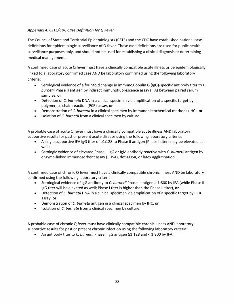
Appendix 4: CSTE/CDC Case Definition for Q Fever
The Council of State and Territorial Epidemiologists (CSTE) and the CDC have established national case
definitions for epidemiologic surveillance of Q fever. These case definitions are used for public health
surveillance purposes only, and should not be used for establishing a clinical diagnosis or determining
medical management.
A confirmed case of acute Q fever must have a clinically compatible acute illness or be epidemiologically
linked to a laboratory confirmed case AND be laboratory confirmed using the following laboratory
criteria:
Serological evidence of a four‐fold change in immunoglobulin G (IgG)‐specific antibody titer to C. burnetii Phase II antigen by indirect immunofluorescence assay (IFA) between paired serum samples, or
Detection of C. burnetii DNA in a clinical specimen via amplification of a specific target by polymerase chain reaction (PCR) assay, or
Demonstration of C. burnetii in a clinical specimen by immunohistochemical methods (IHC), or Isolation of C. burnetii from a clinical specimen by culture.
A probable case of acute Q fever must have a clinically compatible acute illness AND laboratory supportive results for past or present acute disease using the following laboratory criteria:
A single supportive IFA IgG titer of ≥1:128 to Phase II antigen (Phase I titers may be elevated as well).
Serologic evidence of elevated Phase II IgG or IgM antibody reactive with C. burnetii antigen by enzyme‐linked immunosorbent assay (ELISA), dot‐ELISA, or latex agglutination.
A confirmed case of chronic Q fever must have a clinically compatible chronic illness AND be laboratory confirmed using the following laboratory criteria:
Serological evidence of IgG antibody to C. burnetii Phase I antigen ≥ 1:800 by IFA (while Phase II IgG titer will be elevated as well; Phase I titer is higher than the Phase II titer), or
Detection of C. burnetii DNA in a clinical specimen via amplification of a specific target by PCR assay, or
Demonstration of C. burnetii antigen in a clinical specimen by IHC, or Isolation of C. burnetii from a clinical specimen by culture.
A probable case of chronic Q fever must have clinically compatible chronic illness AND laboratory supportive results for past or present chronic infection using the following laboratory criteria:
An antibody titer to C. burnetii Phase I IgG antigen ≥1:128 and < 1:800 by IFA.

23
Appendix 5: Healthcare Provider Alert
(Insert Date, Agency, Contact information) The (insert agency) would like health care providers to be alert for cases of Q fever in patients from (insert region). Within the past (insert timeframe), (insert number of human cases) cases of Q fever have been identified in (insert region), an increase over an average of (insert average number of human cases) cases reported annually in the region. Most, but not all of the cases have reported exposure to (insert animal species and baby of species). Q fever is a zoonotic disease caused by the bacterium Coxiella burnetii. Q fever is transmitted to people most often from inhalation of contaminated dust from infected goats, sheep, and cattle, and their environments, especially during birthing. In addition, Coxiella burnetii can become airborne, and people can be exposed by inhaling bacteria‐contaminated dust miles from animal sources. Q fever is not generally transmitted person‐to‐person. While many people do not experience illness when infected and require no treatment, some do develop disease. The incubation period of Q fever is generally two to three weeks (range, 3 to 30 days). Acute symptoms include sudden onset of high fever, retrobulbar headache, chills, sweats, myalgia, malaise, nausea, vomiting, diarrhea and abdominal or chest pain. Thirty percent to 50% of patients develop pneumonia. Thrombocytopenia and elevated hepatic enzymes are common. A post‐Q fever fatigue syndrome occurs in 20% to 40% of cases. Chronic Q fever manifests primarily as blood‐culture negative endocarditis. It occurs in up to 5% of cases, weeks to years following the acute infection. The three groups at highest risk for developing chronic Q fever are pregnant women, immunosuppressed persons and those with a pre‐existing heart valve defect. Diagnosis is confirmed by a positive PCR assay of whole blood or serum obtained during the first week of acute illness and prior to antibiotic administration, or serologically by a four‐fold rise in IFA titer. There are two antigenic phases to which patients develop antibody responses. In acute infection, an antibody response to C. burnetii Phase II antigen is predominant and is higher than the Phase I antibody response; the reverse is generally true in chronic infection. (Insert agency) can provide assistance with diagnosis of suspect cases. In acute disease, doxycycline administered orally for 14 to 21 days is the treatment of choice in adults, children over age 8, and severely ill or hospitalized children of all ages. In children less than eight years of age and pregnant patients, alternative antibiotic therapy should be utilized. Treatment should not be delayed pending laboratory test results or withheld if laboratory tests are negative during the first week of illness. Prophylactic treatment after a suspected exposure is not recommended. To report suspect cases please call (insert agency and phone number). For more information, please call (insert agency and phone number) or see the Centers for Disease Control and Prevention Q fever website: http://www.cdc.gov/qfever/

24
Appendix 6: Veterinary Alert (Insert date)
Veterinary Alert: Coxiellosis (Coxiella burnetii) cases in (insert livestock species) in (insert county) Dear Veterinarian: The (insert department name) is asking veterinarians to be alert for goat or cattle herds and sheep flocks that may be experiencing abortions due to Coxiella burnetii infection. Since (insert date), abortions, stillbirths and weak neonates caused by Coxiella burnetii have been diagnosed in (insert number of herds/flocks) within (insert jurisdiction). In addition to the positive herd/flocks, (insert number) human Q fever cases have been identified among animal caretakers. Transmission: Coxiellosis (Q fever in people) is a zoonotic infection caused by Coxiella burnetii which can cause a febrile flu‐like illness in humans and abortions in affected animal species. (Insert number) herds/flocks with C. burnetti abortion are diagnosed in our state every year. Goats, sheep, cattle and cats are the domestic species most clinically affected and most likely to transmit the infection to people. Transmission is most often through aerosolization of the bacterium from contaminated birth products, either fresh or desiccated. Please take precautions and use appropriate personal protective equipment,
including a properly fitted respirator mask*(N95 or higher rated), gloves, rubber boots, and protective
clothing when coxiellosis is suspected. Biosecurity: Aborting and parturient animals are an important source of infection to other animals and humans. For this reason, isolation of these animals indoors is recommended to prevent wind‐borne spread of aerosolized bacteria. Placenta and fetuses should be disposed of promptly by burning, burying, or bagging in a leak‐proof container. It is recommended to compost bedding and manure for a minimum of 90 days before spreading to decrease the C. burnetii burden in these materials and to only spread manure on still days away from the residential housing. Diagnosis: If you suspect abortions due to Coxiella burnetii contact (insert state animal health official) and (insert veterinary diagnostic laboratory) to submit appropriate diagnostic specimens. Consult the attached document for additional information on diagnostics and interpretation. Be aware that many infected animals show no signs of disease but may be shedding large quantities of bacteria in the birth fluids and are also a risk to humans. Treatment: Based upon current data, antibiotic usage in the absence of ongoing abortions is not warranted. When abortions are ongoing, parenteral treatment of individual animals (two injections of long‐acting oxytetracycline at 20 mg/kg, given 20 days apart) in late gestation to prevent adverse pregnancy events may be helpful, although data is inconclusive. Human disease: The incubation period of Q fever is two to three weeks following exposure. While many people do not experience illness when infected and require no treatment, some do develop disease.Symptoms of acute Q fever include persistent high fever (104º ‐ 105º F), severe retrobulbar headache, chills, night sweats, muscle pain, nausea, vomiting, diarrhea, fatigue, non‐productive cough, and chest or abdominal pain. Pneumonia and hepatitis can occur in severe cases.
* Talk to your doctor first about whether it is safe for you to wear a respirator.

25
Reporting: We ask your assistance in promptly reporting suspect and confirmed cases of coxiellosis in animals to (insert agency) by telephone at (insert phone number). (insert agency) is available for consultation regarding diagnostics, interpretation, and management of cases. Please direct questions about human health to (insert agency) at (insert phone number). Persons with symptoms of Q fever should contact their healthcare provider. If you have any questions, please call (insert name) at (insert phone number). For more information on Coxiella infection, review the Prevention and Control of Coxiella burnetii Infection among Humans and Animals: Guidance for a Coordinated Public Health and Animal Health Response, 2013. Sincerely, (Insert contact information)

26
Appendix 7: Coxiellosis Diagnostics and Interpretation for Veterinarians This guidance was developed for veterinarians to assist with diagnosis of C. burnetii as the etiologic agent of abortions and other adverse pregnancy events. Diagnosis of coxiellosis is complicated by the fact that C. burnetii is ubiquitous in the environment and asymptomatic infection in animals is common. Additionally, the organism may be also identified in tissues following adverse pregnancy events caused by other abortion agents. While infection without disease is important when considering zoonotic risk, correctly identifying C. burnetii as the cause of disease is important when considering management of an abortion event in a flock or herd. Therefore, distinguishing exposure and infection from disease is crucial when interpreting C. burnetii diagnostics. Serological Tests Serological testing methods available for the detection of C. burnetii in animals include complement fixation (CF), enzyme‐linked immunosorbent assay (ELISA) and indirect fluorescent antibody (IFA) tests. ELISA and IFA are more sensitive than CF and are the preferred diagnostic tests. Serology is best utilized on a herd‐ or flock‐wide basis to demonstrate C. burnetii infection or exposure and is one tool that can be used to determine if infection is likely present in the herd or flock. Serology is less valuable in an individual animal, either to absolutely determine if C. burnetii was the etiologic agent of an abortion or to identify individual infected animals as part of a control program. This is because some animals may require two to three weeks to produce a serological response and others may have persistent serological titers that last for years. In addition, some animals (10‐20%) remain serologically negative while shedding C. burnetii while other animals that test positive may not be shedding the organism. Therefore, in the individual animal, negative serologic test results do not rule out infection, and positive serologic test results do not necessarily indicate shedding or risk of transmission. Antigen Detection and Interpretation of Findings Coxiella burnetii antigen can be detected using PCR, immunohistochemistry, histology or culture. Immunohistochemistry (IHC) of placental cotyledons is the preferred test for identifying C. burnetii as the cause of abortion (29,61). PCR performed on tissues is a very sensitive and specific method for detecting C. burnetii infection and can be used to support a diagnosis of C. burnetii abortion. However, some studies have indicated that presence of the organism can frequently occur when C. burnetii is not the cause of abortion. Some work suggests the number of organisms present, as detected by quantitative PCR on placental tissues, is higher when the abortion appears to be due to C. burnetii, as supported by histologic findings (42). Diagnosis of C. burnetii infection in an individual animal: A positive PCR assay performed on birth products, vaginal secretions, milk, or feces indicates that the animal is currently infected. A negative PCR or culture does not rule out infection in the individual animal, herd, or flock. Diagnosis of C. burnetii infection in a herd or flock: For herd level diagnosis of C. burnetii infection of dairy operations (goat, sheep, or cattle), PCR and/or serologic testing of bulk tank milk is rapid and easy. A positive bulk tank milk antibody result suggests that exposed animals are present in the herd. A positive bulk tank milk sample PCR result suggests that infected animals are present and are shedding the organism in the milk or are shedding the organism in the feces which has contaminated the milk. Given the environmental stability of the organism, the farm

27
is likely to remain infected for a prolonged period of time. In small ruminant operations where milk shedding is highest immediately postpartum, it may be advantageous to collect one of the samples soon after lambing, kidding, or calving when the majority of the herd is in early lactation. Such an approach would increase the sensitivity of the assay. Repeated negative bulk tank milk tests significantly decrease the likelihood that C. burnetii is present on the farm. To determine the true status of the herd or flock and when bulk tank milk is not available, serologic individual animal testing of the entire breeding herd provides the best picture of herd status. In cases where testing the entire breeding herd/flock is not possible due to economic or logistical constraints, animals should be selected for testing with a bias towards recently post‐parturient females. Sampling of pre‐pubertal animals should be avoided. The sample size should, at a minimum assure the ability to detect disease at a 10% prevalence level. Programs are available to calculate sample size based on test sensitivity and specificity, population size (herd/flock size), minimum expected prevalence of disease, and desired Type I and Type II error levels (Table 1). One such program, FreeCalc Version 2, is available from AusVet Animal Health Services at: http://www.ausvet.com.au/content.php?page=software#freecalc .
Table 1. Number of animals to randomly select and serologically test by herd/flock size and estimated infection prevalence to detect C. burnetii infection in a herd/flock, assuming test sensitivity of 98% and test specificity of 100%, and with a target Type I and Type II error of 0.05%. (Source: FreeCalc Version 2)
Flock Size Estimated Infection Prevalence
1% 5% 10% 15%
10 10 10 10 8
35 35 28 19 16
60 59 39 24 17
85 83 45 24 17
110 107 44 26 17
135 131 47 26 18
160 127 50 27 18185 147 53 27 18
210 166 50 28 18
235 186 52 27 19
260 167 54 28 19
285 183 55 28 19
310 200 53 28 19
335 216 54 28 19
360 193 55 28 19
385 207 56 28 19
410 220 55 29 19
435 234 56 28 19
460 211 56 29 19
485 223 57 28 19

28
Investigating C. burnetii infection status by species: The species being investigated will influence the type and timing of sample collection (79). Goats As with sheep, C. burnetii is a common cause of abortion in goats, but most infected goats show no clinical signs of disease. Serology is useful for identifying infection in the herd when investigating herd‐level abortion problems or determining infection status of the herd. Coxiella burnetii seroprevalence is generally higher in aborting goats than in infected goats that have normal birth outcomes (85). In an infected herd, both goats that abort and goats that do not abort shed the organism. However goats that abort have a tendency to shed the organism in higher numbers in birth fluids and vaginal secretions (41,83,84). Shedding in milk in this species is persistent and may continue for weeks (85). Bulk tank milk PCR tests can be used to detect infected goat herds with excellent specificity and good sensitivity. Higher sensitivity can be obtained with sampling closer to the time of parturition and with repeat sampling (85). Sheep Coxiella burnetii is a common cause of abortion in sheep, but most infected sheep show no clinical signs of disease. Sheep will shed organisms in vaginal secretions for several weeks after lambing as well as in the feces for weeks to months (81). Shedding in milk is intermittent and not related to the time of lambing (81). As with cattle, seropositivity (as detected by CF, IFA or ELISA) indicates the presence of infection with C. burnetii, but does not necessarily indicate the organism as the cause of abortion. Coxiella burnetii abortion should be confirmed by examination of the placenta for evidence of inflammation associated with confirmed C. burnetii intracellular organisms by immunohistochemistry. Additionally, sheep may abort due to C. burnetii but be serologically negative (16). Vaginal shedding and placental PCR results give a good indication of flock status. Sheep shed C. burnetii intermittently in the milk; therefore bulk tank milk PCR results may be negative when, in fact, the flock is infected. However a positive bulk tank milk PCR test result is highly specific for flock infection, and repeated testing will increase sensitivity (33). Cattle In cattle, particularly dairy cattle, infection with C. burnetii is common, but abortion is uncommon. PCR or ELISA testing of bulk tank milk or serology of the adult herd is a reliable method for screening dairy herds for C. burnetii infection (10,67). Vaginal shedding is short term in cattle; thus, vaginal secretions for PCR testing in cows should be collected within two weeks of calving or aborting (34). Using serology of the herd or bulk tank milk samples to determine whether abortions are due to infection with C. burnetii is not reliable. Milk PCR is often positive if an abortion is due to C. burnetii, but milk shedding is also common in healthy infected cows. There is poor correlation between serologic or bulk tank milk ELISA results and the ability to detect C. burnetii via PCR in placenta, vaginal mucus, or milk (34,37). Necropsy of fetus and placenta is most reliable as a diagnostic tool for determining C. burnetii as the cause of abortion. Histological findings in aborted fetal membranes from cows typically reflect only mild inflammation, so these findings must be interpreted in conjunction with PCR results. Determining herd/flock “low risk” status with respect to C. burnetii infection: It is sometimes necessary to determine if a group of animals is free from infection with C. burnetii. Freedom from infection is difficult to determine with perfect assurance but herds/flocks can often be designated as “low risk”. Low risk usually means there is no evidence of infection in the population and

29
measures are in place to prevent introduction of the disease agent. To determine whether the flock or herd is at low risk of C. burnetii infection, several measures can be taken:
Serological testing of the entire active breeding herd/flock using CF, ELISA or IFA. Testing should be performed at least annually.
For dairy animals, periodic PCR testing of bulk tank milk samples and higher frequency of testing to increase sensitivity. There are no published data on the sensitivity of bulk tank milk testing at different herd prevalence levels of C. burnetii infection; thus, this method may fail to detect infected herds with low prevalence of infection. Full investigation of all abortions using PCR and immunohistochemistry.
For herds and flocks without abortion issues, placenta can be submitted for C. burnetii PCR from a sample of normal births.
Note that test results are only valid for the time period in which the testing is performed because of the highly infectious nature of this organism. Any C. burnetii infection risk status program should require all of these approaches, as well as general biosecurity measures and good animal husbandry to minimize risk of introduction of C. burnetii to the herd or flock.

30
Appendix 8: Manure Management and Carcass Disposal Considerations Related to Coxiella burnetti
Personal Protective Equipment and General Considerations
Wear a properly fitted respirator mask* (N95 or higher rated), gloves, rubber boots, and protective clothing when cleaning out buildings, moving manure, managing compost piles, or handling animal carcasses or placentas and other birthing products. Coxiella bacteria are resistant to many disinfectants commonly used in barns, so check the product label to ensure that the product is effective.
*Talk to your doctor first about whether it is safe for you to wear a respirator. Avoid using high pressure hoses for cleaning, as this may cause aerosolization of dust and bacteria.
Cover manure, bedding, and other potentially contaminated materials during transport to help prevent spread of C. burnetii.
Ensure that rodent and scavenger control methods are in place. Manure management Composting is the preferred method for handling manure and bedding. Composting for a minimum of 90 days prior to disposal decreases the C. burnetii burden and therefore reduces the risk of spread.
State and local composting regulations exist and should be followed.
Resources describing proper composting techniques are widely available. Examples include: o http://www1.agric.gov.ab.ca/$department/deptdocs.nsf/all/agdex8875 o http://www.omafra.gov.on.ca/english/engineer/facts/05‐021.htm
If manure composting is not feasible, the following measures may decrease the risk of transmission of C. burnetii to people and animals:
Cover manure during transport to reduce spread of the bacteria.
Store manure in a location where run‐off and the potential for water contamination is minimal.
Transport and land‐apply manure on non‐windy days to decrease aerosolization of bacteria.
Land‐apply manure promptly after facilities are cleaned out and plow‐in the manure promptly after application.
Sell only manure that has been fully composted to the public for use in gardens. Carcass disposal Promptly and properly dispose of animal carcasses and placentas and other birthing products. Carcass disposal options include composting, burial, incineration, rendering, and transport to a landfill. The appropriate state or local agency can provide guidance on local and state regulations regarding disposal.
Dispose of carcasses onsite whenever possible, because transporting carcasses and contaminated materials can present a transmission risk. Composting or deep burial is recommended.
Prevent scavenging.
If carcasses are temporarily stored prior to disposal, use closed containers with absorbent materials to prevent splashing and spilling.
If you must send animals suspected to be infected with C. burnetii to a landfill, inform landfill staff so that they can take proper precautions to protect themselves.