prevaunceo fg eneralisedc onvulsives tatos …
TRANSCRIPT

PREVAUNCE Of GENERALISED CONVULSIVE STATOS
EPILEPTICUS IN PATIENTS AOMITTED TO KENYAUA NATIONAL
HOSPITAL UNO THE SHORT TERM OUTCOMES
A DESSERTATION SUBMITTED IN PART FULLILMENT OF THE REQUIREMENTS
FOR THE AWARD OF THE DEGREE OF MASTER OF MEDICINE IN INTERNAL
MEDICINE BY:
GEORGE M. NYALE
University of NAIROBI Library
0535!THE FACULTY OF MEDICINE, UNIVERSITY OF NAIROBI
NOVEMBER 2006
UNWEPr '“'(VltOtOAL
r r »>
LIBRARY
(ME IM TRC LICG/.RY 3HI. t

DECLARATION
I certify that this dissertation is my own original work and has not been presented for a
degree at any other university
2

This dissertation has been submitted with our approval as supervisors:
PROF. ADAM A M
Consultant Neurologist, Associate Professor o f Medicine
Department of Medicine, University o f Nairobi
Signed Date
PROF AM AYOE.O
Consultant Neurologist, Associate Professor of Medicine
Department of Medicine, University o f Nairobi
....................... DateSigned.
DRJOWIJ.O
Consultant Neurologist, chief Medical Specialist
Department of Medicine. Kenyatta National Hospital
ASigned Date D
3

dedication
To my l o v e l y w i f e J a c k i e and son Ian
f o r b e a r i n g w i th my a b s e n c e s d u r in g
th e s t u d y and t h e i r c o n s t a n t
u n d e r s ta n d in g and encouragem en t!
4
A CKNO WLEDGEMENTS
To my supervisors for their fatherly advice and guidance through this
whole project right from writing the proposal to this finished
dissertation.
To my assistants for the dedication they showed in assisting me
follow up and arrange the data. Really I would not have done it
without you!
To my statistician, ‘kudos’ for the swift and precise analysis. It was a
joy working with you!
To Sanofi Aventis and Astra Zeneca for the financial and material
support which enabled me to carry out this study in good time and to
such a completion.
Thank you very much!
5
Table of contents
PageItem
• Declaration ---------------------------------------------------- 2
• Supervisors----------------------------------------------------- 3
• Dedication ----------------------------------------------------- 4
• Acknowledgements------------------------------------------- 5
• Table of contents---------------------------------------------- 6
• List of tables --------------------------------------------------- 8
• List of figures -------------------------------------------------- 9
• List of abbreviations ----------------------------------------- 10
• Abstract --------------------------------------------------------- 11
• Introduction --------------------------------------------------- 13
• Literature review -------------------------------------------- 16
o Classification ---------------------------------------- 16
o Pathophysiology ------------------------------------ 29
• Study justification ------------------------------------------- 34
• Objectives ----------------------------------------------------- 36
• Study Definitions --------------------------------------------- 37
• Methodology -------------------------------------------------- 40
o Methods and tools of the study------------------- 40
• Ethical considerations------------------------------------------- 43
• Statistical methods----------------------------------------------- 44
• Results-------------------------------------------------------------- 46
6

Discussion 61
Conclusion-------------------------------------------------------- 70
Recommendations---------------------------------------------- 71
Study limitations----------------------------------------------- 72
References-------------------------------------------------------- 73
Appendices------------------------------------------------------ 81
7

f TST of tables page
Table 1: GCSE patients: ward distribution 47
Table 2: Serum urea, Creatinine & Electrolytes of GCSE patients 55
Table 3: Hematological investigations done on GCSE patients 56
Table 4: Length of hospital stay for patients with GCSE 59
Table 5: Mortality data of the GCSE patients 59
8
I 1ST OF FIGURES PAGE
FIGURE 1: Pathophysiology of status epilepticus 33
FIGURE 2: Screening and recruitment flow diagram 46
FIGURE 3: Gender distribution of GCSE patients 48
FIGURE 4: GCSE patients Referral status 48
FIGURE 5: Reasons for referral 49
FIGURE 6: Previous convulsive state of GCSE patients 50
FIGURE 7: Adherence to AEDs by persons living with epilepsy 50
FIGURE 8: Primary causes of GCSE 51
FIGURE 9: Specific infections causing GCSE 52
FIGURE 10: ILAE proposed classification of causes of GCSE 53
FIGURE 11: HIV sero-status of GCSE patients 54
FIGURE 12: Emergency drugs prescribed to patients presenting in GCSE 57
FIGURE 13: Discharge medications given to GCSE patients 58
9

I 1ST OF ABBREVIATIONS
GCSE Generalized Convulsive Status epilepticus
NCSE Non- Convulsive Status Epilepticus
K.NH Kenyatta National Hospital
CNS Central Nervous System
EEG Electroencephalogram
SE Status Epilepticus
ILAE International League against Epilepsy
HIV Human Immunodeficiency Virus
AIDS Acquired Immunodeficiency Syndrome
PET Positron Emission Tomography
MRI Magnetic Resonance Imaging
BC Before Christ
AEDs Anti Epileptic Drugs
HAART Highly Active Anti-Retroviral Therapy
SOL Space occupying lesion
WBCs White blood cells
HB Hemoglobin level
Mm 3 Cubic millimeter
TDM Therapeutic dose monitoring
nm d a N-Methyl-D-Aspartate receptors
ELISA Enzyme linked immunosorbant assay
10
ABSTRACT
Background: Generalized Convulsive Status Epilepticus (GCSE) is a sub-group of status
epilepticus that is accompanied by visible body movements and loss of consciousness. It has a
high complication rate including disability or death. Generalized convulsive status epilepticus is
known to occur in both known epileptics and non epileptics, where it may be a manifestation of
another underlying primary cause. Other factors that have been shown to influence outcomes in
patients with GCSE are age and the underlying primary etiology.
No studies have been done in Africa hence its magnitude and its influence on patient’s outcomes
is unknown.
Objectives: This prospective study was primarily aimed at determining the prevalence of GCSE
in patients admitted to KNH and the length of admission. Secondary objectives were to
document the primary etiological causes and treatment given to these patients.
Study design: This is a longitudinal prospective study. The subjects were screened and those
fitting the criteria for GCSE recruited and followed up to either death or discharge.
Study period: The study was carried out over a three calendar month period between 1/1/2006
and 1/4/06 both days inclusive.
Sampling method: Patients were recruited consecutively.
Study location: The study was carried out at Kenyatta National Hospital. The wards involved
included; all medical wards, all surgical wards, the obstetric and gynecology wards, the
maternity unit, the renal unit, the burns unit, the intensive care unit and the high dependency unit.
Methodology: All patients admitted to KNH between January and April 2006 with suspected
convulsions, coma or confusional states were screened and those with GCSE recruited. They
11
were followed up on a daily basis with documentation of the primary etiology, investigations and
treatment given till death or discharge.
Approval to carry out the study was obtained from the KNH ethics and research committee.
Participation was on a voluntary basis following an informed written consent.
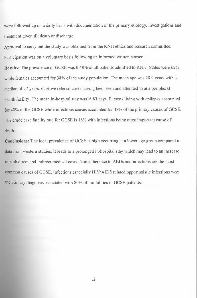
Results: The prevalence of GCSE was 0.48% of all patients admitted to KNH. Males were 62%
while females accounted for 38% of the study population. The mean age was 28.9 years with a
median of 27 years. 62% we referral cases having been seen and attended to at a peripheral
health facility. The mean in-hospital stay was 16.83 days. Persons living with epilepsy accounted
for 40% of the GCSE while infectious causes accounted for 38% of the primary causes of GCSE.
The crude case fatality rate for GCSE is 16% with infections being most important cause of
death.
Conclusions: The local prevalence of GCSE is high occurring at a lower age group compared to
data from western studies. It leads to a prolonged in-hospital stay which may lead to an increase
in both direct and indirect medical costs. Non adherence to AEDs and infections are the most
common causes of GCSE. Infections especially HIV\AIDS related opportunistic infections were
the primary diagnosis associated with 80% of mortalities in GCSE patients.
12
INTRODUCTION
Status epilepticus is a relatively common neuromedical emergency whose worldwide
epidemiology is still being written but data from developing countries is minimal. A few
differences in the etiology and incidence between developed and developing countries have been
noted. While Studies done in developed countries show a low burden of CNS infections, the
developing countries including Kenya have infections high up in their lists. Some examples
include the ‘hot water epilepsy’ which is a kind of reflex epilepsy induced by immersion in hot
water baths a common practice in south India and the higher burden of chronic CNS infections,
especially cysticercosis, seen in developing countries (1). It is a fact that one of the
complications of cerebral malaria includes seizure episodes; more recent data suggest the
existence of a higher prevalence of epilepsy in malaria endemic regions in the Kenyan coast
amongst children (2). Another factor that is thought to increase occurrence of seizure illness is
the increased incidence of irritative focal brain lesions in HIV / AIDS patients. Meningitis and
encephalitis are proven etiological causes of status epilepticus in adults (3).
Both community and hospital-based studies have mainly been recruiting elderly subjects, with
most of them being above 60 yrs (4, 5, 6, 7, 8). This can be explained by the fact that the
population in these countries is aging. It follows that with aging the prevalence co-morbid
conditions, especially cerebrovascular, increase and these are known to cause GCSE. The
incidence of status epilepticus has been shown to be higher among the elderly above 60 yrs (54.5
/100,000) than in young adults (4.2 / 100,000) (4). This is explained by the higher rates of
cerebrovascular diseases in those above 60years. Locally the presumed causes include CNS
mfections that are in high incidence in younger age groups as a result of the HIV/AIDS
pandemic.
13

Mortality was significantly high in the elderly persons due to mortality from cerebrovascular
diseases. Childhood febrile seizures, which are generally less harmful, contributed to the higher
incidence rates. The mortality difference can be explained by the etiological differences, so with
a change in the profile of causes one can expect a change in mortality!
In a review article on ‘Emergency department drug therapy for status epilepticus’, Lockey et al
notes that the most common and dangerous type is the GCSE with an incidence of between 180
to 280 cases per million in the U.K and a mortality of about 10 to 15% (9).
In Africa, a study done in Mbam valley, Cameroon showed that epileptics had a 6.2 higher risk
of death when compared to the general population, with the major contributor being status
epilepticus (56.6% of deaths in the epileptic cohort) (10).
A 30- year cohort study of epileptics in rural Tanzania by Jilek - Aall et al, found that epileptics
had a death rate that was twice that of the general population. Further analysis showed that 50%
of the deaths were epilepsy- related such as status epilepticus, drowning, burns and dying in or
after a seizure (11).
In Kenya the prevalence and mortality of this condition is scanty. A community-based
investigation in Kilifi district by Snow R.W et al showed that 77% of deaths in epileptics had
occurred while patient was in status epilepticus (12).
The prevalence of generalized convulsive status epilepticus has been evaluated in five studies
three done in the U.S.A and two in Europe. Groups that demonstrated a high incidence of GCSE
m these studies were the children below five years, the elderly over 60 yrs and blacks. The major
14
predictors of mortality were etiology and age. Seizure duration was only significant in those that
lasted over 24 hours (13, 14, 15, 16, 17, 18).

Literature review
Modes of Classification
The worldwide co-ordination of all antiepileptic activities falls under the auspices of ILAE. In
1981 a sitting of ILAE defined status epilepticus as; ‘a seizure that persists for a prolonged
period of time or is repeated frequently such that recovery between attacks does not occur’.
Obviously this definition was very ambiguous and difficult to use since it omitted a very
important detail in the specific time duration of the seizure. This void was filled by various
physicians to be between 20 - 30 minutes of seizure activity, being basically the estimated period
of time necessary to cause injury to the central nervous system. It is for obvious reasons that the
emergency physicians have picked the shorter duration of 20 minutes (19). Pro-active thinkers
argue that status epilepticus is an emergency whose treatment is initiated well before the period
of 20 - 30 minutes is over if we are to lower its morbidity and mortality. This means the time
guideline given is impractical for initiating treatment. It is on this basis that many scholars
including Daniel H. Lowenstein are advocating for the newer operational definition of status
epilepticus as either: continuous seizures lasting at least five minutes or two or more discrete
seizures between which there is incomplete recovery of consciousness (20).
According to the guidelines for epidemiological studies on epilepsy seizure types, status
epilepticus is classified as follows:
a) Generalized status epilepticus: where there is no indication of an anatomic localization or
clinical evidence of a focal onset.
Subgroups:
• Convulsive: Tonic, clonic, or tonic-clonic
16

1
• Non convulsive: absence
• Myoclonic
b) Partial status epilepticus: where there is evidence of focal onset.
Subgroups:
• Simple: no impairment of consciousness
• Complex: consciousness impaired
• Secondarily generalizing
c) Unclassified status epilepticus
Neurophysiologists classify status epilepticus based on the EEG pattern. Classes include:
a) Spike wave form - this will basically be the EEG tracing of absent
Vepilepticus and is associated will low risk of brain injury. \
b) Non-spike waveform - these have a higher risk of brain injury esp[v
the generalized convulsive status epilepticus variety.
The obvious down side with this kind of classification is the difficulty in getting intra- ict»
EEG’s (21).
Prevalence studies on status epilepticus
There are only a handful of studies examining the incidence of status epilepticus, all of
from the developed world. The oldest is a retrospective population based study in Roches
Minnesota by Hersdorffer et al (9). This study covered the period 1st January 1965 to 31
December 1984. They used the Rochester Epidemiology project records-linkage system^
ascertain all first episodes of status epilepticus receiving medical attention in Rochester,\
17

the primary service area of the Mayo clinic. In total they sampled 199 patients with first episode
of S.E, making the incidence of S.E in this population to be 18.3 / 100,000. GCSE accounted for
45% of all S.E and had an annual incidence of 8.3/100,000. It is worth noting that the population
is 96% white and non-whites constituted a merge 4% of the study population. Secondly, over
50% of the study subjects were older than 60 years. This is a reflection of the population pyramid
of developed countries with a narrow base. As expected the annual incidence was higher in those
above 65 yrs i.e. 62.5 / 100,000, which can be explained by the higher rates of cerebrovascular
diseases in this age group. Males were shown to have a higher incidence of S.E when compared
to females though there was no explanation for the gender difference. This was a community
based study since it captured information from out-patient clinics, home visits and in-patient
hospitalizations. It was also established that this population was not optimally treated with anti
epileptic drugs.
Another study is a prospective study in Richmond, Virginia by Delorenzo et al (6). Here a
systematic review of all cases above one month of age admitted to the medical college of
Virginia was done. In total they sampled 253 adults with S.E and hence put the annual incidence
of S.E in this population at 41/100,000 with a bi-modal distribution which has an early peak in
those aged less than five years and another in those over 60 years. This was more than twice the
incidence found at Rochester especially bearing in mind that both studies used the same case
definitions. There was also no credible explanation on any new factors that could have come into
play to explain a difference of this magnitude between the Rochester data covering a period
starting 1965 - 1984 and this Richmond data covering 1994. One possible explanation to this
leaP in incidence has to do with the study design. The Rochester study was a retrospective study
18

based on data entered into a linkage-record system while this study was a systematic prospective
review. All retrospective studies have the limitation of the amount of data collected, the
collector’s error and the lack of real-time validation systems. This simply means that there could
have been a lot of omitted data. This is further supported by another retrospective study done in
California which also reported lower incidence rates of S.E (10). The annual incidence of GCSE
was three times higher at 29/100,000 and accounted for 74 % of all S.E patients. More
interestingly, a sub-analysis showed that blacks had a significantly higher incidence of GCSE at
39.9/100,000. Blacks made up over 50% of the study population compared to the Rochester
study where whites accounted for over 96% of the study population (9). This racial difference
could explain the difference in incidence of GCSE in the two populations. This epidemiological
finding has not been supported by any concrete reason/s which makes blacks more susceptible to
GCSE than their white counterparts in the same environments. Further more race did not
influence mortality but age and etiology did clearly predict mortality.
In comparison to the Rochester and Richmond studies, the retrospective study looking at the
incidence and mortality of GCSE in California by Wu et al found a lower incidence of GCSE at
6 18/100,000(5). This is due to the fact that they sampled a different age group which did not
include pediatric patients despite other studies showing that they have a high incidence of S.E.
Since their methodology involved reviewing discharge codes of S.E they missed out on
outpatient and home visit data. Thirdly in their study population blacks accounted for only 15.3%
while whites were 58.7% and as a result the incidence rates of GCSE had dipped down to
CSemble those of white dominated Rochester population. In tandem with the Richmond study
acks were shown to have a higher incidence of GCSE at 13.35/100,000. This is about twice for
19

the general population in California. Again when data was controlled for age and etiology, race
had no influence on mortality. As expected incidence rates were higher in the older age groups
(22.32/100,000 in the group over 75 years and a 15-fold increase in case fatality), which is a
reflection of cardiovascular diseases in this population and its importance in causation of GCSE.
The results of a prospective study by the status epilepticus study group Hessen (SESGH),
looking at the incidence and case fatality rate of status epilepticus in Hessen were similar to
those found in Rochester despite the difference in methodology(5). The study populations were
quite similar in that both consisted mainly of whites and most of the study subjects were above
65 years. In their methodology the investigators used a prospective design where a systematic
review of all patients admitted in the 16 hospitals within zip code area 35 was done. Despite the
difference in study design they got a low crude annual incidence of 17.1/100,000 for S.E in this
population, which was similar to the 18.3 /100,000 reported in Rochester but lower than another
prospective study done in Richmond(6). This could be explained in part by the different times
when the studies were done which will basically translate to an improvement in the primary
control of diseases that lead to seizure disorders. The primary underlying etiologies did not differ
much but it is interesting that 50% of the patients with S.E had a previous diagnosis of epilepsy.
So by extrapolation one can assume that better control of epilepsy could account for the
reduction in the incidence of S.E in this population. They also found a higher incidence in older
roen, which was reflective of the prevalence of cerebrovascular disease in this age group. There
was no sub-analysis according to race.
20
Another prospective study carried out in tertiary care academic hospitals in Geneva estimating
the annual incidence of status epilepticus in French speaking Switzerland by Coeytaux et al
sampled yet another elderly white dominated population(8). By doing a systematic review of all
patients admitted with status epilepticus in all hospitals in that region, they found an age adjusted
annual incidence of 10.3/100,000 for S.E. This is even lower than that found in other white
dominated populations though this could be attributed to the high rate of under ascertainment.
For example Geneva which is the most developed of the six cantons enlisted in the study had the
highest incidence of S.E at 16.3 / 100,000. This is because it had a better ascertainment due
availability of 24 hour EEG facilities and adequate man power than other cantons. They also left
out post anoxic encephalopathies which in Rochester study had accounted for about 10% of the
study population. Just like in Rochester males had a tendency of a higher risk for S.E but this
was found not to be significant after correction for etiology. Also a bimodal distribution with two
peaks was shown, one being in the under fives and the other in the elderly of over 60 years. One
interesting finding was the higher risk for S.E in urban as compared to rural populations for
which there were no proper explanation.
In Africa, studies on status epilepticus are lacking and the incidence/prevalence rates of this
condition are unknown. However some studies on epilepsy did capture some scanty data on
status epilepticus. Examples of such studies include a study done in Mbam Valley, Cameroon by
Kamgno et al that found a majority of epileptic patients described a seizure pattern fitting a
generalized convulsive seizure (101 out of 125) (13).
A 30-year follow up of an epileptic cohort in Mahenge, rural Tanzania found only 21.9% were
still alive. Causes of death were epilepsy-related (status epilepticus, drowning, burns and dying
21

in or after a seizure) in over 50%. These epilepsy-related deaths were higher in the sub-groups
that had poor seizure control, those receiving drugs irregularly and those who had stopped taking
the drugs (20).
A two year community- based investigation of mortality in Kilifi, Kenya targeting deaths in
individuals above 5 years as reported by bereaved relatives revealed that 3.5% of all deaths had
occurred in epileptics. Out of which 77% had occurred while the patient was in status epilepticus
(14).
Etiology
The initiators of status epilepticus can be viewed as acute or chronic processes (20).
• Acute causes include: CNS infections, stroke, head injury, drug toxicity, metabolic
derangements and brain hypoxia from any source.
• The chronic causes include: status in known epileptics which is usually due to lack of
compliance or breakthrough seizures, chronic alcoholism, or other slow or remote CNS
afflictions such as tumors etc.
Generally the slow or chronic processes have a better response to anticonvulsant therapy.
The importance assumed by each etiological agent will also depend on its magnitude in a given
geographical area. This explains why studies done in the west where obesity, diabetes,
hypertension and other cardiovascular risk factors are prevalent show a predominance of
cerebrovascular diseases (7, 13, 14). Other reasons that might account for the low prevalence of
infective causes include:
■ Many were done in the pre HIV era where aids related opportunistic infections were not
a major problem.
22

■ Their study population mainly consists of elderly individuals who have more
cardiovascular co-morbidities.
It is known that CNS infections lead to seizure disorders, status epilepticus included, but new
data seems to show that it may also lead to increased prevalence of epilepsy. Annegers et al
looked at the risk of unprovoked seizures after encephalitis and meningitis and concluded that
the prevalence of epilepsy increases for up to 20 years after infection (3). Recent literature in
Kenya suggests that the incidence of encephalitis and meningitis has risen due to the HIV/AIDS
pandemic. Carter et al working at the centre for geographical medicine at Kilifl, Kenya showed
that about one third of the children with cerebral malaria have status epilepticus of the
generalized tonic- clonic variety.
The other big group consists of poorly controlled epileptics. Those who omit doses or are given
sub-optimal drug doses can relapse back to convulsive disease. Even more interesting is the fact
that an overdose of some antiepileptic drugs e.g. phenytoin can result in repeated convulsions
(19). Aminoff et al in their review of 98 patients with generalized major motor status epilepticus
found that the single most common cause to be non compliance with anticonvulsant drug which
accounted for 53% of patients with previous seizure disorder.
Local data estimate that over 50% of epileptics are poorly controlled. In his thesis Peter Mativo,
showed that only 60% of epileptics attending KNH adult neurology clinic were well controlled
(22). With such a high incidence of poor control of epileptics attending a specialized clinic in a
tertiary institution, one can only imagine its contribution to status epilepticus in our set-up.
23

Neurological diseases at KNH constitute about 7.5% of all cases admitted to the medical wards
or seen at the medical clinics. Out of these, over 50% is accounted for by meningitis (23.1%),
epilepsy (16.6%) and cerebrovascular disease at 15.0% (23).
For easy of analysis, the commission of epidemiology and prognosis of ILAE, has introduced a
classification of etiological into:
i. Acute symptomatic / provoked: these are basically recent onset conditions resulting in
status epilepticus within 7 days of onset.
ii. Remote symptomatic / unprovoked: encompasses all past or slowly progressing
conditions which can eventually lead to status epilepticus e.g. a past head injury
iii. Undetermined causes / unprovoked of unknown origin
EFFECTS OF STA TUS EPILEPTICUS
Mortality and morbidity
Status epilepticus is a medical emergency with a high mortality and morbidity. In a review article
of current concepts in status epilepticus Lowestein et al put the mortality at about 20 % in the
USA. Long term follow up of those who survived the initial 30 days, showed an increased
mortality of up to three times that of the general population at ten years (20). The major
contributors to mortality in S.E are the etiological cause as acute symptomatic status epilepticus,
myoclonic type of S.E, and S.E lasting over 24 hours (24).
Status epilepticus may be complicated by hyperthermia, brisk peripheral leucocytosis, cerebral
sPinal fluid pleocytosis, and a systemic acidosis. These complications have not been shown to
influence the mortality (13).
24

Kamgno et al while looking at the demographic impact of epilepsy in Mbam Valley, Cameroon,
found that the risk of dying was 6.2 times higher in epileptics than the control cohort of non
epileptics. The major causes of death were epilepsy related such as; status epilepticus (56.6%),
sudden unexpected death during an epileptic seizure (18.9%) and drowning during a seizure
(10.8%) (10). This was corroborated by a study in Kenya and one in Tanzania which showed
that over 50% of deaths in known epileptics were epilepsy related and occurred mostly when
patient was in status epilepticus(l 1,12). We did not find any hospital-based study that has
estimated the mortality from status epilepticus in Kenya.
Other Factors influencing outcomes
Questions have always arisen on the magnitude of injury caused by the seizure activity and more
so does it vary in immature and adult brains? Haut et al looked at the susceptibility of immature
brains to seizure effects and concluded that features of seizure induced injury in the immature
brain were different from those in adults. Other factors that influence the outcome include; the
duration of seizures, the number of seizures, cause of seizures, presence of pre-existing
abnormalities and genetics (25).
Claassen et al reviewed the predictors of functional disability and mortality after an episode of
status epilepticus, and found that age and acute symptomatic seizures were the main predictors of
mortality. Functional deterioration was at about 23% upon discharge with the main predictors
being increased length of hospitalization and acute symptomatic seizures (15).
The higher prevalence of status epilepticus seen in males has not been seen in all studies but
Possible explanations to a lower female risk include:
25

/. Males have higher rates o f cerebrovascular disease
ii. Some o f the etiological causes e.g. head trauma are mostly seen in men due to their
behaviour patterns
Hi. Gender difference in seizure threshold: Hormonal influence on the GABA- sensitive
region o f the substansia nigra(it is associated with the regulation o f seizure threshold)
The higher incidence of status epilepticus in the black population has been consistently seen in
most studies but no good explanation has been found and most studies are not geared to assess
this racial bias (6, 8).
Contribution of Non convulsive status epilepticus (NCSE)
It is estimated that about 8% of all comatose patients develop status epilepticus, out of which
NCSE accounts for between 20 - 23%. In about 14% of patients, GCSE patients convert to
NCSE following therapy (26).
A study that looked at critically ill patients, in an intensive care setting by Young et al, reported a
mortality of 50% (22). This was similar to what Litt et al found in critically ill elderly patients
with NCSE i.e. 52 %. The major predictors of mortality were; a primary acute medical cause and
presence of severe mental status impairment (27).
In most of these population-based studies the contribution of NCSE was pretty low. De Lorenzo
R.J et al working in Richmond, Virginia found NCSE to represent about 5% of all status
epilepticus cases. In a hospital-based study in French speaking Switzerland it accounted for up
to 33% of all S.E episodes.
26

This is a relatively difficult diagnosis to make since an intra-ictal EEG is mandatory for
diagnosis thus studies in hospital setting report higher prevalence’s than community-based
studies. We found no studies on this form of status epilepticus in Africa.
Economic and social costs
Perberthy et al looked at the economic burden of status epilepticus to the health care system.
They concluded that the median reimbursement from insurance for a patient with status
epilepticus was $8417 with the average length of admission being 12.9 days. This was a 30 -
60% higher refund as compared with other acute health problems including acute myocardial
infarction or congestive heart failure. Major factors that influenced cost were mainly age and
etiology (29).
An economic evaluation of epilepsy in Kiremba, Burundi showed that the costs of treating an
epileptic averaged US$ 48.4 which is more than a seven fold increase compared to the US$ 7.3
needed for treatment of other medical conditions. This presented a 600% increase in direct costs
and 10.2 disrupted days to patient and family as indirect costs annually (30).
Refractory status epilepticus
Some cases of status epilepticus fail to remit with treatment i.e. refractory status epilepticus
(RSE). RSE has been estimated to occur in about 30% of all S.E cases where it leads to an
mcreased length of hospital stay and functional disability, though curiously does not lead to an
mcreased mortality. Other studies show that status epilepticus lasting more than 24 hours was an
lmP°rtant predictor of long term mortality (24).
27

This is a relatively difficult diagnosis to make since an intra-ictal EEG is mandatory for
diagnosis thus studies in hospital setting report higher prevalence’s than community-based
studies. We found no studies on this form of status epilepticus in Africa.
Economic and social costs
Perberthy et al looked at the economic burden of status epilepticus to the health care system.
They concluded that the median reimbursement from insurance for a patient with status
epilepticus was $8417 with the average length of admission being 12.9 days. This was a 30 -
60% higher refund as compared with other acute health problems including acute myocardial
infarction or congestive heart failure. Major factors that influenced cost were mainly age and
etiology (29).
An economic evaluation of epilepsy in Kiremba, Burundi showed that the costs of treating an
epileptic averaged USS 48.4 which is more than a seven fold increase compared to the US$ 7.3
needed for treatment of other medical conditions. This presented a 600% increase in direct costs
and 10.2 disrupted days to patient and family as indirect costs annually (30).
Refractory status epilepticus
Some cases of status epilepticus fail to remit with treatment i.e. refractory status epilepticus
(RSE). RSE has been estimated to occur in about 30% of all S.E cases where it leads to an
increased length of hospital stay and functional disability, though curiously does not lead to an
increased mortality. Other studies show that status epilepticus lasting more than 24 hours was an
important predictor of long term mortality (24).
27

Where do these patients get admitted?
Ward admission distribution shows that most of status epilepticus patients needing admission are
admitted to internal medicine group wards (50%), followed by the intensive care unit (30%),
then specialized neurological ward (10%) and the rest of the other wards the remaining 10% (9).
Usefulness of the EEG
In 1971 the EEG was first introduced in Kenya but its use has remained confined to the large
urban centers where the facility and expertise exists. A review of the importance of this
diagnostic tool at KNH showed that it was mainly ordered in suspected cases of epilepsy (58.5%
of the requests) and convulsions of uncertain cause (11.8% of requests). Of note was that
epilepsy did not seem to benefit maximally from inter-ictal EEG’s in diagnosis with a positive
rate of only 36.0% (31).
Falope et al working on Nigerian epileptics found that only 30 to 50 % of probable epileptics had
epileptiform patterns on the first EEG. They noted that to increase this yield multiple EEG’s
were needed, a fact that was not practicable in developing countries where resources are lacking.
It should be noted a normal EEG does not exclude the diagnosis of epilepsy and that non
epileptics are know to have abnormal EEG’s. They also found no relationship between the
frequency of seizures and epileptiform patterns on EEG. Hence they concluded that the diagnosis
of epilepsy, and for that matter other convulsive disorders status epilepticus included, was a
clinical matter. This underscores the importance of history, physical examination and eyewitness
accounts in the diagnosis of such conditions (32).
28

p a t h o p h y s io l o g y
Kindling mechanisms
In a normal brain there occurs a synchronous discharge in restricted groups that accounts for the
normal EEG. The spread of these electrical activities usually occurs in a restricted pattern, these
are the normal conduction pathways.
During seizures large groups of neurons are activated repetitively and hyper synchronously
leading to the high voltage spike and wave pattern on EEG’s. At the same time the spread occurs
in a haphazard manner.
The origin of this increased neuronal discharge can be localized or affect the whole brain. Such
areas that are origins of this seizure phenomenon are termed epileptogenic focci. It is postulated
that such areas may arise:
i. Congenital: where one is bom with neurons predisposed to hyper-excitability. This may
be as a result of things like; abnormal migration of cells in utero, intra-uterine infections
etc
ii. Acquired: where an event modifies the function of pre-existing neurons making them
more prone to hyper-excitability e.g. trauma, tumours, infections etc
At a cellular and molecular level status epilepticus is a dynamic process that evolves over time in
a predictable manner with an established sequence of EEG, motor, physiologic and cellular
changes (33).
Kindling, which refers to the initial event that starts the seizure process, has been a subject of
many scientific researches. Currently numerous mechanisms are thought to be involved
Spending on the primary etiology of the seizure.
29
The results of Akcali et al, in looking at the role of apoptosis in a temporal lobe epilepsy
kindling model, showed an increased expression of apoptotic proteins Bax and Bax-XL but no
DNA fragmentation was noted. Thus they concluded that perhaps neuronal death might be both a
cause and a result of epileptic changes (34).
Genetic predispositions are seen in up to 30% of epileptics, who usually have a history of
seizures in first-degree relatives. The exact modes of inheritance are uncertain, but well over 200
genetic disorders have been documented to have seizures as part of their manifestations.
Chemical neuronal transmission has fuelled the thinking that the basic underlying mechanism
may be associated with abnormal neuronal connections, poor synaptic connections,
neurotransmitter abnormalities or perhaps a receptor defect. Mathew E. Barton et al looked at the
role of the NR2B subunit of NMDA receptors on acquisition and expression of kindled seizures,
and concluded that they contribute to the expression of fully kindled secondarily generalized
seizures though other mechanisms seem to play a larger role (35).
This kindling riddle continues to stir many neuroscientists especially as the world of neuro
imaging continues to evolve.
Syn ch ron nation mech an is ms
What ever the seizure kindling mechanism, these single neuronal bursts have to be added up
together in a process termed synchronization to make up the giant epileptic discharges seen on
EEG’s. Neuroscientists have always been tussling as to whether chemical or electrical
transmission forms the main communication channel between neurons (36). Theoretically four
Mechanisms are thought to add to this phenomenon as shown below:
30

Recurrent excitatory chemical synapses: this is believed to be the most important
mechanism of neuronal communication under normal conditions and the basis of most
neurological diseases. Its existence is supported by:
❖ Direct dual intracellular recordings from the hippocampus (37).
❖ The discovery of numerous neurotransmitters, chemical receptors and their strong
association to most neurological diseases (37).
❖ Computer simulation models. Traub et al found that the field potentials and
intracellular recordings observed during inter-ictal spikes of penicillin treated
hippocampus slices were reproducible by a mathematical model of a network of
100 hippocampus neurons (38).
❖ A further review of the penicillin mechanisms in seizure causation, by Phillip et al
at Stanford University, showed that penicillin caused a block in cellular inhibitory
postsynaptic potentials hence allowing remote intrinsic excitatory events to enter
the cell soma and trigger action potentials (39).
Electronic coupling via gap junctions: Bennet in 1997 described the ultra-structural
specialized nature of gap junctions capable of conducting electrical signals between
neurons. This is further supported with the find that blocking chemical synaptic
transmission using low calcium solutions did not completely stop the fast synchronization
of action potentials by pyramidal cells (21,40). Though the participation of these
structures is not in doubt there importance is thought to be minimal due to:
❖ These structures are rare in adult mammalian brains.
31

❖ Dye coupling and electrophysiological evidence suggest that electronic
coupling occurs to one or a few neighboring neurons hence can’t explain
the fast wide spread synchronization.
Ephatic interactions i.e. electrical field effects: basically this refers to neuronal
interactions mediated by electrical current flow through the extra cellular space (36).
Taylor et al in 1982 used differential recording (i.e. intracellular recording minus extra
cellular recording) to show that electrical fields associated with synchronized action
potentials create a field effect depolarization in inactive pyramidal cells hence
synchronizing activity of neurons into an epileptic discharge.
Changes in the concentration of extra cellular ions e.g. potassium that causes cell
swelling or shrinkage that alters cell function: anatomical studies have for long shown
that hippocampus pyramidal cells and dentate granule cells have extraordinary tight
packing. This phenomenon led Green and Maxwell, in 1964, to hypothesize that this
close association could lead to electrical interactions that could potentially contribute to
synchronization of electrical activity. Dudek et al proved this in 1990 when they
discovered that the hyperkalemia induced synchronous bursts can be blocked by
hyperosmolar solutions that cause cell shrinkage. Roper et al in 1992 went further to
show that, dilute media that caused cell swelling and reduction in the extra cellular space
induced seizure activity.
32

Figure 1:Pathophysiology of status epilepticus
kindling
Vsync iromzation
v 7seizure
XZ .Status epilepticus
" Failure to control repetitive synchronous neuronal hyper
i excitability
Failure to abort seizures
Status epilepticus
Status epilepticus principally results when there occurs a failure in the body’s’ mechanisms that
normally abort a single seizure (see figure 1). This altered balance may result from either
excessive/ persistent excitation or a failure of effective recruitment of inhibition mechanisms.
The exact nature, sequence and relative contribution of these processes are poorly understood.
Experimental studies suggest the existence of reverberating seizure circuits between different
brain regions, which produce distinct electrographic changes (20).
Physiological mechanisms that abort a seizure include:
• Neuronal fatigue
• Active inhibition from inhibitory neurons stimulated by the seizure.
Most notable insights in the pathophysiology of status were gathered in 1987 following the
lngestion of mussels contaminated with domoic acid in the USA. Domoic acid is an analogue of
Blutarnate, which is the principal brain excitatory neurotransmitter. This led to some patients
33

getting prolonged seizures hence suggesting excessive excitatory amino acids have a causative
role. Other compounds e.g. penicillin that diminish gamma- amionobutyric acid, which is the
main inhibitory neurotransmitter result in prolonged seizures. So it is likely that numerous
mechanisms are involved and the relative contribution of each will depend on the underlying
cause.
STUDY JUSTIFICATION
Status epilepticus is a medical and neurological emergency whose epidemiology is in course of
definition. Most studies have been from the developed countries with data from developing
countries being scarce and far between. These realizations lead to the Indo-U.K workshop held in
Chennai India under the auspices of the international league against epilepsy (ILAE). It was
noted that developing countries showed higher incidences of epileptic syndromes, different
population distribution and differences in etiology. The epidemiology of status epilepticus in
Kenya is no exception. Factors that may influence the local occurrence of GCSE include: the
population distribution, high burden of tropical infections such as malaria and the HIV/AIDS
pandemic e.t.c. It was against this background that the workshop set research priorities for
developing nations that included:
i. Urged for more and better quality research output from the developing world. This study
will not only add to the number of studies from developing countries but will adopt
internationally recommended guidelines to ensure quality.
ll- The need to develop uniform research protocols. This purely hospital-based study design
adds a novel and cheap way for clinicians to gather data at their sites of work. Data
which is reliable and can be replicated in other areas easily.
34
iii. Twinning of developed and developing nations for research. As part of our
recommendations, we hope other countries in Africa and Europe can in future have
similar studies running concurrently for better comparison of results.
iv. Training of more personnel in developing countries with a view of increasing research
output in future. This is a good eye opener for the principal investigator to gather
research skills.
The prevalence, morbidity and mortality of GCSE in Kenya is unknown though it may contribute
to causing loss of life and increased medical costs. It is estimated that status epilepticus increases
medical costs by between 30 - 60 % compared to other acute health conditions such as acute
myocardial infarction and congestive heart failure (25). This study will quantify the magnitude of
the problem forming a basis for proper medical planning and budgeting.
On the international arena the definition of status epilepticus has been changing with more
pressing for the decrease in the operative duration from thirty to five minutes. This has been
fuelled by the need to treat aggressively before the thirty minutes are over so as to minimize
occurrence of complications. Though being a very plausible idea data to support such a move is
lacking. This study has been designed to look at the magnitude of the problem in our setting and
the in-hospital mortality of this condition using the current standard definition of status
epilepticus (seizures of half an hour or longer) and will follow them up to death or discharge.
UNIVERSITY op " n,!R0BIMEDiCAL LlonARY
35

Study objectives
MAIN OBJECTIVES
1. To determine the prevalence of generalized convulsive status epilepticus
(GCSE) in patients admitted in KNH.
2. To determine the in-hospital mortality rate of patients admitted with GCSE
at KNH
SECONDARY OBJECTIVES
3. To document the etiological causes of GCSE in patients admitted to KNH
4. To document the treatment given to patients admitted to KNH with GCSE.
36

STUDY DEFINITIONS
Generalized convulsive status epilepticus: Refers to a type of status epilepticus that is
associated with visible external whole body movements which may or may not occur in persons
previously diagnosed to suffer from epilepsy.
Status epilepticus: Refers to a single epileptic seizure of more than 30 minute duration or a
series of seizures during which consciousness is not regained between ictal events in a period
exceeding 30 minutes. (I.e. according to the guidelines for epidemiological studies on epilepsy as
proposed by the commission on epidemiology and prognosis of the International league Against
Epilepsy)
** Operative diagnosis o f epileptic seizure will be ascertained in the following cases:
i. An epileptic motor phenomenon that is observed by a consultant neurologist (9).
ii. An epileptic motor phenomenon is observed by the principal investigator or
medically trained assistant and one other eye witness (32)
Hi. An epileptic motor phenomenon is clearly described by at least two eyewitnesses
orally or well reported in casualty doctors’ patient notes (5, 32).
iv. Impairment o f consciousness will be a pre-requisite and its description clearly
documented
** Operative definition o f the duration will be demonstrated and measured by:
i. By direct witness present from the beginning o f the seizure to the end of a
period exceeding 30 minutes (9).
37

ii. By direct witness present after onset o f the seizure or before the real end
o f a group o f seizures lasting more than 30 minutes (9)
Epilepsy: refers to two or more seizure episodes in ones life time (9)
Non-adherence of AEDs: this was from history, only where one is reported to have missed any
single drug for a period exceeding two days.
In-hospital fatality ratio: refers to death occurring with in hospital in any patient known to have
had GCSE. The primary cause of such a death need not be directly attributed to the convulsive
episodes. As a conservative principle all patients still alive by the thirtieth day shall be deemed to
be survivors (6, 13, 16).
** As a rule one will be deemed to have died when the primary doctor certifies the body as dead
and documents the same in the patients file.
Prevalence rate: Refers to the number of GCSE patients found during the 3 month study period
compared to the total number of patients above 13 years of age admitted to KNH.
** Prevalence rate shall be expressed as a percentage.
The length of hospital stay: refers to the number of days spent in hospital by each patient. This
Wl,l be calculated as the difference in days between the date of death or discharge and the date of
^mission.
38

** Date of admission shall be defined as the first day the patients lands in the A&E department
and not the day the patient enters a ward.
** Date of death shall be taken to be the date that the primary doctor certifies the patient dead by
writing the same in the patients file.
** Date of discharge shall be taken to mean the day that the primary doctor allows the patient
home and not the day that the patient clears the hospital bill and leaves the ward.
39

METHODOLOGY
This was a prospective study carried out over a three calendar month period between 1/1/2006
and 30/3/06. The study was carried out at Kenyatta National Hospital general medical wards,
oncology ward, dermatology ward, chest medicine ward, infectious diseases ward, all surgical
wards, ENT wards, neurosurgery ward, cardiothoracic surgery ward, the obstetric and
gynecology wards, the maternity unit, the renal unit, the burns unit, the intensive care unit and
the high dependency unit. The subjects were screened and those fitting the criteria for GCSE
recruited consecutively then followed up to either death or discharge. We included all Patients
aged 13 years and above, who fitted the operational definition of generalized convulsive status
epilepticus and gave an informed written consent. We excluded all Patients who did not meet the
criteria for generalized convulsive status epilepticus and those who were seen at KNH and
admitted to the private wing or other hospitals.
Methods and tools
Screening and recruitment
Prior to starting the study, the primary doctors working in A&E departments and wards were
mformed of the study, its starting date and requested to report to the principal investigator any
suspected cases with coma, confusion or convulsions.
Posters soliciting for patients pinned in all the wards, the A&E departments and the intensive
Care unit. Regular interviews with the primary physicians in these wards were conducted to
40
encourage them to report all cases with a presumptive diagnosis of seizures, confusional state or
loss of consciousness
Methods at A&E departments
At the A&E departments the medical officers held on to relatives or care givers of the suspected
convulsing patients for further interview by the principal investigator.
The principal investigator then screened all patients with suspected seizure disorder. This was
achieved by taking a medical history and performing a comprehensive physical examination. To
ensure a full history was obtained, a pre-coded questionnaire was used as a guide (see appendix I
for details). Eye witness accounts were carefully documented and corroborated whenever
possible.
All findings noted on the physical examination were entered in a prepared proforma and
discussed with the primary doctor.
All investigations and the results as requested by the primary doctor were logged into the
patients’ follow-up sheets. The ward into which the patient was admitted was noted for purposes
of continuing follow up. A log was kept detailing the total number of admissions and those that
were admitted to the private wing or other hospitals.
Methods in the wards
All ward doctors were asked to report any old ward patients who developed a seizure, coma or
confusional state to the principal investigator, who then screened them by:
l- A full medical history taken from eye witnesses or the care givers as guided by a pre-
coded questionnaire.
41

ii. A comprehensive physical examination
iii. Careful documentation of eyewitness accounts
All findings were discussed with the primary doctor with a view of improving the patient care.
Patients' follow up
All patients who fulfilled the criteria of GCSE were recruited and followed up on a daily basis
with the help of four medically trained assistants. At each follow up visit the following were
documented:
i. All investigations requested
ii. The investigations done and their results
iii. Treatments prescribed
iv. Treatments given
v. The day that the ward doctor discharges the patient or the date of death.
All this information was recorded in daily tables in each patient follow-up sheet.
Case ascertainment
To avoid missing any cases and to confirm cases, the principal investigator carried out all the
screening. Where difficulties arose, the cases were referred to one of the supervisors who were
also consultant neurologists.
To ensure uniformity the study adopted ILAE case definitions and requirements.
42

Ethical considerations
Prior to commencing the study approval from the Kenyatta National Hospital ethics and research
committee was obtained.
Participation was on a voluntary basis. An explanation of the study was given to each patient or
the next of kin where the patient was not in a position to do so. Informed consent was obtained in
a written format (see appendix I for sample of consent forms) both in English and Swahili. For
minors and the unconscious, consent was obtained from parents, guardians or the next of kin. A
further verbal explanation and consent was sought prior to carrying out any investigations.
All data and materials obtained from the study were treated with utmost respect and
confidentiality. The sole purpose was in writing this thesis and for scientific publications only.
The study findings were shared with the primary care physician for purposes of improving
patient care.
43
STATISTICAL METHODS
DATA MANAGEMENT
All data collected was entered into a computer, cleaned, verified and analyzed using the SPSS
10.0 software.
Study outcomes:
1. Prevalence of GCSE in patients admitted to KNH expressed as a percentage: this will be
computed by taking the total number of GCSE cases found during the study period
divided by the total number of patients admitted to all the wards listed in the study area
during the study period. This will then be multiplied by 100
2. Length of in-hospital stay: is expressed as the average number of days a GCSE patient
stays in hospital during the study period. This was arrived at by totaling all in-hospital
days of each patient starting on the day of admission to the date of death or discharge.
This will then be added up to get the cumulative total stay before dividing it with the
number of GCSE patients to arrive at the mean in-hospital stay.
3. Crude case fatality rate of GCSE expressed as a percentage: this was computed by
dividing the number of patients that died during the study period by the number of GCSE
patients then multiplied by 100. All causes of death will be included and it may not be
necessarily related to GCSE.
44

4. Documentation of treatment was done on a daily basis where the drugs prescribed by the
primary doctor were noted. A summary of the number of drugs needed to control the
seizures per a patient was noted. Seizures that needed more than three agents were termed
as refractory seizures.
5. Documentation of the primary etiology was lifted from the impressions of the primary
doctor. Though the investigations and results were noted, the diagnosis was only changed
after the primary doctor had revised it in the patient’s notes. Any diagnosis entered by the
primary physician was accepted as the patient’s diagnosis whether it was proved or not so
as to cater for diagnostic dilemmas in the third world. All referral notes were reviewed
for the primary diagnosis and reason for referral.
45

RESULTS
During the three month study period there were 10,416 patients admitted to KNH general wards.
This is per the hospital records kept at the casualty and accident & emergency departments. The
recruitment process is as displayed in figure 2 below.
Figure 2: screening and recruitment flow diagram
Total admissions (n = 10,416)
I IComa or convulsion Hx (n = 203)
► Declined consent (n = 14 } .
Patients screened (n = 189 )
Those who had a history of co nvulsion or coma or unexplained changes in mental condition were
203. Fourteen patients decline-d to give consent hence were excluded. These were thirteen
females and one male patient. Seven were in ICU and HDU, two in bums unit, one in maternity
46

and four in the medical wards. Majority of the declines were by the next of kin, reason being
they found it inappropriate to consent to a study as a proxy. Other reasons included the next of
kin felt the patients were too sick for any study and some not wanting to participate in any study.
This number was very small compared to the total number of screened patients.
There were 189 patients who were screened after consenting. Fourteen were excluded because
they did not have convulsions. Of these 11 had tremors and tremulousness, 2 had twitches and
one had tetanus. This left 175 patients who had seizures disorders. Seventeen patients were
excluded for having eclampsia leaving 50 patients with GCSE. This gave a GCSE period
prevalence of 0.48%.
Table 1: G CSE patients: ward d istribution
W ard Number of patients Percentage
Medical 4 5 9 0 %
HDU 3 6 %
Surgery I 2 %
ICU 1 2 %
Ninety percent of the patients who fulfilled the criteria of GCSE were in the medical wards with
the other departments contributing only 10 % as shown in table 1 above.
47

Figure 3 : Gender distribution of GCSE patientsn = 50
Males accounted for 62 % of all patients with GCSE and females were 38%. The overall mean
age was 28.9 + 1 years with a range of 14 to 68 years. The males had a mean age of 26.6 years
while the females had a mean age of 32.2
Figure 4: GCSE patients referral status

Sixty two percent of the GCSE patients who presented to the A & E departments had presented
elsewhere during the acute presentation and ended up at K.N.H as referrals from such out centers
(see figure 4 above). This is a reflection of the tertiary nature of this hospital which people come
to from all over the country for specialized care. Majority of the patients were referred mainly
for imaging investigations mainly CT scanning. The reasons for referral are as shown in the pie-
chart below.
Figure 5: Reasons for referral
■ N euro-im ag ing □ R e fracto ry se izu res
□ S e lf referral m No reason given
49
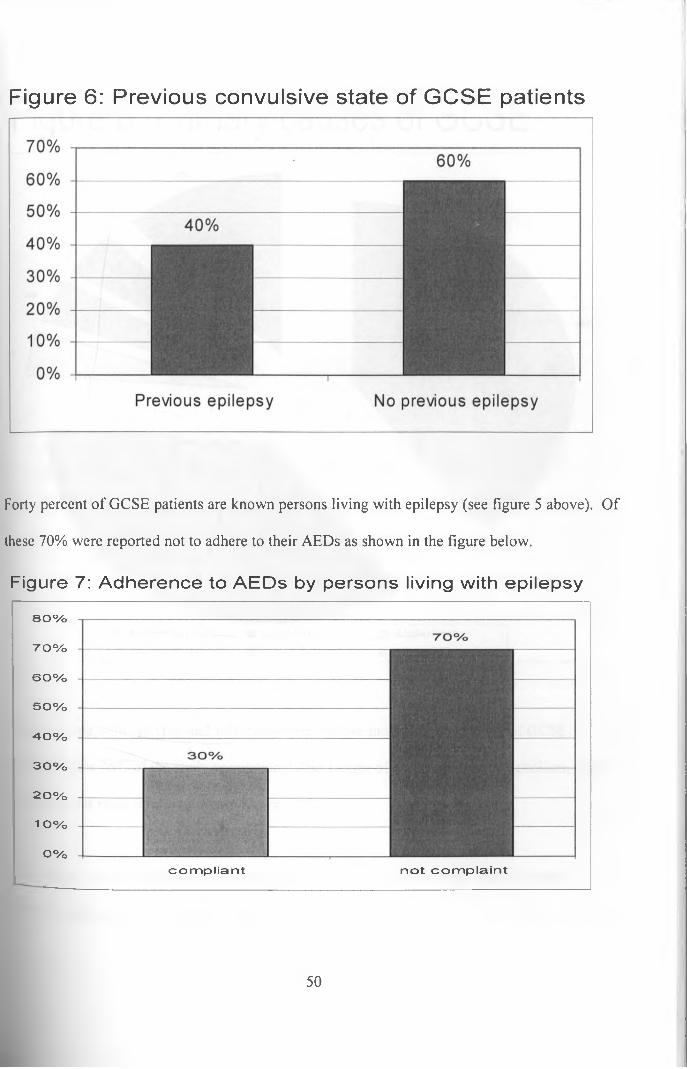
Figure 6: Previous convulsive state of GCSE patients
Forty percent of GCSE patients are known persons living with epilepsy (see figure 5 above). Of
these 70% were reported not to adhere to their AEDs as shown in the figure below.
Figure 7: Adherence to A E D s by persons living with epilepsy
80%
70%
60%
50%
40%
30%
2 0 %
1 o%
o%com pliant not com pla in t
50

Figure 8: Primary causes of GCSE
□ known e p ile p tics u in fec tions □ A lch o lism□ acute p o iso n in g m rena l fa ilu re □ others
Persons living with epilepsy and infections were the two most common causes of GCSE
accounting for 76% of the study population. Other causes of GCSE included alcoholism, acute
poisoning and renal failure as shown above.
Infections as a group contributed to 36% of the primary causes of GCSE. The individual
Sections are as shown in figure 8 below.
51

Figure 9: Infections causing GCSE
Toxoplasmosis
Cryptococcal | meningitis
T.B.M
Non specific Meningoencephalitis
0% 2% 4% 6% 8% 10% 12%
Focal infective processes which presented as space occupying lesions were the most common
accounting for 14 % of all GCSE patients, followed by meningoencephalitis at 10%.
Tuberculous meningitis, cryptococcal meningitis and toxoplasmosis which are AIDS defining
illnesses accounted for 12%. There were 14% of the patients who presented with space
occupying lesions that a conclusive diagnosis was not reach due to investigative difficulties such
as carrying out brain biopsies.
52
L

Figure 10: ILAE proposed Classification of causes of GCSE
The international league against Epilepsy (ILAE) has proposed classification of causes of GCSE
based on the perceived time duration the cause has been operational in the patient. It basically
has three classes with the acute symptomatic having been present for seven days or less while
those present for over one week are considered to be remote symptomatic causes and those in
whom causes are unknown or time is difficult to estimate are classified as undetermined causes.
In this study acute symptomatic causes accounted for about 62% with remote symptomatic
contributing 28% as shown in figure 9 above.
53
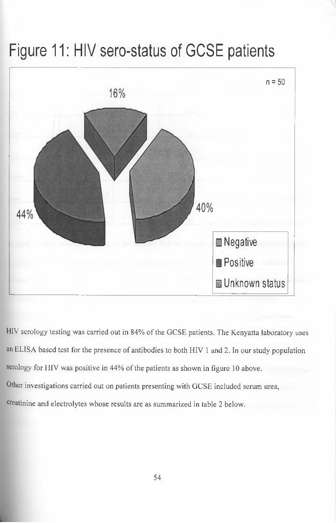
Figure 11: HIV sero-status of GCSE patients
16%n = 50
□ Negative
B Positive
□ Unknown status
HIV serology testing was carried out in 84% of the GCSE patients. The Kenyatta laboratory uses
an ELISA based test for the presence of antibodies to both HIV 1 and 2. In our study population
serology for HIV was positive in 44% of the patients as shown in figure 10 above.
Other investigations carried out on patients presenting with GCSE included serum urea,
creatinine and electrolytes whose results are as summarized in table 2 below.
54
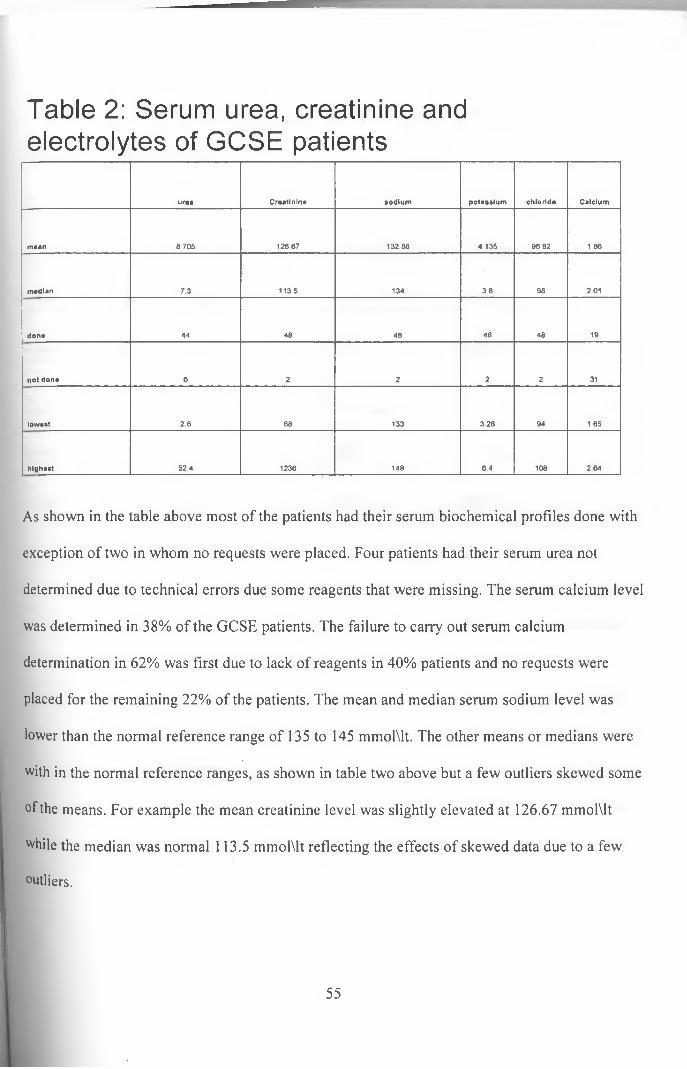
Table 2: Serum urea, creatinine and electrolytes of GCSE patients
urea Creatinine sodium potassium chloride Calcium
mean 8.705 126 67 132 88 4 135 96.82 1.86
median 7.3 113.5 134 3 8 98 201
done 44 48 48 48 48 19
not done 6 2 2 2 2 31
lowest 2.6 68 133 328 94 1 65
highest 52 4 1236 148 8.4 108 2 64
As shown in the table above most of the patients had their serum biochemical profiles done with
exception of two in whom no requests were placed. Four patients had their serum urea not
determined due to technical errors due some reagents that were missing. The serum calcium level
was determined in 38% of the GCSE patients. The failure to carry out serum calcium
determination in 62% was first due to lack of reagents in 40% patients and no requests were
placed for the remaining 22% of the patients. The mean and median serum sodium level was
lower than the normal reference range of 135 to 145 mmolMt. The other means or medians were
with in the normal reference ranges, as shown in table two above but a few outliers skewed some
of the means. For example the mean creatinine level was slightly elevated at 126.67 mmolMt
while the median was normal 113.5 mmolMt reflecting the effects of skewed data due to a few
outliers.
55

Table 3: Hematological investigations done on GCSE patients
Hb (m g\d l) W B C \m m 3 P la te le ts \ m m 3
m ean 12.22 9212.5 245 ,000
m edian 10.3 7050 234 ,000
n 50 50 50
low est 6.8 2 ,800 125,000
highest 15.6 28 ,000 437 ,000
A summary of the hematology investigations done on the patients presenting with GCSE is as
shown in table 3 above. All the 50 GCSE patients had their complete blood counts determined.
The means and the median levels of the hematological investigations were all in the normal
ranges as shown in table 3 above. Hemoglobin level was 12.22 gms\dl with a median of 10.3
gm\dl, the normal ranges being 10-16 gms\dl. The white blood count mean was 9212.50 cells/
mm 3 with a median of 7050 cells/ mm 3 the normal range is 3500 - 10,500 cells/ mm 3. The
mean platelet count was 245,000 particles/ mm 3 and a median of 234,000 particles/ mm 3 both
°f which are with in the normal range of 150,000 - 450,000 particles/ mm 3.
56

Figure 12: Emergency drugs prescribed for patientspresenting in GCSE
The emergency treatment given to patients with GCSE is as shown above. All the 50 patients
received a bolus injection of a benzodiazepine. The benzodi£zeP*ne use< in all these situations
was diazepam. Further to this, 86% of the patients got two 0r 1X101-6 repeat doses of diazepam.
Thirty two percent got a second drug which was a loading d< se °f phenytoin while a third drug, a
loading dose of phenobarbitone, was prescribed in 8% of the patients.
57
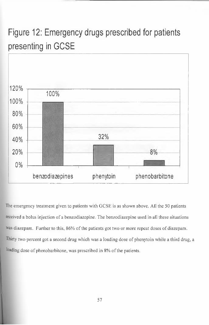
Figure 12: Emergency drugs prescribed for patientspresenting in GCSE
120%
100% 1
80%
60%
40% -
20% -
0%
100%
32%”
benzodiazepines phenytoin phenobarbitone
The emergency treatment given to patients with GCSE is as shown above. All the 50 patients
received a bolus injection of a benzodiazepine. The benzodiazepine used in all these situations
was diazepam. Further to this, 86% of the patients got two or more repeat doses of diazepam.
Thirty two percent got a second drug which was a loading dose of phenytoin while a third drug, a
loading dose of phenobarbitone, was prescribed in 8% of the patients.
57

Figure 13: Discharge medications for patients withGCSE
clonazepam
valproic acid
phenobarbitone
phenytoin
carbarn ezapine
0% 10% 20% 30% 40% 50%
Eighty percent of patients received oral maintenance medication, of these 8% received two or
more drugs. Carbamezapine was the most prescribed oral maintenance drug having been
prescribed to 40% of the patients followed by phenytoin prescribed in 34%. Clonazepam was the
least prescribed at 2% as shown in figure 12 above.
58
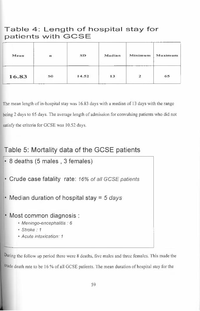
Table 4: Length of hospital stay for patients with GCSE
Mean n SO IVledian Minimum Maxim um
1 6 .8 3 50 14.52 13 2 65
The mean length of in-hospital stay was 16.83 days with a median of 13 days with the range
being 2 days to 65 days. The average length of admission for convulsing patients who did not
satisfy the criteria for GCSE was 10.52 days.
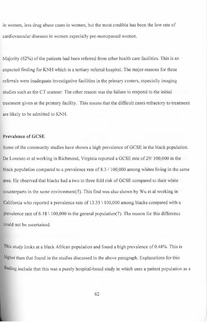
Table 5: Mortality data of the G C S E patients
during the follow up period there were 8 deaths, five males and three females. This made the
Crude death rate to be 16 % of all GCSE patients. The mean duration of hospital stay for the
• 8 deaths (5 males , 3 females)
• Crude case fatality rate: 16% of all GCSE patients
• Median duration of hospital stay = 5 days
• Most common diagnosis :• Meningo-encephalitis: 6• Stroke: 1• Acute intoxication: 1
59

patients who died was 5 days. The primary pathology that was associated with death was
infection to the brain and meninges which was responsible for six out of the eight deaths.

DISCUSSION
Generalized Convulsive Status Epilepticus (GCSE) is a medical emergency with a high mortality
and morbidity. Very few studies have been done in Africa on this disease. The knowledge of the
prevalence, causes, morbidity and mortality factors will fill the current knowledge gap and lead
to better management of this condition. We recruited 50 patients from those admitted to KNH
general wards, intensive care and high dependency units during the three month study period and
followed them to either discharge or death.
Demographic profile
The mean age of the patients was 28.96 years with a median of 27 years. This is younger than the
mean ages in the studies done in western countries where the mean and median ages range
between 50 - 60 years (4, 5, 6, 7, 8). The reason for this apparent age difference could be due to
the differences in the population distribution where the western countries have a larger aging
population leading to a narrow-based population pyramid.
Furthermore cerebrovascular diseases, an etiology associated with the elderly population,
contributed only to 2% of the primary causes. Western studies have also shown that the risk for
GCSE increases with advancing age (4, 5, 6, 7). Wu et al found a prevalence rate of 22.32 \
100,000 in the age group over 75 years compared with 6.18 \ 100,000 in the general population.
The explanation for the above was the high rate of cerebrovascular diseases in the elderly.
The male to female ratio was found to be 1.6 to 1. This male predominance has been observed in
other studies (4, 6, 8). Various theories have been advanced including lower rates of head injury
61
in women, less drug abuse cases in women, but the most credible has been the low rate of
cardiovascular diseases in women especially pre-menopausal women.
Majority (62%) of the patients had been referred from other health care facilities. This is an
expected finding for KNH which is a tertiary referral hospital. The major reasons for these
referrals were inadequate investigative facilities in the primary centers, especially imaging
studies such as the CT scanner. The other reason was the failure to respond to the initial
treatment given at the primary facility. This means that the difficult cases refractory to treatment
are likely to be admitted to KNH.
Prevalence of GCSE
Some of the community studies have shown a high prevalence of GCSE in the black population.
De Lorenzo et al working in Richmond, Virginia reported a GCSE rate of 29/ 100,000 in the
black population compared to a prevalence rate of 8.3 / 100,000 among whites living in the same
area. He observed that blacks had a two to three fold risk of GCSE compared to their white
counterparts in the same environment^). This find was also shown by Wu et al working in
California who reported a prevalence rate of 13.35 \ 100,000 among blacks compared with a
prevalence rate of 6.18 \ 100,000 in the general population(7). The reason for this difference
could not be ascertained.
This study looks at a black African population and found a high prevalence of 0.48%. This is
higher than that found in the studies discussed in the above paragraph. Explanations for this
finding include that this was a purely hospital-based study in which uses a patient population as a
denominator for comparison as opposed to community studies which use the general population
as the reference point.
The study was prospective in design unlike the earlier community studies that have been
retrospective. Retrospective studies have the problems of missing out many cases due to lost
records and low case finding attributable to lack of an active screening process.
Coeytaux et al did a study in six cantons in the French speaking Switzerland and concluded that
urban areas had higher prevalence’s of GCSE than rural populations(8). Our study was set in an
urban area.
Duration of stay
A Study done in French speaking Geneva by Coeytaux et al showed mean admission duration of
13 in patient days while Wu et al found with 6.8 days by in California(7,8). Coeytaux explained
that the difference in in-hospital duration was mainly attributable to the better and faster
investigation rate in California. Fie gave the example of imaging studies such as computerized
tomography scanning (CT scan) and magnetic resonance imaging (MRI) which we available in
California 24 hours a day compared to the rural cantons that lacked such facilities and patients
had to be referred to Geneva for investigations.
This study found a mean admission duration of 16.83 in patient days which is longer than any of
the two earlier studies. One of the reasons contributing to a prolonged in-hospital stay was lack
°f adequate neuro-imaging facilities especially CT scanning. The major reason for referral in
this study was for neuroimaging studies which accounted for about half of the reasons for
63
referral. It was also noted that neuro-imaging studies delayed the time taken to reach a
conclusive final diagnosis of the primary cause of the GCSE. Up to 14% of the GCSE patients
had space occupying lesions that had not been completely characterized due to the inadequacy of
imaging facilities by the time they were discharged.
About 20% of the referral was due to refractory GCSE. These represent a group of patients with
a severe disease that has failed to respond adequately to treatment. These will most likely require
higher or combinations of medications, further investigations all of which will lengthen their in-
hospital stay.
By definition loss of consciousness was a must for one to be included as a case of GCSE. This
translated to low level of consciousness at admission. Coma states are associated with various
complications such as aspiration pneumonia and physical injuries that contribute to a prolonged
in-hospital stay.
Our primary etiologies were different from those found in western studies that reported shorter
in-hospital duration. It is a fact that certain diagnoses are associated with longer hospital stay
than others. For example a diagnosis of cryptococcal meningitis in a HIV sero-positive patient is
likely to be longer than that of an alcoholic presenting in hypoglycemia.
The emergency management was done by registrars not consultant neurologists. It was observed
that about 86% of the GCSE patients got repeat doses of diazepam. This reflected poor
adherence to standard protocols of treating GCSE in the emergency setting which results in a
64

slower rate of seizure activity control. Eventually 40% of the GCSE patients needed a second or
third anticonvulsant drug for adequate control. This could represent a group of patients with
refractory seizures taking a longer duration to control or a group that was treated less
aggressively.
It may have been possible that GCSE is a marker of severe illness which needs a longer duration
of hospital stay.
Etiology of GCSE
Up to 40% of those who presented in GCSE were known to be persons living with epilepsy. This
finding is similar to that of Knake et al in Hessen Germany who observed that up to 50% of the
patients with GCSE were known persons living with epilepsy(4). The major reason for the GCSE
in persons known to suffer from epilepsy was that up to 70% of them were not compliant to
prescribed antiepileptic drugs. This is a known factor that contributes to poor seizure control and
may predispose such individuals to GCSE.
Mativo in a master’s dissertation titled ‘factors causing poor compliance among epileptic patients
presenting at the KNH neurology clinic’ observed a 62% non compliance rate in patients on long
term anti epileptic medication. The individual factors responsible for the poor compliance were
poverty, low level of education and low social economic status (22). The current study did not
look at factors contributing to poor compliance but the results suggest an increased GCSE risk in
persons living with epilepsy who do not adhere to their AEDs. This is an area where a major
reduction can be undertaken to reduce this high prevalence. Interventions such as patient
65

education and availing drugs will lead to better seizure control which eventually leads to a
lowering of the prevalence.
Furthermore most of the patients had poor control as judged by frequent seizures. Whether this
was due to the poor drug adherence or the primary condition was outside the scope of this study.
Infections were the second commonest cause of GCSE accounting for 36% of all patients.
Infection with a focal presentation accounted for 26% compared to 10% causing diffuse brain
and meningeal infections. This is in contrast to the etiologies from western studies where the
leading cause is cerebrovascular diseases with infections playing an insignificant role. This
difference can be explained in several ways. Firstly our study population was younger with a
lower burden of cerebrovascular diseases. Secondly it could be due to a low incidence of
infectious causes in the western countries such that non infectious causes like cerebrovascular
diseases predominate. Also the huge burden can represent a failure of health preventative
measures which is common in developing countries.
Known AIDS defining illnesses including tuberculous meningitis, cryptococcal meningitis and
toxoplasmosis accounted for 12% of the primary etiologies associated with GCSE. This coupled
with the finding of a 44% sero positivity rate for HIV in patients presenting in GCSE
underscores the importance of neurological manifestations of HIV\AIDS.
Nature of investigations
As expected investigations usually focus on the perceived likely cause of the ailment and were
skewed towards causes prevalent locally. This explains the active search for infective causes on
66
all GCSE patients by carrying out complete blood counts. Despite infections ranking second as a
common cause of GCSE the mean and median white blood cell counts were in the normal
ranges. Kenya is at the epicenter of the HIVVAIDS pandemic, this can explain the 84% screening
rates in the GCSE patients which led to the finding of a 44% HIV sero-positivity in the GCSE
patients. HIV/AIDS manifestations in the central nervous system are protean ranging from mild
confusion, acute psychotic episodes to myelopathies and unconscious states. All these can either
be attributed to the HIV itself, opportunistic infections or treatment. From this preliminary data it
would seem that HIV\AIDS contributes significantly to the cause of GCSE. This would be an
area where a prospective study on the relationship between the two would be necessary.
The biochemical parameters were generally normal. However the screening rate for serum
calcium levels was low at 38%. Hypocalcaemia is known to cause convulsions and failure to
determine serum calcium levels in 62% of the patients may have missed many patients. The
ILAE guidelines recommend calcium screening in all convulsing patients. Out of the 19 patients
who had their serum calcium level determined two were found to have hypo-calcemia after
correction for the serum albumin level. The diagnosis of hypocalcaemia associated GCSE was
entered in one patient who had symptomatic hypocalcaemia with a corrected serum calcium level
of less than 1.65 rnMolMt. This makes the prevalence of hypocalcaemia to be 5.26%. This is
higher than what has been reported in other studies and could have contributed by the small
number of patients (4, 8, 10).
Nature of treatment
The emergency treatment decisions of GCSE patients were done by registrars and some did not
conform to the universally laid down guidelines. This can explain the less aggressive use of
second and third drug loading doses and the repeated loading doses of diazepam given to GCSE
patients. Two patients got a diazepam drip for their continued convulsions. Diazepam is oil based
hence immiscible in fluids thus putting it in a drip is not recommended (28, 44, 46). This is not
the best clinical practice for standard protocols have emphasized the use of the above drugs. The
main reason for non use was the unavailability of the drugs. Furthermore clinicians rarely
prescribed it in the treatment sheet. Adherence to the treatment protocol, availability of
phenytoin sodium will go a long way to reduce the mortality and duration of hospital stay
The majority (90%) of the GCSE patients were managed in the general medical wards with 8%
admission into the intensive care unit or the high dependency unit. This is in contrast to the
western studies where over 50% of the patients were in ICU or specialised neurological wards (4,
5, 7). GCSE is a medical emergency which should be closely monitored and intervention done
on a real time basis to improve outcomes. The major reason that the patients never got to such
units was mainly due to a restricted bed space. This would definitely contribute to the high
mortality we observed.
Mortality
The crude fatality rate was 16% which is higher than the 10.7% found by Wu et al in California
(7). This has to be taken in the context of the small sample. The high mortality rate can be
68
largely explained by the differences in the primary causes of GCSE. In the California study a
primary diagnosis of cerebral anoxia was associated with a high risk of death followed by
infective causes. However the total burden of infections was low due to the low prevalence of
infective causes in their locality ranking as the tenth cause of GCSE. This is in contrast the
current study where infections were 38% of the primary causes.
Mean duration of hospital stay before death was five days. This is similar to that found by Wu et
al in California who found a mean hospital stay of 6.7 days before death in patients presenting
with GCSE (7).
Six out of the eight fatalities were due to meningoencephalitis. They had also tested positive for
HIV on ELISA examination done at the KNH laboratories. Two out of the six patients who died
during the study were on antiretroviral therapy for HIV. This goes further to emphasize the role
of HIV/AIDS not just in causing GCSE but in increasing to its mortality.

Conclusion
This study has shown that:
i. The prevalence of GCSE is relatively higher in our setting and that it occurs in relative
younger age groups.
ii. Epilepsy is the single most common etiological factor in GCSE. Most of those result
from poor drug compliance.
iii. Infections are the second most important primary cause of GCSE.
iv. GCSE contributes to a longer in hospital stay in our set up than in other parts of the
world.
v. Providing adequate neuro-imaging facilities in other hospitals can significantly reduce t
referral rate of GCSE patients and improve their management.
vi. HIV/AIDS contributes significantly to both causation and mortality of GCSE. An area
that needs urgent further studies in view of the scaling up program of antiretroviral
therapy.
70

RECOMMENDATIONS
1. Counseling on living with epilepsy, drug adherence, destigmitisation and availing
antiepileptic drugs will ago along way in reducing non compliance rates in persons living
with epilepsy and consequently reduce the prevalence of GCSE.
2. Improving and expanding intensive care facilities will improve management of medical
emergencies GCSE included.
3. Adhering to standard treatment protocols will standardize and improve care for patients
of GCSE.
4. More studies are needed to look at the role of specific diseases in causation of GCSE
especially HIV/AIDS in our set up.
71
STUDY LIMITATIONS
1. The study was carried in a short duration of time (three months) which is may not be a
good indicator on the true prevalence.
2. The study did not carry out postmortem to ascertain the causes of death in the patients
who presented with GCSE.
3. The study did not follow the patients discharged on a long time which could uncover
minor disabilities such as learning difficulties that could be attributed to GCSE that might
not be picked up by a short term mortality study.

REFERENCES
1. Krishnamoorthy E.S, satishchandra P, Sander J.W: research in epilepsy: development
priorities for developing nations - Epilepsia 2003, 44(suppl 1): 5 - 8.
2. Julie A.C, Brian G.R.N, Steven W, Amanda J.R, Godfrey 0, Neema M, Chris M, Charles
R.J.C.N: Increased prevalence of epilepsy associated with severe falciparum malaria in
children - Epilepsia 2004;45(8): 978 - 981
3. Annegers J.F, Hauser W.A, Beghi E. et al: The risk of unprovoked seizures after
encephalitis and meningitis - Neurology 1998;38:1407 - 10
4. Susanne K, Felix R, Mathias V, Wolfgang H ., Hans-Helge M, Angelika W, Niki K and
Hajo M.H, for the epilepticus study group Hessen (SESGH): Incidence of status
epilepticus in adults in Germany; A prospective, population- based study - Epilepsia
2001, 42(6): 714-718.
5. DeLorenzo R.J, Hauser W.A, Towne A.R, et al : A prospective, population based
epidemiologic study of status epilepticus in Richmond, Virginia - Neurology
1996;46:1029- 1035
6. Hesdorffer D.C, Logroscino G, Cascino G, Annagers J.F, Hauser W.A: Incidence of
status epilepticus in Rochester, Minnesota, 1965 - 1984 -Neurology 1998; 50:735 - 741
73
7. Wu Y.W, shek D.W, Garcia P.A, Zhao S, Johnston S.C: incidence and mortality of
generalized convulsive status epilepticus in California - Neurology 2002; 58:1070 - 1076
8. Coeytaux A., Jallon P., Galobardes B.and Morabia A.: Incidence of status epilepticus in
French speaking Switzerland - Neurology September 2000; 55:693 - 697
9. Lockey A.S: Emergency department drug therapy for status epilepticus in adults -
Journal o f emergency medicine 2002,19:96 - 100
10. Joseph K, Se'bastien D.S. and Michel B: Demographic impact of Epilepsy in Africa:
Results of a 10- year cohort study in a rural area of Cameroon - Epilepsia 2003; 44(7):
956 - 963
11. Jilek - Aall L, Rwiza H.T: Prognosis of epilepsy in a rural African community. A 30-year
follow up of 164 patients in an outpatient clinic in rural Tanzania - Epilepsia July -
august 1992;33(4): 645-650
12. Snow R.W, Williams R.E, Rogers J.E, Mung’ala V.O, and Peshu N: The prevalence of
epilepsy among a rural Kenyan population. Its association with premature mortality -
Tropical geographical Medicine 1994;46(3): 175 - 179
13. Aminoff M.J, Simon R.P: Status epilepticus, Causes, clinical features and consequences
in 98 patients - American journal o f Medicine November 1980; 69(5): 657 - 666
74

14. Sung C.Y, Chu N.S: status epilepticus in the elderly: etiology, seizure type and outcome
- Acta Neurol Scand 1989;80:51 - 56
15. Claassen J, Lokin J.K, Fitzsimmons B.F.M, mendelsohn F.A, Mayer S.A: predictors of
functional disability and mortality after status epilepticus - Archives o f neurology
February 2002 59(2): 205-210
16. Mayer S.A, Claasen J, Lokin J, Mendelsohn F, Dennis L.J, Fitzsimmons B.F: Refractory
status epilepticus: frequency, risk factors, and impact on outcome - Archives o f
neurology February 2002;59(2): 205 - 210
17. Towne A.R, Pellock J.M, Ko D, Delorenzo R.J: Determinants of mortality in status
epilepticus - Epilepsia Jan - Feb. 1994;35( 1): 27 - 34
18. Logroscino G, Hesdorffer D.C, Cascino G, Annegers J.F, Hauser W.A: short term
mortality after the first episode of status epilepticus - Epilepsia December 1997; 38(12):
1344- 1349
19. Graham C.A, Gordon M.W.G: status epilepticus in accident and emergency: a difficult
case - Emergency medical journal 2001 ;18:492 - 493
20. Daniel H.L and Brian K.A: Status Epilepticus; review article of current concepts - New
England Journal o f Medicine. Number 14 April 2 1998; Vol 338:970 - 976
75

21. Fountain N.B, Lotham E.W: Pathophysiology of status epilepticus - Journal o f clinical
neurophysiology July 1995; 12(4): 326 - 342
22. Peter mativo: Mmed dissertation: factor lead to poor control among epileptics -
University o f Nairobi, 2004
23. Kwasa T.O.O: The pattern of neurological disease at Kenyatta National Hospital - East
African medical journal may 1992;69(5): 236 - 239
24. Logroscino G, Hesdorffer D.C, Cascino G.D, Annegers J.F, Bagiella E, Hauser W.A:
long- term mortality after a first episode of status epilepticus - Epilepsia December 1997
38(12): 1344- 1349
25. Sheryl R.H, Jana V and Solomon L.M: susceptibility of immature and adult brains to
seizure effects: Neurology 1998, 38:1407 - 1410.
26. Murthy J.M.K: Non-convulsive status epilepticus: An under diagnosed and potentially
treatable condition - Neurology India December 2003;51(4): 453 - 454
27. Young G.B, Jordan K.G, Doig G.S: An assessment of non-convulsive seizures in the
intensive care unit using continuous EEG monitoring: An investigation of variables
associated with mortality - Neurology 1996;47: 83 - 89
76

28. Mark M.S (author): Management of status epilepticus - 1 - Up to date version 12.3.
September 8, 2004
29. Penberthy L.T, Towne A, Garnett L.K, Perlin J.B, Delorenzo R.J: Estimating the
economic burden of status epilepticus to the health care system; Epilepsia 1999; 41:
1923- 1934
30. Georges N, Michel D, Leopold N, Pierre- Marie P, Alain V: Economic evaluation of
Epilepsy in Kiremba (Burundi): A case control study - Epilepsia 2004;45(6): 673 - 677
31. Kwasa T .0.0 and Muthingi P.M: The experience with electroencephalography at the
Kenyatta National Hospital, Nairobi - East African medical journal may 1992;69(5): 259
-261
32. Oduori M.L, Shah S.K The pattern of neurological disease in African children in Kenya -
East African medical journal may 1973;50(5): 253 - 260
33. Coggon D, Geoffrey R, barker D.J.P (authors): Epidemiology for the uninitiated 3rd
edition - BMJ publication, 1993 Fountain N.B, Lothman E.W: Pathophysiology of status
epilepticus - Journal o f clinical neurophysiology July 1995; 12(4): 326 - 342.
34. Kamil C.A, Melike S and Turker S: The role of bcl-2 family of genes during kindling -
Epilepsia, 2005;46(2): 217-223
77

35. Matthew E.B, White H.S: The effect of cgx-1007 and Cl-1041, novel NMDA receptor
antagonists, on kindling acquisition and expression - Epilepsy research 2004; 59:1-12
36. Dudek F.E, Thomas Y and John E.R: Non synaptic mechanisms in seizures and
epileptogenesis - Cell Biology International 1998;vol 22(No 11112): 793 - 805
37. Dudek F.E, Snow R.W, Taylor C.P: Role of electrical interactions in synchronization of
epileptiform bursts -Advancedneurology 1986;44:593 - 617
38. Traub R.D, Wong R.K: Cellular mechanism of neuronal synchronization in epilepsy -
Science 14,h may 1982;216(4547): 745 - 747
39. Phillip A.S and David A.P: changes in excitatory and inhibitory synaptic potentials
leading to epileptogenic activity - Brain research 183:61 - 76
40. Hauser W.A: status epilepticus: epidemiological considerations - Neurology may
1990;40(5 suppl 2): 9 - 1 3
41. Walker M.C, Smith S.J, shorvon S.D: the intensive care treatment of convulsive status
epilepticus in the UK: Results of a national survey and recommendations - Anesthesia
Feb. 1995;50(2): 130-135
78

42. Litt B, Wityk R.J, Herz S.H et al: Non-convulsive status epilepticus in the critically ill
elderly - Epilepsia 1998; 39: 1194- 1202
43. Falope Z.F, Ogunniyi A, Osuntokun B.O: factors associated with epileptiform EEG
patterns in Nigerian epileptics - East African medical journal may 1993;70(5): 294 -
296
44. Recommendations of the epilepsy foundation of the America are working group on status
epilepticus: treatment of convulsive status epilepticus -JAMA Aug 18, 1993; 270(7): 854
- 859.
45. Tatum W.O, Alavi A, Stecker M.M: technetium-99m-HMPAO SPECT in partial status
epilepticus - Journal o f nuclear medicine July 1994;35(7): 1087 - 1094
46. Treiman D.M, Meyers P.D, Walton N.Y, Collins J.F, Colling C, Rowan A.J, Handforth
A, Faught E, Calabrese V.P, Uthman B.M, Ramsay R.E, Mamdani M.B: A comparison of
four treatments generalized convulsive status epilepticus; Veterans affairs status
epilepticus co-operative study group - NEMJ sep 1998;339(12): 792 - 798.
47. Bassel F.S and Nathan B.F: assessment of acute morbidity and mortality in
nonconvulsive status epilepticus -Journal o f clinical neurophysiology July 199512(4 ):
346-352
79

48. De Lorenzo R.J, Pellock J.M, Towne A.R, and Boggs J.G: Epidemiology of status -
Journal o f clinical neurophysiology July 199512(4): 316 - 325
80

Appendix I
Part one
Patient questionnaire client number__________
Prevalence of generalized convulsive status epilepticus in patients admitted to Kenyatta
National Hospital and their short-term outcomes
PATIENT INFORMATION SHEET (English version)
Dear Sir / madam / master / ms
I’m conducting a study on the prevalence of generalized convulsive status epilepticus and
the short-term outcomes in patients admitted to K.N.H. This small note is to seek to inform
you about the study and seek your support.
Your patient has been admitted to this hospital following the harrowing experience of
losing consciousness accompanied by uncoordinated movements of the limbs. This is called
a seizure in medical terms. Such activity results from uncontrolled excessive discharges
from neurons in his brain.
The exact cause of this phenomenon is not known but it is a documented fact that an insult
to the brain can predispose to this condition. Such brain insults can take the form of a head
injury, infection, disturbance in body electrolytes etc. we don’t know what is contributing
to such disease in your patient and our community at large. By participating in this study
you can help uncover what tour patient and others who get similar seizures have so that it
will be easier to plan their treatment in future.
Further more if this seizure activity continues for a long time it can cause brain damage.
Specifically any such seizure lasting over half an hour is thought to be dangerous to life,
and constitute a medical emergency called status epilepticus. Many medical people insist
that one should treat such condition before the lapse of thirty minutes but evidence is
81

scanty for such a decision. So your participation will help resolve this quagmire and may
just be one of the voices of evidence to support early intervention in your patient and all
future patients.
To gather all this data we will need to do various tests on you or your patient which will
include both radiological where the patient will be exposed to x-rays and laboratory
methods which will involve taking blood or other body fluids for analysis. Any risks for
such procedures such as pain at injection will be explained to you or your patient prior to
taking the said samples.
Lastly you / your patient should know the following:
1. Your participation in the study is very important to all patients who suffer seizure
illnesses.
2. Standard medical treatment will be used as prescribed by your doctor. There no
drugs which will be administered in this study.
3. All information acquired during this study shall be treated confidentially and only
used for scientific purposes.
4. Participation in the study is voluntary
5. You are free to withdraw from the study at any point and there are no consequences
for such action
6. All the results will be made available to you and your doctor for purposes of
managing your disease
7. For any extra questions you may have my contacts are:
Dr George M. Nyale
University of Nairobi
Department of medicine
P.O BOX
Nairobi
Tel:
My supervisors are:
82

Prof. Adam - Consultant Neurologist, Department of Medicine, University
of Nairobi
Prof Amayo - Consultant Neurologist, Department of Medicine, University
of Nairobi
Dr Jowi - Consultant Neurologist, Department of Medicine, Kenyatta
National Hospital
CONSENT FORM FOR NEXT OF KIN
I _______ __________________________________ being the parent / legal guardian of
__________________________________ do affirm that I have read the patient
information sheet and that I have fully understood the study as explained to me by
__________________________________________ . I have appended my signature below on a
voluntary basis and as evidence of my consent to participate in the study. I also have the right to
withdraw from the study at any stage without any dire consequences to my patient or myself. I
have done so on:
Date:
Signature:
Witness: date
Signature:
ASSENT FORM FOR MINORS
I _____________________________________________ _ _ do allow my parents / guardian to
consent on my behalf and that I have also been explained to the purpose for this study. I also
know my rights that I can withdraw from the study even if my parents / guardians have
consented to it and that I shall not be required to give an explanation.
Signature:
Date:
83
CONSENT FORM FOR SUBJECT
I __________________________________________ consent to participate in this study on
generalized convulsive status epilepticus as has been explained by
____________________________________________. I fully understand the purpose, benefits
and risks of this study. Further to this I know I can withdraw from this study at any stage without
any dire consequences.
Subjects: signature:
Date:
Witness: signature:
Date
Part two
PROFORMA FORM
Age: gender: male female
DoA: ward:
1. Is it the first episode? Yes no
2. Is the patient a known epileptic? Yes no
a. If yes has he / she been taking his medication as prescribed?
Yes NO
84

CONSENT FORM FOR SUBJECT
I ___________________________________________consent to participate in this study on
generalized convulsive status epilepticus as has been explained by
____________________________________________. I fully understand the purpose, benefits
and risks of this study. Further to this I know I can withdraw from this study at any stage without
any dire consequences.
Subjects: signature:
Date:
Witness: signature:
Date
Part two
PROFORMA FORM
Age: gender: male female
DoA: ward:
1. Is it the first episode? Yes no
2. Is the patient a known epileptic? Yes no
a. If yes has he / she been taking his medication as prescribed?
Yes NO
84
b. Do you know the drugs he is taking?
Yes No
3. How many people witness today’s seizure?
4. Did he / she:
(a) Loose consciousness? Yes
(b) Throw hands and legs? Yes
(c) Pass urine / stool on themselves? Yes
(d) Bite their tongue? Yes
(e) Froth at the mouth? Yes
no
no
no
no
no
5. How long did this convulsive movements last?
a. Below thirty minutes
b. Between thirty minutes to one hour
c. Over one hour
6. After the convulsions were over what happened to the patient?
a. Ran off
b. Went to sleep
c. Was confused
85
d. Another convulsion started
7. Was this episode preceded by any fever or illness in the past one week?
Yes No
8. Do you attribute this condition to any incidents / illnesses in the past?
Yes ------------- No -------------
If yes please state which:
9. What other illnesses / disabilities does this patient have?
Examination findings
General condition of patient: coma
jaundice
contused alert
Pallor
Dehydration
cyanosis edema
oral thrush
Any neurological deficit:( 5 part neurological examination)
86

1) Look at the patient
a. General demeanour
b. Speech
c. Gait
d. Arm swinging
2) Examine the head
a) Facial movements
b) Tongue
c) Eye movements
d) Pupils
e) Fundi
3) Examine the upper limbs
a) Posture of outstretched arms
b) Wasting, fasciculation
c) Power, tone
d) Co-ordination
e) Reflexes
87

Part three
Day of discharge:
Functional level at discharge (circle on the karnofsky score chart)
THE KARNOFSKY PERFORMANCE INDEX
Performance
status
Functional capability of patient
100 Normal, no complaints, no evidence of disease
90 Able to carry on normal activity; minor signs or
symptoms of disease
80 Normal activity with effort; some signs or
symptoms of disease
70 Cares for self; unable to carry on normal activity
or do active work
60 Requires occasional assistance but is able to care
for most needs
50 Requires considerable assistance and frequent
medical care
40 Disabled; requires special care and assistance
30 Severely disabled; hospitalization is indicated
although death is not imminent
20 Very sick; hospitalization necessary, active
supportive treatment necessary
10 Moribund; fatal processes progressing rapidly
0 Dead
91
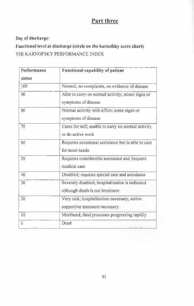