prevalence of leptospirosis in donkey and associated risk
TRANSCRIPT

Prevalence of leptospirosis in donkey
and associated risk factors
KK Daddy
Orcid.org 0000-0002-0349-8030
Dissertation accepted in fulfilment of the requirements for
the degree Master of Science in Animal Health at the
North West University
Supervisor: Dr L Ngoma
Co-supervisor: Prof M Mwanza
Graduation ceremony: April 2020
Student number: 29769183

ii
DECLARATION
I the undersigned, declare that the dissertation titled “PREVALENCE OF
LEPTOSPIROSIS IN DONKEYS AND ASSOCIATED RISK FACTORS” hereby
submitted to the North West University-Mafikeng campus for the degree of Master of
Science in Agriculture, Animal Health represents my original work in design and execution
and has not otherwise been submitted in any form for any degree or diploma to any
university. All materials used have been acknowledged.
Signed……………………………….this………………….day of …………….2019

iii
DEDICATION
This thesis is dedicated to my God, Almighty first, then my parents: Mr. Jean Pierre Kiayima
Kibambe and Mrs. Bernadette Shima Kilolo

iv
ACKNOWLEDGEMENTS
This MSc. dissertation would not have been possible without the collaboration, dedication
and friendship of many people who have given their precious support and assistance towards
this final outcome.
In particular, I am profoundly grateful to my supervisors Dr. Lubanza Ngoma and Prof
Mulunda Mwanza, for their vital part in my learning experience throughout these years as an
MSc. student at North West University. I sincerely appreciate the support, guidance and
opportunities they gave me in this process to develop my own research skills in a very
motivating work environment. During all these years they kept their doors open with a smile
on their faces regardless of how many interruptions, or how inopportune my visits were.
Thank you for all the advices given, and sharing your knowledge and friendship with me. My
appreciation goes to all the donkey farmers, who generously agreed to take part in this
research. This including giving consent for collecting samples, and for freely giving their
time to enable the completion of the research questionnaires. Without their selfless
collaboration, none of the output of this dissertation would have been possible.
I am very grateful to Mr Vuyani Mjekula, the technician in the Department of Animal Health,
North West University, Mr Makhosini and Mr Joseph Tsepo inspectors at the Highveld Horse
Care Unit, who drove all over the district collecting samples. They did a tremendous job in
coordinating farm visits, transporting samples, and ensuring that all questionnaires were
completed properly.
My appreciation also goes to the North West University for their financial support and for
providing me with the much needed facilities used during this project.
My time living in South Africa has been one of the greatest in my life and I want to thank all
the people that made me feel at home. During these years, I crossed paths with so many

v
beautiful friends from so many different places and backgrounds. These friends have
enriched my own life so much and made me realize how lucky I was for living these
experiences. I would name them all in these lines if I was confident of not leaving anyone
out.
Thanks to my parents Jean Pierre Kiayima Kibambe and Bernadette Shima Kilolo for all the
love and support that they have given me, and for encouraging me to pursue my studies at
this level. In addition, I want to extend my gratitude to all my family members for their
support from their respective distant places where they are based.

vi
ABSTRACT
Leptospirosis is a neglected zoonosis of global importance with a complex epidemiology that
affects humans, domestic and wild mammals. Several studies have addressed Leptospira
sero-prevalence and risk factors in horses worldwide, including South Africa. Nevertheless,
sero-prevalence of Leptospira spp. in donkeys around Ngaka Modiri Molema district
(NMMD), North West Province is unknown. The main purpose of the present study was to
estimate the sero-prevalence of eight Leptospira serovars and identify factors associated with
the infection in the NMMD of the North West Province, South Africa. The study was carried
out between March 2017 and October 2018 across NMMD. A cross-sectional study was
adopted for this study. A total of 365 blood samples were collected from healthy donkeys and
sera were tested with live antigen suspensions of leptospiral serovars including serovars
Canicola, Bratislava Hardjo, Grippotyphosa, Icterohaemorrahagiae Szwajizak, Tarassovi and
Pomona using the microscopic agglutination test (MAT). Furthermore, a questionnaire was
used to collect data on the risk factors for Leptospira sero-status. Factors associated with the
presence of Leptospira antibodies were assessed using univariate and multivariate logistic
regression analysis, Odds ratio and their 95% confidence interval were computed. Of the 365
donkeys tested for the presence of Leptospira antibodies, the majority (29.6%; n=108/365)
were from Mafikeng local municipality and the rest (19.7%; n=72/365) were from Ratlou.
The lowest proportion (8.2 %; n=30/365) of donkeys was from Ditsobotla. Just over half,
(58.1%; n=212/365) of donkeys tested were female and the remaining 41.9% were male. In
addition, most donkeys (41.9%; n=153/365) were between 6-12 years old, followed by those
between 0-5 years (37%; n=135/365), and only 20.3% (n=74/365) were above 12 years.
Antibodies against Leptospira were found in 11.5% (95% CI: 4.86-18.14) healthy looking
donkeys. The most common leptospiral serovar against which serum antibodies were detected
was serovar Bratislava (81%; n=34/42) followed by Serovars Tarassovi (19.04%; n=8/42).

vii
Based on the final logistic regression model, presence of horses and agricultural activities in
the vicinity of donkey housing properties were negatively significantly associated with the
presence of antibodies against Leptospira sero-positivity. Furthermore, males were over four
times more likely to test positive than were females (OR = 4.88; p ≤ 0.0001; 95% CI [2.01-
11.82]), there was a statistical significant difference between males and females. Although,
donkeys within the vicinity of fruits and vegetables farming (OR = 0.093; p ≤ 0.0001; 95%
CI [0.031-0.27]) and those with horses in the vicinity (OR = 0.226; p ≤ 0.002; 95% CI
[0.089-0.57]) had a lower odd of testing positive, the differences were negatively significant.
The present study concluded that the sero-prevalence of Leptospira in donkeys was relatively
low 11.5% (95% CI: 4.86-18.14) in the NMMD and that Leptospira interrogans serovars
Bratislava and Tarassovi were the most commons serovars and hence, donkeys may serve as
reservoir for Leptospira bacteria. Furthermore, this study found that donkeys in the study area
are reservoirs for the predominant serovar Bratislava and the less dominant serovar
Tarassovi. Gender of the donkey is a risk factor for Leptospira seroprevalence. Further
studies are needed to investigate the role of agricultural activities in the vicinity of the
dwellings of donkeys in the occurrence of Leptospira in the study area.
Keywords: leptospirosis, sero-prevalence, donkeys, risk factors

viii
TABLE OF CONTENTS
DECLARATION............................................................................................................................ ii
DEDICATION ...............................................................................................................................iii
ACKNOWLEDGEMENTS .......................................................................................................... iv
ABSTRACT ................................................................................................................................... vi
LIST OF FIGURE .......................................................................................................................xiii
LIST OF ABREVIATIONS AND ACRONYMS ..................................................................... xiv
CHAPTER I .................................................................................................................................... 1
INTRODUCTION .......................................................................................................................... 1
1.1 Background ........................................................................................................................... 1
1.2 Problem statement ................................................................................................................ 3
1.3 Justification ........................................................................................................................... 4
1.4 Study hypothesis ................................................................................................................... 4
1.5 Study Aim ............................................................................................................................. 5
1.6 Study Objectives ................................................................................................................... 5
CHAPTER 2 ................................................................................................................................... 6
LITERATURE REVIEW............................................................................................................... 6
2.1 The causative agent of leptospirosis .................................................................................... 6
2.2 Classification of the genus Leptospira ................................................................................ 6
2.2.1 Serological classification of the Leptospira spp .......................................................... 6
2.2.2 Genotypic classification of the genus Leptospira ........................................................ 7
2.3 Pathogenesis .......................................................................................................................... 7
2.4 Virulence factors in pathogenic Leptospira spp ................................................................. 8
2.4.1 Loa22 gene ..................................................................................................................... 8
2.4.2 LipL32 gene.................................................................................................................... 9
2.4.3 Leptospiral immunoglobulin-like (Lig) proteins ......................................................... 9
2.5 Environmental transmission cycling and risk factors for exposure................................. 10

ix
2.6 Important risk factors associated with Leptospira infection in animals.......................... 10
2.6.1 Water, Agriculture and Landscape risks .................................................................... 10
2.6.2 Socio economic factors ............................................................................................... 11
2.6.3 Host reservoirs and load of infectious Leptospira as risk factors............................. 12
2.7 Leptospirosis in South Africa ............................................................................................ 13
2.7.1 Animal leptospirosis .................................................................................................... 13
2.7.2 Human leptospirosis reported cases in South Africa................................................. 14
2.8 Leptospira spp infecting donkeys around the world ........................................................ 14
2.9 Epidemiology of leptospirosis ........................................................................................... 15
2.10 Methods of detection and identification for Leptospira spp .......................................... 16
2.10.1 Detection of anti Leptospira antibodies ................................................................... 16
2.10.2 Molecular identification of Leptospira spp.............................................................. 18
2.11 Prevention and control of animal leptospirosis .............................................................. 19
2.11.1 Prophylactic administration of antibiotics ............................................................... 20
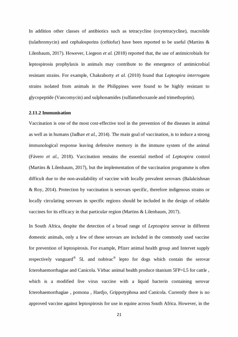
2.11.2 Immunisation ............................................................................................................. 21
2.11.3 Specific management environmental alterations ..................................................... 23
CHAPTER 3 ................................................................................................................................. 24
MATERIAL AND METHODS ................................................................................................... 24
3.1. Study design ....................................................................................................................... 24
3.2 Study area and village selection ........................................................................................ 24
3.3 Population, sample size and animal selection ................................................................... 26
3.4 Data collection, processing and storage ............................................................................ 27
3.4.1 Questionnaire interview .............................................................................................. 27
3.4.2 Blood samples collection ............................................................................................ 28
3.4.3 Ethical Considerations ................................................................................................. 28
3.5 Serology testing (Macroscopic Agglutination Test)......................................................... 29
3.5.1 Live antigen.................................................................................................................. 29
x
3.5.2 Leptospira Ellinghausen–McCullough–Johnson–Harris (EMJH) Liquid media .... 30
3.5.3 Leptospira EMJH semi solid media ........................................................................... 30
3.5.4 Dilution buffer ............................................................................................................. 30
3.5.5 Leptospira spp subcultures.......................................................................................... 31
3.5.6 Controls ........................................................................................................................ 31
3.5.7. Procedure..................................................................................................................... 31
3.6 Statistical analysis............................................................................................................... 33
3.6.1 Data management ........................................................................................................ 33
3.6.2 Descriptive statistics .................................................................................................... 33
3.6.3 Univariate and multivariate analyses.......................................................................... 33
CHAPTER 4 ................................................................................................................................. 35
RESULTS...................................................................................................................................... 35
4.1 Summary Statistics ............................................................................................................. 35
4.2 Sero-prevalence of leptospirosis in the NMMD ............................................................... 37
4.3 Distribution of Leptospira serovar according to MAT titres ........................................... 38
4.4 Inferential statistics ............................................................................................................. 39
4.4.1 Univariate analysis of risk factors association with the presence of Leptospira
antibodies ............................................................................................................................... 39
4.4.2 Results of the multivariate logistic regression analysis: assessment of significant
risk factors for Leptospira seropositivity ............................................................................. 44
CHAPTER 5 ................................................................................................................................. 46
DISCUSSION ............................................................................................................................... 46
5.1 Leptospira sero-prevalence ................................................................................................ 46
5.2 Predominant serovars ......................................................................................................... 47
5.3 Risk factors for donkey exposure ...................................................................................... 49
CONCLUSION AND RECOMMENDATIONS ....................................................................... 51
6.1 Conclusion........................................................................................................................... 51
6.2 Recommendation ................................................................................................................ 52

xi
6.3 Limitation of the study ....................................................................................................... 52
REFERENCES ............................................................................................................................. 53
APPENDIXES .............................................................................................................................. 67
Appendix 1: Participant questionnaire interview.................................................................... 67
Appendix 2: Participant informed consent form ..................................................................... 72
Appendix 3 Reservoir map with 8 tested serovars ................................................................. 74
Appendix 4: Orientation of the U-bottom serum plate ........................................................... 75

xii
LIST OF TABLES
Table 3. 1: Distribution of donkeys within the NMMD, number of villages and samples size
........................................................................................................................................................ 26
Table 3.2: Panel of Leptospira reference serovars and strains used in as antigen in the MAT.
........................................................................................................................................................ 29
Table 4. 1: Demographic, geographic, and management factors distribution of donkeys in the
NMMD who were included in the risk factor study, 2018 36
Table 4. 2: Sero-prevalence of leptospirosis in the NMMD per local municipalities .............. 38
Table 4. 3: Distribution of Leptospira serovars according to MAT titres ................................ 39
Table 4.4: Univariate results and apparent prevalence (AP) of seropositive Donkeys to
Leptospira by demographic factors ............................................................................................. 40
Table4.5 : Univariate results and apparent prevalence (AP) of seropositive Donkeys to
Leptospira by geographic and environmental factors (n=365) .................................................. 41
Table 4.6 : Univariate results and apparent prevalence (AP) of Leptospira seropositive
associated with management factors. .......................................................................................... 43
Table4.7: Univariate results and apparent prevalence (AP) of Leptospira seropositive
associated with disease clinical signs, antibiotics treatment and vaccination history. ............. 44
Table 4.8: Odds Ratios and 95% CI from Multivariate logistic regression analysis of
significant risk factors associated with seropositivity (n=365, np=42)..................................... 45

xiii
LIST OF FIGURE
Figure2. 1 : Trinomial elements involved in the success of the control of animal leptospirosis,
adapted from Martins & Lilenbaum, 2017 .................................................................................. 20
Figure 3.1: Map showing the distribution of the Sampling sites in the five local municipalities
of NMMD. 25

xiv
LIST OF ABREVIATIONS AND ACRONYMS
MAT Macroscopic agglutination test
LIP Lipopolyssacharides
IG Immunoglobuline
DNA Deoxyribonucleiotides acid
qPCR Quantitative polymerase chain reaction
OVI Ondersterpoort Veterinary Institute
CAAT Cross agglutination absorption test
RPM Rotation per minute
SPSS Statistical package of social sciences
NMMD Ngaka Modiri Molema District
OMPs Out membrane proteins
DAFF Department of Agriculture Forestry and Fisheries
USDA United State Department of Agriculture
EMJH Ellinghausen–McCullough–Johnson–Harris
PSI Pound per square inch
ELISA Enzyme Linked Immunosorbent Assay
µL Microliter
Lig Leptospiral immunoglobulin-like

1
CHAPTER I
INTRODUCTION
1.1 Background
According to the Faostat (2014), the world equine population is estimated to be over 113
million of which donkeys comprise over 39% (around 44 million). More than 97% of
donkeys are found in developing countries specifically kept for work.
In South Africa as in most developing countries, donkeys are some of the most important
domestic animals contributing to many social and economic sectors of the resource-limited
communities especially these living in rural areas where modern means of transportation are
absent, unaffordable, unsuitable or inaccessible (Marufu, 2014). Despite their significant
contribution to the national economy, the attention given to study the infectious diseases of
donkeys is minimal. Among the diseases infecting donkeys, leptospirosis is one of the more
common bacterial zoonosis and also an important emerging global economic and public
health problem (Bharti et al., 2003; Burriel, 2010; Derne et al., 2011).
The disease is caused by pathogenic species grouped within the “interrogans” complex. This
group contains approximately 250 identified serovars (Haake, 2009; Loureiro et al., 2013).
The bacteria may be maintained in the environment by reservoir hosts such as rodents which
can have the bacteria without being infected and showing any symptoms of illness (Bharti et
al., 2003). The reservoir host would be harbouring and shedding the pathogen in their urine
(Adler & de la Peña Moctezuma, 2010). Transmission is mainly influenced by several
exposure risk factors, more commonly inadequate management practices, environmental
conditions and exposure to asymptomatic carriers have been reported (Martins & Lilenbaum,

2
2017). Donkeys become infected through contact with water or soil contaminated with urine
from infected rodents, wild or domestic animals. In incidental hosts such as humans,
symptoms are variable and the infection can result in a severe and potentially fatal disease
with clinical manifestations which include fever, renal and hepatic injury, pulmonary
haemorrhage and reproductive failure (Adler & de la Peña Moctezuma, 2010; Utzinger et al.,
2012). Clinical leptospirosis is difficult to recognize in equine. However, recurrent uveitis is
the most recognized clinical signs (Barsoum et al., 1978). Additional and commonly reported
signs include periodic ophthalmia as well as reproductive conditions such as abortions and
stillbirth (Hamond et al., 2014b; Simbizi et al., 2016). Available studies on animal
leptospirosis across South Africa show that agglutinating antibodies may occur in apparently
healthy animals infected with host adapted serovars. For example, dairy cattle may harbour
serovars Hardjo, Pomona, and Grippotyphosa (Hesterberg et al., 2009); pigs may harbour
Pomona, Tarassovi, or Bratislava (Potts et al., 2013) and dogs may harbour Canicola (Roach
et al., 2010). A recent study conducted by Simbizi et al. (2016) shows that Bratislava is the
dominant serovars in horses in South Africa. But it is likely that these animals represent only
a fraction of likely hosts.
No research has been carried out to study the role donkeys may play in the epidemiology of
this disease in South Africa. Furthermore, the infecting serovars and potential risk factors
associated with the seropositivity in donkeys have not yet been fully investigated.
Therefore, the main purpose of the present study was to estimate the sero-prevalence of eight
of the known Leptospira serovars that occur in different animal species in South Africa using
the microscopic agglutination test (MAT), and identify risk factors associated with the
seropositivity.

3
1.2 Problem statement
Donkeys are used in smallholder farming as traction animals and for transportation of goods.
Thus they have close contact with humans and this close contact with human beings poses a
great risk of transmission of leptospirosis from donkeys to human (Guerra, 2013;
Grevemeyer et al., 2017).
Leptospirosis is characterized by infertility, lowered milk production, renal involvement, and
spontaneous abortion and recurrent uveitis (Rathinam, 2005; Grevemeyer et al., 2017). Renal
dysfunction and neonatal mortality have also been reported (Xue et al., 2009). The pathogen
can readily be transmitted between species, and between animals and humans through
infected urine, contaminated soil or water, or other body fluids (Bharti et al., 2003; Haake &
Levett, 2015; Tsegay et al., 2016).
The disease is most commonly found in rural and urban areas of tropical and subtropical parts
of the world (Derne et al., 2011). Several serological studies have showed wide-spread
evidence of leptospiral infection in horses (Simbizi et al., 2016), cattle (Hesterberg et al.,
2009) and dogs (Roach et al., 2010) in South Africa. Furthermore, information on the
outbreak of leptospirosis in donkeys has been reported by several researchers from different
parts of the world (Barsoum et al., 1978; Grubišic´ et al., 2011; Grevemeyer et al., 2017).
However, evidence regarding its prevalence from the rural areas in South Africa is scarce, or
poorly reported.
From extensive literature review, it can be deduced that this is the first time that this kind of
study is done in South Africa on donkeys. This study was therefore undertaken to contribute
to our understanding of the epidemiology of the disease. This information will contribute to
the development of effective control measures.
4
1.3 Justification
According to the World Health Organization, leptospirosis is considered to be a neglected
tropical disease and is one of the most widespread zoonosis. However data on its prevalence
in the donkey population is scarce (WHO, 2003).
A recent study conducted on leptospirosis in horses of South Africa showed that the horse
breed and the presence of a forest in the vicinity of horses’ property constituted potential risk
factors for infection (Simbizi et al., 2016).
Several outbreaks of human or/and animal leptospirosis are usually associated with exposure
or contact with urine of contaminated animal (Adler & de la Peña Moctezuma, 2010). In a
recent study, Grevemeyer et al. (2017) reported the evidence of positive leptospiral DNA in
urine of donkeys.
Due to the large number of variables involved in the epidemiology of leptospirosis, the
development of public health strategies to control, prevent and lower the risk of new infection
in humans and animals depends on the knowledge of Leptospira survival, and transmission
dynamics in the environment and among its animal hosts (Derne et al., 2011).
Therefore understanding the variability in prevalence and serovar diversity in donkeys as well
as risk factors for leptospirosis incidence is a pressing need.
1.4 Study hypothesis
Based on previous leptospiral studies conducted on domestic animals across South Africa we
hypothesize that:
Different Leptospira serovars are sources of infection in donkeys in the NMMD
Geographical, environmental and management factors are not associated with
Leptospira infection in donkeys in the NMMD
5
1.5 Study Aim
The purpose of the study was to determine the Leptospira sero-prevalence in donkeys and
also to identify possible risk factors associated with Leptospira infection in the rural and peri-
urban areas of the NMMD, North West Province of South Africa.
1.6 Study Objectives
The specific objectives of this study were:
To determine the Leptospira serovars infecting donkeys in the study area
To estimate the sero-prevalence of leptospirosis in donkeys within the NMMD
To identify risk factors associated with Leptospira infection

6
CHAPTER 2
LITERATURE REVIEW
2.1 The causative agent of leptospirosis
Leptospires are thin, helically coiled, motile spirochetes usually 6-20 μm in length which
differ from other spirochaetes by the presence of end hooks. They have surface structures that
share features of both Gram-positive and negative bacteria (Adler & de la Peña Moctezuma,
2010; Evangelista & Coburn, 2010). These bacteria are fastidious and aerobic growing in
simple media enriched with vitamins B1 and B12, long-chain fatty acids, and ammonium salts
at an optimum growth temperature of 29±1oC (Adler & de la Peña Moctezuma, 2010).
Leptospires have a typical double membrane structure, within the outer membrane expression
of the surface-exposed epitopes lipopolysaccharide (LPS) constitute the basis of the
serological classification.
2.2 Classification of the genus Leptospira
The classifications system of the genus Leptospira spp is very complex, and both serological
and genotypic methods are used for taxonomic purposes (Cerqueira & Picardeau, 2009).
2.2.1 Serological classification of the Leptospira spp
Historically, serological classification using microscopic agglutination, divides leptospires
into two groups: pathogenic leptospires grouped within the “interrogans” complex
(Leptospira interrogans sensu lato) and saprophytic leptospires grouped in the “biflexa”
complex (Leptospira biflexa sensu lato) (Haake, 2009; Loureiro et al., 2013). In both
complexes, the antigenically related strains have been grouped into serovars and serogroups
(Bharti et al., 2003; Haake, 2009; Loureiro et al., 2013). This method, classified Leptospira
species into 24 serogroups. These serogroups consist of more than 250 identified serovars
(Postic et al., 2000; Langoni et al., 2016) of which more than 200 are considered pathogenic

7
(Loureiro et al., 2013). Furthermore, one serovar can be isolated from both pathogenic and
non-pathogenic species (Bharti et al., 2003; Haake, 2009; Haake & Levett, 2015). Serological
assays can detect antibodies against one serovar that are cross- reacting with infecting
serovar. Hence, de Abreu Fonseca et al. (2006) suggested that in addition to serological
methods, molecular approaches should be used in order to confirm unknown isolates.
2.2.2 Genotypic classification of the genus Leptospira
To date, speciation of new isolates is based on genotypic classification, mainly DNA
relatedness (Loureiro et al., 2013). This classification system has revealed that the genus
Leptospira is divided into three groups including pathogenic, non-pathogenic as well as
intermediate species. More importantly the pathogenic Leptospira group comprise 13 species
including L. alexanderi, L. alstonii L.borgpetersenii, L. inadai, L. interrogans, L. fainei, L.
kirschneri, L. licerasiae, L. noguchi, L. santarosai, L. terpstrae L. weilii, L. wolffii, (Bharti et
al., 2003; Vijayachari et al., 2008; Adler & de la Peña Moctezuma, 2010; Langoni et al.,
2016). These species are the main causes of leptospirosis in animals and humans. However,
the mechanism by which they cause the disease is not fully understood (Ghazaei, 2018).
2.3 Pathogenesis
The initial step for infection and pathogenesis is the adhesion of leptospires to host tissue
components (Evangelista & Coburn, 2010). Like other microbial pathogens, leptospires
require chemotactic mechanisms to adhere and penetrate abraded tissues such as conjunctival
and genital mucous or skin following contact with infected urine (Rathinam, 2005; Xue et al.,
2009). After penetration, the pathogen is harboured in the blood during the bacteraemic stage
which can last up to a week (Marinho & Cardoso, 2015).
The organisms undergo primary multiplication in the liver and other visceral tissues
accompanied by the rise in the concentrations of specific immunoglobulin (IgM and IgG) in

8
the blood (Loureiro et al., 2013). These immunoglobulins ingest the bacteria with their
subsequent removal from blood circulation except in the kidneys in which the organisms
persist and are shed into the urine for several months (Loureiro et al., 2013).
Clinical manifestations of leptospirosis in humans and animals are widely documented.
However, the molecular mechanisms through which the pathogen causes disease remain
undetermined (Ko et al., 2009). Recent research in advanced molecular profiling of
pathogenic Leptospira spp revealed that they possess genes and gene variants that mediate
mechanisms through which pathogenesis occurs (Ghazaei, 2018).
2.4 Virulence factors in pathogenic Leptospira spp
Hill (2012) described virulence factors as structures and mechanisms used by
microorganisms to establish and maintain an infection in the host. These structures involve
the presence of capsules, flagella and other potential factors that may contribute to virulence
have been analysed by whole genome sequencing of pathogenic Leptospira interrogans (Ren
et al., 2003; Jorge et al., 2018), the intermediately pathogenic leptospiral species (Ricaldi et
al., 2012) and the saprophytic Leptospira biflexa (Picardeau et al., 2008). The sequenced
genomes reveal that Leptospira species have several virulence-related factors, such as the out
membrane proteins OMPs (LipL32, Loa22 and the Lig) which are essential in virulence.
2.4.1 Loa22 gene
The Loa22 gene is one of seven leptospiral OmpA-like proteins (Haake & Zückert, 2015). It
is the most abundant protein (Malmström et al., 2009) that is genetically essential for
leptospiral virulence (Ristow et al., 2007). The Loa22 remains the unique gene to date that
fulfils Koch's postulates that require that a virulence gene confers a certain phenotype to the
studied bacteria, inactivation of the gene abolishes the phenotype, and reintroduction of the

9
gene restores the wild type to the mutant (Foulongne et al., 2004). However Loa22 mutant
gene has not been proved to be virulent in hamsters and guinea pigs (Ristow et al., 2007).
2.4.2 LipL32 gene
The Lipl32 gene codes for the major outer membrane protein most recognised as antigen in
sera during infection (Murray et al., 2009). The gene is abundantly conserved in all
pathogenic Leptospira. Nevertheless, the gene is considered as a paradox of leptospiral
biology (Murray, 2013). Studies conducted by Hoke et al. (2008) revealed that LipL32 gene
is necessary for virulence during the infection due to its ability to bind and adhere to the host
tissues.
However, Murray et al. (2009) constructed a LipL32 mutant in Leptospira interrogans using
transposon mutagenesis, it was surprisingly observed that the transposon mutant deficient for
LipL32 gene was adherent to host tissues, and no differences in the virulence was noted. This
suggests that this gene may not be required in the virulence of Leptospira interrogans.
2.4.3 Leptospiral immunoglobulin-like (Lig) proteins
Pathogenic Leptospira species express surface-exposed proteins belonging to the bacterial
immunoglobulin-like substances (Matsunaga et al., 2003). These proteins were thought to
mediate host mammalian cell invasion or attachment during Leptospira pathogenesis (Lin &
Chang, 2007). Two Lig genes designed as LigA and LigB, and one pseudo-gene LigC, were
previously identified as putative Leptospira virulence factors (Matsunaga et al., 2003). LigA
protein is exceptionally found in strains of Leptospira kirschneri (McBride et al., 2009) and
LigB is found in all pathogenic Leptospira species (Haake & Zückert, 2015), making it an
ideal candidate for vaccine and diagnostic applications (McBride et al., 2009).
These two proteins (LigA and B) expressions were studied by using transcription activator-
like effector (TALE) (Pappas & Picardeau, 2015). A reduced expression level of both
proteins in the TALElig transformants confer low virulence in hamsters, this suggests a

10
cumulative role of LigA and B in pathogenesis. However some studies show that disruption of
the LigB protein does not affect virulence in animal models of leptospirosis (Croda et al.,
2008). Variables such as temperature (Lo et al., 2006) and osmolarity (Matsunaga et al.,
2005) are key environmental factors known to affect and control the expression of the Lig
proteins during environmental cycling of Leptospira spp.
2.5 Environmental transmission cycling and risk factors for exposure
The complex interaction which exists between living organisms and their environment is well
documented and the role played by a contaminated environment as a risk factor for human
and animal leptospirosis has been established in traditional epidemiological studies
(Guernier et al., 2018).
Even though the exact mechanisms through which infection occurs, prior contact with such
environment is not well understood (Bharti et al., 2003). Therefore advanced molecular
approaches are needed to trace the source of Leptospira infection by establishing contact and
genotypic identification of different clinical cases with environmental sources (Barragan et
al., 2017b).
To better understand spatial and temporal patterns of Leptospira incidence, Mwachui et al.
(2015) reviewed possible relationships between ecological sources of the disease and its
prevalence. Water associated exposures such as floods and rainfall, recreational water
activities, including contact with rodents and livestock plus poor sanitation were correlated
with an increased Leptospira risk.
2.6 Important risk factors associated with Leptospira infection in animals
2.6.1 Water, Agriculture and Landscape risks
Leptospira bacterial DNA have been found in freshwater and soil from a wide range of
environments in South Africa (Saif, 2013). After excretion in the environment the pathogen

11
can persist and survive up to twenty months in water and soil while maintaining its
pathogenicity (Barragan et al., 2017b). Exposure to these contaminated sources is likely to
increase risk of infection for humans as well as animals (Lau et al., 2010). In a their study,
Mwachui et al. (2015) show that floods were the direct contributory influence to the
occurrence of an epidemic rather than a risk factor. Studies conducted in India (Mumbai,
Kerala) have documented the association of leptospirosis outbreaks with heavy rain fail
(Pappachan et al., 2004; Maskey et al., 2006).
Heavy rainfalls, related to La Niña favoured an outbreak in New Caledonia (Goarant et al.,
2009). Furthermore, fluctuations in climate specifically, changes in rainfall amounts have
also been linked to leptospirosis outbreak in Italy (Pellizzer et al., 2005). Seasonal pattern of
leptospirosis have been observed in Thailand and peak incidence cases were correlated with
peaks in rainfall and temperature (Chadsuthi et al., 2012). In the United State and Canada,
Ward (2002) observed that the rainy season (late summer to fall) can predict the outbreak of
leptospirosis in dogs. On the other hand, changes in local topography due to land use,
urbanisation, deforestation, agricultural practices, irrigation, may increases the risk of
transmission of leptospirosis (Eisenberg et al., 2007; Lau et al., 2010).
2.6.2 Socio economic factors
Socio-economic circumstances such as sanitation, poverty, and overcrowding provide
suitable habitats not only for the spirochetes but also increase the population of reservoir
hosts such as rodents (Bharti et al., 2003; Derne et al., 2011). In Brazil for example Dias et
al. (2007) detected Leptospira interrogans serovar Icterohaemorrhagiae among a population
of humans living in the sewage drainage basins with poorest social and economic conditions.

12
2.6.3 Host reservoirs and load of infectious Leptospira as risk factors
In rural settings the increased number and diversity of peri-domestic and wild animals may
play a particular role in transmission of the disease (Barragan et al., 2017b). However, the
risk of contagion prior contact with the infected animal is associated not only with renal
Leptospira loads but also depend on the amount of urine excreted, the popularity of hosts,
and the local host concentrations (Barragan et al., 2017b).
In a recent study, Costa et al. (2015b) reported that rodents were the primary reservoir hosts
of leptospires in different settings. In South Africa Taylor et al. (2008) isolated the pathogen
in rodents. Recent studies revealed that rats excrete approximately 5.7 × 10⁶ of Leptospira
cells per millilitre of urine while large animals excrete large volumes of urine containing
about 5.1 × 10⁸ to 1.3 × 10⁹ Leptospira cells per day (Barragan et al., 2017a; Barragan et al.,
2017b). Such animals can spread the pathogen in the environment through contaminated
urine for several months or years. Yet, it is important to note that pathogen concentrations in
the urine is crucial in determining infection especially when such load is diluted into stagnant
water formed after high rainfall, putting the entire population at risk (Barragan et al., 2017b).

13
2.7 Leptospirosis in South Africa
2.7.1 Animal leptospirosis
Leptospirosis has a fascinating history among researchers in South Africa. Leptospira
antibodies have been detected in domestic as well as wild animals across the country. A study
conducted by Potts et al. (2013) reported that 22.2% of pigs showed antibodies to serovar
Icterohaemorrhagiae and Bratislava.
Hesterberg et al. (2009) reported that the most likely infecting serovars in cattle in KwaZulu-
Natal were Pomona (22%), Tarassovi (19%), Bratislava (15%), Canicola (13%), Hardjo
(13%), and Icterohaemorrhagiae (12%). A study done by Roach et al. (2010) revealed a
preponderance of reactions to serovar Canicola. This serovars has been traditionally the most
associated with leptospirosis in dogs. More recently, Simbizi et al. (2016) conducted a study
to detect leptospiral serovars prevailing in horses and assess factors linked with the infection.
The result revealed that more than twenty serovars (17 serogroups) were identified including
serovar Bratislava, as the sero-dominant.
Apart from serological studies, research on isolation and identification of Leptospira
pathogens in South Africa is limited. This may be due to the slow growing rate of certain
Leptospira species, making its culture and isolation extremely difficult from biological
samples (Liegeon et al., 2018). However, Te Brugge and Dreyer (1985) successfully isolated
Serovar Hardjo from urine of dairy cattle around Onderstepoort area, Pretoria while
Leptospira interrogans serovar pomona was isolated in aborted Bonsmara cattle around
Zeerust district in the North West province (Herr et al., 1982) .

14
2.7.2 Human leptospirosis reported cases in South Africa
Leptospirosis continues to be an important zoonotic disease in human beings around South
Africa. The first South African case of leptospirosis was diagnosed in a Cape Town fish
hawker who died of typical Weil’s disease due to Leptospira interrogans serovar
Icterohaemorrhagiae. Further cases of leptospirosis in dockworkers were published from
Cape Town, and KwaZulu-Natal Province (NICD, 2015).
Based on IgM detection using ELISA test, the apparent incidence was found to be
moderately high (20%) in Cato Crest-Durban (Taylor et al., 2008). Currently this disease
remains underreported and underdiagnosed. Of the few reported cases, information on the
infecting serovars is lacking (Tan, 2006).
2.8 Leptospira spp infecting donkeys around the world
There is limited literature about leptospirosis and risk factors for endemic infection around
the world in general (Adler & de la Peña Moctezuma, 2010),with little to no recent data
available on the disease in equine in South Africa in particular (Simbizi et al., 2016).
However, in Morocco Javanica and Australis serogroups were found to be predominant in
donkeys (Benkirane et al., 2016). In a comparative survey between horses and donkeys
conducted in Ahvaz, South-west of Iran by Hajikolaei et al. (2005), serovars Grippotyphosa
(49.51%), Icterohaemorrhagiae, Ballum, Pomona, Hardjo and Canicola were found to be
predominant. In addition, that study showed that there was a higher prevalence of leptospiral
antibody rate in donkeys (40%) than in horses (27.88%).
Barsoum et al. (1978) reported that 72% (90/125) hospitalised donkeys in Egypt show
leptospiral antibodies in their sera at a titre of 1: 128 or more. The study also revealed that
sera were mostly reacting to serovars Butembo, Pomona, Icterohaemorrhagiae and Canicola.
In Croatia, Grubišic ́et al. (2011) found that the most common confirmed and likely infective
15
serovars in donkeys were Bratislava, Pomona and serovar Icterohaemorrhagiae. In the
Caribbean, urine samples from a herd of donkeys on the island of St. Kitts Grevemeyer et al.
(2017) found that 18% (n=22/124) clinically healthy donkeys had leptospiral DNA in their
urine. It can therefore be deduced that donkeys can spread the pathogen in the environment
though their urine.
2.9 Epidemiology of leptospirosis
The epidemiology of Leptospira spp. infection is complex since the organism is widely
distributed in the environment with more than 250 identified serovars. One serovar may be
adapted to several hosts, while one host might carry several distinct serovars. The natural
maintenance host ensures the continuous circulation of particular leptospiral serovars in a
geographical area (Adler & de la Peña Moctezuma, 2010).
Distribution and infection patterns may thus change both by adaptation of serovars to other
hosts and by the introduction of new host animals into a new area (Bharti et al., 2003; Adler
& de la Peña Moctezuma, 2010).
Climatic and ecological changes affect the distribution of Leptospira serovars and
consequently the prevalence and their clinical features (Hartskeerl et al., 2011). Prevention
and control, as well as treatment, require serovar recognition (Hamond et al., 2014b). More
than 1.03 million human cases of severe leptospirosis occur worldwide annually (Costa et al.,
2015a).
In the past years, leptospirosis has emerged as a globally important infectious disease. It
occurs in urban environments of industrialised and developing countries, as well as in rural
regions worldwide with high incidence in the tropics (Bharti et al., 2003).
Sub-clinical infection is common in endemic regions; an infected animal can remain
symptom-free and shed infectious organisms in the urine for its entire lifetime (Bharti et al.,

16
2003). The prevalence of different leptospiral serovars depends on the presence of reservoir
animals and the serovars that they carry as well as local environmental conditions (Bharti et
al., 2003; Burriel, 2010).
2.10 Methods of detection and identification for Leptospira spp
The progression of the infection is a crucial component in leptospirosis diagnostics (de Abreu
Fonseca et al., 2006). The natural course of the disease development influences the choice of
diagnostic test that can be used and the type of clinical sample that should be collected
According to Loureiro et al. (2013) leptospires can be found in blood samples of the diseased
animal during the early stages of the infection and methods such as culture and polymerase
chain reaction (PCR) are more appropriate than serology due to low immunologic response
and undetectable serological titres at this stage.
2.10.1 Detection of anti Leptospira antibodies
The antibodies produced during leptospiral infection have been found to be agglutinating IgM
and IgG antibodies (Evangelista & Coburn, 2010). The detection of these specific
immunoglobulins can help in the rapid diagnosis of the disease in endemic regions (Terpstra
et al., 1985). Methods detecting the body’s immunologic response to the organism, such as
the MAT and enzyme linked immunosorbent assay (ELISA) have proven to be useful (Adler
& de la Peña Moctezuma, 2010).
2.10.1.1 Macroscopic Agglutination Test (MAT)
The macroscopic agglutination test is the gold standard serological test recommended for
diagnosis of leptospirosis for both human (WHO, 2003) and animal hosts (Tan, 2006). The
basis of the test is the detection of agglutination reactions between the animal serum
antibodies and outer membrane antigens of live leptospires panel belonging to different
serovars including locally circulating serovars (Loureiro et al., 2013; Haake & Levett, 2015).

17
Despite the high specificity (95%), MAT usually requires paired acute and convalescent
phase samples, thus this method has been reported to be insensitive when samples are
collected at the beginning of the disease (Lessa-Aquino et al., 2013). The presence of clinical
signs and appropriate history of animal contacts, a high single MAT titre of ≥400 or four-fold
rise in titre in paired serum samples is considered indicative of current or recent infection or a
conversion from sero-negativity to a titre of 1/100 or above (Adler & de la Peña Moctezuma,
2010). Although commonly used and recommended, MAT has well known weakness and
limitations including the need to cultivate and maintain panels of live leptospires.
With respect to the ability to identify the infectious serovars, there is consensus that MAT can
reliably identify the presumptive serogroup (Bharti et al., 2003; Picardeau, 2013). However,
due to the high degree of cross-reaction among different serovars in each serogroup, it cannot
be considered as serovar-specific (Levett, 2003).
In addition, over interpretation of MAT serologic findings in the absence of a sound
knowledge of the locally prevalent serovars may limit its value (Levett, 2003). In Kenya for
example, Mgode et al. (2015) observed that inclusion of local serovars particularly serovar
Sokoine in MAT increased Leptospira sero-prevalence from 1.9% to 16.9% in rodents and
0.26% to 10.75% in humans beings.
2.10.1.2 Enzyme Linked Immunosorbent Assay (ELISA)
Rapid screening of IgM antibodies using the ELISA in the early phase of Leptospira infection
has been shown to be more sensitive than MAT. However, its specificity is not only affected
by the antigen used in the assay but also by the presence of antibodies due to previous
exposure (Ahmad et al., 2005).
ELISA test has been developed using a wide variety of antigen preparations, the assay does
not require the maintenance of live cultures and is amenable to automation (WHO, 2003).

18
There are significant limitations to early diagnosis using ELISA. The test only detects genus-
specific antibodies and as a result, it is not suitable for identification of serovars (Bharti et al.,
2003). In view of this, dependence on ELISA alone as diagnostic method of Leptospira spp is
not recommended (WHO, 2003).
2.10.2 Molecular identification of Leptospira spp
Bacterial culture has been the most important technique for diagnosis and detection of
bacterial infections including Leptospira. However, culturing Leptospira is very difficult due
to the slow growing rates of some Leptospira strains and the long incubation periods
(13weeks) before an isolate is established in culture (Millar et al., 2007; Adler & de la Peña
Moctezuma, 2010; Lessa-Aquino et al., 2013).
The introduction of molecular diagnostic technics has significantly enhanced the ability to
detect culture-negative infections, and highlight non-specific serological false-positive results
(Millar et al., 2007). Among the various molecular assays available, techniques including
partial or whole genome sequencing, molecular typing, microarrays, broad range PCR have
emerged as important tools for identification of Leptospira spp. This method is based on
DNA target sequencing for routine identification of species or genus using universally
conserved genes (5S rRNA, 16S rRNA and 23S rRNA). The targeted genes are less affected
by gene transfer and they contain hyper variable regions flanked by high sequence
conservation (Woo et al., 2008; Sibley et al., 2012).
Janda and Abbott (2007) reported that, the use of 16S rRNA gene sequence can provide in
more than 90% genus identification and between 65 and 83% species identification. Because
some species may share 99-100% 16S rRNA identities, alternative gene targets for
Leptospira speciation have been used including LipL32 gene (Stoddard et al., 2009; Hamond
et al., 2014a; Tarigan, 2016; Hsu et al., 2017), rrs gene (Kurilung et al., 2017), secY

19
(Hamond et al., 2014a), lig A and lig B gene (Matsunaga et al., 2003; Hamond et al., 2014a;
Ali et al., 2017) and the gyrB encoding gene (Slack et al., 2006). Most of these genes help to
differentiate pathogenic and non-pathogenic Leptospira spp. However, because 16S rRNA-
based bacteria detection system is prone to DNA contamination problems, Millar et al.
(2007) suggested that strict contamination controls need to be established to avoid the
occurrence of such problems.
2.11 Prevention and control of animal leptospirosis
The control of leptospirosis involves all measures intended to interfere with its unrestrained
occurrence. Although the agent may still be circulating among the animals, it has been
suggested that the major control procedures should include trinomial elements (Figure 2.1)
such as antibiotic therapy, immunization as well as ecological management (Mughini-Gras et
al., 2014; Martins & Lilenbaum, 2017).

20
Figure 2.1 : Trinomial elements involved in the success of the control of animal
leptospirosis, adapted from (Martins & Lilenbaum, 2017)
2.11.1 Prophylactic administration of antibiotics
The use of antibiotics has shown some efficacy in minimizing the severity of the infection by
eliminating leptospires from tissues and preventing the shedding of the bacteria in the urine.
Antibiotics also provide a means of protection by limiting the degree of environmental load
of leptospires until immunity is induced by vaccination (Mughini-Gras et al., 2014).
Antibiotic treatment has been shown to minimize the risk of introducing leptospires onto the
farm (Sykes et al., 2011). Among the commonly used antibiotics, aminoglycosides (e.g.
streptomycin) have been reported to be efficient in cleaning leptospires from the renal tubes.
21
In addition other classes of antibiotics such as tetracycline (oxytetracycline), macrolide
(tulathromycin) and cephalosporins (ceftiofur) have been reported to be useful (Martins &
Lilenbaum, 2017). However, Liegeon et al. (2018) reported that, the use of antimicrobials for
leptospirosis prophylaxis in animals may contribute to the emergence of antimicrobial
resistant strains. For example, Chakraborty et al. (2010) found that Leptospira interrogans
strains isolated from animals in the Philippines were found to be highly resistant to
glycopeptide (Vancomycin) and sulphonamides (sulfamethoxazole and trimethoprim).
2.11.2 Immunisation
Vaccination is one of the most cost-effective tool in the prevention of the diseases in animal
as well as in humans (Jadhav et al., 2014). The main goal of vaccination, is to induce a strong
immunological response leaving defensive memory in the immune system of the animal
(Fávero et al., 2018). Vaccination remains the essential method of Leptospira control
(Martins & Lilenbaum, 2017), but the implementation of the vaccination programme is often
difficult due to the non-availability of vaccine with locally prevalent serovars (Balakrishnan
& Roy, 2014). Protection by vaccination is serovars specific, therefore indigenous strains or
locally circulating serovars in specific regions should be included in the design of reliable
vaccines for its efficacy in that particular region (Martins & Lilenbaum, 2017).
In South Africa, despite the detection of a broad range of Leptospira serovar in different
domestic animals, only a few of these serovars are included in the commonly used vaccine
for prevention of leptospirosis. For example, Pfizer animal health group and Intervet supply
respectively vanguard® 5L and nobivac® lepto for dogs which contain the serovar
Icterohaemorrhagiae and Canicola. Virbac animal health produce titanium 5FP+L5 for cattle ,
which is a modified live virus vaccine with a liquid bacterin containing serovar
Icterohaemorrhagiae , pomona , Hardjo, Grippotyphosa and Canicola. Currently there is no
approved vaccine against leptospirosis for use in equine across South Africa. However, in the
22
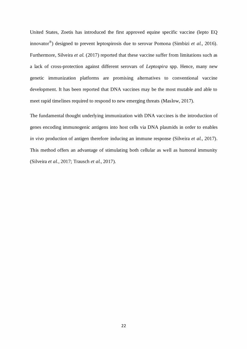
United States, Zoetis has introduced the first approved equine specific vaccine (lepto EQ
innovator®) designed to prevent leptospirosis due to serovar Pomona (Simbizi et al., 2016).
Furthermore, Silveira et al. (2017) reported that these vaccine suffer from limitations such as
a lack of cross-protection against different serovars of Leptospira spp. Hence, many new
genetic immunization platforms are promising alternatives to conventional vaccine
development. It has been reported that DNA vaccines may be the most mutable and able to
meet rapid timelines required to respond to new emerging threats (Maslow, 2017).
The fundamental thought underlying immunization with DNA vaccines is the introduction of
genes encoding immunogenic antigens into host cells via DNA plasmids in order to enables
in vivo production of antigen therefore inducing an immune response (Silveira et al., 2017).
This method offers an advantage of stimulating both cellular as well as humoral immunity
(Silveira et al., 2017; Trausch et al., 2017).

23
2.11.3 Specific management environmental alterations
Because of the long-term survival of pathogenic leptospires in soil and water and the
abundance of wild and domestic animal reservoirs, environmental control measures are
difficult to implement. However, correcting risk factors associated with environmental and
management is of great importance for control of leptospirosis in the herd (Nascimento et al.,
2004).
Uncontrollable factors such as rainfall can extensively be recognised in most endemic areas
and also provide a framework for disease development (Martins & Lilenbaum, 2017).
Additional factors may be corrected; proper hygiene has been demonstrated to significantly
reduce animal exposure to leptospirosis infection. Furthermore, access to infected water,
puddles or swampy areas and co-grazing between animal species should be avoided or
minimized (Martins & Lilenbaum, 2017). More importantly, control of the rodents is the key
to eradication of Leptospira in the environment. Rodenticides provide the basis for the
current management strategies in most countries; unfortunately, these robust control
strategies do not prevent leptospiral transmission or eradication in the herd Therefore,
vaccination remains the essential method of Leptospira spp control.

24
CHAPTER 3
MATERIAL AND METHODS
3.1. Study design
A cross-sectional study design was adopted to estimate the sero-prevalence of Leptospira and
assess the risk factors associated with Leptospira serologic status of donkeys within the
Ngaka Modiri Molema district (NMMD) of the North West Province in South Africa. Multi-
stage simple random sampling was applied:
- Primary stage: selection of villages
- Second stage: Animal selection
3.2 Study area and village selection
The research was carried out in rural and peri-urban areas of the NMMD, within which
Mafikeng, the capital city of the province falls. This district is one of the four district
municipalities of the North West province, and is one of the areas with a large donkey
population. The area is divided into five local municipalities, namely Mafikeng, Ratlou,
Moilwa, Ditsobotla and Tswaing.
The vegetation type in the entire North-west Province encloses two major biomes: the
Grassland and the Savanna Biome (Mucina & Rutherford, 2006). According to the South
African Weather Services, the rainfall is erratic and varies from an average of 600mm per
annum, to exceptional occurrences of more than 700mm per annum (SAWS,2017). The area
receives its lowest rainfall (0.52mm) in June and highest (252mm) in February. The NMMD
is characterised by large seasonal and daily variations in temperature, from very hot in
summer (daily average temperatures may exceed 32 °C in January), to mildly cold in winter,
with average minimum monthly temperatures of up to 12°C.

25
The smallholder village farming system in the NMMD usually has many donkey owners in
one village. Therefore, for the purpose of this study, all the animals in the selected villages
were considered to belong to one large herd, although owned by many different farmers.
To draw a simple random sample from the population of animals in the village was difficult,
because although there are many owners of donkeys, there was no sampling frame. In
addition, donkeys were not individually identified. To overcome these problems, a two-stage
sampling procedure was adopted: a random sample of villages was selected in the first stage
and in the second, animals were randomly selected from each selected village (Cameron,
1999). The list of villages in each local municipality of the NMMD was considered as a
sampling frame (Stats, 2012). Below Figure 3.2 highlights the study area and the sampling
site.
Figure 3.1: Map showing the distribution of the Sampling sites in the five local
municipalities of NMMD.

26
In total there were 187 villages in the District, out of which 19 (approximately 10%) were
selected by means of simple random sampling and were included in the study (Cameron,
1999).
3.3 Population, sample size and animal selection
According to the department of Agriculture Forestry and Fisheries DAFF (2016) the total
population of the donkeys in the NMMD was estimated to be 6 125. The probability
proportion to size was used to determine the numbers of samples in each local municipality
(Table 3.1) which was divided per village that was selected in stage one. The required sample
size was calculated using EpiInfo version 6.04 software (CDC, Atlanta, GA, USA) assuming
5% acceptance error, 50% expected prevalence and the confidence level of 95%. The
estimated sample size was determined to be 365. Visits were organized to each selected
village; systematic random sampling was used to select donkeys for inclusion in the study.
All donkeys in the village were held in the community crush, and a number was assigned to
each donkey from one up to the last donkey in the crush pen and the sampling interval was
calculated based on the population size of donkeys in the village (present in the crush pen)
divided by the required samples for that specific village (Table 3.1).
Table 3. 1: Distribution of donkeys within the NMMD, number of villages and samples size
Area Population
N. of
village
Selected
villages
Samples per
village
Total samples
required
Mafikeng 1875 83 8 13.5 108
Tswaing 1400 26 3 27.6 83
Ratlou 1240 23 2 24.3 73
Moilwa 1200 38 4 17.7 71
Ditsobotla 500 17 2 15 30
Total 6125 187 19 365

27
Randomly, the first sample (not necessarily from the first donkey lined up in the crush) was
selected and then the fixed sampling interval was used to select other donkeys until the
number of required samples in the village was reached. This process was repeated for each
selected village.
3.4 Data collection, processing and storage
3.4.1 Questionnaire interview
In order to identify risk factors related to seropositive donkeys, a structured questionnaire was
designed to collect data on possible risk factors (Appendix 1). The questionnaire was
completed by the owners of donkeys through structured interviews on the same day that
donkeys were sampled.
Consent in this study was sought from each respective owner prior to sample collections
(Appendix 2).
Demographic information (breed, gender, and age), geographical and environmental
information (village name, average annual temperature and rain-fall, agricultural practices) as
well as management information (presence of rodents, others animal in the vicinity, water
source) were recorded. Any symptoms (such as abortions or recurrent uveitis), if observed for
the past two years were recorded including the vaccination history and recent antibiotic
treatment.
Donkeys ages were estimated by the incisor teeth eruption, they were categorised into three
age groups (1- 5 years, 6-11 years and 12 and above). The breeds were characterised as local,
cross breed and others and based on the activities performed by donkeys, they were classified
as working, racing, breeding, or others.
Geographic and environmental factors involve the average annual rain and temperature,
agricultural activities around the donkey villages and the municipalities where they were

28
sampled. The average temperature in the study area ranged from 12-34℃ and based on this
temperature, donkeys were clustered in three groups: low temperature group with annual
average temperature ranging between 2-12℃, medium temperature group with annual
average temperature ranging from 13 to 25℃ and the high temperature reaching 26℃ and
above (South African Weather Services, 2017).
The average rain-falls received in each village where donkeys were sampled ranged from 0
mm-700mm, and was clustered into three groups according to the quantity of rain each
village received. The average rain-fall was categorised as follow: low rain-fall group (0-
100mm), medium rain-fall group (110-300mm and high rain-fall group (310-700mm) (South
African Weather Services, 2017).
3.4.2 Blood samples collection
Between March 2017 and October 2018, a total of 365 blood samples were aseptically
collected from healthy donkeys via the jugular vein using a sterile 18G needle and 10 ml
tubes containing clot activator. Samples were transported to the Animal Health Laboratory,
North West University Mafikeng Campus. Upon arrival samples were centrifuged at 3500
rpm for 10 minutes at room temperature. Sera were kept in labeled cryotubes and stored at -
20ºC before shipment while frozen on ice to the Agricultural Research Council-
Onderstepoort Veterinary Institute (ARC-OVI) for analysis.
3.4.3 Ethical Considerations
This Study was approved by the Animal Care, Health and Safety in Research Ethics
Committee (AnimCare) of the North-West University, Ethics number: NWU-00182-18-S5.
Act 35 (Animal Diseases Act) of 1984, Section 20 approval to perform research was obtained
from DAFF: Directorate of Animal Health (Reference Number 12/11/1/3) of the Republic of
South Africa.

29
3.5 Serology testing (Macroscopic Agglutination Test)
The macroscopic agglutination test is the gold standard serological test Picardeau (2013)
stated that the most conclusive criterion for diagnosis of leptospirosis is a four-fold rise in
MAT titre or sero-conversion to a titre of 1/100 or above . Thus, to confirm a diagnosis with
conviction, paired samples were required, which was difficult to obtain in this study. Adler
and de la Peña Moctezuma (2010) suggested in such circumstances that a single high MAT
titre of 1:100 can be considered as a diagnostic criterion. Shivakumar and Krishnakumar
(2006) reported titre of 1:100 as a significant criterion for diagnosis. The test was done at
ARC-OVI in Pretoria, South Africa. This test was used to identify the infecting Leptospira
serovar or antigenically related serovars using eight live Leptospira antigens (Table 3.2)
maintained in liquid media. These serovars were presumed to be circulating in domestic
animals in South Africa (Hesterberg et al., 2009; Roach et al., 2010; Potts et al., 2013).
3.5.1 Live antigen
Four to eight day cultures of Leptospira reference serovars and strains were originally
obtained from the Royal Tropical Institute (KIT) in Amsterdam, Netherlands and sub-
cultured weekly on liquid medium, cultures showing macroscopic growth (silky cloud when
tube is shaken) were used for testing.
Table 3.2: Panel of Leptospira reference serovars and strains used in as antigen in the MAT.
Species Strains Serovars
Leptospira interrogans Australis Bratislava
Leptospira interrogans Canicola Canicola
Leptospira interrogans Ictero 1 Icterohaemorrhagiae
Leptospira interrogans Pomona Pomona
Leptospira interrogans Szwjizak Szwjizak
Leptospira borgpersenii Perepelitsin Tarassovi
Leptospira kirschneri Moskav v Grippotyphosa
Leptospira interrogans Hardjo type prajitno Hardjo type prajitno

30
3.5.2 Leptospira Ellinghausen–McCullough–Johnson–Harris (EMJH) Liquid media
About 2.3 g of Leptospira Ellinghausen–McCullough–Johnson–Harris liquid medium
(EMJH) (Difco-USA) were dissolved in 900 mL of deionized distilled water and autoclaved
for 15 minutes at 121oC at 15psi. The media was allowed to cool and 100 mL EMJH media
Difco enrichment was added to the solution and mixed well at the lamina Flow bench.
Approximately 5 mL prepared media was dispensed into 10 mL tubes with screw tops and
the media were used for subculture.
3.5.3 Leptospira EMJH semi solid media
Approximately 2.3 g of Leptospira EMJH medium base was dissolved in 500 mL distilled
water. A solution of 1.5 g Bacto-agar was prepared in 400 mL distilled water and then both
solutions were sterilized by autoclaving at 121℃ for 15 minutes at 15psi. After sterilisation,
the solutions were placed in a water bath at 50℃ for 1 hour. The two solutions were mixed
and the EMJH enrichment was added. About 5mL of the medium was dispensed in 10 mL
sterile tubes with screw tops and the media were used for subculture.
3.5.4 Dilution buffer
Sorensen’s buffer stock solution was prepared by adding about 10.44 g of disodium
phosphate (Na2HPO42H2O) and 1.09 g potassium phosphate (monobasic) (KH2PO4). The
solution was dissolved in 1L of deionized distilled water (the pH was adjusted to 7.2) and
stored at 4°C.± 0.3ºC. The Sorensen’s buffer working solution was prepared by adding 2 L
deionized water in 0.85 % saline solution (17 g NaCl in 2 000 mL deionized water) and 840
mL of the saline solution was sterilized by autoclaving for 15 minutes at 121°C at 15psi. The
solution was allowed to cool to room temperature and 160 mL Sorensen’s buffer stock
solution was added. The solution was stored at 4°C.± 0.3ºC.

31
3.5.5 Leptospira spp subcultures
Approximately 0.5-1mL of each serovar were dispensed into tubes containing ±5-10mL
liquid media, back up cultures were maintained on semi solid medium and incubated at
29±1oC in dark. Cultures with clear growth zone (Dinger’s zone) were used for testing.
Contaminated culture was purified using 0.45 or 0.22µm Millipore filter depending on the
level of contamination.
3.5.6 Controls
Positive control sera were obtained from (USDA), the United State Department of
Agriculture (USDA, Ames USA) and stored at -20 ± 5oC, in the ultra-freezer. Negative
control serum was obtained from a negative cow at ARC-OVI and stored at -20 ± 5oC.
After each experiment, the positive sera were kept at 4±3oC together with the negative
control. Contaminated or haemolysed sera not suitable for testing were recorded on the final
test report.
3.5.7. Procedure
The MAT procedure was done according to OIE Manual with minor modifications
(Epizooties, 2004).
3.5.7.1 Screening test
All the samples to be tested were recorded according to the screening test form (BS/RT 005)
The U and the flat bottom micro titre plates were marked according to the (BS/RT 005) form.
Every row of serum on the U bottom micro titre plate represents a flat bottom test plate (see
appendix 4 for orientation of the plate). Positive and negative controls were included in every
batch.

32
Approximately 20 µL of the test serum were pipetted into the U bottom microtitre plate and
140 µL Sorensens’s buffer was dispensed into the wells of the U bottom micro titre plate in
order to obtain 1/8 sera dilution.
In all the wells of the columns and row of the flat bottom microtitre plate, 25 µL Sorensens’s
buffer was dispensed using the 8-channel electronic pipette.
From the U bottom plate, 40 µL of the diluted (1/8) test sera were picked up using the electric
pipette. Approximately 5 µL of test sera were dispensed in rows A-H, wells 3-10 and 5 µL
positive control sera equivalent to the related antigen were dispensed into well 9 (A-H) of
each row of the flat bottom micro titre plate.
A reservoir with 8 wells (appendix 4).was used, and each well was filled with corresponding
serovar according to the screening test form. A 300 µL 8-Channel pipette was used to pick up
25 µL of antigen from the reservoir and dispense in wells A (3-1) to H (3-10) on stepper
mode.
Every plate was softly shaken for up to 10 seconds and stacks on top of each other and the
plates were incubated at 29℃ for 2 hours. After incubation agglutinations was viewed using
Leitz Ortholux, dry condenser dark-field microscope (DFM) with long range objective. A
level of 50% agglutination and greater was considered as positive response and all sera which
reacted positively to the screening test were submitted for titration with the corresponding
serovar.
3.5.7.2 Titration test
Sera reacting in the screening test (≥50% agglutination) were subjected to the titration test
according to the titration test form (BS/RT 004). The examined samples were recorded and
each flat bottom plate was marked as per the titration test form.

33
Approximately 10 µL test serum were pipetted and transferred to column 2 wells of the micro
titre plate, in order to obtain the serum dilution (1/12.5), 115 µL of Sorensens’s buffer were
added into the wells of column 2 (A-H) using the electronic 300 µL 8-channel pipette on
stepper mode. Into wells 3 to10 of rows A to H, 25 µL buffers were dispensed. About 25 µL
of the diluted serum (1/12.5) was picked up from column 2(A-H), and further diluted to
column 9 having a final dilution of (1/3200). Using a new set of tips for each plate 25 µL
were pipetted from the column 9 (A to H), and discarded.
A total of 25 µL antigens were dispensed into wells 3-10 of each row, using the serovars
according to the titration test form. For each serovar new tips were used. The plates were
gently shaken for ± 10 seconds and stacked on top of each other and incubated in the dark at
29±1 for 2 hours ± 10 minutes. Positive and negative reactions were recorded on the titration
test result form (BS/RT 004). The end-point was the dilution where 50% agglutination of the
leptospires was seen. A titre of 1/50 was considered as negative, 1/100 or higher was
considered positive.
3.6 Statistical analysis
3.6.1 Data management
Data from the laboratory tests and the questionnaire were compiled and captured into a single
Microsoft Excel file. The dataset were then assessed for inconsistencies, and analysed using
SPSS v 20 for Windows (SPSS Inc., Chicago IL, USA).
3.6.2 Descriptive statistics
Percentages and 95% Confidence Intervals (CI) were computed for all categorical variables.
3.6.3 Univariate and multivariate analyses
The univariate multinomial logistic regression model was fitted to assess the relationships
between the potential risk factors and the polytomous outcome variable (Leptospira

34
seropositivity). This was performed in two steps. In the first step of the analysis, a univariable
logistic regression model was fitted to identify potential risk factors at a p-value ≤ 0.20 were
considered for inclusion in the multivariate model.
In the second step, a multivariate logistic regression model was fitted using manual
backwards selection containing all variables that had significant univariate associations
(p<0.2) with polytomous Leptospira seropositivity variable as the outcome. At this step,
statistical significance was assessed at α=0.05 based on the Wald Chi-Square. Confounding
and interaction terms among variables in the final model were assessed. The odd ratios (OR)
and their corresponding 95% confidence intervals were computed for all variables included in
the final model. The reference category was specified for each categorical variable before the
model was run. Hosmer-Lemeshow goodness-of-fit test was used to assess the model fit.

35
CHAPTER 4
RESULTS
4.1 Summary Statistics
The results obtained from this study are shown in Table 4.1 which displays the summary of
the demographic, geographic, and management factors. Of the 365 donkeys tested for the
presence of Leptospira antibodies, the majority (29.6%) were from Mafikeng local
municipality. Approximately (19.7%) were from Ratlou whereas the lowest proportion
(8.2%) of donkeys was from Ditsobotla. Over 50% of donkeys tested were female (58.1%),
with males accounting for the remaining (41.9%). In addition, most donkeys (42.7%) were
between 6-12 years old, followed by those between 0-5 years (37%), only 20.3% of donkeys
were above 12 years. Of the two identified breeds, 99.7% were local breed and 0.3% cross
breed. The majority of these donkeys were used for work (92.9%), followed by those used for
racing (3.8%) and other (3.8%). More than 70% of the donkeys that were tested (80.3%) were
from a medium rain-fall zone while the remainder (19.7%) were from a low rain fall areas.
Approximately 27.4% of the tested donkeys were sampled with maize in the vicinity of
housing properties as the main agricultural activities compare to 10.7% with fruit and
vegetables in the vicinity. The sources of drinking water for the majority (26.6%) of the
tested donkeys were boreholes and dams, followed by rivers (31.5%), municipality supplied
(16.4%), dams water (10.7%) and the remaining 14.8% drink water from the boreholes.

36
Table 4. 1: Distribution of donkeys by demographic, geographic, and management factors
(n= 365)
Variables *Number (n) % (95% CI)
Demographic factors
Gender
Male 153 41.9 (31.2; 51.9)
Female 212 58.1 (45.9; 70.3)
Age
0-5 years 135 37 (25.1; 48.9)
6-12 years 156 42.7 (30; 55.5)
12 years and above 74 20.3 (11.5; 29.1)
Breed
Locale breed 364 99.7 (80.1; 119.2)
Cross breed 1 0.3 (-0.77; 1.3)
Geographic factors
Average annual rain fall
Low 72 19.7 (11.0; 28.3)
Medium 293 80.3 (62.7; 97.8)
Agricultural activities
Maize 100 30.7 (17.1; 37.6)
Fruit and vegetables 39 12 (4.2; 17.1)
None 187 57.4 (42.5; 42.5)
Municipalities of origin
Mafikeng 108 29.6 (18.9; 40.2)
Ratlou 72 19.7 (10.7; 28.3)
Tswaing 83 22.7 (13.4; 32)
Ditsobotla 30 8.2 (2.5; 13.8)
Moilwa 72 19.7 (10.7; 28.3)
Management factors
Water source
River 115 31.5 (20.5; 42.5)
Municipality supplier 60 16.4 (8.1; 24.3)
Dam 39 10.7 (4.2; 17.1)
Borehole 54 14.8 (7.2; 22.3)
Borehole and dam 97 26.6 (16.5; 36.1)
Donkeys' use
Racing 14 3.8 (0.0; 7.6)
Working 339 92.9 (74; 111.8)
Other 12 3.3 (-0.2; 6.8)
(Continued)

37
Table 4 .1: (Continued)
Variables *Number (%) 95% CI
Other animals in donkey's vicinity
Cattles
Yes 66 18.1 (9.7; 26.4)
No 299 81.9 (64.1; 99.6)
Horses
Yes 74 20.3 (11.4; 29.1)
No 291 79.7 (61.6; 96.5)
Pigs
Yes 10 2.7 (-0.5; 5.9)
No 313 96.3 (77.6; 116.6)
Sheep & goat
Yes 55 15.1 (10.4; 22.7)
No 310 84.9 (66.8; 102.9)
Presence of rodent on farm
Yes 253 71.3 (54.7; 87.8)
No 102 28.8 (18.26; 39.31)
*Column subtotals may not sum to the total due to missing data
CI: Confidence interval
4.2 Sero-prevalence of leptospirosis in the NMMD
Of the 365 donkeys tested, 11.5% (42/365) were Leptospira seropositive (cut-off MAT titer
of ⩾1:100) and included serovar Bratislava (n=34) and serovar Tarassovi (n=8). Seropositive
donkeys were not found in all municipalities that were assessed. The highest sero-prevalence
was observed in Mafikeng local municipality (24.07%; n=26/108), followed by (21.68%;
n=15/83) in the Tswaing local municipality. A lower sero-prevalence (3.33%; n=1/30) was
recorded in the Ditsobotla local municipality. Samples collected from Ratlou and Moilwa
local municipalities were negatives for Leptospira serovars. The apparent sero-prevalence
and their relative 95% confidence intervals for each municipality are given in (Table 4.2).

38
Table 4. 2: Sero-prevalence of leptospirosis in the NMMD per local municipalities
Municipalities Number of tested Positive AP (%) [95%CI]
Mafikeng 108 26 24[14.4-33.7]
Tswaing 83 15 21.6[12.7-30.7]
Ratlou 73 0 0[00-00]
Moilwa 71 0 0[00-00]
Ditsobotla 30 1 3.33[-0.24 - 6.9
Total 365 42 11.5%[4.8-18.1]
AP : Apparent prevalence
C I : Confidence Interval
4.3 Distribution of Leptospira serovar according to MAT titres
The results obtained on the distribution of Leptospira serovars based on MAT titres (Table
4.3) showed that the majority of positive reactions had recorded titre values of 1: 100 or 1:
200. Of the 42 samples that tested positive, (47.6%; n=20/42) had a titre of 1:100 followed by
(35.7%; n=15/42) at MAT titre of 1:200. It was noted that only (7.1%; n=3/42) of the
donkeys that tested positive had a titre of 1:400, the high MAT titre of 1:3200 was observed
in (7.1%; n=3/42) of the positive.
The most common leptospiral serovar against which serum antibodies (MAT titre values of
>1:100) were detected was Bratislava 81% (95% CI: 63.3-98.6) followed by serovars
Tarassovi 19.04% (95% CI: 10.4-27.5) (Table 4.3). None of the donkeys tested had
antibodies against serovar Grippotyphosa, Canicola, Hardjo type, Icterohaemorrahagiae,
Szwajizak and Pomona.

39
Table 4.3: Distribution of Leptospira serovars according to MAT titres.
Serovars
Titres
95% CI
n [AP]
LCL UCL
1:100 1:200 1:400 1:800 1:1600 1:3200
Bratislava 18 12 3 1 - - 34[81] 63.36 98.64
Tarassovi 2 3 - - - 3 8[19.04] 10.48 27.52
Canicola - - - - - - 0[00] 0.00 0.00
Hardjo type - - - - - - 0[0.00] 0.00 0.00
Icterohaemorrahagiae - - - - - - 0[0.00] 0.00 0.00
Szwajizak - - - - - - 0[0.00] 0.00 0.00
Pomona - - - - - - 0[0.00] 0.00 0.00
Grippotyphosa - - - - - - 0[0.00] 0.00 0.00
Overall 20 15 3 1 0 3 42
AP: Apparent prevalence, CI: Confidence Interval, LCL: lower confidence limit, UCL: upper
confidence limit, n: number
4.4 Inferential statistics
4.4.1 Univariate analysis of risk factors association with the presence of Leptospira
antibodies
4.4.1. 1 Demographic factors
Gender was significantly associated with Leptospira sero positive donkeys (X2= 3.216, df: 1,
p value=0.096). The highest sero-prevalence 23% (23/153) was observed in males compared
to females (19%, 19/212).
This study showed an association between the age and Leptospira sero-prevalence in donkeys
(x = 3.412, dl = 2 p value 0.193) (Table 4.4). The variable “use” was removed from the
analysis due to the frequencies count being too small in others categories.

40
Table 4.4: Univariate results and apparent prevalence (AP) of seropositive Donkeys to
Leptospira by demographic factors.
Variables No of tested
No of
positive
AP
(%) X df P value
Demographic factors
Gender 3.216 1 0.0093
Male 153 23 15
Female 212 19 9
Age 3.4112 2 0.193
0-5 years 135 18 13.33
6-12 years
156 20 14.81
12 years and above 74 4 5.4
Breed
Locale breed 364 42 42
Cross breed 1 0 0
Donkey's use
Other 12 0 0
Working 339 37 10.9
Racing 14 5 35.7
4.4.1. 2 Geographic and environmental factors
The distribution of the donkeys sampled in each category of average rain-fall and annual
average temperature are given in (Table 4.5). The results obtained in this study on the
correlation between the environmental factors such as average rain-fall and annual average
temperature and the sero-prevalence of leptospirosis among donkeys (Table 4.5) showed that
the average rain-fall was significantly associated with the sero-prevalence (X2 =11.663, df =
1, P < 0.001). With donkeys from villages with a medium average rain-fall having the highest
sero-prevalence as compared to those from villages with low rain (Table 4.5).
This study also showed that there was significant correlation between the agricultural
activities in the vicinity of donkeys’ village and Leptospira sero-prevalence (X2 = 20.185, df
= 3, p < 0.001) (Table 4.5). The study revealed that agricultural activities such as maize
farming, fruits and vegetables favoured more the occurrence of the disease. The results

41
obtained in this study showed a correlation between the sampling areas (municipalities) and
Leptospira sero-prevalence. The chi-square analysis revealed that the sampling area was
significantly associated with Leptospira sero-prevalence (x= 40.957, df=4, p-value <0.001).
With donkeys sampled in the Mafikeng local municipality having the highest sero-prevalence
(24.07%) followed by the Tswaing local municipality (18.07%) and Ditsobotla local
municipality (3.44%) respectively (Table 4.5).
Table4.5 : Univariate results and apparent prevalence (AP) of seropositive Donkeys to
Leptospira by geographic and environmental factors (n=365)
Variables
No. of
donkeys
tested
No. of positive
donkeys
≥1:100 Ap (%) X2 df p-value
Temperature
High
Low
Medium
0
0
365
0
0
42
0.00
0.00
11.5
Avg annual rainfall
High
Low
Medium
0
72
293
0
0
42
0.00
0.00
14.33
11.663 1 <0.001*
Agricultural activity
Fruits& vegetable
Sugarcane
Maize farms
None
…Missing
39
0
100
187
39
10
0
15
9
25.64
0.00
15
4.81
18.211 3 <0.0001*
Municipality
Mafikeng
Ratlou
Tswaing
Moilwa
Ditsobotla
108
72
83
72
30
26
0
15
0
1
24.07
0.00
18.07
0.00
3.44
40.957 4 <0.0001*
Avg: Average
4.4.1.3 Management factors
Other animals in the vicinity of the donkeys’ housing structures were analysed for their
possible association with the presence of antibodies to Leptospira spp. The univariate

42
analysis revealed that the presence of cattle (X2=0.359, df:1 p-value 0.670), dogs (X2=0.011,
df:1 p-value 1.000), the presence of horses (X2=3.348, df:1 p-value 0.100), goat and sheep
((X2=3.939, df:1 p-value 0.063) around donkeys’ housing properties were not associated with
the presence of Leptospira antibodies in donkeys (Table 4.6) shows proportions of positive
donkeys for each of the categories.
The water source was significantly associated with the presence of Leptospira spp antibodies
(X2=38.679, df: 4, p-value <0.001). It was noted that 33% of positive donkeys had a
communal drinking water point at the dam compared to other water sources (Table 4.6).
Rodent presence around donkey’s housing properties was significantly associated with
Leptospira infection (X2=10.843, df: 1, p-value<0.001). It was observed that 15.41% of
positive donkeys were those whose owner reported the presence of the rodent in or around
the donkey’s premises (Table 4.6).
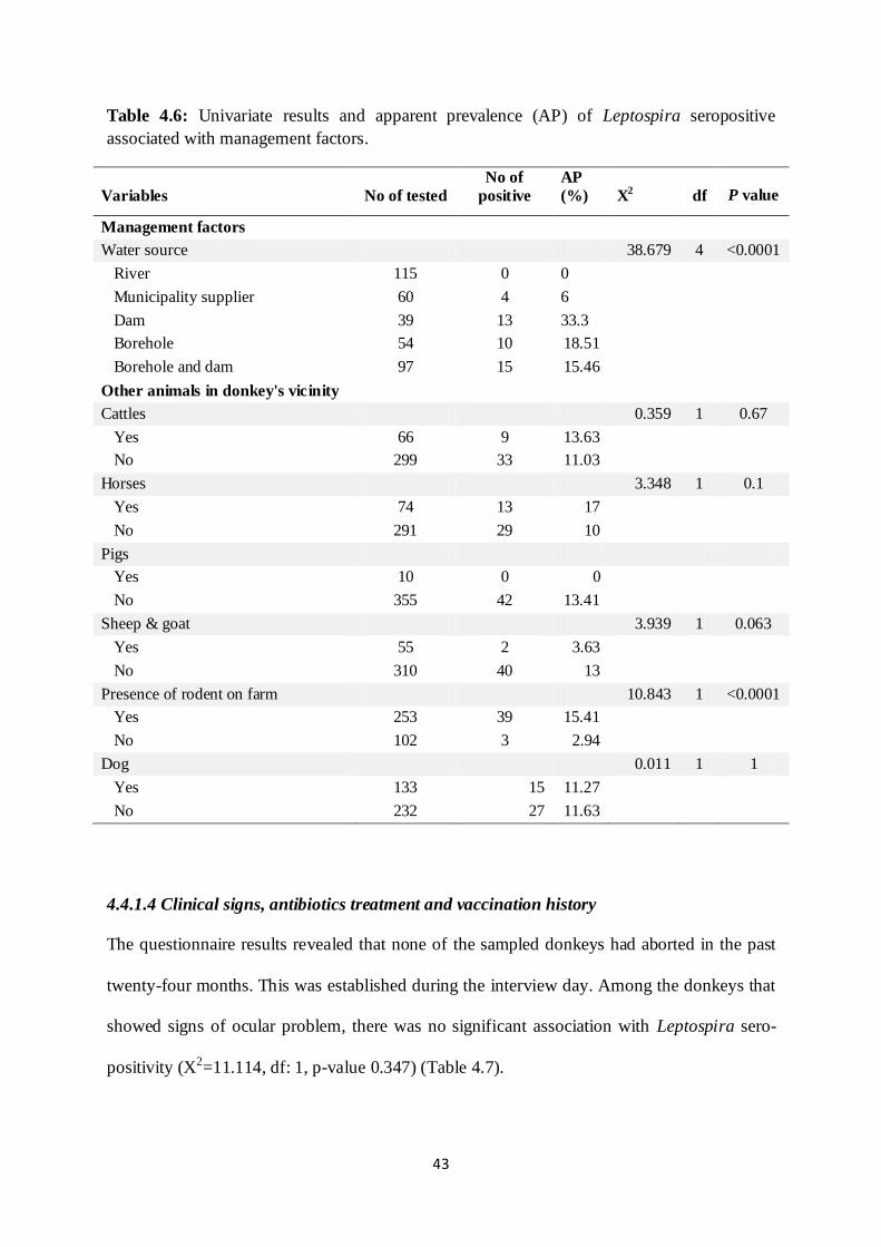
43
Table 4.6: Univariate results and apparent prevalence (AP) of Leptospira seropositive
associated with management factors.
Variables No of tested
No of
positive
AP
(%) X2 df P value
Management factors
Water source 38.679 4 <0.0001
River 115 0 0
Municipality supplier 60 4 6
Dam 39 13 33.3
Borehole 54 10 18.51
Borehole and dam 97 15 15.46
Other animals in donkey's vicinity
Cattles 0.359 1 0.67
Yes 66 9 13.63
No 299 33 11.03
Horses 3.348 1 0.1
Yes 74 13 17
No 291 29 10
Pigs
Yes 10 0 0
No 355 42 13.41
Sheep & goat 3.939 1 0.063
Yes 55 2 3.63
No 310 40 13
Presence of rodent on farm 10.843 1 <0.0001
Yes 253 39 15.41
No 102 3 2.94
Dog
0.011 1 1
Yes 133 15 11.27
No 232 27 11.63
4.4.1.4 Clinical signs, antibiotics treatment and vaccination history
The questionnaire results revealed that none of the sampled donkeys had aborted in the past
twenty-four months. This was established during the interview day. Among the donkeys that
showed signs of ocular problem, there was no significant association with Leptospira sero-
positivity (X2=11.114, df: 1, p-value 0.347) (Table 4.7).

44
Table4.7: Univariate results and apparent prevalence (AP) of Leptospira seropositive
associated with disease clinical signs, antibiotics treatment and vaccination history.
Variables N.of
donkeys
tested
N.of positive
donkeys
≥1:100 Ap (%) X2 df p-
value
Ocular disease
Yes No
22
343
1
41
4.54
12
1.114 1 0.291
Abortion
Yes
No
Missing
0
118
94
0.00
10
0.00
8.47
Stillbirth
Yes
No
0
118
0
10
0.00
8.47
Vaccination
Yes
No
Missing
0
210
155
0
14
0.00
6.66
Antibiotic treatment
Yes No
Missing
0
330
35
0
34
0.00
10.3
4.4.2 Results of the multivariate logistic regression analysis: assessment of significant
risk factors for Leptospira seropositivity
After assessment of factors significantly associated with Leptospira seropositivity at
univariable level analyses using the univariate model, the municipality, gender, average
annual rainfall, agricultural activities, water sources, presence of rodents, horses, sheep and
goats in the donkey neighbourhood were considered for inclusion in the multivariate model
based on a liberal p-value of 0.20.
Table 4.8 presents the results of the multivariate logistic regression models predicting the
odds of testing positive. The results displayed in Table 4.8, revealed that male donkeys were
over 4 times more likely to test positive than were females (OR = 4.88; p ≤ 0.0001; 95% CI
[2.01-11.82]).
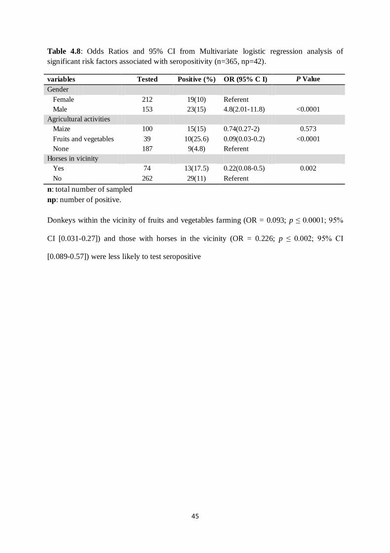
45
Table 4.8: Odds Ratios and 95% CI from Multivariate logistic regression analysis of
significant risk factors associated with seropositivity (n=365, np=42).
variables
Tested Positive (%) OR (95% C I) P Value
Gender
Female
212 19(10) Referent
Male
153 23(15) 4.8(2.01-11.8) <0.0001
Agricultural activities
Maize
100 15(15) 0.74(0.27-2) 0.573
Fruits and vegetables 39 10(25.6) 0.09(0.03-0.2) <0.0001
None
187 9(4.8) Referent
Horses in vicinity
Yes
74 13(17.5) 0.22(0.08-0.5) 0.002
No
262 29(11) Referent
n: total number of sampled
np: number of positive.
Donkeys within the vicinity of fruits and vegetables farming (OR = 0.093; p ≤ 0.0001; 95%
CI [0.031-0.27]) and those with horses in the vicinity (OR = 0.226; p ≤ 0.002; 95% CI
[0.089-0.57]) were less likely to test seropositive
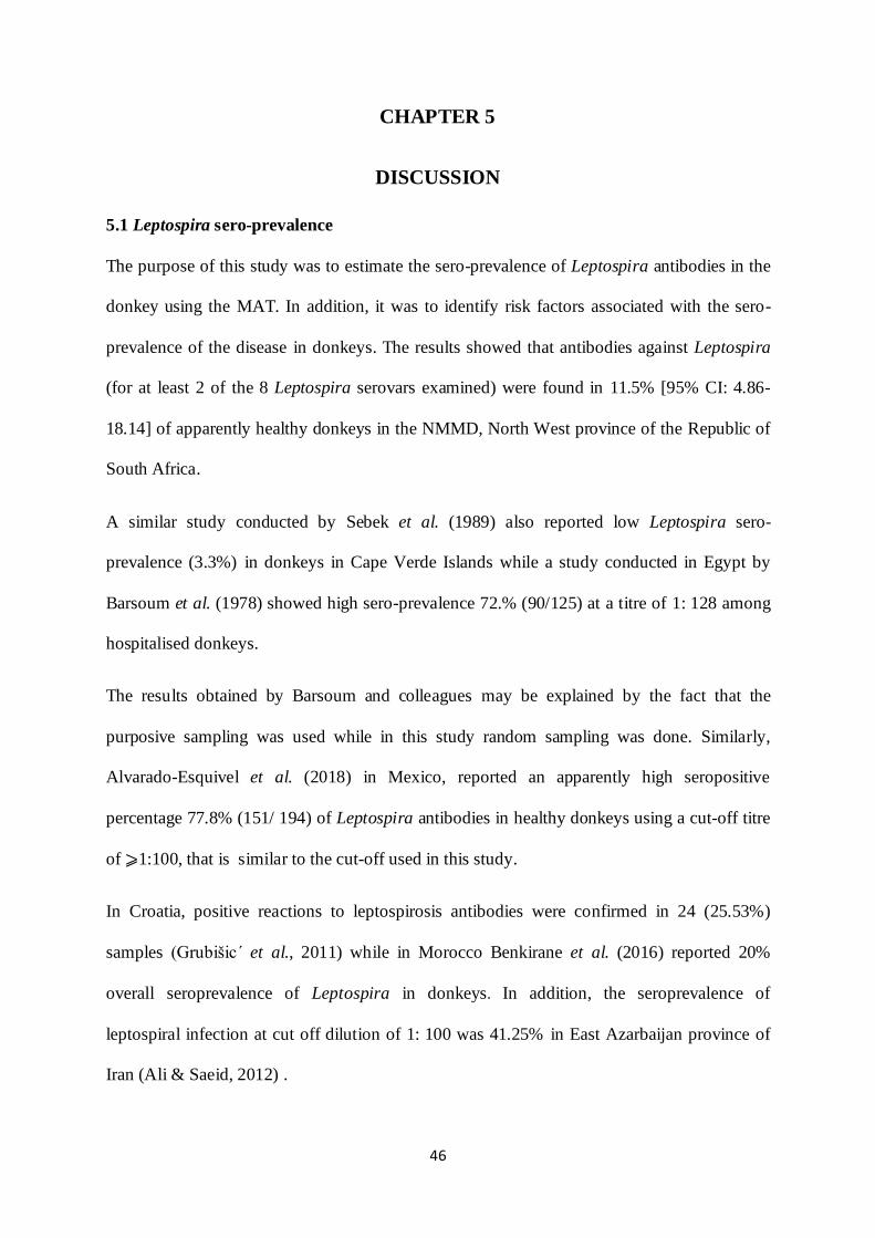
46
CHAPTER 5
DISCUSSION
5.1 Leptospira sero-prevalence
The purpose of this study was to estimate the sero-prevalence of Leptospira antibodies in the
donkey using the MAT. In addition, it was to identify risk factors associated with the sero-
prevalence of the disease in donkeys. The results showed that antibodies against Leptospira
(for at least 2 of the 8 Leptospira serovars examined) were found in 11.5% [95% CI: 4.86-
18.14] of apparently healthy donkeys in the NMMD, North West province of the Republic of
South Africa.
A similar study conducted by Sebek et al. (1989) also reported low Leptospira sero-
prevalence (3.3%) in donkeys in Cape Verde Islands while a study conducted in Egypt by
Barsoum et al. (1978) showed high sero-prevalence 72.% (90/125) at a titre of 1: 128 among
hospitalised donkeys.
The results obtained by Barsoum and colleagues may be explained by the fact that the
purposive sampling was used while in this study random sampling was done. Similarly,
Alvarado-Esquivel et al. (2018) in Mexico, reported an apparently high seropositive
percentage 77.8% (151/ 194) of Leptospira antibodies in healthy donkeys using a cut-off titre
of ⩾1:100, that is similar to the cut-off used in this study.
In Croatia, positive reactions to leptospirosis antibodies were confirmed in 24 (25.53%)
samples (Grubišic ́ et al., 2011) while in Morocco Benkirane et al. (2016) reported 20%
overall seroprevalence of Leptospira in donkeys. In addition, the seroprevalence of
leptospiral infection at cut off dilution of 1: 100 was 41.25% in East Azarbaijan province of
Iran (Ali & Saeid, 2012) .

47
The results of Leptospira sero-prevalence in donkeys obtained from different municipalities
in the North West Province (Mafikeng, Tswaing, Ratlou, Moilwa, Ditsobotla) showed low
percentage of sero-positives compared to those reported in other countries. It is possible that
the observed disparity may be due to differences in environmental conditions in different
parts of the world (Barragan et al., 2017b). The occurrence of sero-prevalence in donkeys
varied from one area to the other as seen in this study with Mafikeng having the highest sero-
prevalence (Table 4. 2). This may be due to climatic and geographical difference which was
also confirmed in this study. This results are in line with the one of Adler and de la Peña
Moctezuma (2010) on horses which showed variations in the sero-prevalence from 1-95%.
These variations depended on geographical location and serovars assessed. This underlines
the results obtained in this study which showed that the percentage of donkeys infected with
Leptospira spp differed among municipalities.
The result obtained in the current study also suggest that beside the inconsistence in the MAT
panel used in different studies, the reading of the result in relation with the cut-off used in
each study may explain the differences observed in the sero-prevalence of Leptospira
antibodies among studies done on donkeys around the world.
5.2 Predominant serovars
Serovars tested in this study were presumed to be circulating in other domestic animals in
South Africa (Hesterberg et al., 2009; Roach et al., 2010; Potts et al., 2013). In the current
study Leptospira interrogans serovar Bratislava was the most common infecting serovar in
donkeys around the NMMD (Table 4. 3). This result is in agreement with study of Grubišic´
et al. (2011) who also reported serovar Bratislava as being the most abundant in donkeys in
Croatia.

48
The results obtained in this study were not in line with the majority of studies conducted in
different parts of the globe. For example in Morocco, Benkirane et al. (2016) found that
Javanica and Australis serogroups were the most predominant in donkey. In Iran
Grippotyphosa and Icterohaemorrhagiae, were found to be predominant (Hajikolaei et al.,
2005), while the serovars Butembo, Pomona, Icterohaemorrhagiae and Canicola were the
most reacting serovars in hospitalised donkeys in Egypt (Barsoum et al., 1978).
In Mexico Alvarado-Esquivel et al. (2018) found Icterohaemorrhagiae and Sejroe serovars to
be prevailing in donkeys. For most of these previous studies the MAT panel used was not
consistent among the studies. The predominate serovar in a region is likely not to be the same
in other regions (Bharti et al., 2003).
Observations in the current study revealed that Leptospira interrogans serovar Bratislava was
the dominant found in donkeys around the NMMD. This serovar was not included in the
panel of serovars tested in different studies around the world (Alvarado-Esquivel et al.,
2018). Other serovars used and found to be predominant in donkeys in other studies
elsewhere were not included this study. Therefore, this lack of serovar inclusion in the MAT
panel might explain the differences in predominate serovars in different regions.
A few previous surveys conducted in South Africa on leptospirosis in domestic animals
revealed that serovar Bratislava has been reported to cause subclinical disease in domestic
animal such as pigs (Gummow et al., 1999; Potts et al., 2013) , horses (Simbizi et al., 2016)
and cattle (Hesterberg et al., 2009) and dogs (Roach et al., 2010). These findings show that
this serovars appears to be widespread countrywide and is also maintained by donkeys in the
NMMD North West Province of South Africa.

49
5.3 Risk factors for donkey exposure
It was observed that donkeys within the vicinity of fruit and vegetable farming as main
agricultural activity were significantly less likely to test seropositive for Leptospira (Adjusted
OR = 0.093; 95% CI: [0.031-0.27; p ≤ 0.0001) compared to those that were not in the vicinity
of an agricultural activity. Similarly donkeys that were within the vicinity of maize garden
were less likely to test seropositive (Adjusted OR 0.74; 95%CI 0.27-2; p value=0.573)
compared to donkeys that were not in close vicinity of gardening. This is contrary to what
was expected because agricultural activities like fruits and vegetable fields irrigated with
water tend to be contaminated by rodent urine and hence act as reservoirs for Leptospira
infections for donkeys living nearby such fields. This is supported by the findings by Azócar-
Aedo et al. (2014) who reported that animals like cats that dwell in places where agricultural
activities were carried out are 30 times more likely to be seropositive to Leptospira. This was
also consistent with findings of the study by Alavi and Khoshkho (2014) who observed
similar findings among rice farmers in Iran.
In the present study, it was observed that male animals were 5 times (Adjusted OR = 4.88;
95% CI: 2.01-11.82; p ≤ 0.0001) more likely to test positive than females donkeys. This is in
conformity with findings reported by Alvarado-Esquivel et al. (2018) and those of Kikuti et
al. (2012), . Kikuti et al. (2012) also reported gender as a risk factor for Leptospira in dogs
(OR=0.439, 95% CI: 0.62-0.89; P= 0.009). The authors of the present work are of the view
that the most plausible explanation for the results reported here, is that male donkeys are
usually more engaged in working activities, and therefore spend a lot of time outside of their
premises where they get exposed to the infected environment (areas). The later increasing the
risk of Leptospira infection among male donkeys. However, there are studies that report
findings that are contrary those observed in the present study, in that they concluded that
50
there is no statistically significant differences between the proportions of prior Leptospira
infection in males and females (Meeyam et al., 2006; Dias et al., 2007).
Several studies have identified horses as a reservoirs of serovar Bratislava around the world
(Hamond et al., 2014a) and also in South Africa (Simbizi et al., 2016). In view of this, our
observation of donkeys living in the vicinity of horses of co-grazing with horses not being at
a higher risk of testing seropositive, and instead it was rather a protective factor (OR = 0.226;
p ≤ 0.002; 95% CI [0.089-0.57] was not expected. This is important considering that the
serovar Bratislava was predominant among donkeys living in the vicinity of horse, and that
according to Schoonman and Swai (2010) co-grazing between different species increases the
exposure risk resulting in the spread of the disease in the herds
51
CHAPTER 6
CONCLUSION AND RECOMMENDATIONS
6.1 Conclusion
Leptospirosis in donkeys is of epidemiologic importance. The aim of this study was to
estimate the sero-prevalence of Leptospira infecting serovars in donkeys using the
macroscopic agglutination test (MAT) and to investigate the effects of demographics,
environmental and management factors as potential risk factors in the occurrence of
leptospirosis in donkeys.
It was found that 11.5% of clinically healthy donkeys had anti Leptospira antibodies in their
blood. Two serovars (Bratislava and Tarassovi) were detected in this study. Serovar
Bratislava was the most widespread and has also been reported in several surveys of domestic
animals in South Africa. The majority of the donkeys sampled were positive at the MAT titre
of ≥1/100 or ≥1/200, high MAT titre ≥1/3200 were also recorded.
This study concluded that donkeys were routinely infected by a variety of circulating
Leptospira species and may act as reservoirs for Leptospira bacteria. This makes them to be a
potential source of infection for human and others animals. Furthermore, this study found that
the gender of the donkeys was the significant risk factors associated with Leptospira sero-
prevalence in the study area.
52
6.2 Recommendation
Recognition and appropriate management of factors impacting leptospirosis incidence in
donkeys such co-grazing donkeys and horses nearby fruit and vegetable farming should be
avoided in order to decrease the disease incidence. Donkey owners should be aware of the
disease in donkeys since those donkeys are in daily contact with human beings during the
period when they are being used for work. Prevention is better than cure, vaccination should
be encouraged for all donkeys but more specifically for males.
6.3 Limitation of the study
There was few missing information in the data due to the fact that some donkey owners did
not keep the record of their animals. In addition; a lot of variables were removed from the
analysis due to the small numbers in some categories. Furthermore, the use of sero-
surviellance could lead to over or under reporting because being seropositive does not mean
the animal is infected but rather exposure at some time. The use of DNA based methods such
as PCR is a promising alternative for carrier identification.

53
REFERENCES
Adler, B. & de la Peña Moctezuma, A. 2010. Leptospira and leptospirosis. Veterinary
Microbiology, 140(3–4):287-296.
Ahmad, S., Shah, S. & Ahmad, F.H. 2005. Laboratory diagnosis of leptospirosis. Journal of
Postgraduate Medicine, 51(3):195.
Alavi, S.M. & Khoshkho, M.M. 2014. Seroprevalence study of leptospirosis among rice
farmers in khuzestan province, South west iran, 2012. Jundishapur Journal of Microbiology,
7(7).
Ali, H. & Saeid, S. 2012. Seroprevalence of leptospiral infection in horses, donkeys and
mules in East Azerbaijan province. African Journal of Microbiology Research, 6(20):4384-
4387.
Ali, S.A., Kaur, G., Boby, N., Sabarinath, T., Solanki, K., Pal, D. & Chaudhuri, P. 2017.
Rapid and visual detection of Leptospira in urine by LigB-LAMP assay with pre-addition of
dye. Molecular and Cellular Probes, 36(2017):29-35.
Alvarado-Esquivel, C., Cruz-Romero, A., Romero-Salas, D., Alvarado-Félix, Á.O., Aguilar-
Domínguez, M., Ochoa-Valencia, J.L., Alvarado-Esquivel, D., Hernández-Tinoco, J.,
Zamarripa-Barboza, J.A. & Sánchez-Anguiano, L.F. 2018. Apparently high Leptospira
antibody seropositivity in donkeys for slaughter in three municipalities in Durango, Mexico.
Journal of Veterinary Diagnostic Investigation, 15:1-4.
Azócar-Aedo, L., Monti, G. & Jara, R. 2014. Leptospira spp. in domestic cats from different
environments: prevalence of antibodies and risk factors associated with the seropositivity.
Animals, 4(4):612-626.
Balakrishnan, G. & Roy, P. 2014. Comparision of efficacy of two experimental bovine
leptospira vaccines under laboratory and field. Veterinary Immunology and
Immunopathology, 159(1):11-15.

54
Barragan, V., Nieto, N., Keim, P. & Pearson, T. 2017a. Meta-analysis to estimate the load of
Leptospira excreted in urine: beyond rats as important sources of transmission in low-income
rural communities. BMC Research Notes, 10(1):71.
Barragan, V., Olivas, S., Keim, P. & Pearson, T. 2017b. Critical Knowledge Gaps in Our
Understanding of Environmental Cycling and Transmission of Leptospira spp. Applied and
Environmental Microbiology, 83(19):e01190-01117.
Barsoum, I., Botros, B. & Morcos, M. 1978. Equine leptospirosis with some clinical
observations. Annals of Veterinary Research, 9:115-118.
Benkirane, A., Noury, S., Hartskeerl, R.A., Goris, M.G.A., Ahmed, A. & Nally, J.E. 2016.
Preliminary Investigations on the Distribution of Leptospira Serovars in Domestic Animals in
North-west Morocco. Transboundary And Emerging Diseases, 63(2):e178-e184.
Bharti, A.R., Nally, J.E., Ricaldi, J.N., Matthias, M.A., Diaz, M.M., Lovett, M.A., Levett,
P.N., Gilman, R.H., Willig, M.R., Gotuzzo, E. & Vinetz, J.M. 2003. Leptospirosis: a
zoonotic disease of global importance. The Lancet Infectious Diseases, 3(12):757-771.
Burriel, A.R. 2010. Leptospirosis: an important zoonotic diseasesis. Current Research,
Technology and Education Topics in Applied Microbiology and Microbial Biotechnology.
Badajoz: Formatex:687-693.
Cameron, A. 1999. Survey toolbox: a practical manual and software package for active
surveillance of livestock diseases in developing countries. Australia: Australian Centre for
International Agricultural Research
Cerqueira, G.M. & Picardeau, M. 2009. A century of Leptospira strain typing. Infection,
Genetics and Evolution, 9(5):760-768.
Chadsuthi, S., Modchang, C., Lenbury, Y., Iamsirithaworn, S. & Triampo, W. 2012.
Modeling seasonal leptospirosis transmission and its association with rainfall and temperature

55
in Thailand using time–series and ARIMAX analyses. Asian Pacific Journal of Tropical
Medicine, 5(7):539-546.
Chakraborty, A., Miyahara, S., Villanueva, S.Y., Gloriani, N.G. & Yoshida, S.-i. 2010. In
vitro sensitivity and resistance of 46 Leptospira strains isolated from rats in the Philippines to
14 antimicrobial agents. Antimicrobial Agents and Chemotherapy, 54(12):5403-5405.
Costa, F., Hagan, J.E., Calcagno, J., Kane, M., Torgerson, P., Martinez-Silveira, M.S., Stein,
C., Abela-Ridder, B. & Ko, A.I. 2015a. Global morbidity and mortality of leptospirosis: a
systematic review. PLoS Neglected Tropical Diseases, 9(9):e0003898.
Costa, F., Wunder Jr, E.A., De Oliveira, D., Bisht, V., Rodrigues, G., Reis, M.G., Ko, A.I.,
Begon, M. & Childs, J.E. 2015b. Patterns in Leptospira shedding in Norway rats (Rattus
norvegicus) from Brazilian slum communities at high risk of disease transmission. PLoS
Neglected Tropical Diseases, 9(6):e0003819.
Croda, J., Figueira, C.P., Wunder, E.A., Santos, C.S., Reis, M.G., Ko, A.I. & Picardeau, M.
2008. Targeted mutagenesis in pathogenic Leptospira species: disruption of the LigB gene
does not affect virulence in animal models of leptospirosis. Infection and Immunity,
76(12):5826-5833.
de Abreu Fonseca, C., Teixeira de Freitas, V.L., Caló Romero, E., Spinosa, C., Arroyo
Sanches, M.C., Da Silva, M.V. & Shikanai‐Yasuda, M.A. 2006. Polymerase chain reaction
in comparison with serological tests for early diagnosis of human leptospirosis. Tropical
Medicine & International Health, 11(11):1699-1707.
Derne, B.T., Fearnley, E.J., Lau, C.L., Paynter, S. & Weinstein, P. 2011. Biodiversity and
leptospirosis risk: a case of pathogen regulation? Medical Hypotheses, 77(3):339-344.
Dias, J.P., Teixeira, M.G., Costa, M.C.N., Mendes, C.M.C., Guimarães, P., Reis, M.G., Ko,
A. & Barreto, M.L. 2007. Factors associated with Leptospira sp infection in a large urban

56
center in northeastern Brazil. Revista da Sociedade Brasileira de Medicina Tropical,
40(5):499-504.
Eisenberg, J.N., Desai, M.A., Levy, K., Bates, S.J., Liang, S., Naumoff, K. & Scott, J.C.
2007. Environmental determinants of infectious disease: a framework for tracking causal
links and guiding public health research. Environmental Health Perspectives, 115(8):1216-
1223.
Epizooties, O.I.d. 2004. Manual of Diagnostic Tests and Vaccines for Terrestrial
Animals:(Mammals, Birds and Bees). Vol. 130: OIE.
Evangelista, K.V. & Coburn, J. 2010. Leptospira as an emerging pathogen: a review of its
biology, pathogenesis and host immune responses. Future Microbiology, 5(9):1413-1425.
Faostat, F. 2014. Food and Agriculture Organization statistical database. Retrieved Feb.
Fávero, J.F., Fritzen, A., Lovato, L.T., Martins, P., Baldissera, M.D., Stefani, L.M. & Da
Silva, A.S. 2018. Immune response of a commercial vaccine against Leptospira interrogans:
Antibodies and cytokine levels. Microbial Pathogenesis, 114:46-49.
Foulongne, V., Michaux-Charachon, S., Jumas-Bilak, E., O'Callaghan, D. & Ramuz, M.
2004. Strategies for bacterial virulence genes identification. Pathologie-Biologie, 52(2):104-
114.
Ghazaei, C. 2018. Pathogenic Leptospira: Advances in understanding the molecular
pathogenesis and virulence. Open Veterinary Journal, 8(1):13-24.
Goarant, C., Laumond‐Barny, S., Perez, J., Vernel‐Pauillac , F., Chanteau, S. & Guigon, A.
2009. Outbreak of leptospirosis in New Caledonia: diagnosis issues and burden of disease.
Tropical Medicine & International Health, 14(8):926-929.
Grevemeyer, B., Vandenplas, M., Beigel, B., Cho, E., Willingham, A.L. & Verma, A. 2017.
Detection of Leptospiral DNA in the Urine of Donkeys on the Caribbean Island of Saint
Kitts. Veterinary Sciences, 4(1):2.
57
Grubišic´, M., Milas, Z., Cvetnic´, Z., Habuš, J., Perko, V.M., Majetic ,́ Z.Š. & Turk, N.
2011. Serological research of the distribution of leptospirosis in donkeys in the Republic of
Croatia. / Serološko istrazivanje rasprostranjenosti leptospiroze u magaraca u Republici
Hrvatskoj. Veterinarska Stanica, 42(4):307-315.
Guernier, V., Goarant, C., Benschop, J. & Lau, C.L. 2018. A systematic review of human
and animal leptospirosis in the Pacific Islands reveals pathogen and reservoir diversity. PLoS
Neglected Tropical Diseases, 12(5):e0006503.
Guerra, M.A. 2013. Leptospirosis: Public health perspectives. Biologicals, 41(5):295-297.
Gummow, B., Myburgh, J., Thompson, P., Van Der Lugt, J. & Spencer, B. 1999. Three case
studies involving Leptospira interrogans serovar pomona infection in mixed farming units:
case report. Journal of the South African Veterinary Association, 70(1):29-34.
Haake, D.A. 2009. Spirochetes A2 - Schaechter, Moselio. Encyclopedia of Microbiology
(Third Edition). Oxford: Academic Press. p. 278-292).
Haake, D.A. & Levett, P.N. 2015. 241 - Leptospira Species (Leptospirosis) A2 - Bennett,
John E. (In Dolin, R. & Blaser, M.J., eds. Mandell, Douglas, and Bennett's Principles and
Practice of Infectious Diseases (Eighth Edition). Philadelphia: Content Repository Only! p.
2714-2720.e2712).
Haake, D.A. & Zückert, W.R. 2015. The leptospiral outer membrane. Leptospira and
Leptospirosis. Springer. p. 187-221).
Hajikolaei, M.R.H., Haidari, M. & Abdollapour, G. 2005. Comparison of leptospiral
infection in the horse and donkey. Bull Vet Inst Pulawy, 49:175-178.
Hamond, C., Martins, G., Bremont, S., Medeiros, M.A., Bourhy, P. & Lilenbaum, W. 2014a.
Predominance of Leptospira interrogans serovar Bratislava DNA in vaginal fluid of mares
suggests sexual transmission of leptospirosis. Animal Reproduction Science, 151(3):275-279.

58
Hamond, C., Pinna, A., Martins, G. & Lilenbaum, W. 2014b. The role of leptospirosis in
reproductive disorders in horses. Tropical Animal Health & Production, 46(1):1-10.
Hartskeerl, R.A., Collares-Pereira, M. & Ellis, W.A. 2011. Emergence, control and re-
emerging leptospirosis: dynamics of infection in the changing world. Clinical Microbiology
and Infection, 17(4):494-501.
Herr, S., Riley, A., Neser, J., Roux, D. & De Lange, J. 1982. Leptospira interrogans serovar
pomona associated with abortion in cattle: isolation methods and laboratory animal
histopathology. Onderstepoort Journal of Veterinary Research, 49:57-62.
Hesterberg, U., Bagnall, R., Bosch, B., Perrett, K., Horner, R. & Gummow, B. 2009. A
serological survey of leptospirosis in cattle of rural communities in the province of KwaZulu-
Natal, South Africa. Journal of the South African Veterinary Association, 80(1):45-49.
Hill, C. 2012. Virulence or niche factors: what's in a name? Journal of Bacteriology,
194(21):5725-5727.
Hoke, D.E., Egan, S., Cullen, P.A. & Adler, B. 2008. LipL32 is an extracellular matrix-
interacting protein of Leptospira spp. and Pseudoalteromonas tunicata. Infection and
Immunity, 76(5):2063-2069.
Hsu, Y.-H., Chou, S.-J., Chang, C.-C., Pan, M.-J., Yang, W.-C., Lin, C.-F. & Chan, K.-W.
2017. Development and validation of a new loop-mediated isothermal amplification for
detection of pathogenic Leptospira species in clinical materials. Journal of Microbiological
Methods, 141:55-59.
Jadhav, S., Gautam, M. & Gairola, S. 2014. Role of vaccine manufacturers in developing
countries towards global healthcare by providing quality vaccines at affordable prices.
Clinical Microbiology and Infection, 20(s5):37-44.

59
Janda, J.M. & Abbott, S.L. 2007. 16S rRNA gene sequencing for bacterial identification in
the diagnostic laboratory: pluses, perils, and pitfalls. Journal of Clinical Microbiology,
45(9):2761-2764.
Jorge, S., Kremer, F.S., Oliveira, N.R.d., Navarro, G.d.O.S.V., Guimarães, A.M., Sanchez,
C.D., Woloski, R.D.d.S., Ridieri, K.F., Campos, V.F. & Pinto, L.d.S. 2018. Whole-genome
sequencing of Leptospira interrogans from southern Brazil: genetic features of a highly
virulent strain. Memórias do Instituto Oswaldo Cruz, 113(2):80-86.
Kikuti, M., Langoni, H., Nobrega, D., Corrêa, A. & Ullmann, L. 2012. Occurrence and risk
factors associated with canine leptospirosis. Journal of Venomous Animals and Toxins
including Tropical Diseases, 18(1):124-127.
Ko, A.I., Goarant, C. & Picardeau, M. 2009. Leptospira: the dawn of the molecular genetics
era for an emerging zoonotic pathogen. Nature Reviews Microbiology, 7(10):736-747.
Kurilung, A., Chanchaithong, P., Lugsomya, K., Niyomtham, W., Wuthiekanun, V. &
Prapasarakul, N. 2017. Molecular detection and isolation of pathogenic Leptospira from
asymptomatic humans, domestic animals and water sources in Nan province, a rural area of
Thailand. Research in Veterinary Science, 115:146-154.
Langoni, H., Kuribara, I.Y., Correa, A.P.F.L., Ullmann, L.S., Sánchez, G.P. & Lucheis, S.B.
2016. Anti-leptospirosis agglutinins in Brazilian capybaras (hydrochoerus hydrochaeris).
Journal of Venomous Animals and Toxins including Tropical Diseases, 22(1):4.
Lau, C.L., Smythe, L.D., Craig, S.B. & Weinstein, P. 2010. Climate change, flooding,
urbanisation and leptospirosis: fuelling the fire? Transactions of the Royal Society of
Tropical Medicine and Hygiene, 104(10):631-638.
Lessa-Aquino, C., Rodrigues, C.B., Pablo, J., Sasaki, R., Jasinskas, A., Liang, L., Wunder Jr,
E.A., Ribeiro, G.S., Vigil, A. & Galler, R. 2013. Identification of seroreactive proteins of

60
Leptospira interrogans serovar copenhageni using a high-density protein microarray
approach. PLoS Neglected Tropical Diseases, 7(10):e2499.
Levett, P.N. 2003. Usefulness of serologic analysis as a predictor of the infecting serovar in
patients with severe leptospirosis. Clinical Infectious Diseases, 36(4):447-452.
Liegeon, G., Delory, T. & Picardeau, M. 2018. Antibiotic susceptibilities of livestock
isolates of leptospira. International Journal of Antimicrobial Agents, 51(5):693-699.
Lin, Y.-P. & Chang, Y.-F. 2007. A domain of the Leptospira LigB contributes to high
affinity binding of fibronectin. Biochemical and Biophysical Research Communications,
362(2):443-448.
Lo, M., Bulach, D.M., Powell, D.R., Haake, D.A., Matsunaga, J., Paustian, M.L., Zuerner,
R.L. & Adler, B. 2006. Effects of temperature on gene expression patterns in Leptospira
interrogans serovar Lai as assessed by whole-genome microarrays. Infection and Immunity,
74(10):5848-5859.
Loureiro, A.P., Martins, G., Thomé, S. & Lilenbaum, W. 2013. Laboratorial diagnosis of
animal leptospirosis. Revista Brasileira de Ciência Veterinária, 20(3):119-126.
Malmström, J., Beck, M., Schmidt, A., Lange, V., Deutsch, E.W. & Aebersold, R. 2009.
Proteome-wide cellular protein concentrations of the human pathogen Leptospira interrogans.
Nature, 460(7256):762.
Marinho, M. & Cardoso, T.C. 2015. Pathogenesis of leptospirosis: important issues.
Journal of Medical Microbiology & Diagnosis, 4(1):1.
Martins, G. & Lilenbaum, W. 2017. Control of bovine leptospirosis: Aspects for
consideration in a tropical environment. Research in Veterinary Science, 112:156-160.
Marufu, M. 2014. Socio-economic importance, health and welfare aspects of donkeys
(Equus asinus) used for carting in a peri-urban area of South Africa. Alice, Eastern Cape,
South Africa: University of Fort Hare. (Dissertation).

61
Maskey, M., Shastri, J., Saraswathi, K., Surpam, R. & Vaidya, N. 2006. Leptospirosis in
Mumbai: post-deluge outbreak 2005. Indian journal of Medical Microbiology, 24(4):337.
Maslow, J.N. 2017. Vaccine development for emerging virulent infectious diseases.
Vaccine.
Matsunaga, J., Barocchi, M.A., Croda, J., Young, T.A., Sanchez, Y., Siqueira, I., Bolin, C.A.,
Reis, M.G., Riley, L.W. & Haake, D.A. 2003. Pathogenic Leptospira species express
surface‐exposed proteins belonging to the bacterial immunoglobulin superfamily. Molecular
Microbiology, 49(4):929-946.
Matsunaga, J., Sanchez, Y., Xu, X. & Haake, D.A. 2005. Osmolarity, a key environmental
signal controlling expression of leptospiral proteins LigA and LigB and the extracellular
release of LigA. Infection and Immunity, 73(1):70-78.
McBride, A.J., Cerqueira, G.M., Suchard, M.A., Moreira, A.N., Zuerner, R.L., Reis, M.G.,
Haake, D.A., Ko, A.I. & Dellagostin, O.A. 2009. Genetic diversity of the Leptospiral
immunoglobulin-like (Lig) genes in pathogenic Leptospira spp. Infection, Genetics and
Evolution, 9(2):196-205.
Meeyam, T., Tablerk, P., Petchanok, B., Pichpol, D. & Padungtod, P. 2006. Seroprevalence
and risk factors associated with leptospirosis in dogs. Southeast Asian Journal of Tropical
Medicine and Public Health, 37(1):148.
Mgode, G.F., Machang’u, R.S., Mhamphi, G.G., Katakweba, A., Mulungu, L.S., Durnez, L.,
Leirs, H., Hartskeerl, R.A. & Belmain, S.R. 2015. Leptospira serovars for diagnosis of
leptospirosis in humans and animals in Africa: common Leptospira isolates and reservoir
hosts. PLoS Neglected Tropical Diseases, 9(12):e0004251.
Millar, B.C., Xu, J. & Moore, J.E. 2007. Molecular diagnostics of medically important
bacterial infections. Molecular Diagnostics: Current Technology and Applications, 9(1):21-
39.

62
Mucina, L. & Rutherford, M.C. 2006. The vegetation of South Africa, Lesotho and
Swaziland: South African National Biodiversity Institute.
Mughini-Gras, L., Bonfanti, L., Natale, A., Comin, A., Ferronato, A., La Greca, E.,
Patregnani, T., Lucchese, L. & Marangon, S. 2014. Application of an integrated outbreak
management plan for the control of leptospirosis in dairy cattle herds. Epidemiology &
Infection, 142(6):1172-1181.
Murray, G.L. 2013. The lipoprotein LipL32, an enigma of leptospiral biology. Veterinary
Microbiology, 162(2-4):305-314.
Murray, G.L., Srikram, A., Hoke, D.E., Wunder, E.A., Henry, R., Lo, M., Zhang, K.,
Sermswan, R.W., Ko, A.I. & Adler, B. 2009. Major surface protein LipL32 is not required
for either acute or chronic infection with Leptospira interrogans. Infection and Immunity,
77(3):952-958.
Mwachui, M.A., Crump, L., Hartskeerl, R., Zinsstag, J. & Hattendorf, J. 2015.
Environmental and behavioural determinants of leptospirosis transmission: a systematic
review. PLoS Neglected Tropical Diseases, 9(9):e0003843.
Nascimento, A.L.T.O.d., Verjovski-Almeida, S., Van Sluys, M., Monteiro-Vitorello, C.,
Camargo, L., Digiampietri, L., Harstkeerl, R., Ho, P., Marques, M. & Oliveira, M. 2004.
Genome features of Leptospira interrogans serovar Copenhageni. Brazilian Journal of
Medical and Biological Research, 37(4):459-477.
National Institute for Communicable Diseases. 2015. Leptospirosis.
http://www.nicd.ac.za/assets/files/Leptosprirosis.pdf. Date of access: 19 July 2019.
Pappachan, M., Sheela, M. & Aravindan, K. 2004. Relation of rainfall pattern and epidemic
leptospirosis in the Indian state of Kerala. Journal of Epidemiology & Community Health,
58(12):1054-1054.

63
Pappas, C.J. & Picardeau, M. 2015. Control of gene expression in Leptospira spp. by
transcription activator-like effectors demonstrates a potential role for LigA and LigB in
Leptospira interrogans virulence. Applied and Environmental Microbiology, 81(22):7888-
7892.
Pellizzer, P., Todescato, A., Benedetti, P., Colussi, P., Conz, P. & Cinco, M. 2005.
Leptospirosis following a flood in the Veneto area, North-east Italy. Annali di Igiene:
Medicina Preventiva e di Comunita, 18(5):453-456.
Picardeau, M. 2013. Diagnosis and epidemiology of leptospirosis. Médecine et Maladies
Infectieuses, 43(1):1-9.
Picardeau, M., Bulach, D.M., Bouchier, C., Zuerner, R.L., Zidane, N., Wilson, P.J., Creno,
S., Kuczek, E.S., Bommezzadri, S. & Davis, J.C. 2008. Genome sequence of the saprophyte
Leptospira biflexa provides insights into the evolution of Leptospira and the pathogenesis of
leptospirosis. PloS One, 3(2):e1607.
Postic, D., Merien, P., Perolat, P. & Baranton, G. 2000. Diagnostic Biologique
Leptospirose-Borreliose de Lyme: Biological Diagnosis Leptospirosis-Lyme Borreliosis:
Institut Pasteur.
Potts, A., Lotter, C. & Robinson, J. 2013. Serological prevalence of leptospiral antibodies in
pigs in South Africa. Onderstepoort Journal of Veterinary Research, 62:281-284.
Rathinam, S. 2005. Ocular manifestations of leptospirosis. Journal of Postgraduate
Medicine, 51(3):189.
Ren, S.-X., Fu, G., Jiang, X.-G., Zeng, R., Miao, Y.-G., Xu, H., Zhang, Y.-X., Xiong, H., Lu,
G. & Lu, L.-F. 2003. Unique physiological and pathogenic features of Leptospira
interrogans revealed by whole-genome sequencing. Nature, 422(6934):888.
Ricaldi, J.N., Fouts, D.E., Selengut, J.D., Harkins, D.M., Patra, K.P., Moreno, A., Lehmann,
J.S., Purushe, J., Sanka, R. & Torres, M. 2012. Whole genome analysis of Leptospira

64
licerasiae provides insight into leptospiral evolution and pathogenicity. PLoS Neglected
Tropical Diseases, 6(10):e1853.
Ristow, P., Bourhy, P., da Cruz McBride, F.W., Figueira, C.P., Huerre, M., Ave, P., Saint
Girons, I., Ko, A.I. & Picardeau, M. 2007. The OmpA-like protein Loa22 is essential for
leptospiral virulence. PLoS Pathogens, 3(7):e97.
Roach, J., Van Vuuren, M. & Picard, J. 2010. A serological survey of antibodies to
Leptospira species in dogs in South Africa. Journal of the South African Veterinary
Association, 81(3):156-159.
Saif, A.N. 2013. The Detection of Burkholderia spp. and pathogenic Leptospira spp. in
South Africa. Johannesburg, SOUTH AFRICA: University of the Witwatersrand
(Dissertation).
Schoonman, L. & Swai, E.S. 2010. Herd-and animal-level risk factors for bovine
leptospirosis in Tanga region of Tanzania. Tropical animal health and production,
42(7):1565-1572.
Sebek, Z., Miorini, I., Brosch, R., Buchrieser, C., Buchrieser, V., Sixl, W. & Valova, M.
1989. A survey of leptospirological studies carried out on the Cape Verde Islands.
Geographia Medica. Supplement= Geographia medica. Sonderband, 5:153-160.
Shivakumar, S. & Krishnakumar, B. 2006. Diagnosis of leptospirosis-role of MAT. Journal
of the Association of Physicians of India, 54:338-339.
Sibley, C.D., Peirano, G. & Church, D.L. 2012. Molecular methods for pathogen and
microbial community detection and characterization: current and potential application in
diagnostic microbiology. Infection, Genetics and Evolution, 12(3):505-521.
Silveira, M.M., Oliveira, T.L., Schuch, R.A., McBride, A.J.A., Dellagostin, O.A. & Hartwig,
D.D. 2017. DNA vaccines against leptospirosis: A literature review. Vaccine, 35(42):5559-
5567.

65
Simbizi, V., Saulez, M.N., Potts, A., Lötter, C. & Gummow, B. 2016. A study of
leptospirosis in South African horses and associated risk factors. Preventive Veterinary
Medicine, 134:6-15.
Slack, A.T., Symonds, M.L., Dohnt, M.F. & Smythe, L.D. 2006. Identification of
pathogenic Leptospira species by conventional or real-time PCR and sequencing of the DNA
gyrase subunit B encoding gene. BMC Microbiology, 6(1):95.
South African Weather Services. 2017. Average of the daily temperatures (°c) and total of
the monthly daily rain (mm) form 2008-2017 in different station in the North-west Province.
Pretoria.
Stats, S. 2012. Census 2011 statistical release: P0301. 4. Statistics South Africa. Pretoria:
Statistics South Africa: South Africa.
Stoddard, R.A., Gee, J.E., Wilkins, P.P., McCaustland, K. & Hoffmaster, A.R. 2009.
Detection of pathogenic Leptospira spp. through TaqMan polymerase chain reaction targeting
the LipL32 gene. Diagnostic Microbiology and Infectious Disease, 64(3):247-255.
Sykes, J., Hartmann, K., Lunn, K., Moore, G., Stoddard, R. & Goldstein, R. 2011. 2010
ACVIM small animal consensus statement on leptospirosis: diagnosis, epidemiology,
treatment, and prevention. Journal of Veterinary Internal Medicine, 25(1):1-13.
Tan, S. 2006. OIE quality standard and guidelines for veterinary laboratories: infectious
diseases. The Canadian Veterinary Journal, 47(12):1184.
Tarigan, S. 2016. Recombinant LipL32 Protein for Leptospirosis Detection in Indonesia.
Procedia Chemistry, 18:18-25.
Taylor, P.J., Arntzen, L., Hayter, M., Iles, M., Frean, J. & Belmain, S. 2008. Understanding
and managing sanitary risks due to rodent zoonoses in an African city: beyond the Boston
Model. Integrative Zoology, 3(1):38-50.

66
Te Brugge, L.A. & Dreyer, T. 1985. Leptospira interrogans serovar hardjo associated with
bovine abortion in South Africa. Onderstepoort Journal of Veterinary Research, 52:51-52.
Terpstra, W.J., Ligthart, G. & Schoone, G. 1985. ELISA for the detection of specific IgM
and IgG in human leptospirosis. Microbiology, 131(2):377-385.
Trausch, J.J., Shank-Retzlaff, M. & Verch, T. 2017. Replacing antibodies with modified
DNA aptamers in vaccine potency assays. Vaccine, 35(41):5495-5502.
Tsegay, K., Potts, A., Aklilu, N., Lötter, C. & Gummow, B. 2016. Circulating serovars of
Leptospira in cart horses of central and southern Ethiopia and associated risk factors.
Preventive Veterinary Medicine, 125:106-115.
Utzinger, J., Becker, S.L., Knopp, S., Blum, J., Neumayr, A.L., Keiser, J. & Hatz, C.F. 2012.
Neglected tropical diseases: diagnosis, clinical management, treatment and control. Swiss
medical weekly: official journal of the Swiss Society of Infectious Diseases, the Swiss Society
of Internal Medicine, the Swiss Society of Pneumology, 142:1-24.
Vijayachari, P., Sugunan, A. & Shriram, A. 2008. Leptospirosis: an emerging global public
health problem. Journal of Biosciences, 33(4):557-569.
Ward, M.P. 2002. Seasonality of canine leptospirosis in the United States and Canada and
its association with rainfall. Preventive Veterinary Medicine, 56(3):203-213.
Woo, P., Lau, S., Teng, J., Tse, H. & Yuen, K.Y. 2008. Then and now: use of 16S rDNA
gene sequencing for bacterial identification and discovery of novel bacteria in clinical
microbiology laboratories. Clinical Microbiology and Infection, 14(10):908-934.
World Health Organization. 2003. Human leptospirosis: guidance for diagnosis, surveillance
and control: World Health Organization.
Xue, F., Yan, J. & Picardeau, M. 2009. Evolution and pathogenesis of Leptospira spp.:
lessons learned from the genomes. Microbes and Infection, 11(3):328-333.

67
APPENDIXES
Appendix 1: Participant questionnaire interview
Prevalence of Leptospirosis in donkeys and associated risk factors
Participant questionnaire
The research team appreciates your involvement in this study. The information from this
questionnaire will help us to assess the risk of donkeys to contract leptospirosis and to
develop control strategies.
This study and questionnaire will be approved by the Animal Care, Health and Safety in
Research Ethics Committee (AnimCare) Faculty of Health Sciences, North-West University
Personal information included in the questionnaire will be treated in confidence and will not
be published or disclosed to any third parties by the research team in a manner that would
allow identification of participants.
Results will be communicated to you either by phone call or email.
Date of interview: ____/____/2019 Interviewer’s name: ______________________

68
Participant general information
Farmer’s Name & surname: ____________________________________________
Did you fill in a consent and confidentiality form?
□ Yes
□ No (Please get form filled in now)
What is your Municipality of origin?
□Mafikeng □ Ratlou □ Ditsobotla
□ Tswaing □ Ramotshere Moilwa
Farmer’s contact :
email address : ________________________
cell phone number : ______________________
How do you want us to communicate the result to you
□ cell phone □ email

69
2. Donkey’s demographics information
What is the breed of your Donkey? _________________________
What is the age of your Donkey? □ 0-5years
□ 6-11 years
□ 12 years and above
What is the gender of your donkey? □ Male □ Gelding
□ Female
What is the use of your donkey? □ Breeding □ Working
□ Racing □ Others
3. Donkey’s environmental and management information
The average annual rain fall in your farm □ Low (0-600mm)
□Medium (600-1000mm)
□ High (>1000mm)
What is the average annual temperature □ Low (21.5-23.9℃)
□ Medium (24-25.9℃)
□ High (≥25℃)
What is the main agricultural activity around the donkey farm?
□ Fruits and vegetables farming
□Maize farming
□ Sugarcane
How is your donkeys feed and housed?
□ Pasture access with little or no stable feeding

70
□ Stable access with little or no pasture feeding
□ Combination of stable and pasture feeding
Do you have a concrete floor in the stable?
□ Yes
□ No
Do you see rodents in the farm □ Yes
□ No
What are other animals in the vicinity?
□ Dog □ Horse
□ Cattles □ Sheep & Goat
□Pig
Where do your donkeys commonly drink water?
□ Dam water □ River
□Bore hole water □ Municipality
Which of the following sign did you observe on your donkey for the past 12 months?
□ Abortion
□ Stillbirth
□ Ocular disease (ulcers, watering red eyes, swollen
eyes)
Did your donkeys get vaccinated against Leptospirosis?
□ Yes (date: mm/yy)
_________________
□ No

71
Did your donkeys receive antibiotics treatment for the last 12 months?
□ Yes (date: mm/yy)
_________________
□ No
……………………………… ……………………………………………………………
THANK YOU!
This is the end of the questionnaire. The research team appreciates your involvement in this
Study of Leptospirosis and is committed to privacy of all personal information.
The lab will check your Donkey’s blood for previous exposure to Leptospira spp. and we will
notify you of the result as soon as possible.

72
Appendix 2: Participant informed consent form
INFORMED CONSENT FORM
Project Title: Prevalence of leptospirosis in donkeys and associated risk factors
Researcher’s name: ______________________
Date of interview: ____/____/2019
Interviewer’s name: ______________________
The research team appreciates your involvement in this study of leptospirosis.
The information from this questionnaire will help us to assess the risk of contracting
leptospirosis in donkeys and to develop control strategies.
This study and questionnaire will be submitted for approval by the North West University
Animal Care, Health and Safety in Research Ethics Committee (AnimCare). This consent
form is necessary for us to ensure that you understand the purpose of your involvement and
that you agree to the conditions of your participation.
I will read and explain the question to you and then you can sign this form to certify that you
approve the following:
The interview will be recorded and transcript will be produced.
The transcript of the interview will be analysed by Dr Kibambe Kiayima Daddy as the
research investigator

73
Access to the interview will be limited to Dr Kibambe Kiayima Daddy and academic
colleagues and researchers with whom he might collaborate as part of the research process
Personal information included in the questionnaire will be treated in confidence and will not
be published or disclosed to any third parties by the research team in a manner that would
allow identification of participants
The actual recording will be kept until the completion of the study
All or part of the content of your interview may be used;
In academic papers, policy or news article
On our website and in other media that we may produce such as spoken presentations
On other feedback event
By signing this form I agree that:
I am voluntarily taking part in this project. I understand that I do not have to take part, and I
can stop the interview at any time
The transcribed interview or extract from it may be used as described above
I can request a copy of the transcript of my interview and my make edits I fell necessary to
ensure the effectiveness of any agreement made bout confidentiality;
I have been able to ask any question I might have and I understand that I am free to contact
the researcher with any question I may have in future
Farmer’s consent
I ………………………………………………….hereby give permission for blood collection
from my donkey (s) and agree to participate in this study freely without pressure. I declare
that information provided in this questionnaire is correct.
Signed ………………………..at……………………………..on………………………..
……………………………………..END…………………………………………………….

74
Appendix 3 Reservoir map with 8 tested serovars
Bra = Bratislava antigen
Cani = Canicola antigen
Ict = Icterohaemorraghiae
Pom = Pomona antigen
Tar = Tarassovi antigen
Szw = Szwajizak antigen
Gri = Grippotyphosa
Har = Hardjo antigen

75
Appendix 4: Orientation of the U-bottom serum plate