prevalence of goblet cell metaplasia in …journal.usm.my/journal/mjms-14-1-056.pdf · prevalence...
TRANSCRIPT
56
ORIGINAL ARTICLE
PREVALENCE OF GOBLET CELL METAPLASIA INENDOCERVICAL AND ENDOMETRIAL ADENOCARCINOMA : A
HISTOCHEMICAL STUDY
Lauren Nieuwenhuizen, Mohd Khairy Khalil, Venkatesh R. Naik, Nor Hayati Othman
Department of Pathology,School of Medical Sciences, Universiti Sains Malaysia, Health Campus
16150 Kubang Kerian, Kelantan, Malaysia
To determine the prevalence of goblet cell metaplasia in endocervical andendometrial adenocarcinomas by histochemial staining and to investigate the mostsensitive histochemical staining method to detect this metaplasia, a total of 90 tissueblocks representing 30 non-neoplastic cervix, 30 non-neoplastic endometrium, 30endocervical and endometrial adenocarcinoma cases were obtained forhistochemical staining with Toluidine Blue (TB), Methylene Blue (MB),Mucicarmine (MUC), Periodic Acid Schiff before and after Diastase digestion (PAS,PAS-D), Alcian Blue pH 2.5 (AB), and Periodic Acid Schiff after Alcian Blue pH2.5 (PAB). The cases were blinded and evaluated by a pathologist [NHO] for thepresence of goblet cell metaplasia, the amount of goblet cells present and thehistochemical differentiation of the goblet cells compared with its surroundingglandular epithelium. Goblet cell metaplasia was present in 2 out of 30 cases innon-neoplastic cervix, 0 out of 30 cases in non-neoplastic endometrium, 7 out of 15cases in endocervical adenocarcinoma and in 2 out of 15 cases in endometrialadenocarcinoma. Relatively few goblet cells were seen in endometrialadenocarcinoma, few to moderate amounts were seen in endocervicaladenocarcinoma and relatively more goblet cells were seen in non-neoplastic cervix.The differentiation of the goblet cells with its surrounding glandular epitheliumwas moderate to strong in non-neoplastic cervix and endocervical adenocarcinoma,while the differentiation in endometrial adenocarcinoma was weak to moderate.The various staining methods showed differences in presence, amount anddifferentiation of the goblet cells. Goblet cell metaplasia of the reproductive organsis not as rare as previously reported. There was no statistical difference in presence,amount and differentiation of goblet cells according to the various cases. The mustoptimum staining methods for staining goblet cells in non-neoplastic cervix,endocervical adenocarcinoma and endometrial adenocarcinoma were PAS, PAS-D and AB.
Key words : endocervical adenocarinoma, endometrial adenocarcinoma, histochemical staining, intestinalmetaplasia, goblet cell metaplasia, prevalence.
Introduction
Goblet cell metaplasia is often synonymouslyreferred as intestinal metaplasia. It has beendescribed in endocervical (1-4) and endometrialadenocarcinomas (5-7). Savargaonkar et al (1) foundintestinal metaplasia in 32% of endocervicaladenocarcinoma cases, while McCluggage et al (6)
found intestinal metaplasia in 17% of endometrialadenocarcinoma cases.
Intestinal metaplasia has also been reportedin non-neoplastic cervix (8,9) and endometrium (10),mucinous tumors of the ovary (11, 12),adenocarcinomas of the vulva (13) and in the vagina(14), in adenoma malignum of the endocervix (15,16).
Submitted-20-02-2004, Accepted-03-12-06
Malaysian Journal of Medical Sciences, Vol. 14, No. 1, January 2007 (56-61)
57
The aim of our study was to determine theprevalence of goblet cell metaplasia in non-neoplastic endocervix and endometrium and theirmalignant counterparts. We also wanted toinvestigate the most sensitive histochemical stainingmethod to detect this metaplasia. To the best of ourknowledge similar studies has not been carried outbefore.
Materials and methods
Endocervix and endometrium tissues from theyear 1998 to 2004 were obtained from the archivedtissue block registry at the Department of Pathology,Universiti Sains Malaysia (USM). We randomlychose 30 non-neoplastic cervix, 15 non-neoplastic
endometrium, 15 endocervical adenocarcinoma and15 endometrial adenocarcinoma cases. The non-neoplastic cervix used in this study was acute/chronic endocervicitis and the non-neoplasticendometrium was proliferative/secretoryendometrium.
Seven 4-µm sections were obtained for eachcase. The sections were then stained with ToluidineBlue (TB), Methylene Blue (MB), Mucicarmine(MUC), Periodic Acid Schiff before and afterDiastase digestion (PAS, PAS-D), Alcian Blue pH2.5 (AB), and Periodic acid schiff after Alcian BluepH 2.5 (PAB) based on the standard techniques foreach stain (17).
The sections were examined under 100Xmicroscopic magnification by a pathologist (NHO).
PREVALENCE OF GOBLET CELL METAPLASIA IN ENDOCERVICAL AND ENDOMETRIAL ADENOCARCINOMA ; A HISTOCHEMICAL STUDY
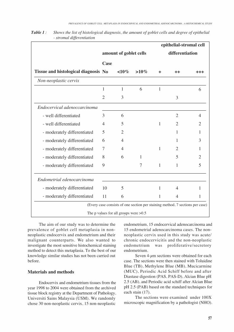
Table 1 : Shows the list of histological diagnosis, the amount of goblet cells and degree of epithelial- stromal differentiation
Tissue and histological diagnosis
Non-neoplastic cervix
Endocervical adenoccarcinoma
- well differentiated
- well differentiated
- moderately differentiated
- moderately differentiated
- moderately differentiated
- moderately differentiated
- moderately differentiated
Endometrial edenocarcinoma
- moderately differentiated
- moderately differentiated
amount of goblet cells
Case
No
1
2
3
4
5
6
7
8
9
10
11
<10%
1
3
6
5
2
4
4
6
5
6
>10%
6
1
7
+
1
1
1
1
1
1
++
3
2
2
1
1
2
5
1
4
4
+++
6
4
2
1
3
1
2
5
1
1
epithelial-stromal cell
differentiation
(Every case consists of one section per staining method, 7 sections per case)
The p values for all groups were >0.5
58
Goblet cell metaplasia was defined as the presenceof goblet cells in the glandular epithelium. Theresults were graded as negative (-) or positive (+).In cases of positive results the degree of severity ofthe metaplasia and the staining intensity of the gobletcells were evaluated. The degree of severity ofmetaplasia was defined as the amount of goblet cellspresent compared to the amount of normal glandularcells. This was arbitrarily expressed as <10% or>10%. The differentiation of the goblet cellscompared with its surrounding glandular epitheliumwas evaluated as weak (+), moderate (++), or strong(+++). In endocervical and endometrialadenocarcinoma the histological grade of thecarcinoma was also evaluated. This was graded aswell, moderate or poor. Statistical analysis was doneby Chi-Square test and Mann-Whitney test.
Results
PrevalenceA total of 630 sections representing 90 cases
were evaluated. Goblet cell metaplasia was presentin 56 (0.09%) sections, representing 11 cases. Gobletcell metaplasia was present in 2 out of 30 (6.67%)cases in non-neoplastic cervix, 0 out of 30 cases innon-neoplastic endometrium, 7 out of 15 (46.7%)cases in endocervical adenocarcinoma and in 2 outof 15 (13.3%) cases in endometrial adenocarcinoma.Correlation for histological grade and presence ofgoblet cell metaplasia was not statisticallysignificant. The endometrial adenocarcinomas,which displayed goblet cell metaplasia were gradedas moderately differentiated adenocarcinomas. Someexamples of the positive cases are depicted inFigure1. There was no statistical difference in the
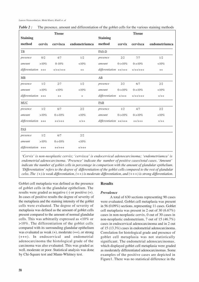
‘Cervix’ is non-neoplastic cervix; ‘cervixca’ is endocervical adenocarcinoma; ‘endometriumca’ isendometrial adenocarcinoma. ‘Presence’ indicate the number of positive cases/total cases. ‘Amount’indicate the number of goblet cells in percentage in comparison with the amount of glandular epithelium.‘Differentiation’ refers to the degree of differentiation of the goblet cells compared to the rest of glandularcelss. The (+) is weak differentiation, (++) is moderate differentiation, and (+++) is strong differentiation.
Table 2 : The presence, amount and differentiation of the goblet cells for the various staining methods
Lauren Nieuwenhuizen, Mohd Khairy Khalil et. al
Staining
method
Tissue Tissue
cervix cervixca endometriumca
Staining
method cervix cervixca endometriumca
TB
presence
amount
differentiation
0/2
>10%
+++
4/7
0-10%
+/++/+++
1/2
<10%
++
PAS-D
presence
amount
differentiation
2/2
0->10%
++/+++
7/7
0->10%
+/++/+++
1/2
<10%
++
MB
presence
amount
differentiation
1/2
>10%
+++
2/7
<10%
++
1/2
<10%
+
AB
presence
amount
differentiation
2/2
0->10%
+/+++
6/7
0->10%
+/++/+++
2/2
<10%
+/++
MUC
presence
amount
differentiation
1/2
>10%
+++
6/7
0->10%
++/+++
2/2
<10%
+/++
PAB
presence
amount
differentiation
1/2
0->10%
++/+++
4/7
0->10%
++/+++
2/2
<10%
+/++
PAS
presence
amount
differentiation
1/2
>10%
+++
6/7
0->10%
++/+++
2/2
<10%
+/+++
59
amount of goblet cells of various histologicaldiagnosis (Table 1).
Histochemical stainingThe staining differentiation of the goblet cells
with its surrounding glandular epithelium wasmoderate to strong in non-neoplastic cervix andendocervical adenocarcinoma, while the stainingdifferentiation in endometrial adenocarcinoma wasweak to moderate. The various staining methodsshowed differences in presence, amount anddifferentiation of the goblet cells (Table 2).
Discussion
The presence of goblet cell metaplasia inendometrium and endocervix is often overlookedby pathologists unlike in the stomach. Our studyshowed a prevalence of intestinal metaplasia to be6.67% (2/30 cases) in non-neoplastic cervix, 46.7%
in endocervical adenocarcinoma (7/15cases), 13.3%in endometrial adenocarcinoma (2/15 cases) andnone in non-neoplastic endometrium (0/30 cases).This confirms that intestinal metaplasia is not as rareas previously reported. This is supported by the studyof Savargaonkar et al (1) and McCluggage et al (6).Our study showed no correlation between theprevalence of goblet cell metaplasia and thehistological grade in endocervical adenocarcinoma.McCluggage et al (7) noted similar findings but forendometrial adenocarcinoma.
The amount of goblet cells seemed to differamong the various histological groups. In this studyrelatively few goblet cells were seen in endometrialadenocarcinoma, few to moderate amounts wereseen in endocervical adenocarcinoma and relativelymore goblet cells were seen in non-neoplastic cervix.The differentiation of the goblet cells with itssurrounding glandular epithelium was moderate tostrong in non-neoplastic cervix and in endocervical
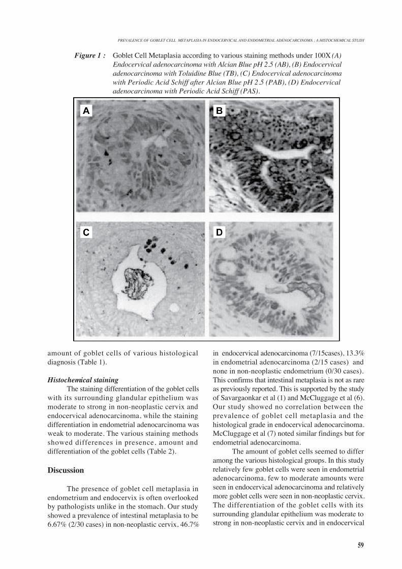
Figure 1 : Goblet Cell Metaplasia according to various staining methods under 100X (A)Endocervical adenocarcinoma with Alcian Blue pH 2.5 (AB), (B) Endocervicaladenocarcinoma with Toluidine Blue (TB), (C) Endocervical adenocarcinomawith Periodic Acid Schiff after Alcian Blue pH 2.5 (PAB), (D) Endocervicaladenocarcinoma with Periodic Acid Schiff (PAS).
PREVALENCE OF GOBLET CELL METAPLASIA IN ENDOCERVICAL AND ENDOMETRIAL ADENOCARCINOMA ; A HISTOCHEMICAL STUDY
60
adenocarcinoma, while the differentiation inendometrial adenocarcinoma was weak to moderate.There was no statistical difference in differentiationbetween the various histological groups. Suchfindings were also shown by Savargaonkar et al (1)using PB/KOH/PAS-staining.
In this study we demonstrated that the mostoptimal overall staining methods for staining gobletcells in non-neoplastic cervix, endocervicaladenocarcinoma and endometrial adenocarcinomawere PAS, PAS-D and AB. These stains highlightedthe cells exhibiting goblet cell metaplasia from theadjacent glandular epithelium. Mucincarmine has arelatively strong detection rate, but thedifferentiation is of a lower degree compared to theother staining methods. Our study is in line with thefindings of Mikami et al (15), who described a strongstaining of the intestinal metaplasia in endocervicalglandular hyperplasia for AB.
In conclusion, the presence of goblet cellmetaplasia in reproductive organs is not as rare aspreviously reported. Routine histochemical stainscould be used to highlight its presence. Althoughthe presence of goblet cell metaplasia in neoplasticcervix and endometrium gives no added prognosticimplication, when in abundance could lead todiagnostic difficulty.
Footnote : Drs. Laurens Nieuwenhuizen wasa postdoctoral (hon) researcher at the department ofpathology, USM, Kubang Kerian, Malaysia. Hishome institution is Faculty of Medicine, UniversityMaastricht, Netherlands. This research wasconducted under sponsorship of grant number 305/PPSP/6112246 of the Ministry of Science,Technology and Innovation, Malaysia
Corresponding Author :
Professor Dr Nor Hayati Othman (MBBS; MPath;MIAC; FAMM)Deputy Dean (Research) & Professor of Pathology,School of Medical Sciences,Universiti Sains Malaysia, Health Campus,16150 Kubang Kerian, Kelantan, MalaysiaTel : 609 7664002 (office), 609 765 3712 ,fax: 609 7656532Email : [email protected]” [email protected]
References
1. Savargaonkar PR, Hale RT, Pope R, Fox H, BuckleyH. Enteric differentiation in cervical adenocarcinomaand its prognostic significance. Histopathology 1993;
23: 275-277.2. Fox H, Wells M, Harris M, McWilliam LJ, Anderson
GS. Enteric tumours of the lower female genital tract:a report of three cases. Histopathology 1988; 12:167-176.
3. Lapertosa G, Baracchini P, Fulcher E, Tanzi R. Patternsof mucous secretion in normal and pathologicalconditions of the endocervix. Eur J Gynecol Oncol1986; 7:113-119.
4. Azzopardi JG, Hou LT. Intestinal metaplasia withargentaffin cells in cervical adenocarcinoma. J PatholBacteriol 1965; 90:686-690.
5. Wells M, Tiltman A. Intestinal metaplasia of theendometrium. Histopathology 1989; 15:431-3.
6. McCluggage WG, Roberts N, Bharucha H. Entericdifferentiation in endometrial adenocarcinomas: amucin histochemical study. Int J of GynecolPathol1995; 14:250-254.
7. Zheng W, Yang GC, Godwin TA, Caputo TA, ZunaRE. Mucinous adenocarcinoma of the endometriumwith intestinal differentiation: a case report. HumPathol 1995; 26:1358-1368.
8. Trowell JE. Intestinal metaplasia with argentaffin cellsin the uterine cervix. Histopathology 1985; 9:551-559.
9. Michael H, Sutton G, Hull MT, Roth LM. Villousadenoma of the uterine cervix associated with invasiveadenocarcinoma: a histochemical, ultrastructural andimmunohistochemical study. Int J Gyaecol Pathol1986; 5:163-169.
10. Moore WF, Bentley RC, Kim KR, Olatidoye B, GraySR, Robboy SJ. Goblet-cell mucinous epitheliumlining the endometrium and endocervix: evidence ofmetastasis from an appendiceal primary tumourthrough the use of cytokeratin-7 and -20 immunostains.Int J Gynecol Pathol 1998; 17:363-367.
11. Szymanska K, Szamborski J, Miechowiecka N,Czerwinski W. Malignant transformation of mucinousovarian cystadenomas of intestinal epithelial type.Histopathology 1986; 7:497-509.
12. Lapertosa G, Baracchini P, Fulcher E, Tanzi R. O-acetylated sialic acid variants in mucinous tumours ofthe ovary. Histopathol 1986; 10: 707-712.
13. Tiltman AJ, Knutzen VK. Primary adenocarcinoma ofthe vulva originating in misplaced cloacal tissue. ObstetGynecol 1978; 51:30-3.
14. Fox H, Wells M, Harris M, McWilliam LJ, AnderssonGS. Enteric tumours of the lower female genital tract:a report of three cases. Histopathology 1988; 12:167-176.
15. Mikami Y et al. Florid endocervical glandularhyperplasia with intestinal and pyloric glandmetaplasia: worrisome benign mimic of “adenomamalginum”. Gynecol Oncol 1999; 74:504-511.
16. Ishii K et al. A new view of the so-called adenomamalignum of the uterine cervix. Virchows Arch 1998;
Lauren Nieuwenhuizen, Mohd Khairy Khalil et. al
61
PREVALENCE OF GOBLET CELL METAPLASIA IN ENDOCERVICAL AND ENDOMETRIAL ADENOCARCINOMA ; A HISTOCHEMICAL STUDY
432:315-322.17. Bancroft J, De Steven A. Theory and Practice of
Histological Techniques. New York: ChurchillLivingstone, 1996.