prevalence of and risk factors for serum antibodies ... · pdf fileleptospirosis was...
TRANSCRIPT

938 ScientificReports JAVMA,Vol234,No.7,April1,2009
SP
EC
IAL
RE
PO
RT
Because of their frequent contact with multiple animal species, veterinarians are at risk of contracting
zoonotic infections.1 In 2005, the CDC and the AVMA conducted a survey2 of veterinarians that included their perceived risk of acquiring zoonotic diseases. Leptospirosis was reportedly a concern for 33.7% of small animal veterinarians and 59.0% of large animal veterinarians.
Leptospirosis is a bacterial disease affecting hu-mans and other animals. Fewer than 100 human cases of leptospirosis/y were reported in the United States from 1984 through 1994, the last year in which lepto-spirosis was a nationally notifiable disease.3 Symptoms may include headache, fever, myalgia, conjunctivitis, nausea, vomiting, and diarrhea or constipation.4 In-fection with Leptospira spp ranges from subclinical to mild, influenza-like illness to serious multisystemic and hepatic disease (Weil’s disease). Meningitis, me-ningoencephalitis, or pulmonary hemorrhage with re-spiratory failure may also result.5 Leptospiral infection during pregnancy may cause abortion.6 Mild infec-
Prevalence of and risk factors for serum antibodies against Leptospira serovars in US veterinarians
Ellen A. Spotts Whitney, mph; Elizabeth Ailes, mph; Lee M. Myers, dvm, mph, dacvpm; Jeremiah T. Saliki, dvm, phd, dacvm; Ruth L. Berkelman, md
Objective—To determine the seroprevalence of antibodies against Leptospira serovarsamongveterinariansandidentifyriskfactorsforseropositivityinveterinarycaresettings.Design—Seroepidemiologicsurvey.Study Population—Veterinariansattendingthe2006AVMAAnnualConvention.Procedures—Bloodsampleswerecollectedfrom511veterinarians,andserumwashar-vestedforamicrocapsuleagglutinationtest(MAT)todetectantibodiesagainst6serovarsofLeptospira.Aggregatedataanalysiswasperformedtodeterminetheratiooftheoddsofagivenexposure (eg, typesofanimals treatedorbiosafetypractices) inseropositiveindividualstotheoddsinseronegativeindividuals.Results—Evidence of previous leptospiral infection was detected in 2.5% of veterinar-ians.MostveterinariansreportedmultiplepotentialexposurestoLeptospira spp andotherpathogensintheprevious12months,includingunintentionalneedlestickinjuries(379/511[74.2%]),animalbites(345/511[67.5%]),andanimalscratches(451/511[88.3%]).Treatmentofadogwithaninfluenza-likeillnesswithinthepastyearwasassociatedwithseropositivityforantibodiesagainstLeptospira spp.Conclusions and Clinical Relevance—Veterinariansareatriskforleptospirosisandshouldtakemeasurestodecreasepotentialexposuretoinfectiousagentsingeneral.Diagnostictests for leptospirosisshouldbeconsideredwhenveterinarianshave febrile illnessesofunknownorigin.(J Am Vet Med Assoc2009;234:938–944)
tions may also lead to future chronic disease, includ-ing chronic fatigue, neuropsychiatric symptoms, and eye infections.5,7 Leptospirosis is often not diagnosed because symptoms may be nonspecific and laboratory diagnosis is difficult.5 The mortality rate for infected humans ranges from < 5% to 30% in various parts of the world.5
Leptospires, excreted in animal urine or tissues of parturition, can survive for weeks to months after be-coming established in soil or water. Animals and hu-mans become infected after contact with this soil or water, which may happen by ingestion of contaminated
From the Center for Public Health Preparedness and Research and the Department of Epidemiology, Rollins School of Public Health, Emory University, Atlanta, GA 30322 (Whitney, Ailes, Berkelman); the Georgia Department of Agriculture, 19 Martin Luther King Jr Dr SW, Atlanta, GA 30334 (Myers); and the Athens Veterinary Diagnostic Laboratory, University of Georgia, Athens, GA 30602 (Saliki). Dr. Myers’ present address is National Veterinary Stockpile, USDA, APHIS, Veterinary Services, 2637 Nunnally Shoals Rd, Good Hope, GA 30641.
Supported by the O. Wayne Rollins Foundation through a grant to the Center for Public Health Preparedness and Research, Rollins School of Public Health, Emory University.
The authors thank Julie Kearney and Jeff Marshall for assistance with implementation of the study; Doris Miller-Liebl and Pamela Currin for performing laboratory tests; Susan Lance, Corrie Brown, and Christopher Woods for assistance with protocol design; Craig Greene for techni-cal assistance; Christine Moe, Juan Leon, and Melissa Dowd for processing and storage of specimens; Colleen Spellen for data entry; and Lance Waller for assistance with the statistical analysis.
Address correspondence to Ms. Whitney.
Abbreviations
CI ConfidenceintervalGHLIT GroupHealth&LifeInsuranceTrustIgM ImmunoglobulinMMAT MicrocapsuleagglutinationtestNVSL NationalVeterinaryServicesLaboratoriesOR Oddsratio

JAVMA,Vol234,No.7,April1,2009 ScientificReports 939
SP
EC
IAL R
EP
OR
T
food or water or through contact with abraded skin and mucosal surfaces, such as an eye or nose. Veterinar-ians and others may also become infected as a result of contact with infected tissues, body fluids, and organs from infected animals through direct contact or aero-sol transmission. Veterinarians and others exposed to infected animals (ie, dogs, rodents, cattle, swine, goats, sheep, and horses), animal products, or contaminated soil or water as part of their work can acquire leptospi-rosis occupationally.7–10
Of particular concern to veterinarians is the in-creasing incidence of diagnosis of leptospirosis in dogs and other animals.11,12 In the lower peninsula of Michi-gan, > 20% of healthy dogs were found to have anti-bodies against Leptospira serovars, most of which were historically uncommon serovars.13 Researchers have also reported a higher incidence of leptospirosis in dogs that have access to potentially contaminated bodies of water.12
Despite the known risk of infection to veterinar-ians in the United States, the incidence and prevalence of leptospirosis in this population remains largely un-defined. Few seroepidemiologic studies have been conducted, and we are unaware of any national se-roepidemiologic surveys of veterinary workers in the United States. The purpose of the study reported here was to estimate the prevalence of antibodies against Leptospira spp and identify factors that influence the risk of infection.
Materials and Methods
Study subjects―The study population was healthy, practicing veterinarians, 18 years of age or older, at-tending the 143rd AVMA Annual Convention from July 15 through July 19, 2006, in Honolulu.
During the convention, health and wellness screen-ing for attendees was performed at the AVMA GHLIT wellness booth. The booth was open to all veterinar-ians, their spouses, and veterinary students attending the convention. Health screenings available to attend-ees included full hematologic and serum biochemical analyses, determination of serum concentration of pros-tate-specific antigen for males > 40 years of age, and determination of blood hemoglobin concentration for females. Measurement of serum antibody titer against rabies virus was also available to booth attendees when they had not been evaluated for that titer within the last 3 years. The study protocol was reviewed and approved by the Institutional Review Board at Emory University.
Specimen and data collection―Informed consent and authorization under the Health Insurance Portabil-ity and Accountability Act was acquired from each par-ticipant. Afterward, a 10-mL blood sample was collect-ed from each participant into a serum separator tube by experienced laboratory personnel. The serum separator tube was centrifuged at 1,800 X g for 8 minutes. Serum was removed and stored at 4oC (39oF) or frozen until it was shipped by expedited delivery to Emory University, where samples were stored at –70oC (–94oF).
Veterinarians were asked to complete a self-admin-istered survey. The standardized survey was designed to obtain information on participant demographics,
reported recent exposure to zoonotic diseases, animal specialty, clinic experience, accidental vaccination, re-cent illness and injury, and frequency of use of personal protective equipment and other infection control prac-tices (eg, handwashing, not recapping needles prior to disposal, or not eating, drinking, or smoking in animal-handling areas). The survey and serum sample of each participant were coded with the same unique identifier to maintain confidentiality.
Serum antibody testing―An aliquot of each serum sample was sent from Emory University to the Athens Veterinary Diagnostic Laboratory at the University of Georgia, where antibody testing was performed. An MAT was used to detect antibodies against Leptospira serovars Bratislava, Canicola, Grippotyphosa, Hardjo, Icterohem-orrhagiae, and Pomona, as described elsewhere.14,15 All testing and reading of results were performed by the same technician who had been trained and had successfully completed the 2006 USDA NVSL proficiency testing for the leptospirosis MAT. A cutoff titer of 1:100 was adapted to the testing conditions of the Athens Veterinary Diag-nostic Laboratory by standardizing the endpoint readings with serum samples of known endpoints supplied by the NVSL in the 2006 leptospirosis proficiency serum panel. The MAT included 1 positive control sample of known titer and 1 negative control sample (titer < 1:100). A par-ticipant was considered seropositive for antibody against Leptospira spp when the antibody titer against any of the aforementioned serovars was ≥ 1:100; all others were considered seronegative.5,16–18
Subanalyses were performed on the basis of informa-tion from other reports and results from initial analysis. Because other researchers have used a seropositive cutoff titer of 1:200, we also considered individuals with a se-rum titer of 1:200 as seropositive and considered all other veterinarians (including those with an antibody titer of 1:100) as seronegative.17 Initial laboratory findings indi-cated that the predominant reactive serovar was Bratislava; therefore, individuals with a 1:100 titer against any serovar other than Bratislava were excluded from the subanalysis. Individuals with a titer against Bratislava of ≥ 1:100 were considered seropositive and compared with the remaining seronegative individuals.
Participants were not informed of their results for anti-Leptospira antibodies because the laboratory used for testing is not certified through the Clinical Labora-tory Improvement Amendments program. Furthermore, there was no anticipated clinical benefit to providing serologic results because antibodies are indicative of previous rather than acute or recent infection.
Statistical analysis―Data were entered into a da-tabasea and analyzed in aggregate form with commer-cially available statistical software.b Medians of ordinal variables were calculated, and these variables were di-chotomized at their medians for use in additional analy-ses. Comparisons between seropositive and seronegative veterinarians were evaluated with a Fisher exact test for ordinal and dichotomous variables, and a value of P < 0.05 was considered significant.
Exact logistic regression was used to determine ORs and 95% CIs, comparing the odds of a given exposure among veterinarians seropositive for anti-Leptospira
940 ScientificReports JAVMA,Vol234,No.7,April1,2009
SP
EC
IAL
RE
PO
RT
antibodies with the odds among veterinarians seronega-tive for anti-Leptospira antibodies.19 An OR met signifi-cance when the associated P value was < 0.05, indicat-ing that the 95% CI for the OR did not include the null value of 1.0.20,21 Variables that achieved a significance of P < 0.10 in the univariate analyses, variables that have been associated with leptospirosis in other stud-ies, and biologically plausible variables were considered for inclusion in a multivariate logistic regression model of risk factors for seropositivity. A backward selection method was used to create the final multivariate logistic regression model; because of the small sample size, ex-act estimates were used for the ORs and 95% CIs.
Results
General characteristics of study veterinarians―During the AVMA Annual convention in 2006, 1,112 individuals (49.4% female) attended the AVMA GHLIT wellness booth. The status of these individuals (eg, vet-erinarian, spouse of veterinarian, or veterinary student) was unknown. Of these, 535 (48.1%) were enrolled in the study. Twenty-four (4.5%) were excluded because their serum sample was missing, they did not have a DVM degree, or they spent ≤ 50% of their working time in clinical veterinary practice. Therefore, 511 veterinar-ians were ultimately included. The median age of veteri-narians was 46 years (range, 25 to 81 years; interquartile range, 35 to 54 years). Two hundred fifty-seven (50.4%) veterinarians were female, and 253 (49.6%) were male.
Veterinarians practiced in various geographic re-gions of the United States as follows: southeastern, 36.6%; western, 24.5%; midwestern, 22.9%; and north-eastern, 13.9%. The remaining 2.1% of veterinarians indicated they worked outside the United States. One hundred seventy-five of 494 (35.4%) veterinarians indicated they had traveled internationally. Most vet-erinarians (353 [69.2%]) reported that they worked in small (companion) animal practice, followed by mixed small and large animal (105 [20.6%]), exotic animal (26 [5.1%]), equine (9 [1.8%]), food animal (includ-ing livestock and poultry; (5 [1.0%]), or another prac-tice type (12 [2.4%]). Nearly all veterinarians reported treating dogs (479 [93.7%]) and cats (479 [93.7%]). Less than a third had treated small ruminants (164 [32.1%]), horses (163 [31.9%]), cattle (148 [29.0%]), exotic mammals (138 [27.0%]), mammalian wildlife (121 [23.7%]), swine (107 [20.9%]), exotic livestock (100 [19.6%]), or poultry (98 [19.2%]).
Thirty-three of 487 (6.8%) veterinarians believed they had contracted a zoonotic disease through their work, with most of these individuals (20 [60.6%]) re-porting they were exposed through skin or mucosal sur-faces. Other suspected routes of transmission were fe-cal-oral (7 [21.2%]), inhalation (7 [21.2%]), parenteral inoculation (1 [3.0%]), and other (5 [15.2%]). Thirty-five of 486 (7.5%) veterinarians reported having inad-vertently inoculated themselves with the vaccine against Leptospira. Of the 268 of 464 (57.8%) veterinarians who reported treating an animal with leptospirosis, 86.2% re-ported treating a dog, 19.8% treated a cow, 5.2% treated a pig, 0.4% treated a rodent, and 3.7% treated another type of animal. The median number of animals with leptospi-rosis treated per year was 3 (interquartile range, 2 to 6).
In response to questions regarding general mea-sures taken to prevent infection, most veterinarians re-ported sometimes or always wearing disposable gloves (86.7%), a laboratory coat or equivalent (71.0%), and a surgical mask (66.3%). Most veterinarians (94.7%) re-ported sometimes or always washing hands before eat-ing, drinking, or smoking, and 96.9% reported washing hands after evaluating an animal. Fewer than half of the veterinarians (42.5%) reported sometimes or always wearing eye protection. Approximately half of the vet-erinarians (55.0%) reported sometimes or always eat-ing, drinking, or smoking in animal handling areas.
Over the previous year (July 2005 to July 2006), 345 (67.5%) veterinarians were bitten by an animal such that the skin was broken at least once and 220 (43.1%) were bitten at least twice. During the same period, 451 (88.3%) veterinarians were scratched by an animal such that the skin was broken at least once and 411 (80.4%) reported at least 2 animal scratches. Most veterinarians reported at least 1 unintentional needlestick injury (379 [74.2%]) and almost half (248 [48.5%]) reported ≥ 2 in the previous year. Most veteri-narians (431 [84.3%]) reported that they sometimes or always recapped needles before disposal. One hundred eighty-eight (36.8%) veterinarians reported being cut by a surgical or necropsy instrument at least once and 97 (19.0%) reported being cut ≥ 2 times within the pre-vious year.
Serum antibody testing―Thirteen of the 511 (2.5%) participating veterinarians had a titer of ≥ 1:100 for antibody against Leptospira, and 7 veterinarians had a titer that exceeded 1:100. The most common leptospiral serovar against which serum antibodies were detected was Bratislava, which was detected in 10 veterinarians. Five of these had a titer of 1:100, 1 had a titer of 1:200, 1 had a titer of 1:400, and 3 others had a titer of 1:800. Two of the veterinarians that were seropositive for se-rovar Bratislava with a titer of 1:800 were also seroposi-tive for serovar Icterohemorrhagiae (titer, 1:400). Of the remaining 3 veterinarians, one had antibody against serovar Icterohemorrhagiae (titer, 1:800), the second had antibody against serovar Hardjo (titer, 1:800), and the third had antibodies against serovars Pomona (titer, 1:200), Icterohemorrhagiae (titer, 1:800), and Grippoty-phosa (titer, 1:100). None of the seropositive individuals had antibody against serovar Canicola.
Of the 13 seropositive veterinarians, 6 were female. All 13 reported treating dogs or cats in the past year, and 6 reported treating an animal with a diagnosis of leptospirosis. None of the 13 reported having a previ-ous diagnosis of leptospirosis, and none reported hav-ing inadvertently inoculated themselves with vaccine against Leptospira (Table 1). Although 1 of 494 (0.2%) veterinarians reported having a previous diagnosis of leptospirosis, this individual was not seropositive at the time of testing.
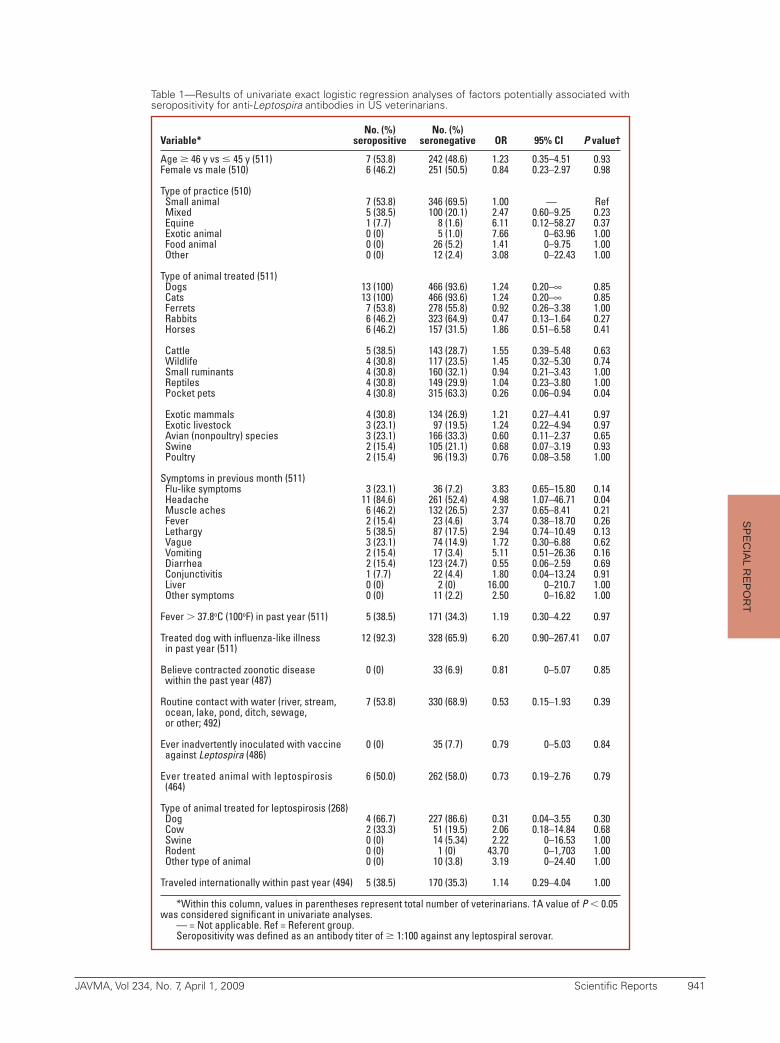
Factors associated with a serum anti-Leptospira antibody titer of ≥ 1:100―Results of univariate analy-ses indicated that, among the 511 participating veteri-narians, neither gender nor primary type of practice was associated with an anti-Leptospira antibody titer ≥ 1:100 (Table 1). With the exception of a report of
JAVMA,Vol234,No.7,April1,2009 ScientificReports 941
SP
EC
IAL R
EP
OR
T
No. (%) No. (%) Variable* seropositive seronegative OR 95% CI P value†
Age 46 y vs 45 y (511) 7 (53.8) 242 (48.6) 1.23 0.35–4.51 0.93Female vs male (510) 6 (46.2) 251 (50.5) 0.84 0.23–2.97 0.98 Type of practice (510) Small animal 7 (53.8) 346 (69.5) 1.00 — Ref Mixed 5 (38.5) 100 (20.1) 2.47 0.60–9.25 0.23 Equine 1 (7.7) 8 (1.6) 6.11 0.12–58.27 0.37 Exotic animal 0 (0) 5 (1.0) 7.66 0–63.96 1.00 Food animal 0 (0) 26 (5.2) 1.41 0–9.75 1.00 Other 0 (0) 12 (2.4) 3.08 0–22.43 1.00 Type of animal treated (511) Dogs 13 (100) 466 (93.6) 1.24 0.20–∞ 0.85 Cats 13 (100) 466 (93.6) 1.24 0.20–∞ 0.85 Ferrets 7 (53.8) 278 (55.8) 0.92 0.26–3.38 1.00 Rabbits 6 (46.2) 323 (64.9) 0.47 0.13–1.64 0.27 Horses 6 (46.2) 157 (31.5) 1.86 0.51–6.58 0.41 Cattle 5 (38.5) 143 (28.7) 1.55 0.39–5.48 0.63 Wildlife 4 (30.8) 117 (23.5) 1.45 0.32–5.30 0.74 Small ruminants 4 (30.8) 160 (32.1) 0.94 0.21–3.43 1.00 Reptiles 4 (30.8) 149 (29.9) 1.04 0.23–3.80 1.00 Pocket pets 4 (30.8) 315 (63.3) 0.26 0.06–0.94 0.04 Exotic mammals 4 (30.8) 134 (26.9) 1.21 0.27–4.41 0.97 Exotic livestock 3 (23.1) 97 (19.5) 1.24 0.22–4.94 0.97 Avian (nonpoultry) species 3 (23.1) 166 (33.3) 0.60 0.11–2.37 0.65 Swine 2 (15.4) 105 (21.1) 0.68 0.07–3.19 0.93 Poultry 2 (15.4) 96 (19.3) 0.76 0.08–3.58 1.00 Symptoms in previous month (511) Flu-like symptoms 3 (23.1) 36 (7.2) 3.83 0.65–15.80 0.14 Headache 11 (84.6) 261 (52.4) 4.98 1.07–46.71 0.04 Muscle aches 6 (46.2) 132 (26.5) 2.37 0.65–8.41 0.21 Fever 2 (15.4) 23 (4.6) 3.74 0.38–18.70 0.26 Lethargy 5 (38.5) 87 (17.5) 2.94 0.74–10.49 0.13 Vague 3 (23.1) 74 (14.9) 1.72 0.30–6.88 0.62 Vomiting 2 (15.4) 17 (3.4) 5.11 0.51–26.36 0.16 Diarrhea 2 (15.4) 123 (24.7) 0.55 0.06–2.59 0.69 Conjunctivitis 1 (7.7) 22 (4.4) 1.80 0.04–13.24 0.91 Liver 0 (0) 2 (0) 16.00 0–210.7 1.00 Other symptoms 0 (0) 11 (2.2) 2.50 0–16.82 1.00 Fever 37.8oC (100oF) in past year (511) 5 (38.5) 171 (34.3) 1.19 0.30–4.22 0.97 Treated dog with influenza-like illness 12 (92.3) 328 (65.9) 6.20 0.90–267.41 0.07 in past year (511) Believe contracted zoonotic disease 0 (0) 33 (6.9) 0.81 0–5.07 0.85 within the past year (487) Routine contact with water (river, stream, 7 (53.8) 330 (68.9) 0.53 0.15–1.93 0.39 ocean, lake, pond, ditch, sewage, or other; 492) Ever inadvertently inoculated with vaccine 0 (0) 35 (7.7) 0.79 0–5.03 0.84 against Leptospira (486) Ever treated animal with leptospirosis 6 (50.0) 262 (58.0) 0.73 0.19–2.76 0.79 (464) Type of animal treated for leptospirosis (268) Dog 4 (66.7) 227 (86.6) 0.31 0.04–3.55 0.30 Cow 2 (33.3) 51 (19.5) 2.06 0.18–14.84 0.68 Swine 0 (0) 14 (5.34) 2.22 0–16.53 1.00 Rodent 0 (0) 1 (0) 43.70 0–1,703 1.00 Other type of animal 0 (0) 10 (3.8) 3.19 0–24.40 1.00 Traveled internationally within past year (494) 5 (38.5) 170 (35.3) 1.14 0.29–4.04 1.00
*Within this column, values in parentheses represent total number of veterinarians. †A value ofP , 0.05 was considered significant in univariate analyses.
— = Not applicable. Ref = Referent group. Seropositivity was defined as an antibody titer of 1:100 against any leptospiral serovar.
Table1—Resultsofunivariateexactlogisticregressionanalysesoffactorspotentiallyassociatedwithseropositivityforanti-LeptospiraantibodiesinUSveterinarians.

942 ScientificReports JAVMA,Vol234,No.7,April1,2009
SP
EC
IAL
RE
PO
RT
a headache, those with anti-Leptospira antibodies were no more likely to have been symptomatic in the pre-vious month than other veterinarians. International travel within the past year was not an independent risk factor for seropositivity. A greater proportion of sero-positive veterinarians reported treating a dog with an influenza-like illness in the past year, compared with the proportion of seronegative veterinarians, although the difference was not significant. Treatment of pocket pets was the only factor significantly associated with seropositivity.
Being bitten or scratched by an animal, having re-ceived an unintentional needlestick injury, or being cut by a surgical or necropsy instrument did not increase the odds of leptospiral seropositivity (data not shown). There were no associations between reported biosafety practices and use of personal protective equipment and seropositivity (data not shown).
On the basis of results of the univariate analyses, a full multivariate model was created, including the vari-ables treatment of pocket pets and treatment of a dog with influenza-like illness in the past year. Because report of a headache within the month prior to the survey lacked clinical specificity for previous Leptospira infection, it was not included. Results of the multivariate model in-dicated that treatment of pocket pets (OR, 0.21; 95% CI, 0.05 to 0.76; P = 0.01) and treatment of a dog with in-fluenza-like illness in the past year (OR, 8.21; 95% CI, 1.17 to 358.00; P = 0.03) were significant predictors of a positive antibody response to Leptospira.
Factors associated with a serum anti-Leptospira antibody titer of ≥ 1:200―Eight (1.6%) veterinarians had a high antibody titer (≥ 1:200). All 8 (100%) report-ed that they had treated a dog with influenza-like illness in the past year, compared with 332 of 503 (66.0%) se-ronegative veterinarians (including the 5 veterinarians with antibody titers of 1:100; P = 0.08). Seven (87.5%) veterinarians with a high antibody titer were > 46 years of age, compared with 242 (48.1%) seronegative veteri-narians (P = 0.06). Three (37.5%) veterinarians with a high antibody titer reported influenza-like symptoms in the past month, compared with 36 (7.2%) seronegative veterinarians (exact OR, 7.72; 95% CI, 1.15 to 41.5; P = 0.04). One (12.5%) veterinarian with a high antibody titer reported treating pocket pets, whereas 318 (63.2%) seronegative veterinarians (exact OR, 0.083; 95% CI, 0.002 to 0.658; P = 0.01) reported the same thing.
Factors associated with a serum antibody titer of 1:100 against Bratislava―Ten (2.0%) veterinarians had a serum antibody titer of 1:100 against Leptospira serovar Bratislava. Of various influenza-like symptoms, only headache was significantly associated with sero-positivity for Bratislava (exact OR, 8.15; 95% CI, 1.11 to 359.68; P = 0.03).
Discussion
In the present study of veterinarians attending the 143rd AVMA Annual Convention in 2006, 13 of 511 (2.5%) veterinarians had evidence of previous lep-tospiral infection. Other estimates of the seropreva-lence of leptospirosis include 1.8% among all practic-
ing veterinarians in Illinois between 1956 and 1972,22 1% among veterinarians in New Zealand in 1974,23 and 2.9% among Austrian veterinarians in 1994.24 We might have expected fewer leptospiral infections given advances in and greater use of vaccines against Lepto-spira in veterinary medicine. At the same time, there are reports11,12 of increases in leptospiral infections in wild animals in the United States and a concomitant increase in infections in domestic animals, particularly dogs.
The first veterinary vaccine, developed for dogs in the 1950s, protected against Leptospira serovars Ictero-hemorrhagiae and Canicola. The first trivalent vaccine containing distemper virus, infectious canine hepatitis virus, and Leptospira serovar Canicola for dogs was li-censed in 1961.25 In 2000, a new leptospirosis vaccine was introduced, which included not only Leptospira se-rovars Icterohemorrhagiae and Canicola but also Pomo-na and Grippotyphosa, to address emerging changes in the distributions of infections by the various serovars.26 In the study reported here, 10 of 13 seropositive vet-erinarians had antibody against Leptospira serovar Bratislava, which is not included in the canine vaccine. This serovar predominantly infects pigs and horses but has been associated with illness in dogs as well.4,16
Four of the 13 seropositive veterinarians in the present study had antibody against Leptospira serovar Icterohemorrhagiae, which has been included in the canine vaccine since original production.27 Rodents are the primary reservoir of this serovar, and they may serve as a source of infection for humans as well as oth-er animals.16
Only 1 veterinarian had antibodies against Lepto-spira serovars Pomona and Grippotyphosa, and none were seropositive for antibodies against Canicola. Be-cause dogs are the primary reservoir for Leptospira se-rovar Canicola,16 and this serovar has been included in the old and new vaccines used in dogs, our results were not surprising. Vaccinated animals may still become subclinically infected and shed leptospires in their urine.4,28,29 The American Animal Hospital Association Canine Vaccine Task Force has recommended that vac-cination of dogs against Leptospira be limited to use in areas where there is a reasonable risk of exposure, pri-marily because of lack of information on prevalence of various serovars in different geographic areas as well as the risk of postvaccination reactions.30 In cows, sheep, and pigs, annual vaccinations against Leptospira are rec-ommended for confined animals, whereas semiannual vaccination should be considered for open herds.31
There are several rapid serologic assays for the diagnosis of leptospirosis. These include the slide ag-glutination assay,32 indirect hemagglutination assay,33 MAT,34 immunofluorescence assay,35 ELISA for IgM,36,37 IgM dipstick assay,38 and IgM dot-ELISA dipstick test.39 Limitations of these assays include low sensitivity in subjects tested during the first week of illness (whole-cell based serologic assays), the requirement for spe-cialized laboratory equipment such as a fluorescence microscope (immunofluorescence assay), the need for skilled personnel to perform the assay (ELISA), and the inability to detect the infecting serogroup. The gold standard is the MAT, with a sensitivity of 98.2% (95% CI, 95.8% to 100.6%) and specificity of 96.4% (95% CI,

JAVMA,Vol234,No.7,April1,2009 ScientificReports 943
SP
EC
IAL R
EP
OR
T
95.0% to 97.9%).14 The MAT allows for the differentia-tion of infecting serogroups. However, few laboratories perform MATs because of the expense and technical ex-pertise required.14 Typically, positive results from acute serum samples (from blood samples collected ≤ 14 days after the onset of symptoms of leptospirosis) and con-valescent serum samples (from blood samples collected ≥ 15 days after onset of symptoms) are used to make a definitive diagnosis. A positive result from a single se-rum sample (≥ 1:100 MAT titer) may indicate previous infection because antibodies against Leptospira spp may persist for many years.7,39–43
Because we used an MAT on 1 serum sample from each participating veterinarian, we are unable to deter-mine the time frame during which they were exposed to Leptospira. It is possible that the proportion of veterinar-ians who were exposed to Leptospira during their life-time was greater than the proportion that we detected. Additionally, we only tested for antibodies against 6 lep-tospiral serovars. If we had tested for antibodies against a broader spectrum of serovars, we may have detected a higher seroprevalence than we did. Cross-reaction be-tween serovars may also have occurred.
As of December 31, 2006, there were 81,468 veteri-narians practicing in the United States, 47.1% of whom were female.44 This percentage is similar to our study sample, in which 50% of veterinarians were female. The proportion of veterinarians in our study who worked with small animals (69%) was similar to that of the gen-eral veterinarian population in the United States (66%)44; however, the proportion of veterinarians who worked at mixed animal practices was not (21% in our study vs 7.8% in the whole United States). Given these findings, caution should be used when attempting to generalize our results to the US population of veterinarians.
Treatment of pocket pets and dogs with influenza in the previous year were the only factors significantly asso-ciated with leptospiral seropositivity when multivariate analysis was performed in the present study. The protective association with pocket pets was either spurious or related to other characteristics of small animal veterinarians that were not measured. For example, veterinarians who treated pocket pets may have been more likely to work in urban areas and may consequently have been less likely to have come in contact with animals infected with Leptospira.
Veterinarians who treated dogs with influenza-like illness in the previous year were more likely to have an antibody response to the Leptospira serovars evaluated. Some signs of leptospirosis and influenza-like illness in dogs are similar. Dogs with leptospirosis may have non-specific signs including fever, depression, lethargy, an-orexia, arthralgia or myalgia, and oculonasal discharge. As the disease progresses, clinical signs may include vomiting, dehydration, lumbar pain from renomegaly and nephritis, tongue-tip ulceration and necrosis, in-tussusception, pulmonary hemorrhage, uveitis, pneu-monitis, chronic hepatitis, and reproductive failure.31 Influenza-like illness in dogs may also be evident as fe-ver, nasal discharge, and persistent cough.45,46 The simi-larity between the signs of leptospirosis and those of in-fluenza-like illness suggests that veterinarians treating ill dogs may be putting themselves at risk for exposure to zoonotic pathogens such as Leptospira.
The potential for the veterinarians in our study to have been exposed to zoonotic pathogens is remarkable. Indeed, 74.2% of 511 participating veterinarians reported an unintentional needlestick injury at least once in the past year. In 1998 and 1999, the frequency of needlestick injuries was reportedly 0.45/person/y among companion animal veterinarians and 2.03/person/y among large ani-mal veterinarians.47,48 Most veterinarians (84.3%) in our study reported that they sometimes or always recapped needles before disposal, a known risk factor for pathogen transmission in veterinary settings.49 Another study re-vealed similar findings in that 63% of veterinarians mostly or always recapped needles prior to disposal.2 This prac-tice has been regulated in human medicine through the implementation of the Bloodborne Pathogens Regulation (1910.1030) since 199250; the practice has been discour-aged in veterinary animal medicine since 2006.51 Thus, veterinarians should be encouraged to always dispose of uncapped needles in an approved sharps container.
In another study,2 < 5% of veterinarians reported wearing appropriate respiratory or eye protection when handling products of conception and only 6.3% of small animal veterinarians reported wearing appropriate per-sonal protective equipment when examining an animal with respiratory signs. In the present study, frequency of use of personal protective equipment was poor, yet most veterinarians reported treating an animal with a diagnosis of leptospirosis. Although no association was evident between lack of protective equipment use and evidence of leptospiral infection, this may have been attributable to the fact that most veterinarians did not use such protection; thus, the power to detect an as-sociation was limited. It is essential that personal pro-tective equipment such as gloves, barrier gowns, and eye protection be worn when handling animals with leptospirosis or products (eg, urine or tissue) from ani-mals suspected of having leptospirosis.52 In the event of a high-risk exposure (eg, direct contact with urine or blood from an infected animal via mucosal membranes or broken skin), the World Health Organization recom-mends postexposure prophylaxis with doxycycline.53,54
Although it is impossible to eliminate the occupa-tional risk of exposure to zoonotic pathogens in vet-erinary practice, the risk of infection can be mitigated through early recognition and appropriate management of infected or potentially infected animals, use of good personal hygiene and personal protective equipment, avoidance of recapping needles, as well as proper ani-mal handling and housing. The Compendium of veteri-nary standard precautions: zoonotic disease prevention in veterinary personnel51 provides valuable information and guidelines for the prevention of the transmission of zoo-notic pathogens from animals to veterinary personnel.
a. Microsoft Access 2003, Microsoft Corp, Redmond, Wash.b. SAS, version 9.1, SAS Institute Inc, Cary, NC.
References1. Langley R, Pryor WJ, O’Brien K. Health hazards among veterinar-
ians: a survey and review of the literature. J Agromed 1995;2:23–52.2. Wright JG, Jung S, Holman RC, et al. Infection control practices
and zoonotic disease risks among veterinarians in the United States. J Am Vet Med Assoc 2008;232:1863–1872.
944 ScientificReports JAVMA,Vol234,No.7,April1,2009
SP
EC
IAL
RE
PO
RT
3. The Leptospirosis Information Center. Leptospirosis infection rates. 2004/2005. Available at: www.leptospirosis.org/topic.php?t=44. Accessed Oct 25, 2007.
4. Pan American Health Organization. Leptospirosis. In: Acha PN, Szyfres B, eds. Zoonoses and communicable diseases common to man and animals: bacterioses and mycoses. 3rd ed. Washington, DC: Pan American Health Organization, 2003;157–168.
5. World Health Organization/International Leptospirosis Soci-ety. Human leptospirosis: guidance for diagnosis, surveillance and control. Geneva: World Health Organization, 2003. Available at: www.who.int/csr/don/en/WHO_CDS_CSR_EPH_2002.23.pdf. Accessed Oct 25, 2007.
6. Shaked Y, Shpilberg O, Samra D, et al. Leptospirosis in preg-nancy and its effect on the fetus: case report and review. Clin Infect Dis 1993;17:241–243.
7. Faine S. Leptospirosis. In: Hausler WJ, Sussman M, eds. Topley and Wilson’s microbiology and microbial infections. London: Ar-nold, 1998;849–869.
8. Farr RW. Leptospirosis. Clin Infect Dis 1995;21:1–6.9. Feigin RD, Anderson DC. Human leptospirosis. Crit Rev Clin
Lab Sci 1975;5:413–467.10. Heath CW Jr, Alexander AD, Galton MM. Leptospirosis in the
United States. Analysis of 483 cases in man, 1949–1961. N Engl J Med 1965;273:915–922.
11. Bolin CA. Diagnosis of leptospirosis: a reemerging disease of compan-ion animals. Semin Vet Med Surg (Small Anim) 1996;11:166–171.
12. Ghneim GS, Viers JH, Chomel BB, et al. Use of a case-control study and geographic information systems to determine envi-ronmental and demographic risk factors for canine leptospiro-sis. Vet Res 2007;38:37–50.
13. Stokes JE, Kaneene JB, Schall WD, et al. Prevalence of serum antibodies against six Leptospira serovars in healthy dogs. J Am Vet Med Assoc 2007;230:1657–1664.
14. Bajani MD, Ashford DA, Bragg SL, et al. Evaluation of four com-mercially available rapid serologic tests for diagnosis of lepto-spirosis. J Clin Microbiol 2003;41:803–809.
15. World Organisation for Animal Health (OIE). Leptospirosis. In: Manual of diagnostic tests and vaccines for terrestrial animals. 5th ed. Paris: World Organisation for Animal Health (OIE), 2004;316–327.
16. Bharti AR, Nally JE, Ricaldi JN, et al. Leptospirosis: a zoonotic disease of global importance. Lancet Infect Dis 2003;3:757–771.
17. Levett PN. Leptospirosis. Clin Microbiol Rev 2001;14:296–326.18. Faine S. Guidelines for the control of leptospirosis. Geneva, Swit-
zerland: World Health Organization, 1982.19. Allison PD. Logistic regression using the SAS system: theory and
application. Cary, NC: SAS Institute Inc, 1999.20. Hosmer D, Lemeshow S. Applied logistic regression. 2nd ed. New
York: Wiley & Sons, 2000.21. Kleinbaum DG. Logistic regression: a self-learning text. 2nd ed.
New York: Springer-Verlag, 2002.22. Schnurrenberger PR, Grigor JK, Walker JF, et al. The zoonosis-
prone veterinarian. J Am Vet Med Assoc 1978;173:373–376.23. Robinson RA, Metcalfe RV. Zoonotic infections in veterinarians.
N Z Vet J 1976;24:201–210.24. Nowotny N, Deutz A, Fuchs K, et al. Prevalence of swine influ-
enza and other viral, bacterial, and parasitic zoonoses in veteri-narians. J Infect Dis 1997;176:1414–1415.
25. Zuschek F, Jungk N, King D, et al. Noninterference with im-munity by a trivalent vaccine containing distemper virus, infec-tious canine hepatitus virus, and Leptospira canicola. Cornell Vet 1961;51:68–76.
26. Foster & Smith Inc. Fort Dodge releases new leptospirosis vac-cine 2000. Available at: www.peteducation.com/article_print.cfm?c=0+1275&aid=1267. Accessed Dec 11, 2007.
27. Brunner KT, Meyer KF. Immunization of hamsters and dogs against experimental leptospirosis. J Immunol 1950;64:365–372.
28. Bolin CA, Alt DP. Use of a monovalent leptospiral vaccine to prevent renal colonization and urinary shedding in cattle ex-posed to Leptospira borgpetersenii serovar hardjo. Am J Vet Res 2001;62:995–1000.
29. Feigin RD, Lobes LA Jr, Anderson D, et al. Human leptospirosis from immunized dogs. Ann Intern Med 1973;79:777–785.
30. American Animal Hospital Association Canine Vaccine Task Force, Paul MA, Carmichael LE, et al. 2006 AAHA canine vac-cine guidelines. J Am Anim Hosp Assoc 2006;42:80–89.
31. Merck & Co Inc. The Merck veterinary manual. 2006. Avail-able at: www.merckvetmanual.com/mvm/index.jsp?cfile=htm/bc/51202.htm&word=leptospirosis. Accessed Dec 11, 2007.
32. Galton MM, Powers DK, Hall AD, et al. A rapid microscopic-slide screening test for the serodiagnosis of leptospirosis. Am J Vet Res 1958;19:505–512.
33. Sulzer CR, Glosser JW, Rogers F, et al. Evaluation of an indirect hemagglutination test for the diagnosis of human leptospirosis. J Clin Microbiol 1975;2:218–221.
34. Arimitsu Y, Kmety E, Ananyina Y, et al. Evaluation of the one-point microcapsule agglutination test for diagnosis of leptospi-rosis. Bull World Health Organ 1994;72:395–399.
35. Appassakij H, Silpapojakul K, Wansit R, et al. Evaluation of the immunofluorescent antibody test for the diagnosis of human leptospirosis. Am J Trop Med Hyg 1995;52:340–343.
36. Adler B, Murphy AM, Locarnini SA, et al. Detection of specific anti-lep-tospiral immunoglobulins M and G in human serum by solid-phase en-zyme-linked immunosorbent assay. J Clin Microbiol 1980;11:452–457.
37. Winslow WE, Merry DJ, Pirc ML, et al. Evaluation of a com-mercial enzyme-linked immunosorbent assay for detection of immunoglobulin M antibody in diagnosis of human leptospiral infection. J Clin Microbiol 1997;35:1938–1942.
38. Gussenhoven GC, van der Hoorn MA, Goris MG, et al. LEPTO dip-stick, a dipstick assay for detection of Leptospira-specific immunoglob-ulin M antibodies in human sera. J Clin Microbiol 1997;35:92–97.
39. Levett PN, Branch SL, Whittington CU, et al. Two methods for rapid serological diagnosis of acute leptospirosis. Clin Diagn Lab Immunol 2001;8:349–351.
40. Cacciapuoti B, Vellucci A, Ciceroni L, et al. Prevalence of lepto-spirosis in man. Pilot survey. Eur J Epidemiol 1987;3:137–142.
41. Cumberland P, Everard CO, Wheeler JG, et al. Persistence of anti-leptospiral IgM, IgG and agglutinating antibodies in pa-tients presenting with acute febrile illness in Barbados 1979–1989. Eur J Epidemiol 2001;17:601–608.
42. Finsterer J, Stollberger C, Sehnal E, et al. Mild leptospirosis with three-year persistence of IgG- and IgM-antibodies, initially mani-festing as carpal tunnel syndrome. J Infect 2005;51:E67–E70.
43. Lupidi R, Cinco M, Balanzin D, et al. Serological follow-up of patients involved in a localized outbreak of leptospirosis. J Clin Microbiol 1991;29:805–809.
44. AVMA. Reference, Market research statistics, US veterinarians—2007. Available at: www.avma.org/reference/marketstats/usvets.asp. Accessed Jun 26, 2007.
45. Dubovi EJ, Njaa BL. Canine influenza. Vet Clin North Am Small Anim Pract 2008;38:827–835.
46. AVMA. Control of canine influenza in dogs—questions, answers, and interim guidelines 2005. Available at: www.avma.org/public_health/influenza/canine_guidelines.asp. Accessed Jul 14, 2008.
47. Poole AG, Shane SM, Kearney MT, et al. Survey of occupa-tional hazards in large animal practices. J Am Vet Med Assoc 1999;215:1433–1435.
48. Poole AG, Shane SM, Kearney MT, et al. Survey of occupational hazards in companion animal practices. J Am Vet Med Assoc 1998;212:1386–1388.
49. Artenstein AW, Hicks CB, Goodwin BS, et al. Human infection with B virus following a needlestick injury. Rev Infect Dis 1991;13:288–291.
50. Occupational Safety & Health Administration. Bloodborne patho-gens—1910.1030 1992. Available at: www.osha.gov/pls/oshaweb/owadisp.show_document?p_table=STANDARDS&p_id=10051. Accessed Jul 7, 2007.
51. National Association of State Public Health Veterinarians (NASPHV). Compendium of veterinary standard precautions: zoonotic disease pre-vention in veterinary personnel, 2006. Available at: www.cfsph.iastate.edu/BRM/resources/stationary/CompendiumofVeterinaryStandard-Precautions.pdf. Accessed Jun 26, 2007.
52. Weese JS, Peregrine AS, Armstrong J. Occupational health and safety in small animal veterinary practice: part I—nonparasitic zoonotic diseases. Can Vet J 2002;43:631–636.
53. Takafuji ET, Kirkpatrick J, Miller R, et al. An efficacy trial of dox-ycycline chemoprophylaxis against leptospirosis. N Engl J Med 1984;310:497–500.
54. World Health Organization. Leptospirosis. WHO recommended standards and strategies for surveillance, prevention and control of communicable diseases. Available at: www.who.int/zoonoses/diseases/Leptospirosissurveillance.pdf. Accessed Feb 25, 2008.