prevalence and clinical relevance of exocrine pancreatic ... c/hall c sunday/ppthardt.pdf ·...
TRANSCRIPT

First World Congress on Controversies in Obesity, Diabetes and
HypertensionBerlin, October 26-29, 2006
PrevalencePrevalence And And ClinicalClinical RelevanceRelevance Of Of
ExocrineExocrine PancreaticPancreatic InsufficiencyInsufficiency In In
Diabetes MellitusDiabetes Mellitus
Hardt PD, MD
University Hospital Giessen and Marburg
Giessen, Germany

1. What is the prevalence of exocrine pancreatic
insufficiency in diabetes mellitus?
2. Is diabetes mellitus secondary to pancreaticdiseases (type-3) a frequent phenomenon?
3. What are the clinical consequences of exocrine pancreatic insufficiency in diabetes
mellitus?

1. What is the prevalence of exocrinepancreatic insufficiency in diabetes
mellitus?

Author Year Subjects Methods Results Pollard H et al.
1943
13 Amylase and lipase after pancreozymin-secretin stimulation
62% reduced
Chey WY et al. 1963 50 diabetic patients; 13 juvenile type
Amylase and lipase after pancreozymin-secretin stimulation
Low amylase output in diabetes 36% Juvenile diabetes 77%
Vacca JB et al 1964 55 diabetic patients (22 insulin treated)
Diastase and bicarbonate after secretin stimulation; fecal fat
73% abnormal; correlation with age, no correlation with fecal fat
Frier BM et al. 1976 20 IDDM, 7 NIDDM, 13 controls
Stimulation with iv secretin and CCK-PZ
PEI: 80% IDDM; correlation with duration
Harano Y et al. 1978 53 NIDDM, 4 IDDM, 18 controls
Secretin-pancreozymin test
Diabetes: 69% deficient enzyme output; correlation with diabetes control
Lankisch PG et al. 1982 53 IDDM Secretin-pancreozymin test
Diabetes: 43% impaired function
Bretzke G et al. 1984 60 insulin-treated type 2 diabetic patients
Secretin-pancreozymin test
Diabetes 27% „mild PEI“
El Nehwihi H et al. 1988 10 type 2 diabetic patients with diarrhea and neuropathy
Secretin and CCK test Enzyme and bicarbonate reduction in all subjects
Semakula C 1996 307 IDDM, 303 non-diabetic siblings, 207 controls
Serum amylase and lipase
Reduced enzyme levels in 18% IDDM, 6% siblings and 2% controls
The prevalence of exocrine pancreatic insufficiencyin diabetes mellitus
Studies on function by direct function tests:Abnormal in 52.4% (18-100%)

Author Year Subjects Methods Results
Hardt et al.
1999
128
type 1+2
Fecal
chymotrysin
Fecal elastase 1
45% <6 U7l
46% < 200 µg/g
Hardt et al. 2000 39 type 1
77 type 2
Fecal elastase 1 74% < 200 µg/g
36%
Icks et al. 2001 112 type 1 Fecal elastase 1 54,5% < 200 µg/
Rathmann et al. 2001 544 type 2 Fecal elastase 1 30,3% < 200 µg/
Hardt et al. 2003 323 type 1
697 type 2
Fecal elastase 1 51%< 200 µg/
35%< 200 µg/
Nunes et al. 2003 42 type 1+2 Fecal elastase 1 36% < 200 µg/g
Yilmaztepe et al. 2005 32 type 2 Fecal elastase 1 28% < 200 µg/
Cavalot et al. 2006 66 type 1 Fecal elastase 1 26% < 200 µg/g
The prevalence of exocrine pancreatic insufficiencyin diabetes mellitus
Studies on function by indirect function tests:Abnormal in type 1 diabetes: 51% (26-74%)Abnormal in type 2 diabetes: 32% (28-36%)


Hardt PD et al., Pancreatology 3: 395-402, 2003
Characteristics and clinical findings in type 1 (a) and type 2 (b) diabetes mellitus patients

Hardt PD et al., Pancreatology 3: 395-402, 2003
Exocrine function in diabetes mellitus as determined by fecal elastase 1 concentrations
Type 1 (n = 323) Type 2 (n = 697)
(E1 < 100 µg/g)
(E1 100 - 200 µg/g)
(E1 < 100 µg/g)
(E1 100 - 200 µg/g)

Author Year Subjects Methods Results Blumenthal H et al. 1963 3.821 autopsy cases Morphology Prevalence of
pancreatitis: in diabetes: 11,2% nondiabetes: 5,3%
Putzke HP et al. 1986 100 diabetic and 100 non-diabetic autopsy cases
Histopathology Differences in several items; lipomatosis 75% in diabetics; 60% in controls
Warren et LeCompte 1952 ? Histology 50% pancreas fibrosis Foulis A et al. 1997 type 1 Histology Fibrosis, atrophy, fatty
infiltration Klöppel et al. 1996 type 1 Histology Fibrosis, atrophy, fatty
infiltration Gilbeau et al. 1992 20 type 1, 37 type 2 CT-Scans Pronounced lobulation,
small size compared to controls
Altobelli et al. 1998 60 type 1 Ultrasound small size compared to controls; dependant on duration
Alzaid et al. 1993 14 type 1, 43 type 2 Ultrasound small size compared to controls; type 1<type2<controls; not dependent on duration
Nakanishi et al. 1994 36 type 1, 43 type 2 ERCP Changes like CP type 1 40%, type 2 9%
Hardt et al. 2002 38 type 1, 118 type 2 ERCP Changes like CP type 1>Type 2, up to 75%
The prevalence of morphologic changesof the exocrine pancreas in diabetes mellitus
Morphologic changes of the exocrinepancreas in diabetes mellitus arefrequent
The prevalence is about 40% (11-75%)
Changes are more pronounced in type 1

Summary I:
The prevalence of exocrine pancreatic insufficiencyis very high in diabetic populations:
NIDDM/Type 2: about 35% (15-73) exocrine insufficiencyIDDM/Type 1: about 50% (40-80) exocrine insufficiency
using indirect as well as direct function tests.
The prevalence of morphologic changes is also rather common and affects up to 40%.

2. Is diabetes mellitus secondary to pancreatic disease (type-3) a frequent
phenomenon?

(1) Insulin has a trophic effect on pancreatic acinar tissue(insulin-acinar portal system) and its lack may cause pancreatic atrophyWilliams JA, Goldfine ID (1985); Korc M (1993)
(2) Islet hormones have regulatory functions on exocrine tissuewhich may be impairedLazarus SS and Volk BW (1961); Adler G, Kern HF (1975)
(3) Diabetic autonomic neuropathy may lead to impairedenteropancreatic reflexes and exocrine dysfunctionEl Newihi H et al. (1988)
Exocrine dysfunction in diabetes mellitus: Pathophysiological concepts - 1
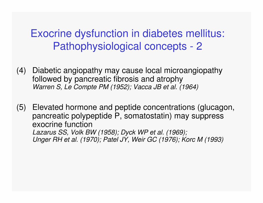
(4) Diabetic angiopathy may cause local microangiopathyfollowed by pancreatic fibrosis and atrophyWarren S, Le Compte PM (1952); Vacca JB et al. (1964)
(5) Elevated hormone and peptide concentrations (glucagon, pancreatic polypeptide P, somatostatin) may suppressexocrine functionLazarus SS, Volk BW (1958); Dyck WP et al. (1969); Unger RH et al. (1970); Patel JY, Weir GC (1976); Korc M (1993)
Exocrine dysfunction in diabetes mellitus: Pathophysiological concepts - 2
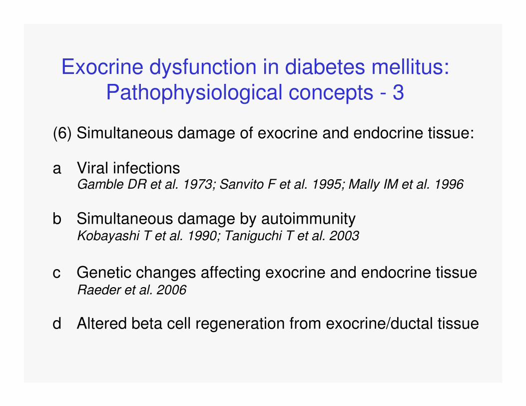
(6) Simultaneous damage of exocrine and endocrine tissue:
a Viral infectionsGamble DR et al. 1973; Sanvito F et al. 1995; Mally IM et al. 1996
b Simultaneous damage by autoimmunityKobayashi T et al. 1990; Taniguchi T et al. 2003
c Genetic changes affecting exocrine and endocrine tissueRaeder et al. 2006
d Altered beta cell regeneration from exocrine/ductal tissue
Exocrine dysfunction in diabetes mellitus: Pathophysiological concepts - 3

(7) „...it is also possible that some of the diabetics are actuallypatients with chronic pancreatitis. It is this that
we would emphasize.“[Chey et al. 1963]
Exocrine dysfunction in diabetes mellitus – pathophysiological concepts
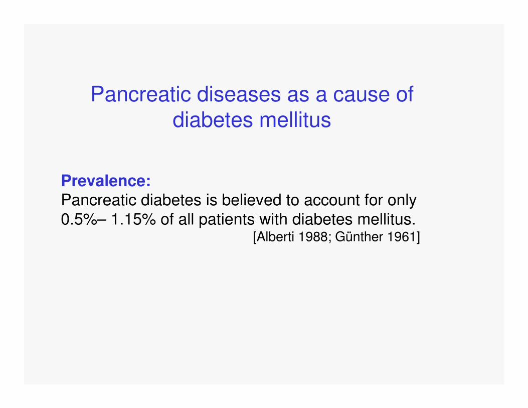
Pancreatic diseases as a cause of diabetes mellitus
Prevalence:Pancreatic diabetes is believed to account for only0.5%– 1.15% of all patients with diabetes mellitus.
[Alberti 1988; Günther 1961]

IV. Gestational diabetes mellitus (GDM)
H. Other genetic syndromes sometimes associated with diabetes
G. Uncommon forms of immune-mediated diabetes
F. Infections
E. Drug- or chemical-induced
D. Endocrinopathies
C. Diseases of the exocrine pancreas
B. Genetic defects in insulin action
A. Genetic defects of ß-cell function
III. Other specific types
II. Type 2 diabetes* (may range from predominantly insulin
resistance with relative insulin deficiency to a predominantlysecretory defect with insulin resistance)
B. Idiopathic
A. Immune mediated
I. Type 1 diabetes* (ß-cell destruction, usually leading to absolute insulin deficiency)
Report of the Expert Committee on the Diagnosis and Classification of Diabetes Mellitus
[Diabetes Care 26:S5-S20, 2003]

Pancreatic diseases as a cause of diabetes mellitus
- acute pancreatitis- chronic pancreatitis- carcinoma- surgery- hemochromatosis- cystic fibrosis

Diabetes mellitus in pancreatitis
Prevalence of diabetes mellitus in acute pancreatitis:- about 50% impaired glucose tolerance during acute phase- persisting diabetes in 1-5%
[Warren et al. 1950, Scuro et al. 1984]
Prevalence of diabetes mellitus in chronic pancreatitis:- 40-70%- in chronic-calcifying pancreatitis up to 90%
[Bank et al. 1975]

Is pancreatic diabetes (type-3 diabetes) more common than believed so far?
The definition of chronic pancreatitis includes persisting/progressive changes of exocrine function and morphology.
A significant number of diabetes patients not only showfunctional changes, but morphologic changes matchingdiagnostic criteria for chronic pancreatitis.

Reclassification study
- Retrospective analysis of 1922 patients with diabetes mellitus
- Screening patients records for parameters of exocrine function and morphology, diabetes duration, diabetes therapy, age at onset, C-peptide levels, HbA1c levels, and diabetes associated antibodies
- Diagnosis type 1: Presence of autoantibodies, early onset, early insulin requirementDiagnosis type 2: absence of autoantibodies, no (late) insulin requirement, insulin resistanceDiagnosis type 3: Absence of autoantibodies, exocrine pancreatic insufficiency, typical morphologic findings [Ewald et al., submitted]

8%
92%
Diabetes mellitus Typ 3c
Übriges diabetischesKrankengut
Prevalence of pancreatic diabetes (Type IIIC)in 1922 patients of a university hospital
[Ewald et al., submitted]
Type 3 diabetes mellitus
Type 1 or type 2 diabetes mellitus

76%
8%
9%4% 3%
Chronische Pankreatitis
HereditäreHämochromatose
Pankreas-NPL
Zystische Fibrose
Z.n. Pankreas-teilresektion
Etiology of pancreatic diabetes (Type IIIc)in 1922 patients of a university hospital
[Ewald et al., submitted]
Chronic pancreatitis
Hemochromatosis
Pancreatic cancer
Cystic fibrosis
Pancreas surgery

Detailierte Aufschlüsselung der Missklassifikation
der Diabetes mellitus Typ 3c Patienten
80%
12%
8%
Missklassifikation alsTyp 2
Missklassifikation alsTyp 1
Missklassifikation als"nicht klassifizierbar"
[Ewald et al., submitted]
Invalid classification of patients with type 3 diabetesmellitus:
Classified as type 2
Classified as type 1
No classification

How can type-3 diabetes mellitus be frequent if
the incidence of chronic pancreatitis is believed to be as
low as 0.2 to 8 per 1000 inhabitants in clinical studies?
[Gullo et al. 1977; Andersen et al. 1982]

Chronic pancreatitis and type-3 diabetes mellitusappear to be underestimated in clinical studiescompared to autopsies:
3821 autopsy cases:- 5.3% CP of non-diabetic persons at autopsy- 11.2% CP of diabetic persons at autopsy
[Blumenthal et al, Arch Surg 1963]
394 autopsy cases:- 2 (0.5%) had clinical chronic pancreatitis13% had CP at autopsy19% of patients with CP at autopsy had clinical diabetes7% of cases without CP at autopsy had clinical diabetes
[Olsen, Acta Path Mic Scand 1978]

- Symptoms of exocrine disease are not specific in theearly stages of chronic pancreatitis
- Since diagnostic procedures have historically been ratherinvasive (ERCP, direct function tests) their use has beenrestricted to obvious indications
- Endocrine dysfunction should be expected to be diagnosedearly and might be the first symptom recognized bypatients and physicians
Could the diagnosis of chronic pancreatitisbe missed in clinical practice?

- In pancreatic diabetes, the clinical diagnosis of endocrine disease might be made first
- Patients might be misclassified as type-1 ortype-2 diabetes mellitus
Could the diagnosis of chronic pancreatitisbe missed in clinical practice?

73 patients with morphological signs of CP and moderate exocrine insufficiency (secretin test)
- 40% diabetic- 54% impaired glucose tolerance- 20% steatorrhea
[Aparisi et al. Med Clin Barc 2001]
Could the diagnosis of chronic pancreatitisbe missed in clinical practice?

Summary II
Is diabetes mellitus secondary to pancreatic disease a frequent phenomenon?
Yes!Type-3 diabetes mellitus appears to bemuch more frequent than 0.5-1.15% of diabetescases. According to recent data it might represent8% of all cases with diabetes mellitus!

Consequences:
- Diagnostic concepts should be changed to detectexocrine disease at earlier stages
- Prevention and early treatment of exocrine diseasemight prevent progression to diabetes mellitus
- Type-3 diabetes should be studied more carefullyto consider peculiarities of this disease

3. What are the clinical consequences of exocrine pancreatic insufficiency in
diabetes mellitus?


- 101 patients (30 type-1; 71 type-2) fecal elastase 1
concentrations < 100 µg/g
- fat standardized nutrition (100 g/die) for 4 days
- complete stool collection last 72 hrs
- documentation of clinical parameters and symptoms
- analysis of fat excretion according to Van de Kamer
Study protocol

Hardt PD et al., Dig Dis Sci 48: 1688–1692, 2003
Patient Characteristics

Clinical Findings in Diabetic Patients withFecal Elastase 1 Concentrations < 100 µg/g
Hardt PD et al., Dig Dis Sci 48: 1688–1692, 2003

Fat digestion in Diabetes mellitusProportion of normal and pathologic fat digestion in 101 patients with diabetes mellitus and fecal elastase 1 concentrations < 100 µg/g
< 7 g/die (normal)40,6%
7-10 g/die19,8%
>10 g/die 39,6%
[Hardt et al., Dig Dis Sci 2003]
Fecal fat excretion [g/d]403020100
Sto
ol w
eigh
t [g/
d]
700
600
500
400
300
200
100
0

Follow-up of exocrine pancreatic functionin type-1 diabetes mellitus.
- Follow up of 20 out of 53 patients from an earlier study- Pancreatic insufficiency by secretin-pancreozymin test- no correlation between diabetes duration and test results- in 2 cases progress to PEI- in 2 patients normalisation
„Therefore, this finding is of minor clinical importance and expensive pancreatic
enzyme substitution will not be required.“
[Creutzfeldt et al., Digestion. 2005]

FRANCO CAVALOT, KATIA BONOMO, ELISA FIORA, ELISA BACILLO, PAOLA SALACONE, MASSIMO CHIRIO, EZIO GAIA, MARIELLA TROVATI
DIABETES CARE, VOLUME 29, NUMBER 3, MARCH 2006
Figure 1—A: Linear regression plot of PE-1 in stools versus FFE in 66
type 1 diabetic subjects (r0.360, P 0.0033).
„The main messages of this study are that
1) 29% of asymptomatic type 1 diabetic patients negative for
autoantibodies associated with celiac disease present
steatorrhea,
2) FFE inversely correlates to PE-1, and
3) steatorrhea occurs in 22% of patients with normal PE-1.
Our study therefore shows that PE-1 is associated with
steatorrhea but to an extent too weak to justify
measurement of FFE only in patients with low PE-1.“

Steatorrhea is common in patients withdiabetes mellitus and exocrine
insufficiency!!!

Steatorrhea in diabetes mellitus –concequences?
- Do diabetic patients show clinical symptoms and do symptoms correlate with the degree of steatorrhea?
- Can steatorrhea in diabetes mellitus be treated with enzyme replacement therapy?
- Do diabetic patients show qualitative malnutrition (deficiency of fat-soluble nutritions, e.g. vitamin D)?
- Is there an impact of fat maldigestion on glucose metabolism?
- Should steatorrhea in diabetes mellitus be treated with enzyme replacement therapy?

[Hardt et al., submitted]
Steatorrhea and symptomsin 101 diabetic patients with fecal elastase < 100 µg/g
no abdominal pain
mild abdominal pain
moderate abdominal pain
fat excretion
< 7 g/d
82.9% 12.2% 2.4%
fat excretion 7-10 g/d
95% 5.0% 0%
fat excretion > 10 g/d
85% 10% 2.5%
p > 0,05
Abdominalpain
number of stools 1
number of stools 2
number of stools 3
number of stools 4
fat excretion
< 7 g/d
78.0% 19.5% 0% 0%
fat excretion 7-10 g/d
70% 30% 0% 0%
fat excretion > 10 g/d
72.5% 17.5% 5% 2%
p > 0,05
Stoolfrequency
stool consistency formed/normal
stool consistency soft
stool consistency hard
stool consistency watery
fat excretion
< 7 g/d
75.6% 19.5% 2.4% 0%
fat excretion 7-10 g/d
75% 20% 5% 0%
fat excretion > 10 g/d
47.5% 40% 10% 0%
p < 0,05
Stool consistency
no mild moderate severe
fat excretion
< 7 g/d
48.8% 31.7% 12.2% 4.9%
fat excretion 7-10 g/d
50% 45.0% 5%
fat excretion > 10 g/d
30% 55% 12.5%
p < 0,05
Flatulence/meteorism
Abdominal Pain:
No correlation
Meteorism/Flatulence:
sign. correlation
Stool frequency
No correlation
Stool consistency:
sign. correlation

There is an association between clinical symptoms of exocrine insufficiency and the
degree of steatorrhea

Can steatorrhea be treated in diabeticpatients?
[Hardt et al., submitted]
German multicenter study:
- 29 patients with fecal fat excretion > 10 g/day
- Run-in-phase: fat standardized nutrition (100 g/die) for 4 days
- Treatment-phase: fat standardized nutrition (100 g/die) for 4 days
- Randomization to receive either minimicropheres pancreatin (10000 lipase units) (n = 16) or placebo (n = 13) 15 capsules/die
- Complete stool collection last 72 hrs
- Documentation of clinical parameters and symptoms
- Analysis of fat excretion according to Van de Kamer

Can steatorrhea be treated in diabeticpatients?
[Hardt et al., unpublished data]
11.4 g/d (+/-5.7)6.9 g/d (+/- 2.3)fecal fat(treatment)
12.4 g/d (+/-3.2)13.4 g/d (+/-3.9)fecal fat
(run-in)
Placebo
(n=13)
Pancreatin
(n=16)
p < 0,05
no relevant side-effects or adverse events observed

In patients with diabetes mellitus and exocrine insufficiency steatorrhea can be treated by enzyme replacement therapy.
The treatment appears to be safe.
[Hardt et al., unpublished data]

Symptoms and treatment (n = 29)
[Hardt et al., submitted]
Flatulence
/
meteorism
Nonen (%)
Mildn (%)
Moderaten (%)
Severen (%)
Run in:Control groupPancreatingroup
5 (41.7)4 (26.7)
4 (33.3)9 (60.0)
3 (25.0)1 (6.7)
0 (0)0 (0)
Treatment:Control groupPancreatingroup
4 (33.3)7 (46.7)
3 (25.0)5 (33.3)
5 (41.7)3 (20.0)
0 (0)0 (0)
p > 0,05
Stool consistency
hardn (%)
normaln (%)
softn (%)
wateryn (%)
Run in:Control groupPancreatingroup
0 (0)2 (13.3)
8 (66.7)10 (66.7)
4 (33.3)3 (20.0)
0 (0)0 (0)
Treatment:Control groupPancreatingroup
1 (8.3)1 (6.7)
8 (66.7)13 (86.7)
3 (25)1 (6.7)
0 (0)0 (0)
p > 0,05
Abdominalpain
Nonen (%)
Mildn (%)
Moderaten (%)
Severen (%)
Run in:Control groupPancreatingroup
11 (91.7)13 (86.7)
1 (8.3)2 (13.3)
0 (0)0 (0)
0 (0)0 (0)
Treatment:Control groupPancreatingroup
11 (91.7)13 (86.7)
1 (8.3)2 (13.3)
0 (0)0 (0)
0 (0)0 (0)
p > 0,05

Clinical symptoms improved under enzyme therapy, however the difference was not
statistically significant due to small patient numbers

A significant correlation between reduced fecal elastase 1 levels and low vitamin D levels has been demonstratede.g. in osteoporosis
[Mann et. al. 2003]
Vitamin D plays a role in the regulation/function of theimmune system and vitamin D deficiency might be involvedin the pathogenesis of type 1 diabetes
[Holick 2004; Virtanen et al. 2003]
Steatorrhea and qualitative malnutrition:

Patients with insulin treated diabetes and fecal elastase 1 < 100 µg/g (n=80); 16 weeks of treatment with pancreatin vs placebo
Levels of vitamins E,D and A within the normal range
Vitamin A levels unchanged in both groups
Vitamin D levels increased in treatment group
Vitamin E levels increased in both groups
No significant differences between groups
Steatorrhea and qualitative malnutrition in diabetes mellitus: “The Canada study“
[Hardt et al., submitted]

Studies on enzyme replacement therapy in patientswith diabetes mellitus and exocrine pancreaticinsufficiency with regard to glucose metabolism:
- No positive effect on HbA1c; less stable control in patients with diabetes in chronic pancreatitis[O´Keefe et al. J Clin Gastroenterol 2001]
- Positive effect on HbA1c and more stable diseasein patients with diabetes mellitus in tropical pancreatitis[Mohan et al. Int J Pancreatol 1998]
- No positive effect on HbA1c but more stable controlin patients classified as insulinopenic diabetes in CP[Glasbrenner et al. Z Gastroenterol 1990]

Enzyme replacement therapy in patients withdiabetes mellitus and exocrine pancreaticinsufficiency: “The Canada Study“
- Patients with insulin treated diabetes andfecal elastase 1 < 100 µg/g
- 39 pat. randomized to pancreatin; 41 pat. to placebo
- 16 weeks enzyme therapy and follow up
[Hardt et al., submitted]

3338 3338 3338N =Pancreatin
Placebo
HbA
1c [%
]
12
11
10
9
8
7
6
5
week 0
week 4
week 163540 3540 3540N =
PancreatinPlacebo
Insu
lin [U
/day
]140
120
100
80
60
40
20
0
week 0week 4week 16
3237 3237 3237N =PancreatinPlacebo
2 hr
spp
gluc
ose
[mm
ol/l]
40
30
20
10
0
week 0week 4week 16
3540 3540 3540N =PancreatinPlacebo
num
ber
of m
ild h
ypog
lyce
mia
s
12
10
8
6
4
2
0
week 0week 4week 16
The Canada Study
[Hardt et al., submitted]

In insulin-treated patients with diabetes mellitus and exocrine pancreatic insufficiency no obvious benefit nor negative effect regarding the control of glucose
metabolism could be observed under enzyme replacement therapy

What about glucose metabolismin patients with preserved beta-cell function
and exocrine pancreatic insufficiency?

The incretin-effect
- Incretins (e.g. GLP-1 and GIP) are secreted in responseto oral feeding by special gut cells (K-duodenum (GIP); L-ileum/colon (GLP-1))
- Incretins augment beta cell insulin secretion after oral feeding(glucose, fat, protein) as compared to i.v. application of glucose
- In type-2-diabetes, GLP-1 secretion is reduced, the effectsnormal. GIP-secretion is normal, while effects are reduced
[Holst et al. 2003]

The incretin-effect in pancreatic steatorrhea:
- 16 patients with chronic pancreatitis and steatorrhea(>25g/d)
- Liquid test meal with and without pancreatin
- Measurement of GIP, insulin and glucose
[Ebert and Creutzfeldt, Diabetologia 1980]

[Ebert and Creutzfeldt, Diabetologia 1980]

- 28 patients with chronic pancreatitis/12 healthycontrols
- GIP response to a test meal reduced as comparedto healthy controls
- Lowest GIP-values in patients with CP anddiabetes mellitus
[Gomez-Cerezo et al. 1996]
Incretin levels in pancreatitis compared tocontrols

- In patients with steatorrhea the GIP-response afteringestion of nutritients is reduced
- Substitution of pancreatic enzymes improves digestionand GIP secretion
- The insulin response and glucose tolerance are also improved
[Creutzfeldt and Nauck 1992]
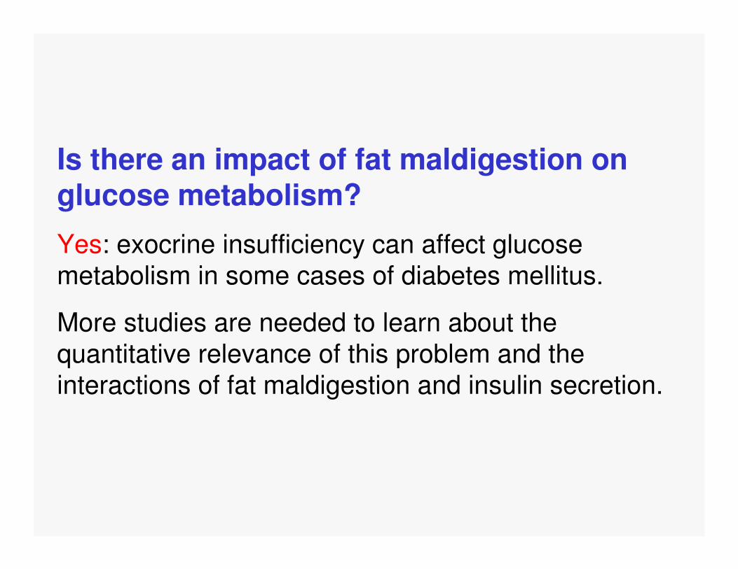
Is there an impact of fat maldigestion on glucose metabolism?
Yes: exocrine insufficiency can affect glucose metabolism in some cases of diabetes mellitus.
More studies are needed to learn about the quantitative relevance of this problem and the interactions of fat maldigestion and insulin secretion.

Should exocrine insufficiency andsteatorrhea be treated byenzyme therapy in diabetes mellitus?
- No general recommendation so far
- Treatment, if steatorrhea and symptoms
are present
- More studies on the impact on glucosemetabolism and on qualitative malnutritionneeded

Summary III
What are clinical consequences of exocrinepancreatic insufficiency in diabetes mellitus?
- Steatorrhea, GI-symptoms, impaired controlof glucose metabolism, qualitative malnutrition

1. What is the prevalence of exocrine pancreatic
insufficiency in diabetes mellitus?It is as high as 35% in patients previously classified as „type 2“ and
about 50% in patients previously classified as „type 1“.
2. Is diabetes mellitus secondary to pancreaticdiseases (type-3) a frequent phenomenon?It appears to affect at least 8% of all diabeteic patients. A careful
workup of every diabetic patient concerning the exocrine pancreas isrecommended. More studies are needed in this field.
3. What are the clinical consequences of exocrine pancreatic insufficiency in diabetes
mellitus?Steatorrhea is frequent in patients with diabetes mellitus. Fecal
elastase 1 measurement can be used as a screening tool. PEI mightbe associated with symptoms and can affect glucose control. More
studies are of great interest and clinical relevance.

23
Thank you for the Audience
Third Medical Department and Policlinic,Rodthohl 6, D-35392 Giessen, Germany
http://www.uniklinikum-giessen.de/med3/