pretreatment of mice with streptomycin provides a salmonella … · intestinal colonization by...
TRANSCRIPT

INFECTION AND IMMUNITY, May 2003, p. 2839–2858 Vol. 71, No. 50019-9567/03/$08.00�0 DOI: 10.1128/IAI.71.5.2839–2858.2003Copyright © 2003, American Society for Microbiology. All Rights Reserved.
Pretreatment of Mice with Streptomycin Provides a Salmonella entericaSerovar Typhimurium Colitis Model That Allows Analysis
of Both Pathogen and HostManja Barthel,1,2 Siegfried Hapfelmeier,1,2 Leticia Quintanilla-Martínez,3 Marcus Kremer,3,4
Manfred Rohde,5 Michael Hogardt,2 Klaus Pfeffer,6 Holger Russmann,2and Wolf-Dietrich Hardt1*
Institute of Microbiology, ETH Zurich, 8092 Zurich, Switzerland,1 and Max von Pettenkofer-Institut, Ludwig MaximiliansUniversitat, 80336 Munich,2 Institute of Medical Microbiology, Immunology, and Hygiene6 and Institute of Pathology,4
Technical University of Munich, 81675 Munich, GSF-Research Center for Environment and Health,85764 Neuherberg,3 and GBF, 38124 Braunschweig,5 Germany
Received 25 November 2002/Returned for modification 16 January 2003/Accepted 6 February 2003
Salmonella enterica subspecies 1 serovar Typhimurium is a principal cause of human enterocolitis. For un-known reasons, in mice serovar Typhimurium does not provoke intestinal inflammation but rather targets thegut-associated lymphatic tissues and causes a systemic typhoid-like infection. The lack of a suitable murinemodel has limited the analysis of the pathogenetic mechanisms of intestinal salmonellosis. We describe herehow streptomycin-pretreated mice provide a mouse model for serovar Typhimurium colitis. Serovar Typhi-murium colitis in streptomycin-pretreated mice resembles many aspects of the human infection, includingepithelial ulceration, edema, induction of intercellular adhesion molecule 1, and massive infiltration of PMN/CD18� cells. This pathology is strongly dependent on protein translocation via the serovar Typhimurium SPI1type III secretion system. Using a lymphotoxin �-receptor knockout mouse strain that lacks all lymph nodesand organized gut-associated lymphatic tissues, we demonstrate that Peyer’s patches and mesenteric lymphnodes are dispensable for the initiation of murine serovar Typhimurium colitis. Our results demonstrate thatstreptomycin-pretreated mice offer a unique infection model that allows for the first time to use mutants of boththe pathogen and the host to study the molecular mechanisms of enteric salmonellosis.
Salmonella spp. are gram-negative enterobacteria that causediseases ranging from a self-limiting enterocolitis to systemicinfection (typhoid fever). Salmonella enterica serovar Typhi-murium evokes a common form of nonsystemic enterocolitis inhumans and cattle, whereas mice are intrinsically resistant toserovar Typhimurium enterocolitis (68, 81). Although resistantto intestinal salmonellosis, certain susceptible mouse strainsthat carry mutations in the NRAMP gene develop a diseasesimilar to typhoid fever (30, 75).
After oral infection of susceptible mice, serovar Typhimu-rium does not replicate efficiently in the intestine but pene-trates the epithelial barrier by invasion of M cells (12, 41, 64)or (less efficiently) by transport via CD18�/dendritic cells (67,80) and possibly by penetration of enterocytes (72). After pen-etration of the epithelial barrier, Salmonella spp. colonize Pey-er’s patches and mesenteric lymph nodes and then spread tothe liver and spleen, and the mice finally succumb to systemicinfection (10, 35, 75, 81). However, mice show few signs of theintestinal inflammation observed in cattle or humans.
Due to the lack of a versatile animal model, much less isknown about the mechanisms of the enteric salmonellosis (21,33, 62, 75, 81). To overcome these limitations, the pathogenesisof enteric salmonellosis has been studied by extrapolating datafrom tissue culture (review by Galan [26]) or from intestinalorgan culture (1) or by infection of ligated murine and rabbit
ileal loops (11, 12, 20, 41, 63, 64). However, it remains unclearhow these results relate to enteric salmonellosis.
For this reason, bovine infection models with serovar Typhi-murium (and serovar Dublin) have been established recentlyto study Salmonella-associated enterocolitis (reviewed previ-ously [68, 75, 81]). Bovine models have allowed the identifica-tion of several Salmonella-associated factors required to evokeenterocolitis, including flagella (71), aroAD (42, 73), lipopoly-saccharide (LPS) (73), and the SPI1 type III secretion system(75, 81, 86). This type III secretion system allows Salmonellaspp. to inject (translocate) bacterial toxins (effector proteins)directly into the cytosol of host cells (13, 83) to manipulate hostresponses (23, 26) in order to invade the bovine intestinalepithelium and the Peyer’s patches and induce inflammatoryresponses (28, 73, 74, 82). However, due to severe technicallimitations, the bovine model has allowed only limited analysisof the contribution of the host in the complex interplay withserovar Typhimurium leading to enterocolitis. For example, itis still a matter of dispute whether intestinal inflammation isinitiated by direct interaction with epithelial cells (reviewed inreference 26) or by colonization of the gut-associated lym-phatic tissues (GALT; see reviews in references 79 and 81).
The gut-associated immune system is composed of diffuselydistributed (dendritic, B, cytotoxic T, and NK) cells and orga-nized lymphoid tissues (i.e., Peyer’s patches and mesentericlymph nodes) and coordinates appropriate responses to anti-gens derived from food, commensal bacteria, and pathogenicmicroorganisms (32, 58). Organogenesis of the gut-associatedlymphoid tissues is determined by developmental programs,
* Corresponding author. Mailing address: Institute of Microbiology,ETH Zurich, Schmelzbergstr. 7, 8092 Zurich, Switzerland. Phone:41-1-632-5143. Fax: 41-1-632-1129. E-mail: [email protected].
2839
on July 16, 2019 by guesthttp://iai.asm
.org/D
ownloaded from
cytokines, and the exposure to antigens (46). Gene targetinghas shown that the interleukin-7 receptor, the tumor necrosisfactor (TNF), lymphotoxin (LT �3, LT �2�1, and LT �1�2),and the TNF receptor (TNFR) superfamily are of fundamentalimportance in this process (24). However, only lymphotoxin �receptor (LT�R; a member of the TNFR superfamily) knock-out mice (LT�R�/�) are completely devoid of Peyer’s patches,colon-associated lymphoid tissues, the cecum-associated lym-phatic patch, and all lymph nodes (25). Therefore, LT�R�/�
mice allow one to analyze the functional requirement of orga-nized GALT in bacterial infection.
We have found that streptomycin-pretreated mice developcolitis upon infection with serovar Typhimurium. This inflam-mation is evoked by specific bacterial virulence factors. Fur-thermore, our data show that the organized GALT are dis-pensable for murine serovar Typhimurium colitis. Due to theavailability of a broad variety of knockout mouse strains, thestreptomycin-pretreated mouse model will be of great advan-tage compared to existing animal models: it allows study of therole of the host defense mechanisms in intestinal salmonellosisin great detail.
MATERIALS AND METHODS
Bacterial strains. The naturally streptomycin-resistant wild-type strain S. en-terica serovar Typhimurium SL1344 (36) and the isogenic mutants SB161(SL1344, �invG [43]), SB302 (SL1344, invJ::aphT [15]), and SB241 (SL1344,sipD::aphT [44]) were generously provided by J. E. Galan. The nonpathogenicLactobacillus strain was a gift from A. Macpherson.
Serovar Typhimurium strains were grown for 12 h at 37°C in Luria-Bertanibroth supplemented with 0.3 M NaCl, diluted 1:20 in fresh medium, and sub-cultured for 4 h under mild aeration. Bacteria were washed twice in ice-coldphosphate-buffered saline (PBS) and then suspended in cold PBS (2 � 103 or 108
CFU/50 �l).Lactobacillus spp. were grown on MRS agar (Biolife, Milano, Italy) at 37°C
under an anaerobic CO2-enriched atmosphere (Anaerocult A; Merck, Darm-stadt, Germany). Bacteria were scraped from the plates, washed with cold PBS,and resuspended in PBS to yield a final concentration of 108 CFU/50 �l.
Animal experiments. Specific-pathogen-free (SPF) female mice were fromHarlan Winkelmann (C57BL/6; 6 to 8 weeks old; Borchen, Germany) or CharlesRiver (LT�R�/� mice, C57BL/6 background; 6 to 8 weeks old; Sulzfeld, Ger-many [25]). Genotypes were verified by PCR typing with the primers 5�-CGGGTC TCC GAC CTA GAG ATC-3� and 5�-GAG GTG GGT GGA TTG GAAAGA G-3�.
For the experiments, animals were housed individually or in groups of up tofive animals under standard barrier conditions in individually ventilated cages(Tecniplast, Buguggiate, Italy) equipped with steel grid floors and autoclavedfilter paper at the Max von Pettenkofer Institute (Munich, Germany) or BZL(Zurich, Switzerland).
Water and food were withdrawn 4 h before per os (p.o.) treatment with 20 mgof streptomycin (75 �l of sterile solution or 75 �l of sterile water [control]).Afterward, animals were supplied with water and food ad libitum. At 20 h afterstreptomycin treatment, water and food were withdrawn again for 4 h before themice were infected with 108 CFU of serovar Typhimurium (50-�l suspension inPBS p.o.) or treated with sterile PBS (control). Thereafter, drinking water adlibitum was offered immediately and food 2 h postinfection (p.i.). At the indi-cated times p.i., mice were sacrificed by CO2 asphyxiation, and tissue samplesfrom the intestinal tracts, mesenteric lymph nodes, spleens, and livers wereremoved for analysis.
Animal experiments were approved by the German and Swiss authorities andperformed according to the legal requirements.
Analysis of serovar Typhimurium loads in intestines, mesenteric lymph nodes,spleens, and livers. Two fresh fecal pellets were placed in 500 �l of 4°C cold PBSand suspended homogeneously on ice by vortexing and pipetting. Intestinalcontents from the ileum, cecum, or colon were collected at the indicated timesp.i. and weighed before resuspending them in 500 �l of 4°C cold PBS. Thenumbers of CFU were determined by plating appropriate dilutions on Mac-Conkey agar plates (streptomycin at 50 �g/ml). The minimum detectable value
was 5 CFU/fecal pellet and 10 CFU/sample (between 25 and 150 mg) of intes-tinal content.
Intestinal colonization by Lactobacillus spp. was determined by resuspending50 mg of cecal content in 500 �l of ice-cold PBS, plating appropriate dilutions onMRS agar (Biolife, Milano, Italy) without delay, and incubation for 48 h at 37°Cunder an anaerobic CO2-enriched atmosphere (Anaerocult A).
To analyze the colonization, mesenteric lymph nodes, spleens, and livers wereremoved aseptically and homogenized in 4°C cold PBS (0.5% Tergitol, 0.5%bovine serum albumin) by using a Potter homogenizer. The numbers of CFUwere determined by plating appropriate dilutions on MacConkey agar plates(streptomycin at 50 �g/ml). The minimal detectable values were 20 CFU/organin the spleen, 100 CFU/organ in the liver, and 10 CFU/organ in the mesentericlymph nodes.
Histological procedures. Segments of the ileum, cecum, and colon were fixedand embedded in paraffin according to standard procedures. Alternatively, tissuesamples were embedded in O.C.T. (Sakura, Torrance, Calif.), snap-frozen inliquid nitrogen, and stored at �80°C. Cryosections (5 or 30 �m) were mountedon glass slides, air dried for 2 h at room temperature, and stained with hema-toxylin and eosin (H&E). Pathological evaluation was performed by two pathol-ogists in a blinded manner.
Based on an earlier study (49), we developed a scoring scheme for quantitativepathological analysis of cecal inflammation. H&E-stained sections (5 �m) werescored independently by two pathologists in a blinded manner as follows.
(i) Submucosal edema. Submucosal edema was scored as follows: 0 nopathological changes; 1 mild edema (the submucosa is 0.20 mm wide andaccounts for 50% of the diameter of the entire intestinal wall [tunica muscu-laris to epithelium]); 2 moderate edema; the submucosa is 0.21 to 0.45 mmwide and accounts for 50 to 80% of the diameter of the entire intestinal wall; and3 profound edema (the submucosa is �0.46 mm wide and accounts for �80%of the diameter of the entire intestinal wall). The submucosa widths were deter-mined by quantitative microscopy and represent the averages of 30 evenly spacedradial measurements of the distance between the tunica muscularis and thelamina mucosalis mucosae.
(ii) PMN infiltration into the lamina propria. Polymorphonuclear granulo-cytes (PMN) in the lamina propria were enumerated in 10 high-power fields(�400 magnification; field diameter of 420 �m), and the average number ofPMN/high-power field was calculated. The scores were defined as follows: 0 5 PMN/high-power field; 1 5 to 20 PMN/high-power field; 2 21 to60/high-power field; 3 61 to 100/high-power field; and 4 �100/high-powerfield. Transmigration of PMN into the intestinal lumen was consistently observedwhen the number of PMN was �60 PMN/high-power field.
(iii) Goblet cells. The average number of goblet cells per high-power field(magnification, �400) was determined from 10 different regions of the cecalepithelium. Scoring was as follows: 0 �28 goblet cells/high-power field (mag-nification, �400; in the cecum of the normal SPF mice we observed an averageof 6.4 crypts/high-power field and the average crypt consisted of 35 to 42 epi-thelial cells, 25 to 35% of which were differentiated into goblet cells); 1 11 to28 goblet cells/high-power field; 2 1 to 10 goblet cells/high-power field; and 3 1 goblet cell/high-power field.
(iv) Epithelial integrity. Epithelial integrity was scored as follows: 0 nopathological changes detectable in 10 high-power fields (�400 magnification);1 epithelial desquamation; 2 erosion of the epithelial surface (gaps of 1 to10 epithelial cells/lesion); and 3 epithelial ulceration (gaps of �10 epithelialcells/lesion; at this stage, there is generally granulation tissue below the epithe-lium).
We averaged the two independent scores for submucosal edema, PMN infil-tration, goblet cells, and epithelial integrity for each tissue sample. The combinedpathological score for each tissue sample was determined as the sum of theseaveraged scores. It ranges between 0 and 13 arbitrary units and covers thefollowing levels of inflammation: 0 intestine intact without any signs of in-flammation; 1 to 2 minimal signs of inflammation (this was frequently foundin the ceca of SPF mice; this level of inflammation is generally not considered asa sign of disease); 3 to 4 slight inflammation; 5 to 8 moderate inflammation;and 9 to 13 profound inflammation.
Statistical analysis. Statistical analysis of the cecum weight, the individualpathological scores for submucosal edema, PMN infiltration, goblet cells, andepithelial integrity and for the combined pathological score was performed byusing the exact Mann-Whitney U test and SPSS version 11.0 software. P valuesof 0.05 were considered statistically significant. This procedure was adoptedfrom that of Madsen et al. (49).
To allow the statistical analysis of the bacterial loads, values for animalsyielding “no CFU” were set to the minimal detectable value (fecal pellet 5CFU; spleen 20 CFU; liver 100 CFU; mesenteric lymph nodes 10 CFU;
2840 BARTHEL ET AL. INFECT. IMMUN.
on July 16, 2019 by guesthttp://iai.asm
.org/D
ownloaded from

intestinal content between 67 and 400 CFU [see above]). Afterward, themedian values were calculated by using Microsoft Excel XP, and statisticalanalysis was performed by using the exact Mann-Whitney U test and the SPSSversion 11.0 software. P values of 0.05 were considered statistically significant.
Immunohistological procedures. Cryosections (5 �m) of OCT-embedded tis-sue samples from the ileum, cecum, and colon of each animal were mounted onglass slides (Sigma), air dried at room temperature for 2 h, fixed (PBS, 3.7%formaldehyde; 1 h at room temperature), washed in PBS, permeabilized withTriton X-100 (0.1% in PBS, 10 min at room temperature), washed in PBS, andblocked with goat serum (20% in PBS, overnight at 4°C). Monoclonal rat �CD18(1:100), hamster �-intercellular adhesion molecule-1 (ICAM-1; 1:100), and poly-clonal rabbit �-Salmonella O antigen group B (factors 1, 4, 5, and 12) antiserum(1:300) were from Becton Dickinson (San Diego, Calif.). Fluorescein isothiocya-nate (FITC)-conjugated goat �-rabbit (1:200), FITC-conjugated goat �-rat (1:100), and Cy3-conjugated goat �-hamster (1:200) polyclonal antibodies werefrom Dianova (Hamburg, Germany). Controls with appropriate species- andisotype-matched monoclonal antibodies (Becton Dickinson) were performed toensure specific detection of the antigens. DAPI (4�,6�-diamidino-2-phenylindole;0.5 �g/ml; Sigma) was used for nucleic acid staining. Staining was performed withPBS (20% goat serum). Sections were washed with PBS and mounted withVectashield (Vector Laboratories, Inc., Burlingame, Calif.). Images (see Fig.11E and F and 14) were obtained with a Leica DM epifluorescence microscopeand a Visitron spot charge-coupled device camera system. Other images (see Fig.9I to P and 11G and H) were recorded by using a Perkin-Elmer Ultraviewconfocal imaging system and a Zeiss Axiovert 200 microscope: red and greenfluorescence was recorded confocally, and the blue fluorescence was determinedby epifluorescence microscopy. Afterwards, the red, green, and blue images weresuperimposed by using Adobe Photoshop software, ensuring that all panels fromeach figure were processed in the same way.
RESULTS
Serovar Typhimurium colonizes the lower intestine of strep-tomycin-pretreated mice and causes colitis. Murine salmonel-losis is generally not associated with efficient colonization ofthe intestine and overt pathological changes of the intestinalmucosa. However, it has long been known that oral treatmentwith streptomycin reduces the oral 50% infectious dose ofserovar Enteritidis or serovar Typhimurium by 105- to 106-foldand greatly improves intestinal colonization (2, 3, 9, 53, 54, 57,66). This effect has been attributed to the elimination of com-mensal intestinal bacteria (4, 5, 55, 65). However, to our
knowledge it has never been determined whether streptomy-cin-pretreated mice colonized with serovar Typhimurium de-velop enterocolitis.
In a first experiment, five streptomycin-pretreated (20 mgp.o.) and five water-pretreated C57BL/6 mice were placed inseparate cages. At 24 h after the pretreatment the mice wereinfected p.o. with a dose of 2 � 103 CFU of the virulent serovarTyphimurium strain SL1344. In agreement with earlier reports(2, 3, 9, 53, 54, 57, 66), we found that the streptomycin-pre-treated mice were excreting significantly larger quantities (107
to 1010 CFU/fecal pellet) of serovar Typhimurium than thewater-pretreated control mice at days 1 and 2 p.i. (Fig. 1A; P 0.032). However, one of the five streptomycin-pretreatedmice was not excreting detectable quantities (5 CFU/fecalpellet) of serovar Typhimurium (Fig. 1A). Therefore, we re-peated the experiment with six animals per group with a higherinoculum (108 CFU of serovar Typhimurium p.o.; Fig. 1B).Again, we found that the streptomycin-pretreated mice wereexcreting significantly higher amounts (107 to 1010 CFU/fecalpellet; P 0.002) of serovar Typhimurium than the water-pretreated control mice at days 1 and 2 p.i. (Fig. 1B). In thisexperiment we detected large quantities of serovar Typhi-murium in the feces of all six streptomycin-pretreated animals.Therefore, we have used an inoculum of 108 CFU p.o. through-out the rest of the present study.
To analyze serovar Typhimurium-induced intestinal pathol-ogy, two of the streptomycin-pretreated mice (Fig. 1B) and onemouse from the control group (Fig. 1B) were sacrificed 2 daysp.i., and the internal organs were fixed and embedded in par-affin (see Materials and Methods). Histopathological analysisof 5-�m thin sections revealed pronounced inflammation ofthe cecum (Fig. 2A and C) of the streptomycin-pretreated micecolonized with serovar Typhimurium. In the cecum of bothmice, we observed pronounced edema in the submucosa,edematous changes in the lamina propria, crypt elongation,disruption of the crypt architecture, reduced numbers of goblet
FIG. 1. Streptomycin pretreatment allows efficient colonization of the murine intestine by serovar Typhimurium. (A) Infection with aninoculum of 2 � 103 serovar Typhimurium. Five mice (C57BL/6) were treated with 20 mg of streptomycin (F) or water (E) p.o. After 24 h, theywere infected with 2 � 103 CFU of serovar Typhimurium SL1344. (B) Infection with an inoculum of 108 CFU of serovar Typhimurium. Six mice(C57BL/6) were treated with 20 mg of streptomycin (F) or water (E) p.o. After 24 h, they were infected with 108 CFU of serovar TyphimuriumSL1344. We monitored the excretion of serovar Typhimurium (CFU per fecal pellet) at days 1 and 2 p.i. (see Materials and Methods). The dashedline indicates the limit of detection; bars indicate the median; and “P” refers to the P value (Mann-Whitney U test; see Materials and Methods).Arrows indicate mice selected for histopathological analysis (see Fig. 2).
VOL. 71, 2003 MURINE SEROVAR TYPHIMURIUM COLITIS MODEL 2841
on July 16, 2019 by guesthttp://iai.asm
.org/D
ownloaded from
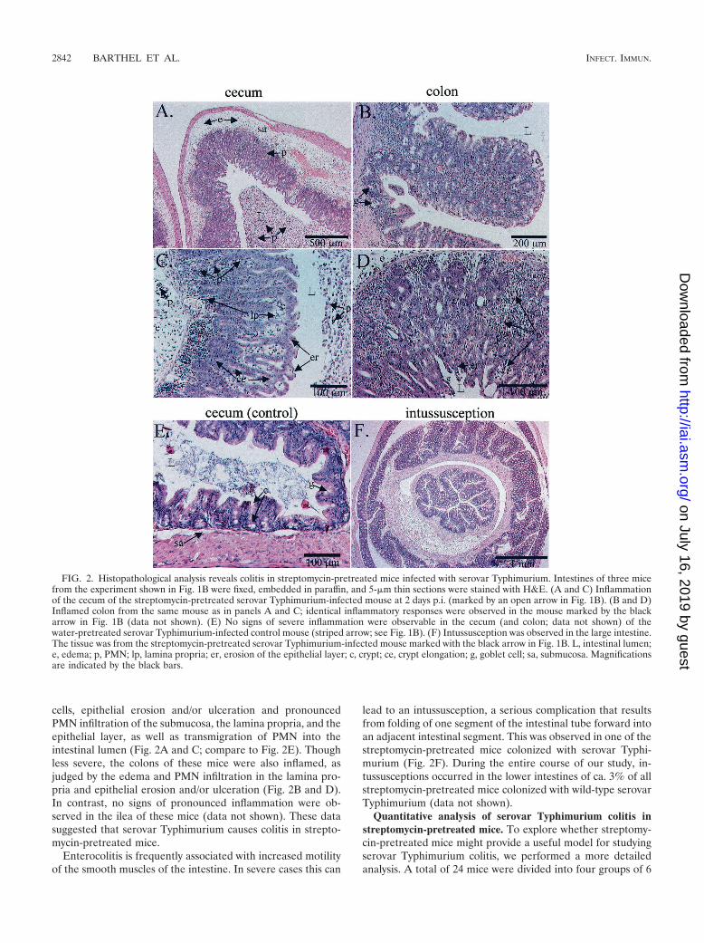
cells, epithelial erosion and/or ulceration and pronouncedPMN infiltration of the submucosa, the lamina propria, and theepithelial layer, as well as transmigration of PMN into theintestinal lumen (Fig. 2A and C; compare to Fig. 2E). Thoughless severe, the colons of these mice were also inflamed, asjudged by the edema and PMN infiltration in the lamina pro-pria and epithelial erosion and/or ulceration (Fig. 2B and D).In contrast, no signs of pronounced inflammation were ob-served in the ilea of these mice (data not shown). These datasuggested that serovar Typhimurium causes colitis in strepto-mycin-pretreated mice.
Enterocolitis is frequently associated with increased motilityof the smooth muscles of the intestine. In severe cases this can
lead to an intussusception, a serious complication that resultsfrom folding of one segment of the intestinal tube forward intoan adjacent intestinal segment. This was observed in one of thestreptomycin-pretreated mice colonized with serovar Typhi-murium (Fig. 2F). During the entire course of our study, in-tussusceptions occurred in the lower intestines of ca. 3% of allstreptomycin-pretreated mice colonized with wild-type serovarTyphimurium (data not shown).
Quantitative analysis of serovar Typhimurium colitis instreptomycin-pretreated mice. To explore whether streptomy-cin-pretreated mice might provide a useful model for studyingserovar Typhimurium colitis, we performed a more detailedanalysis. A total of 24 mice were divided into four groups of 6
FIG. 2. Histopathological analysis reveals colitis in streptomycin-pretreated mice infected with serovar Typhimurium. Intestines of three micefrom the experiment shown in Fig. 1B were fixed, embedded in paraffin, and 5-�m thin sections were stained with H&E. (A and C) Inflammationof the cecum of the streptomycin-pretreated serovar Typhimurium-infected mouse at 2 days p.i. (marked by an open arrow in Fig. 1B). (B and D)Inflamed colon from the same mouse as in panels A and C; identical inflammatory responses were observed in the mouse marked by the blackarrow in Fig. 1B (data not shown). (E) No signs of severe inflammation were observable in the cecum (and colon; data not shown) of thewater-pretreated serovar Typhimurium-infected control mouse (striped arrow; see Fig. 1B). (F) Intussusception was observed in the large intestine.The tissue was from the streptomycin-pretreated serovar Typhimurium-infected mouse marked with the black arrow in Fig. 1B. L, intestinal lumen;e, edema; p, PMN; lp, lamina propria; er, erosion of the epithelial layer; c, crypt; ce, crypt elongation; g, goblet cell; sa, submucosa. Magnificationsare indicated by the black bars.
2842 BARTHEL ET AL. INFECT. IMMUN.
on July 16, 2019 by guesthttp://iai.asm
.org/D
ownloaded from

mice. The first group was pretreated with streptomycin 24 hprior to p.o. infection with 108 CFU of serovar Typhimurium(Fig. 3). One control group was pretreated with water 24 hprior to infection p.o. with 108 CFU of serovar Typhimurium.The two other control groups were pretreated either withstreptomycin or with water 24 h prior to mock infection by oraltreatment with sterile PBS. The mice were killed 2 days p.i.,and we analyzed the bacterial colonization (Fig. 3) and patho-logical changes in the ceca (Fig. 4, 5, and 6).
As expected, serovar Typhimurium was only detectable inthe intestine and organs of the mice that had been infected(Fig. 3). We detected no serovar Typhimurium in the contentsof the medial and terminal ilea of most of the water-pretreatedmice infected with serovar Typhimurium. Low bacterial loads(�105 CFU/g) were detected in the medial ileum of one, in the
terminal ilea of two, and in the ceca of five of the six water-pretreated mice (Fig. 3A).
In streptomycin-pretreated mice we observed a slight but notsignificant (P 0.24; P 0.065) increase in the bacterial loadsin the medial (P 0.24) and terminal (P 0.065) ilea andsignificantly increased (ca. 105-fold) bacterial loads in the ce-cum contents (P 0.002; Fig. 3).
We observed significantly increased loads of serovar Typhi-murium in the mesenteric lymph nodes of streptomycin-pre-treated mice (P 0.002; Fig. 3B). Slightly increased coloniza-tion was also detected in the spleens (P 0.002) and livers(P 0.009) of streptomycin-pretreated mice. However, even inthe streptomycin-pretreated mice bacterial loads in the spleensand livers were only ca. 10-fold above the detection limit.Overall, these data are in line with earlier observations (57)
FIG. 3. Effect of the streptomycin pretreatment on the colonization of the intestine and internal organs by serovar Typhimurium. Four groupsof six mice (C57BL/6) were pretreated with streptomycin (solid symbols) or sterile water (open symbols) and infected with 108 CFU of serovarTyphimurium SL1344 (circles) or mock infected with sterile PBS (triangles; see Materials and Methods). The mice were sacrificed 2 days p.i., andwe analyzed the bacterial colonization. (A) Bacterial loads in the intestinal contents of the medial ileum (left panel), the terminal ileum (middlepanel), and the cecum (right panel). (B) Bacterial loads in the mesenteric lymph nodes (left panel), the spleen (middle panel), and the liver (rightpanel; see Materials and Methods). The dashed line indicates the limit of detection; the bars indicate the median bacterial load; and “P” indicatesthe P value (Mann-Whitney U test; differences between the water and streptomycin-pretreated mice infected with serovar Typhimurium). NS, notstatistically significant. Symbols: ‚, water-pretreated mice mock infected with PBS; E, water-pretreated mice infected with serovar Typhimurium;Œ, streptomycin-pretreated mice mock infected with PBS; F, streptomycin-pretreated mice infected with serovar Typhimurium.
VOL. 71, 2003 MURINE SEROVAR TYPHIMURIUM COLITIS MODEL 2843
on July 16, 2019 by guesthttp://iai.asm
.org/D
ownloaded from

and suggest that even in streptomycin-pretreated mice the sys-temic infection is still at a very early stage at 48 h p.i. There-fore, the systemic infection in these mice is unlikely to affectthe pathological changes observed in the lower intestine.
During the dissection we did not observe macroscopic signsof inflammation of the ilea, ceca, and colons of the water-pretreated mice that were infected with serovar Typhimuriumor mock infected with sterile PBS. In accordance with earlierresults (69), streptomycin-pretreated mice that were mock in-fected with sterile PBS had enlarged ceca. In contrast to thethree control groups, fecal pellet formation was impaired inthe proximal colons of all six streptomycin-pretreated miceinfected with serovar Typhimurium, and the tissues of theproximal colons appeared pale and swollen in five of the sixanimals (data not shown). Furthermore, the ceca of all sixstreptomycin-pretreated serovar Typhimurium-infected micewere shriveled to a small size, pale, and filled with purulentexudate (Fig. 4A, right side). To document these macroscopicdifferences, we removed the cecum from one mouse of each
group for photographic documentation (Fig. 4A) and deter-mined the weight of the cecum, including the contents of theremaining five animals from each group (Fig. 4B). In water-pretreated mice, infection with serovar Typhimurium did notsignificantly affect the total weight of the cecum (Fig. 4B; P 0.458). However, in streptomycin-pretreated mice we observeda significant reduction of the cecal weights in mice infectedwith serovar Typhimurium (Fig. 4B, right panel; P 0.002).
For histopathologic analysis, intestinal tissue samples werecryoembedded, and 5-�m thin sections were stained with H&E(see Materials and Methods). The intestines of all three con-trol groups appeared histologically normal (Fig. 5A to C and Eto G and data not shown). However, in line with our initialobservations (Fig. 2), pronounced inflammation was detectedin the ceca and, to a lesser extent, also in the colons of allstreptomycin-pretreated mice colonized with serovar Typhi-murium (Fig. 5D and H; data not shown). This included pro-nounced edema in the submucosa and the lamina propria,crypt elongation, disruption of the crypt architecture, reducednumbers of goblet cells, epithelial erosion, and pronouncedPMN infiltration of the submucosa, the lamina propria, and theepithelial layer, as well as transmigration of PMN into theintestinal lumen. No signs of severe inflammation were ob-served in the ilea of these mice (data not shown).
To improve comparison of the inflammatory responses be-tween the different groups of mice, we devised a histopatho-logic scoring scheme for H&E-stained cecal tissue sections(see Materials and Methods). This scheme considers the extentof the submucosal edema (0 to 3 [arbitrary units]), PMN infil-tration (0 to 4), loss of goblet cells (0 to 3), and the epithelialintegrity (0 to 3) and yields a total pathological score of 0 to 13U (Materials and Methods). Using this scoring scheme, wehave found no significant differences between the water-pre-treated mice that had been infected with serovar Typhimuriumor mock infected with sterile PBS (Fig. 6, left panel). Theminimal signs of cecal inflammation observed in these miceand in the streptomycin-pretreated mice that had been mockinfected with sterile PBS are frequently found in untreatedSPF mice and are generally not considered as a sign of disease.In contrast, the streptomycin-pretreated mice infected withserovar Typhimurium displayed profound inflammation thatwas significantly stronger than in the animals of the controlgroup (Fig. 6, right panel; P 0.008). These data are in linewith the macroscopic observations (Fig. 4) and demonstratethat streptomycin-pretreated mice offer a robust model forstudying serovar Typhimurium colitis.
Lactobacillus spp. do not induce colitis in streptomycin-pre-treated mice. Streptomycin treatment is known to diminish theintestinal flora and to render mice susceptible to intestinalcolonization by various microorganisms. It was conceivablethat any dramatic change in the composition of the intestinalmicroflora might lead to inflammation and that the colitisinduced by serovar Typhimurium might not be attributable tospecific virulence factors. To test this hypothesis, we pretreatedtwo groups of mice with 20 mg of streptomycin given p.o. 24 hprior to oral infection with 108 CFU of nonpathogenic Lacto-bacillus spp. or serovar Typhimurium SL1344. The mice weresacrificed 2 days p.i., and we verified efficient colonization byboth bacterial species by plating samples of the cecum contentson suitable agar plates (see Materials and Methods; ��104
FIG. 4. Serovar Typhimurium induces macroscopic changes in thececum of streptomycin-pretreated mice at 2 days p.i. We removed thececa of mice from the experiment shown in Fig. 3 for macroscopicanalysis. (A) Cecum morphology. The cecum of one mouse from eachgroup was taken for photographic documentation. The photograph isrepresentative of all six animals from each group. The centimeter scaleindicates the magnification. (B) Total weight of the cecum. We deter-mined the weight of the ceca of the remaining five mice from eachgroup. P, P value (Mann-Whitney U test; differences between animalswhich were infected with serovar Typhimurium or mock infected withsterile PBS); NS, not statistically significant. “S. Tm � or �” and “sm� or �” indicate whether the mice were pretreated with streptomycin(sm) or water and whether the animals were infected with serovarTyphimurium (S. Tm). Bars indicate the median weight of the cecum.Symbols: ‚, water-pretreated mice mock infected with PBS; E, water-pretreated mice infected with serovar Typhimurium; Œ, streptomycin-pretreated mice mock infected with PBS; F, streptomycin-pretreatedmice infected with serovar Typhimurium.
2844 BARTHEL ET AL. INFECT. IMMUN.
on July 16, 2019 by guesthttp://iai.asm
.org/D
ownloaded from

CFU/g of Lactobacillus spp. or Salmonella spp.). Intestinal tis-sue samples were cryoembedded, and we analyzed 5-�m thinH&E-stained sections for signs of inflammation. In the Lacto-bacillus-infected mice we did not observe significant inflamma-tion in the small or large intestine (Fig. 7A, C, and E; data notshown), whereas serovar Typhimurium-infected mice showedsevere inflammation of the cecum and the proximal colon (Fig.7B, D, and E). This indicates that colitis in streptomycin-pretreated mice is a specific response to colonization by sero-var Typhimurium.
Time course of serovar Typhimurium colitis in streptomy-cin-pretreated mice. To analyze the early stages of serovarTyphimurium colitis, we investigated the time course of theinfection. Groups of five mice were pretreated with streptomy-cin, infected with 108 CFU of serovar Typhimurium SL1344(see Materials and Methods), and sacrificed at 2, 8, 20, and48 h p.i. We analyzed pathological changes, as well as bacterialloads in the liver, mesenteric lymph nodes, and the intestinalcontent.
At 2 h p.i. the highest bacterial loads were present in thesmall intestine, and only low loads (104 CFU/g) were presentin the cecal contents (Fig. 8A and B). In accordance withearlier results (66), bacterial loads in the cecum increasedsignificantly between 2 h and 8 h p.i. (P 0.008). Even higherbacterial loads were detected in the cecum at 20 h p.i. (P 0.008; median 4.4 � 107 CFU/g) (Fig. 8B; Table 1). Between20 h and 48 h p.i. we did not observe any further increase in theserovar Typhimurium loads in the cecum (P 0.841; Table 1),which indicated that colonization of the ceca of streptomycin-pretreated mice by serovar Typhimurium had reached asteady-state level within 20 h after oral infection. We did notobserve a specific enrichment of serovar Typhimurium at theintestinal epithelium at any time between 8 h and 48 h p.i. and
FIG. 5. Cecal inflammation at 2 days p.i. The ceca of five of the six animals from each group of the experiment shown in Fig. 3 werecryoembedded, and 5-�m thin sections were stained with H&E for histopathological analysis. (A and E) Cecum of a water-pretreated mouse mockinfected with PBS; (B and F) cecum of a water-pretreated mouse infected with serovar Typhimurium; (C and G) cecum of a streptomycin-pretreated mouse mock infected with PBS; (D and H) streptomycin-pretreated mouse infected with serovar Typhimurium. The images arerepresentative for all animals from each group. Boxes in panels A, B, C, and D indicate the area shown at the higher magnification. L, intestinallumen; e, edema; p, PMN; er, erosion of the epithelial layer; g, goblet cell; sa, submucosa. Magnifications are indicated by the black bars.
FIG. 6. Scoring of inflammatory changes at 2 days p.i. In order toquantify pathological changes, we scored H&E-stained sections of thececa from five of the six mice from each group of the experiment shownin Fig. 3 as described in Materials and Methods. Edema in the sub-mucosa (black), PMN infiltration (medium gray), reduction of thenumber of goblet cells (dark gray), and erosion or ulceration of theepithelial layer (light gray) were scored separately and plotted asstacked vertical bars. The combined score equals the sum of the sep-arate scores. “S. Tm � or �” and “sm � or �” indicate whether themice were pretreated with streptomycin (sm) or water and whether theanimals were infected with serovar Typhimurium (S. Tm). Statisticalanalyses are shown for the separate scores and for the combined score.P, P value (Mann-Whitney U test; see Materials and Methods; differ-ence between mice infected with serovar Typhimurium and mice mockinfected with sterile PBS); NS, not statistically significant.
VOL. 71, 2003 MURINE SEROVAR TYPHIMURIUM COLITIS MODEL 2845
on July 16, 2019 by guesthttp://iai.asm
.org/D
ownloaded from

observed that only a small minority of the bacteria (5%)were located in crypts (Fig. 9J, K, and L).
At 2 h p.i. macroscopic inspection did not reveal any signs ofinflammation in the small and the large intestine (medianweight of the cecum 0.59 g; Fig. 8D; data not shown). Thiswas confirmed by histopathological analysis (Fig. 8F and 9Aand E). At 8 h p.i. two of the mice appeared to be normal, andhistopathological analysis did not reveal signs of inflammatorydisease (Fig. 8F). Interestingly, these two mice also carried thelowest loads of serovar Typhimurium in their cecal contents(Fig. 8C), suggesting that a slower bacterial colonization mightresult in a delayed onset of the inflammatory response. Thececa of the three other animals sacrificed 8 h p.i. were pale,shriveled to a small size, and filled with purulent exudate (Fig.8E). Histopathological analysis confirmed that the ceca ofthese mice were inflamed, as judged by submucosal edema,PMN infiltration into the lamina propria, epithelial erosion orulceration, and a reduced number of goblet cells (Fig. 8F and9B and F). Substantial infiltration of PMN (CD18�, segmentednuclei) into the lamina propria was confirmed by immunoflu-orescence microscopy (Fig. 9N). In addition, we observed in-
creased expression of ICAM-1 (compare Fig. 9M and N), a keytransmembrane receptor that binds CD18–�2-integrin dimersand is involved in the extravasation of leukocytes from capil-lary blood vessels into tissues.
At 20 h p.i. the ceca of all five mice showed characteristicsigns of inflammation, including a significantly reduced cecumsize (Fig. 8E and Table 1) and histopathologic changes suchas submucosal edema, a reduced number of goblet cells, andepithelial erosion or ulceration (Fig. 8F and 9C and G). Incontrast to the three mice that showed intestinal inflammationalready at 8 h p.i., PMN were present in high numbers not onlyin the lamina propria but also in the submucosa and in theintestinal lumen (Fig. 9G and O), and we observed a concom-itant increase in ICAM-1 expression (compare Fig. 9O and N).
In accordance with our first observations, the ceca of all fivemice sacrificed at 48 h p.i. showed signs of profound inflam-mation (Fig. 8E and F and 9D, H, and P). Compared to thefindings at 20 h p.i., we observed a further progression of thedisruption of the villus architecture (compare Fig. 9C and Gwith 9D and H), increased ICAM-1 expression, and a signifi-cant exacerbation of the PMN infiltration (P 0.032) and the
FIG. 7. Infection of streptomycin-pretreated mice with Lactobacillus spp. does not lead to colitis. Groups of three streptomycin-pretreated mice(C57BL/6) were infected with 108 CFU of Lactobacillus spp. or 108 CFU of serovar Typhimurium. At 48 h p.i. the animals were sacrificed and 5-�mcryosections of the cecum were stained with H&E. (A and C) Cecum of streptomycin-pretreated mouse infected with Lactobacillus spp.; (B andD) cecum of streptomycin-pretreated mouse infected with serovar Typhimurium. The images are representative for all three animals from eachgroup. The boxes in panels A and B indicate the areas shown at the higher magnification in panels C and D. L, intestinal lumen; e, edema; p, PMN;er, erosion of the epithelial layer; g, goblet cell; sa, submucosa. Magnifications are indicated by black bars. (E) Scoring of inflammatory changesas described in Materials and Methods. Edema in the submucosa (black), PMN infiltration (medium gray), reduction of the number of goblet cells(dark gray), and erosion or ulceration of the epithelial layer (light gray) were scored separately and are plotted as stacked vertical bars. The combinedscore equals the sum of the separate scores. S. Tm, mice infected with serovar Typhimurium; sm, mice were pretreated with streptomycin.
2846 BARTHEL ET AL. INFECT. IMMUN.
on July 16, 2019 by guesthttp://iai.asm
.org/D
ownloaded from

loss of goblet cells (P 0.008) (Fig. 8F; Table 1; see also Fig.9L and P).
To assess a possible correlation between intestinal inflam-mation and the onset of systemic infection, we monitored thetime course of colonization of mesenteric lymph nodes andliver. Serovar Typhimurium could not be detected in the mes-enteric lymph nodes and liver of any mice up to 8 h p.i. (Fig. 8Cand D). However, at 8 h p.i. three of the five mice already hadan inflamed cecum (see above). This suggests that colonizationof the mesenteric lymph nodes or spread to internal organs isnot a prerequisite for intestinal inflammation. At 20 h p.i. wedetected a significant increase in the median serovar Typhi-murium load in the mesenteric lymph nodes (P 0.032) and afurther increase at 48 h p.i. (P 0.008; Fig. 8C; Table 1). In thelivers we detected serovar Typhimurium in only one of fiveanimals at 20 h p.i. (P 0.690; Fig. 8D; Table 1). However,bacterial loads in the liver were significantly increased at 48 hp.i. (Fig. 8D and Table 1). In conclusion, colonization of the
FIG. 8. Time course of serovar Typhimurium colitis in streptomycin-pretreated mice. Groups of five streptomycin-pretreated mice wereinfected for 2, 8, 20, or 48 h with 108 CFU of serovar Typhimurium SL1344 p.o. We determined bacterial colonization and pathological changesas described in Materials and Methods. (A) Bacterial loads in different regions of the intestine at 2 h p.i. (left panel) and 8 h p.i. (right panel).(B) Time course of the bacterial loads in the cecum content. (C) Time course of bacterial loads in the mesenteric lymph nodes. (D) Time courseof bacterial loads in the liver. (E) Time course of the total weight of the cecum (determined before the removal of samples for bacteriological orhistopathologic analysis). (F) Time course of the histopathologic changes. Samples of the cecal tissue were cryoembedded and 5-�m thin sectionswere stained with H&E. Edema in the submucosa (black), PMN infiltration (medium gray), reduction of the number of goblet cells (dark gray),and erosion or ulceration of the epithelial layer (light gray) were scored separately (see Materials and Methods) and are plotted as stacked verticalbars. The combined score equals the sum of the separate scores. For the statistical analysis, see Table 1. S. Tm, serovar Typhimurium; dashed line,limit of detection; bars, median bacterial load; gray arrows, values determined for the two mice without cecal inflammation at 8 h p.i. The blackarrows indicate the pathological scores of the tissue sections shown in Fig. 9.
TABLE 1. Statistical analysis of disease parameters in thetime course of serovar Typhimurium colitis in
streptomycin-pretreated micea
ComparisonP at time (h) p.i.
2 vs 8 2 vs 20 2 vs 48 8 vs 20 20 vs 48
Cecum CFU 0.008 0.008 0.008 0.008 NSmLN CFU NS 0.032 0.008 0.032 0.008Liver CFU NS NS 0.008 NS 0.032Cecum wt NS 0.008 0.008 0.032 NSCombined score NS 0.008 0.008 NS NSEdema 0.032 0.008 0.008 NS NSPMN infitration NS 0.008 0.008 NS 0.032Goblet cells NS 0.008 0.008 NS 0.008Epithelium NS 0.008 0.008 NS NS
a The data shown in Fig. 8 were analyzed by using the Mann-Whitney U test(see Materials and Methods). NS, not significant; mLN, mesenteric lymph nodes.
VOL. 71, 2003 MURINE SEROVAR TYPHIMURIUM COLITIS MODEL 2847
on July 16, 2019 by guesthttp://iai.asm
.org/D
ownloaded from

mesenteric lymph nodes and liver does not seem to be requiredfor the initiation of cecal inflammation. However, we cannotrule out that colonization of the mesenteric lymph nodes or theearly systemic infection might play a role in the exacerbation ofthe inflammatory response observed between 8 h and 48 h p.i.
Overall, the observed pathological changes correlated wellwith the kinetics of intestinal colonization of streptomycin-pretreated mice by serovar Typhimurium. In some animals weobserved cecal inflammation as early as 8 h p.i. These animalscarried bacterial loads of ca. 107 CFU/g in the cecal contents.At 20 h p.i. the cecum was fully colonized (median 4 � 108
CFU/g; Fig. 8B), and the maximum levels of cecal inflamma-tion were reached by between 20 and 48 h p.i. These observedsymptoms are indicative of inflammation and a rapid regener-ation of the cecal epithelium in response to colonization byserovar Typhimurium and resemble many aspects of human(nontyphoid) salmonellosis and bovine and rabbit models of
Salmonella enterocolitis. Therefore, streptomycin-pretreatedmice might offer a versatile alternative model for studying thepathogenesis of gastrointestinal serovar Typhimurium infec-tions.
Role of SPI1 type III secretion in murine serovar Typhi-murium colitis. The SPI1 type III secretion system plays a keyrole in the early, gut-associated stages of the infection in sus-ceptible animals (75, 81). In calves, the SPI1 type III secretionsystem plays a key role in the induction of inflammatory diar-rhea. However, it has remained unclear whether the SPI1 typeIII secretion system can play a similar role in mice. To explorethis question, we used SB161 (SL1344, �invG), an isogenicmutant that lacks an essential subunit of the SPI1 type IIIapparatus and is incapable of secreting and translocating anyproteins via this route (14, 43). Eight streptomycin-pretreatedC57BL/6 mice were infected with wild-type serovar Typhi-murium SL1344, and eight mice were infected with SB161 (108
FIG. 9. Analysis of the time course of murine serovar Typhimurium colitis by histology and immunofluorescence microscopy. Cecal tissues ofthe mice from the experiment described in Fig. 8 (sacrificed at 2, 8, 20, or 48 h p.i.) were cryoembedded, sectioned, and stained as described inMaterials and Methods. (A to H) Histopathology of thin sections (5 �m) of the cecum of mice infected for 2 h (A and E), 8 h (B and F), 20 h(C and G), or 48 h (D and H; marked by black arrows in Fig. 8F). (I to L) Localization of the bacteria. Thin sections (30 �m) of the cecum at2 h (I), 8 h (J), 20 h (K), or 48 h (L) p.i. (marked by black arrows in Fig. 8F) were stained with DAPI, TRITC (tetramethyl rhodamine iso-thiocyanate)-phalloidin, a rabbit �-Salmonella LPS antiserum, and a secondary �-rabbit-FITC conjugate (DNA blue; actin red; serovarTyphimurium green fluorescence). (M to P) Expression of ICAM-1 and infiltration of CD18� cells. Thin sections (5 �m) of the cecum at 2 h(M), 8 h (N), 20 h (O), or 48 h (P) p.i. (marked by black arrows in Fig. 8F) were stained with DAPI, rat �-mouse CD18, hamster �-mouse ICAM-1,and polyclonal preadsorbed �-rat IgG-FITC and �-hamster IgG-Cy3 antibodies (DNA blue; ICAM-1 red, CD18 green fluorescence; seeMaterials and Methods). The boxes in panels A, B, C, and D indicate areas shown at a higher magnification in panels E, F, G, and H. The insetin panel K is a higher magnification of the area marked by the white box in the upper right corner of the panel. L, intestinal lumen; e, edema; p,PMN; er, erosion of the epithelial layer; g, goblet cell; sa, submucosa; c, crypt. Magnifications are indicated by bars.
2848 BARTHEL ET AL. INFECT. IMMUN.
on July 16, 2019 by guesthttp://iai.asm
.org/D
ownloaded from

CFU p.o.). At 48 h p.i., the mice were sacrificed and analyzed.Bacterial loads in the cecum contents, the mesenteric lymphnodes, and in the livers did not differ significantly betweenmice infected with wild-type serovar Typhimurium and thoseinfected with SB161 (Fig. 10A and B). However, significantlylower loads of SB161 than of wild-type serovar Typhimuriumwere detected in the spleens (P 0.015; Fig. 10B, middlepanel). These observations are in line with earlier findings (27,57) and indicate that the SPI1 type III secretion system plays arole in establishing systemic infection in streptomycin-pre-treated mice. However, in spite of similar interaction patternswith the intestinal wall during the early stages of colonization(Fig. 11G and H) and similar Salmonella loads in the cecalcontents (Fig. 10A), only mild symptoms of colitis were de-tected in the ceca (and proximal colons) of all eight mice col-onized with SB161. This included significantly higher cecalweights (P 0.001; Fig. 10C) and reduced histopathologicalsigns of inflammation (combined score P 0.001; Fig. 10D,compare Fig. 11A and C with 11B and D; Table 2). Infiltrationand transmigration of CD18� cells/PMN and expression ofICAM-1 were also alleviated in the ceca of mice infected with
the serovar Typhimurium mutant SB161 (compare Fig. 11Eand F). This demonstrates that serovar Typhimurium requiresa functional SPI1 type III secretion apparatus in order to elicitprofound intestinal inflammation.
Tissue culture experiments have suggested that mere secre-tion (and not direct injection into the host cell) of some effec-tor proteins (i.e., SipA) might be sufficient to elicit proinflam-matory responses in polarized epithelial monolayers (47). Inorder to assess whether protein secretion or direct injection(translocation) of bacterial effector proteins into host cells isrequired for the induction of colitis, we analyzed two serovarTyphimurium mutants that are incapable of injecting effectorproteins into host cells but still retain the capacity to secretesome or all proteins across the bacterial cell envelope. Incontrast to SB161, strain SB302 (SL1344, invJ::aphT) can se-crete a few proteins (InvJ and SpaO [14]) and strain SB241(SL1344, sipD::aphT) is even more efficient than wild-type se-rovar Typhimurium at secreting all known effector proteinsinto bacterial culture supernatants (44).
Groups of five C57BL/6 mice were infected with the iso-genic mutants SB161, SB302 (SL1344, invJ::aphT), or SB241
FIG. 10. Role of the SPI1 type III secretion system in serovar Typhimurium colitis. Eight streptomycin-pretreated C57BL/6 mice were infectedp.o. with 108 CFU of wild-type serovar Typhimurium SL1344 (black circles in panels A, B, and C; left side of panel D) or with SB161 (SL1344,�invG; open circles in panels A, B, and C; right side of panel D) and analyzed at 2 days p.i. (A) Loads of serovar Typhimurium present in the cecumcontent. (B) Colonization of mesenteric lymph nodes (left panel), spleen (middle panel), and liver (right panel). (C) Total weight of the cecum,including the cecal contents (determined before the removal of samples for bacteriological or histopathologic analysis). (D) Histopathologicalanalysis. Samples of the cecal tissue were cryoembedded, and 5-�m thin sections were stained with H&E. Edema in the submucosa (black), PMNinfiltration (medium gray), reduction of the number of goblet cells (dark gray), and erosion or ulceration of the epithelial layer (light gray) werescored separately (see Materials and Methods) and plotted as stacked vertical bars. The combined score equals the sum of the separate scores.Black and white arrows indicate the pathological scores of the tissue sections from the wild-type- and SB161-infected mice, respectively, shown inFig. 11. For the statistical analysis, see Table 2. P, P value (Mann-Whitney U test; see Materials and Methods). NS, not statistically significant;S. Tm, serovar Typhimurium; dashed line, limit of detection; bars, median bacterial load or weight.
VOL. 71, 2003 MURINE SEROVAR TYPHIMURIUM COLITIS MODEL 2849
on July 16, 2019 by guesthttp://iai.asm
.org/D
ownloaded from

(SL1344, sipD::aphT) or with wild-type serovar Typhimurium(108 CFU p.o.). At 48 h p.i. the mice were sacrificed foranalysis of bacterial loads in internal organs, intestinal coloni-zation, and pathological symptoms. The capacity of SB302 and
SB241 to colonize the cecum, the lymph nodes, and the liverand to cause inflammatory changes in the cecal tissue did notdiffer significantly from that of the secretion- and transloca-tion-deficient serovar Typhimurium mutant SB161 (Fig. 12A
FIG. 11. Role of the SPI1 type III secretion system in cecal inflammation. (A to F) The ceca of all 16 mice from the experiment shown in Fig.10 were cryoembedded and sectioned for histopathological evaluation and analysis by immunofluorescence microscopy. (A to D) SB161 causes aless-pronounced cecal inflammation. Thin sections (5 �m) were stained with H&E. (A and C) Representative image of mice infected for 2 dayswith wild-type serovar Typhimurium SL1344 (marked by white arrow in Fig. 10D); (B and D) representative image of mice infected for 2 days withthe SPI1 mutant SB161 (marked by black arrow in Fig. 10D); (E and F) induction of ICAM-1 and infiltration of CD18� cells. Thin sections (5 �m)of the ceca of mice infected for 48 h with wild-type serovar Typhimurium SL1344 (E) or SB161 (F) were stained with DAPI, rat �-mouse CD18,hamster �-mouse ICAM-1, and polyclonal preadsorbed �-rat IgG-FITC and �-hamster IgG-TRITC antibodies (DNA blue; ICAM-1 red,CD18 green fluorescence; see Materials and Methods). (G and H) Early interaction of wild-type serovar Typhimurium and SB161 with theintestinal epithelium. Groups of two streptomycin-pretreated mice were infected with 108 CFU of wild-type serovar Typhimurium (G) or SB161(H) and sacrificed at 8 h p.i. Macroscopic inspection and histopathologic scoring confirmed that the ceca of all mice were not inflamed (data notshown). Thin sections (30 �m) of the ceca were stained with DAPI, TRITC-phalloidin, a rabbit �-Salmonella LPS antiserum, and a secondary�-rabbit-FITC conjugate (DNA blue, actin red; serovar Typhimurium green fluorescence). Insets show higher-magnification views of theareas marked by the white arrows. L, intestinal lumen; e, edema; p, PMN; er, erosion or ulceration of the epithelial layer; g, goblet cell; sa,submucosa; c, crypt; f, autofluorescence of food particles. Magnifications are indicated by bars.
2850 BARTHEL ET AL. INFECT. IMMUN.
on July 16, 2019 by guesthttp://iai.asm
.org/D
ownloaded from
to E and Table 3). Altogether, these data are in accordancewith observations from bovine models (75, 81, 86) and dem-onstrate that the SPI1 type III secretion system is dispensablefor establishing colonization of the murine intestine andspread to the mesenteric lymph nodes. However, efficient dis-semination to systemic sites and the elicitation of colitis aredependent on a functional SPI1 type III secretion apparatus.Furthermore, the results obtained with SB241 indicate that invivo mere secretion is insufficient but that the SPI1 effectorproteins inducing the inflammatory response must be translo-cated into host cells in order to exert their biological function.
Role of GALT in murine serovar Typhimurium colitis. It isstill a matter of dispute how inflammation is primed in entericsalmonellosis. Data from tissue culture experiments suggestthat the direct interaction of serovar Typhimurium with epi-thelial cells can trigger inflammatory responses, chloride se-cretion, and PMN chemotaxis (19, 34, 47, 50, 51, 61; for areview, see reference 26). Alternatively, it has been argued thatthe inflammation is primed within the GALT (79) that arecolonized early on during infection and that are of generalimportance in the initiation of innate and acquired immuneresponses (58, 60). However, due to the lack of a suitableanimal model, this issue could not be addressed directly.
We used LT�R�/� knockout mice, which lack Peyer’spatches, colon-associated lymphoid tissues, and all lymphnodes (25), to analyze the role of the gut-associated organizedlymphatic tissues in the induction of serovar Typhimuriumcolitis. Eight streptomycin-pretreated LT�R�/� mice andeight wild-type C57BL/6 mice were infected p.o. with 108 CFUof serovar Typhimurium SL1344. In addition, seven water-pretreated LT�R�/� mice and seven wild-type C57BL/6 micewere infected p.o. with 108 CFU of serovar TyphimuriumSL1344. Furthermore, two streptomycin-pretreated LT�R�/�
mice and two wild-type C57BL/6 mice were mock infected withsterile PBS. The mice were sacrificed at 48 h p.i. and analyzedwith respect to bacterial colonization and intestinal inflamma-tion (see Materials and Methods).
In streptomycin-pretreated C57BL/6 and LT�R�/� mice in-fected with serovar Typhimurium we did not detect significantdifferences of the bacterial loads in the feces, spleens, andlivers (left panels of Fig. 13A to C). In all eight streptomycin-pretreated C57BL/6 and LT�R�/� mice infected with serovarTyphimurium the cecum was pale, shriveled to a small size, and
showed signs of inflammation. No significant differences weredetected between the cecal weights and the pathological scoresof both mouse strains (left panels of Fig. 13D and E; Table 4).In both strains we observed edema, epithelial erosion or ulcer-ation, disruption of the villus architecture, loss of goblet cells(Fig. 13 and 14A and C), migration of large numbers of PMN/CD18� cells into the lamina propria and the intestinal lumen(Fig. 14E and G), and increased ICAM-1 expression (Fig. 14Iand K). Only the distribution of the B220high B cells differedbetween C57BL/6 and LT�R�/� mice. In the cecal submuco-sae of uninfected C57BL/6 control mice we detected smallclusters of B220high B cells that were absent in LT�R�/� mice(compare Fig. 14P and N). At 48 h p.i. with serovar Typhi-murium these small B-cell clusters had disappeared in theC57BL/6 mice (Fig. 14O). We have repeatedly observed sig-nificant numbers of B220high B cells in the exudates ofC57BL/6 mice colonized with wild-type serovar Typhimurium(data not shown), suggesting that transmigration into the in-testinal lumen might play a role in the disappearance of thececal B-cell clusters. In LT�R�/� mice, however, we observedmassive infiltration of B220high B cells into the lamina propria(Fig. 14M), whereas no B220high B cells were present in theintestinal lumina of these mice. Therefore, LT�R signalingmight play a role not only in the formation of Peyer’s patchesand mesenteric lymph nodes, but it might also affect B-cell (butnot PMN) recruitment and/or function during acute inflamma-tory responses. Nonetheless, our data clearly demonstrate thatorganized GALT are dispensable for the induction of murineserovar Typhimurium colitis.
In the control experiments we analyzed the effect of theLT�R�/� mutation on the serovar Typhimurium infection inwater-pretreated mice. As expected, intestinal colonizationwas low, and we did not detect signs of inflammatory disease inall seven LT�R�/� and wild-type C57BL/6 mice (Fig. 13A, D,and E). Interestingly, in spite of the lack of Peyer’s patches,and all lymph nodes in the LT�R�/� animals (25), the bacte-rial loads in the livers and spleens did not differ significantlybetween the two mouse strains (Fig. 13B and C; Table 4).However, it should be noted that the variance of the bacterialloads in the spleens and livers was higher in this experimentthan in the experiment described in Fig. 3. Furthermore, themedian bacterial load in the water-pretreated mice was some-what higher than in the experiment shown in Fig. 3B (compareresults for wild-type C57BL/6 mice). The reason for this resultis unclear. Nevertheless, our data indicate that colonization ofthe Peyer’s patches and mesenteric lymph nodes is dispensablefor the initiation of systemic infection.
DISCUSSION
It has long been noted that different S. enterica serotypesoften have different host preferences and that the type ofdisease (i.e., inflammatory diarrhea or systemic infection) de-pends on the S. enterica serotype, as well as on the host species(75, 78). This concept has often been illustrated by the obser-vation that oral infection of humans and calves with serovarTyphimurium results in enterocolitis, whereas susceptible micesuccumb to a systemic typhoid-like disease without overt in-testinal inflammation. The latter phenomenon has prohibiteddetailed analysis of intestinal salmonellosis. Here, we demon-
TABLE 2. Comparison of the infection with the secretion-deficientmutant SB161 and wild-type serovar Typhimurium:
statistical analysisa
Comparison P
Cecum CFU..................................................................................... NSmLN CFU........................................................................................ NSSpleen CFU ..................................................................................... 0.015Liver CFU........................................................................................ NSCecum wt .........................................................................................0.001Combined score...............................................................................0.001Edema ..............................................................................................0.001PMN infitration ...............................................................................0.001Goblet cells ......................................................................................0.001Epithelium .......................................................................................0.001
a The data shown in Fig. 10 were analyzed by using the Mann-Whitney U test(see Materials and Methods). NS, not significant; mLN, mesenteric lymph nodes.
VOL. 71, 2003 MURINE SEROVAR TYPHIMURIUM COLITIS MODEL 2851
on July 16, 2019 by guesthttp://iai.asm
.org/D
ownloaded from

strate that streptomycin-pretreated mice offer a versatile ani-mal model for studying serovar Typhimurium colitis.
Earlier reports had demonstrated that oral treatment withstreptomycin renders mice highly susceptible to oral infectionwith the S. enterica serovars Typhimurium and Enteritidis (2, 3,9, 53, 54, 57, 66). This effect has been attributed to the elimi-nation of commensal intestinal bacteria (4, 5, 55, 65). Indeed,gnotobiotic mice are also highly susceptible to infection withvarious bacteria, including Salmonella spp. (16, 22, 38, 59).Many of the early studies have not used well-defined inbredmouse strains, and the animals were housed under conven-
tional hygiene conditions. Nevertheless, our results with inbredSPF C57BL/6 mice housed under barrier conditions are inperfect agreement with the earlier reports. The former studieshad focused on the effect of streptomycin treatment on colo-nization by Salmonella spp. Our work extends these studies bydemonstrating that streptomycin-pretreated C57BL/6 mice de-velop colitis upon oral infection with serovar Typhimurium.
The pathology of the murine serovar Typhimurium colitis isstrikingly similar to that observed in bovine models, infectionsof rabbit ileal loops, rhesus monkeys, and humans (20, 42, 68,73, 82, 84). This includes edema in the submucosa and lamina
FIG. 12. Effect of different mutations compromising effector protein translocation on murine serovar Typhimurium colitis. Groups of fivestreptomycin-pretreated C57BL/6 mice were infected p.o. with 108 CFU of wild-type serovar Typhimurium SL1344 (black circles), SB161 (SL1344,�invG; white circles), SB302 (SL1344, invJ::aphT; gray circles), or SB241 (SL1344, sipD::aphT; striped circles), sacrificed 48 h p.i. and analyzed asdescribed in Materials and Methods. (A) Bacterial load in the cecal content; (B) bacterial load in the mesenteric lymph nodes (mLN); (C) bacterialload in the liver; (D) total weight of the cecum, including the cecal contents (determined before the removal of samples for bacteriological orhistopathologic analysis); (E) histopathological analysis. Samples of the cecal tissue were cryoembedded, and 5-�m thin sections were stained withH&E. Edema in the submucosa (black), PMN infiltration (medium gray), reduction of the number of goblet cells (dark gray), and erosion orulceration of the epithelial layer (light gray) were scored separately (see Materials and Methods) and plotted as stacked vertical bars. The combinedscore equals the sum of the separate scores. S. Tm, serovar Typhimurium. For the statistical analysis, see Table 3. Dashed line, limit of detection;bars, median bacterial load.
2852 BARTHEL ET AL. INFECT. IMMUN.
on July 16, 2019 by guesthttp://iai.asm
.org/D
ownloaded from

propria, typical symptoms associated with fast regenerationof the intestinal epithelium (i.e., crypt elongation and loss ofgoblet cells), ulceration of the epithelial layer, upregulation ofICAM-1 expression (37), and pronounced PMN infiltrationof the submucosa, lamina propria, and the epithelial layer, aswell as inflammatory exudates in the intestinal lumen.
However, it should be noted that some differences from thebovine, rabbit, and human infections may also exist. In rabbits,calves, and primates, the infection is often associated withmassive luminal fluid secretion (75, 81). In contrast, strepto-mycin-pretreated mice infected with serovar Typhimuriumhave a rather mild secretory response restricted to impairedformation of fecal pellets in the colon. Furthermore, it shouldbe mentioned that the intestinal compartments most affectedby the serovar Typhimurium infection often differ betweendifferent hosts. In cattle, both the ileum and the colon areaffected (76), whereas streptomycin-pretreated mice developcolitis. Even though systematic analyses are lacking, anecdotalreports of the human disease have also identified the mostsevere pathological changes in the large intestine (8, 17, 52). Insummary, our data show that streptomycin-pretreated miceprovide an accurate model for many aspects of enteric salmo-nellosis.
Which serovar Typhimurium virulence factors induce intes-tinal inflammation? We found that serovar Typhimurium mu-tants with a defective SPI1 type III secretion system are stillable to colonize the lower intestinal tract of streptomycin-pretreated mice but that the mutants cause much milder colitisthan the isogenic wild-type strain. Similar observations havebeen made in bovine models and rabbit ileal loops, in whichthe SPI1 type III secretion system is required for intestinalinflammation and the induction of massive fluid secretion (20,28, 73, 82). No evidence is available for the human infection,but work on human tissue culture models suggests that theserovar Typhimurium SPI1 type III secretion system can trig-ger proinflammatory responses such as cytokine expressionand PMN transmigration (19, 34, 47, 51). Therefore, the at-tenuation of serovar Typhimurium mutants with a defectiveSPI1 type III secretion system further supports that strepto-mycin-pretreated mice are useful “surrogate” hosts to studythe pathogenetic mechanisms of enteric salmonellosis.
The SPI1 type III secretion system allows serovar Typhi-
murium to translocate effector proteins directly into the cytosolof host cells. A large body of evidence suggests that the effectorproteins exert their biological function inside the host cells.However, serovar Typhimurium can also secrete effector pro-teins into the culture supernatant via the SPI1 type III secre-tion system (39, 43–45, 83). Recently, it has been reported thataddition of the purified effector protein SipA to the culturemedia is sufficient to induce PMN transmigration across po-larized human intestinal epithelial cell monolayers (47). How-ever, in streptomycin-pretreated C57BL/6 mice the transloca-tion-deficient serovar Typhimurium mutant SB241 (SL1344,sipD::aphT), which secretes effector proteins such as SipA evenmore efficiently than the wild-type strain (44), is attenuated tothe same extent as SB161 (SL1344, �invG), a mutant incapableof secreting and translocating effector proteins (43). In bovineinfections a sipD mutant is also highly attenuated (74). Thesedata cannot completely rule out that some SPI1 effector pro-teins can also exert functions from the outside. However, theystrongly suggest that the effector proteins involved in murinecolitis must be translocated directly into host cells.
Recognition of pathogen-associated molecular patterns byreceptors of the innate immune system is thought to play animportant role during the early responses to bacterial infection(40, 77). Indeed, serovar Typhimurium is known to express andrelease large amounts of LPS and flagellar subunits, and thesebacterial factors have been shown to elicit “defensive” re-sponses in a variety of in vitro and in vivo assays (29, 31, 85).Do these mechanisms contribute to serovar Typhimurium co-litis in streptomycin-pretreated C57BL/6 mice? Serovar Typhi-murium mutants with a defective type III secretion systeminduce only mild inflammation after 2 days of infection (Fig.10D and 12E). However, these mutants release or expressidentical amounts of LPS and flagellar components (44; un-published observations), and they colonize the murine intes-tine as efficiently as the isogenic wild-type strain (Fig. 10 and12; Tables 2 and 3). These data are in line with results frombovine and rabbit ileal loop models (20, 28, 73, 82) and sug-gest that responses of the innate immune system to Salmo-nella pathogen-associated molecular patterns are insufficient tocause pronounced intestinal inflammation, at least in the ab-sence of a functional SPI1 type III secretion system. However,serovar Typhimurium mutants with a defective SPI1 type IIIsecretion system seem to retain a residual capacity to causeinflammation in streptomycin-pretreated C57BL/6 mice (com-pare Fig. 6 and 13E with Fig. 10D and 12E). Future experi-ments will have to determine whether innate immune re-sponses might contribute to the SPI1-dependent or residualSPI1-independent inflammation.
What is the role of organized lymphatic tissues in systemicdisease? In the murine model several different mechanismshave been identified that allow serovar Typhimurium to breachthe intestinal barrier and cause systemic disease. In particular,penetration of M cells, with subsequent colonization of Peyer’spatches and mesenteric lymph nodes (10–12, 35, 41, 64) andactive sampling of luminal serovar Typhimurium by CD18�
phagocytes/dendritic cells (67, 80), has been discussed recently.However, their relative contributions have remained unclear.We did not find significant differences in the efficiency of sys-temic infection between wild-type C57BL/6 mice and LT�R�/�
mice (Fig. 13C; Table 4). Since LT�R�/� mice lack Peyer’s
TABLE 3. Statistical analysis of disease parameters instreptomycin-pretreated mice infected with different
translocation-deficient serovar Typhimurium mutantsa
Comparison
P
wt vsSB161
wt vsSB302
wt vsSB241
SB161 vsSB302
SB161 vsSB241
Cecum CFU NS NS NS NS NSmLN CFU NS NS NS NS NSLiver CFU 0.016 0.016 NS NS NSCecum wt. 0.008 0.008 0.008 NS NSCombined score 0.008 0.008 0.008 NS NSEdema 0.008 0.008 0.008 NS NSPMN infitration 0.008 0.008 0.008 NS NSGoblet cells 0.008 0.008 0.008 NS NSEpithelium NS NS NS NS NS
a The data shown in Fig. 12 were analyzed by using the Mann-Whitney U test(see Materials and Methods). NS, not significant (P � 0.05); wt, wild type.
VOL. 71, 2003 MURINE SEROVAR TYPHIMURIUM COLITIS MODEL 2853
on July 16, 2019 by guesthttp://iai.asm
.org/D
ownloaded from

FIG. 13. Serovar Typhimurium colitis in streptomycin-pretreated LT�R�/� mice. (A to D) On the left side of each panel, eight wild-typeC57BL/6 and eight LT�R�/� mice (genetic C57BL/6 background) were pretreated with streptomycin and infected with 108 CFU of serovarTyphimurium SL1344 p.o. (black circles and black triangles). In the middle of each panel, seven C57BL/6 and seven LT�R�/� mice were pretreatedwith water and infected with 108 CFU of serovar Typhimurium SL1344 p.o. (open circles and open triangles). On the right side of each panel, twoC57BL/6 and two LT�R�/� mice were pretreated with streptomycin and mock infected with sterile PBS (gray circles and gray triangles). The micewere sacrificed 48 h p.i. and then analyzed as described in Materials and Methods. (A) Bacterial loads in the feces; (B) bacterial loads in thespleens; (C) bacterial loads in the livers; (D) total weight of the cecum (determined before the removal of samples for bacteriological orhistopathologic analysis); (E) histopathological analysis. Samples of the cecal tissue were cryoembedded, and 5-�m thin sections were stained withH&E. Edema in the submucosa (black), PMN infiltration (medium gray), reduction of the number of goblet cells (dark gray), and erosion orulceration of the epithelial layer (light gray) were scored separately (see Materials and Methods) and plotted as stacked vertical bars. The combinedscore equals the sum of the separate scores. For the statistical analysis, see Table 4. NS, not statistically significant; dashed line, limit of detection;bars, median bacterial loads.
2854 BARTHEL ET AL. INFECT. IMMUN.
on July 16, 2019 by guesthttp://iai.asm
.org/D
ownloaded from

patches and mesenteric lymph nodes, colonization of theseGALT seems to be dispensable for the initiation of systemicinfection. It is a bit more complicated to judge the involvementof M cells: due to the lack of M-cell markers, it has beendifficult to formally prove the absence of all M cells inLT�R�/� mice. In addition, a rapid increase in the number ofM cells might occur shortly after bacterial infection (6, 7, 70).However, LT�R�/� mice presumably have at least significantlyreduced numbers of M cells (18, 25), which suggests that al-ternative pathways such as CD18� phagocyte/dendritic celltransport (67, 80) might represent the major route for breach-ing the intestinal barrier in the initiation of systemic disease.
What is the role of organized lymphatic tissues in serovarTyphimurium colitis? A multitude of defense mechanisms, in-cluding the mucous layer, bactericidal peptides, innate immuneresponses, and the gut-associated immune system, ensure thatpotentially pathogenic intestinal microorganisms are detectedand eliminated (48, 58, 60). As discussed above, it has been amatter of dispute whether serovar Typhimurium colitis is in-
FIG. 14. Cecal inflammation in serovar Typhimurium-infected LT�R�/� and wild-type C57BL/6 mice. Representative images of the ceca of theLT�R�/� and wild-type C57BL/6 mice from the experiment in Fig. 13 are shown. Each cecum was cryoembedded, and 5-�m thin sections werestained with H&E (A to D) or processed for immunofluorescence microscopy (E to P). (A, E, I, and M) Streptomycin-pretreated LT�R�/� miceinfected with serovar Typhimurium; (B, F, J, and N) streptomycin-pretreated LT�R�/� mice mock infected with PBS; (C, G, K, and O)streptomycin-pretreated wild-type C57BL/6 mice infected with serovar Typhimurium; (D, H, L, and P) streptomycin-pretreated wild-type C57BL/6mice mock infected with sterile PBS. In panels A to D, the cecum sections of LT�R�/� and wild-type C57BL/6 mice were stained with H&E. Inpanels E to H, infiltration and transmigration of CD18� cells is shown. Cecum sections were stained with DAPI, rat �-mouse CD18, and �-ratIgG-FITC antibodies (DNA blue; CD18 green). In panels I to L, induction of ICAM-1 expression is shown. Cecum sections were stained withDAPI, hamster �-mouse ICAM-1, and �-hamster IgG-TRITC antibodies (DNA blue, ICAM-1 red). In panels M to P, the distribution ofB220high B cells is shown. Cecum sections were stained with DAPI, rat �-mouseB220, and �-rat IgG-TRITC antibodies (DNA blue, B220 red).L, intestinal lumen; e, edema; p, PMN; er, erosion or ulceration of the epithelial layer; g, goblet cell; sa, submucosa; lp, lamina propria; c, crypt.Magnifications are indicated by bars. “S. Tm � or �” indicates whether the mice were infected with serovar Typhimurium SL1344 or mock infectedwith sterile PBS; “Sm � or �” indicates whether the mice were pretreated with streptomycin or with water.
TABLE 4. Statistical analysis of disease parameters in wild-typeC57BL/6 and LT�R�/� mice infected for 2 days with
wild-type serovar Typhimurium SL1344a
ComparisonP in C57BL/6 vs LT�R�/� miceb
� �
Feces CFU NS NSSpleen CFU NS NSLiver CFU NS NSCecum wt NS NSCombined score NS NSEdema NS NSPMN infitration NS NSGoblet cells NS NSEpithelium NS NS
a The data shown in Fig. 13 were analyzed by using the Mann-Whitney U test(see Materials and Methods). NS, not significant (P � 0.05).
b �, streptomycin pretreated; �, not streptomycin pretreated.
VOL. 71, 2003 MURINE SEROVAR TYPHIMURIUM COLITIS MODEL 2855
on July 16, 2019 by guesthttp://iai.asm
.org/D
ownloaded from
duced by direct interaction of the bacteria with intestinal epi-thelial cells or as a consequence of colonization of Peyer’spatches and mesenteric lymph nodes (79). We found that cecalinflammation actually precedes colonization of the mesentericlymph nodes (Fig. 8). Furthermore, we found that serovarTyphimurium colitis in LT�R�/� mice, which lack all orga-nized GALT, is just as strong as in wild-type C57BL/6 mice(Fig. 13 and Table 4). This suggests that Peyer’s patches andmesenteric lymph nodes are not essential for the initiation ofserovar Typhimurium colitis.
In conclusion, our results provide the first direct evidencethat neither colonization of Peyer’s patches and mesentericlymph nodes nor host responses dependent upon these lym-phoid structures are required for intestinal inflammation orspread of serovar Typhimurium to internal organs. This isfurther supported by preliminary results obtained with im-munodeficient mice (SCID, BALB/c genetic background; un-published data). However, the presence of bacteria (and pre-sentation of their antigenic determinants) in the GALT isrequired later for the efficient development of protective im-mune responses (56, 60). The effects of such a response are notexpected to affect our short-term infection experiments be-cause adaptive immune responses generally take longer tocome into effect.
In summary, pretreatment of SPF mice with streptomycinrenders them susceptible to serovar Typhimurium colitis thatclosely resembles the inflammatory responses observed in thehuman colon and animal models for intestinal salmonellosis.The virulence defects that we have observed with serovar Ty-phimurium SPI1 mutants lend further support to this notion.In contrast to the other models that have been used so far tostudy the pathogenesis of enteric salmonellosis, streptomycin-pretreated mice offer several important advantages. (i) Thereis a wide variety of tools for immunohistologic analyses ofinflammatory responses. (ii) A multitude of knockout mousestrains lacking specific organs, cell types, or proteins are avail-able. (iii) Innate and adaptive immune responses have beenstudied in great detail in mice. This allows for the first timedetailed analysis of the role of host responses in enteric sal-monellosis. Our data demonstrate that the possibility to com-bine manipulation of both serovar Typhimurium and the mu-rine host provides a very powerful system for unraveling themolecular pathogenesis of serovar Typhimurium colitis.
ACKNOWLEDGMENTS
M.B. and S.H. contributed equally to this study.We are grateful to Andrew Macpherson for scientific discussion,
help with the histopathological evaluation, and comments on themanuscript; Burkhardt Seifert for advice on the statistical analysis; C.Kunzel and S. Brinkmann for expert assistance with the animal exper-iments; and J. E. Galan and J. Heesemann for continued support. Wethank R. Zinkernagel and H. Hengartner for generous support of ouranimal work at the BZL Zurich.
The work described here was funded in part by grants from the DFGand the Volkswagenstiftung (to W.-D.H.).
REFERENCES
1. Baumler, A. J., R. M. Tsolis, and F. Heffron. 1996. The lpf fimbrial operonmediates adhesion of Salmonella typhimurium to murine Peyer’s patches.Proc. Natl. Acad. Sci. USA 93:279–283.
2. Bohnhoff, M., B. L. Drake, and C. P. Miller. 1954. Effect of streptomycin onsusceptibility of intestinal tract to experimental Salmonella infection. Proc.Soc. Exp. Biol. 86:132–137.
3. Bohnhoff, M., and C. P. Miller. 1962. Enhanced susceptibility to Salmonellainfection in streptomycin-treated mice. J. Infect. Dis. 111:117–127.
4. Bohnhoff, M., C. P. Miller, and W. R. Martin. 1964. Resistance of the mouseintestinal tract to experimental Salmonella infection. (I) Factors which in-terfere with the initiation of infection by oral inoculation. J. Exp. Med.120:805–816.
5. Bohnhoff, M., C. P. Miller, and W. R. Martin. 1964. Resistance of the mouseintestinal tract to experimental Salmonella infection. II. Factors responsiblefor its loss following streptomycin treatment. J. Exp. Med. 120:817–828.
6. Borghesi, C., M. Regoli, E. Bertelli, and C. Nicoletti. 1996. Modifications ofthe follicle-associated epithelium by short-term exposure to a non-intestinalbacterium. J. Pathol. 180:326–332.
7. Borghesi, C., M. J. Taussig, and C. Nicoletti. 1999. Rapid appearance of Mcells after microbial challenge is restricted at the periphery of the follicle-associated epithelium of Peyer’s patch. Lab. Investig. 79:1393–1401.
8. Boyd, J. F. 1985. Pathology of the alimentary tract in Salmonella typhimuriumfood poisoning. Gut 26:935–944.
9. Brown, K. J., G. W. Tannock, R. A. Eyres, R. B. Elliott, and D. R. Lines. 1979.Colonization by Salmonella typhimurium and Shigella flexneri III of the gas-trointestinal tract of mice treated with �-2-thienylalanine and streptomycin.Antonie Leeuwenhoek 45:531–546.
10. Carter, P. B., and F. M. Collins. 1974. The route of enteric infection innormal mice. J. Exp. Med. 139:1189–1203.
11. Clark, M. A., B. H. Hirst, and M. A. Jepson. 1998. Inoculum composition andSalmonella pathogenicity island 1 regulate M-cell invasion and epithelialdestruction by Salmonella typhimurium. Infect. Immun. 66:724–731.
12. Clark, M. A., M. A. Jepson, N. L. Simmons, and B. H. Hirst. 1994. Prefer-ential interaction of Salmonella typhimurium with mouse Peyer’s patch Mcells. Res. Microbiol. 145:543–552.
13. Collazo, C. M., and J. E. Galan. 1997. The invasion-associated type IIIsystem of Salmonella typhimurium directs the translocation of Sip proteinsinto the host cell. Mol. Microbiol. 24:747–756.
14. Collazo, C. M., and J. E. Galan. 1996. Requirement for exported proteins insecretion through the invasion-associated type III system of Salmonella ty-phimurium. Infect. Immun. 64:3524–3531.
15. Collazo, C. M., M. K. Zierler, and J. E. Galan. 1995. Functional analysis ofthe Salmonella typhimurium invasion genes invI and invJ and identification ofa target of the protein secretion apparatus encoded in the inv locus. Mol.Microbiol. 15:25–38.
16. Collins, F. M., and P. B. Carter. 1978. Growth of salmonellae in orallyinfected germfree mice. Infect. Immun. 21:41–47.
17. Day, D. W., B. K. Mandal, and B. C. Morson. 1978. The rectal biopsyappearances in Salmonella colitis. Histopathology 2:117–131.
18. Debard, N., F. Sierro, J. Browning, and J. P. Kraehenbuhl. 2001. Effect ofmature lymphocytes and lymphotoxin on the development of the follicle-associated epithelium and M cells in mouse Peyer’s patches. Gastroenterol-ogy 120:1173–1182.
19. Eckmann, L., M. F. Kagnoff, and J. Fierer. 1993. Epithelial cells secrete thechemokine interleukin-8 in response to bacterial entry. Infect. Immun. 61:4569–4574.
20. Everest, P., J. Ketley, S. Hardy, G. Douce, S. Khan, J. Shea, D. Holden, D.Maskell, and G. Dougan. 1999. Evaluation of Salmonella typhimurium mu-tants in a model of experimental gastroenteritis. Infect. Immun. 67:2815–2821.
21. Fierer, J., and D. G. Guiney. 2001. Diverse virulence traits underlying differentclinical outcomes of Salmonella infection. J. Clin. Investig. 107:775–780.
22. Filho-Lima, J. V., E. C. Vieira, and J. R. Nicoli. 2000. Antagonistic effect ofLactobacillus acidophilus, Saccharomyces boulardii, and Escherichia coli com-binations against experimental infections with Shigella flexneri and Salmo-nella enteritidis subsp. typhimurium in gnotobiotic mice. J. Appl. Microbiol.88:365–370.
23. Finlay, B. B., and J. H. Brumell. 2000. Salmonella interactions with host cells:in vitro to in vivo. Philos. Trans. R. Soc. Lond. B 355:623–631.
24. Fu, Y. X., and D. D. Chaplin. 1999. Development and maturation of sec-ondary lymphoid tissues. Annu. Rev. Immunol. 17:399–433.
25. Futterer, A., K. Mink, A. Luz, M. H. Kosco-Vilbois, and K. Pfeffer. 1998. Thelymphotoxin beta receptor controls organogenesis and affinity maturation inperipheral lymphoid tissues. Immunity 9:59–70.
26. Galan, J. E. 2001. Salmonella interactions with host cells: type III secretionat work. Annu. Rev. Cell Dev. Biol. 17:53–86.
27. Galan, J. E., and R. Curtiss III. 1989. Cloning and molecular characteriza-tion of genes whose products allow Salmonella typhimurium to penetratetissue culture cells. Proc. Natl. Acad. Sci. USA 86:6383–6387.
28. Galyov, E. E., M. W. Wood, R. Rosqvist, P. B. Mullan, P. R. Watson, S.Hedges, and T. S. Wallis. 1997. A secreted effector protein of Salmonelladublin is translocated into eukaryotic cells and mediates inflammation andfluid secretion in infected ileal mucosa. Mol. Microbiol. 25:903–912.
29. Gewirtz, A. T., T. A. Navas, S. Lyons, P. J. Godowski, and J. L. Madara.2001. Cutting edge: bacterial flagellin activates basolaterally expressed TLR5to induce epithelial proinflammatory gene expression. J. Immunol. 167:1882–1885.
2856 BARTHEL ET AL. INFECT. IMMUN.
on July 16, 2019 by guesthttp://iai.asm
.org/D
ownloaded from

30. Govoni, G., and P. Gros. 1998. Macrophage NRAMP1 and its role in resis-tance to microbial infections. Inflamm. Res. 47:277–284.
31. Hayashi, F., K. D. Smith, A. Ozinsky, T. R. Hawn, E. C. Yi, D. R. Goodlett,J. K. Eng, S. Akira, D. M. Underhill, and A. Aderem. 2001. The innateimmune response to bacterial flagellin is mediated by Toll-like receptor 5.Nature 410:1099–1103.
32. Hayday, A., and J. L. Viney. 2000. The ins and outs of body surface immu-nology. Science 290:97–100.
33. Hensel, M. 2000. Salmonella pathogenicity island 2. Mol. Microbiol. 36:1015–1023.
34. Hobbie, S., L. M. Chen, R. J. Davis, and J. E. Galan. 1997. Involvement ofmitogen-activated protein kinase pathways in the nuclear responses andcytokine production induced by Salmonella typhimurium in cultured intesti-nal epithelial cells. J. Immunol. 159:5550–5559.
35. Hohmann, A. W., G. Schmidt, and D. Rowley. 1978. Intestinal colonizationand virulence of Salmonella in mice. Infect. Immun. 22:763–770.
36. Hoiseth, S. K., and B. A. Stocker. 1981. Aromatic-dependent Salmonellatyphimurium are non-virulent and effective as live vaccines. Nature 291:238–239.
37. Huang, G. T., L. Eckmann, T. C. Savidge, and M. F. Kagnoff. 1996. Infectionof human intestinal epithelial cells with invasive bacteria upregulates apicalintercellular adhesion molecule-1 (ICAM-1) expression and neutrophil ad-hesion. J. Clin. Investig. 98:572–583.
38. Hudault, S., H. Bewa, C. Bridonneau, and P. Raibaud. 1985. Efficiency ofvarious bacterial suspensions derived from cecal floras of conventional chick-ens in reducing the population level of Salmonella typhimurium in gnoto-biotic mice and chicken intestines. Can. J. Microbiol. 31:832–838.
39. Hueck, C. J., M. J. Hantman, V. Bajaj, C. Johnston, C. A. Lee, and S. I.Miller. 1995. Salmonella typhimurium secreted invasion determinants arehomologous to Shigella Ipa proteins. Mol. Microbiol. 18:479–490.
40. Janeway, C. A., Jr. 2001. How the immune system works to protect the hostfrom infection: a personal view. Proc. Natl. Acad. Sci. USA 98:7461–7468.
41. Jones, B. D., N. Ghori, and S. Falkow. 1994. Salmonella typhimurium initiatesmurine infection by penetrating and destroying the specialized epithelial Mcells of the Peyer’s patches. J. Exp. Med. 180:15–23.
42. Jones, P. W., G. Dougan, C. Hayward, N. Mackensie, P. Collins, and S. N.Chatfield. 1991. Oral vaccination of calves against experimental salmonello-sis using a double aro mutant of Salmonella typhimurium. Vaccine 9:29–34.
43. Kaniga, K., J. C. Bossio, and J. E. Galan. 1994. The Salmonella typhimuriuminvasion genes invF and invG encode homologues of the AraC and PulDfamily of proteins. Mol. Microbiol. 13:555–568.
44. Kaniga, K., D. Trollinger, and J. E. Galan. 1995. Identification of two targetsof the type III protein secretion system encoded by the inv and spa loci ofSalmonella typhimurium that have homology to the Shigella IpaD and IpaAproteins. J. Bacteriol. 177:7078–7085.
45. Kaniga, K., S. Tucker, D. Trollinger, and J. E. Galan. 1995. Homologs of theShigella IpaB and IpaC invasins are required for Salmonella typhimuriumentry into cultured epithelial cells. J. Bacteriol. 177:3965–3971.
46. Kraehenbuhl, J. P., and M. R. Neutra. 2000. Epithelial M cells: differenti-ation and function. Annu. Rev. Cell Dev. Biol. 16:301–332.
47. Lee, C. A., M. Silva, A. M. Siber, A. J. Kelly, E. Galyov, and B. A. McCor-mick. 2000. A secreted Salmonella protein induces a proinflammatory re-sponse in epithelial cells, which promotes neutrophil migration. Proc. Natl.Acad. Sci. USA 97:12283–12288.
48. Lehrer, R. I., and T. Ganz. 2002. Defensins of vertebrate animals. Curr.Opin. Immunol. 14:96–102.
49. Madsen, K., A. Cornish, P. Soper, C. McKaigney, H. Jijon, C. Yachimec, J.Doyle, L. Jewell, and C. De Simone. 2001. Probiotic bacteria enhance murineand human intestinal epithelial barrier function. Gastroenterology 121:580–591.
50. McCormick, B. A., S. P. Colgan, C. Delp-Archer, S. I. Miller, and J. L.Madara. 1993. Salmonella typhimurium attachment to human intestinal ep-ithelial monolayers: transcellular signalling to subepithelial neutrophils.J. Cell Biol. 123:895–907.
51. McCormick, B. A., P. M. Hofman, J. Kim, D. K. Carnes, S. I. Miller, andJ. L. Madara. 1995. Surface attachment of Salmonella typhimurium to intes-tinal epithelia imprints the subepithelial matrix with gradients chemotacticfor neutrophils. J. Cell Biol. 131:1599–1608.
52. McGovern, V. J., and L. J. Slavutin. 1979. Pathology of salmonella colitis.Am. J. Surg Pathol. 3:483–490.
53. Meynell, G. G. 1955. Some factors affecting the resistance of mice to oralinfection by Salmonella typhimurium. Proc. R. Soc. Med. 48:916–918.
54. Meynell, G. G., and T. V. Subbaiah. 1963. Antibacterial mechanisms of themouse gut. I. Kinetics of infection by Salmonella typhimurium in normal andstreptomycin-treated mice studied with abortive transductants. Br. J. Exp.Pathol. 44:197–208.
55. Miller, C. P., and M. Bohnhoff. 1963. Changes in the mouse’s enteric mi-croflora associated with enhanced susceptibility to Salmonella infection fol-lowing streptomycin treatment. J. Infect. Dis. 113:59–66.
56. Mowat, A. M., and J. L. Viney. 1997. The anatomical basis of intestinalimmunity. Immunol. Rev. 156:145–166.
57. Murray, R. A., and C. A. Lee. 2000. Invasion genes are not required forSalmonella enterica serovar Typhimurium to breach the intestinal epithe-
lium: evidence that Salmonella pathogenicity island 1 has alternative func-tions during infection. Infect. Immun. 68:5050–5055.
58. Nagler-Anderson, C. 2001. Man the barrier! Strategic defences in the intes-tinal mucosa. Nat. Rev. Immunol. 1:59–67.
59. Nardi, R. M., M. E. Silva, E. C. Vieira, E. A. Bambirra, and J. R. Nicoli. 1989.Intragastric infection of germfree and conventional mice with Salmonellatyphimurium. Braz. J. Med. Biol. Res. 22:1389–1392.
60. Neutra, M. R., N. J. Mantis, and J. P. Kraehenbuhl. 2001. Collaboration ofepithelial cells with organized mucosal lymphoid tissues. Nat. Immunol.2:1004–1009.
61. Norris, F. A., M. P. Wilson, T. S. Wallis, E. E. Galyov, and P. W. Majerus.1998. SopB, a protein required for virulence of Salmonella dublin, is aninositol phosphate phosphatase. Proc. Natl. Acad. Sci. USA 95:14057–14059.
62. Ohl, M. E., and S. I. Miller. 2001. Salmonella: a model for bacterial patho-genesis. Annu. Rev. Med. 52:259–274.
63. Pascopella, L., B. Raupach, N. Ghori, D. Monack, S. Falkow, and P. L.Small. 1995. Host restriction phenotypes of Salmonella typhi and Salmonellagallinarum. Infect. Immun. 63:4329–4335.
64. Penheiter, K. L., N. Mathur, D. Giles, T. Fahlen, and B. D. Jones. 1997.Non-invasive Salmonella typhimurium mutants are avirulent because of aninability to enter and destroy M cells of ileal Peyer’s patches. Mol. Microbiol.24:697–709.
65. Que, J. U., S. W. Casey, and D. J. Hentges. 1986. Factors responsible forincreased susceptibility of mice to intestinal colonization after treatmentwith streptomycin. Infect. Immun. 53:116–123.
66. Que, J. U., and D. J. Hentges. 1985. Effect of streptomycin administration oncolonization resistance to Salmonella typhimurium in mice. Infect. Immun.48:169–174.
67. Rescigno, M., M. Urbano, B. Valzasina, M. Francolini, G. Rotta, R. Bonasio,F. Granucci, J. P. Kraehenbuhl, and P. Ricciardi-Castagnoli. 2001. Den-dritic cells express tight junction proteins and penetrate gut epithelial mono-layers to sample bacteria. Nat. Immunol. 2:361–367.
68. Santos, R. L., S. Zhang, R. M. Tsolis, R. A. Kingsley, L. G. Adams, and A. J.Baumler. 2001. Animal models of Salmonella infections: enteritis versustyphoid fever. Microbes Infect. 3:1335–1344.
69. Savage, D. C., and R. Dubos. 1968. Alterations in the mouse cecum and itsflora produced by antibacterial drugs. J. Exp. Med. 128:97–110.
70. Savidge, T. C., M. W. Smith, P. S. James, and P. Aldred. 1991. Salmonella-induced M-cell formation in germ-free mouse Peyer’s patch tissue. Am. J.Pathol. 139:177–184.
71. Schmitt, C. K., J. S. Ikeda, S. C. Darnell, P. R. Watson, J. Bispham, T. S.Wallis, D. L. Weinstein, E. S. Metcalf, and A. D. O’Brien. 2001. Absence ofall components of the flagellar export and synthesis machinery differentiallyalters virulence of Salmonella enterica serovar Typhimurium in models oftyphoid fever, survival in macrophages, tissue culture invasiveness, and calfenterocolitis. Infect. Immun. 69:5619–5625.
72. Takeuchi, A. 1967. Electron microscope studies of experimental Salmonellainfection. I. Penetration into the intestinal epithelium by Salmonella typhi-murium. Am. J. Pathol. 50:109–136.
73. Tsolis, R. M., L. G. Adams, T. A. Ficht, and A. J. Baumler. 1999. Contribu-tion of Salmonella typhimurium virulence factors to diarrheal disease incalves. Infect. Immun. 67:4879–4885.
74. Tsolis, R. M., L. G. Adams, M. J. Hantman, C. A. Scherer, T. Kimbrough,R. A. Kingsley, T. A. Ficht, S. I. Miller, and A. J. Baumler. 2000. SspA isrequired for lethal Salmonella enterica serovar Typhimurium infections incalves but is not essential for diarrhea. Infect. Immun. 68:3158–3163.
75. Tsolis, R. M., R. A. Kingsley, S. M. Townsend, T. A. Ficht, L. G. Adams, andA. J. Baumler. 1999. Of mice, calves, and men: comparison of the mousetyphoid model with other Salmonella infections. Adv. Exp. Med. Biol. 473:261–274.
76. Tsolis, R. M., S. M. Townsend, E. A. Miao, S. I. Miller, T. A. Ficht, L. G.Adams, and A. J. Baumler. 1999. Identification of a putative Salmonellaenterica serotype Typhimurium host range factor with homology to IpaH andYopM by signature-tagged mutagenesis. Infect. Immun. 67:6385–6393.
77. Underhill, D. M., and A. Ozinsky. 2002. Toll-like receptors: key mediators ofmicrobe detection. Curr. Opin. Immunol. 14:103–110.
78. Uzzau, S., D. J. Brown, T. Wallis, S. Rubino, G. Leori, S. Bernard, J.Casadesus, D. J. Platt, and J. E. Olsen. 2000. Host adapted serotypes ofSalmonella enterica. Epidemiol. Infect. 125:229–255.
79. Vazquez-Torres, A., and F. C. Fang. 2000. Cellular routes of invasion byenteropathogens. Curr. Opin. Microbiol. 3:54–59.
80. Vazquez-Torres, A., J. Jones-Carson, A. J. Baumler, S. Falkow, R. Valdivia,W. Brown, M. Le, R. Berggren, W. T. Parks, and F. C. Fang. 1999. Extrain-testinal dissemination of Salmonella by CD18-expressing phagocytes. Nature401:804–808.
81. Wallis, T. S., and E. E. Galyov. 2000. Molecular basis of Salmonella-inducedenteritis. Mol. Microbiol. 36:997–1005.
82. Watson, P. R., E. E. Galyov, S. M. Paulin, P. W. Jones, and T. S. Wallis.1998. Mutation of invH, but not stn, reduces Salmonella-induced enteritis incattle. Infect. Immun. 66:1432–1438.
83. Wood, M. W., R. Rosqvist, P. B. Mullan, M. H. Edwards, and E. E. Galyov.1996. SopE, a secreted protein of Salmonella dublin, is translocated into the
VOL. 71, 2003 MURINE SEROVAR TYPHIMURIUM COLITIS MODEL 2857
on July 16, 2019 by guesthttp://iai.asm
.org/D
ownloaded from
target eukaryotic cell via a sip-dependent mechanism and promotes bacterialentry. Mol. Microbiol. 22:327–338.
84. Wray, C., and W. J. Sojka. 1978. Experimental Salmonella typhimuriuminfection in calves. Res. Vet. Sci. 25:139–143.
85. Yang, R. B., M. R. Mark, A. Gray, A. Huang, M. H. Xie, M. Zhang, A.Goddard, W. I. Wood, A. L. Gurney, and P. J. Godowski. 1998. Toll-like
receptor-2 mediates lipopolysaccharide-induced cellular signalling. Nature395:284–288.
86. Zhang, S., R. L. Santos, R. M. Tsolis, S. Stender, W. D. Hardt, A. J. Baumler,and L. G. Adams. 2002. The Salmonella enterica serotype Typhimuriumeffector proteins SipA, SopA, SopB, SopD, and SopE2 act in concert toinduce diarrhea in calves. Infect. Immun. 70:3843–3855.
Editor: D. L. Burns
2858 BARTHEL ET AL. INFECT. IMMUN.
on July 16, 2019 by guesthttp://iai.asm
.org/D
ownloaded from