pressure distribution in mammography. mechanical imaging ... · pressure distribution in...
TRANSCRIPT

LUND UNIVERSITY
PO Box 117221 00 Lund+46 46-222 00 00
Pressure distribution in mammography. Mechanical imaging and implications forbreast compression
Dustler, Magnus
2016
Document Version:Publisher's PDF, also known as Version of record
Link to publication
Citation for published version (APA):Dustler, M. (2016). Pressure distribution in mammography. Mechanical imaging and implications for breastcompression. Lund: Lund University: Faculty of Medicine.
General rightsCopyright and moral rights for the publications made accessible in the public portal are retained by the authorsand/or other copyright owners and it is a condition of accessing publications that users recognise and abide by thelegal requirements associated with these rights.
• Users may download and print one copy of any publication from the public portal for the purpose of private studyor research. • You may not further distribute the material or use it for any profit-making activity or commercial gain • You may freely distribute the URL identifying the publication in the public portalTake down policyIf you believe that this document breaches copyright please contact us providing details, and we will removeaccess to the work immediately and investigate your claim.
Pressure distribution in mammographyMechanical imaging and implications for breast compressionMAGNUS DUSTLER
MEDICAL RADIATION PHYSICS | FACULTY OF MEDICINE | LUND UNIVERSITY

Department of Translational MedicineMedical Radiation Physics
Lund University, Faculty of Medicine Doctoral Dissertation Series 2016:112
ISBN 978-91-7619-338-9 ISSN 1652-8220
Printed by Media-Tryck, Lund U
niversity 2016 Nordic Ecolabel 3041 0903
978
9176
1933
89
Pressure distribution in mammographyMechanical imaging and implications for breast compression
This thesis describes the use of pressure sensors to measure the distribution of compression force on the surface of the breast, both for compression optimization in mammography and to investigate the diagnostic use of the pressure sensors as a mechanical breast imaging system.

Pressure distribution in mammography.
Mechanical imaging and implications for breast compression
Magnus Dustler
DOCTORAL DISSERTATION by due permission of the Faculty of Medicine, Lund University, Sweden. To be defended at room 2005-7, DC, SUS, Malmö, 2016-10-07, 9.00.
Faculty opponent Professor Nico Karssemeijer, Radboud University, Nijmegen

Organization LUND UNIVERSITY
Document name DOCTORAL DISSERTATION
Date of issue
Author(s) Magnus Dustler Sponsoring organization
Title and subtitle: Pressure distribution in mammography – Mechanical imaging and implications for breast compression
Breast cancer screening with mammography has proven effective in reducing breast cancer mortality, though it is not without
limitations. Compression of the breast is seen as a requirement for a high quality, low-dose mammogram. Studies have shown that
pain or expectance of pain from compression is one of the main factors for screening non-attendance, yet reductions of compression
force seem to have little effect on the thickness of the breast or on image quality; no optimal level of compression has been determined.
An issue with any screening method is that the majority of those screened are healthy and that the specificity is not perfect. In the
case of breast cancer screening, roughly 80-90% of those recalled for clinical work-up are false positives, and are later classified as
benign using additional modalities, e.g. ultrasound. False positive women suffer anxiety and other psychosocial consequences and
are expensive for the healthcare system. Further data with which to better characterize suspicious findings at the initial screening
stage would be valuable. This thesis describes the use of pressure sensors to measure the distribution of compression force on the
surface of the breast, both for compression optimization and to investigate the diagnostic use of the pressure sensors as a mechanical
imaging system. Measurements on compressed breasts showed that there are distinct variations in the distribution of pressure for
different women and also substantial variations across the breast. Notably, there is almost always high to very high pressure on the
juxtathoracic parts of the breast, close to the chest wall, especially in the medio-lateral oblique (MLO) projection. Quite often there
is no measurable pressure on the breast itself. This indicates that the stiff juxtathoracic tissue hinders compression of the more central
parts of the breast by absorbing much of the applied force. Data shows that the juxtathoracic area may be well-compressed at half
the standard force, and further compression fails to substantially affect the overall thickness of the breast or the pressure on the
central breast. The pressure distribution is improved though the use of a flexible compression plate, as it redistributes compression
force from the juxtathoracic area to the more central parts of the breast, with the experienced pain remaining equivalent. Malignant
breast lesions are known to be stiffer than benign lesions and normal breast tissue, and this thesis shows that this difference can be
measured with pressure sensors. The results from a study of women recalled with suspicious mammography findings show that there
is a significant and substantial difference in the normalized pressure over malignant and benign lesions, and that there is a pressure
threshold below which no suspicious findings are malignant. Results indicate that the implementation of this form of mechanical
imaging as an adjunct to mammography screening could potentially reduce recalls by 36% without impairing sensitivity.
Key words
Classification system and/or index terms (if any)
Supplementary bibliographical information Language: English
ISSN and key title: ISSN 1652-8220 Lund University, Faculty of Medicine Doctoral Dissertation Series 2016:112
ISBN 978-91-7619-338-9
Recipient’s notes Number of pages rice
Security classification
I, the undersigned, being the copyright owner of the abstract of the above-mentioned dissertation, hereby grant to all reference sources permission to publish and disseminate the abstract of the above-mentioned dissertation.
Signature Date: 2016-08-31

Pressure distribution in mammography.
Mechanical imaging and implications for breast compression
Magnus Dustler

Coverphoto by Berndt Hildebrandt
Copyright Magnus Dustler
Faculty of Medicine
Department of Translational Medicine
Medical Radiation Physics
ISSN 1652-8220
ISBN 978-91-7619-338-9
Lund University, Faculty of Medicine Doctoral Dissertation Series 2016:112
Printed in Sweden by Media-Tryck, Lund University Lund 2016

Till mina två flickor
-We cannot solve our problems with the same thinking we used when we created them.
Albert Einstein


7
List of papers
Original Papers
The following four original papers are the basis for this thesis. They are all appended at the end of the main body of the thesis.
Paper I.
Breast compression in mammography: pressure distribution patterns
Magnus Dustler, Ingvar Andersson, Håkan Brorson, Patrik Fröjd, Sören Mattsson, Anders Tingberg, Sophia Zackrisson and Daniel Förnvik
Acta Radiologica 2012: 53(9), 973-980
Paper II.
Distribution of pressure on the breast in mammography using flexible and rigid compression plates – implications on patient handling
Magnus Dustler, Daniel Förnvik, Pontus Timberg, Sophia Zackrisson, Serge Muller
Submitted
Paper III.
No evidence for shedding of circulating tumor cells to the peripheral venous blood as a result of mammographic breast compression
Daniel Förnvik, Ingvar Andersson, Magnus Dustler, Roy Ehrnström, Lisa Rydén, Anders Tingberg, Sophia Zackrisson and Kristina Aaltonen
Breast cancer research and treatment 2013: 141(2), 187-189

8
Paper IV.
Can mechanical imaging increase the specificity of mammography screening?
Magnus Dustler, Daniel Förnvik, Pontus Timberg, Ingvar Andersson, Hannie Petersson, Håkan Brorson, Anders Tingberg, Sophia Zackrisson
Submitted to European Radiology, undergoing requested revisions
Related publications and preliminary reports
The Effect of Breast Positioning on Breast Compression in Mammography: a Pressure Distribution Perspective
Magnus Dustler, Ingvar Andersson, Daniel Förnvik, Anders Tingberg
SPIE Medical Imaging 2012: 8313(83134M), 1-6
Pressure distribution in mammography: compression of breasts with malignant tumor masses
Daniel Förnvik, Magnus Dustler, Ingvar Andersson, Håkan Brorson, Pontus Timberg, Sophia Zackrisson and Anders Tingberg
SPIE Medical Imaging 2013: 8668(86684E), 1-8
The Characteristics of Malignant Breast Tumors Imaged Using a Prototype Mechanical Imaging System as an Adjunct to Mammography
Magnus Dustler, Daniel Förnvik, Pontus Timberg, Hannie Petersson, Anders Tingberg and Sophia Zackrisson
International Workshop on Digital Mammography 2016: 9699, 282-288

9
Other publications
Visibility of single spiculations in digital breast tomosynthesis
Pontus Timberg , Magnus Dustler, Daniel Förnvik and Sophia Zackrisson
SPIE Medical Imaging 2013: 8673(86731B), 1-6
A Study of the Feasibility of using slabbing to reduce Tomosynthesis Review Time
Magnus Dustler, Martin Andersson, Daniel Förnvik, Pontus Timberg and Anders Tingberg
SPIE Medical Imaging 2013: 8673(86731L), 1-6
Image Quality of Thick Average Intensity Pixel Slabs Using Statistical Artifact Reduction in Breast Tomosynthesis
Magnus Dustler, Pontus Timberg, Anders Tingberg and Sophia Zackrisson
International Workshop on Digital Mammography 2014: 8539, 544-549
Detection of calcification clusters in digital breast tomosynthesis slices at different dose levels utilizing a SRSAR reconstruction and JAFROC
Pontus Timberg, Magnus Dustler, Hannie Petersson, Anders Tingberg and Sophia Zackrisson
SPIE Medical Imaging 2015: 9416(941604), 1-6
Monte Carlo simulation of breast tomosynthesis: visibility of microcalcifications at different acquisition schemes
Hannie Petersson, Magnus Dustler, Anders Tingberg and Pontus Timberg
SPIE Medical Imaging 2015: 9412(94121H), 1-7
Application of the fractal Perlin noise algorithm for the generation of simulated breast tissue
Magnus Dustler, Predrag Bakic, Hannie Petersson, Pontus Timberg, Anders Tingberg and Sophia Zackrisson
SPIE Medical Imaging 2015: 9412(94123E), 1-9

10
Evaluation of the possibility to use thick slabs of reconstructed outer breast tomosynthesis slice images
Hannie Petersson, Magnus Dustler, Anders Tingberg and Pontus Timberg
SPIE Medical Imaging 2016: 9787(97871M), 1-6
ESTIMATES OF BREAST CANCER GROWTH RATE FROM MAMMOGRAMS AND ITS RELATION TO TUMOUR CHARACTERISTICS
Daniel Förnvik, Kristina Lång, Ingvar Andersson, Magnus Dustler, Signe Borgquist and Pontus Timberg
Radiation Protection Dosimetry 2016: 169(1-4), 151-157
VALIDATION OF A SIMULATION PROCEDURE FOR GENERATING BREAST TOMOSYNTHESIS PROJECTION IMAGES
Hannie Petersson, Lucy M. Warren, Anders Tingberg, Magnus Dustler and Pontus Timberg
Radiation Protection Dosimetry 2016: 169(1-4), 386-391
VOLUMETRIC LOCALISATION OF DENSE BREAST TISSUE USING BREAST TOMOSYNTHESIS DATA
Magnus Dustler, Hannie Petersson and Pontus Timberg
Radiation protection dosimetry 2016: 169(1-4), 392-397
Breast Density Assessment Using Breast Tomosynthesis Images
Pontus Timberg, Andreas Fieselmann, Magnus Dustler, Hannie Petersson, Hanna Sartor, Kristina Lång, Daniel Förnvik and Sophia Zackrisson
International Workshop on Digital Mammography 2016: 9699, 197-202
11
Populärvetenskaplig sammanfattning
Bröstcancer är bland de vanligaste cancerformerna för kvinnor i världen och i Sverige. I Sverige dör årligen ca 1 400 kvinnor i bröstcancer. En metod som har använts i ett försök att minska dödligheten är bröstcancerscreening med mammografi – bröströntgen. Alla kvinnor i Sverige mellan 40 och 74 har möjlighet att gå på regelbunden mammografi för att på ett tidigt stadium upptäcka bröstcancer så att den kan behandlas effektivt. Kvinnor som har misstänkta förändringar i brösten återkallas och undersöks vidare. Ungefär 10-20% av dessa har cancer; för de övriga orsakar återkallningen onödig oro och innebär också en stor kostnad för sjukvården. En annan negativ aspekt är smärtan. Det är viktigt att bröstet komprimeras under mammografiundersökningen, både för att få bättre bilder och för att minska stråldosen. Tyvärr har flera studier visat att smärtan som uppkommer vid kompressionen är en av de vanligaste orsakerna till att kvinnor inte deltar i screeningprogrammet. Det finns studier som visar att det verkar vara möjligt att minska kompressionskraften utan att försämra bildkvalitén.
I den här avhandlingen har vi undersökt hur trycket fördelas på bröstet när det komprimeras. Detta har gjorts genom att fästa små trycksensorer på kompressionsplattan. Detta har gjorts av två anledningar: dels för att undersöka vad som kan göras för att förbättra kompressionen, och dels för att undersöka trycket över cancertumörer i bröstet.
Mätningarna har visat att trycket fördelas mycket ojämnt över bröstet. Den tjocka och styva vävnaden närmast bröstkorgsväggen tar upp en stor del av kompressionskraften, vilket leder till att trycket över de centrala delarna av bröstet blir betydligt lägre än förväntat, i vissa fall obefintligt. Eftersom det är de centrala delarna som är viktigast att avbilda så är det också de som är viktigast att komprimera, vilket innebär att kompressionen inte uppnår sitt syfte. Att minska kompressionskraften med hälften har liten påverkan på både tjockleken och trycket över bröstets centrala delar. Våra mätningar visar också att flexibla kompressionsplattor, som anpassar sig efter bröstets lutning, omfördelar tryck genom att minska kompressionen av vävnad vid bröstkorgsväggen och öka kompressionen av de centrala delarna. Smärtupplevelsen är den samma med flexibla och rigida plattor om samma kompressionskraft används. En flexibel kompressionsplatta kan därför antingen användas för att förbättra bildkvalitén utan att påverka smärtan, eller för att minska smärtan genom att minska kompressionen utan att påverka bildkvalitén.

12
Cancertumörer är styvare än annan bröstvävnad, och våra studier visar att trycket över dem när bröstet komprimeras därför är högre. Genom att undersöka 155 kvinnor som återkallats från screeningen med misstänkt bröstcancer kunde vi se att godartade bröstförändringar som t.ex. cystor hade ett lägre tryck över sig än de elakartade cancertumörerna. Den lägsta tryckförändringen som sågs för cancer var högre än motsvarande värde för 36% av de godartade tumörerna, vilket betyder att man genom att lägga till en tryckmätning (mekanisk avbildning) till mammografiscreeningen skulle kunna minska återkallningarna avsevärt utan att missa några cancrar.
13
Abstract
Breast cancer screening with mammography has proven effective in reducing breast cancer mortality, though it is not without limitations. Compression of the breast is seen as a requirement for a high quality, low-dose mammogram. Studies have shown that pain or expectance of pain from compression is one of the main factors for screening non-attendance, yet reductions of compression force seem to have little effect on the thickness of the breast or on image quality; no optimal level of compression has been determined.
An issue with any screening method is that the majority of those screened are healthy and that the specificity is not perfect. In the case of breast cancer screening, roughly 80-90% of those recalled for clinical work-up are false positives, and are later classified as benign using additional modalities, e.g. ultrasound. False positive women suffer anxiety and other psychosocial consequences and are expensive for the healthcare system. Further data with which to better characterize suspicious findings at the initial screening stage would be valuable.
This thesis describes the use of pressure sensors to measure the distribution of compression force on the surface of the breast, both for compression optimization and to investigate the diagnostic use of the pressure sensors as a mechanical imaging system.
Measurements on compressed breasts showed that there are distinct variations in the distribution of pressure for different women and also substantial variations across the breast. Notably, there is almost always high to very high pressure on the juxtathoracic parts of the breast, close to the chest wall, especially in the medio-lateral oblique (MLO) projection. Quite often there is no measurable pressure on the breast itself. This indicates that the stiff juxtathoracic tissue hinders compression of the more central parts of the breast by absorbing much of the applied force. Data shows that the juxtathoracic area may be well-compressed at half the standard force, and further compression fails to substantially affect the overall thickness of the breast or the pressure on the central breast. The pressure distribution is improved though the use of a flexible compression plate, as it redistributes compression force from the juxtathoracic area to the more central parts of the breast, with the experienced pain remaining equivalent.
Malignant breast lesions are known to be stiffer than benign lesions and normal breast tissue, and this thesis shows that this difference can be measured with pressure sensors. The results from a study of women recalled with suspicious mammography findings

14
show that there is a significant and substantial difference in the normalized pressure over malignant and benign lesions, and that there is a pressure threshold below which no suspicious findings are malignant. Results indicate that the implementation of this form of mechanical imaging as an adjunct to mammography screening could potentially reduce recalls by 36% without impairing sensitivity.
15
Content
List of papers ............................................................................................................. 7 Original Papers ..................................................................................................... 7 Related publications and preliminary reports ........................................................ 8 Other publications ................................................................................................ 9
Populärvetenskaplig sammanfattning ....................................................................... 11
Abstract ................................................................................................................... 13
Content ................................................................................................................... 15
Acknowledgements .................................................................................................. 17
List of figures ........................................................................................................... 19
List of Abbreviations ................................................................................................ 21
Introduction ............................................................................................................ 23 Objectives ........................................................................................................... 24
Scientific and technical background ......................................................................... 25 Overview of breast cancer screening and diagnosis .............................................. 25
Breast cancer ................................................................................................. 25 Mammography .............................................................................................. 28 Other imaging modalities .............................................................................. 29 Mammography screening recall rates and false positives ................................. 33 Attendance .................................................................................................... 34
Breast compression in mammography ................................................................. 34 Breast composition ........................................................................................ 35 Image quality ................................................................................................. 36 Pain ............................................................................................................... 37 Interventions to improve compression ........................................................... 37

16
Mechanical imaging ............................................................................................ 38 Mechanical properties of breast tissue ............................................................ 40 Mechanical imaging of the breast................................................................... 41
Tekscan pressure sensors ..................................................................................... 43 Technical description .................................................................................... 43 Scientific use .................................................................................................. 44
Summary of papers .................................................................................................. 47 Optimization of breast compression – Papers I + II............................................. 47
Paper I – Breast Compression in Mammography: Pressure Distribution Patterns ....................................................................... 47 Paper II – Distribution of pressure on the breast in mammography using flexible and rigid compression plates ............................ 50
Diagnostic aspects of mechanical imaging – Papers III + IV ................................ 52 Paper III – No evidence for shedding of circulating tumor cells into the peripheral venous blood as a result of mammographic breast compression ............................................................... 52 Paper IV – Can mechanical imaging increase the specificity of mammography screening? ......................................................... 53
Discussion and concluding remarks ......................................................................... 57 Optimization of breast compression .................................................................... 57 Diagnostic aspects of mechanical imaging ........................................................... 62 Futures Aspects ................................................................................................... 65
Image quality studies ..................................................................................... 65 Defining an optimal level of compression ...................................................... 66 Training of radiographers .............................................................................. 66 Design of new compression plates.................................................................. 66 Integration of sensors ..................................................................................... 66 Sensitivity of mechanical imaging as an adjunct to screening ......................... 67 Randomized screening studies ....................................................................... 67
Conclusions ............................................................................................................. 69
References ................................................................................................................ 71

17
Acknowledgements
Conducting research is probably one of the most rewarding and stimulating jobs I can imagine. Having worked with projects related to this thesis since 2010 I can also say that it is at times tough, but almost never boring. For those who have never worked with research I guess it also seems like an odd profession, with a whole lot more freedom than you will find almost anywhere else. It’s not for everyone of course, and I wouldn’t call myself the ideal scientist, but it’s a very special feeling to see a project theorised about and planned eventually bearing fruit. If I could say just one thing which I feel is most important for good science, it is to discuss not only the answers, but also the questions. Those of you who’ve read Douglas Adams’ The Hitchhikers Guide to the Galaxy know that it’s of very little use to know that the ultimate answer to life, the universe and everything is 42, if you can’t define the ultimate question. So, in short, I would like to thank my supervisors and colleagues for creating an environment where I’ve been allowed to (mostly) define my own questions.
In slightly more words I wish to thank everyone who has contributed to this thesis:
Special thanks to my supervisors Anders Tingberg, Sophia Zackrisson and Pontus Timberg. I think we make a great team, as we complement each other in so many ways. Anders, thank you for allowing me the freedom to find my own way, and for always being available if I’ve needed comments, advice or simply some support, even when you haven’t really had the time. And thank you also for teaching me how to be a (hopefully) great congress-goer. Without you I wouldn’t have 1/10 of the contacts and friends in the breast imaging community that I now count myself fortunate to have. Sophia, thank you for being generous with your time, your experience and your “pondus”, and for injecting a much needed dose of clinical reality. Also, thank you very much for keeping the thesis on track when it has been in danger of veering off. Pontus, I would be nothing as a researcher without your humour, wits and know-how. Thank you for always being ready to discuss my ideas (and you own) in an open, friendly and very interesting manner.
Further special thanks to Daniel, for recruiting me and bringing me onto this scientific path to begin with. You have been my collaborator, friend and an extra supervisor in all but name. Good luck in Kyoto! Hannie, I admire you for managing to survive in a room inhabited by three loud-mouthed young(ish) men with a will to discuss just about any irrelevant topic in detail. I’ve enjoyed our daily brain-storming sessions, and hope

18
I will continue doing so for a long time. Sören, without you to kick-start everything and forever being youthfully enthusiastic I don’t know where I would be today. Håkan, thank you for being a greatly optimistic and inspiring person, and thank you for trusting me with some very expensive hardware. And of course a very special thank you to the one and only Ingvar Andersson. I think you know how much you have meant to my research and to me. Thank you for your dignitas, gravitas and virtus.
I would also like to thank my LUCI colleagues Stina, Hanna, Aldana and Kristin, my friends and colleagues from Medical Radiation Physics and SUS (Martin, Mattias, Therese, Hanna, Leo, Mikael, Marcus, Jonas and Jonas, Sigrid, David, Marcus (the other one), Ünal, Mats I, Mats II, Tommy, Simon, Pernilla, Calle, Kalle, Lena, Lars, Viveca, Christopher, Christian, Peder, Maria, Marie, Sofie, Fredrik and others who’ve almost certainly slipped my mind) and all the fantastic doctors, nurses and other staff at the Unilabs Breast centres in both Malmö and Lund. And of course Patrik Fröjd for co-authoring the master thesis which started it all with me.
Last but for far from least, to my family. Thank you for all your love, support and trust.

19
List of figures
1. Illustration of the two most common mammography projection views, the cranio-caudal (CC) and the medio-lateral oblique (MLO). The MLO view is performed at an angle of 45-55° and includes more of the upper and lateral juxtathoracic parts of the breast in the field-of-view.
2. Schematic illustration of the mammographic compression device, used in mammography and breast tomosynthesis. The breast is positioned on the solid breast support and then compressed by the descending compression plate (also called a compression paddle). The compression plate is constructed from relatively thin, strong plastic, such as polycarbonate, and should ideally have a homogenous thickness so as to provide equal x-ray absorption across the entire field-of-view. The breast support usually houses the x-ray detector, but in some units the breast support is instead similarly constructed to the compression plate, and a separate, movable detector is provided.
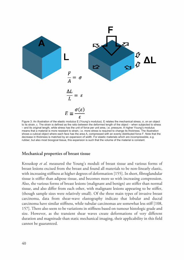
3. An illustration of the elastic modulus E (Young’s modulus). E relates the mechanical stress, , on an object to its strain, . The strain is defined as the ratio between the deformed length of the object – when subjected to stress – and its original length, while stress has the unit of force per unit area, i.e. pressure. A higher Young’s modulus means that a material is more resistant to strain, i.e. more stress is required to change its thickness. The illustration shows a cubical object where each face has the area A, compressed with an evenly distributed force F. Note that the decrease in thickness is matched by an expansion of width. For elastic materials which are incompressible, e.g. rubber, but also most biological tissue, this expansion is such that the volume of the material is constant.
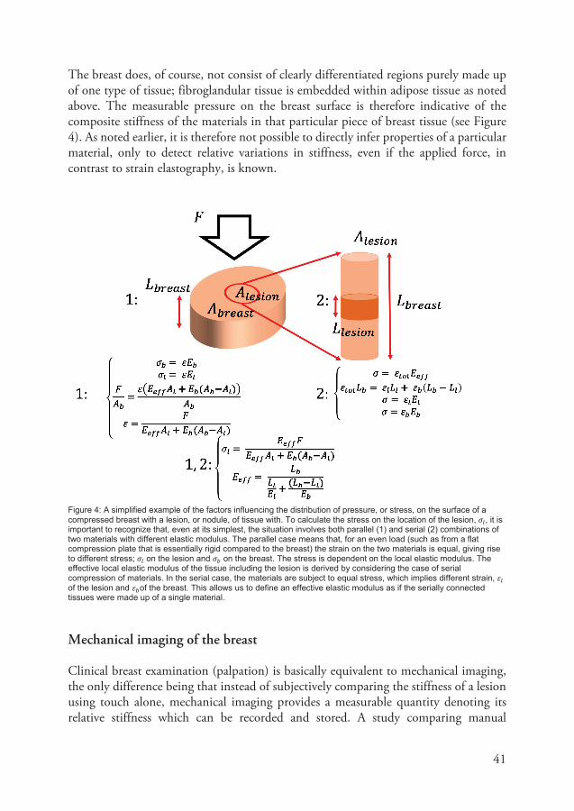
4. A simplified example of the factors influencing the distribution of pressure, or stress, on the surface of a compressed breast with a lesion, or nodule, of tissue with. To calculate the stress on the location of the lesion, , it is important to recognize that, even at its simplest, the situation involves both parallel (1) and serial (2) combinations of two materials with different elastic modulus. The parallel case means that, for an even load (such as from a flat compression plate that is essentially rigid compared to the breast) the strain on the two materials is equal, giving rise to different stress; on the lesion and on the breast.
20
The stress is dependent on the local elastic modulus. The effective local elastic modulus of the tissue including the lesion is derived by considering the case of serial compression of materials. In the serial case, the materials are subject to equal stress, which implies different strain, of the lesion and of the breast. This allows us to define an effective elastic modulus as if the serially connected tissues were made up of a single material.
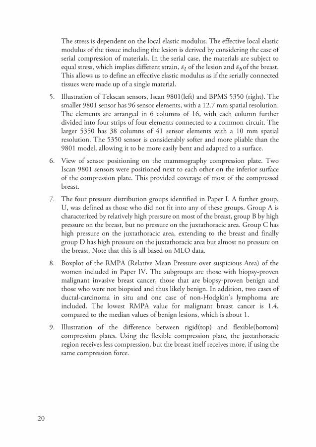
5. Illustration of Tekscan sensors, Iscan 9801(left) and BPMS 5350 (right). The smaller 9801 sensor has 96 sensor elements, with a 12.7 mm spatial resolution. The elements are arranged in 6 columns of 16, with each column further divided into four strips of four elements connected to a common circuit. The larger 5350 has 38 columns of 41 sensor elements with a 10 mm spatial resolution. The 5350 sensor is considerably softer and more pliable than the 9801 model, allowing it to be more easily bent and adapted to a surface.
6. View of sensor positioning on the mammography compression plate. Two Iscan 9801 sensors were positioned next to each other on the inferior surface of the compression plate. This provided coverage of most of the compressed breast.
7. The four pressure distribution groups identified in Paper I. A further group, U, was defined as those who did not fit into any of these groups. Group A is characterized by relatively high pressure on most of the breast, group B by high pressure on the breast, but no pressure on the juxtathoracic area. Group C has high pressure on the juxtathoracic area, extending to the breast and finally group D has high pressure on the juxtathoracic area but almost no pressure on the breast. Note that this is all based on MLO data.
8. Boxplot of the RMPA (Relative Mean Pressure over suspicious Area) of the women included in Paper IV. The subgroups are those with biopsy-proven malignant invasive breast cancer, those that are biopsy-proven benign and those who were not biopsied and thus likely benign. In addition, two cases of ductal-carcinoma in situ and one case of non-Hodgkin’s lymphoma are included. The lowest RMPA value for malignant breast cancer is 1.4, compared to the median values of benign lesions, which is about 1.
9. Illustration of the difference between rigid(top) and flexible(bottom) compression plates. Using the flexible compression plate, the juxtathoracic region receives less compression, but the breast itself receives more, if using the same compression force.
21
List of Abbreviations
BT or DBT – (Digital) Breast Tomosynthesis
CC – Cranio Caudal projection
CT – Computed Tomography
CTC – Circulating Tumour Tells
DCIS – Ductal Carcinoma in Situ
DM or FFDM – (Full-Field) Digital Mammography
FSR – Force Sensing Resistors
IDC – Invasive Ductal Cancer
ILC – Invasive Lobular Cancer
LM – Latero Medial projection
MI – Mechanical Imaging
MLO – Medio Lateral Oblique projection
MRI – Magnetic Resonance Imaging
PET – Positron Emission Tomography
RMPA – Relative Mean Pressure over lesion Area
SPECT – Single Photon Emission Computed Tomography
WHO – the World Health Organization

23
Introduction
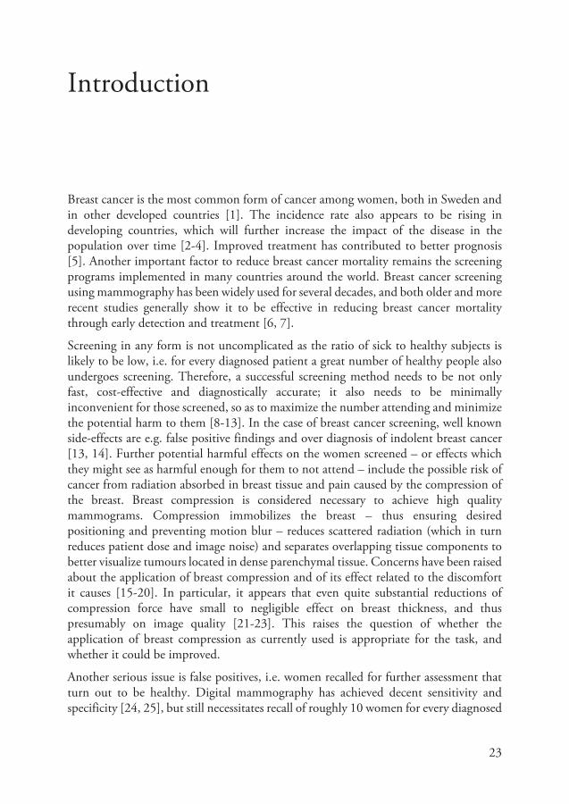
Breast cancer is the most common form of cancer among women, both in Sweden and in other developed countries [1]. The incidence rate also appears to be rising in developing countries, which will further increase the impact of the disease in the population over time [2-4]. Improved treatment has contributed to better prognosis [5]. Another important factor to reduce breast cancer mortality remains the screening programs implemented in many countries around the world. Breast cancer screening using mammography has been widely used for several decades, and both older and more recent studies generally show it to be effective in reducing breast cancer mortality through early detection and treatment [6, 7].
Screening in any form is not uncomplicated as the ratio of sick to healthy subjects is likely to be low, i.e. for every diagnosed patient a great number of healthy people also undergoes screening. Therefore, a successful screening method needs to be not only fast, cost-effective and diagnostically accurate; it also needs to be minimally inconvenient for those screened, so as to maximize the number attending and minimize the potential harm to them [8-13]. In the case of breast cancer screening, well known side-effects are e.g. false positive findings and over diagnosis of indolent breast cancer [13, 14]. Further potential harmful effects on the women screened – or effects which they might see as harmful enough for them to not attend – include the possible risk of cancer from radiation absorbed in breast tissue and pain caused by the compression of the breast. Breast compression is considered necessary to achieve high quality mammograms. Compression immobilizes the breast – thus ensuring desired positioning and preventing motion blur – reduces scattered radiation (which in turn reduces patient dose and image noise) and separates overlapping tissue components to better visualize tumours located in dense parenchymal tissue. Concerns have been raised about the application of breast compression and of its effect related to the discomfort it causes [15-20]. In particular, it appears that even quite substantial reductions of compression force have small to negligible effect on breast thickness, and thus presumably on image quality [21-23]. This raises the question of whether the application of breast compression as currently used is appropriate for the task, and whether it could be improved.
Another serious issue is false positives, i.e. women recalled for further assessment that turn out to be healthy. Digital mammography has achieved decent sensitivity and specificity [24, 25], but still necessitates recall of roughly 10 women for every diagnosed

24
cancer case, depending on screening centre and country [26]. Various modalities are used for further assessment of recalled women, most commonly additional mammography imaging and breast ultrasound, with biopsy of the suspicious lesion required in a substantial number of cases. These false positives result in both anxiety for the recalled women [27] and considerable expenditure in time and money for the healthcare system, especially in the case of those women who require biopsies. Broadly, recalled women who do not have breast cancer can be divided into two groups: those who have a benign lesion (cyst, fibroadenoma, papilloma etc.) and those who turn out not to have any findings/lesions. For the latter group, recall is usually caused by a component (or components) of normal tissue masquerading as a suspicious-looking lesion. To correctly distinguish these different cases, other modalities are used in breast diagnosis in addition to mammography. One modality used mainly in the work-up is ultrasound (and the related technique of ultrasound elastography which provides information about the acousto-mechanical properties of the tissue) which provides further information which the radiologist can use to distinguish between malignant lesions, benign lesions and various types of normal tissue. Mechanical imaging, which involves measuring the stress (pressure) on compressed tissue to determine its stiffness, is a form of elasticity imaging distinct in some aspects from elastography, but provides similar information [28]. If mechanical imaging data would be available already at initial breast screening (and not substantially impact the time or complexity of the screening examination), a substantial number of false positives – and biopsies – could potentially be avoided.
Objectives
The objective of this thesis was to investigate the pressure distribution on the breast during breast compression in mammography from various aspects:
• Investigate the distribution of pressure on the breast with standard and reduced compression and correlate with experienced pain and other factors (Paper I)
• Investigate how the use of different compression plates influence the compression of the breast (Paper II)
• Determine the pressure over cancer tumours during compression, and whether compression spreads tumour cells (Paper III)
• Quantify the difference in pressure between malignant and benign breast lesions and estimate its potential for screening (Paper IV)

25
Scientific and technical background
Overview of breast cancer screening and diagnosis
Screening a predominantly healthy population for disease is an endeavour which puts a multitude of specific demands on the technique used [8-11]. This is particularly true for breast cancer screening. The ability of the method to correctly classify diseased (sensitivity) as well as non-diseased (specificity) individuals must be high. Harmful side-effects of the method must be minimized. Lastly, it must be efficient from a health-economic point-of-view.
In the case of breast cancer screening, the current standard is digital mammography, though other alternative techniques have been proposed, and breast tomosynthesis in particular is showing promising results.
Breast cancer
Overview Breast cancer is one of the most common forms of female cancer in the world, especially in developed countries, but also on the rise in the developing world [1-4, 29, 30]. Although mortality has dropped steadily due to both improved treatments and the widespread adoption of screening, in 2012 breast cancer still overall caused the 5th largest number of cancer deaths worldwide, and the largest number of cancer deaths among women [1]. In Sweden, official statistics show that between 2010 and 2014, an average of 1421 women died annually from breast cancer [31]. Incidence rates have doubled since the 1960s, probably due to both changes in risk factor distribution and increased detection following introduction of screening. Five-year survival was 88%, up from 65% in the 60s, with the caveat that such numbers may possibly be inflated by over diagnosis from screening. Breast cancer is a complex disease with many known of suspected risk factors, such as genetic factors and high breast density [1]. Lifestyle and environmental factors are thought to cause 27% of breast cancers, according to UK data [32].

26
Types Breast cancer can be categorized using many different histological schemes and this thesis makes no attempt to describe these in detail. Roughly, most breast cancers can be categorized in a grid, as seen in Table 1. The dividing lines are invasive/non-invasive, ductal/lobular. Non-invasive or in-situ cancer is a growth of abnormal cells which has not infiltrated tissue beyond its place of origin/basal membrane. There has been discussion of whether this should actually be labelled as cancer, and whether in-situ carcinomas will eventually progress to an invasive stage or not, with their being differences between low and high grade lesions [33-35]. Invasive cancer refers to malignant cell growths that have infiltrated surrounding tissue. The division of breast cancer into lobular and ductal stems from the fact that it was thought that these two forms of cancer began respectively in the breast lobules and the epithelial tissue of the ducts, but this has now generally been discounted in favour of both types originating in the terminal part of the breast gland, and the names kept to refer to distinct microscopic growth patterns [36].
Invasive ductal carcinoma (IDC) is the most common form of breast carcinoma, followed by invasive lobular carcinoma (ILC) and tubular carcinoma (invasive cancer characterized by growing in tubular patterns). Ductal carcinoma in-situ (DCIS) is the most common form of non-invasive breast cancer.
Table 1.
Breast cancer types
Appearance As mammography is by far the most widespread breast imaging technique, appearance of tumours is here mainly focused on their radiological appearance and somewhat on the appearance of excised tumours.
Breast cancer often presents as a stellate lesion, which is a star-shaped object with spiculations extending into the surrounding tissue. These spicules are a combination of
Ductal Lobular Other
Invasive Invasive ductal
carcinoma
Invasive lobular
carcinoma
Tubular carcinoma, medullary carcinoma
In situ Ductal carcinoma
in-situ
Lobular carcinoma
in-situ
-

27
strands of infiltrating cancer and reactive fibrosis. The core of the lesion is an irregular mass with diffuse borders. Invasive ductal, invasive lobular and tubular carcinoma can all have a stellate appearance. The apparent size of such tumours on a mammogram is often smaller than their palpable size, suggesting that the tissue reaction to its presence is more widespread than what is visible from fibrous reactions. This is corroborated by ultrasonic data, where a hyper-echogenic border is seen around the tumour, presumably caused by tissue fibrosis. Spiculated lesions, especially tubular carcinomas, can be confused with radial scars, a form of benign breast lesion.
Non-spiculated cancers can be diffuse (which are often difficult to locate), well-circumscribed (having a well-defined border) or multi-focal (having several nodular foci). This indicates less reactive components in surrounding tissue. Well-circumscribed lesions can look similar to benign cysts, fibroadenomas (a benign epithelial lesion) and papillomas (a benign ductal lesion). Some lesions are detected not as a solid mass but by the architectural distortion of tissue, which is harder to notice [37].
Microcalcifications, typically caused by calcification of necrotic material in the ducts can be a characteristic sign of cancer and accompany some of the above mentioned lesion types, though many microcalcifications are benign. DCIS is characteristically identified through detection of clusters of malignant microcalcifications, with no apparent accompanying mass.
Stellate lesions tend to be lower grade, more receptive to hormone treatments and generally have a better prognosis [38]. A recent study however found that, in the Japanese population, there does not appear to be any difference in survival between women with stellate and well-circumscribed lesions [39].
Circulating tumour cells The number of circulating tumour cells (CTC) per unit volume of blood is an independent prognostic marker of both progression and disease-free survival of metastatic and non-metastatic breast cancer [40-43]. Measuring CTC count is an emerging technique, which is mainly investigated for use in cancer treatment staging, such as deciding whether a patient will benefit from adjuvant therapy. These results pertain to an equilibrium level of CTCs caused by continuous release of cells from the primary tumour and possible metastases. Evidence persists that physical damage to tumours, including from palpation, causes a considerable outflow of CTCs [44, 45]. Whether such a transient spike of CTCs can contribute to the spread of cancer is poorly understood, but can potentially be of critical importance for both the use of compression in mammography and clinical breast palpation.

28
Mammography
Mammography is the gold standard of breast screening in most of the world, with full-field digital mammography (FFDM) being available since 2000. Using dedicated breast x-ray units for screening was investigated in several large randomised, controlled trials during the 70s and 80s [7, 46]. Largely positive results (although widely debated with regards to estimations of reduced breast cancer mortality and over diagnosis of indolent lesions) from such prospective screening studies lead to the establishment of population-based mammography breast cancer screening programs in many developed countries.
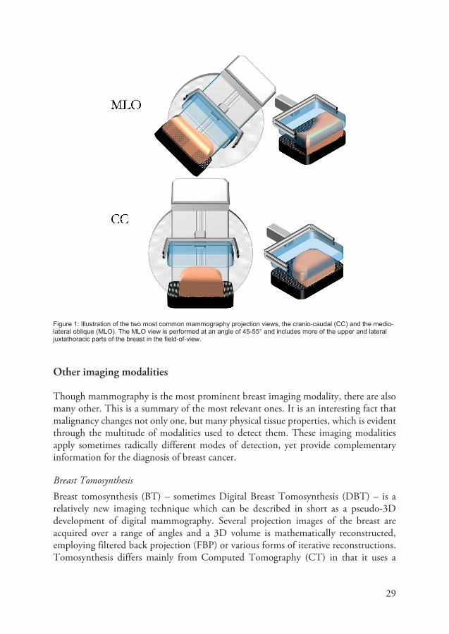
Later publications have highlighted the number of missed cancers, the substantial number of false-positives, and also the potentially large proportion of overdiagnosis, i.e. detection, diagnosis and treatment of asymptomatic cancers which would not have been detected in the absence of screening [46, 47]. Recent reviews of available data by the Independent UK Panel on Breast Cancer Screening [7] and the American Cancer Society [6] both assert the effectiveness of mammography screening and recommend women to attend regular breast screening, respectively according to the UK recommendations of screening between age 50 and 70, and annually from 45 years to 54 and then biennially. In Sweden, women are screened from the age of 40, at intervals of 18-24 months, until they reach age 74 [48]. Two standard projection views are used, the cranio-caudal (CC) and medio-lateral oblique (MLO) (Figure 1). These two views are complementary; the MLO-view covers more tissue and provides better visualization of the upper juxtathoracic part of the breast while the CC-view suffers less from overlapping dense tissue and can potentially provide better visualization of centrally located lesions as [49-51]. A latero-medial (LM) view is usually added in clinical work-up of recalled women.
Mammography is required to be able to distinguish between small differences in absorption between different breast tissues. The entire x-ray system is therefore tailored for soft tissue diagnosis, with anode-filter combinations providing comparatively low-energy x-rays that are detected by high resolution detectors, while the dose is kept low. Young et al. estimated that a woman undergoing the UK screening program would receive an average mean glandular dose of 60 mGy during one decade, and roughly 2 mGy per view per screening occasion [52, 53]. This level is estimated to lead to only a small increase in risk of cancer death, which is outweighed by the reduced breast cancer mortality of screening [54].
Two modern studies of screening with full field digital mammography are largely in agreement concerning sensitivity and specificity. Pisano et al. found that in a screening study on 42 760 US and Canadian women showed 70% sensitivity and 92% specificity [24]. Skaane et al. reported 77.4% sensitivity and 96.5% specificity in a randomized screening trial of 23 929 Norwegian women [25, 55].
29
Figure 1: Illustration of the two most common mammography projection views, the cranio-caudal (CC) and the medio-lateral oblique (MLO). The MLO view is performed at an angle of 45-55° and includes more of the upper and lateral juxtathoracic parts of the breast in the field-of-view.
Other imaging modalities
Though mammography is the most prominent breast imaging modality, there are also many other. This is a summary of the most relevant ones. It is an interesting fact that malignancy changes not only one, but many physical tissue properties, which is evident through the multitude of modalities used to detect them. These imaging modalities apply sometimes radically different modes of detection, yet provide complementary information for the diagnosis of breast cancer.
Breast Tomosynthesis Breast tomosynthesis (BT) – sometimes Digital Breast Tomosynthesis (DBT) – is a relatively new imaging technique which can be described in short as a pseudo-3D development of digital mammography. Several projection images of the breast are acquired over a range of angles and a 3D volume is mathematically reconstructed, employing filtered back projection (FBP) or various forms of iterative reconstructions. Tomosynthesis differs mainly from Computed Tomography (CT) in that it uses a

30
limited angular range of 15 – 50 degrees (in diagnostically used systems) rather than a full 180 degree set of projections [56, 57]. The reconstructed volume can perhaps more accurately be referred to as pseudo-3D, as its depth resolution is far less than its in-plane resolution due to the artefacts caused by this limited angle.
Standard mammography and BT use broadly similar imaging units, with BT units capable of acquiring standard mammography as well. Some manufacturers offer the same units for both purposes, with a software upgrade enabling tomosynthesis. In comparison to mammography, BT has the advantage of generating multiple thin slices of the breast, which may reveal breast tumours that would be obscured by overlying tissue on a 2D image. BT was envisaged as a tool for both clinical breast imaging and breast cancer screening [58, 59]. Experimental studies and simulations imply that it can substantially improve sensitivity compared to mammography [60-62]. Opposing its implementations in general screening is concerns about increased radiation dose (depending on manufacturer and imaging protocol) and increased examination time, and more importantly, reading time. In practice, the time it takes a radiologist to review a BT image volume is estimated to be at least twice that required for the corresponding 2D mammogram [63-65].
Four large prospective screening trials show similar results, in that BT used in a screening situation increases cancer detection by ~30-40% [66-68]. All trials (The Norwegian Oslo trial, the Italian STORM 1 and 2 trials and the Swedish MBTST) also appear to show increased recall rates compared to mammography. Notably, the MBTST compares one-view BT with 2-view mammography, while the others compare the combination of two-view BT and 2-view mammography (or synthetic mammograms in STORM 2) with 2-view mammography on its own, implying that mammography and an additional BT view does not add substantial extra information compared to a single BT volume of the breast. The trials reported data from only one screening round, and have to date not reported follow-up interval cancer data. These studies are thus essentially reporting results from a prevalence round, meaning that the effect on both recalls and extra cancer detection might be different (in all likelihood, lower) in subsequent screening rounds.
Numerous US retrospective screening studies have also investigated BT, with largely different results from the mentioned European trials [69-75]. Comparing women screened with BT to women screened with mammography – without taking into account possible selection bias – results support a substantial reduction of recall rates, with little effect on cancer detection. One must here remember the substantially greater proportion of recalls in the USA (around 10-15%) compared to Europe (3-7%), the difference between opportunistic and population based screening and also, as mentioned, possible selection bias; one can speculate that women with a higher socio-economic status are more likely to have access to BT.
31
Ultrasound Breast ultrasound is an important diagnostic modality. It is essential in the work-up of symptomatic or asymptomatic women, where it is used to investigate suspicious lesions detected on screening mammography or through e.g. self-examinations or clinical examination. Ultrasound adds valuable information in the characterization of malignant and benign lesions [76-78]. It is often used to guide biopsies. It is capable of finding mammographically occult tumours, as they differ from normal dense breast tissue in echogenicity, even if radiological density is similar. Breast ultrasound requires a trained operator, usually a radiologist or other physician or a dedicated sonographer, to perform the examination.
The use of breast ultrasound as a screening modality has been proposed and discussed, but has been mostly dismissed due to several issues, mainly cost-effectiveness and the high false-positive rate [79-82]. The high cost is a result of the long examination time (compared to mammography or breast tomosynthesis) and the above mentioned need for a trained operator. Still, the large J-START Japanese screening trial has investigated the use of ultrasound in screening. Japanese women have denser breasts than is found in European or American populations, limiting the applicability of mammography [83, 84]. The J-START trial used ultrasound as an adjunct to mammography to screen > 70 000 women, finding it to detect more cancers than mammography alone (especially early stage cancers). The trial did not investigate the health economic aspects of ultrasound screening.
Recent developments of various forms of automatically scanned ultrasound which dispenses with or limits the need for a trained operator may potentially warrant revaluation of ultrasound screening [85-87]. The SoftVue (Delphinus Medical Technologies, Novi, MI, USA) breast ultrasound tomography system is one example, in which the woman lies prone on a bed with the breast suspended in a water-filled hemispherical container lined with ultrasound transducers [88-90]. An image volume is reconstructed, looking very similar to CT or MRI slices.
MRI Magnetic resonance imaging (MRI) has a higher sensitivity than mammography for detecting breast cancer, although the specificity has been an issue [91, 92]. For breast cancer imaging, gadolinium contrast enhanced MRI is most commonly employed, differentiating lesions based on their uptake of the contrast agent due to the neovascularization. It provides an effective means of evaluating difficult cases in clinical work-up, especially so in dense breasts where other modalities might be insufficiently sensitive. Though potentially valuable as a screening modality for women with a very high risk of breast cancer (e.g. carriers of the BRCA mutation), MRI’s combination of high cost and long examination time makes it unsuitable for use in primary screening [93]. MRI is useful as a tool for pre-operative staging before excision of breast cancer,

32
as it can accurately delineate tumour margins [94, 95]. Shorter MRI protocols are being investigated for use in screening [96, 97].
Elastography Elastography is a form of elasticity imaging in which a static or transient force is applied and some means is used to detect the deformation of different tissues subject to that force. The degree of deformation is indicative of tissue stiffness, which can in turn be indicative of possible malignancy. In essence, both breast palpation in the clinic and breast self-examination can be considered a form of unaided elastography [28].
Elastography is mainly defined by the modality used to evaluate tissue strain. The two main types are ultrasound elastography and MRI elastography, although other techniques have also been described, e.g. tomosynthesis breast elastography [98] and optical coherence elastography [99].
Ultrasound elastography can be broadly divided into two types: compression elastography and shear-wave elastography, with many subtypes [100, 101]. Compression elastography, also known as static or strain elastography or RTE (real-time elastography), requires the user to compress the examined area (i.e. breast in this case) with the ultrasound probe, while the tissue deformation is measured, translated into a colour map and combined with a B-mode ultrasound image. This is the most widely used form of elastography, providing qualitative data on the stiffness of evaluated tissues relative to each other.
In shear-wave elastography the source of strain is instead a focused pulse of ultrasound generated by the probe, allowing quantification of tissue stiffness either directly through deformation measurements or by measuring the propagation speed of the shear-wave [102]. Only shear-wave elastography provides quantitative measurements of tissue stiffness [103].
Ultrasound elastography is an adjunct modality to standard ultrasound, increasing the ability to distinguish between benign and malignant solid breast masses [104-108].
MRI elastography, or MRE, uses the same principal as shear-wave elastography to induce a dynamic shear-wave in the tissue to be imaged [109, 110]. This wave is synchronized with the MR pulse-sequence, and through phase-contrast MRI the motion of tissue can be recovered. The technique offers sub-micrometre sensitivity. MRE of the breast is being investigated as a means of improving the poor specificity of breast MRI [111-113].
Additional Modalities Spectral mammography uses an injection of iodine contrast medium and a dual-energy x-ray acquisition (or a photon counting detector) to visualize areas of increased blood flow, allowing better detection of subtle or obscured tumours [114-116]. It can be
33
compared to breast MRI in that it requires a contrast injection to achieve a better sensitivity.
Less prominent breast imaging modalities, many of them at an experimental level, include breast CT [117-120], breast SPECT [121-123] and PET [124], and synchrotron mammography [125, 126].
Mammography screening recall rates and false positives
In breast cancer screening, a very important measure of screening efficiency is the proportion of women recalled for further clinical assessment. Published data suggests that the rate of screening detected cancers in developed countries is about 0.5% for women screened [26]. In the ideal situation, with 100% sensitivity and specificity, recall rates would thus also be 0.5%. In reality, breast cancer screening with mammography has recently been estimated to have a sensitivity of 77.4% and specificity of 92.0% in Europe (Norway) [25] compared to 70.0% and 92.0%, respectively, in North America [24].
The European Guidelines for Quality Assurance in Mammography recommend that, in the prevalence round, recall rates should be 7%, and then between 3-5% in following screening rounds [127]. Assuming a 0.5% cancer detection rate, this means that ~90% of recalled women are false positives. Actual data from the Norwegian and Spanish breast screening programs show that 6.1 and 10.2 women, respectively, were recalled for every detected cancer, while in the US only 1 in 20.3 recalled women had cancer [26, 128]. The three programs reported similar sensitivity values.
False positive mammography has been linked with long- and short term psychosocial consequences, such as anxiety, with up to 30% of women with false positive screening results still experiencing such problems after one year [27]. Also, false positives are expensive for the healthcare system. Assessment of the breast involves various modalities, usually additional mammography images and often ultrasound, breast tomosynthesis and various forms of biopsies [13, 127]. In difficult cases, MRI may be used. All false positives represent a significant investment of time both for the woman recalled and for healthcare professionals. Hofvind et al. estimated that the life-time risk for at least one false positive result of a woman attending screening biennially from age 50 to 68 was 20% [129].

34
Attendance
For a population based screening program it is important that the rate of attendance is as high as possible. For Europe, recently published data report attendance rates above 90% in Sweden [130] and 50-80% in Spain [131]. In the US, where screening is opportunistic rather than population-based, but still recommended by healthcare authorities and required to be fully covered by healthcare plans, it is harder to estimate attendance rates. The American cancer society reported that, 69% of women aged between 40-49 had had a mammogram during the last two years (with the Society recommending annual screening from age 40) [132].
Several studies link non-attendance with socio-economic factors and with pain [19, 130, 133-135]. In particular, factors indicative of a lower socio-economic status (low income, cramped living conditions, foreign born etc.) were predictive of non-attendance. Elwood et al. found that of 121 women who had not attended screening subsequent to their initial screening round, 46% stated pain as their reason for non-attendance [136].
Breast compression in mammography
Compression of the breast is considered necessary to achieve high quality images in mammography and to limit absorbed radiation dose. There is no established universal guideline as to the level to which the breast should be compressed. The European guidelines for quality assurance in breast cancer screening and diagnosis [127] makes the following recommendations:
“The breast should be properly compressed, but no more than is necessary to achieve a good image quality. “
“The compression of the breast tissue should be firm but tolerable. There is no optimal value known for the force…”
The breast is compressed between a movable compression plate (or paddle) and the fixed breast support, which usually houses the x-ray detector (see Figure 2). The radiographer positions the breast on the breast support and lowers the compression plate either by a foot-operated pedal or by a button. The compression plate is gradually lowered, applying an even load on the breast. Recommendations, as those quoted above from the European Guidelines, are vague in terms of the force that should be used and the degree of compression that should be attained. Though compression should presumably take into account breast size, with smaller breasts requiring less compression force, data indicates that actual force used is relatively constant. 100-130

35
N is reported in many screening programs [20-23, 137] but, e.g., uses of 180 N or more as a standard have been reported [138]. Maximum force for the mammography unit is ~200 N, with the actual force used also subject to radiographer variability [139].
Figure 2: Schematic illustration of the mammographic compression device, used in mammography and breast tomosynthesis. The breast is positioned on the solid breast support and then compressed by the descending compression plate (also called a compression paddle). The compression plate is constructed from relatively thin, strong plastic, such as polycarbonate, and should ideally have a homogenous thickness so as to provide equal x-ray absorption across the entire field-of-view. The breast support usually houses the x-ray detector, but in some units the breast support is instead similarly constructed to the compression plate, and a separate, movable detector is provided.
Breast composition
Radiologically speaking, the breast mainly consists of two types of tissue with distinctly different X-ray absorption: adipose tissue and fibroglandular tissue. While adipose tissue consists mainly of fat, fibroglandular tissue – often simply called dense tissue – is a more complex mix of tissue components, including the ductal and glandular networks and fibrous connective tissue, such as Cooper’s ligaments. The skin and the pectoral muscle are not included in these groups. The amount of dense tissue and proportion of dense tissue in the breast is predictive of breast cancer risk, women with denser

36
breasts having an increased risk of breast cancer [140]. A widely used division into four risk groups according to radiological density is the BIRADS density score, ranging from A (lowest risk) to D (highest risk) [141].
When compressed, the compression plate – which is rigid in comparison to breast tissue – thus compresses a number of different tissue types with different stiffness and other mechanical properties. In the MLO-view one criterion for a successful image is that the pectoral muscle is prominent on the image, extending from the axillary area down to roughly 50% of the breast extent. This is to make sure that the juxtathoracic part of the breast is included so as to not miss a potential tumour in this area. This of course also necessitates compression of the pectoral muscle.
Image quality
As noted, there are two reasons for breast compression: reduced patient dose, and increased image quality. Image quality is a broadly defined term, but in the case of breast compression, the improvement comes from two angles: physics and anatomy. The physics side of the issue is that when the breast is compressed, its thickness decreases. This means that there is less tissue for x-ray photons to penetrate and interact with, resulting in less absorbed dose and less scattered radiation, even though there will be an increase of breast area. The reduction of scatter will improve the signal-to-noise ratio, thereby providing a less noisy image. Saunders and Samei [142] suggests that for thickness differences of less than 5 mm there is no clinically discernible effect on image quality caused by photon interactions, as long as exposure settings are adapted to the new thickness. The anatomical side is that when the breast is compressed, tissue is forced to move perpendicular to the applied force, being spread out on the breast support of the mammography device (the tissue of the breast has a Poisson’s ratio, , close to the perfect value of 0.5, meaning that it does not change volume while under compression). Spreading out tissue, i.e. separating overlaying structures, allows better visualization of lesions, particularly those located in areas of dense tissue. Better separation of structures is one of the reasons cited to explain the fact that some tumours are better visible in the CC-view than in the MLO-view [49, 50].
Compression is also employed in breast tomosynthesis, apparently for the same reasons. However, as a quasi-3D technique, it does not straightforwardly follow that tomosynthesis, despite its many similarities with mammography, would benefit in the same way. For example, although compression separates structures in a plane perpendicular to the applied force, it also decreases separation in depth. It is currently unknown to what extent breast compression thus improves the image quality in breast tomosynthesis and whether it is necessary to maintain the same levels of compression force.

37
Pain
Some women consider breast compression to be painful. Studies have reported that 25-42% of women undergoing mammography experience either discomfort or pain [19]. Peipins et al. reported that 25% of US women rated their experience of screening 30 months after their latest mammogram as at least moderately painful [143]. It would thus appear plausible that remembered pain can possibly dissuade women from attending later screening (see Attendance above).
Interventions to improve compression
Various interventions designed to limit pain for examined women and/or better standardize the application of breast compression have been proposed. As current practice varies, there is no recognized optimal level of compression, either based on applied force, thickness reduction or estimated pressure
Poulos and McLean argued for measuring the actual degree to which the breast is compressed, i.e. compressibility based compression [17, 144]. By applying an initial compression of 30 N they were able to predict the compressibility of the breast, and from that estimate the minimum breast thickness that could be achieved. They noted that for 74% of women, minimum achievable breast thickness was not achieved with the force applied by the radiographer.
A similar approach is implemented by Siemens, employing the OPCOMP system which continually measures the ratio between thickness decrease and applied force, automatically stopping compression when further force application no longer meaningfully affects breast thickness. This follows the principal that for many materials (see Mechanical Properties of Breast Tissue, below), the greater the degree of deformation (compression) the greater the resistance to further deformation becomes, i.e. the system is intended to halt compression when further application of force does not improve compression.
Standardization based on mean breast pressure has been proposed by a group at the University of Amsterdam [137, 138, 145]. The basic idea is that through measuring the contact area between the breast and compression plate and recording the compression force, one can calculate the mean pressure on the breast, or rather the mean pressure on the contact area. They have recommended 10 kPa as a suitable mean pressure, citing that pressure should not exceed diastolic blood pressure so as to not constrict blood flow. According to published data, contact area correlates with breast area, though not strongly. In earlier publications by the same group various different ways of estimating contact area were employed, at first retrospectively using data from breast density assessment software. Currently, the group collaborates with Sigmascreening (Amsterdam, the Netherlands) which manufactures a compression

38
plate that continually measures contact area through changes in electrical conductivity and stops application of compression force once a pre-set pressure level is reached.
As mentioned, breast tomosynthesis potentially requires different considerations with regard to breast compression. Förnvik et al. investigated reducing compression force by half from a base level of ~100 N [22]. The results indicated that not only was there relatively little difference in thickness (5.8 mm on average) but there was no substantial difference in image quality of the resulting tomosynthesis images. Similar results have been reported both with breast tomosynthesis and mammography [21, 23]. Regarding pain, results of reducing compression force varies. Concerning screen-film mammography, Poulos and McLean found, in a study on 114 women taking part in the Australian breast screening program, that reducing compression force by 30 N did not affect breast thickness for 24% of imaged women [17]. Reduced image quality was reported.
Several manufacturers have implemented so-called “flexible” compression plates [146-149]. This can refer to both flexible (hinged) mountings or to the plate itself being made of more pliant forms of plastic, or to a combination of the two. In any case, the flexible plate is intended to conform to the breast and thereby improve compression. While a conventional rigid compression plate forces the breast to assume an essentially flat profile parallel to the detector, using a flexible plate results in a thickness gradient from nipple to chest wall, with the compressed breast being at its thinnest at the nipple. For one mammography unit with a flexible compression plate, Selenia Dimensions (Hologic, inc., Marlborough, MA, USA), the median tilt angle has been estimated to be ~2° [148]. Broeders et al. [146] suggests that at least one manufacturer’s flexible compression plate unacceptably impairs the diagnostic quality of mammograms as the retroglandular (juxtathoracic) tissue is either not included or imaged well, relying on retrospective data from the Dutch screening program. There was no evident difference in pain or discomfort.
Other interventions have included using soft cushions on the breast support [150-152], and allowing the woman to compress the breast herself [153]. Such interventions generally decrease discomfort at the cost of image quality [154].
Mechanical imaging
Mechanical imaging, also known as tactile imaging refers to the method of applying a set force to an object and acquiring quantitative or qualitative data about the mechanical properties of the materials comprising the object. This can be done through measuring the resultant mechanical stress (pressure) on the surface of the compressed object.

39
Elastic materials, i.e. materials that regain their original shape after deformation – to at least some degree of deformation – are defined by the following expression:
E is Young’s modulus, is the stress exerted on the material and is the material strain (Figure 3). Young’s modulus thus defines the relationship between the applied pressure on the material and the deformation of the material due to that pressure; Young’s modulus can therefore be said to denote the stiffness of a material when subjected to uniaxial stress. The related shear modulus, G, denotes the stiffness of a material with regard to shear stress (such as in shear-wave elastography), though in the case of isotropic materials, i.e. materials which respond identically regardless of the axis of applied stress, these quantities are not independent of each other but related by the simple equation
where is the Poisson’s ratio, which approaches 0.5 for most forms of breast tissue [155]. This means that these two quantities can be used essentially interchangeably to denote the stiffness of soft tissue.
Most materials that are defined as elastic behave linearly elastic only in a certain strain range; above that strain level they become plastic, i.e. the deformation is no longer reversible. For certain materials, denoted non-linearly elastic, E is a function of , i.e. the stiffness of the material depends on the degree of deformation.
When compressed with an even load, an object consisting of several materials with distinct Young’s moduli receives equal strain, but the stress – pressure – on a material is proportional to the difference between the moduli of the materials making up the object. This is valid under the assumption that the load is applied with an object essentially rigid compared to the compressed materials (had it instead been essentially flexible in comparison to the compressed object the stress would have been even and the strain would be proportional to E). Mechanical imaging used in this way cannot therefore determine any absolute value of E, only a relative difference between the moduli of different tissue types. Mechanical imaging is thus similar to, but distinct from, elastography. As described above, elastography directly measures the elastic modulus by quantifying the deformation of individual components within the imaged object. Sarvazyan [28, 156], considering both mechanical imaging (abbreviated as MI) and elastography as forms of elasticity imaging, noted that:
“…MI may be called “stress imaging”, in contrast to other elasticity imaging techniques, which are estimating tissue displacement and referred to as “strain imaging”.”
40
Figure 3: An illustration of the elastic modulus E (Young’s modulus). E relates the mechanical stress, , on an object to its strain, . The strain is defined as the ratio between the deformed length of the object – when subjected to stress – and its original length, while stress has the unit of force per unit area, i.e. pressure. A higher Young’s modulus means that a material is more resistant to strain, i.e. more stress is required to change its thickness. The illustration shows a cubical object where each face has the area A, compressed with an evenly distributed force F. Note that the decrease in thickness is matched by an expansion of width. For elastic materials which are incompressible, e.g. rubber, but also most bioogical tissue, this expansion is such that the volume of the material is constant.
Mechanical properties of breast tissue
Krouskop et al. measured the Young’s moduli of breast tissue and various forms of breast lesions excised from the breast and found all materials to be non-linearly elastic, with increasing stiffness at higher degrees of deformation [155]. In short, fibroglandular tissue is stiffer than adipose tissue, and becomes more so with increasing compression. Also, the various forms of breast lesions (malignant and benign) are stiffer than normal tissue, and also differ from each other, with malignant lesions appearing to be stiffer, (though sample sizes were relatively small). Of the three main types of invasive breast carcinoma, data from shear-wave elastography indicate that lobular and ductal carcinoma have similar stiffness, while tubular carcinomas are somewhat less stiff [108, 157]. There also seem to be variations in stiffness based on tumour histologic grade and size. However, as the transient shear waves create deformations of very different duration and magnitude than static mechanical imaging, their applicability in this field cannot be guaranteed.
41
The breast does, of course, not consist of clearly differentiated regions purely made up of one type of tissue; fibroglandular tissue is embedded within adipose tissue as noted above. The measurable pressure on the breast surface is therefore indicative of the composite stiffness of the materials in that particular piece of breast tissue (see Figure 4). As noted earlier, it is therefore not possible to directly infer properties of a particular material, only to detect relative variations in stiffness, even if the applied force, in contrast to strain elastography, is known.
Figure 4: A simplified example of the factors influencing the distribution of pressure, or stress, on the surface of a compressed breast with a lesion, or nodule, of tissue with. To calculate the stress on the location of the lesion, , it is important to recognize that, even at its simplest, the situation involves both parallel (1) and serial (2) combinations of two materials with different elastic modulus. The parallel case means that, for an even load (such as from a flat compression plate that is essentially rigid compared to the breast) the strain on the two materials is equal, giving rise to different stress; on the lesion and on the breast. The stress is dependent on the local elastic modulus. The effective local elastic modulus of the tissue including the lesion is derived by considering the case of serial compression of materials. In the serial case, the materials are subject to equal stress, which implies different strain, of the lesion and of the breast. This allows us to define an effective elastic modulus as if the serially connected tissues were made up of a single material.
Mechanical imaging of the breast
Clinical breast examination (palpation) is basically equivalent to mechanical imaging, the only difference being that instead of subjectively comparing the stiffness of a lesion using touch alone, mechanical imaging provides a measurable quantity denoting its relative stiffness which can be recorded and stored. A study comparing manual
42
palpation of spherical nodules in a phantom with mechanical imaging of the same found mechanical imaging to detect lesions at a greater tissue depth [158]. For example, a 6 mm nodule was detected by palpation at a maximum depth of 10 mm, while mechanical imaging detected it at a depth of 17.5 mm.
Egorov et al. has studied and described the utility of a handheld clinical mechanical imaging device intended specifically for breast imaging, the SureTouch system (Medical Tactile, Inc., Los Angeles, CA, USA). The system looks similar to a standard ultrasound probe, but substitutes the ultrasound array for a small matrix of capacitive pressure sensors [158, 159]. The operator presses the probe against the breast and moves it around to scan as much as possible of the breast volume while specialized software records data and composites it into a full relative stiffness map of the breast. One feature of the system is attempted differentiation of benign and malignant lesions through a number of measured quantities, e.g. hardness, shape and mobility. In a study on 179 women from four sites presenting with suspected breast cancer, the group demonstrated a sensitivity of 87.5% and a specificity of 84.4% when using a Bayesian classifier to combine six lesion differentiation features [159]. Theoretical and phantom studies show that the detectability of masses in soft tissue are dependent on lesion depth, size and hardness [28]. Though a study of breast cancer cost effectiveness by Sarvazayan et al. [82] optimistically indicates that mechanical imaging using a hand-held probe could be a highly cost-effective screening modality, the technique still requires a trained operator and examination times broadly equivalent to that of breast ultrasound. Data on its clinical performance is limited, especially in a screening setting.
Other similar devices have been documented, such as the Breast View System (Breast View System; Assurance Medical Corp, Hopkinton, Mass, USA). Wellman et al. proposed the use of this device to improve the accuracy of clinical breast palpation, stating that:
“In a limited clinical trial on 24 surgical patients, lump size was estimated with less than 17% mean absolute error when compared with ex-vivo size measurements. This is more than twice as accurate as either clinical breast examination or ultrasound examination of the same lumps.” [160]
Similar devices have been used for mechanical vaginal and prostate imaging [161, 162].
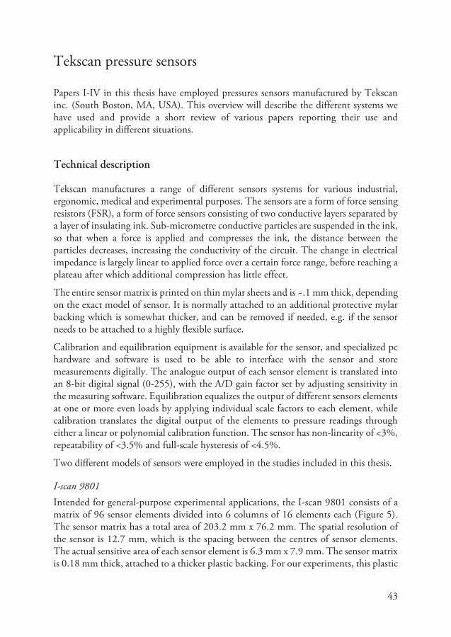
43
Tekscan pressure sensors
Papers I-IV in this thesis have employed pressures sensors manufactured by Tekscan inc. (South Boston, MA, USA). This overview will describe the different systems we have used and provide a short review of various papers reporting their use and applicability in different situations.
Technical description
Tekscan manufactures a range of different sensors systems for various industrial, ergonomic, medical and experimental purposes. The sensors are a form of force sensing resistors (FSR), a form of force sensors consisting of two conductive layers separated by a layer of insulating ink. Sub-micrometre conductive particles are suspended in the ink, so that when a force is applied and compresses the ink, the distance between the particles decreases, increasing the conductivity of the circuit. The change in electrical impedance is largely linear to applied force over a certain force range, before reaching a plateau after which additional compression has little effect.
The entire sensor matrix is printed on thin mylar sheets and is ~.1 mm thick, depending on the exact model of sensor. It is normally attached to an additional protective mylar backing which is somewhat thicker, and can be removed if needed, e.g. if the sensor needs to be attached to a highly flexible surface.
Calibration and equilibration equipment is available for the sensor, and specialized pc hardware and software is used to be able to interface with the sensor and store measurements digitally. The analogue output of each sensor element is translated into an 8-bit digital signal (0-255), with the A/D gain factor set by adjusting sensitivity in the measuring software. Equilibration equalizes the output of different sensors elements at one or more even loads by applying individual scale factors to each element, while calibration translates the digital output of the elements to pressure readings through either a linear or polynomial calibration function. The sensor has non-linearity of <3%, repeatability of <3.5% and full-scale hysteresis of <4.5%.
Two different models of sensors were employed in the studies included in this thesis.
I-scan 9801 Intended for general-purpose experimental applications, the I-scan 9801 consists of a matrix of 96 sensor elements divided into 6 columns of 16 elements each (Figure 5). The sensor matrix has a total area of 203.2 mm x 76.2 mm. The spatial resolution of the sensor is 12.7 mm, which is the spacing between the centres of sensor elements. The actual sensitive area of each sensor element is 6.3 mm x 7.9 mm. The sensor matrix is 0.18 mm thick, attached to a thicker plastic backing. For our experiments, this plastic

44
backing was generally retained to protect the sensor, as it was used attached to an even more solid backing, either the compression plate or the detector cover of the mammography device.
The 9801 sensors that we used had a recommended maximum pressure range of 35 kPa (meaning that higher values than 35 kPa are less reliable). Equilibration and calibration was performed using a device supplied by the manufacturer which employed a Kevlar membrane to apply an even pressure to the sensor (inserted beneath the membrane and locked into place). Pressure is measured by an attached manometer, and set by putting weights on a platform connected to the membrane. The calibration equipment is rated for a full-scale gauge accuracy of 3%.
BPMS 5350 The BPMS (Body Pressure Measurement System) sensor is intended for measuring interface pressure between skin and other materials, used in applications such as seating and bedding analysis (Figure 5). It is more flexible and robust than the I-scan sensors and is encased in a rubbery protective material. The model 5350 sensor has 41 columns of 38 elements, for a total of 1558 sensor elements with a 10.0 mm spatial resolution. The sensor has recommended maximum pressure of 41 kPa. Both calibration and equilibration are performed using a vacuum calibration device capable of applying evenly distributed pressure up to 180 mmHg = 24 kPa. The full-scale gauge accuracy of the calibration equipment is 1%.
Scientific use
The reproducibility and accuracy of measurements of the Tekscan system has been investigated by, among others, Brimacombe et al. who found the measurement accuracy of the system to be highly dependent on the calibration method used [163]. Multi-point calibrations (in which more than two known pressure levels are used) proved to yield better accuracy than linear and two-point calibrations, and in general using calibration pressures closer to the saturation limit were found to increase accuracy. Tekscan sensors have been found to be more accurate than the Fuji Film Prescale pressure measurement system, which has been a “…standard tool in orthopaedic and bioengineering research.” [164]
Other notable scientific uses of the Tekscan system have been plantar pressure measurements in shoes and of barefoot feet [165, 166], measurements of internal pressures in artificial joints [164, 167] and in studies of railroads [168]. More pertinent to the scope of this thesis, several studies have used various Tekscan sensors to study the distribution of pressure on different areas of the body from backpacks and other load-bearing devices [169-171].

45
Figure 5: Illustration of Tekscan sensors, Iscan 9801(left) and BPMS 5350 (right). The smaller 9801 sensor has 96 sensor elements, with a 12.7 mm spatial resolution. The elements are arranged in 6 columns of 16, with each column further divided into four strips of four elements connected to a common circuit. The larger 5350 has 38 columns of 41 sensor elements with a 10 mm spatial resolution. The 5350 sensor is considerably softer and more pliable than the 9801 model, allowing it to be more easily bent and adapted to a surface.


47
Summary of papers
The papers making up this dissertation can be divided into two topics covering the optimization of breast compression using mechanical imaging data, and the diagnostic use of mechanical imaging to detect and characterize tumours. All studies were approved by the Regional Ethical Review Board of Lund University and the local radiation safety committee at Skåne University Hospital and Unilabs AB (if applicable).
Optimization of breast compression – Papers I + II
Paper I – Breast Compression in Mammography: Pressure Distribution Patterns
This study was intended to attempt to explain the fact that reduction in breast compression force seem to have little effect on compressed breast thickness by directly measuring the distribution of pressure – or more accurately stress – on the breast using FSR (Force Sensitive Resistor) pressure sensors designed by Tekscan, specifically I-scan 9801. Patients were enrolled, after informed consent, from the breast cancer screening program and asked to have two extra compressions of one breast (the left, in the MLO view), one with standard compression force and one with roughly half the standard compression force, while the distribution of pressure on the compression plate was measured using two pressure sensors, each with 6x16 sensor elements with a spatial resolution of 12.7 mm. The sensors covered most of the compression plate, placed for maximum breast coverage (Figure 6). The breast thickness displayed on the mammography unit was also recorded. The women were asked to rate their experience of pain during the two compressions on two separate visual analogue scales (VAS). The pressure images were then matched with the corresponding mammogram acquired as part of standard screening mammography in order to further match pressure distribution with anatomy, dividing the breast into three regions (inner, middle and outer breast) and individually measuring force on these regions.
A total of 103 women were included in the study, with women with breast implants being excluded. Later investigation found no biopsy-proven breast cancers in the group.
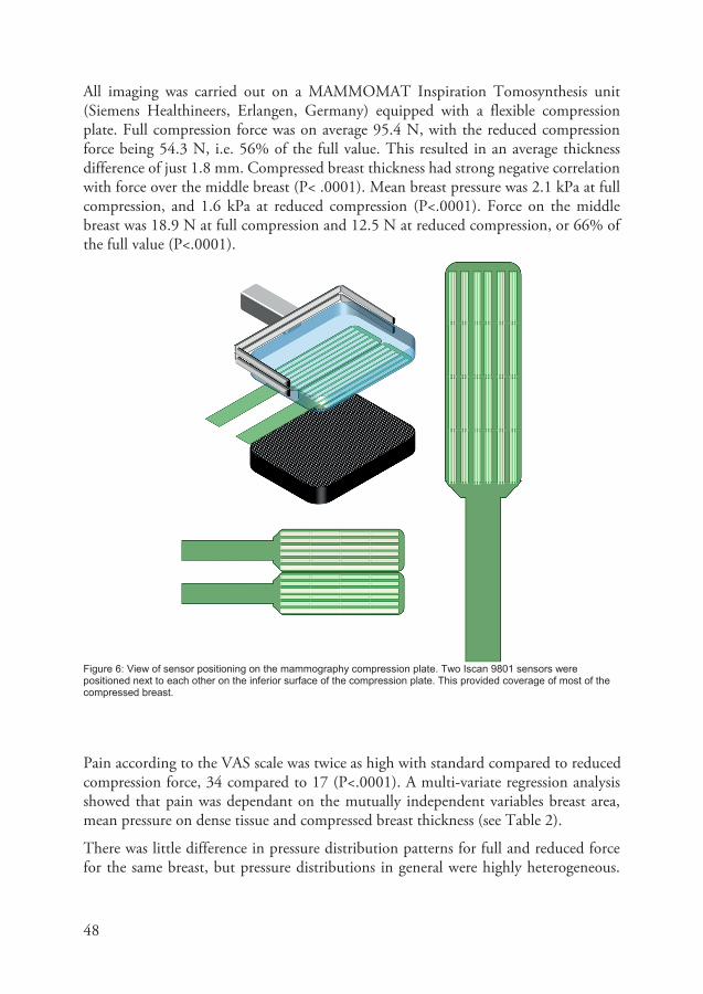
48
All imaging was carried out on a MAMMOMAT Inspiration Tomosynthesis unit (Siemens Healthineers, Erlangen, Germany) equipped with a flexible compression plate. Full compression force was on average 95.4 N, with the reduced compression force being 54.3 N, i.e. 56% of the full value. This resulted in an average thickness difference of just 1.8 mm. Compressed breast thickness had strong negative correlation with force over the middle breast (P< .0001). Mean breast pressure was 2.1 kPa at full compression, and 1.6 kPa at reduced compression (P<.0001). Force on the middle breast was 18.9 N at full compression and 12.5 N at reduced compression, or 66% of the full value (P<.0001).
Figure 6: View of sensor positioning on the mammography compression plate. Two Iscan 9801 sensors were positioned next to each other on the inferior surface of the compression plate. This provided coverage of most of the compressed breast.
Pain according to the VAS scale was twice as high with standard compared to reduced compression force, 34 compared to 17 (P<.0001). A multi-variate regression analysis showed that pain was dependant on the mutually independent variables breast area, mean pressure on dense tissue and compressed breast thickness (see Table 2).
There was little difference in pressure distribution patterns for full and reduced force for the same breast, but pressure distributions in general were highly heterogeneous.

49
Four distinct pressure patterns were defined, and the women were assigned to the most appropriate group, or to a fifth group making up those who could not otherwise be classified. Figure 7 shows the different groups, with the mean pressure distribution of all women included in that group.
From the patterns, it was clear that a large proportion of women undergoing mammography have almost no pressure on the breast itself, as seen clearly in group D but also in group C. Group A showed considerable pressure on the central breast, but also very high pressure along the chest wall and on the pectoral muscle; this was also the largest group. The smallest group, B, was the only group to have high pressure on the central breast without correspondingly high pressure on the chest wall. This group also showed the highest levels of pain by a wide margin, 70 on the VAS scale compared to 34 for the whole population, at full compression.
In short, the results showed that a large amount of the compression force is absorbed in the juxtathoracic area, with in many cases little or no pressure on the breast itself. Reducing compression by 50% had little effect on breast thickness, but reduced experienced pain.
Table 2 Multiple linear regression pain model. A snall breast area, a high compressed breast thickness and high pressure over dense breast tissue are assosciated with increased pain.
Coefficient 95% CI P value
Constant 39.24 N/A N/A
Breast area -0.209 -0.334 – -0.078 0.0023
Breast thickness 0.630 0.053 – 1.208 0.035
Mean pressure over dense tissue 3.346 0.891 – 5.801 0.0088

50
Figure 7: The four pressure distribution groups identified in Paper I. A further group, U, was defined as those who did not fit into any of these groups. Group A is characterized by relatively high pressure on most of the breast, group B by high pressure on the breast, but no pressure on the juxtathoracic area. Group C has high pressure on the juxtathoracic area, extending to the breast and finally group D has high pressure on the juxtathoracic area but almost no pressure on the breast. Note that this is all based on MLO data.
Paper II – Distribution of pressure on the breast in mammography using flexible and rigid compression plates
This study investigated whether the choice of rigid or flexible compression plate would impact the distribution of pressure on the breast. The working hypothesis was that a flexible compression plate which conforms somewhat to the breast should show a relatively higher pressure on the outer breast, and a relatively lower pressure closer to the chest wall compared to a rigid plate, i.e. it should show a redistribution of force from the stiff juxtathoracic region to the softer breast. In addition, a dynamic recording of the pressure distribution during the entire positioning, compression and imaging
51
process was made for both compression plates, to gain a better understanding both of the two types of plates and of the compression process in general.
The study was carried out on a Senographe Essentials mammography unit (GE Healthcare, Buc, France), using included flexible and rigid compression plates. It enrolled 28 women recruited from among those recalled from screening for further clinical work-up. They were subjected to two additional breast compressions (of the same breast), with both the flexible and rigid paddle in randomized order, using the same compression force (as far as possible). The first 15 women were examined in the CC-position, and the latter 13 in the MLO position.
A radiographer supported the woman during the change of compression plates, and care was taken to maintain the position of the breast for both compressions. An FSR pressure sensor matrix was attached to the detector cover – of a different model than that used in Paper I, Tekscan BPMS 5350 – and set to dynamically record the pressure distribution at a 10 Hz frequency. Recording was started as soon as the compression plate made contact with the breast and stopped when the breast was released. In order to be able to match pressure readings with breast contours and anatomical features low-dose mammograms (5 mAs, same kVp as used for the corresponding standard mammogram) were acquired of the breast compressed with both plates.
As in paper I, each woman was asked to rate her experience of pain during the two compressions separately on two VAS scales.
For both the CC and MLO view, the flexible compression plate managed to achieve a significantly higher mean pressure on the breast, 3.0 kPa compared to 2.5 kPa in CC and 1.1 kPa compared to 0.7 kPa in MLO. Breast thickness was likewise substantially lower with the flexible plate, 50.8 mm compared to 61.5 mm with rigid plate. The ratio of the force applied to the force measured over the breast was significantly higher with the flexible plate, both in CC, 33% vs 27%, and in MLO, 26% vs 17%.
Experienced pain was rated similarly for both plates. Twelve women rated the plates as equally painful, 8 women rated the flexible as more painful and 8 women rated the rigid as more painful. Six women rated the rigid plate as causing moderate or higher pain (>50% of VAS scale maximum) compared to 5 women on the flexible plate.
By comparing the peak mean breast pressure during compression – usually achieved immediately prior to the end of force application – to the stable pressure level maintained during the imaging phase, it was found that the flexible compression plate maintained similar ratios on both CC and MLO, 86% and 81% respectively. The rigid plate showed an equivalent ratio on CC, 87%, but a significantly lower level on MLO, 57%.
The results of the study suggest that neither the flexible nor the rigid plate manage to distribute more than a third of the applied compression force to the breast, with the rest absorbed in the juxtathoracic region. The flexible plate however achieves a
52
substantially lower thickness (though because of the non-flat profile of the breast, the effective difference is lower) and greater compression pressure on the clinically relevant parts of the breast, particularly for the MLO view. In addition, results show that in the MLO view, the rigid compression plate is unable to maintain the desired level of compression set by radiographer through the imaging process. As there was no apparent difference in pain between the two compression plates, the study strongly suggests that the flexible plate is superior, allowing either improved compression at the same level of discomfort or equivalent compression at a lower force level, likely reducing discomfort.
Diagnostic aspects of mechanical imaging – Papers III + IV
Paper III – No evidence for shedding of circulating tumor cells into the peripheral venous blood as a result of mammographic breast compression
The main goal of this study was to investigate whether breast compression leads to the release of circulating tumour cells (CTC) into peripheral venous blood if a cancer is present in the breast. The number of CTC per unit volume in peripheral venous blood has been established as an independent prognostic factor of breast cancer, meaning that such a release could potentially be harmful, though establishment of any actual effects of e.g. mortality was beyond the scope of the study. In order to investigate if breast compression could potentially disseminate breast cancer, it was necessary to measure the distribution of pressure on the breast to ensure pressure on the breast tumour, as results from Paper I showed that large parts of the breast remain essentially uncompressed even at the application of substantial compression force. A further goal was to possibly establish a correlation between tumour pressure and the amount of detected CTCs.
Women recalled from breast cancer screening or presenting with symptomatic breast cancer were recruited to the study. In order to avoid having to investigate a large number of benign cases, a radiologist pre-selected patients that by virtue of their screening mammography or their clinical report were deemed to have a high risk of malignancy. In total 24 women were included, 13 were screening recalls and 11 were clinical patients. 23 had malignant lesions and 1 had a benign cyst.
Women had an extra mammogram acquired for the purposes of the study, with two of the I-scan 9801 sensors used in Paper I attached to the compression plate. The extra full-dose mammogram was seen as necessary in order to be able to directly match sensor data with radiographic area and thus accurately be able to measure pressure on the tumour. Blood samples were drawn prior to and after compression, and CTC count measured using the CellSearch system (Veridex, Raritan, NJ, USA).

53
Measurements showed that four women were CTC positive prior to compression, and two of them were also positive subsequent to compression. All women who were CTC negative prior to compression were also CTC negative after compression. Blood samples were drawn on average 5.1 minutes after compression.
Average mean tumour pressure was 6.8 kPa, with a minimum value of 1.0 kPa, which was significantly higher than the mean breast pressure of 3.4 kPa (P<0.001).
The results of the study indicate that the pressures experienced by tumours during breast compression are not high enough to cause any release of CTCs to peripheral venous blood. Further, the study shows that the presence of a malignant tumour influences local stiffness enough to cause the local pressure to increase to about twice the mean pressure on the breast.
Paper IV – Can mechanical imaging increase the specificity of mammography screening?
This study investigated whether the addition of adjunct mechanical imaging to screening mammography could be used to better characterize malignant and benign breast lesions, specifically as a means to reduce the number of recalls and biopsies.
Paper III showed that the stiffness differential between malignant lesions and normal breast tissue was detectable as a relatively substantial difference in pressure on the lesion site using pressure sensors attached to the compression plate. This study was thus undertaken in order to investigate whether the same was true for benign lesions, and whether malignant lesions could be distinguished from them.
Two of the same I-scan 9801 pressure sensors used in Papers I and III were used to measure breast pressure during compression. A low-dose mammogram (5 mAs) was acquired to be able to match pressure readings with suspicious locations on mammography. We defined the quantity Relative Mean Pressure on lesion Area (RMPA), as the mean pressure on 3x3 sensor elements centred on the suspicious lesion.
Women were recruited from among those recalled from mammography screening, excluding those with breast implants and those scheduled for stereotactic biopsy (to avoid adding additional examination time to an already lengthy procedure). In total, 155 women were included in the study. After acquisition of x-rays images and mechanical imaging, one suspicious lesion that was the reason for recall was identified for each woman, and RMPA was recorded as described above. In the case of more than one lesion, the one with the highest RMPA was chosen.
For 45 women, no RMPA value could be established, mainly due to technical problems related to the prototype sensor system, and in some cases because the lesion was outside of the field-of-view or located on the pectoral muscle or on the chest wall, not present
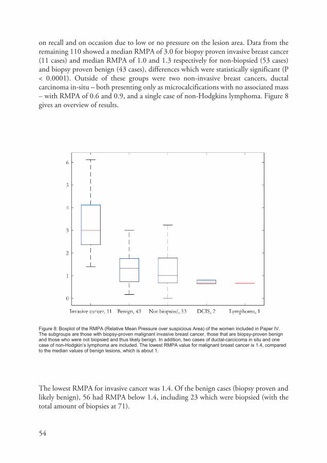
54
on recall and on occasion due to low or no pressure on the lesion area. Data from the remaining 110 showed a median RMPA of 3.0 for biopsy proven invasive breast cancer (11 cases) and median RMPA of 1.0 and 1.3 respectively for non-biopsied (53 cases) and biopsy proven benign (43 cases), differences which were statistically significant (P < 0.0001). Outside of these groups were two non-invasive breast cancers, ductal carcinoma in-situ – both presenting only as microcalcifications with no associated mass – with RMPA of 0.6 and 0.9, and a single case of non-Hodgkins lymphoma. Figure 8 gives an overview of results.
Figure 8: Boxplot of the RMPA (Relative Mean Pressure over suspicious Area) of the women included in Paper IV. The subgroups are those with biopsy-proven malignant invasive breast cancer, those that are biopsy-proven benign and those who were not biopsied and thus likely benign. In addition, two cases of ductal-carcicoma in situ and one case of non-Hodgkin’s lymphoma are included. The lowest RMPA value for malignant breast cancer is 1.4, compared to the median values of benign lesions, which is about 1.
The lowest RMPA for invasive cancer was 1.4. Of the benign cases (biopsy proven and likely benign), 56 had RMPA below 1.4, including 23 which were biopsied (with the total amount of biopsies at 71).

55
So, if adjunct mechanical imaging was implemented and available at breast cancer screening, and women were only recalled if they had a suspicious finding and RMPA readings on that suspicious finding (whether an actual lesion or not) were over the minimum threshold for malignancy, the results from this study suggests that a 36% reduction in recalls and 32% reduction in biopsies would be possible, without missing any additional invasive cancers.

-However beautiful the strategy, you should occasionally look at the results.
Winston Churchill

57
Discussion and concluding remarks
Optimization of breast compression
There are two main items of new knowledge gained from Papers I and II which helps confirm, explain or refute a number of major points of previous publications in the field, some raised in conjunction with our publications. These are both the result of analysis of the pressure distribution on the breast in different situations. Firstly, the distribution of pressure on the breast is heterogeneous when an even load is applied to it and, secondly, the distribution of pressure varies widely between different individuals.
The groups qualitatively identified in Paper I show that it is not possible to state recommendations on breast compression simply on breast area, contact area with the compression plate and compression force. The force was essentially the same in all cases and both breast area and contact area were very similar for all groups, implying that no simple size difference separates the groups. It is rather the location of the pressurized area that is important: group B and D might have the same pressurized area, but it seems more beneficial to have that area centred on the breast rather than in the juxtathoracic area.
The heterogeneity of the pressure distribution can be clearly exemplified by contrasting the mean pressure of the pressurized area of the breast actually measured by the pressure sensors with the predicted pressure arrived at by dividing the applied force by breast area. Paper I showed a mean pressure on the breast of 2.1 kPa (at full compression), and 5.6 kPa over pressurized areas. Simply dividing applied force by breast area yields a very different result, 4.9 kPa, and the same is true even if we instead base calculations on the pressurized area, 10.9 kPa (the pressurized area of the breast here is defined as the part of the breast showing pressure values above the noise level of the sensor). Previous publications have similarly suggested that the mean pressure on the compressed area is 14 kPa or higher, and should be limited to diastolic blood pressure, 10 kPa, to avoid constricting blood flow [138, 145]. Though the size of the compressed area and the pressurized area might differ, this still shows a large discrepancy with the actual pressure values found on the breast.
Partly, this is explained by the fact that some sensor elements on the breast are saturated, causing underestimation of the pressure values and also to a lesser degree by the partial area effect. It is of course impossible to determine how high the pressure values on

58
saturated elements actually are, but observing the fact that the total measured force on the breast in Paper I is roughly 50% of the applied force gives an idea.
There are two possibilities for the missing force (discounting measurement errors, discussed in more detail in the next section): either the force is in saturated elements, or the force is distributed outside of the sensor. The I-scan 9801 sensors used in Paper I covered the breast in the majority of cases, except for a narrow strip at the edges of the panel, both because of the geometry of the sensor (there is a ~3 mm wide gap from the sensor edge to the sensing elements) and because of the geometry of compression plate with its slightly rounded edge. The only region outside of the sensor is thus bordered by, almost always, high pressure values and the vast majority of saturated sensor elements, suggesting that it experiences similarly high pressure values.
The hypothesis that this narrow region accounts for the missing force is strongly supported by the fact that in Paper II, which uses the BPMS 5350 sensor that has a wider gap of ~10 mm, the ratio of force applied to force measured is even lower, 26% for the flexible plate and 17% for the rigid plate in the MLO projection (also used in Paper I). The juxtathoracic and axillary regions making up this area are normally thicker than the breast (if they are not, the likely result is group B of Paper I) and prominently include the pectoral muscle and proportionally high levels of connective tissue, meaning that they are stiff in relation to the rest of the breast. In a conference paper we repositioned breasts to partially avoid compression of the juxtathoracic area by moving them 1 cm further back supports this explanation, as mean breast pressure and pressurized breast area both increased [172]. This is very similar to the results of Paper II, as the fact that a higher force is measured on the breast using the flexible plate, even though the applied force is the same, is strongly implied to be caused by redistribution of force from a previously more compressed region, i.e. the juxtathoracic area (Figure 9).

59
Figure 9: Illustration of the difference between rigid (top) and flexible (bottom) compression plates. Using the flexible compression plate, the juxtathoracic region receives less compression, but the breast itself receives more, if using the same compression force.
Our data shows that pressures in the juxtathoracic area are very high, 40 kPa or higher in many cases, with actual pressure likely to be higher, especially outside the sensors area. Even if only the pressure actually on the breast is considered, it is clear that some regions experience very high pressures, substantially more than the 10 kPa suggested to constrict arterial blood flow [138, 145].
Several studies have shown that increasing compression above a certain level causes mostly pain and suggested that reduction of compression force have little effect on breast thickness [17, 21-23]. A high distribution of compression force to the thicker juxtathoracic area means that its stiffness increases proportionally to the less compressed breast at an equal compressed thickness. This means that to decrease the thickness of the relatively soft breast, the much stiffer, already compressed, juxtathoracic area must be compressed just as much, limiting the effect of additional compression force on thickness, but disproportionately increasing pressure on this area and therefore causing additional pain. Paper II suggests that using a flexible compression plate can alleviate this by allowing the thickness of the juxtathoracic area to be higher than that of the breast itself, and thus allowing a more even degree of compression at the prize of a non-uniform breast profile. However, pain was equivalent with either plate, which can

60
potentially have a number of explanations. We theorize that, for most women, the pain arises mainly from the juxtathoracic area using the rigid plate and the breast using the flexible plate. As the breast is the more tender of the two areas, as evidenced by the pain model of Paper I which showed a strong dependency with pressure over dense tissue, even a relatively small increase in pressure, as the one seen in Paper II will thus increase pain there and counter a reduction in pain in the overly compressed juxtathoracic area. Paper I also uses a flexible compression plate, though from a different manufacturer, and shows similar pressure value levels in the central breast.
Pain results in Paper I can have been systematically affected by always using the same order: full compression followed by reduced compression. This can be argued to have potentially affected the results in either direction: either the relief of not having as high a force on the second round gave rise to a feeling of relief, or residual tenderness from the first compression made the second one disproportionately painful. The first explanation can be considered more likely, all the more so as women were not blinded to the order in which they were compressed. Paper II used a superior methodology of randomizing the order, and those results should be more reliable.
Though it is difficult to directly compare the thickness readouts from different mammography units and models of compression plates, Paper I does show a noticeably lower differential in thickness from reduced compression force compared to a previous study with equivalent force levels but using a rigid compression plate [22]. The measured 1.8 mm difference in thickness implies a higher value close to the chest wall. Data from Kallenberg et al. suggests that the median tilt angle of the Hologic Selenia Dimensions flexible compression plate is ~2° [148]. Assuming that the same is true for the Siemens system, this means that the thickness at the chest wall would be roughly 7 mm greater than at the plate edge (plate dimensions being 20 x 28 cm).
While it must thus be kept in mind that thickness is necessarily defined differently for a flexible plate, this still seems to show that once compressed to a certain degree (showing mean pressures of ~3-5 kPa) further application of force has little effect on either pressure on, or thickness of, the breast itself. One can speculate that because the flexible plate is inherently designed with a maximum degree of flex, once it has reached this inclination it behaves essentially like a rigid plate and, to further reduce thickness, must now compress both the now already well-compressed breast and the moderately compressed juxtathoracic area. This might mean that the overall stiffness, or elastic modulus, of this combination of tissues is greater than that of a poorly compressed breast and an overly compressed juxtathoracic region, explaining the divergent results. The mean pressure over the pressurized area of the breast (on the sensor) at reduced compression in Paper I is 82% of the same value at full compression, implying that most of the additional force is absorbed elsewhere. Thus, a reduction of compression force would seem prudent, as it reduces experienced pain and substantially affects

61
neither the thickness of nor the pressure on the clinically most relevant parts of the breast.
Paper II shows that the compression equipment is unable to keep the level of compression desired by the radiographer, as the mean pressure drops after the peak value is achieved. This is often seen during examinations, as we saw during the studies: the force readout on the machine drops after the radiographer stops applying force and walks to the operating terminal to acquire images. This is analogous to the end of the “cinch” phase of compression described in other literature [173]. The problem is especially pronounced for MLOs acquired on the rigid compression plate, where the peak force is twice as high as the force during imaging. The reasons for this was clearly observed on the dynamic pressure recordings as the hand of the radiographer can be observed in contact with the pressure sensors on the breast support. Once the hand is removed, pressures drop. As long as one side of the breast is supported by the radiographer’s hand it cannot expand in that direction when compressed, becoming effectively stiffer. As the hand is removed, that support disappears and the breast can expand in that direction. Because of the oblique angle, gravity makes the breast sag further. This is probably part of the explanation for the appearance of the group D breasts from Paper I: after the supporting hand is removed, contact is lost between large parts of the breast and the compression plate, while the juxtathoracic region and pectoral muscle is kept compressed. The flexible compression plate seems to handle this situation better than the rigid, likely because of it making better contact with the breast during the “cinch” phase.
Taking all the above points into mind, it is quite clear that flexible compression plates provides “better” compression than rigid compression plates using the same amount of force. This is accomplished by redistributing force from clinically less relevant areas, to clinically more relevant ones. Pain is constant. Though Broeders et al. [146] do conclude that flexible compression plates decrease image quality in the juxtathoracic area, it can be argued that the image quality of this area is not as critical as the image quality of the breast itself. Reducing compression force by half likely has little effect on image quality in the breast itself as pressure is not much affected. Earlier literature suggests the same to be true for the rigid compression plate.
Neither Paper I nor II has directly studied the effects on image quality of the various compression modes investigated. This is of course a limitation as to the validity of the results. As noted in the background section, thickness differences in the ranges seen in especially Paper I should have little effect on image quality purely from the point-of-view of radiation absorption and scatter. Any substantial effects would thus be the result of tissue and lesion separation. From this, it can be construed that image quality would be better with higher pressures measured on the breast, indicating higher stress and thus higher strain – compression. So, even if no direct studies of image quality have been carried out, the evidence still supports better image quality with flexible plates and of

62
women in group A and B compared to group C and D. At least group D is unlikely to be improved by more vigorous compression, as so little force is distributed to the breast. On the other hand, group B, where a much greater degree of force is applied to the breast, should not need to be subjected to as high a force as the other groups, as equivalent pressures over the clinically most relevant parts are reached at a lower level.
Diagnostic aspects of mechanical imaging
The potential usefulness of mechanical imaging of the breast has been investigated before [158, 160], but not in a setting where it is explicitly intended to be used as adjunct screening, without requiring a trained operator or making the examination longer. The difference in elastic modulus between benign and malignant lesions has also been established [104, 155, 174]. The most important point of this thesis on the basis of mechanical imaging of the breast is thus that this difference can, in contrast with earlier results, be registered by a technique that is relatively simple, does not require a trained operator (as in ultrasound) or additional detection algorithms. Further, the difference in stiffness between malignant and benign breast lesions suggests a threshold for malignancy can be established, and be used to potentially increase the specificity of breast screening.
Detectability of nodules using mechanical imaging have been reported to vary by the size and depth of the nodule [158]. Though no data on lesion depth is available from our studies, the fact that all invasive malignant lesions (except for those where readings were inconclusive) showed higher RMPA than the background mean pressure in Paper IV suggests that the effect is relatively minor. Data from Paper III and additional data published in conference proceedings [175] further suggests that malignant tumours are generally detectable despite their depth, as only four of 22 lesions had mean lesion pressure below mean breast pressure, one of which was benign, one of which had very low pressure over the breast – including the lesion location – one was a non-invasive cancer and one was very small. Neither Paper III nor IV found the measured lesion pressures to correlate with lesion size. As radiological and pathologic size often diverges, and in addition, the palpable size of the lesion further differs, it is difficult to conclude the exact cause of this effect. Speculatively, it could be that the palpable size does not correlate closely with the actual size of the carcinoma. It could also be that smaller carcinomas are generally stiffer than larger carcinomas, or that most of the pressure increase detected with mechanical imaging arises over the stiffer core of the lesion and that the size of the core – sometimes containing necrotic tissue – is relatively independent of the tumour size.
Another aspect is what is actually represented by the pressure increase. At least one study has connected the stiffness of the breast with the risk of breast cancer [176]. This

63
can be seen as a simple consequence of denser breasts having a higher risk of breast cancer, but it can also be construed to mean that areas of increased stiffness have a higher risk of giving rise to carcinomas than softer areas. In that case, it might not be that the tumour itself is the main cause of stiffness increase but that the stiffness implies a high concentration of tissue components from which a tumour might originate.
Regarding possible leakage of CTCs, the results from Paper III seem to preclude a major release of cells from a compressed breast carcinoma. What evidence there exists on the magnitude of a release seemed to suggest that it could be in the region of thousands of cells per ml of blood [177, 178]. It seems possible that a release on that scale could spread the cancer, but as not even an increase of 1 cell per 7.5 ml could be seen, any “forced metastasis” of breast carcinoma caused by mammography can be tentatively ruled-out. In fact, the strain on breast tumours caused by manual breast palpation, for diagnostic purposes or otherwise, is very likely higher than that caused by mammography and would be of a greater risk of spreading the cancer. The possibility remains that an outflow of CTCs occurs, but that it never reaches the venous blood. In fact, there is evidence that CTCs are too large to pass the capillaries of the lung, which raises the question of the actual origin of CTCs [179].
The method we propose in Papers III and IV could be used fully automatically as long as a purpose-built array of sensors and readout-out electronics could be integrated in the compression plate and/or breast support. The methodology described in Paper IV could in essence – and with further investigation, particularly in automatic determination of inconclusive readings – allow the mechanical imaging data to be used without requiring any user input; once the radiologist makes a recommendation to recall and marks the location of a suspicious lesion, software could independently determine whether the marked location is above the threshold for recall based on measured surface stress. The main exception seems to be ductal carcinoma in-situ, specifically when presenting only as microcalcification clusters, as they do not seem to cause any increase in local stiffness.
For the purposes of this analysis, false-positives can be divided into two groups which we can call, lesion and non-lesion. The lesion group consists of benign lesions (cysts etc.) mistaken for malignant lesions, while the non-lesion group consists of all cases where the recall is due to e.g. over-projection of tissue and other forms of suspicious-looking normal breast tissue. Presumably, one can see the biopsy-proven benign group from Paper IV to mainly represent the former group, and the other benign group to represent the latter, likely with a number of exceptions. It could be expected that the system would work better for non-biopsied cases, as over-projection of tissue should not lead to local pressure increases. Contrary to expectations, the mechanical imaging system seems to work better for the biopsied group, with 46% being below the recall threshold compared to 39% for the non-biopsied group. This is perhaps explained by the overrepresentation of inconclusive readings among the non-biopsied group, especially

64
the cases in which the suspicious feature was not apparent on recall. This is exactly the kind of cases where the system would be most useful as the lack of any lesion, benign or malignant, to increase local pressure would mean that these cases would fall below the recall threshold.
To put the effect of reducing recalls by 36% into perspective, Erhard et al. reported that spectral mammography could potentially reduce recalls by 20% through being able to correctly differentiate benign cysts from well-circumscribed carcinomas, at the cost of 1 missed cancer per 625 correctly diagnosed benign cysts [180]. Egorov et al. suggested that the SureTouch mechanical imaging system used on recalled women in addition to or instead of ultrasound could reduce the biopsy rate by 23% without missing additional cancers, and by 50% at the cost of 4.6 % missed cancers [159]. For any such additional information to be useful, it is of course vital that the radiologist can trust the results, both objectively and subjectively. Objective proof of the reliability can be obtained from trials and studies, but it is likely more difficult to make radiologists comfortable with counter-intuitively not recalling suspicious looking findings based on, e.g. a numerical value evaluated by software.
Though, as noted in the Background section, studies on breast tomosynthesis have shown somewhat divergent results, the most optimistic appraisals foresee a 40% reduction of recalls. These results are from a US setting, i.e. the number of recalls is much higher than in European screening programs though the number of detected cancers is not. With this lower specificity, a greater number of the recalled cases should be in the non-lesion group discussed above, so in addition to the recall reduction already affecting a greater proportion of women screened, the ratio of cases below the malignancy threshold might also be greater. Mechanical Imaging might thus potentially have a greater effect in a US setting. In a future of breast tomosynthesis used as a general screening modality, mechanical imaging might also see use as an adjunct to breast tomosynthesis, further improving the method. There is evidence that the increased recalls seen with breast tomosynthesis are partly a result of some better visualized subtle structures looking similar to subtle malignant changes (ref). If that is the case, mechanical imaging could potentially be even more useful in such a setting than together with mammography.
From a practical point-of-view, our mechanical imaging system has both advantages and disadvantages compared to probe-based systems, such as the ones employed by Egorov and Wellman. The chief disadvantage is the inability to selectively examine arbitrary parts of the breast, which leads to the problem seen in Paper IV, i.e. mechanical imaging readings of a substantial number of suspicious lesions are inconclusive as they are located either outside of the field-of-view of the sensors (or for that matter the mammogram) or alternately in poorly compressed regions of the breast. It is thus important to keep the results of both Paper I and II in mind when interpreting the results of Paper III and IV. It would be preferable to have a device which could

65
more reliably apply compression to the entire breast. On the other side, the two main advantages of using the compression plate as a source of compression is that there is an even load over the entire breast with the result that values from different parts of the breast are directly comparable and that the examination is much quicker, and can even acquire simultaneous dynamic readings on the entire breast as in Paper II.
The threshold for malignancy defined in Paper IV worked well for the lesions identified in that study, but that is not necessarily true for a larger population. One issue is the one already identified in the paper, namely that DCIS has no measurable increase in pressure. The simple solution is for suspicious microcalcifications to always be recalled despite pressure values, at least when seen in isolation from any identifiable mass. The number of DCIS in the study was limited, partly because DCIS in combination with an invasive carcinoma was not defined separately, but also because stereoscopic biopsies were excluded for reasons of expediency. Suspicious microcalcifications are commonly biopsied this way.
Biopsies of benign lesions were generally reported such detail by the pathologists as malignant biopsies. This is the reason why benign lesions are not stratified according to type in Paper IV; data was limited in many cases, with pathologist reports simply stating “benign”. It is also unknown how many of the non-biopsied benign cases represented benign lesions and if some were, in fact, cancers. A false positive diagnosis is in itself known to entail an increased risk of future cancer [181, 182], which raises the question of whether a subgroup of seemingly benign lesions might represent pre-malignant stages of breast cancer. As of May 2016 no women classified as benign in Paper IV had presented with interval- or screening detected breast cancer, though in a future screening study it would be very interesting to see if the location of interval cancers could be correlated with increased pressure and if benign lesions (biopsied or not) with high pressure values would be at an increased risk of developing into malignant lesions.
Futures Aspects
Image quality studies
It is necessary to directly establish the difference in image quality suggested in various points of this thesis, preferably both for mammography and breast tomosynthesis. Both the effect of using reduced compression and of using flexible compression plates are important in this regard, as are the various groups seen in Paper I.
66
Studies on recalled women, or even only on women with diagnosed breast cancer would probably be most prudent, as using the system on a screening material would result in too few cancer cases.
Defining an optimal level of compression
Though a broad subject matter, defining a reliable standard of compression is very important. As neither vague older recommendations, rate or magnitude of thickness decrease nor newer ideas about setting a standardized pressure take into account the compression of the most clinically relevant parts of the breast, a different framework needs to be developed. The relevant quantity would be the strain of the fibroglandular tissue, more easily measured as the pressure on that tissue. If pressure measurements can be integrated with the compression plate and used routinely, this could be accomplished. In cases where there is no pressure on dense tissue, a standardized level of force could be used.
Training of radiographers
The loss of compression following the “cinch” phase seen in Paper II can perhaps be remedied through training. It would be valuable to study whether knowledge of this and various interventions would lessen this effect.
Design of new compression plates
The flexible compression plate appears to quite effectively redistribute compression force from the juxtathoracic area to the central breast. Still, most of the compression force is not distributed to the breast. This is most prominent on MLO. Designing a new compression plate that is flexible in two degrees of freedom rather than just one might be one step on the way to better compression, as it would allow force redistribution from the axillary region as well. Integrating pressure sensors would further allow compression to be adjusted to find an adequate compression level.
Integration of sensors
The mechanical imaging system used during the course of this doctoral thesis is an improvised combination of off-the-shelf sensors and software applied to a standard compression plate. Of the various technical issues limiting the scope of studies, perhaps the most serious is that the sensors are prominent on the mammogram used to match

67
anatomy. It is of paramount importance to compensate for this, either by subtraction imaging or through the use of sensors that have low enough absorption as to not appear on the mammogram.
Sensitivity of mechanical imaging as an adjunct to screening
Paper IV has established that adjunct mechanical imaging might increase the specificity of screening, but an equally important aspect is if it could possibly improve sensitivity as well. In a follow-up study, further data on the included patients in Paper IV will be analysed. This includes bilateral imaging and attempts at using asymmetries in pressure distribution to detect cancers. A future embodiment might be a system similar to mammography computer-assisted detection (CAD), where potential sites of tumours are marked on the mammogram based on elevated and/or asymmetric pressure readings.
Randomized screening studies
Papers III and IV suggest that mechanical imaging of the breast is plausible and useful, but the results are based entirely on enriched populations, i.e. recalls. Ideally, the same method should be used on a screening material. This sounds easy in theory, but because of the comparatively low rate of cancers and benign lesions, the required amount of examinations is high. Such a study should include investigation of both sensitivity and specificity, and aim to find an optimal threshold for malignancy. A further aspect would be to see if the mechanical imaging system could be used predictively, i.e. if areas of anomalously high pressure represent possible future cancers.


69
Conclusions
• Currently used compression procedures in mammography cannot guarantee adequate or justifiable compression. The heterogeneity of pressure distribution, both on a population level and on an individual level, makes it difficult to define a reproducible optimal compression level.
• Much of the compression force is distributed to the juxtathoracic area, resulting in lower than expected pressures on the breast itself.
• Use of flexible compression plates manages to redistribute a substantial amount of force from the juxtathoracic area, thus improving compression of the clinically most relevant parts of the breast.
• The mean pressure on malignant lesions is significantly different both from the mean breast pressure and from the corresponding value on benign lesions.
• Implementing mechanical imaging to detect the stiffness difference between malignant and benign breast lesions allows a threshold pressure for malignancy to be established.
• Mechanical imaging as an adjunct to mammography in breast screening can substantially reduce false positives without reducing sensitivity with regards to invasive breast cancer.

71
References
1. Cancer Research UK, http://www.cancerresearchuk.org/health-professional/cancer-statistics/worldwide-cancer
2. Bray, F., Jemal, A., Grey, N., et al.: Global cancer transitions according to the Human Development Index (2008-2030): a population-based study. Lancet Oncol 13, 790-801 (2012)
3. Okonkwo, Q.L., Draisma, G., Kinderen, A., et al.: Breast cancer screening policies in developing countries: A cost-effectiveness analysis for India. J Natl Cancer I 100, 1290-1300 (2008)
4. Parkin, D.M., Bray, F., Ferlay, J., et al.: Estimating the world cancer burden: GLOBOCAN 2000. Int J Cancer 94, 153-156 (2001)
5. Abe, O., Abe, R., Enomoto, K., et al.: Effects of chemotherapy and hormonal therapy for early breast cancer on recurrence and 15-year survival: an overview of the randomised trials. Lancet 365, 1687-1717 (2005)
6. Oeffinger, K.C., Fontham, E.T.H., Etzioni, R., et al.: Breast Cancer Screening for Women at Average Risk 2015 Guideline Update From the American Cancer Society. Jama-J Am Med Assoc 314, 1599-1614 (2015)
7. Independent, U.K.P.o.B.C.S.: The benefits and harms of breast cancer screening: an independent review. Lancet 380, 1778-1786 (2012)
8. Wilson, J.M.G., Jungner, G.: Principles and practice of screening for disease. World Health Organization, Geneva, (1968)
9. Cole, P., Morrison, A.S.: Basic Issues in Population Screening for Cancer. J Natl Cancer I 64, 1263-1272 (1980)
10. Morrison, A.S.: Screening. In: Rothman, K.J., Greenland, S. (eds.) Modern epidemiology. Lippincott-Raven, Philadelphia (1998)
11. WHO (ed.): Cancer control: Early detection WHO, Geneva (2007) 12. Sankaranarayanan, R.: Screening for cancer in low- and middle-income countries.
Ann Glob Health 80, 412-417 (2014) 13. WHO: WHO position paper on mammography screening. (2014) 14. Gotzsche, P.C., Nielsen, M.: Screening for breast cancer with mammography.
Cochrane Database Syst Rev CD001877 (2011) 15. Hofvind, S.S., Wang, H., Thoresen, S.: The Norwegian Breast Cancer Screening
Program: re-attendance related to the women's experiences, intentions and previous screening result. Cancer Causes Control 14, 391-398 (2003)

72
16. Orton, M., Fitzpatrick, R., Fuller, A., et al.: Factors affecting women's response to an invitation to attend for a second breast cancer screening examination. Br J Gen Pract 41, 320-322 (1991)
17. Poulos, A., McLean, D., Rickard, M., et al.: Breast compression in mammography: how much is enough? Australas Radiol 47, 121-126 (2003)
18. Keemers-Gels, M.E., Groenendijk, R.P., van den Heuvel, J.H., et al.: Pain experienced by women attending breast cancer screening. Breast Cancer Res Treat 60, 235-240 (2000)
19. Whelehan, P., Evans, A., Wells, M., et al.: The effect of mammography pain on repeat participation in breast cancer screening: a systematic review. Breast 22, 389-394 (2013)
20. Miller, D., Livingstone, V., Herbison, P.: Interventions for relieving the pain and discomfort of screening mammography. Cochrane Db Syst Rev (2008)
21. Chida, K., Komatsu, Y., Sai, M., et al.: Reduced compression mammography to reduce breast pain. Clin Imag 33, 7-10 (2009)
22. Fornvik, D., Andersson, I., Svahn, T., et al.: The effect of reduced breast compression in breast tomosynthesis: human observer study using clinical cases. Radiat Prot Dosim 139, 118-123 (2010)
23. Suhaimi, S.A.A., Mohamed, A., Ahmad, M., et al.: Effects of Reduced Compression in Digital Breast Tomosynthesis on Pain, Anxiety, and Image Quality. Malays J Med Sci 22, 40-46 (2015)
24. Pisano, E.D., Gatsonis, C., Hendrick, E., et al.: Diagnostic performance of digital versus film mammography for breast-cancer screening. N Engl J Med 353, 1773-1783 (2005)
25. Skaane, P., Hofvind, S., Skjennald, A.: Randomized trial of screen-film versus full-field digital mammography with soft-copy reading in population-based screening program: follow-up and final results of Oslo II study. Radiology 244, 708-717 (2007)
26. Domingo, L., Hofvind, S., Hubbard, R.A., et al.: Cross-national comparison of screening mammography accuracy measures in U.S., Norway, and Spain. Eur Radiol 26, 2520-2528 (2016)
27. Bolejko, A., Hagell, P., Wann-Hansson, C., et al.: Prevalence, Long-term Development, and Predictors of Psychosocial Consequences of False-Positive Mammography among Women Attending Population-Based Screening. Cancer Epidem Biomar 24, 1388-1397 (2015)
28. Sarvazyan, A., Egorov, V.: Mechanical Imaging - a Technology for 3-D Visualization and Characterization of Soft Tissue Abnormalities: A Review. Curr Med Imaging Rev 8, 64-73 (2012)
29. Ferlay, J., Soerjomataram, I., Dikshit, R., et al.: Cancer incidence and mortality worldwide: sources, methods and major patterns in GLOBOCAN 2012. Int J Cancer 136, E359-386 (2015)
30. Bray, F., Ferlay, J., Laversanne, M., et al.: Cancer Incidence in Five Continents: Inclusion criteria, highlights from Volume X and the global status of cancer registration. Int J Cancer 137, 2060-2071 (2015)

73
31. Engholm G, F.J., Christensen N, Kejs AMT, Hertzum-Larsen R, Johannesen TB, Khan S, Leinonen MK, Ólafsdóttir E, Petersen T, Schmidt LKH, Trykker H, Storm HH.: NORDCAN: Cancer Incidence, Mortality, Prevalence and Survival in the Nordic Countries, Version 7.3 (08.07.2016). Association of the Nordic Cancer Registries. Danish Cancer Society (2016)
32. Parkin, D.M., Boyd, L., Walker, L.C.: 16. The fraction of cancer attributable to lifestyle and environmental factors in the UK in 2010. Br J Cancer 105 Suppl 2, S77-81 (2011)
33. Foucar, E.: Do pathologists play dice? Uncertainty and early histopathological diagnosis of common malignancies. Histopathology 31, 495-502 (1997)
34. Kerlikowske, K., Molinaro, A.M., Gauthier, M.L., et al.: Biomarker expression and risk of subsequent tumors after initial ductal carcinoma in situ diagnosis. J Natl Cancer Inst 102, 627-637 (2010)
35. Molinaro, A.M., Sison, J.D., Ljung, B.M., et al.: Risk prediction for local versus regional/metastatic tumors after initial ductal carcinoma in situ diagnosis treated by lumpectomy. Breast Cancer Res Treat 157, 351-361 (2016)
36. Andersson, I.: Invasive Breast Cancer. In: Gourtsoyiannis, N.C.R., Pablo R. (ed.) Radiologic-Pathologic Correlations from Head to Toe. Springer-Verlag, Heidelberg (2005)
37. Shaheen, R., Schimmelpenninck, C.A., Stoddart, L., et al.: Spectrum of Diseases Presenting as Architectural Distortion on Mammography: Multimodality Radiologic Imaging with Pathologic Correlation. Semin Ultrasound Ct 32, 351-362 (2011)
38. Evans, A.J., Pinder, S.E., James, J.J., et al.: Is mammographic spiculation an independent, good prognostic factor in screening-detected invasive breast cancer? AJR Am J Roentgenol 187, 1377-1380 (2006)
39. Moriuchi, H., Yamaguchi, J., Hayashi, H., et al.: Cancer Cell Interaction with Adipose Tissue: Correlation with the Finding of Spiculation at Mammography. Radiology 279, 56-64 (2016)
40. Cristofanilli, M., Budd, G.T., Ellis, M.J., et al.: Circulating tumor cells, disease progression, and survival in metastatic breast cancer. N Engl J Med 351, 781-791 (2004)
41. Bidard, F.C., Mathiot, C., Delaloge, S., et al.: Single circulating tumor cell detection and overall survival in nonmetastatic breast cancer. Ann Oncol 21, 729-733 (2010)
42. Budd, G.T., Cristofanilli, M., Ellis, M.J., et al.: Circulating tumor cells versus imaging--predicting overall survival in metastatic breast cancer. Clin Cancer Res 12, 6403-6409 (2006)
43. Franken, B., de Groot, M.R., Mastboom, W.J., et al.: Circulating tumor cells, disease recurrence and survival in newly diagnosed breast cancer. Breast Cancer Res 14, R133 (2012)
44. Watmough, D.J., Quan, K.M.: X-ray mammography and breast compression. Lancet 340, 122 (1992)
45. Choy, A., McCulloch, P.: Induction of tumour cell shedding into effluent venous blood breast cancer surgery. Br J Cancer 73, 79-82 (1996)

74
46. Gotzsche, P.C., Jorgensen, K.J.: Screening for breast cancer with mammography. Cochrane Database Syst Rev CD001877 (2013)
47. Gotzsche, P.C., Jorgensen, K.J.: The benefits and harms of breast cancer screening. Lancet 381, 799 (2013)
48. http://www.socialstyrelsen.se/SiteCollectionDocuments/screening-brostcancer-rekommendation.pdf
49. Andersson, I., Hildell, J., Muhlow, A., et al.: Number of Projections in Mammography - Influence on Detection of Breast Disease. Am J Roentgenol 130, 349-351 (1978)
50. Ikeda, D.M., Sickles, E.A.: Second-screening mammography: one versus two views per breast. Radiology 168, 651-656 (1988)
51. Kim, S.J., Moon, W.K., Cho, N., et al.: Computer-aided detection in digital mammography: comparison of craniocaudal, mediolateral oblique, and mediolateral views. Radiology 241, 695-701 (2006)
52. Young, K.C.: Radiation doses in the UK trial of breast screening in women aged 40-48 years. Br J Radiol 75, 362-370 (2002)
53. Young, K.C., Burch, A., Oduko, J.M.: Radiation doses received in the UK Breast Screening Programme in 2001 and 2002. Br J Radiol 78, 207-218 (2005)
54. Armstrong, K., Moye, E., Williams, S., et al.: Screening mammography in women 40 to 49 years of age: a systematic review for the American College of Physicians. Ann Intern Med 146, 516-526 (2007)
55. Skaane, P.: Studies comparing screen-film mammography and full-field digital mammography in breast cancer screening: updated review. Acta Radiol 50, 3-14 (2009)
56. Sechopoulos, I.: A review of breast tomosynthesis. Part II. Image reconstruction, processing and analysis, and advanced applications. Med Phys 40, 014302 (2013)
57. Sechopoulos, I.: A review of breast tomosynthesis. Part I. The image acquisition process. Med Phys 40, 014301 (2013)
58. Niklason, L.T., Christian, B.T., Niklason, L.E., et al.: Digital Tomosynthesis in breast imaging. Radiology 205, 399-406 (1997)
59. Wu, T., Stewart, A., Stanton, M., et al.: Tomographic mammography using a limited number of low-dose cone-beam projection images. Medical Physics 30, 365-380 (2003)
60. Houssami, N., Skaane, P.: Overview of the evidence on digital breast tomosynthesis in breast cancer detection. Breast 22, 101-108 (2013)
61. Gur, D., Abrams, G.S., Chough, D.M., et al.: Digital Breast Tomosynthesis: Observer Performance Study. Am J Roentgenol 193, 586-591 (2009)
62. Timberg, P., Bath, M., Andersson, I., et al.: In-plane visibility of lesions using breast tomosynthesis and digital mammography. Medical Physics 37, 5618-5626 (2010)
63. Bernardi, D., Ciatto, S., Pellegrini, M., et al.: Application of breast tomosynthesis in screening: incremental effect on mammography acquisition and reading time. Br J Radiol 85, e1174-1178 (2012)

75
64. Dustler, M., Andersson, M., Fornvik, D., et al.: A Study of the Feasibility of using slabbing to reduce Tomosynthesis Review Time. Proc Spie 8673, (2013)
65. Skaane, P., Bandos, A.I., Gullien, R., et al.: Comparison of Digital Mammography Alone and Digital Mammography Plus Tomosynthesis in a Population-based Screening Program. Radiology 267, 47-56 (2013)
66. Skaane, P., Bandos, A.I., Eben, E.B., et al.: Two-view digital breast tomosynthesis screening with synthetically reconstructed projection images: comparison with digital breast tomosynthesis with full-field digital mammographic images. Radiology 271, 655-663 (2014)
67. Ciatto, S., Houssami, N., Bernardi, D., et al.: Integration of 3D digital mammography with tomosynthesis for population breast-cancer screening (STORM): a prospective comparison study. Lancet Oncol 14, 583-589 (2013)
68. Lang, K., Andersson, I., Rosso, A., et al.: Performance of one-view breast tomosynthesis as a stand-alone breast cancer screening modality: results from the Malmo Breast Tomosynthesis Screening Trial, a population-based study. European Radiology 26, 184-190 (2016)
69. Haas, B.M., Kalra, V., Geisel, J., et al.: Comparison of tomosynthesis plus digital mammography and digital mammography alone for breast cancer screening. Radiology 269, 694-700 (2013)
70. Rose, S.L., Tidwell, A.L., Bujnoch, L.J., et al.: Implementation of breast tomosynthesis in a routine screening practice: an observational study. AJR Am J Roentgenol 200, 1401-1408 (2013)
71. Friedewald, S.M., Rafferty, E.A., Rose, S.L., et al.: Breast cancer screening using tomosynthesis in combination with digital mammography. JAMA 311, 2499-2507 (2014)
72. Greenberg, J.S., Javitt, M.C., Katzen, J., et al.: Clinical performance metrics of 3D digital breast tomosynthesis compared with 2D digital mammography for breast cancer screening in community practice. AJR Am J Roentgenol 203, 687-693 (2014)
73. McCarthy, A.M., Kontos, D., Synnestvedt, M., et al.: Screening outcomes following implementation of digital breast tomosynthesis in a general-population screening program. J Natl Cancer Inst 106, (2014)
74. Durand, M.A., Haas, B.M., Yao, X., et al.: Early clinical experience with digital breast tomosynthesis for screening mammography. Radiology 274, 85-92 (2015)
75. Lourenco, A.P., Barry-Brooks, M., Baird, G.L., et al.: Changes in recall type and patient treatment following implementation of screening digital breast tomosynthesis. Radiology 274, 337-342 (2015)
76. Stavros, A.T., Thickman, D., Rapp, C.L., et al.: Solid breast nodules: use of sonography to distinguish between benign and malignant lesions. Radiology 196, 123-134 (1995)
77. Flobbe, K., Nelemans, P.J., Kessels, A.G., et al.: The role of ultrasonography as an adjunct to mammography in the detection of breast cancer. a systematic review. Eur J Cancer 38, 1044-1050 (2002)

76
78. Britton, P., Warwick, J., Wallis, M.G., et al.: Measuring the accuracy of diagnostic imaging in symptomatic breast patients: team and individual performance. Br J Radiol 85, 415-422 (2012)
79. Berg, W.A., Blume, J.D., Cormack, J.B., et al.: Combined screening with ultrasound and mammography vs mammography alone in women at elevated risk of breast cancer. JAMA 299, 2151-2163 (2008)
80. Sprague, B.L., Stout, N.K., Schechter, C., et al.: Benefits, Harms, and Cost-Effectiveness of Supplemental Ultrasonography Screening for Women With Dense Breasts. Annals of Internal Medicine 162, 157-U133 (2015)
81. Melnikow, J., Fenton, J.J., Whitlock, E.P., et al.: Supplemental Screening for Breast Cancer in Women With Dense Breasts: A Systematic Review for the US Preventive Services Task Force. Annals of Internal Medicine 164, 268-+ (2016)
82. Sarvazyan, A., Egorov, V., Son, J.S., et al.: Cost-effective screening for breast cancer worldwide: current state and future directions. Breast Cancer (Auckl) 1, 91-99 (2008)
83. Ohuchi, N., Ishida, T., Kawai, M., et al.: Randomized controlled trial on effectiveness of ultrasonography screening for breast cancer in women aged 40-49 (J-START): research design. Jpn J Clin Oncol 41, 275-277 (2011)
84. Ishida, T., Suzuki, A., Kawai, M., et al.: A randomized controlled trial to verify the efficacy of the use of ultrasonography in breast cancer screening aged 40-49 (J-START): 76 196 women registered. Jpn J Clin Oncol 44, 134-140 (2014)
85. Kelly, K.M., Dean, J., Comulada, W.S., et al.: Breast cancer detection using automated whole breast ultrasound and mammography in radiographically dense breasts. European Radiology 20, 734-742 (2010)
86. Wojcinski, S., Gyapong, S., Farrokh, A., et al.: Diagnostic performance and inter-observer concordance in lesion detection with the automated breast volume scanner (ABVS). Bmc Med Imaging 13, (2013)
87. Giger, M.L., Inciardi, M.F., Edwards, A., et al.: Automated Breast Ultrasound in Breast Cancer Screening of Women With Dense Breasts: Reader Study of Mammography-Negative and Mammography-Positive Cancers. Am J Roentgenol 206, 1341-1350 (2016)
88. Ranger, B., Littrup, P.J., Duric, N., et al.: Breast ultrasound tomography versus MRI for clinical display of anatomy and tumor rendering: preliminary results. AJR Am J Roentgenol 198, 233-239 (2012)
89. O'Flynn, E., Fromageau, J., Ledger, M., et al.: Breast density measurements with ultrasound tomography: a comparison with non-contrast MRI. Breast Cancer Res 17, (2015)
90. Duric, N., Boyd, N., Littrup, P., et al.: Breast density measurements with ultrasound tomography: A comparison with film and digital mammography. Medical Physics 40, (2013)
91. Heywang-Kobrunner, S.H.: Magnetic Resonance Imaging: The Evolution of Breast Imaging. Breast 22, S8-S9 (2013)
92. Peters, N.H.G.M., Rinkes, I.H.M.B., Zuithoff, N.P.A., et al.: Meta-analysis of MR imaging in the diagnosis of breast lesions. Radiology 246, 116-124 (2008)

77
93. Lauby-Secretan, B., Scoccianti, C., Loomis, D., et al.: Breast-Cancer Screening - Viewpoint of the IARC Working Group. New Engl J Med 372, 2353-2358 (2015)
94. Mann, R.M., Loo, C.E., Wobbes, T., et al.: The impact of preoperative breast MRI on the re-excision rate in invasive lobular carcinoma of the breast. Breast Cancer Res Tr 119, 415-422 (2010)
95. Caramella, T., Chapellier, C., Ettore, F., et al.: Value of MRI in the surgical planning of invasive lobular breast carcinoma: a prospective and a retrospective study of 57 cases - Comparison with physical examination, conventional imaging, and histology. Clin Imag 31, 155-161 (2007)
96. Mango, V.L., Morris, E.A., David Dershaw, D., et al.: Abbreviated protocol for breast MRI: are multiple sequences needed for cancer detection? Eur J Radiol 84, 65-70 (2015)
97. Kuhl, C.K., Schrading, S., Strobel, K., et al.: Abbreviated breast magnetic resonance imaging (MRI): first postcontrast subtracted images and maximum-intensity projection-a novel approach to breast cancer screening with MRI. J Clin Oncol 32, 2304-2310 (2014)
98. Engelken, F.J., Sack, I., Klatt, D., et al.: Evaluation of tomosynthesis elastography in a breast-mimicking phantom. Eur J Radiol 81, 2169-2173 (2012)
99. Sun, C., Standish, B., Yang, V.X.: Optical coherence elastography: current status and future applications. J Biomed Opt 16, 043001 (2011)
100. Goddi, A., Bonardi, M., Alessi, S.: Breast elastography: A literature review. J Ultrasound 15, 192-198 (2012)
101. Gennisson, J.L., Deffieux, T., Fink, M., et al.: Ultrasound elastography: principles and techniques. Diagn Interv Imaging 94, 487-495 (2013)
102. Bercoff, J., Chaffai, S., Tanter, M., et al.: In vivo breast tumor detection using transient elastography. Ultrasound Med Biol 29, 1387-1396 (2003)
103. Tanter, M., Bercoff, J., Athanasiou, A., et al.: Quantitative assessment of breast lesion viscoelasticity: Initial clinical results using supersonic shear imaging. Ultrasound Med Biol 34, 1373-1386 (2008)
104. Garra, B.S., Cespedes, E.I., Ophir, J., et al.: Elastography of breast lesions: initial clinical results. Radiology 202, 79-86 (1997)
105. Itoh, A., Ueno, E., Tohno, E., et al.: Breast disease: Clinical application of US elastography for diagnosis. Radiology 239, 341-350 (2006)
106. Zhi, H., Ou, B., Luo, B.M., et al.: Comparison of ultrasound elastography, mammography, and sonography in the diagnosis of solid breast lesions. J Ultras Med 26, 807-815 (2007)
107. Skerl, K., Vinnicombe, S., Thomson, K., et al.: Anisotropy of Solid Breast Lesions in 2D Shear Wave Elastography is an Indicator of Malignancy. Acad Radiol 23, 53-61 (2016)
108. Evans, A., Sim, Y.T., Thomson, K., et al.: Shear wave elastography of breast cancer: Sensitivity according to histological type in a large cohort. Breast 26, 115-118 (2016)
109. Mariappan, Y.K., Glaser, K.J., Ehman, R.L.: Magnetic Resonance Elastography: A Review. Clin Anat 23, 497-511 (2010)

78
110. Low, G., Kruse, S.A., Lomas, D.J.: General review of magnetic resonance elastography. World J Radiol 8, 59-72 (2016)
111. Sinkus, R., Tanter, M., Catheline, S., et al.: Imaging anisotropic and viscous properties of breast tissue by magnetic resonance-elastography. Magnet Reson Med 53, 372-387 (2005)
112. Sinkus, R., Siegmann, K., Xydeas, T., et al.: MR elastography of breast lesions: Understanding the solid/liquid duality can improve the specificity of contrast-enhanced MR mammography. Magnet Reson Med 58, 1135-1144 (2007)
113. Siegmann, K.C., Xydeas, T., Sinkus, R., et al.: Diagnostic value of MR elastography in addition to contrast-enhanced MR imaging of the breast-initial clinical results. Eur Radiol 20, 318-325 (2010)
114. Fredenberg, E., Hemmendorff, M., Cederstrom, B., et al.: Contrast-enhanced spectral mammography with a photon-counting detector. Med Phys 37, 2017-2029 (2010)
115. Fallenberg, E.M., Dromain, C., Diekmann, F., et al.: Contrast-enhanced spectral mammography versus MRI: Initial results in the detection of breast cancer and assessment of tumour size. Eur Radiol 24, 256-264 (2014)
116. Lobbes, M.B., Lalji, U., Houwers, J., et al.: Contrast-enhanced spectral mammography in patients referred from the breast cancer screening programme. Eur Radiol 24, 1668-1676 (2014)
117. Prionas, N.D., Lindfors, K.K., Ray, S., et al.: Contrast-enhanced dedicated breast CT: initial clinical experience. Radiology 256, 714-723 (2010)
118. Lindfors, K.K., Boone, J.M., Nelson, T.R., et al.: Dedicated breast CT: initial clinical experience. Radiology 246, 725-733 (2008)
119. Kuzmiak, C.M., Cole, E.B., Zeng, D., et al.: Dedicated Three-dimensional Breast Computed Tomography: Lesion Characteristic Perception by Radiologists. J Clin Imaging Sci 6, 14 (2016)
120. Kalender, W.A., Kolditz, D., Steiding, C., et al.: Technical feasibility proof for high-resolution low-dose photon-counting CT of the breast. Eur Radiol (2016)
121. Maublant, J., deLatour, M., Mestas, D., et al.: Technetium-99m-sestamibi uptake in breast tumor and associated lymph nodes. J Nucl Med 37, 922-925 (1996)
122. Pieper, B.C., Bowsher, J.E., Tornai, M.P., et al.: Breast tumor imaging using a tiltable head SPECT camera. Ieee T Nucl Sci 48, 1477-1482 (2001)
123. Perez, K.L., Cutler, S.J., Madhav, P., et al.: Characterizing the contribution of cardiac and hepatic uptake in dedicated breast SPECT using tilted trajectories. Phys Med Biol 55, 4721-4734 (2010)
124. Moliner, L., Gonzalez, A.J., Soriano, A., et al.: Design and evaluation of the MAMMI dedicated breast PET. Medical Physics 39, 5393-5404 (2012)
125. Castelli, E., Tonutti, M., Arfelli, F., et al.: Mammography with Synchrotron Radiation: First Clinical Experience with Phase-Detection Technique. Radiology 259, 684-694 (2011)
126. Longo, R., Arfelli, F., Bellazzini, R., et al.: Towards breast tomography with synchrotron radiation at Elettra: first images. Phys Med Biol 61, 1634-1649 (2016)

79
127. Perry, N., Broeders, M., de Wolf, C., et al.: European guidelines for quality assurance in breast cancer screening and diagnosis. Fourth edition--summary document. Ann Oncol 19, 614-622 (2008)
128. Domingo, L., Hofvind, S., Hubbard, R.A., et al.: Cross-national comparison of screening mammography accuracy measures in U.S., Norway, and Spain. Eur Radiol (2015)
129. Hofvind, S., Ponti, A., Patnick, J., et al.: False-positive results in mammographic screening for breast cancer in Europe: a literature review and survey of service screening programmes. J Med Screen 19, 57-66 (2012)
130. Lagerlund, M., Sontrop, J.M., Zackrisson, S.: Psychosocial factors and attendance at a population-based mammography screening program in a cohort of Swedish women. Bmc Womens Health 14, (2014)
131. Esteva, M., Ripoll, J., Leiva, A., et al.: Determinants of non attendance to mammography program in a region with high voluntary health insurance coverage. BMC Public Health 8, 387 (2008)
132. Society, A.C.: Breast Cancer Facts & Figures 2015-2016. (2015) 133. Zackrisson, S., Andersson, I., Manjer, J., et al.: Non-attendance in breast cancer
screening is associated with unfavourable socio-economic circumstances and advanced carcinoma. Int J Cancer 108, 754-760 (2004)
134. Tatla, R.K., Paszat, L.F., Bondy, S.J., et al.: Socioeconomic status & returning for a second screen in the Ontario breast screening program. Breast 12, 237-246 (2003)
135. Aro, A.R., de Koning, H.J., Absetz, P., et al.: Psychosocial predictors of first attendance for organised mammography screening. J Med Screen 6, 82-88 (1999)
136. Elwood, M., McNoe, B., Smith, T., et al.: Once is enough--why some women do not continue to participate in a breast cancer screening programme. N Z Med J 111, 180-183 (1998)
137. Branderhorst, W., de Groot, J.E., Highnam, R., et al.: Mammographic compression--a need for mechanical standardization. Eur J Radiol 84, 596-602 (2015)
138. de Groot, J.E., Branderhorst, W., Grimbergen, C.A., et al.: Towards personalized compression in mammography: a comparison study between pressure- and force-standardization. Eur J Radiol 84, 384-391 (2015)
139. Mercer, C.E., Hogg, P., Lawson, R., et al.: Practitioner compression force variability in mammography: a preliminary study. Br J Radiol 86, 20110596 (2013)
140. Boyd, N.F., Martin, L.J., Yaffe, M.J., et al.: Mammographic density and breast cancer risk: current understanding and future prospects. Breast Cancer Res 13, (2011)
141. D'Orsi, C.J.S., E.A; Mendelson, E.B.; Morris, E.A.; et al. : ACR BI-RADS® Atlas, Breast Imaging Reporting and Data System. American College of Radiolog, Reston, VA (2013)
142. Saunders, R.S., Jr., Samei, E.: The effect of breast compression on mass conspicuity in digital mammography. Med Phys 35, 4464-4473 (2008)
143. Peipins, L.A., Shapiro, J.A., Bobo, J.K., et al.: Impact of women's experiences during mammography on adherence to rescreening (United States). Cancer Causes Control 17, 439-447 (2006)

80
144. Poulos, A., McLean, D.: The application of breast compression in mammography: a new perspective. Radiography 10, 131-137 (2004)
145. de Groot, J.E., Broeders, M.J.M., Branderhorst, W., et al.: A novel approach to mammographic breast compression: Improved standardization and reduced discomfort by controlling pressure instead of force. Medical Physics 40, (2013)
146. Broeders, M.J.M., ten Voorde, M., Veldkamp, W.J.H., et al.: Comparison of a flexible versus a rigid breast compression paddle: pain experience, projected breast area, radiation dose and technical image quality. European Radiology 25, 821-829 (2015)
147. Kallenberg, M.G., van Gils, C.H., Lokate, M., et al.: Effect of compression paddle tilt correction on volumetric breast density estimation. Phys Med Biol 57, 5155-5168 (2012)
148. Kallenberg, M.G., Karssemeijer, N.: Compression paddle tilt correction in full-field digital mammograms. Phys Med Biol 57, 703-715 (2012)
149. Mawdsley, G.E., Tyson, A.H., Peressotti, C.L., et al.: Accurate estimation of compressed breast thickness in mammography. Med Phys 36, 577-586 (2009)
150. Dibble, S.L., Israel, J., Nussey, B., et al.: Mammography with breast cushions. Womens Health Issues 15, 55-63 (2005)
151. Markle, L., Roux, S., Sayre, J.W.: Reduction of discomfort during mammography utilizing a radiolucent cushioning pad. Breast J 10, 345-349 (2004)
152. Tabar, L., Lebovic, G.S., Hermann, G.D., et al.: Clinical assessment of a radiolucent cushion for mammography. Acta Radiol 45, 154-158 (2004)
153. Kornguth, P.J., Rimer, B.K., Conaway, M.R., et al.: Impact of patient-controlled compression on the mammography experience. Radiology 186, 99-102 (1993)
154. Miller, D., Livingstone, V., Herbison, P.: Interventions for relieving the pain and discomfort of screening mammography. Cochrane Database Syst Rev CD002942 (2008)
155. Krouskop, T.A., Wheeler, T.M., Kallel, F., et al.: Elastic moduli of breast and prostate tissues under compression. Ultrasonic Imaging 20, 260-274 (1998)
156. Sarvazyan, A.P., Egorov, V.: Mechanical Imaging in Medical Applications. Ieee Eng Med Bio 1975-1978 (2009)
157. Evans, A., Whelehan, P., Thomson, K., et al.: Differentiating benign from malignant solid breast masses: value of shear wave elastography according to lesion stiffness combined with greyscale ultrasound according to BI-RADS classification. Brit J Cancer 107, 224-229 (2012)
158. Egorov, V., Sarvazyan, A.P.: Mechanical imaging of the breast. IEEE Trans Med Imaging 27, 1275-1287 (2008)
159. Egorov, V., Kearney, T., Pollak, S.B., et al.: Differentiation of benign and malignant breast lesions by mechanical imaging. Breast Cancer Res Treat 118, 67-80 (2009)
160. Wellman, P.S., Dalton, E.P., Krag, D., et al.: Tactile imaging of breast masses: first clinical report. Arch Surg 136, 204-208 (2001)
161. Weiss, R.E., Egorov, V., Ayrapetyan, S., et al.: Prostate mechanical imaging: a new method for prostate assessment. Urology 71, 425-429 (2008)

81
162. Egorov, V., van Raalte, H., Sarvazyan, A.P.: Vaginal tactile imaging. IEEE Trans Biomed Eng 57, 1736-1744 (2010)
163. Brimacombe, J.M., Wilson, D.R., Hodgson, A.J., et al.: Effect of calibration method on Tekscan sensor accuracy. J Biomech Eng 131, 034503 (2009)
164. Bachus, K.N., DeMarco, A.L., Judd, K.T., et al.: Measuring contact area, force, and pressure for bioengineering applications: using Fuji Film and TekScan systems. Med Eng Phys 28, 483-488 (2006)
165. Zammit, G.V., Menz, H.B., Munteanu, S.E.: Reliability of the TekScan MatScan(R) system for the measurement of plantar forces and pressures during barefoot level walking in healthy adults. J Foot Ankle Res 3, 11 (2010)
166. Koch, M.: [Measuring plantar pressure in conventional shoes with the TEKSCAN sensory system]. Biomed Tech (Berl) 38, 243-248 (1993)
167. Wirz, D., Becker, R., Li, S.F., et al.: [Validation of the Tekscan system for statistic and dynamic pressure measurements of the human femorotibial joint]. Biomed Tech (Berl) 47, 195-201 (2002)
168. Rose, J.G., Stith, J.C.: Pressure measurements in railroad trackbeds at the rail/tie interface using Tekscan sensors. In: American Railway Engineering and Maintenance-of-Way Assoc. 2004 Annual Conference PROCEEDINGS, Nashville, TN, September. (Year)
169. Macias, B.R., Murthy, G., Chambers, H., et al.: Asymmetric Loads and Pain Associated With Backpack Carrying by Children. J Pediatr Orthoped 28, 512-517 (2008)
170. Jones, G.R., Hooper, R.H.: The effect of single- or multiple-layered garments on interface pressure measured at the backpack-shoulder interface. Appl Ergon 36, 79-83 (2005)
171. Wettenschwiler, P.D., Stampfli, R., Lorenzetti, S., et al.: How reliable are pressure measurements with Tekscan sensors on the body surface of human subjects wearing load carriage systems? Int J Ind Ergonom 49, 60-67 (2015)
172. Dustler, M., Andersson, I., Fornvik, D., et al.: The Effect of Breast Positioning on Breast Compression in Mammography: a Pressure Distribution Perspective. Medical Imaging 2012: Physics of Medical Imaging 8313, (2012)
173. de Groot, J.E., Broeders, M.J., Grimbergen, C.A., et al.: Pain-preventing strategies in mammography: an observational study of simultaneously recorded pain and breast mechanics throughout the entire breast compression cycle. Bmc Womens Health 15, 26 (2015)
174. Krouskop, T.A., Wheeler, T.M., Kallel, F., et al.: Elastic moduli of breast and prostate tissues under compression. Ultrason Imaging 20, 260-274 (1998)
175. Fornvik, D., Dustler, M., Andersson, I., et al.: Pressure distribution in mammography: compression of breasts with malignant tumor masses. Proc Spie 8668, (2013)
176. Boyd, N.F., Li, Q., Melnichouk, O., et al.: Evidence that breast tissue stiffness is associated with risk of breast cancer. PLoS One 9, e100937 (2014)

82
177. Romsdahl, M.M., McGrath, R.G., Hoppe, E., et al.: Experimental Model for the Study of Tumor Cells in the Blood. Acta Cytol 9, 141-145 (1965)
178. Liotta, L.A., Kleinerman, J., Saidel, G.M.: Quantitative relationships of intravascular tumor cells, tumor vessels, and pulmonary metastases following tumor implantation. Cancer Res 34, 997-1004 (1974)
179. Weiss, L., Orr, F.W., Honn, K.V.: Interactions of cancer cells with the microvasculature during metastasis. FASEB J 2, 12-21 (1988)
180. Erhard, K., Kilburn-Toppin, F., Willsher, P., et al.: Characterization of Cystic Lesions by Spectral Mammography: Results of a Clinical Pilot Study. Invest Radiol 51, 340-347 (2016)
181. Roman, M., Castells, X., Hofvind, S., et al.: Risk of breast cancer after false-positive results in mammographic screening. Cancer Med 5, 1298-1306 (2016)
182. Castells, X., Tora-Rocamora, I., Posso, M., et al.: Risk of Breast Cancer in Women with False-Positive Results according to Mammographic Features. Radiology 280, 379-386 (2016)