presidential address transposable elemen ts,...
TRANSCRIPT
9 NOVEMBER 2012 VOL 338 SCIENCE www.sciencemag.org 758
ASSOCIATION AFFAIRS
Transposable genetic elements (TEs) com-
prise a vast array of DNA sequences, all
having the ability to move to new sites in
genomes either directly by a cut-and-paste
mechanism (transposons) or indirectly
through an RNA intermediate (retrotrans-
posons). First discovered in maize plants by
the brilliant geneticist Barbara McClintock
in the mid-1940s, they were initially con-
sidered something of a genetic oddity (1,
2). Several decades later, TEs acquired the
anthropomorphic labels of “selfish” and
“parasitic” because of their replicative
autonomy and potential for genetic disrup-
tion (3, 4). However, TEs generally exist in
eukaryotic genomes in a reversibly inactive,
genetically undetectable form we now call
“epigenetically silenced,” whose discovery
can also be traced to McClintock’s elegant
genetic studies (5, 6). As the underlying bio-
chemical mechanisms emerged from obscu-
rity and epigenetics became popular toward
the end of the 20th century, it was proposed
that epigenetic silencing evolved to control
the proliferation of TEs and their perceived
destructive potential (5, 6).
Today, we know that TEs constitute more
than half of the DNA in many higher eukary-
otes. We know, too, that the fi ngerprints of
TEs and transposition are everywhere in
their genomes, from the coarsest features of
genomic landscapes and how they change
through real and evolutionary time to the
fi nest details of gene structure and regula-
tion. My purpose here is to challenge the
current, somewhat pejorative, view of TEs
as genomic parasites with the mounting
evidence that TEs and transposition play
a profoundly generative role in genome
evolution. I contend that it is precisely the
elaboration of epigenetic mechanisms from
their prokaryotic origins as suppressors
of genetic exchanges that
underlies both the genome
expansion and the pro-
liferation of TEs charac-
teristic of higher eukary-
otes. This is the inverse of
the prevailing view that
epigenetic mechanisms
evolved to control the dis-
ruptive potential of TEs.
The evidence that TEs
shape eukaryotic genomes
is by now incontrovertible.
My thesis, then, is that TEs
and the transposases they
encode underlie the evolvability of higher
eukaryotes’ massive, messy genomes.
Although my examples in this essay
are largely from plants, I believe that the
inferences drawn apply to higher eukary-
otes in general, among which plants tend
toward exaggeration in
genome size, TE abundance,
and epigenetic complex-
ity. Perhaps because they
have no recourse to behav-
ioral responses in coping
with stressful environments,
plants appear to have honed
genetic and epigenetic strate-
gies for adaptation to a much
greater extent than animals.
How Transposons Came to Be Called “Selfi sh” DNAThe invention of DNA
sequencing techniques in
the late 1970s and their sub-
sequent mechanization led
to an explosion of knowl-
edge about the structure,
gene content, and organiza-
tion of genomes. The 1960s
had seen the development
of nucleic acid reassociation
techniques whose applica-
tion revealed the presence of much repetitive
DNA in eukaryotic genomes (7, 8). As DNA
sequencing became a reality, a good deal of
discussion arose over the
value of sequencing entire
genomes, particularly that
of humans (9–12), in view
of the calculation that only a
tiny fraction of the genome
consisted of genes in the
then-conventional sense
of protein- and structural
RNA–coding sequences
and their associated regula-
tory sequences (13).
A pair of papers pub-
lished in Nature in 1980
solidif ied the idea that
much of eukaryotic DNA, including trans-
posons, was “junk”—a designation con-
ferred a decade earlier by Ohno, who argued
that our genomes were replete with nonfunc-
tional DNA (3, 4, 14). The objective of the
Nature papers was to get beyond the then
P R E S I D E N T I A L A D D R E S S
Nina V. Fedoroff is Distinguished Professor of Biosciences at the King Abdullah University of Science and Technol-ogy, Saudi Arabia, and Evan Pugh Professor in the Huck Institutes of the Life Sciences, Pennsylvania State Univer-sity. She served as the Science and Technology Adviser to the Secretary of State and to the Administrator of the U.S. Agency for International Development (USAID) from 2007 to 2010. She was president of AAAS from February 2011 to February 2012. This article is based on the Presidential Address she delivered at the AAAS annual meeting in Van-couver, BC, on 16 February 2012. E-mail: [email protected]
Transposable Elements, Epigenetics, and Genome EvolutionNina V. Fedoroff
Flowering plants
Birds
Mammals
Reptiles
Amphibians
Bony fish
Cartilaginous fish
Echinoderms
Crustaceans
Insects
Mollusks
Worms
Molds
Algae
Fungi
Gram-positive bacteria
Gram-negative bacteria
Mycoplasma
Genome size (kilobases)
103 104 105 106 107 108
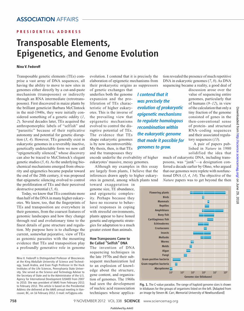
Fig. 1. The C-value paradox. The range of haploid genome sizes is shown in kilobases for the groups of organisms listed on the left. [Adapted from an image by Steven M. Carr, Memorial University of Newfoundland]
I contend that it
was precisely the
evolution of prokaryotic
epigenetic mechanisms
to regulate homologous
recombination within
the eukaryotic genome
that made it possible for
genomes to grow.
Published by AAAS
on June 29, 2018
http://science.sciencemag.org/
Dow
nloaded from

www.sciencemag.org SCIENCE VOL 338 9 NOVEMBER 2012 759
2012
still-prevalent view that every
bit of an organism’s DNA has
a specifi c function crafted by
selection. Thus, both papers
promoted Dawkins’ concept
of “selfi sh DNA”—the notion
that DNA capable of prolifer-
ating within a genome, as TEs
do, may need no other expla-
nation for its survival (15).
Orgel and Crick asserted,
“The spread of selfi sh DNA
sequences within the genome
can be compared to the spread
of a not-too-harmful parasite
within its host” (3).
The self ish DNA con-
cept was initially offered to
explain the long-standing
C-value paradox that organ-
isms of similar evolutionary
complexity differ vastly in
their DNA content (16), and
this it did. The C value, which
is the DNA content per haploid genome,
varies widely among closely related organ-
isms of apparently comparable complexity
(Fig. 1); this has for some time been attrib-
uted to the repetitive portion of the genome
(17). Such variation is especially striking in
angiosperms, whose highest and lowest C
values differ by a factor of 2000 (18, 19).
The explanation of the C-value paradox
does indeed reside largely in the profound
differences among genomes in the abun-
dance of TEs, primarily retrotransposons,
even as gene numbers remain relatively con-
stant. The Arabidopsis genome, for exam-
ple, contains about 27,000 genes and 20
to 25 Mb of retrotransposons, whereas the
maize genome contains about 40,000 genes
and more than 1800 Mb of retrotransposon
sequences (20–22). What the selfi sh DNA
hypothesis does not attempt to explain, how-
ever, is how genomes can accumulate such
vast amounts of repetitive sequences, given
the ease of eliminating them by homologous
recombination.
The Selfi sh DNA Label Stuck
Fortunately, genome sequencing raced for-
ward, and today we have a vastly more com-
plex understanding of genome structure
and organization than we might have had if
sequencing had been restricted to protein-
and RNA-coding sequences. But we fi nd
ourselves neck deep in TEs: Transposon
and retrotransposon sequences constitute
two-thirds of our own genome and 85% of
the corn genome (21, 23). Ideas about junk
DNA have evolved substantially over the
past two decades, with growing knowledge
of the regulatory roles fulfi lled by noncod-
ing sequences and their transcripts (24, 25).
However, the transposon monikers from the
selfish DNA papers have persisted. Hav-
ing cloned the fi rst mammalian DNA meth-
yltransferase gene and realizing that its
methyltransferase domain resembles those
of prokaryotic restriction
methyltransferases, Bestor
proposed in 1990 that
eukaryotic DNA methyla-
tion had evolved to regu-
late gene expression in
development and chro-
mosome structure, much
along the lines of earlier
proposals (26–28). But
by the end of the decade,
he and his colleagues had concluded that
because cytosine methylation is predomi-
nantly found in TEs, methylation was more
likely to represent a nuclear defense sys-
tem that had evolved precisely to “con-
trol” the destructive potential of “parasitic
sequences,” mostly transposons and ret-
rotransposons (5, 29). This view was widely
accepted, and transposons are today almost
universally referred to as “invaders,” “para-
sites,” or “parasitic sequences” (30–33).
Does the notion that epigenetic mecha-
nisms evolved to control invading “par-
asitic” transposons still f it the facts in
the light of the many that
have accumulated since
this hypothesis was f irst
advanced? Perhaps not. The
diff iculty starts with the
question of where such para-
sites might have come from.
It turns out that genes encod-
ing transposases, which all
have certain common struc-
tural motifs in their catalytic
cores, are present through-
out eukaryotes (34) and can
be traced back into prokary-
otic organisms that do not
have the elaborate epigen-
etic regulatory superstruc-
ture of eukaryotes (35). That
is, transposons were around
long before the eukaryotic
lifestyle, with its bloated
genomes, appeared on the
evolutionary scene. This
implies that transposons
coevolved with all the rest of the eukary-
otic genome’s inhabitants. Moreover, pro-
karyotic transposition is minimized and
regulated by mechanisms that are simi-
lar to those in eukaryotes, including weak,
enhancer-insensitive promoters, transpo-
son-encoded regulatory proteins and regu-
latory RNAs, and DNA methylation (36–
39). Yet prokaryotic genomes carry only
modest numbers of transposons.
It is true that the ability
of eukaryotic transposons
and retrotransposons to
accumulate in large num-
bers, together with their
highly generic transposi-
tion mechanisms, means
that the proliferation of
a transposon introduced
into a genome lacking it—
whether by a genetic cross
or a virus—makes it resemble an “invader”
(40, 41). And indeed, there is growing appre-
ciation that transposons are subject to hori-
zontal transfer in eukaryotes, in some cases
through host-parasite interactions (42–46).
But the same is true of prokaryotic trans-
posons (37). As well, new transposons can
arise within a genome and silent transposons
can be mobilized anew by a variety of physi-
ological and genetic stresses, undergoing
“bursts” of transposition to expand genomes
over millennia before being silenced and
decaying (47) or being silenced quickly
within a generation (48).
I argue that transposable
elements accumulate
in eukaryotic genomes
because of, not despite,
epigenetic silencing
mechanisms.
Replication
Triplication
Deletion
Duplication
Fig. 2. Generation and elimination of duplications by unequal crossing over. Broken lines
trace the recombination event.
Published by AAAS
on June 29, 2018
http://science.sciencemag.org/
Dow
nloaded from

9 NOVEMBER 2012 VOL 338 SCIENCE www.sciencemag.org 760
ASSOCIATION AFFAIRS
How Do Genomes Get So Fat?
There’s perhaps a deeper problem than the
ancient origin of transposons. What distin-
guishes the organization of higher eukary-
otic genomes from that of prokaryotes is
the presence of vast amounts of duplicated
DNA. It has long puzzled me that we almost
universally take this for granted. But then,
eukaryotes also have a markedly more com-
plex, largely epigenetic system than do pro-
karyotes for managing the transcription,
reproduction, and recombination of genetic
material, as well as its distribution to daugh-
ter cells during mitotic and meiotic divi-
sions. Which is cause and which is effect?
Prokaryotes can readily duplicate genome
segments by virtue of small stretches of
homology, but tandem duplications are rap-
idly lost unless retained by selection, and
even then, they are generally interspersed
with nonhomologous sequences (49–51).
This is illustrated diagrammatically in Fig.
2. Absent either selection or a reduction in
homology, tandem duplications are inevita-
bly eliminated by homologous unequal intra-
or interchromosomal crossing over between
duplicated sequences, generating one-copy
organisms (an absorbing state) and organ-
isms with increasing numbers of copies that
in turn throw off singletons (50, 52). Organ-
isms with many copies are quite unstable
and are likely to be eliminated, either by vir-
tue of the energetic drag of the extra DNA or
by a population bottleneck. This is borne out
by the observation that duplicate genes in
prokaryotes are generally acquired by hori-
zontal gene transfer rather than by duplica-
tion (53, 54).
The “selfi sh DNA” argument rests on the
assumption that there exists a category of
DNA that has little or no phenotypic effect,
and hence is not subject to selective pres-
sure, but can nonetheless multiply within
the genome. This is not an unreasonable
inference, because we know that eukaryotic
genomes are packed with repetitive DNA of
all kinds. But I fi nd it quite remarkable that it
passes unremarked. How did eukaryotes tip
the balance between duplication and dele-
tion that keeps genome size small in organ-
isms in which homology-dependent recom-
bination mechanisms predominate? And how
can transposons, whose duplicative mecha-
nisms create dispersed repetitive sequences,
build up in large numbers, given the ability
of homologous recombination among them
to cause major, even catastrophic, chromo-
somal rearrangements?
What Epigenetic Mechanisms Do and How
They Came to Be
I believe that the answer to these questions
lies precisely in the epigenetic mechanisms
that eukaryotes have elaborated to a much
greater extent than prokaryotes. Repres-
sive protein complexes, histone methyla-
tion, RNA interference (RNAi), and RNA-
directed DNA methylation, as well as recom-
binational regulatory complexes, are among
the epigenetic mechanisms that have so far
surfaced (55–59). These serve a variety of
structural and regulatory functions, but per-
haps the essential one for understanding the
evolution of eukaryotic genomes is the mini-
mization of illegitimate and ectopic recom-
bination among homologous sequences dur-
ing DNA replication and the DNA break-
repair processes that maintain genome and
chromosome stability.
Heterochromatin, the highly compacted
chromosome regions rich in repetitive DNA,
is recombinationally inert (60, 61). Although
not all eukaryotes use all of the known epi-
genetic mechanisms, even lower eukaryotes
with relatively small genomes use RNAi to
stabilize repetitive DNAs, such as ribosomal
RNA genes and centromeric repeats (61–
63). In fi ssion yeast, noncoding transcripts
of repetitive sequences initiate a process that
generates small RNAs, which in turn target
further transcripts for degradation and attract
protein complexes that induce heterochro-
matization through histone modification
(64, 65). Disruption of the RNAi machinery
disturbs the repair of double-strand breaks,
stimulating repair by homologous recombi-
nation (66).
The evolutionary origins of the eukaryotic
epigenetic regulatory machinery lie in bac-
terial systems that discriminate endogenous
DNA from that acquired through horizontal
gene transfer and bacteriophage infection
(67–69). Although prokaryotic transposons
can move both by conjugation (commonly
on plasmids) and on bacteriophage, they do
not appear to have been independently tar-
geted for inactivation by either the restric-
tion-modification system or the CRISPR
(clustered regularly interspaced short pal-
indromic repeat)–cas (CRISPR-associated)
interference pathway (70). This recently dis-
covered bacterial pathway confers sequence-
specifi c immunity to phage and plasmids and
exhibits parallels with eukaryotic RNAi sys-
tems, particularly the Piwi-interacting RNA CR
ED
IT: K
. S
UT
LIF
F/S
CIE
NC
E
Histone
modification
Heritable silencing
DNAmethylation
RNA
Histonemethyl-
transferases
Histonedeacetylases
Histonedemethylases
Histoneacetyl-transferases
DNAglycosylases
DNAmethyl-
transferases
Pol IV
RDR2
dsRNA
mCG mCHG mCHH
H3K9me
H3K27me
H3K36me
H3K4me
H3K4ac
H3K9ac
AGO4
Pol V
DCL3
ssRNA
Fig. 3. Plant epigenetic mechanisms include DNA methylation, histone modifi cation, and RNA-directed DNA methylation (RdDM). RdDM involves two plant-specifi c RNA polymerases (Pol IV and Pol V), an RNA-depen-dent RNA polymerase (RDR2), an enzyme that cleaves double-stranded RNA (DCL3), and an Argonaute-family RNA-binding protein (AGO4). [Adapted with permission from (199)]
Published by AAAS
on June 29, 2018
http://science.sciencemag.org/
Dow
nloaded from

www.sciencemag.org SCIENCE VOL 338 9 NOVEMBER 2012 761
2012
system of Drosophila (71, 72). Cytosine
methylation is widespread in both prokary-
otes and eukaryotes, and eukaryotic DNA
methylases evolved from bacterial restric-
tion-modifi cation methylases by acquiring
new recognition and binding modules (69).
The evolution of DNA methylases appears to
have proceeded in parallel with that of his-
tone-modifying enzymes and RNA-based
silencing mechanisms, so that today they
comprise intimately interconnected systems
(68, 69). Some eukaryotes lack either DNA
methylation or the RNAi machinery (or both),
but nonetheless exhibit epigenetic silenc-
ing; hence, there is some redundancy among
silencing mechanisms (73, 74). Precisely
how these disparate systems came together
is not yet known, but the evolutionary genius
of linking RNAi feedback mechanisms to the
heritability of the DNA methylation mecha-
nism means, of course, that silencing can be
inducible, sequence-specifi c, and heritable.
My contention is that it was precisely the
evolution of prokaryotic epigenetic mecha-
nisms, originally limiting recombination
among horizontally exchanged sequences, to
regulate homologous recombination within
the eukaryotic genome that made it possi-
ble for genomes to grow. Interference with
DNA methylation, histone modification,
and the small RNA pathways of contempo-
rary genomes generally destabilizes repeti-
tive regions, both tandem and dispersed
(48, 61, 62). The ability to suppress homol-
ogous recombination might well be what
tipped the balance between duplication and
deletion in favor of sequence endo-redupli-
cation in general and transposon prolifera-
tion in particular. The fact that small diffus-
ible RNA molecules are at the heart of the
silencing machinery also means that new
copies of transposons cannot evade regula-
tion by moving to new locations where the
ability to cause severe chromosomal disrup-
tions through ectopic homologous recombi-
nation might consign them to the scrap heap
of evolution. What I am suggesting, then, is
that TEs accumulate in eukaryotic genomes
because of, not despite, epigenetic silencing
mechanisms. This is exactly the inverse of the
“parasite control” hypothesis, which posits
that epigenetic mechanisms arose to control
invading, parasitic transposons (5).
The ability to retain duplicated sequences
is also arguably a critical step in the evolution
of multicellular organisms, underpinning
the ability to diversify duplicates for expres-
sion in specifi c cells and tissues, at different
developmental moments, and in response to
different environmental stimuli (75). Equally
key is the ability to program genes for differ-
ential expression by a variety of mechanisms,
among which are the relatively stable ones
involving DNA and histone modification,
as well as the more labile small RNA–medi-
ated and transcriptional mechanisms. On bal-
ance, then, the likelihood that contemporary
eukaryotic genomes evolved in the context of
epigenetic mechanisms seems vastly greater
than the likelihood that they were invented as
an afterthought to combat a plague of para-
sitic transposons.
Plant Genomes Do It More
Plants have a more complex and redun-
dant array of epigenetic silencing mecha-
nisms than animals, making use of multiple
DNA methylation mechanisms, chromatin
protein modification, and feedback mech-
anisms involving small noncoding RNAs
(55, 58, 76). Mammals primarily methylate
the C residues in the CG dinucleotide con-
text, whereas plants methylate C residues in
nucleo tides within all sequence contexts (55,
IDN2 Removal of active histone marks
(histone deacetylation, H3K4 demethylation)
Establishment of repressive histone marks
(H3K9 methylation, H3K27 methylation)
Pol IV
Pol VssRNA
dsRNA
siRNA duplexMethylated
siRNA duplex
AGO4-RISC
DRM2
KTF1
CTD
Pol Vtranscript
AGO4
IDN2Po
trans
DRM2DRM2
?
RDR2
DDR complex
DRD1 DMS3
DMS4
RDM1
CLSY1
AGO4
Fi g. 4. The RNA-directed DNA methylation pathway. RNA polymerase IV (Pol IV) initiates RdDM, generating single-stranded RNA (ssRNA) that is then copied into double-stranded RNA (dsRNA) by RNA-DEPENDENT RNA POLYMERASE 2 (RDR2). The putative chromatin remodeler and/or helicase CLASSY 1 (CLSY1) assists in one or more of these steps. DICER-LIKE 3 (DCL3) cleaves the dsRNA into 24-nucleotide small interfering RNA (siRNA) duplexes that are then methylated at their 3′ ends by HUA-ENHANCER 1 (HEN1). A single strand of the siRNA duplex associates with ARGONAUTE 4 (AGO4) to form an RNA-induced silencing complex (RISC)–AGO4 complex. Independently of siRNA biogenesis, Pol V transcription is assisted by the DDR complex [DRD1 (DEFECTIVE IN RNA-DIRECTED DNA METHYLATION 1), DMS3 (DEFECTIVE IN MERISTEM SILENCING 3), and RDM1 (REQUIRED FOR DNA METHYLA-TION 1)] and DMS4. AGO4 binds Pol V transcripts through base-pairing with the
siRNA and is stabilized by AGO4 interaction with the NRPE1 (the largest subunit of Pol V) C-terminal domain (CTD) and KTF1 (KOW DOMAIN-CONTAINING TRANSCRIP-TION FACTOR 1), which also binds RNA. IDN2 may stabilize Pol V transcript–siRNA pairing. The RDM1 protein of the DDR complex binds AGO4 and the de novo cyto-sine methyltransferase DOMAINS REARRANGED METHYLTRANSFERASE 2 (DRM2), bringing them to Pol V–transcribed regions, resulting in DNA methylation. Histone modifi cations resulting from the RdDM pathway include the removal of activating acetylation and methylation marks [deacetylation of multiple Lys of several core histone proteins and demethylation of histone H3 Lys4 (H3K4)] and the establish-ment of alternative, repressive histone methylation marks (such as the methylation of H3K9 and H3K27), thereby facilitating transcriptional silencing. [Adapted with permission from (80)]C
RE
DIT
: K
. S
UT
LIF
F/S
CIE
NC
E
Published by AAAS
on June 29, 2018
http://science.sciencemag.org/
Dow
nloaded from
9 NOVEMBER 2012 VOL 338 SCIENCE www.sciencemag.org 762
ASSOCIATION AFFAIRS
77). DNA methylation stabilizes the silencing
and inactivation of genes and other genetic
elements in many eukaryotes, but is not uni-
versal; Drosophila and budding yeast repre-
sent well-investigated exceptions (55, 78).
Chromosomal protein modifi cation, partic-
ularly histone 3 methylation, is involved in
guiding DNA methylases to their correct tar-
gets in both plants and animals (55, 79). In
plants, unmethylated DNA is methylated by
one DNA methyltransferase, whereas main-
tenance methylation involves two addi-
tional DNA methyltransferases (55). Fig-
ure 3 shows an overview of the several epi-
genetic mechanisms currently known to
exist in plants.
Sequence specificity is imparted to
DNA methylation through a mechanism
called RNA-directed DNA methyla-
tion (RdDM) (57, 58). RdDM involves
two unique plant RNA polymerases,
Pol IV and Pol V, and is mediated by
24-nucleotide small interfering RNAs
(siRNAs) (55, 57, 80). As illustrated in
Fig. 4, RdDM is initiated by conversion
of Pol IV–generated transcripts to RNA
duplexes by an RNA-dependent RNA
polymerase (RDR2). The duplexes are
then cleaved into 24-nucleotide siRNAs
by an RNAse III–family enzyme (DCL3)
and the appropriate strand associates with
the Argonaute family protein AGO4 (55,
80, 81). This leads to the formation of a
complex comprising the AGO4-siRNA
and a number of other proteins (includ-
ing a DNA methylase, DRM2), which
then triggers local DNA methylation (57,
80, 82). As well, activation of RdDM pro-
motes histone deacetylation and meth-
ylation changes that lead to the estab-
lishment of chromatin structures that
repress transcription (57, 80). Plant DNA
demethylation is mediated by one of several
DNA glycosylase activities that removes
the 5-methylcytosine, after which the DNA
backbone is cleaved at the abasic site and
repaired (83).
Why and exactly how the different epi-
genetic systems evolved remains to be under-
stood. But plants use epigenetic systems
today in a variety of developmental con-
texts. Unlike higher animals, plants do not
set aside a germ line early in development;
this imposes more stringent requirements for
maintaining genetic integrity, because differ-
entiated genomes must eventually be repro-
grammed for reproduction. So the elabora-
tion of epigenetic mechanisms may have
made possible the indeterminate lifestyle of
many higher plants and their ability to repro-
duce in response to environmental signals.
Epigenetic mechanisms, for example, regu-
late such environmentally responsive devel-
opmental transitions as vernalization, a cold-
temperature requirement for germination
or the transition to fl owering, the reproduc-
tive phase. For example, Arabidopsis plants
requiring vernalization grow vegetatively
unless exposed to a period of cold to induce
fl owering, as illustrated in Fig. 5 (84, 85).
Vernalization is mediated by cold-induced
epigenetic silencing of the FLOWERING
LOCUS C (FLC) gene that encodes a repres-
sor of fl owering. The silencing increases with
the duration of the cold period, involves pro-
duction of noncoding FLC transcripts, and
results in histone modifi cations that inacti-
vate transcription of the gene (86).
Although transposons are primary tar-
gets for epigenetic silencing, they are far
from the only targets in plants. The fi rst plant
gene silencing mechanism understood at the
molecular level was that underlying the long-
known ability of a viral infection to cross-
protect a plant against infection by a closely
related virus (87–90). Then, in 1994, it was
reported that a wholly artifi cial gene com-
prising a viroid cDNA became methylated
and transcriptionally inactive in the tobacco
genome, but only if viroid RNA replication
had occurred, suggesting a feedback mech-
anism initiated by transcript overabundance
(91). It was subsequently discovered in the
early days of plant molecular modifi cation
that an introduced transgene encoding an
Fig. 5. Vernalization. Arabidopsis plants requiring vernalization grow vegetatively (A) unless exposed to a period of cold to induce fl owering (B). Vernalization involves cold-induced epigenetic silencing of the FLC gene, a repressor of fl owering.
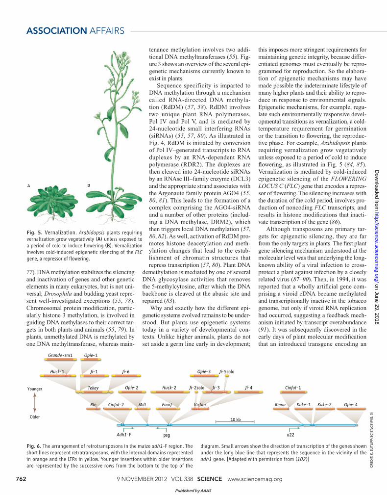
Fig. 6. The arrangement of retrotransposons in the maize adh1-F region. The short lines represent retrotransposons, with the internal domains represented in orange and the LTRs in yellow. Younger insertions within older insertions are represented by the successive rows from the bottom to the top of the
diagram. Small arrows show the direction of transcription of the genes shown under the long blue line that represents the sequence in the vicinity of the adh1 gene. [Adapted with permission from (102)]
Adh1-F
Younger
Older
psg
10 kb
u22
Grande -zm1
Huck-1
Huck-2Tekay
Rle Milt Fourf Victim Reina
Ji-1 Ji-6
Ji-3 Ji-4
Ji-5solo
Ji-2solo
Opie-1
Cinful-1
Cinful-2 Kake-1 Kake-2
Opie-2
Opie-4
Opie-3C
RE
DIT
: K
. S
UT
LIF
F/S
CIE
NC
E (F
IG. 5
)
Published by AAAS
on June 29, 2018
http://science.sciencemag.org/
Dow
nloaded from

www.sciencemag.org SCIENCE VOL 338 9 NOVEMBER 2012 763
2012
enzyme in the pigment biosynthetic pathway
was subject to silencing (92–94). Later stud-
ies found that silencing entails both transcrip-
tional and posttranscriptional mechanisms,
and that these mechanisms share characteris-
tics with those used by plants to control viral
pathogens and are mediated by the produc-
tion of siRNAs (95, 96). Sequence duplica-
tion also underlies a reversible silencing phe-
nomenon, termed “paramutation,” in which
an allele termed “paramutagenic” can herita-
bly silence a susceptible allele termed “para-
mutable” of the same locus residing on the
homolog (97). Paramutation involves a small
RNA feedback mechanism and DNA meth-
ylation triggered by duplication of either cod-
ing or regulatory sequences (98, 99). Thus,
the repetitive character of the sequence is
also a common trigger for siRNA-mediated
gene silencing and methylation (100).
The Contemporary Plant Genome Landscape
Despite the multiplicity of plant epigenetic
silencing mechanisms, the fi ngerprints of
transposition and recombination are evident
at every level of plant genome structure,
organization, and evolution. Maize genes are
clustered in small groups separated by long,
uninterrupted stretches of DNA consisting
of retrotransposons (101, 102). Almost 85%
of the contemporary 2.3-Gb maize (Zea
mays or corn) genome comprises transpo-
sons, more than 75% of which are long ter-
minal repeat (LTR) retrotransposons (21).
Its roughly 40,000 genes, averaging about
3.3 kb in length, form small islands in a sea
of more than a million transposons and ret-
rotransposons belonging to almost 1300 dif-
ferent gene families.
In addition to forming very large blocks,
retrotransposons exhibit a tendency to home
to different neighborhoods. In maize, for
example, gypsy and copia elements are over-
and underrepresented in pericentromeric
regions, respectively (21, 103). Within a
retro transposon block, younger elements are
progressively nested within older elements,
as illustrated in Fig. 6 for a short region near
the maize adh1 gene (21, 102, 103). Such
targeting can occur through the interaction
of retrotransposon-specific proteins and
chromatin proteins, which are themselves
preferentially associated with certain types
of sequences. An example is provided by
the interactions of yeast Sir4p, a structural
protein of heterochromatin, with a 6–amino
acid motif of the Ty5 integrase protein that
targets insertion into telomeric heterochro-
matin (104, 105). An Arabidopsis lyrata
bz stc1 rpl35A znf
cdl1 rlkhypro2,3
tac7077 uce2
tac6058
hypro1
HelANotlNotl
HelB
gagMcC111 kb
bz haplotypesNotl fragment size
hATDOP
Ji-6 Huck1b
Huck1a
Opie2a
Opie2bDeletions
RetrotransposonsTransposons
HelA HelB
B7373 kb
ZsL
TAFT2Grande1Tekay
Xilon2
Mu
gagMo1752 kb
hAT
Grande1CsL
A18855 kb
Ds
Grande1CsL
CACTA
Hel1-4
gagCML25866 kb
Ds
TAFT1 Grande1
Opie2
gagI137TN121 kb
Ds
Opie3
CsL
Elote1
Huck1
Huck2a
Huck2b
Milt
LINE
Chico
Hel1-5Hel1-6
gagNalTel79 kb
Ds hATGrande1
Ji1Milt1a
MuMilt1b
gagCoroico
159kb
TAFT1
Ds
Grande1
Prem1r1
Milt1a
Milt1bElote
Opie2
Opie4P1 s-LTR
X3 s-LTRPrem1r3
Chico
Huck2bJi7
Huck1
Fig. 7. The organization of the sequence adjacent to the bronze (bz) gene in eight different lines (haplotypes) of maize. The genes in this region are shown in the top diagram: bz, stc1, rpl35A, tac6058, hypro1, znf, tac7077, and uce2. The orientation of the gene is indicated by the direction of the green pentagon, pointing in the direc-tion of transcription; exons are represented in dark green and introns in light green. Each haplotype is identifi ed by its name and the size of the cloned NotI fragment. The same symbols are used for gene fragments carried by Helitrons (Hels), which are represented as bidirectional arrows below the line for each haplotype. Vacant sites for HelA and HelB are provided as reference points and marked by short vertical red bars. Dashed lines represent deletions. Retrotransposons are represented by yellow bars. DNA transposons and TAFTs (TA-fl anked transposons), which are probably also DNA transposons, are represented by red triangles; small insertions are represented by light blue triangles. [Redrawn with permission from (113)]
Published by AAAS
on June 29, 2018
http://science.sciencemag.org/
Dow
nloaded from
9 NOVEMBER 2012 VOL 338 SCIENCE www.sciencemag.org 764
ASSOCIATION AFFAIRS
centromeric retrotransposon was reported
to insert preferentially into centromeres in
A. thaliana (106). Because the centromeric
sequences are quite different in the two spe-
cies, targeting is likely to involve an interac-
tion with the highly conserved centromere-
specifi c structural proteins.
Unlike retrotransposons, which replicate
through an RNA intermediate and reinsert
DNA copies, DNA transposons move by a
cut-and-paste mechanism, generally excis-
ing from just one newly replicated sister
chromatid and reinserting into a site either
nearby on the same chromosome or else-
where in the genome (107). Because a copy
of the transposon is retained at the donor site,
such transposition events commonly give
rise to additional transposon copies. DNA
transposons account for a much smaller frac-
tion of the plant genome than retrotranspo-
sons, are generally present in fewer copies,
and tend to be associated with genic regions,
some even inserting preferentially into genes
(108). Mu transposons in maize favor recom-
binationally active regions of the
genome (109), whereas Helitrons
accumulate near but not inside
each other (110). Such cluster-
ing may refl ect the propensity of
some TEs to move to nearby sites,
long documented for the Ac/Ds
(Activator/Dissociation) trans-
poson family of maize (111).
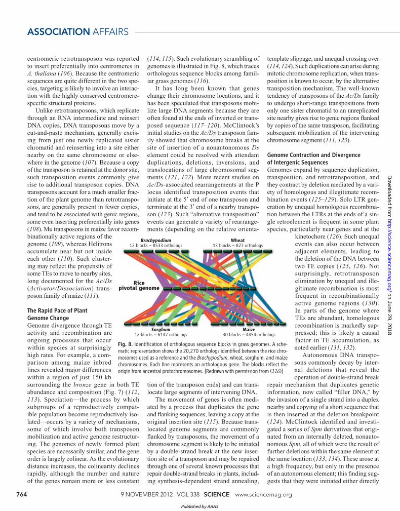
The Rapid Pace of Plant
Genome Change
Genome divergence through TE
activity and recombination are
ongoing processes that occur
within species at surprisingly
high rates. For example, a com-
parison among maize inbred
lines revealed major differences
within a region of just 150 kb
surrounding the bronze gene in both TE
abundance and composition (Fig. 7) (112,
113). Speciation—the process by which
subgroups of a reproductively compat-
ible population become reproductively iso-
lated—occurs by a variety of mechanisms,
some of which involve both transposon
mobilization and active genome restructur-
ing. The genomes of newly formed plant
species are necessarily similar, and the gene
order is largely colinear. As the evolutionary
distance increases, the colinearity declines
rapidly, although the number and nature
of the genes remain more or less constant
(114, 115). Such evolutionary scrambling of
genomes is illustrated in Fig. 8, which traces
orthologous sequence blocks among famil-
iar grass genomes (116).
It has long been known that genes
change their chromosome locations, and it
has been speculated that transposons mobi-
lize large DNA segments because they are
often found at the ends of inverted or trans-
posed sequence (117–120). McClintock’s
initial studies on the Ac/Ds transposon fam-
ily showed that chromosome breaks at the
site of insertion of a nonautonomous Ds
element could be resolved with attendant
duplications, deletions, inversions, and
translocations of large chromosomal seg-
ments (121, 122). More recent studies on
Ac/Ds-associated rearrangements at the P
locus identified transposition events that
initiate at the 5′ end of one transposon and
terminate at the 3′ end of a nearby transpo-
son (123). Such “alternative transposition”
events can generate a variety of rearrange-
ments (depending on the relative orienta-
tion of the transposon ends) and can trans-
locate large segments of intervening DNA.
The movement of genes is often medi-
ated by a process that duplicates the gene
and fl anking sequences, leaving a copy at the
original insertion site (115). Because trans-
located genome segments are commonly
fl anked by transposons, the movement of a
chromosome segment is likely to be initiated
by a double-strand break at the new inser-
tion site of a transposon and may be repaired
through one of several known processes that
repair double-strand breaks in plants, includ-
ing synthesis-dependent strand annealing,
template slippage, and unequal crossing over
(114, 124). Such duplications can arise during
mitotic chromosome replication, when trans-
position is known to occur, by the alternative
transposition mechanism. The well-known
tendency of transposons of the Ac/Ds family
to undergo short-range transpositions from
only one sister chromatid to an unreplicated
site nearby gives rise to genic regions fl anked
by copies of the same transposon, facilitating
subsequent mobilization of the intervening
chromosome segment (111, 123).
Genome Contraction and Divergence
of Intergenic Sequences
Genomes expand by sequence duplication,
transposition, and retrotransposition, and
they contract by deletion mediated by a vari-
ety of homologous and illegitimate recom-
bination events (125–129). Solo LTR gen-
eration by unequal homologous recombina-
tion between the LTRs at the ends of a sin-
gle retroelement is frequent in some plant
species, particularly near genes and at the
kinetochore (126). Such unequal
events can also occur between
adjacent elements, leading to
the deletion of the DNA between
two TE copies (125, 126). Not
surprisingly, retrotransposon
elimination by unequal and ille-
gitimate recombination is most
frequent in recombinationally
active genome regions (130).
In parts of the genome where
TEs are abundant, homologous
recombination is markedly sup-
pressed; this is likely a causal
factor in TE accumulation, as
noted earlier (131, 132).
Autonomous DNA transpo-
sons commonly decay by inter-
nal deletions that reveal the
operation of double-strand break
repair mechanism that duplicates genetic
information, now called “fi ller DNA,” by
the invasion of a single strand into a duplex
nearby and copying of a short sequence that
is then inserted at the deletion breakpoint
(124). McClintock identifi ed and investi-
gated a series of Spm derivatives that origi-
nated from an internally deleted, nonauto-
nomous Spm, all of which were the result of
further deletions within the same element at
the same location (133, 134). These arose at
a high frequency, but only in the presence
of an autonomous element; this fi nding sug-
gests that they were initiated either directly
Fig. 8. Identifi cation of orthologous sequence blocks in grass genomes. A sche-matic representation shows the 20,270 orthologs identifi ed between the rice chro-mosomes used as a reference and the Brachypodium, wheat, sorghum, and maize chromosomes. Each line represents an orthologous gene. The blocks refl ect the origin from ancestral protochromosomes. [Redrawn with permission from (116)]
Wheat13 blocks – 827 orthologs
Brachypodium12 blocks – 8533 orthologs
Maize30 blocks – 4454 orthologs
Sorghum12 blocks – 6147 orthologs
Ricepivotal genome
Published by AAAS
on June 29, 2018
http://science.sciencemag.org/
Dow
nloaded from
www.sciencemag.org SCIENCE VOL 338 9 NOVEMBER 2012 765
2012
by the double-strand cleavage activity of
the transposase encoded by the autonomous
element or indirectly by secondary double-
strand breaks incurred during the transposi-
tion reaction (135).
Analyses of intergenic regions in related
species have revealed that they turn over very
rapidly on an evolutionary time scale. Fine-
grained analysis of the sequence dynamics
shows that the intergenic volatility is indeed
mediated by transposons, which both delete
adjacent DNA sequences and insert filler
DNA from elsewhere in the process of repair-
ing the double-strand breaks in the DNA intro-
duced by transposases (128). Although dele-
tions commonly occur adjacent to a transpo-
son end, they can remove entire transposons.
Statistically significant clustering of such
deletions in the vicinity of transposons sug-
gests that they arise as a result of the double -
strand breaks that initiate transposition.
Traffi c in Genes and Regulatory Sequences
At a much fi ner level of resolution, transpo-
sons contribute to creating genes, modify-
ing them, and programming and reprogram-
ming them. Many transposons and retroele-
ments contain captured gene fragments and
can be part of gene regulatory regions (136–
142). A classic example is provided by the
maize R locus that encodes a transcription
factor necessary for synthesis of anthocy-
anin pigments. The R-r allele comprises four
tandem duplications, including a complete
coding sequence and several truncated ones
in direct and inverted order separated by a
Doppia transposon. The complete coding
sequence is responsible for pigment expres-
sion in the plant, whereas the several trun-
cated copies support pigment expression in
the seed (143).
The traff ic in genes and regulatory
sequences is bidirectional: Transposons pick
up bits and pieces of genes that code for
proteins other than transposases, and trans-
posase genes are pressed into services other
than transposition (144, 145). For example,
the proteins encoded by the FAR1 and FHY3
genes of Arabidopsis are both related to the
MuDR family of transposases (146). FHY3
and FAR3 are transcription factors that reg-
ulate light-dependent chlorophyll biosyn-
thesis in development, the former also gat-
ing phytochrome signaling to the circadian
clock (147, 148). A familiar example is pro-
vided by the human immune system, which
uses recombinase proteins that evolved from
transposases to generate sequence diversity
through V(D)J recombination (149, 150).
Transposons provide the telomeres of some
organisms and jump in to replace them in
others (151, 152). Centromeres contain and
are often surrounded by transposons (22,
153–155). As well, transposons are central to
the epigenetic phenomenon of “imprinting”
that imbues genes with different expression
patterns depending on whether they were
transmitted through male or female gametes;
such differences arise during the major epi-
genetic reprogramming events of gameto-
genesis (156–158).
Driving Evolution
Thus, transposases hold a special place in the
pantheon of genome sculptors. Arguably the
products of the most abundant genes on Earth
(159), transposases are transposon-encoded
enzymes that cleave transposon ends and
attach them to new sequences. The essential
elements are (i) very strict sequence recog-
nition and precise cleavage at the donor site
and (ii) either a relaxed sequence specifi city
or no sequence specifi city at the target site
(160). The prokaryotic Mu element’s trans-
position mechanism appears to be paradig-
matic and common to many members of the
transposase super family (161–165). Multim-
ers of the transposase form a transpososome
complex that recognizes the transposon’s
terminal inverted repeats and brings them
together with the target insertion site (Fig. 9)
(166, 167). The transposon ends are brought
into close juxtaposition with each other at the
donor site for a coupled reaction that cleaves
the transposon termini, introduces a stag-
gered cleavage at the target site, and trans-
fers the 3′ ends of the transposon to the over-
hanging 5′ ends at the target (168, 169). The
gapped duplexes at the two element ends are
then repaired to generate the target site dupli-
cation, whose length is a TE family charac-
teristic determined by the transposase. Sub-
sequent excision of the transposon generally
leaves behind an imperfect version of the
target site duplication, generating sequence
diversity (170, 171).
In both bacteria and plants, recognition
and cleavage of the two hemimethylated ter-
minal inverted repeats of a single transpo-
son ensures genome integrity and confi nes
transposition to just one of the two newly
replicated daughter strands or sister chro-
matids (36, 172). But like other aspects of
transposition, recombination, and DNA
repair, this process is error-prone and can be
fooled by such transposition events as the
insertion of one Ds transposon in inverted
orientation into the center of itself, giving
the chromosome-breaking double Ds trans-
poson that led McClintock to her momen-
tous discoveries about how transposons
move and restructure chromosomes (173,
174). The bottom line for genomes is that
the cleavage and resection of DNA by trans-
posases virtually guarantees sequence vari-
ation, genome scrambling, and the appear-
ance of transposons at rearrangement
breakpoints. Simply put, transposases drive
genome evolution.
Transposaserecognition and
binding
Target DNA
Cleavageand integrationinto target DNA
Formation oftranspososome
Donor DNA
Transposase
Transposon
Fig. 9. DNA “cut-and-paste” transposition mechanism. Transposition is initiated by the site-specifi c recognition and binding of transposase to the transposon DNA ends. Dimerization of the transposase leads to formation of the transpososome,
which brings together the transposon termini and a target site. Concerted cleavage at the donor and target sites is followed by integration of the excised transposon into the target site and resection of the empty donor site (200).
CR
ED
IT: K
. S
UT
LIF
F/S
CIE
NC
E
Published by AAAS
on June 29, 2018
http://science.sciencemag.org/
Dow
nloaded from

9 NOVEMBER 2012 VOL 338 SCIENCE www.sciencemag.org 766
ASSOCIATION AFFAIRS
Genomic Shock and Transgenerational
Epigenetic Inheritance
Both unpredictable stresses, such as irradia-
tion, and predictable abiotic stresses, such
as heat shock, elicit from genomes a highly
programmed response intended to minimize
the impact of the stress. McClintock coined
the term “genomic shock” to refer to such
a response (175). It is by now amply docu-
mented that plant transposons are activated
in response to a variety of DNA-damaging
agents and both biotic and abiotic stresses, as
well as pathogen infection and the passage of
plant cells through tissue culture (176–181).
Other sources of natural chromosomal distur-
bance are provided by interspecifi c hybridiza-
tion and allopolyploidization, both of which
trigger the activation of transposons (182–
184). This appears to be true as well in other
eukaryotes, from yeast to flies to humans.
Telomerases are relatives of retrotransposon-
encoded reverse transcriptases,
and transposons either com-
prise or can fi ll in for missing
telomeres in flies and yeast,
respectively (152, 185–187).
Evidence is accumulat-
ing that both biotic and abi-
otic stresses induce a herita-
ble increase in the ability of
plants to withstand infection
and tolerate stress (188–191).
DNA damage, pathogen infec-
tion, and abiotic stresses also
increase homologous recombi-
nation frequency and chromo-
somal rearrangements, both
somatically and heritably (188, 192–195).
Thus, responses to stress—whether from
pathogens, environmental extremes, or dam-
age to the genetic apparatus—evoke not just
a transcriptional response, but also a pro-
found and to some extent heritable change in
the epigenetic framework. Such changes can
loosen the epigenetic constraints on transpo-
sons, allowing stress-inducible TEs to propa-
gate stress-inducible promoters to other genes
through transposition (181).
Just as McClintock reported that broken
chromosome ends can “heal,” so do trans-
position bursts subside, over both short and
long runs (175). Some of the Arabidopsis
transposons and retrotransposons demethyl-
ated in a genetic background devoid of the
MET1 DNA methylase are gradually remeth-
ylated by RNA-dependent DNA methylation
within several generations after reintroduc-
tion of a wild-type MET1 gene (196–198).
Heat-induced transcription and transposi-
tion of the Arabidopsis ONSEN retrotranspo-
son is rapidly silenced, becoming transgen-
erational only in plants with a compromised
RdDM pathway (181). Thus, transcriptional
activation by demethylation can also trigger
a feedback mechanism that restores methyla-
tion and resilences transposons. Recent years
have seen progress in identifying the compo-
nents of the restructuring response, but we do
not yet know how cells and organisms per-
ceive and initiate epigenetic reorganization in
response to either genetic disruptions or envi-
ronmental stressors.
Evolvability and Its Agents
I have argued that epigenetic mechanisms
not only underpin the management of tran-
scription and chromosome structure, but also
provide the key to understanding the size
and organization of eukaryotic genomes.
They assure the sta-
bility of chromo-
somes, including vast
menageries of TEs,
and manage the rep-
lication and segre-
gation of the genetic
material in both mito-
sis and meiosis. My
argument is that TEs
accumulate because
of, not despite, the
epigenetic mecha-
nisms that control
homology-depen-
dent recombination,
whose dominance keeps the genomes of pro-
karyotes and many lower eukaryotes small.
Absent the existence of such mechanisms,
ectopic, homology-dependent recombina-
tion among dispersed TEs would rapidly
eliminate them, either directly by intrachro-
mosomal deletions or indirectly by creating
nonviable chromosomes.
But although epigenetic mechanisms
slow the pace of genome restructuring to
an evolutionary time scale, the impact of
transposons and retrotransposons on genes
and genomes is inescapable. Indeed, their
ability to move and to move sequences has
shaped higher eukaryotic genomes, from the
structuring and restructuring of genes and
their regulatory sequences to the shaping
and reshaping of the genomic landscape. It
is becoming increasingly diffi cult to escape
the conclusion that eukaryotic genome evo-
lution is driven from within not just by the
gentle breeze of the genetic mechanisms that
replicate and repair DNA, but by the stron-
ger winds (with perhaps occasional gale-
force gusts) of transposon activity. The abil-
ity to evoke rapid genome restructuring is
at the heart of eukaryotic evolvability—the
capacity of organisms with larger and larger
genomes to maintain evolutionary fl exibility.
References 1. N. V. Fedoroff, in Discoveries in Plant Biology, S.-D. Kung,
S.-F. Yang, Eds. (World Scientifi c, Singapore, 1998), vol. 1, pp. 89–104.
2. N. V. Fedoroff, Proc. Natl. Acad. Sci. U.S.A. 0.1073/pnas.1215482109 (2012).
3. L. E. Orgel, F. H. C. Crick, Nature 284, 604 (1980). 4. W. F. Doolittle, C. Sapienza, Nature 284, 601 (1980). 5. J. A. Yoder, C. P. Walsh, T. H. Bestor, Trends Genet. 13, 335
(1997). 6. N. V. Fedoroff, in Plant Transposons and Genome Dynamics
in Evolution, N. V. Fedoroff, Ed. (Wiley-Blackwell, Hoboken, NJ, in press), chap. 4; www.wiley.com/WileyCDA/WileyTitle/productCd-0470959940.html.
7. M. Waring, R. J. Britten, Science 154, 791 (1966). 8. R. J. Britten, D. E. Kohne, Science 161, 529 (1968). 9. D. A. Smith, Science 233, 1246b (1986). 10. R. L. Sinsheimer, Science 233, 1246c (1986). 11. J. B. Walsh, J. Marks, Nature 322, 590 (1986). 12. R. Lewin, Science 233, 620 (1986). 13. J. G. Gall, Science 233, 1367 (1986). 14. S. Ohno, Brookhaven Symp. Biol. 23, 366 (1972). 15. R. Dawkins, The Selfi sh Gene (Oxford Univ. Press, Oxford,
ed. 1, 1976). 16. J. G. Gall, J. Cell Biol. 91, 3s (1981). 17. M. Rosbash, P. J. Ford, J. O. Bishop, Proc. Natl. Acad. Sci.
U.S.A. 71, 3746 (1974). 18. M. D. Bennett, I. J. Leitch, Ann. Bot. 107, 467 (2011). 19. M. D. Bennett, J. B. Smith, Philos. Trans. R. Soc. London Ser.
B 274, 227 (1976). 20. R. Liu, J. L. Bennetzen, New Phytol. 179, 249 (2008). 21. P. S. Schnable et al., Science 326, 1112 (2009). 22. R. S. Baucom et al., PLoS Genet. 5, e1000732 (2009). 23. A. P. J. de Koning, W. Gu, T. A. Castoe, M. A. Batzer, D. D.
Pollock, PLoS Genet. 7, e1002384 (2011). 24. E. Zuckerkandl, G. Cavalli, Gene 390, 232 (2007). 25. Nature ENCODE Explorer (www.nature.com/encode/
threads). 26. R. Holliday, J. E. Pugh, Science 187, 226 (1975). 27. A. D. Riggs, Cytogenet. Cell Genet. 14, 9 (1975). 28. T. H. Bestor, Philos. Trans. R. Soc. London Ser. B 326, 179
(1990). 29. T. H. Bestor, Novartis Found. Symp. 214, 187 (1998). 30. M. G. Kidwell, D. R. Lisch, Nature 393, 22 (1998). 31. R. Martienssen, Trends Genet. 14, 263 (1998). 32. R. K. Slotkin, R. Martienssen, Nat. Rev. Genet. 8, 272
(2007). 33. D. W. Zeh, J. A. Zeh, Y. Ishida, Bioessays 31, 715 (2009). 34. Y. W. Yuan, S. R. Wessler, Proc. Natl. Acad. Sci. U.S.A. 108,
7884 (2011). 35. A. B. Hickman, M. Chandler, F. Dyda, Crit. Rev. Biochem.
Mol. Biol. 45, 50 (2010). 36. D. Roberts, B. C. Hoopes, W. R. McClure, N. Kleckner, Cell
43, 117 (1985). 37. N. Kleckner, Annu. Rev. Cell Biol. 6, 297 (1990). 38. R. Raina, D. Cook, N. Fedoroff, Proc. Natl. Acad. Sci. U.S.A.
90, 6355 (1993). 39. J. Casadesús, D. Low, Microbiol. Mol. Biol. Rev. 70, 830
(2006). 40. W. R. Engels, Bioessays 14, 681 (1992). 41. M. Evgen’ev, H. Zelentsova, L. Mnjoian, H. Poluectova,
M. G. Kidwell, Chromosoma 109, 350 (2000). 42. M. G. Kidwell, Genetica 86, 275 (1992). 43. J. C. Silva, M. G. Kidwell, Mol. Biol. Evol. 17, 1542 (2000). 44. C. Gilbert, S. Schaack, J. K. Pace 2nd, P. J. Brindley,
C. Feschotte, Nature 464, 1347 (2010).
Plant responses to stress—
whether from pathogens,
environmental extremes,
or damage to the genetic
apparatus—evoke not just
a transcriptional response,
but also a profound and
to some extent heritable
change in the epigenetic
framework.
Published by AAAS
on June 29, 2018
http://science.sciencemag.org/
Dow
nloaded from

www.sciencemag.org SCIENCE VOL 338 9 NOVEMBER 2012 767
2012
45. J. Thomas, S. Schaack, E. J. Pritham, Genome Biol. Evol. 2, 656 (2010).
46. C. Gilbert, S. S. Hernandez, J. Flores-Benabib, E. N. Smith, C. Feschotte, Mol. Biol. Evol. 29, 503 (2012).
47. R. S. Baucom, J. C. Estill, J. Leebens-Mack, J. L. Bennetzen, Genome Res. 19, 243 (2009).
48. J. S. Khurana et al., Cell 147, 1551 (2011). 49. R. P. Anderson, J. R. Roth, Annu. Rev. Microbiol. 31, 473
(1977). 50. G. Achaz, E. P. Rocha, P. Netter, E. Coissac, Nucleic Acids
Res. 30, 2987 (2002). 51. D. I. Andersson, D. Hughes, Annu. Rev. Genet. 43, 167
(2009). 52. A. L. Koch, J. Mol. Evol. 14, 273 (1979). 53. H. Isambert, R. R. Stein, Biol. Direct 4, 28 (2009). 54. T. J. Treangen, E. P. Rocha, PLoS Genet. 7, e1001284
(2011). 55. J. A. Law, S. E. Jacobsen, Nat. Rev. Genet. 11, 204 (2010). 56. S. Feng, S. E. Jacobsen, W. Reik, Science 330, 622 (2010). 57. H. Zhang, J. K. Zhu, Curr. Opin. Plant Biol. 14, 142 (2011). 58. S. A. Simon, B. C. Meyers, Curr. Opin. Plant Biol. 14, 148
(2011). 59. G. L. Moldovan et al., Mol. Cell 45, 75 (2012). 60. L. Vanderlyn, Bot. Rev. 15, 507 (1949). 61. J. C. Peng, G. H. Karpen, Curr. Opin. Genet. Dev. 18, 204
(2008). 62. J. C. Peng, G. H. Karpen, Nat. Cell Biol. 9, 25 (2007). 63. J. Torres-Rosell et al., Nat. Cell Biol. 9, 923 (2007). 64. A. Verdel et al., Science 303, 672 (2004). 65. E. H. Bayne et al., Cell 140, 666 (2010). 66. M. Zaratiegui et al., Nature 479, 135 (2011). 67. S. A. Shabalina, E. V. Koonin, Trends Ecol. Evol. 23, 578
(2008). 68. L. Aravind, S. Abhiman, L. M. Iyer, Prog. Mol. Biol. Transl.
Sci. 101, 105 (2011). 69. L. M. Iyer, S. Abhiman, L. Aravind, Prog. Mol. Biol. Transl.
Sci. 101, 25 (2011). 70. J. van der Oost, M. M. Jore, E. R. Westra, M. Lundgren, S. J.
Brouns, Trends Biochem. Sci. 34, 401 (2009). 71. R. Barrangou et al., Science 315, 1709 (2007). 72. A. Szczepankowska, Adv. Virus Res. 82, 289 (2012). 73. T. J. Young, A. L. Kirchmaier, Biochim. Biophys. Acta 1819,
303 (2012). 74. J. H. Brickner, Curr. Opin. Cell Biol. 21, 127 (2009). 75. K. L. Adams, J. Hered. 98, 136 (2007). 76. M. Zaratiegui, D. V. Irvine, R. A. Martienssen, Cell 128, 763
(2007). 77. A. Bird, Genes Dev. 16, 6 (2002). 78. P. Meyer, FEBS Lett. 585, 2008 (2011). 79. H. Cedar, Y. Bergman, Nat. Rev. Genet. 10, 295 (2009). 80. J. R. Haag, C. S. Pikaard, Nat. Rev. Mol. Cell Biol. 12, 483
(2011). 81. E. R. Havecker et al., Plant Cell 22, 321 (2010). 82. J. A. Law, A. A. Vashisht, J. A. Wohlschlegel, S. E. Jacobsen,
PLoS Genet. 7, e1002195 (2011). 83. J. K. Zhu, Annu. Rev. Genet. 43, 143 (2009). 84. M. Groszmann et al., Biochim. Biophys. Acta 1809, 427
(2011). 85. M. W. Yaish, J. Colasanti, S. J. Rothstein, J. Exp. Bot. 62,
3727 (2011). 86. J. Song, A. Angel, M. Howard, C. Dean, J. Cell Sci. 125,
3723 (2012). 87. A. C. Cassells, C. C. Herrick, Virology 78, 253 (1977). 88. P. P. Abel et al., Science 232, 738 (1986). 89. D. C. Baulcombe, Plant Cell 8, 1833 (1996). 90. F. Ratcliff, B. D. Harrison, D. C. Baulcombe, Science 276,
1558 (1997). 91. M. Wassenegger, S. Heimes, L. Riedel, H. L. Sänger, Cell 76,
567 (1994). 92. R. Jorgensen, Trends Biotechnol. 8, 340 (1990). 93. R. Jorgensen, Dev. Genet. 15, 523 (1994). 94. Q. Que, R. A. Jorgensen, Dev. Genet. 22, 100 (1998). 95. J. M. Kooter, M. A. Matzke, P. Meyer, Trends Plant Sci. 4, 340
(1999). 96. X. Chen, Plant J. 61, 941 (2010). 97. R. A. Brink, Cold Spring Harb. Symp. Quant. Biol. 23, 379
(1958). 98. V. L. Chandler, Cell 128, 641 (2007).
99. M. A. Arteaga-Vazquez, V. L. Chandler, Curr. Opin. Genet.
Dev. 20, 156 (2010). 100. S. W. Chan, X. Zhang, Y. V. Bernatavichute, S. E. Jacobsen,
PLoS Biol. 4, e363 (2006). 101. P. SanMiguel et al., Science 274, 765 (1996). 102. P. SanMiguel, B. S. Gaut, A. Tikhonov, Y. Nakajima, J. L.
Bennetzen, Nat. Genet. 20, 43 (1998). 103. A. H. Paterson et al., Nature 457, 551 (2009). 104. X. Gai, D. F. Voytas, Mol. Cell 1, 1051 (1998). 105. P. G. Fuerst, D. F. Voytas, Chromosoma 112, 58 (2003). 106. S. Tsukahara et al., Genes Dev. 26, 705 (2012). 107. T. Wicker et al., Nat. Rev. Genet. 8, 973 (2007). 108. F. Wei et al., PLoS Genet. 5, e1000728 (2009). 109. S. Liu et al., PLoS Genet. 5, e1000733 (2009). 110. L. Yang, J. L. Bennetzen, Proc. Natl. Acad. Sci. U.S.A. 106,
19922 (2009).111. I. M. Greenblatt, R. A. Brink, Genetics 47, 489 (1962).112. H. Fu, H. K. Dooner, Proc. Natl. Acad. Sci. U.S.A. 99, 9573
(2002).113. Q. Wang, H. K. Dooner, Proc. Natl. Acad. Sci. U.S.A. 103,
17644 (2006). 114. T. Wicker, J. P. Buchmann, B. Keller, Genome Res. 20, 1229
(2010). 115. T. Wicker et al., Plant Cell 23, 1706 (2011). 116. M. Abrouk et al., Trends Plant Sci. 15, 479 (2010).117. J. L. Bennetzen, M. Freeling, Genome Res. 7, 301 (1997).118. A. P. Tikhonov et al., Proc. Natl. Acad. Sci. U.S.A. 96, 7409
(1999). 119. J. L. Bennetzen, Plant Cell 12, 1021 (2000).120. T. Wicker et al., Plant J. 41, 184 (2005). 121. B. McClintock, Carnegie Inst. Wash. Yearb. 49, 157 (1950).122. B. McClintock, Proc. Natl. Acad. Sci. U.S.A. 36, 344 (1950). 123. C. Yu, J. Zhang, T. Peterson, Genetics 188, 59 (2011). 124. H. Puchta, J. Exp. Bot. 56, 1 (2005).125. K. M. Devos, J. K. Brown, J. L. Bennetzen, Genome Res. 12,
1075 (2002). 126. J. Ma, K. M. Devos, J. L. Bennetzen, Genome Res. 14, 860
(2004). 127. C. Vitte, J. L. Bennetzen, Proc. Natl. Acad. Sci. U.S.A. 103,
17638 (2006). 128. J. P. Buchmann, T. Matsumoto, N. Stein, B. Keller, T. Wicker,
Plant J. 71, 550 (2012). 129. J. H. Xu, J. L. Bennetzen, J. Messing, Mol. Biol. Evol. 29,
861 (2012). 130. Z. Tian et al., Genome Res. 19, 2221 (2009). 131. H. Fu et al., Proc. Natl. Acad. Sci. U.S.A. 98, 8903 (2001). 132. H. Fu, Z. Zheng, H. K. Dooner, Proc. Natl. Acad. Sci. U.S.A.
99, 1082 (2002).133. Z. Schwarz-Sommer, A. Gierl, R. Berndtgen, H. Saedler,
EMBO J. 4, 2439 (1985).134. P. Masson, R. Surosky, J. A. Kingsbury, N. V. Fedoroff,
Genetics 117, 117 (1987).135. B. McClintock, Carnegie Inst. Wash. Yearb. 66, 20 (1968).136. L. E. Talbert, V. L. Chandler, Mol. Biol. Evol. 5, 519 (1988).137. N. Jiang, Z. Bao, X. Zhang, S. R. Eddy, S. R. Wessler, Nature
431, 569 (2004). 138. N. Jiang, A. A. Ferguson, R. K. Slotkin, D. Lisch, Proc. Natl.
Acad. Sci. U.S.A. 108, 1537 (2011). 139. C. Du, N. Fefelova, J. Caronna, L. He, H. K. Dooner, Proc.
Natl. Acad. Sci. U.S.A. 106, 19916 (2009).140. N. Elrouby, T. E. Bureau, Mol. Genet. Genomics 287, 143
(2012). 141. K. Naito et al., Nature 461, 1130 (2009). 142. S. R. Ludwig, L. F. Habera, S. L. Dellaporta, S. R. Wessler,
Proc. Natl. Acad. Sci. U.S.A. 86, 7092 (1989). 143. E. L. Walker, T. P. Robbins, T. E. Bureau, J. Kermicle,
S. L. Dellaporta, EMBO J. 14, 2350 (1995).144. D. Lisch, J. L. Bennetzen, Curr. Opin. Plant Biol. 14, 156
(2011). 145. D. Lisch, Annu. Rev. Plant Biol. 60, 43 (2009). 146. M. E. Hudson, D. R. Lisch, P. H. Quail, Plant J. 34, 453
(2003). 147. T. Allen et al., Plant Cell 18, 2506 (2006). 148. W. Tang et al., Plant Cell 24, 1984 (2012). 149. V. V. Kapitonov, J. Jurka, PLoS Biol. 3, e181 (2005). 150. K. Hiom, M. Melek, M. Gellert, Cell 94, 463 (1998). 151. M. J. McEachern, J. E. Haber, Annu. Rev. Biochem. 75, 111
(2006).
152. M. Belfort, M. J. Curcio, N. F. Lue, Proc. Natl. Acad. Sci.
U.S.A. 108, 20304 (2011). 153. J. Ma, J. L. Bennetzen, Proc. Natl. Acad. Sci. U.S.A. 103,
383 (2006). 154. L. Kanizay, R. K. Dawe, Funct. Integr. Genomics 9, 287
(2009). 155. R. K. Slotkin, Epigenetics 5, 483 (2010). 156. M. Gehring, Y. Choi, R. L. Fischer, Plant Cell 16 (suppl.),
S203 (2004). 157. T. Kinoshita, Y. Ikeda, R. Ishikawa, Semin. Cell Dev. Biol. 19,
574 (2008). 158. M. Gehring, K. L. Bubb, S. Henikoff, Science 324, 1447
(2009). 159. R. K. Aziz, M. Breitbart, R. A. Edwards, Nucleic Acids Res.
38, 4207 (2010). 160. N. L. Craig, Annu. Rev. Biochem. 66, 437 (1997). 161. P. D. Kaufman, D. C. Rio, Cell 69, 27 (1992). 162. D. H. Dreyfus, Ann. Allergy Asthma Immunol. 97, 567
(2006). 163. S. P. Montaño, P. A. Rice, Curr. Opin. Struct. Biol. 21, 370
(2011). 164. I. V. Nesmelova, P. B. Hackett, Adv. Drug Deliv. Rev. 62,
1187 (2010). 165. J. H. Keith, C. A. Schaeper, T. S. Fraser, M. J. Fraser Jr., BMC
Mol. Biol. 9, 73 (2008). 166. K. Mizuuchi, R. Craigie, Annu. Rev. Genet. 20, 385 (1986). 167. S. Haapa-Paananen, H. Rita, H. Savilahti, J. Biol. Chem.
277, 2843 (2002). 168. H. Savilahti, P. A. Rice, K. Mizuuchi, EMBO J. 14, 4893
(1995).169. T. L. Williams, E. L. Jackson, A. Carritte, T. A. Baker, Genes
Dev. 13, 2725 (1999). 170. L. Scott, D. LaFoe, C. F. Weil, Genetics 142, 237 (1996).171. H. Kim, K. Kim, J. Kim, S. H. Kim, J. Yim, Biochem. Biophys.
Res. Commun. 417, 335 (2012). 172. F. Ros, R. Kunze, Genetics 157, 1723 (2001).173. H. P. Döring, I. Pahl, M. Durany, Mol. Gen. Genet. 224, 40
(1990). 174. J. J. English, K. Harrison, J. D. G. Jones, Plant Cell 7, 1235
(1995).175. B. McClintock, Science 226, 792 (1984). 176. M. A. Grandbastien et al., Genetica 100, 241 (1997). 177. K. Sugimoto, S. Takeda, H. Hirochika, Plant Cell 12, 2511
(2000).178. C. Mhiri et al., Plant Mol. Biol. 33, 257 (1997). 179. C. M. Ki et al., Mol. Cells 14, 231 (2002).180. H. Hirochika, K. Sugimoto, Y. Otsuki, H. Tsugawa, M. Kanda,
Proc. Natl. Acad. Sci. U.S.A. 93, 7783 (1996). 181. H. Ito et al., Nature 472, 115 (2011). 182. A. Madlung et al., Plant J. 41, 221 (2005). 183. K. Kashkush, M. Feldman, A. A. Levy, Nat. Genet. 33, 102
(2003). 184. M. Kenan-Eichler et al., Genetics 188, 263 (2011). 185. T. H. Eickbush, Science 277, 911 (1997). 186. M. L. Pardue, O. N. Danilevskaya, K. L. Traverse, K.
Lowenhaupt, Genetica 100, 73 (1997). 187. M. J. Curcio, M. Belfort, Proc. Natl. Acad. Sci. U.S.A. 104,
9107 (2007). 188. P. Kathiria et al., Plant Physiol. 153, 1859 (2010). 189. A. Slaughter et al., Plant Physiol. 158, 835 (2012). 190. E. Luna, T. J. A. Bruce, M. R. Roberts, V. Flors, J. Ton, Plant
Physiol. 158, 844 (2012). 191. M. Jaskiewicz, U. Conrath, C. Peterhänsel, EMBO Rep. 12,
50 (2011). 192. A. Bilichak, Y. Ilnystkyy, J. Hollunder, I. Kovalchuk, PLoS
ONE 7, e30515 (2012). 193. Y. Yao, I. Kovalchuk, Mutat. Res. 707, 61 (2011). 194. Y. Yao, A. Bilichak, A. Golubov, I. Kovalchuk, Mutat. Res.
709–710, 7 (2011). 195. A. Boyko et al., PLoS ONE 5, e9514 (2010). 196. F. K. Teixeira et al., Science 323, 1600 (2009). 197. H. Saze, T. Kakutani, Curr. Opin. Plant Biol. 14, 81
(2011). 198. J. Paszkowski, U. Grossniklaus, Curr. Opin. Plant Biol. 14,
195 (2011). 199. C. Miguel, L. Marum, J. Exp. Bot. 62, 3713 (2011).200. A. Changela, K. Perry, B. Taneja, A. Mondragon, Curr. Opin.
Struct. Biol. 13, 15 (2003).
Published by AAAS
on June 29, 2018
http://science.sciencemag.org/
Dow
nloaded from
Transposable Elements, Epigenetics, and Genome EvolutionNina V. Fedoroff
DOI: 10.1126/science.338.6108.758 (6108), 758-767.338Science
ARTICLE TOOLS http://science.sciencemag.org/content/338/6108/758
REFERENCES
http://science.sciencemag.org/content/338/6108/758#BIBLThis article cites 197 articles, 61 of which you can access for free
PERMISSIONS http://www.sciencemag.org/help/reprints-and-permissions
Terms of ServiceUse of this article is subject to the
is a registered trademark of AAAS.Sciencelicensee American Association for the Advancement of Science. No claim to original U.S. Government Works. The title Science, 1200 New York Avenue NW, Washington, DC 20005. 2017 © The Authors, some rights reserved; exclusive
(print ISSN 0036-8075; online ISSN 1095-9203) is published by the American Association for the Advancement ofScience
on June 29, 2018
http://science.sciencemag.org/
Dow
nloaded from