preserving esthetics, occlusion and occlusal...
TRANSCRIPT

The Saudi Dental Journal (2016) 28, 203–208
King Saud University
The Saudi Dental Journal
www.ksu.edu.sawww.sciencedirect.com
CASE REPORT
Preserving esthetics, occlusion and occlusal vertical
dimension in a patient with fixed prostheses seeking
dental implant treatment
* Corresponding author at: Department of Prosthetic Dental
Sciences, College of Dentistry, King Saud University, P.O.
Box 60169, King Abdullah Road, Riyadh 11545, Saudi Arabia. Fax:
+966 1 467 8548.
E-mail address: [email protected] (S.R. Habib).
Peer review under responsibility of King Saud University.
Production and hosting by Elsevier
http://dx.doi.org/10.1016/j.sdentj.2016.05.0031013-9052 � 2016 The Authors. Production and hosting by Elsevier B.V. on behalf of King Saud University.This is an open access article under the CC BY-NC-ND license (http://creativecommons.org/licenses/by-nc-nd/4.0/).
Abdulaziz Al Baker, Syed Rashid Habib *, Mohammad D. Al Amri
Department of Prosthetic Dental Sciences, College of Dentistry, King Saud University, Riyadh, Saudi Arabia
Received 25 February 2015; accepted 23 May 2016Available online 18 October 2016
KEYWORDS
Esthetics;
Occlusal vertical dimension;
Dental implants;
Fixed partial dentures;
Occlusion;
Immediate dentures
Abstract The preservation of esthetics and occlusal vertical dimension is critical in patients with
existing full-arch tooth-retained fixed prostheses. This clinical report describes the provision of a
maxillary immediate complete denture in a patient with a maxillary full-arch fixed dental prosthesis
over nonviable teeth. The existing fixed dental prosthesis was used in the fabrication of the maxil-
lary immediate complete denture to preserve esthetics. The technique involved recording and
preservation of the occlusal vertical dimension and occlusion of the existing prosthesis. The tech-
nique is simple, quick, cost-effective and less challenging clinically and technically.� 2016 The Authors. Production and hosting by Elsevier B.V. on behalf of King Saud University. This is
an open access article under theCCBY-NC-ND license (http://creativecommons.org/licenses/by-nc-nd/4.0/).
1. Introduction
The demand for dental treatment from patients with missingteeth is increasing worldwide. Various types of treatment,including the use of conventional complete and partial den-
tures and tooth- and implant-supported fixed and removableprostheses, may be indicated for partially or completely eden-
tulous patients. The purpose of dental treatment is to respond
to unique patients’ needs. Thus, treatment should be highlyindividualized according to the patient and the disease (Allenet al., 2011; Jivraj and Chee, 2006; Nadig et al., 2011;
Shahghaghian et al., 2014; Zitzmann et al., 2010).The treatment of patients seeking dental implants and pre-
senting with failed fixed dental prostheses is challenging. Manyconcerns arise in such cases, including preservation of the
esthetics and occlusal vertical dimension (OVD) of the existingprosthesis, preservation of the horizontal relationship of thedentition, atraumatic removal of the existing prosthesis (alone,
or with the abutment teeth in cases of poor prognosis), surgicalplacement of dental implants, and temporization of the denti-tion with a provisional fixed or removable prosthesis. Each of
these factors is important for the clinical and technical successof treatment (Chaimattayompol et al., 2002; Palmer et al.,2000). With the advent of dental implant–supported prostheses
and the increased life expectancy of the elderly population, the

204 A. Al Baker et al.
restoration of mastication, phonetic function, and esthetics inelderly patients is a challenging task, even for the experiencedclinician. However, the use of implants and restorations has
reached a reasonably predictable level of success (Kammeyeret al., 2002). This case report illustrates a method by which afailed fixed prosthesis was converted into complete denture
in a middle-aged patient.
2. Case report
A 43-year-old man reported to the Department of Prosthodon-tics, College of Dentistry, King Saud University, Riyadh,Kingdom of Saudi Arabia, with the chief complaint of tooth
mobility. On general physical examination, the patient seemedto be in good general health. He had maxillary (14-unit) andmandibular (12-unit) full-arch splinted fixed prostheses
(Fig. 1). Detailed clinical and radiographic examinationsrevealed generalized advanced periodontitis at the 11 maxillaryabutment teeth (Fig. 2). The patient had used the maxillaryprosthesis for 9 years, and was satisfied with its esthetics
despite chipped porcelain at tooth #23 (Fig. 1). The occlusalvertical dimension and horizontal jaw relationship were foundto be satisfactory, but the patient’s oral hygiene status was not
satisfactory. No other intraoral pathology was observed, andsalivary flow was adequate.
A diagnosis of maxillary fixed dental prosthesis failure was
made. The treatment options available initially were removalof the prosthesis and extraction of all maxillary teeth, followedby provision of an immediate complete denture and then a per-manent conventional complete denture; or provision of an
immediate complete denture over implants, followed by provi-sion of a screw-retained implant-supported fixed prosthesis.
The patient was eager to receive implant treatment, but
refused immediate provision of a new denture; he wanted topreserve the esthetics of the existing maxillary fixed prosthesis.The prosthodontist and implant surgeon discussed the case
again in detail, and formulated a new treatment plan basedon the patient’s demand and consideration of his local andgeneral health condition. This plan included the removal of
the existing maxillary fixed prosthesis, extraction of all maxil-lary teeth, placement of six maxillary implants, and utilizationof the existing fixed prosthesis in the fabrication of an immedi-
Figure 1 Preoperative frontal view of the maxillary fixed
prosthesis.
ate maxillary complete denture to be fitted over the implants.This approach preserved the maxillary esthetics and involvedthe provision of an implant-supported fixed prosthesis after
complete healing and osseointegration of the dental implants.The risks and benefits of all options were explained to the
patient, and he accepted the modified treatment option. A final
comprehensive treatment plan was drafted. The objective wasto preserve esthetics and the vertical and horizontal jaw rela-tionships by including the existing fixed dental prosthesis in
the interim complete denture. The initial diagnostic phaseincluded the improvement of oral hygiene and review of thepatient’s history and medical condition.
After the elimination of active disease and potential causes
of future disease, the surgical and prosthetic rehabilitationphase was initiated. The vertical and horizontal dimensionsof occlusion were analyzed thoroughly. Before removal of
the maxillary fixed prosthesis, a silicone bite registration index(ImprintTM Bite Registration Material; 3M ESPE, Minnesota,USA) and facebow record were made to document the jaw
relationships (Fig. 3). Using Niswonger’s method (Milletet al., 2010), marks were placed on the tip of the nose and chinto record the vertical dimension of occlusion. After informing
the patient about possible complications and obtaining hisconsent, the maxillary prosthesis was removed under localanesthesia (XylestesinTM-A, 3M ESPE, Seefeld, Germany)using a crown remover and forceps. The prosthesis was
removed without damage, and teeth #15, 21, and 27 wereextracted along with it (Fig. 4). These teeth were then removedfrom the prosthesis, and the prosthesis was cleaned and disin-
fected in the laboratory and stored for future use (Fig. 5). Allremaining maxillary teeth were extracted (Fig. 5), and thepatient was then transported to the implant surgeon’s clinic,
where six implants were placed (Fig. 6).The patient returned to our clinic for the fabrication of the
immediate maxillary complete denture. The fixed prosthesis
was seated in the patient’s mouth with the help of the previ-ously made silicone putty index (Fig. 7). The patient was askedto close the jaws lightly, and the vertical dimension of occlu-sion was verified with reference to the marks placed before
prosthesis removal. Using polyvinyl siloxane bite registrationmaterial (ImprintTM; 3M ESPE), the relationship between theintaglio surface of the old prosthesis and the maxilla was
recorded with the same vertical dimension of occlusion(Fig. 8). An alginate (Jeltrate�; Dentsply, Surrey, UK) maxil-lary impression was then made carefully (Fig. 9). In the labo-
ratory, maxillary and mandibular casts were poured with typeII gypsum stone (Shera; Werkstoff Technologie GmbH, Lem-forde, Germany). The upper cast was then mounted on anarticulator with the lower cast, utilizing the previously
obtained maxillary and mandibular records, along with themaxillary prosthesis. A wax-up of the immediate maxillarycomplete denture, incorporating the old prosthesis, was made.
Investment of the denture in flasks, de-waxing, packing of themold with heat-cured acrylic resin (Dentsply), and curing ofthe denture using a short cycle (Athar et al., 2009) were then
performed in the laboratory. After finishing and polishing,the denture was ready for insertion (Fig. 10).
In the clinic, the denture was then tried in the patient’s
mouth (Fig. 11). Pressure-indicating paste (Mizzy Inc., NewJersey, USA) was used to identify and address any areas of pres-sure caused by the intaglio surface of the denture. The occlusionwas verified and the immediate denture was delivered to the

Figure 2 Pretreatment panoramic radiograph.
Figure 3 Recording the occlusal relation with the silicone index.
Figure 4 Removal of the maxillary fixed prosthesis.
Figure 5 Extracted teeth along with the prosthesis.
Figure 6 Maxillary arch after extractions and implant placement.
Esthetics, occlusion and occlusal vertical dimension in a patient with fixed prostheses 205
patient with instructions for use (Fig. 12) (Holt, 1986). The
patient was asked to report back for review and follow up24 h and 1 week later. At the follow-up visits, the patient veri-fied that he was satisfied with the overall function, esthetics,
occlusion, and phonetic function of the new denture. Thismethod is simple and utilizes materials and equipment com-
monly available in almost all dental surgery facilities. Thepatient was supposed to use this denture for 3 months, before
the third phase of treatment. The various clinical and technicalstages of the treatment are summarized in Table 1.
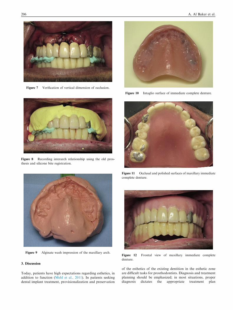
Figure 7 Verification of vertical dimension of occlusion.
Figure 8 Recording interarch relationship using the old pros-
thesis and silicone bite registration.
Figure 9 Alginate wash impression of the maxillary arch.
Figure 10 Intaglio surface of immediate complete denture.
Figure 11 Occlusal and polished surfaces of maxillary immediate
complete denture.
Figure 12 Frontal view of maxillary immediate complete
denture.
206 A. Al Baker et al.
3. Discussion
Today, patients have high expectations regarding esthetics, in
addition to function (Mehl et al., 2011). In patients seekingdental implant treatment, provisionalization and preservation
of the esthetics of the existing dentition in the esthetic zone
are difficult tasks for prosthodontists. Diagnosis and treatmentplanning should be emphasized; in most situations, properdiagnosis dictates the appropriate treatment plan

Table 1 Clinical and Laboratory procedures for conversion of
fixed into removable prosthesis.
Steps Stage Detail Suggestions
1. Clinical Diagnosis and
treatment plan
If the pt. is suitable,
making of primary
impressions
List those aspects of
the existing prosthesis
that will be modified or
used in the future
prosthesis
2. Lab Construct primary
diagnostic casts
3. Clinical Face bow record and
centric relation using
silicone index
Recording and
marking the OVD with
Niswonger’s method
It is important to mark
and measure the
vertical dimension of
occlusion
4. Clinical Fixed Prosthesis
Removal and
extraction of all the
abutment teeth
Implant placement.
Suturing of the
extraction sites if
required
Careful removal of the
fixed prosthesis in
order to preserve its
esthetics
5. Clinical Recording wash
impression of the
upper edentulous arch
Record the wash
impression with a free
flowing elastic
impression material
like alginate
6. Clinical Verifying the vertical
and horizontal jaw
relations with the index
made at step 2
The marking should be
preserved and not
washed with the
surgical procedures
7. Lab Mounting, Final wax
up, Flasking,
Dewaxing, Packing of
acrylic, Curing,
Finishing and
Polishing of dentures
If required, wax can be
added to the palate in
the upper arch and wax
up is finalized for a
removable prosthesis
before flasking
8. Clinical Denture insertion Selective spot grinding
of the teeth intraorally
Removal of the
pressure spots from the
intaglio surface of
denture with the use of
pressure indicating
paste
9. Clinical Patient follow up
Figure 13 Preoperative (left) and post-treatment (right) photographs.
Esthetics, occlusion and occlusal vertical dimension in a patient with fixed prostheses 207
(Chaimattayompol et al., 2002; Palmer et al., 2000). Coordina-
tion among the prosthodontist, the implant surgeon, and thedental technician is critical for successful conversion of anexisting fixed dental prosthesis into an immediate denture in
the esthetic zone. Inadequately planned or executed treatmentfails to meet ideal treatment goals and achieve patient satisfac-tion (Kammeyer et al., 2002; Strong, 2012).
The technique described here preserves the esthetics, occlu-
sion, and occlusal vertical dimension by transforming thepatient’s existing fixed dental prosthesis into an immediate com-plete denture over the submerged dental implants (Fig. 13). It
also eliminates interim implant abutment placement and allowshealing prior to definitive implant abutment selection. Thepatient wore the prosthesis for 3 months, until healing of the tis-
sues and implant osseointegration were completed.An alternative approach would be to fabricate an immedi-
ate maxillary complete denture, which would require majoralteration of the diagnostic cast and might not produce an
esthetic result that is perceivable and acceptable to the patient(St George et al., 2010). The technique described in this casereport gives the patient the option of preserving the same
esthetics, with the added advantage of preserving the occlusionand occlusal vertical dimension. Ideal retention in the maxil-lary immediate denture may not be achievable in such cases.
The goals of treatment should be realistic and achievable,and take into account the patient’s esthetics, confidence, andcomfort, as well as function and cost. The patient should be
fully involved in treatment decisions.
4. Conclusion
This report describes the conversion of a maxillary full-archfixed dental prosthesis into an interim complete denture tofunction during implants healing. The technique is simpleand has the advantages of preserving occlusion, occlusal verti-
cal dimension, and esthetics.
Conflict of interest
There is no conflict of interest.
References
Allen, P.F., McKenna, G., Creugers, N., 2011. Prosthodontic care for
elderly patients. Dent. Update 38. 460-2,465-6,469-70.

208 A. Al Baker et al.
Athar, Z., Juszczyk, A.S., Radford, D.R., Clark, R.K., 2009. Effect of
curing cycles on the mechanical properties of heat cured acrylic
resins. Eur. J. Prosthodont. Restor. Dent. 17 (2), 58–60.
Chaimattayompol, N., Emtiaz, S., Woloch, M.M., 2002. Transforming
an existing fixed provisional prosthesis into an implant supported
fixed provisional prosthesis with the use of healing abutments. J.
Prosthet. Dent. 88, 96–99.
Holt Jr., R.A., 1986. Instructions for patients who receive immediate
dentures. J. Am. Dent. Assoc. 112 (5), 645–646.
Jivraj, S., Chee, W., 2006. Transitioning patients from teeth to
implants. Br. Dent. J. 201 (11), 699–708.
Kammeyer, G., Proussaefs, P., Lozada, J., 2002. Conversion of a
complete denture to a provisional implant-supported screw
retained fixed prosthesis for immediate loading of a completely
edentulous arch. J. Prosthet. Dent. 87, 473–476.
Mehl, C.J., Harder, S., Kern, M., Wolfart, S., 2011. Patients’ and
dentists’ perception of dental appearance. Clin. Oral Invest. 15 (2),
193–199. http://dx.doi.org/10.1007/s00784-010-0393-y. Epub 2010
Mar 16.
Millet, C., Leterme, A., Jeannin, C., Jaudoin, P., 2010. Vertical
dimension in the treatment of the edentulous patient. Rev.
Stomatol. Chir Maxillofac 111 (5–6), 315–330.
Nadig, R.R., Usha, G., Kumar, V., Rao, R., Bugalia, A., 2011.
Geriatric restorative care – the need, the demand and the
challenges. J. Conserv. Dent. 14 (3), 208–214.
Palmer, R.M., Palmer, P.J., Newton, J.T., 2000. Dealing with esthetic
demands in the anterior maxilla. Periodontology 2003 (33), 105–
118.
Shahghaghian, S., Taghva, M., Abduo, J., Bagheri, R., 2014. Oral
health related quality of life of removable partial denture wearers
and related factors. J. Oral Rehabil. http://dx.doi.org/10.1111/
joor.12221.
St George, G., Hussain, S., Welfare, R., 2010. Immediate dentures: 1.
Treatment planning. Dent. Update 37 (2). 82-4, 86-8, 91.
Strong, S.M., 2012. Conversion from fixed bridge to implant-
supported restoration in the esthetic zone. Gen. Dent. 60 (3),
182–185.
Zitzmann, N.U., Krastl, G., Hecker, H., Walter, C., Waltimo, T.,
Weiger, R., 2010. Strategic considerations in treatment planning:
deciding when to treat, extract, or replace a questionable tooth. J.
Prosthet. Dent. 104 (2), 80–91. http://dx.doi.org/10.1016/S0022-
3913(10)60096-0.