presented by alana canfield william goddard ravi abrol and soo-kyung kim socalbsi program august 21,...
Post on 21-Dec-2015
216 views
TRANSCRIPT

Presented by Alana CanfieldWilliam Goddard
Ravi Abrol and Soo-Kyung KimSoCalBSI ProgramAugust 21, 2008
Computational Analysis of the Structure of Human Histamine 2 GPCR

What is a GPCR? What is H2? GPCR : G-Protein Coupled Receptor H2 is a histamine receptor involved in
gastric acid secretion in gastrointestinal system.
Image courtesy of http://www.zmnh.uni-hamburg.de/schaller/gpcr.jpg

Why Study GPCR’s? Why study them computationally? Membrane proteins are involved in a large
number of biological processes.◦ Signal transduction pathways
Lack of crystal structures makes computational approaches necessary.
Structures are needed in therapeutic drug design.◦ More than 50% of marketed drugs target GPCR’s
Image courtesy of http://www.drugstore.com

Proposed binding pocket
Beta-2 Receptor
H2 Receptor: Optimize Asp98
Asp98
Asp98
TM3
TM5
TM3
TM3
TM5
TM5

The Prediction MethodIndividual Helices Helix Bundle
Bundle+Ligand
Determine transmembrane residues. Optimize helix kinks.Predict orientations of helices according to template crystal structure.
Determine helix orientations in space.Analyze final bundle structurally and energetically.
Add extracellular loops.Use final structure to dock various ligands.Analyze possible activation pathways.

Determining Transmembrane Regions Transmembrane (TM) regions predicted using
hydrophobicity profile and homology search.
The ‘cap’ regions (helix ends) must carefully be considered.
REGION CONSENSUS RESIDUES TM1 KITITVVLAVLILITVAGNVVVCLAV TM2 NCFIVSLAITDLLLGLLVLPFSAIYQLSCK TM3 NIYTSLDVMLCTASILNLFMISLD TM4 RVAISLVLIWVISITLSFLSIH TM5 EVYGLVDGLVTFYLPLLIMCITYYRIFK TM6 KATVTLAAVMGAFIICWFPYFTAFVYRGLR TM7 EAIVLWLGYANSALNPILYAALNR
Hydrophobicity Plot
-1.5
-1
-0.5
0
0.5
1
0 100 200 300 400
Residue Number
Hyd
rop
ho
bic
ity
Val
ue
Transmembrane Region 7 H1 : NEHLHMFTIWLGYINSTLNPLIYPLCNE H2 : NEVLEAIVLWLGYANSALNPILYAALNR H3 : PDYWYETSFWLLWANSAVNPVLYPLCHH H4 : KSVWYRIAFWLQWFNSFVNPLLYPLCHK FINAL: EAIVLWLGYANSALNPILYAALNR
1 2 3 4 5 6 7

Character of Individual Helices Helices may have characteristic ‘kinks’. For individual helices, all residues except
Pro, Gly, Thr, and Ser were alanized.◦ This results in conformational sampling while
maintaining the over helix character
Kinked Helix Un-kinked Helix

The Helix Bundle : 24 Methods
Bovine Rhodopsin AND Human Beta-2 Adrenergic Receptor
Minimum Energy AND Minimum RMSD
Area AND Rawmid
AlanizedNot Alanized
De-alanize All De-alanize Raw
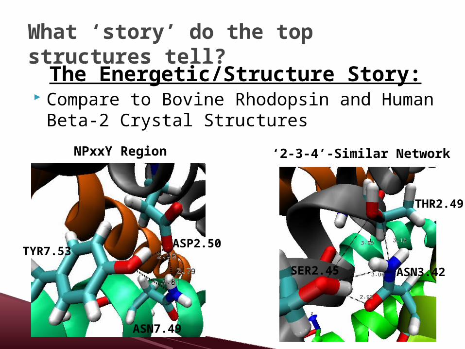
What ‘story’ do the top structures tell?
The Energetic/Structure Story: Compare to Bovine Rhodopsin and Human
Beta-2 Crystal Structures
ASN3.42
THR2.49
SER2.45
‘2-3-4’-Similar Network
TYR7.53ASP2.50
ASN7.49
NPxxY Region

What ‘story’ do the top structures tell?
The Energetic/Chemical Structure Story: Many favorable inter-helical hydrogen bonds.
Bond RMSD/AREA & Dealanize All
RMSD/AREA & Dealanize Raw
Asp186-Arg257 2.64 2.64
Cys102-Tyr250 3.33 3.32
Tyr94-Tyr78 3.32 3.24
Asp64-Tyr288 2.74 2.75
Asp64-Asn284 2.83 2.85
Tyr288-Asn284 2.98 4.06
Thr63-Asn108 2.73 2.73
Tyr278-Gln79 2.72 2.72
Ser59-Asn108 3.10 3.08
Ser59-Thr63 3.98 3.96
Cys246-Asn280 3.36 3.36
Ser105-Asp64 3.77 3.62
Asn108-Ser59 (#2) 2.96 2.97
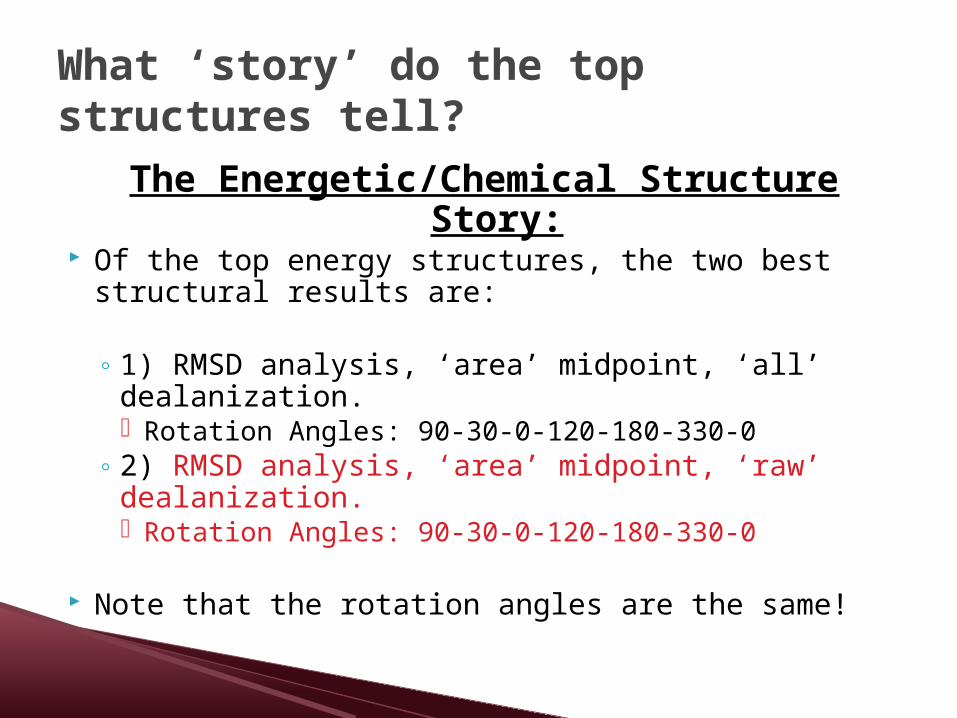
What ‘story’ do the top structures tell?
The Energetic/Chemical Structure Story:
Of the top energy structures, the two best structural results are:
◦ 1) RMSD analysis, ‘area’ midpoint, ‘all’ dealanization. Rotation Angles: 90-30-0-120-180-330-0
◦ 2) RMSD analysis, ‘area’ midpoint, ‘raw’ dealanization. Rotation Angles: 90-30-0-120-180-330-0
Note that the rotation angles are the same!

What ‘story’ do the top structures tell?
Further Structural Analysis - Homology:
Asn1.50 and W4.50
SER2.45
SER2.45
THR2.49
THR2.49
ASN3.42
ASN3.42
TRP4.50
TRP4.50
Rotations: 90-30-0-120-180-330-0 Rotations: 300-30-0-30-180-330-0

Receptors for Docking Docking to four receptors:
◦ 1) Best energetic/chemical structure and 2) best homology-based structure. Both 1) and 2) : with and without rotamer
optimization of Asp98.
Asp98 Asp98
optimize

Possible Ligands for DockingCimitidine (crystal) ICL 162846
Clobenpropit Tiotidine

Initial Spheres

Future Work I am docking the ligand cimitidine.
Final goals for group: Compare the four different histamine receptors in order to gain insight into selectivity.
References Novel Insights Into Histamine H2 Receptor Biology,
John Del Valle and Ira Gantz, Am J Physiol Gastrointest Liver Physiol 273:987-996, 1997.
A study of antagonist affinities for the human histamine H2 receptor, JG Baker, British Journal of Pharmacology (2008) 153, 1011–1021
Structural diversity of G protein-coupled receptors and significance for drug discovery, Malin C. Lagerström and Helgi B. Schiöth, Nature Reviews, Volume 7 , April 2008

Acknowledgements Caltech:
◦ Professor Goddard◦ Ravi Abrol ◦ Soo-Kyung Kim◦ Charlie Seto
SoCal Bioinformatics Research Institute:◦ Core Instructors:
Dr. Jamil Mommand, Dr. Sandra Sharp, Dr. Wendy Johnston Dr. Nancy Warter-Perez, Dr. Beverly Krilowicz Dr. Silvia Heubach, Dr. Jennifer Faust
◦ Program Coordinator: Ronnie Cheng◦ All SocalBSI faculty and students
Funding:◦ NSF, NIH, Economic and Workforce Development