prepared by : reem aldossari. the muscular tissues general characteristics of the muscular tissues:...
TRANSCRIPT
The Muscular Tissues
Prepared by : Reem Aldossari

The Muscular Tissues
General characteristics of the muscular tissues:•The cells of the muscular tissues are elongated elements, named muscle fibers.•The cytoplasm of the muscle fiber, the sarcoplasm, contains myofibrils. These fibrils are made up of the proteins actin and myosin.•The plasma membrane of the muscle fiber is called the sarcolemma.•There are three types of muscle fiber:
oSkeletal muscles, which are voluntary and striated.oSmooth muscle, which are involuntary and non- striated.oCardiac muscles, which are involuntary and striated.
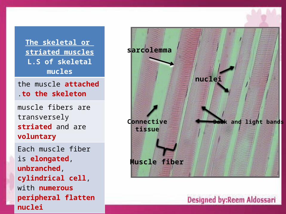
The skeletal or striated muscles
L.S of skeletal mucles
the muscle attached to the skeleton.
muscle fibers are transversely striated and are voluntary
Each muscle fiber is elongated, unbranched, cylindrical cell, with numerous peripheral flatten nuclei Muscle fiber
nuclei
Dark and light bands
sarcolemma
Connectivetissue

T.S of skeletal mucles
Each individual muscle fiber is surrounded by a delicate connective tissue, the endomysium
Bundles or groups of fibers are wrapped by a dense connective tissue called the perimysium
The whole muscle (formed of several bundles) is covered by a dense connective tissue sheath, the epimysium
T.S. of Striated Muscles Fibers (Skeletal Muscles Fibers)
1.Endomysium 2-Perimysium -3- Epimysium
2
3
1

The Smooth Unstriated Muscle
These muscles are present in the wall of blood vessels, and digestive, respiratory, urinary and reproduction systems
The smooth muscle fibers are Unstriated and involuntary
The smooth muscle fibers are elongated, spindle- shaped
cells with pointed ends .
The nucleus is elongated or rod-shaped and centrally located in the cytoplasm at the widest part of the cell.
Muscle fiber
nuclei
Connectivetissue

The Cardiac Muscles
These muscles are present in the heart
The cardiac muscle fibers are striated but are involuntary.
cardiac muscle fibers are elongated, branched, mononucleate or binucleate cells.
The nuclei are oval and centrally located.
At the end to end junction of the cells there are intercalated discs.
Muscle fiber
nucleiDark and light bandsConnective
tissue
intercalated disc

The Nervous Tissues
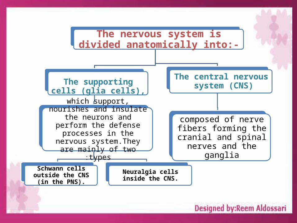
The nervous system is divided anatomically into:-
The supporting cells (glia cells),
which support, nourishes and insulate the neurons and perform
the defense processes in the nervous system.They are mainly
of two types:
Schwann cells outside the CNS (in the PNS).
Neuralgia cells inside the CNS.
The central nervous system (CNS)
composed of nerve fibers forming the cranial and spinal nerves and the
ganglia

Structure of the NeuronThe cell body: it is also called the perikaryon
or soma. It contains the nucleus and much of the metabolic
machinery of the cell
The dendrites: they are multiple
cytoplasmic processes
specialized in receiving
stimuli and transmitting
them to the cell body.
The axon: a single, usually long process specialized in conducting
nerve impulses to other cells

Glial cell nucleus
Neurons
The nervous Tissue
Neurons – Glial cell nucleus

The CELL DIVISTION

The CELL DIVISTIONCell division is a process by which the cellular material is dividedbetween 2 new daughter cells.
Mitosis (or indirect division)• occurs in somatic cells of higher organisms.• it is the means of population growth in unicellular organisms.• results in two daughter cells.•have the same number of chromosomes of the mother cell.
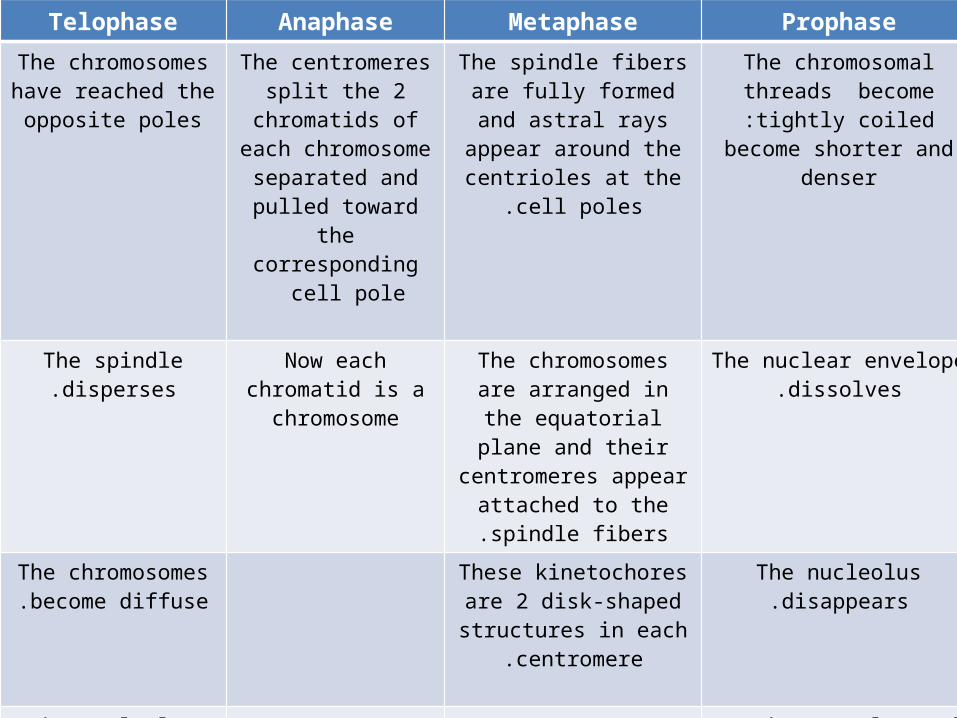
Telophase Anaphase Metaphase ProphaseThe chromosomes have reached the
opposite poles
The centromeres split the 2
chromatids of each chromosome
separated and pulled toward the
corresponding cell pole
The spindle fibers are fully formed and astral rays appear around the
centrioles at the cell poles.
The chromosomal threads become tightly coiled:become shorter and
denser
The spindle disperses.
Now each chromatid is a chromosome
The chromosomes are arranged in the
equatorial plane and their centromeres
appear attached to the spindle fibers.
The nuclear envelope dissolves.
The chromosomes become diffuse.
These kinetochores are 2 disk-shaped structures
in each centromere.
The nucleolus disappears.
The nucleolus reappears.
In the cytoplasm of animal cells the centrioles, with a
surrounding fan of astral rays
A nuclear envelope reforms nucleus is
formed.
The spindle fibers begin to form.
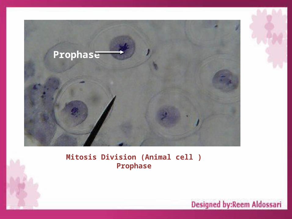
Prophase
Mitosis Division (Animal cell )Prophase

Metaphase
Mitosis Division (Animal cell )Metaphase
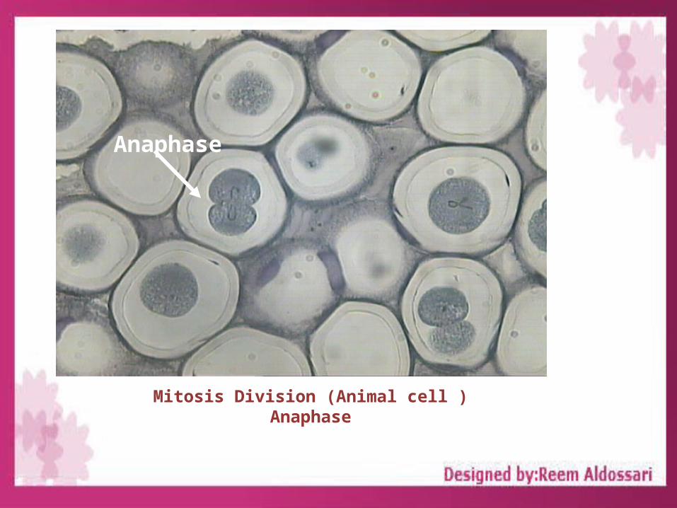
Anaphase
Mitosis Division (Animal cell )Anaphase

Telophase
Mitosis Division (Animal cell )Telophase

Thank you for your attention