predictors of recovery in moderate to severe traumatic
TRANSCRIPT

CLINICAL ARTICLEJ Neurosurg 131:1648–1657, 2019
TraumaTic brain injury (TBI) is a significant cause of death and disabling neurological deficits.2,32 Al-though the Glasgow Coma Scale (GCS) score is
important in assessing the state of consciousness,48 it has been noted to have great limitations and inconsistencies when used in assessing survival and functional outcomes among patients with TBI.7,15,51 Clinical outcomes in pa-tients with TBI can be assessed using the Glasgow Out-come Scale (GOS) score.22 Although current management strategies take into account factors such as the admission GCS score, pupillary reactivity, age of the patient, mecha-
nism of injury,20 CT scan findings,30 intracranial pressure (ICP), and brain tissue oxygen tension (PBO2), little or no information regarding the degree of ischemic cell damage, the level of oxidative stress imbalance, and inflammatory changes can be deduced from these parameters.
Oxidative stress is a disturbance in the equilibrium status of prooxidant/antioxidant systems and accounts for most of the negative consequences of secondary brain in-jury. The process of oxidative stress involves enhancement in the production of free radicals and strong oxidants as well as depletion of body stores of antioxidants following
ABBREVIATIONS AUC = area under the curve; BBB = blood-brain barrier; CI = confidence interval; GCS = Glasgow Coma Scale; GOS = Glasgow Outcome Scale; HR = hazard ratio; ICP = intracranial pressure; IL = interleukin; MDA = malondialdehyde; PBO2 = brain tissue oxygen tension; ROC = receiver operating characteristic; SOD = superoxide dismutase; TAC = total antioxidant capacity; TBARS = thiobarbituric acid reactive substances; TBI = traumatic brain injury; TNFa = tumor necrosis factor–a. SUBMITTED September 10, 2017. ACCEPTED April 5, 2018.INCLUDE WHEN CITING Published online November 9, 2018; DOI: 10.3171/2018.4.JNS172185.
Predictors of recovery in moderate to severe traumatic brain injuryKadhaya David Muballe, MD, PhD,1 Constance R. Sewani-Rusike, MBChB, PhD,2 Benjamin Longo-Mbenza, MD, PhD, DSC,3 and Jehu Iputo, MBChB, PhD2
Departments of 1Neurosurgery, 2Physiology, and 3Public Health, Walter Sisulu University, Mthatha, Eastern Cape Province, South Africa
OBJECTIVE Traumatic brain injury (TBI) is a significant cause of morbidity and mortality worldwide. Clinical outcomes in TBI are determined by the severity of injury, which is dependent on the primary and secondary brain injury processes. Whereas primary brain injury lesions are related to the site of impact, secondary brain injury results from physiological changes caused by oxidative stress and inflammatory responses that occur after the primary insult. The aim of this study was to identify important clinical and biomarker profiles that were predictive of recovery after moderate to severe TBI. A good functional outcome was defined as a Glasgow Outcome Scale (GOS) score of ≥ 4.METHODS This was a prospective study of patients with moderate to severe TBI managed at the Nelson Mandela Aca-demic Hospital during the period between March 2014 and March 2016. Following admission and initial management, the patient demographic data (sex, age) and admission Glasgow Coma Scale score were recorded. Oxidative stress and inflammatory biomarkers in blood and CSF were sampled on days 1–7. On day 14, only blood was sampled for the same biomarkers. The primary outcome was the GOS score—due to its simplicity, the GOS was used to assess clinical out-comes at day 90. Because of difficulty in performing regular follow-up due to the vastness of the region, difficult terrain, and long travel distances, a 3-month follow-up period was used to avoid default.RESULTS Sixty-four patients with Glasgow Coma Scale scores of ≤ 12 were seen and managed. Among the 56 patients who survived, 42 showed significant recovery (GOS score ≥ 4) at 3 months. Important predictors of recovery included antioxidant activity in the CSF (superoxide dismutase and total antioxidant capacity).CONCLUSIONS Recovery after TBI was dependent on the resolution of oxidative stress imbalance.https://thejns.org/doi/abs/10.3171/2018.4.JNS172185KEYWORDS trauma; traumatic brain injury; oxidative stress; inflammatory changes; recovery
J Neurosurg Volume 131 • November 20191648 ©AANS 2019, except where prohibited by US copyright law
Unauthenticated | Downloaded 04/06/22 11:15 PM UTC

J Neurosurg Volume 131 • November 2019 1649
Muballe et al.
trauma or inflammation. Early after TBI, oxidative stress changes occur, leading to increased lipid peroxidation and decreased enzymatic antioxidant activities, which can af-fect clinical outcomes.16,31
The reactive oxygen species generated during oxida-tive stress imbalance cause neuronal damage, dysfunc-tion of the blood-brain barrier (BBB), and brain edema in TBI.19 These reactive oxygen intermediates, including superoxide anions, hydroxyl radicals, hydrogen peroxide, and hypochlorous acid, cause oxidative damage to cellular proteins and nucleic acids in addition to lipid peroxida-tion.26 Lipid peroxidation results in the generation of free radicals including malondialdehyde (MDA) and 4-hy-droxynonenal (4-HNE) or 2-propenol (acrolein), which can cause increased cell membrane permeability, brain edema, and neuronal damage.49,55 Termination of lipid per-oxidation occurs in reactions involving lipid radicals with endogenous antioxidants.
TBI also induces an inflammatory response, with sub-sequent release of inflammatory mediators. Delayed or inadequate management of TBI may propagate an unreg-ulated acute inflammatory response and put in motion a vicious cycle of inflammatory damage8,19, 21, 25, 33, 38, 42, 45, 52, 54,60 caused by inflammatory factors such as tumor necrosis factor–a (TNFa), interleukin (IL)-1b, and IL-18, which are proinflammatory cytokines. The secretion of inflam-matory cytokines, including IL-1b, TNFa, IL-6, and IL-8, which occurs early in TBI, causes the recruitment of leukocytes and increased secretion of nitric oxide during inflammation.9,39 These proinflammatory responses lead to a disruption of the BBB, neuronal tissue damage, and worsening of brain edema.1,17,41
The inflammatory process also involves secretion of antiinflammatory factors, including IL-6 and IL-10. IL-6 has a dual inflammatory role, with both antiinflammatory and proinflammatory effects. The antiinflammatory effect of IL-6 is through inhibition of the TNFa synthesis, pro-motion of release of soluble TNFa receptors, induction of the IL-1 receptor antagonist, and attenuation of oxidative stress.37,57 IL-6 also promotes the release of prostaglandin E2, which stimulates IL-10 secretion.24 The neuroprotec-tive effects of IL-6 occur through neuronal apoptosis, reg-ulation of cytokine secretion, and maintenance of BBB in-tegrity by inhibiting the expression of matrix metallopro-teinase–9 in ischemic cells.14 IL-10 is an immunoregulato-
ry protein produced by macrophages, dendritic cells, and B-lymphocyte cells—it exerts its antiinflammatory effect by limiting or inhibiting adaptive and innate immunity. Actions of IL-10 prevent downregulation of antiapoptotic protein Bcl-2 in ischemic brain tissue and limit excitotoxic brain injury.3 Effects of IL-10 in cerebral ischemia lead to a reduction in volume of infarcted tissue and improve-ment in clinical outcomes. Low levels of IL-10 are associ-ated with impaired antiinflammatory response and poor recovery in stroke patients.4 The antiinflammatory effect of IL-10 attained by limiting the production of TNFa and interferon-g may lead to a reduction in tissue destruction and limitation of tissue necrosis.28,58,59
MethodsThis was a prospective study of patients with moderate
to severe TBI managed at the Nelson Mandela Academic Hospital in the Eastern Cape Province of South Africa during the period between March 2014 and March 2016.
At the accident and emergency department, neurologi-cal evaluation and radiological investigations including CT scans of the brain were performed. The patients re-quiring surgical intervention were then taken to the oper-ating room, where surgical procedures such as burr holes for the insertion of external ventricular drain catheters, ICP probes, PBO2 probes (Licox), and craniotomy or cra-niectomy were performed in patients with intracranial mass lesions for decompression or evacuation of intracra-nial hematoma. Criteria for inclusion or exclusion from the study were defined (Table 1).
Postoperatively, the patients were admitted to the ICU, where ventilation, sedation, analgesia, and monitoring of the ICP, PBO2, and brain tissue temperatures were done. Daily blood and CSF samples were taken at 9:00 am for evaluation of inflammatory and oxidative stress biomark-ers from day 1 to day 7. The blood was again sampled for evaluation of the biomarkers on day 14 of follow-up. The serum and CSF total antioxidant capacity (TAC) and superoxide dismutase (SOD) levels were analyzed. The serum MDA levels were assessed using the thiobarbituric acid reactive substances (TBARS) assay, whereas serum inflammatory biomarkers, including IL-1b, IL-6, and IL-10, were assayed using the BioLegend enzyme-linked im-munosorbent assay (ELISA) method. The patients were reviewed at 2 weeks and 12 weeks, during which time the GOS score was assessed.
Sample Collection and StorageThe blood samples were collected by venipuncture into
sterile vacutainer non–EDTA-containing tubes (Becton Dickinson). Following centrifugation of blood samples for 13 minutes at 3000 rpm, the supernatant (serum) was pipetted into vacutainer tubes. Aliquots were then placed into cryovials bearing the patient’s name, date, and time of collection before storage at -80°C. The CSF samples were also pipetted into cryovials and stored at -80°C.
MDA Assay by Evaluation of TBARSThe OxiSelect TBARS assay kit for MDA quantita-
tion (Cell Biolabs, Inc.) was used. Assay of MDA by the
TABLE 1. Inclusion and exclusion criteria
Inclusion Criteria Exclusion Criteria
Patients w/ moderate to severe TBI (GCS ≤12) in whom neuromonitoring & surgical intervention were indicated
Patients who died while still in the accident & emergency depart-ment before admission
Patients w/ intracranial pathology requiring surgical intervention &/or temporary CSF diversion to reduce ICP
Patients in whom neuromonitoring was not done due to logistical problems
Patients w/ informed consent (from relatives) to participate in the study
Patients w/ no informed consent due to refusal by relatives to participate in the study
Unauthenticated | Downloaded 04/06/22 11:15 PM UTC

Muballe et al.
J Neurosurg Volume 131 • November 20191650
TBARS method is used to monitor lipid peroxidation. The procedure was performed per the manufacturer’s specifi-cations.
SOD AssayA SOD assay kit manufactured by Cayman Chemical
USA was used. The SOD assay kit uses tetrazolium salt for the detection of superoxide radicals generated by xan-thine oxidase and hypoxanthine. One unit of SOD is the amount of enzyme needed to exhibit 50% of dismutation of the superoxide radical. This assay measures all 3 types of SOD (Cu/Zn, Mn, and Fe SOD).
TAC Assay by the Ferric-Reducing Antioxidant Power Method
This method of evaluating the total antioxidant power assesses the ferric-reducing potential of the sample. Dur-ing the procedure, inactivation of oxidants was done in a redox reaction by using reductants as the electron-donat-ing antioxidants. Reduction of ferric tripyridyltriazone to a ferrous state occurs at a low pH to produce an intense blue color, which was measured by the change in absor-bance at 593 nm. The change in absorbance was directly related to the total reduction of the electron-donating anti-oxidants in the reaction.
IL AssayIL-1b, IL-6, and IL-10 assay was performed using Bio-
Legend’s ELISA MAX deluxe sets (ISO 9001:2008 and ISO 13485:2003; BioLegend, Inc.). The assay was per-formed per the manufacturer’s specifications.
Ethical ConsiderationsPrior to initiation of the study, ethical and institutional
approval was obtained from the Walter Sisulu Universi-ty and the department of health according to the ethical guidelines of the Helsinki Declaration of 1975.
Data and Statistical AnalysisData analysis was performed using SPSS version 23.0
for Windows (IBM Corp.). Univariate and multivariate lo-gistic regression models including receiver operating char-acteristic (ROC) curves, Cox regression, and Kaplan-Meier estimates were used to identify the variables that were in-dependently associated with survival and recovery.
ResultsIdentification of Potential and Independent Predictors of Recovery of Cerebral Functional Status in Patients With Moderate to Severe TBI
During the study, 64 patients with moderate to severe TBI were treated. The age of our patients ranged from 10 years to 62 years. The patient age group with the high-est frequency of TBI was the age group 20–39 years (n = 35), the intermediate frequency was in the age group 0–19 years (n = 21), and the lowest frequency was in the age group 40–62 years (n = 8) (Fig. 1). Of the 64 patients, 65.6% (n = 42) sustained injuries from assaults, 21.9% (n = 14) from motor vehicle accidents, 9.4% (n = 6) from falls from heights, and 3.1% (n = 2) for other unclear reasons (“other”) (Table 2).
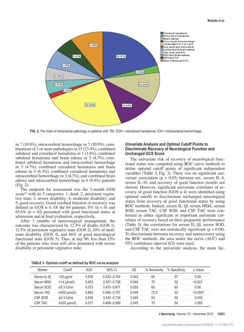
Frequencies of pathology among the intracranial find-ings in 64 patients were as follows: subdural hematoma in 13 (20.3%), extradural hematoma in 2 (3.1%), brain edema
FIG. 1. Distribution of patients with TBI by age groups.
TABLE 2. Mechanism of injury and number of patients
Mechanism of Injury No. of Patients %
Assault 42 65.6Motor vehicle accident 14 21.9Fall from height 6 9.4Other 2 3.1Total 64 100.0
Unauthenticated | Downloaded 04/06/22 11:15 PM UTC
J Neurosurg Volume 131 • November 2019 1651
Muballe et al.
in 7 (10.9%), intracerebral hemorrhage in 7 (10.9%), com-binations of 3 or more pathologies in 15 (23.4%), combined subdural and extradural hematoma in 1 (1.6%), combined subdural hematoma and brain edema in 3 (4.7%), com-bined subdural hematoma and intracerebral hemorrhage in 3 (4.7%), combined extradural hematoma and brain edema in 4 (6.3%), combined extradural hematoma and intracerebral hemorrhage in 3 (4.7%), and combined brain edema and intracerebral hemorrhage in 6 (9.4%) patients (Fig. 2).
The endpoint for assessment was the 3-month GOS score22 with its 5 categories: 1, dead; 2, persistent vegeta-tive state; 3, severe disability; 4, moderate disability; and 5, good recovery. Good cerebral function or recovery was defined as GOS ≥ 4. Of the 64 patients, 0% (n = 0) and 65.6% (n = 42) presented with good functional status at admission and at final evaluation, respectively.
After 3 months of neurosurgical management, the outcome was characterized by 12.5% of deaths (GOS 1), 11.5% of persistent vegetative state (GOS 2), 10% of mod-erate disability (GOS 4), and 66% of good neurological functional state (GOS 5). Thus, at day 90, less than 15% of the patients who were still alive presented with severe disability or persistent vegetative state.
Univariate Analysis and Optimal Cutoff Points to Discriminate Recovery of Neurological Function and Unchanged GCS Score
The univariate risk of recovery of neurological func-tional status was computed using ROC curve methods to define optimal cutoff points of significant independent variables (Table 3, Fig. 3). There was no significant uni-variate association (p > 0.05) between sex, serum IL-6, serum IL-10, and recovery of good function (results not shown). However, significant univariate correlates of re-covery of good function (GOS ≥ 4) were identified using optimal cutoffs to discriminate unchanged neurological status from recovery of good functional status by using ROC methods. Indeed, serum IL-1b, serum MDA, serum SOD, serum TAC, CSF SOD, and CSF TAC were con-firmed as either significant or important univariate cor-relates of recovery based on their prognostic performance (Table 3); the correlations for serum IL-1b, serum SOD, and CSF TAC were not statistically significant (p = 0.08). To discriminate between recovery and nonrecovery using the ROC methods, the area under the curve (AUC) and 95% confidence interval (CI) were used.
According to the univariate analysis, the main fac-
FIG. 2. Pie chart of intracranial pathology in patients with TBI. EDH = extradural hematoma; ICH = intracerebral hemorrhage.
TABLE 3. Optimal cutoff as defined by ROC curve analysis
Marker Cutoff AUC 95% CI SE % Sensitivity % Specificity p Value
Serum IL-1β <35 pg/ml 0.619 0.533–0.704 0.043 66 57 0.08Serum MDA <1.4 µmol/L 0.653 0.567–0.738 0.044 70 52 <0.001Serum SOD ≥0.3 IU/ml 0.573 0.475–0.671 0.050 60 60 0.08Serum TAC ≥450 µmol/L 0.662 0.566–0.757 0.049 72.2 52 <0.0001CSF SOD ≥0.3 IU/ml 0.635 0.547–0.734 0.045 63 52 0.002CSF TAC ≥300 µmol/L 0.577 0.488–0.666 0.045 70 50 0.083
Unauthenticated | Downloaded 04/06/22 11:15 PM UTC

Muballe et al.
J Neurosurg Volume 131 • November 20191652
tors that strongly predicted recovery and improvement of functional status in patients with moderate to severe TBI included serum IL-1b, serum MDA, serum SOD, serum TAC, CSF SOD, and CSF TAC (Tables 3 and 4), as was amplified by the calculated relative risk (Table 4).
Multivariate analysis was then performed to identify the most significant independent predictors of recovery, by using Cox regression analysis (Table 5) and stratified Kaplan-Meier curves (Figs. 4 and 5). In Cox regression analysis, after adjusting for serum SOD, serum TAC, se-rum IL-1b, serum IL-10, and serum MDA, only the CSF SOD and CSF TAC remained as the most significant in-dependent predictors of recovery of good functional sta-tus (Table 5). Indeed, the likelihood of recovery of good functional status—defined as GOS ≥ 4—was multiplied in excess of 90% by both CSF SOD ≥ 0.3 IU/ml and CSF TAC ≥ 300 μmol/L (Table 4).
The comparisons of the mean time to recovery of good functional status according to the log-rank test by strati-fication of CSF SOD (Fig. 4) and CSF TAC (Fig. 5) were obtained by the Kaplan-Meier curves.
Kaplan-Meier Analysis of CSF SOD as an Indicator of Recovery
Based on Kaplan-Meier analysis of the CSF SOD activ-ity, the cumulative proportion of patients with CSF SOD activity ≥ 0.3 IU/ml who recovered good functional status was 95.7%, with a mean time to recovery of 34 ± 1.9 days. This was statistically significant (p < 0.0001 by the log-rank Mantel-Cox test) when compared with the propor-tion of patients with CSF SOD activity < 0.3 IU/ml who
FIG. 3. ROC curves for significant correlates of recovery of good functional status.
TABLE 4. Summary of calculated significant association between correlates and recovery of good functional status
Variable of Interest
% w/ Recovery of Good
Functional Status (no.)
% w/ Disability
(no.)Relative
Risk95% CI
p Value
Serum IL-1β <35 pg/ml 94.7 (36) 5.3 (2) 1.4 1.2–1.5 <0.0001 ≥35 pg/ml 69.2 (18) 30.8 (8)Serum MDA <1.4 μmol/L 94.4 (34) 5.6 (2) 1.4 1.2–1.5 <0.0001 ≥1.4 μmol/L 71.4 (20) 28.6 (8)Serum SOD ≥0.3 IU/ml 93.3 (42) 6.7 (3) 1.5 1.3–1.8 <0.0001 <0.3 IU/ml 57.9 (11) 42.1 (8)Serum TAC ≥450 μmol/L 92.5 (37) 7.5 (3) 1.4 1.2–1.6 <0.0001 <450 μmol/L 66.7 (16) 33.3 (8)CSF SOD ≥0.3 IU/ml 95.7 (44) 4.3 (2) 1.7 1.4–2 <0.0001 <0.3 IU/ml 55.6 (10) 44.4 (8)CSF TAC ≥300 μmol/L 95.5 (42) 4.5 (2) 1.5 1.3–1.8 <0.0001 <300 μmol/L 60 (12) 40 (8)
Unauthenticated | Downloaded 04/06/22 11:15 PM UTC

J Neurosurg Volume 131 • November 2019 1653
Muballe et al.
recovered to GOS ≥ 4 (55.6%, with mean time to recovery of 43 ± 4.4 days). Values for continuous variables are ex-pressed as the mean ± SD throughout.
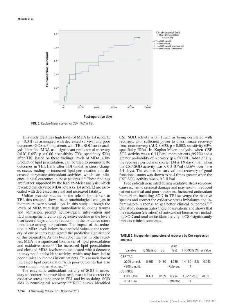
Kaplan-Meier Analysis of CSF TAC as an Indicator of Recovery
Kaplan-Meier analysis was used to estimate the sur-vival of patients based on the biomarkers. The cumulative proportion of patients with CSF TAC ≥ 300 μmol/L sur-viving and recovering to GOS ≥ 4 was 95.5%, with a mean time to recovery of 34.6 ± 1.97 days. This is significant when compared (log-rank Mantel-Cox test, p < 0.001) with the cumulative proportion of patients with CSF TAC < 300 μmol/L who would recover (60%, mean time to recovery 41.4 ± 4.03 days).
DiscussionAs in previous South African studies, assaults were a
predominant mechanism of injury in our study population. This is unlike the findings in North American studies, in which motor vehicle accidents and falls are the most com-mon causes of TBI.10,44,50
In this study, we describe the impact of TBI and neuro-surgical management on clinical and biomarker profiles to predict the likelihood of recovery as defined by the GOS score at 3 months. We attempt to provide an objective as-sessment of the likely outcome by discriminating recovery (GOS ≥ 4) from nonrecovery (poor outcomes) (GOS ≤ 3) after moderate to severe TBI.
The most significant prognostic information was ob-served from CSF TAC and SOD activity. None of these factors have been considered in prognostic models such as the International Mission for Prognosis and Clinical Trials in Traumatic Brain Injury (IMPACT) and Corti-
costeroid Randomisation After Significant Head Injury (CRASH-1).29,47 Indeed, these two characteristics have not been previously studied to determine any relationship with clinical outcomes in TBI.
Following neurosurgical intervention and management, some patients did not recover and were observed to have had an unchanged (“maintained”) neurological functional status (GOS ≤ 3) by day 90. Univariate correlates of recov-ery by ROC curve analysis defined thresholds for serum IL-1b, serum MDA (by TBARS assay), and serum and CSF SOD and TAC.
From this research it is evident that high levels of IL-1b are associated with lack of recovery and poor outcomes after TBI among these patients. This is most likely to be related to the potent proinflammatory effects of IL-1b, which lead to an increase in volume of hemorrhagic contu-sions and brain edema. These pathophysiological changes in TBI result in poor clinical outcomes as manifested by the low GOS score.18,43 Various studies have been done to determine the prognostic significance of some biomark-ers, including the calcium-binding protein (S100B) and neuron-specific enolase as biomarkers in TBI. No univer-sal consensus has been reached to define the role of these biomarkers in predicting outcomes, due to their low speci-ficity and the small sample sizes used.40,53
Analysis using ROC methods revealed that IL-1b levels ≥ 35 pg/ml were associated with poor clinical outcomes (GOS ≤ 3). The proinflammatory effects of IL-1b aggra-vate the pathological changes in TBI and affect the pa-tient’s capacity for survival and recovery. The association of elevated serum IL-1b, a proinflammatory cytokine with poor clinical outcomes, has also been demonstrated in oth-er studies.6,13,46,56 Indeed, the impact of IL-1b on survival and recovery revealed in this study has also been demon-strated in various other studies.12
FIG. 4. Kaplan-Meier curves for CSF SOD in TBI.
Unauthenticated | Downloaded 04/06/22 11:15 PM UTC
Muballe et al.
J Neurosurg Volume 131 • November 20191654
This study identifies high levels of MDA (≥ 1.4 μmol/L; p = 0.041) as associated with decreased survival and poor outcomes (GOS ≤ 3) in patients with TBI. ROC curve anal-ysis identified MDA as a significant predictor of recovery (AUC 0.653; p < 0.001; sensitivity 70%, specificity 52%) after TBI. Based on these findings, levels of MDA, a by-product of lipid peroxidation, can be used to prognosticate outcomes in TBI. Early after TBI oxidative stress chang-es occur, leading to increased lipid peroxidation and de-creased enzymatic antioxidant activities, which can influ-ence clinical outcomes in these patients.16,31 These findings are further supported by the Kaplan-Meier analysis, which revealed that elevated MDA levels (≥ 1.4 μmol/L) are asso-ciated with decreased survival and increased fatality.
Unlike previous studies on the role of biomarkers in TBI, this research shows the chronobiological changes in biomarkers over several days. In this study, although the levels of MDA were high immediately following trauma and admission, prompt neurosurgical intervention and ICU management led to a progressive decline in the levels over several days and to a reduction in the oxidative stress imbalance among our patients. The impact of the reduc-tion in MDA levels below the threshold value on the recov-ery of our patients highlighted the predictive significance of this biomarker. As has been documented in other stud-ies, MDA is a significant biomarker of lipid peroxidation and oxidative stress.36 The increased lipid peroxidation and elevated MDA levels were associated with a decrease in enzymatic antioxidant activity, which may have led to poor clinical outcomes in our patients. This association of increased lipid peroxidation with poor outcomes has also been shown in other studies.16,31
The enzymatic antioxidant activity of SOD is neces-sary to counter the prooxidant response and to correct the oxidative stress imbalance in TBI, and by so doing, SOD aids in neurological recovery.19,60 ROC curves identified
CSF SOD activity ≥ 0.3 IU/ml as being correlated with recovery, with sufficient power to discriminate recovery from nonrecovery (AUC 0.635; p = 0.002; sensitivity 63%, specificity 52%). In Kaplan-Meier analysis, when CSF SOD activity was ≥ 0.3 IU/ml, more patients (95.7%) had a greater probability of recovery (p < 0.0001). Additionally, the recovery period was shorter (34 ± 1.9 days) than when the CSF SOD activity was < 0.3 IU/ml (55.6% over 43 ± 4.4 days). The chance for survival and recovery of good functional status was shown to be 4 times greater when the CSF SOD activity was ≥ 0.3 IU/ml.
Free radicals generated during oxidative stress response cause ischemic cerebral damage and may result in reduced patient survival and poor outcomes. Increased antioxidant biomarkers including SOD in TBI scavenge the reactive species and correct the oxidative stress imbalance and in-flammatory response to get better clinical outcomes.23,35 Our study demonstrates these observations and shows that the resolution (elevation) of antioxidant biomarkers includ-ing SOD and total antioxidant activity in CSF significantly impacts clinical outcomes.
FIG. 5. Kaplan-Meier curves for CSF TAC in TBI.
TABLE 5. Independent predictors of recovery by Cox regression analysis
Variable Β Statistic SEWald Test HR (95% CI) p Value
CSF TAC ≥300 µmol/L 0.363 0.180 4.085 1.4 (1.01–2.1) 0.043 <300 µmol/L Referent 1CSF SOD ≥0.3 IU/ml 0.471 0.189 6.224 1.6 (1.1–2.3) <0.01 <0.3 IU/ml Referent 1
Unauthenticated | Downloaded 04/06/22 11:15 PM UTC

J Neurosurg Volume 131 • November 2019 1655
Muballe et al.
ROC curve analysis furthermore identified better clini-cal outcomes when serum TAC was ≥ 450 μmol/L (AUC 0.662; p < 0.0001). However, analysis of CSF TAC showed that the optimal cutoff value for recovery was ≥ 300 μmol/L (AUC 0.577), which defines the important role of antioxi-dants in averting the oxidative stress imbalance. Further analysis using Cox regression models defined CSF TAC as a major determinant of good outcomes in TBI (hazard ratio [HR] 1.4, 95% CI 1.01–2.1; p = 0.043) (Table 5).
Additionally, Kaplan-Meier analysis showed that when CSF TAC levels were ≥ 300 μmol/L, the cumulative pro-portion of patients surviving was 95.5%, with a shorter time to recovery (34.6 ± 1.97 days) when compared with levels < 300 μmol/L, where the proportion of patients recovering was smaller (60%) and the time to recovery was prolonged (41.4 ± 4 days). Indeed, various studies show that antioxi-dant defenses can prolong survival, enhance recovery, and reduce the destructive effects of free radicals and oxidant factors.34,35 Thus, both Kaplan-Meier and Cox regression analysis demonstrate that the resolution and improved antioxidant activity of CSF SOD and CSF TAC not only shorten the time to recovery but also result in an increased proportion of patients with better outcomes (GOS ≥ 4).
Thus, in multivariate analysis an elevation in CSF TAC and CSF SOD activity was independently associated with recovery of good functional status (GOS ≥ 4). The find-ings in this study reveal that antioxidant factors, as defined by the CSF TAC levels and SOD activity, are important in the recovery of patients with moderate to severe TBI. Multivariate Cox regression analysis revealed that recov-ery (GOS ≥ 4) in TBI was significantly dependent on fac-tors such as increased antioxidant function, manifested by increased CSF SOD ≥ 0.3 IU/ml (HR 1.6, 95% CI 1.1–2.3; p < 0.01) and increased CSF TAC ≥ 300 μmol/L (HR 1.4, 95% CI 1.01–2.1; p = 0.043).
This study reveals that the resolution in levels of anti-oxidants as evidenced by the CSF SOD and CSF TAC is more predictive of functional recovery. The study further justifies the important role of endogenous antioxidants in functional recovery. The impact of antioxidant and antiin-flammatory biomarkers in correcting the oxidative stress imbalance, BBB dysfunction, and cerebral autoregulation has been documented in various other studies.5
Studies on the role of calcium-binding protein (S100B) and neuron-specific enolase show that elevated levels of these biomarkers may be predictive of poor outcomes in patients with TBI.11,27 Our investigation is a comprehensive study that relates the trends of biomarker profiles over sev-eral days to the clinical recovery of patients with moderate to severe TBI. As illustrated in other studies, this research defines the role of antioxidants in reversing the destructive effects of free radicals.34,35
Limitations of the Study and Future PerspectivesThis sample size was relatively small and the study was
conducted in a single institution; therefore, institutional bias may be a valid limitation. It would be preferable to conduct a larger, multicenter, institutional study that evalu-ates the impact of replenishment of antioxidants and re-lates it to clinical outcomes in patients with moderate to severe TBI.
ConclusionsIndependent predictors of recovery in patients with
moderate to severe TBI include CSF SOD and CSF TAC. An elevation in the antioxidant levels in the CSF was as-sociated with better clinical outcomes. Although other studies have evaluated the role of biomarkers in TBI, none of them reviewed the trends in these biomarkers and their impact on survival, as has been demonstrated in this study.
AcknowledgmentsI (K.D.M.) would like to thank the Walter Sisulu University
research office for funding this study. This funding was granted to me during my Doctor of Philosophy in Health Sciences studies.
References 1. Allan SM, Rothwell NJ: Cytokines and acute neurodegenera-
tion. Nat Rev Neurosci 2:734–744, 2001 2. Andelic N, Hammergren N, Bautz-Holter E, Sveen U, Brun-
borg C, Røe C: Functional outcome and health-related qual-ity of life 10 years after moderate-to-severe traumatic brain injury. Acta Neurol Scand 120:16–23, 2009
3. Arimoto T, Choi DY, Lu X, Liu M, Nguyen XV, Zheng N, et al: Interleukin-10 protects against inflammation-mediated degeneration of dopaminergic neurons in substantia nigra. Neurobiol Aging 28:894–906, 2007
4. Barone FC, Price WJ, White RF, Willette RN, Feuerstein GZ: Genetic hypertension and increased susceptibility to cerebral ischemia. Neurosci Biobehav Rev 16:219–233, 1992
5. Batinic-Haberle I, Rajic Z, Tovmasyan A, Reboucas JS, Ye X, Leong KW, et al: Diverse functions of cationic Mn(III) N-substituted pyridylporphyrins, recognized as SOD mimics. Free Radic Biol Med 51:1035–1053, 2011
6. Chiaretti A, Genovese O, Aloe L, Antonelli A, Piastra M, Polidori G, et al: Interleukin 1beta and interleukin 6 rela-tionship with paediatric head trauma severity and outcome. Childs Nerv Syst 21:185–194, 2005
7. Demetriades D, Kuncir E, Murray J, Velmahos GC, Rhee P, Chan L: Mortality prediction of head Abbreviated Injury Score and Glasgow Coma Scale: analysis of 7,764 head inju-ries. J Am Coll Surg 199:216–222, 2004
8. DeWitt DS, Prough DS: Traumatic cerebral vascular injury: the effects of concussive brain injury on the cerebral vascula-ture. J Neurotrauma 20:795–825, 2003
9. Doherty DE, Downey GP, Worthen GS, Haslett C, Henson PM: Monocyte retention and migration in pulmonary inflam-mation. Requirement for neutrophils. Lab Invest 59:200–213, 1988
10. Domingo Z, Peter JC, de Villiers JC: Low-velocity penetrat-ing craniocerebral injury in childhood. Pediatr Neurosurg 21:45–49, 1994
11. Egea-Guerrero JJ, Revuelto-Rey J, Murillo-Cabezas F, Mu-ñoz-Sánchez MA, Vilches-Arenas A, Sánchez-Linares P, et al: Accuracy of the S100b protein as a marker of brain dam-age in traumatic brain injury. Brain Inj 26:76–82, 2012
12. Egerod I, Jensen MB, Herling SF, Welling KL: Effect of an analgo-sedation protocol for neurointensive patients: a two-phase interventional non-randomized pilot study. Crit Care 14:R71, 2010
13. Fan L, Young PR, Barone FC, Feuerstein GZ, Smith DH, McIntosh TK: Experimental brain injury induces expression of interleukin-1 beta mRNA in the rat brain. Brain Res Mol Brain Res 30:125–130, 1995
14. Feng Q, Wang YI, Yang Y: Neuroprotective effect of inter-leukin-6 in a rat model of cerebral ischemia. Exp Ther Med 9:1695–1701, 2015
Unauthenticated | Downloaded 04/06/22 11:15 PM UTC

Muballe et al.
J Neurosurg Volume 131 • November 20191656
15. Foreman BP, Caesar RR, Parks J, Madden C, Gentilello LM, Shafi S, et al: Usefulness of the abbreviated injury score and the injury severity score in comparison to the Glasgow Coma Scale in predicting outcome after traumatic brain injury. J Trauma 62:946–950, 2007
16. Gaetani P, Marzatico F, Rodriguez y Baena R, Pacchiarini L, Viganò T, Grignani G, et al: Arachidonic acid metabolism and pathophysiologic aspects of subarachnoid hemorrhage in rats. Stroke 21:328–332, 1990
17. Goodman JC, Robertson CS, Grossman RG, Narayan RK: Elevation of tumor necrosis factor in head injury. J Neuro-immunol 30:213–217, 1990
18. Hopkins SJ, Rothwell NJ: Cytokines and the nervous system. I: Expression and recognition. Trends Neurosci 18:83–88, 1995
19. Hudome S, Palmer C, Roberts RL, Mauger D, Housman C, Towfighi J: The role of neutrophils in the production of hy-poxic-ischemic brain injury in the neonatal rat. Pediatr Res 41:607–616, 1997
20. Hukkelhoven CW, Steyerberg EW, Habbema JD, Farace E, Marmarou A, Murray GD, et al: Predicting outcome after traumatic brain injury: development and validation of a prognostic score based on admission characteristics. J Neu-rotrauma 22:1025–1039, 2005
21. Ikeda Y, Anderson JH, Long DM: Oxygen free radicals in the genesis of traumatic and peritumoral brain edema. Neuro-surgery 24:679–685, 1989
22. Jennett B, Bond M: Assessment of outcome after severe brain damage. Lancet 1:480–484, 1975
23. Juurlink BH: Response of glial cells to ischemia: roles of reactive oxygen species and glutathione. Neurosci Biobehav Rev 21:151–166, 1997
24. Köller M, Clasbrummel B, Kollig E, Hahn MP, Muhr G: Major injury induces increased production of interleukin-10 in human granulocyte fractions. Langenbecks Arch Surg 383:460–465, 1998
25. Kontos HA, Wei EP, Povlishock JT, Christman CW: Oxygen radicals mediate the cerebral arteriolar dilation from arachid-onate and bradykinin in cats. Circ Res 55:295–303, 1984
26. Kretzschmar M, Pfeiffer L, Schmidt C, Schirrmeister W: Plasma levels of glutathione, alpha-tocopherol and lipid per-oxides in polytraumatized patients; evidence for a stimulating effect of TNF alpha on glutathione synthesis. Exp Toxicol Pathol 50:477–483, 1998
27. Lesko MM, O’Brien SJ, Childs C, Bouamra O, Rainey T, Lecky F: Comparison of several prognostic tools in traumatic brain injury including S100B. Brain Inj 28:987–994, 2014
28. Lisinski TJ, Furie MB: Interleukin-10 inhibits proinflam-matory activation of endothelium in response to Borrelia burgdorferi or lipopolysaccharide but not interleukin-1beta or tumor necrosis factor alpha. J Leukoc Biol 72:503–511, 2002
29. Maas AI, Marmarou A, Murray GD, Teasdale SG, Steyerberg EW: Prognosis and clinical trial design in traumatic brain in-jury: the IMPACT study. J Neurotrauma 24:232–238, 2007
30. Marshall LF, Marshall SB, Klauber MR, Van Berkum Clark M, Eisenberg H, Jane JA, et al: The diagnosis of head injury requires a classification based on computed axial tomogra-phy. J Neurotrauma 9 (Suppl 1):S287–S292, 1992
31. Marzatico F, Gaetani P, Cafè C, Spanu G, Rodriguez y Baena R: Antioxidant enzymatic activities after experimental sub-arachnoid hemorrhage in rats. Acta Neurol Scand 87:62–66, 1993
32. Masson F, Thicoipe M, Aye P, Mokni T, Senjean P, Schmitt V, et al: Epidemiology of severe brain injuries: a prospective population-based study. J Trauma 51:481–489, 2001
33. Matzinger P: The danger model: a renewed sense of self. Sci-ence 296:301–305, 2002
34. McCord JM: Human disease, free radicals, and the oxidant/antioxidant balance. Clin Biochem 26:351–357, 1993
35. McCord JM: Oxygen-derived free radicals. New Horiz 1:70–76, 1993
36. Meagher EA, FitzGerald GA: Indices of lipid peroxidation in vivo: strengths and limitations. Free Radic Biol Med 28:1745–1750, 2000
37. Morganti-Kossmann MC, Rancan M, Otto VI, Stahel PF, Kossmann T: Role of cerebral inflammation after traumatic brain injury: a revisited concept. Shock 16:165–177, 2001
38. Nathan C: Points of control in inflammation. Nature 420:846–852, 2002
39. Oppenheim JJ, Zachariae CO, Mukaida N, Matsushima K: Properties of the novel proinflammatory supergene “inter-crine” cytokine family. Annu Rev Immunol 9:617–648, 1991
40. Papa L, Lewis LM, Silvestri S, Falk JL, Giordano P, Brophy GM, et al: Serum levels of ubiquitin C-terminal hydrolase distinguish mild traumatic brain injury from trauma controls and are elevated in mild and moderate traumatic brain injury patients with intracranial lesions and neurosurgical interven-tion. J Trauma Acute Care Surg 72:1335–1344, 2012
41. Riva-Depaty I, Fardeau C, Mariani J, Bouchaud C, Delhaye-Bouchaud N: Contribution of peripheral macrophages and microglia to the cellular reaction after mechanical or neurotoxin-induced lesions of the rat brain. Exp Neurol 128:77–87, 1994
42. Rosenberg AL: Recent innovations in intensive care unit risk-prediction models. Curr Opin Crit Care 8:321–330, 2002
43. Rothwell NJ, Hopkins SJ: Cytokines and the nervous system II: Actions and mechanisms of action. Trends Neurosci 18:130–136, 1995
44. Rutland-Brown W, Langlois JA, Thomas KE, Xi YL: Inci-dence of traumatic brain injury in the United States, 2003. J Head Trauma Rehabil 21:544–548, 2006
45. Schlag G, Redl H: Mediators of injury and inflammation. World J Surg 20:406–410, 1996
46. Shiozaki T, Hayakata T, Tasaki O, Hosotubo H, Fuijita K, Mouri T, et al: Cerebrospinal fluid concentrations of anti-in-flammatory mediators in early-phase severe traumatic brain injury. Shock 23:406–410, 2005
47. Subaiya S, Roberts I, Komolafe E, Perel P: Predicting intra-cranial hemorrhage after traumatic brain injury in low and middle-income countries: a prognostic model based on a large, multi-center, international cohort. BMC Emerg Med 12:17, 2012
48. Teasdale G, Jennett B: Assessment of coma and impaired consciousness. A practical scale. Lancet 2:81–84, 1974
49. Thorburne SK, Juurlink BH: Low glutathione and high iron govern the susceptibility of oligodendroglial precursors to oxidative stress. J Neurochem 67:1014–1022, 1996
50. Thurman DJ, Alverson C, Dunn KA, Guerrero J, Sniezek JE: Traumatic brain injury in the United States: A public health perspective. J Head Trauma Rehabil 14:602–615, 1999
51. Timmons SD, Bee T, Webb S, Diaz-Arrastia RR, Hesdorffer D: Using the abbreviated injury severity and Glasgow Coma Scale scores to predict 2-week mortality after traumatic brain injury. J Trauma 71:1172–1178, 2011
52. Todo H, Ohta S, Wang J, Ichikawa H, Ohue S, Kumon Y, et al: Impairment in biochemical level of arterial dilative capa-bility of a cyclic nucleotides-dependent pathway by induced vasospasm in the canine basilar artery. J Cereb Blood Flow Metab 18:808–817, 1998
53. Topolovec-Vranic J, Pollmann-Mudryj MA, Ouchterlony D, Klein D, Spence J, Romaschin A, et al: The value of serum biomarkers in prediction models of outcome after mild trau-matic brain injury. J Trauma 71 (5 Suppl 1):S478–S486, 2011
54. Vincent JL, Ferreira F, Moreno R: Scoring systems for as-sessing organ dysfunction and survival. Crit Care Clin 16:353–366, 2000
Unauthenticated | Downloaded 04/06/22 11:15 PM UTC

J Neurosurg Volume 131 • November 2019 1657
Muballe et al.
55. Williams TI, Lovell MA, Lynn BC: Analysis of derivatized biogenic aldehydes by LC tandem mass spectrometry. Anal Chem 77:3383–3389, 2005
56. Winter CD, Iannotti F, Pringle AK, Trikkas C, Clough GF, Church MK: A microdialysis method for the recovery of IL-1beta, IL-6 and nerve growth factor from human brain in vivo. J Neurosci Methods 119:45–50, 2002
57. Xing Z, Gauldie J, Cox G, Baumann H, Jordana M, Lei XF, et al: IL-6 is an antiinflammatory cytokine required for con-trolling local or systemic acute inflammatory responses. J Clin Invest 101:311–320, 1998
58. Yin Z, Braun J, Neure L, Wu P, Eggens U, Krause A, et al: T cell cytokine pattern in the joints of patients with Lyme arthritis and its regulation by cytokines and anticytokines. Arthritis Rheum 40:69–79, 1997
59. Yin Z, Braun J, Neure L, Wu P, Liu L, Eggens U, et al: Cru-cial role of interleukin-10/interleukin-12 balance in the regu-lation of the type 2 T helper cytokine response in reactive arthritis. Arthritis Rheum 40:1788–1797, 1997
60. Zuccarello M, Boccaletti R, Romano A, Rapoport RM: En-dothelin B receptor antagonists attenuate subarachnoid hem-orrhage-induced cerebral vasospasm. Stroke 29:1924–1929, 1998
DisclosuresThe authors report no conflict of interest concerning the materi-als or methods used in this study or the findings specified in this paper.
Author ContributionsConception and design: Muballe. Acquisition of data: Muballe. Analysis and interpretation of data: Muballe. Drafting the article: Muballe. Critically revising the article: Muballe. Reviewed sub-mitted version of manuscript: Muballe. Approved the final version of the manuscript on behalf of all authors: Muballe. Statistical analysis: Longo-Mbenza. Administrative/technical/material sup-port: Iputo. Study supervision: Sewani-Rusike, Longo-Mbenza.
CorrespondenceKadhaya David Muballe: Walter Sisulu University, Mthatha, East-ern Cape Province, South Africa. [email protected].
Unauthenticated | Downloaded 04/06/22 11:15 PM UTC