preclinical development handbook || metabolism kinetics
TRANSCRIPT

697
19 METABOLISM KINETICS
Charles W. Locuson and Timothy S. Tracy University of Minnesota, Minneapolis, Minnesota
Preclinical Development Handbook: ADME and Biopharmaceutical Properties, edited by Shayne Cox GadCopyright © 2008 John Wiley & Sons, Inc.
Contents
19.1 Introduction 19.2 Typical Kinetics
19.2.1 Michaelis – Menten Equation and Its Use in Drug Metabolism 19.2.2 The Case of Multiple Substrates 19.2.3 Use of the Michaelis – Menten Equation and Its Parameters
19.3 Atypical Kinetics Displayed by Drug Metabolizing Enzymes 19.3.1 Sigmoidal Kinetics with a Single Substrate 19.3.2 Biphasic Kinetics (Nonasymptotic) 19.3.3 Hyperbolic (Nonessential) Activation 19.3.4 Substrate Inhibition 19.3.5 Explanations for Atypical Kinetics in P450 Enzymes and Their Detection 19.3.6 Artifactual Sources of Atypical Kinetics
19.4 Conclusion Acknowledgment References
19.1 INTRODUCTION
It is common to apply kinetic models to in vitro data in an attempt to determine parameters, such as intrinsic clearance, that can be used to make correlations to the in vivo situation. These estimations are generally predicated on the data following a hyperbolic profi le such that the Michaelis – Menten equation can be applied. This

698 METABOLISM KINETICS
allows determination of V m and K m and thus the determination of intrinsic clearance. However, it is increasingly being recognized that, for many substrates, these “ typical kinetic ” conditions may not exist and “ atypical kinetics ” may be observed. Unfor-tunately, application of the Michaelis – Menten equation to these “ atypical ” situa-tions results in misestimation of V m and K m and, subsequently, intrinsic clearance. Thus, it is important to understand the assumptions behind typical kinetics in drug metabolism reactions, the different types of atypical kinetic profi les that may also be observed, and what models and equations can be used to estimate kinetic param-eters from each of these types of data.
19.2 TYPICAL KINETICS
19.2.1 Michaelis – Menten Equation and Its Use in Drug Metabolism
One of the most widely taught and used biochemical principles is embodied in an equation, which has remained the very foundation of enzymology for nearly a century. This equation allows quantitative descriptions of many enzyme - catalyzed reactions in terms of rate and concentration and therefore helps us describe the molecular workings of the cell. Enzymology has aided in the diagnoses of diseases as well as the development of therapies used to treat them, and at the heart of enzymology is the Michaelis – Menten equation:
v
VK
= ⋅+
m
m
SS
[ ][ ]
(19.1)
where reaction velocity, ν , is described as a function of substrate concentration, [S], multiplied by a limiting rate, V m (also known as V or V max whose subscript is not to be confused with that of K m ), and divided by the sum of [S] and constant K m , whose subscript refers to its name, the Michaelis constant.
Following up on the theory of A. J. Brown and V. Henri that enzymatic catalysis results from the formation of an intermediate enzyme – substrate complex, Michaelis and Menten reaffi rmed Henri ’ s basic assumption. 1 Mainly, the effect of substrate concentration on velocity could be described by an equilibrium between enzyme and substrate and that their complexation can lead to product.
E S ES E P+ ⎯ →⎯ +−
k
k
k1
1
2� ⇀��↽ ��� (19.2)
Derivation of the rate equation for Eq. 19.2 based on the initial concentration of enzyme and substrate, and the defi nition of the substrate dissociation constant, K S , gives
v
kK
= ⋅ ⋅+
2 0E SSS
[ ][ ]
(19.3)
1 The following details were compiled largely from Cornish - Bowden [6] , and for further details, consulta-tion of this book is highly recommended.

where E 0 is the initial enzyme concentration. By avoiding any assumption about equilibrium in the fi rst step of Eq. 19.2 (i.e., substrate binding to enzyme), Briggs and Haldane actually made the fi nal contribution to the Michaelis – Menten equation with their steady - state treatment. Their steady - state assumption was that the con-centration of the enzyme – substrate intermediate would become constant soon after initiation of the reaction. Using the same scheme (Eq. 19.2 ), K S in Eq. 19.3 is sub-stituted with the Michaelis constant:
K
k kk
m = +−1 2
1 (19.4)
using the steady - state assumption as shown in the derivation available in most bio-chemistry textbooks. This is probably the preferred treatment unless specifi c details regarding substrate binding are known that enable the use of K S . Whether K m or K S is used, the form of the equation is the same:
v
kK
= ⋅ ⋅+
cat
m
E SS
0 [ ][ ]
(19.5)
with the substitution of k 2 for k cat (the observed rate 2 of reaction) so that no assump-tion is made about the number of steps that occur between substrate binding and the chemistry described by k cat × E 0 . In other words, k 2 in Eq. 19.2 does not refl ect the rate of catalysis except for the simplest two - step mechanism such that k cat is used to represent the observed rate of reaction if there are more than two steps, even if the true number of steps is not known. For the same reason K m is often more complex than shown in Eq. 19.4 . E 0 and k cat are then multiplied to produce the parameter V m to yield the familiar Eq. 19.1 .
Equation 19.2 does not consider the reversibility of the k 2 step, which would result in the subtraction of the reverse rate in the numerator ( k P × E 0 × [P]) and a term that includes a K m for product P in the denominator. Even so, the Michaelis – Menten equation in its commonest form (Eq. 19.1 ) adequately describes the kinetics of metabolism for many drugs by enzymes like the cytochromes P450. Ideally, the rate of reaction at each substrate concentration would follow the method of initial rates , meaning several measurements over a limited amount of time (i.e., less than a few minutes) are subjected to linear regression to determine a rate. This prevents excessive amounts of product from building up and reversibly (or irreversibly) inhibiting the enzyme, or undergoing signifi cant reversal back to substrate. However, this is often not feasible if time - expensive methods like liquid - chromatography separation or even extraction into organic solvent are needed to measure metabo-lized drug. In addition, even an incubation carried out with recombinant P450 is
2 k cat , the catalytic constant or turnover number , describes the number of turnovers an enzyme can accomplish in a given unit of time. Determination of k cat requires accurately estimating V m , therefore requiring the use of a suffi cient range of substrate concentration to ensure saturation, and knowledge of E 0 ( k cat = V m /E 0 ). V m is determined preferably by nonlinear regression (although certain linear plots have been used for estimates). The initial enzyme concentration, E 0 or E total , is in the same concentration units as V m (e.g., µ M/time) so that the unit of k cat is time − 1 .
TYPICAL KINETICS 699

700 METABOLISM KINETICS
much more complex than most may realize. Using the steady - state treatment, a P450 incubation may be thought of as possessing a K m not only for drug, but for oxygen, P450 oxidoreductase (which has a K m for NADPH), and possibly any other partially rate - limiting steps required for catalysis. Fortunately, most of these variables are kept constant and/or saturating. Considering the number of variables in P450 – drug incubations and some of the diffi culties involved in analyzing the products of such reactions, the Michaelis – Menten equation nevertheless remains remarkably useful.
Therefore, although proper controls must be carried out (see below), triplicate end - point measurements and their analysis with the Michaelis – Menten equation are probably the most fundamental and useful experiments in drug metabolism.
19.2.2 The Case of Multiple Substrates
Cofactors are heavily relied upon by drug metabolizing enzymes. The UDP - glucuronosyltransferases (UGTs), glutathione S - transferases (GSTs), fl avin mono-oxygenases (FMOs), sulfotransferases (more correctly, sulfonyltransferases), N - acetyltransferases (NATs), and thiopurine S - methyltransferase (TPMT) all utilize nucleotide - containing cofactors for oxygen activation (e.g., FMO) or for conjuga-tion (e.g., all others). FMOs can remain poised for oxidation without drug substrate ever binding as long as saturating levels of NADPH are present. UGTs, on the other hand, require a ternary complex of enzyme and two substrates (UDP - glucuronic acid and the aglycone substrate to be conjugated) to form before catalysis can occur.
Unfortunately, the number of parameters in rate equations expands substantially when describing models involving additional reaction steps. For instance, take the example of the kinetic mechanism of UGT that generally occurs through the ordered binding of two cosubstrates [1] . If the reverse reaction is ignored and any amount of product formed during the course of the reaction is considered negligible, the rate equation requires concentration information and estimation of K m values for both substrates:
v
VK K K K
=+ + +
m
iA mB mB mA
A BA B A B[ ][ ]
[ ] [ ] [ ][ ] (19.6)
where A and B represent the two substrates each with their own K m ( K mA and K mB ) and K iA is the dissociation constant for the EA complex. Even with the aforemen-tioned assumption, a substantial number of data points would be needed to fi t this rate equation due to the number of parameters involved. However, if substrate B is always present in saturating concentrations and care is taken to avoid the buildup of products, Eq. 19.6 reduces eventually to the Michaelis – Menten equation (Eq. 19.1 ). In fact, this experimental and analytical simplifi cation is probably the standard and preferred method for determining V m as well as the K m for each substrate. With further analysis this method also has the benefi t of revealing features of the kinetic mechanism such as whether substrate binding is random or ordered, or if the fi rst substrate is turned over and leaves before the second substrate can be turned over (i.e., Ping - Pong mechanism) [2] .

ATYPICAL KINETICS DISPLAYED BY DRUG METABOLIZING ENZYMES 701
19.2.3 Use of the Michaelis – Menten Equation and Its Parameters
Most are familiar with the rectangular hyperbola that results from plotting reaction velocity ( y - axis) versus substrate concentration ( x - axis) and that these plotted data can be fi t to the Michaelis – Menten equation using nonlinear regression (although the curve itself was never used by Michaelis and Menten and therefore is not called a Michaelis – Menten curve). Therefore, the parameters V m and K m , as best fi t by the nonlinear version of the equation (Eq. 19.1 ), are reviewed.
At low [S], Eq. 19.1 demonstrates that velocity becomes proportional to [S]:
v
kK
VK
≈ ⋅ ⋅ = ⋅cat
m m
E S S0 [ ] [ ]
(19.7)
The middle expression of Eq. 19.7 is important because a very useful parameter is obtained from the ratio of k cat / K m known as the specifi city constant. k cat / K m (units of time − 1 × concentration − 1 ) is an excellent evaluation of substrate specifi city for an enzyme. Imagine two substrates for the same enzyme that can each be turned over without the other present. The substrate with the highest specifi city constant will correctly predict that it will be turned over faster than the other substrate if both were present at any equimolar concentration. Even if one substrate has a larger k cat , a substrate with a higher k cat / K m will be turned over faster if the two are compared at the same concentration.
Low substrate concentrations (i.e., much lower than K m ) are also relevant to in vivo – in vitro correlations. If the therapeutic dosage of a drug results in sub - K m plasma concentrations (the most common situation), where kinetics are largely fi rst order for the enzyme(s) metabolizing it, Eq. 19.7 can be defi ned as the intrinsic clearance ( CL int = V m / K m , units time − 1 ), which is the clearance of drug from a tissue via metabolism independent of blood fl ow, protein binding, and other restrictions. Although the units are different from that for k cat / K m , V m / K m is still a specifi city constant and the comparison of CL int values for different drugs with the same enzyme may prove useful in predicting the reduced CL int for each drug involved in a drug – drug interaction or polypharmacy.
Next, when the substrate concentration is equal to K m , K m can be defi ned opera-tionally as the concentration of substrate where velocity equals 12 V m . Often, K m in this sense is used as a measure of the affi nity of substrate for enzyme as if they were in equilibrium ( K S ); however, without additional information regarding rate constants, K m should never be assumed to be anything more than an estimate of K S with no guarantees attached. Finally, when substrate is said to be saturating so that every enzyme molecule is bound to substrate, K m becomes negligible and ν ≈ V m . In other words, the rate is limited by the amount of enzyme present, and hence, V m is perhaps better described as a limiting rate rather than a maximum.
19.3 ATYPICAL KINETICS DISPLAYED BY DRUG METABOLIZING ENZYMES
Atypical kinetics is a term that has come to be used to describe any in vitro drug metabolism kinetics that do not fi t the hyperbolic function seen when velocity is
702 METABOLISM KINETICS
plotted versus substrate concentration. Allosterism is another term used frequently in the P450 literature that refers to the ability of a ligand (a.k.a. effector) other than the substrate being turned over to bind in a distinct, noncatalytic site on an enzyme and affect the rate of metabolism of the substrate. The effector can be a second (or third or fourth, etc.) substrate molecule binding to the same enzyme or it can be a molecule that is structurally dissimilar from the substrate. Several lines of evidence suggest that some of the allosteric sites in P450s 3A4 and 2C9 are really distinct sites within the same binding pocket that substrate occupies; however, we think allosterism is still a suitable term to defi ne this phenomenon since the P450s most likely do not bind every substrate in the same orientation or subregion of their active sites anyway.
Much attention has been given to the atypical kinetics of P450s in drug metabo-lism, but it is becoming apparent that phase II enzymes and drug transporters may occasionally display atypical kinetics likely stemming from the simultaneous occu-pancy of a single protein by multiple ligand (in addition to the normal substrates, if there are multiple substrates required for catalysis). In fact, almost all of the theo-retical mechanisms that could give rise to atypical kinetics are plausible with drug metabolizing enzymes whether it occurs via one substrate ( homotropic ) or combina-tions of substrate and effector ( heterotropic ). Using P450s as an example, it is com-pletely reasonable to hypothesize atypical kinetics arise from effector binding that modifi es P450 – P450, P450 – P450 reductase, or P450 – cytochrome - b 5 interactions, where the site can be in the same binding pocket as substrate or remote and be either homotropic or heterotropic.
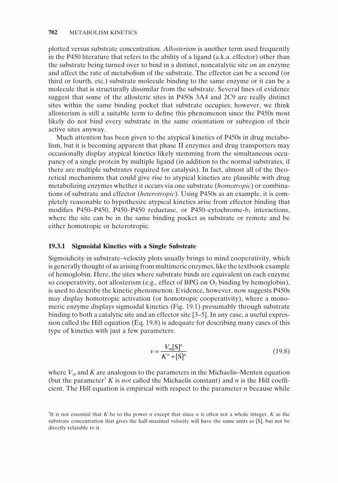
19.3.1 Sigmoidal Kinetics with a Single Substrate
Sigmoidicity in substrate – velocity plots usually brings to mind cooperativity, which is generally thought of as arising from multimeric enzymes, like the textbook example of hemoglobin. Here, the sites where substrate binds are equivalent on each enzyme so cooperativity, not allosterism (e.g., effect of BPG on O 2 binding by hemoglobin), is used to describe the kinetic phenomenon. Evidence, however, now suggests P450s may display homotropic activation (or homotropic cooperativity), where a mono-meric enzyme displays sigmoidal kinetics (Fig. 19.1 ) presumably through substrate binding to both a catalytic site and an effector site [3 – 5] . In any case, a useful expres-sion called the Hill equation (Eq. 19.8 ) is adequate for describing many cases of this type of kinetics with just a few parameters:
v
VK
n
n n=
+m S
S[ ]
[ ] (19.8)
where V m and K are analogous to the parameters in the Michaelis – Menten equation (but the parameter 3 K is not called the Michaelis constant) and n is the Hill coeffi -cient. The Hill equation is empirical with respect to the parameter n because while
3 It is not essential that K be to the power n except that since n is often not a whole integer, K as the substrate concentration that gives the half - maximal velocity will have the same units as [S], but not be directly relatable to it.

ATYPICAL KINETICS DISPLAYED BY DRUG METABOLIZING ENZYMES 703
it measures relative cooperativity, it does not necessarily equal the number of binding sites for substrate because it is often not an integer. A more biochemically logical description of multiple binding sites is obtained with the use of the Adair equation (described in Cornish - Bowden [6] ); however, the parameters of the Hill equation remain practical for most applications, including the study of drug metabo-lizing enzymes. For example, substrates (e.g., testosterone, carbamazepine, pyrene) that induce sigmoidal kinetics by CYP3A4 can all be meaningfully compared to each other using the Hill equation to determine which ones have the highest effi -ciency ( V m / K ) and cooperativity ( n ).
When cooperativity is caused by an effector other than the target substrate in a monomeric enzyme (a.k.a. heterotropic activation ) [7, 8] , for instance, during inhibi-tion screening, the Hill equation can still be used. Or, to better defi ne the V m and K m parameters for substrate when it is known that the effector turnover rate is insignifi cant or much less than it is for substrate, Eq. 19.9 may prove useful by giving K m and V m in the absence or presence of effector (subscripts 1 and 2, respectively):
v
VK
VK K
K K K
=
⋅ + ⋅⋅
+ +⋅
m
m
m
m m
m m m
S S
S S
1
1
22
1 2
1
2
1 2
1
[ ] [ ]
[ ] [ ]
(19.9)
Equation 19.9 , although working under the assumption of equilibrium, will also give the K m and V m of a substrate metabolized in two distinct subsites within the substrate binding pocket, each with different binding affi nities and velocities. More complex rate equations have been developed for sigmoidal kinetics in drug metabolism and more rare mechanisms behind cooperativity can be found in Cornish - Bowden [6] , and these sources should be consulted for a full description.
FIGURE 19.1 Graphical representation of a kinetic profi le exhibiting sigmoidal (autoactivation) kinetics. Inset demonstrates the Eadie – Hofstee plot of a sigmoidal kinetic profi le.

704 METABOLISM KINETICS
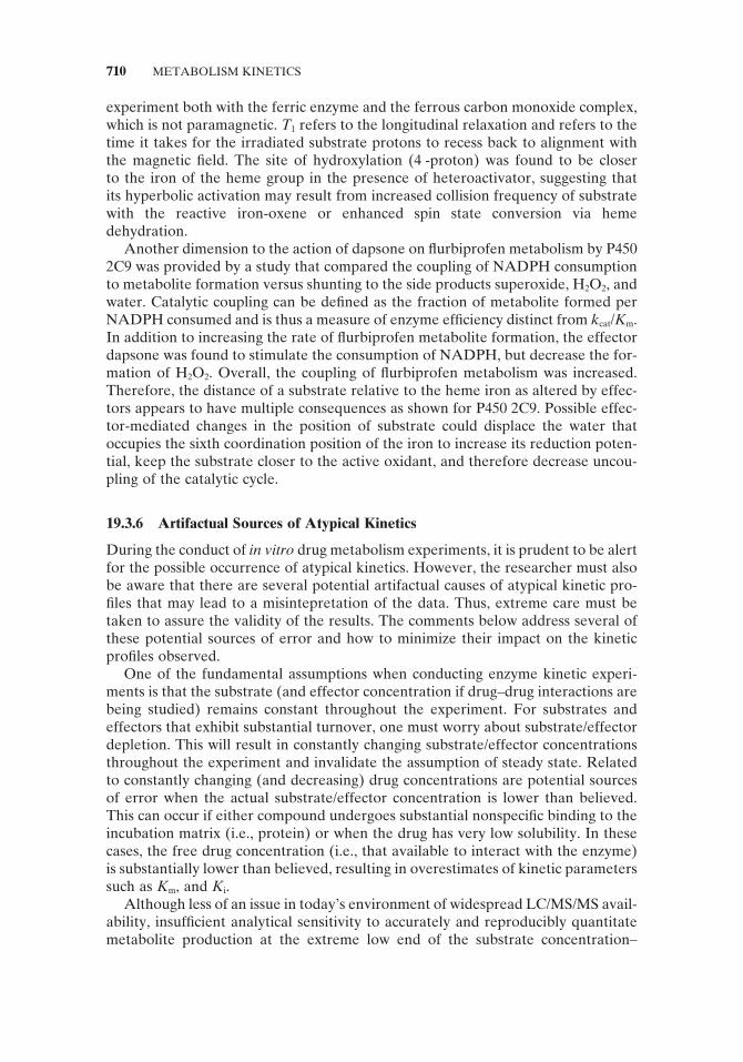
19.3.2 Biphasic Kinetics (Nonasymptotic)
As defi ned here, a biphasic kinetic profi le does not follow saturation kinetics and has two distinct phases. ( Note that sigmoidal kinetics may also be biphasic but exhib-its saturation. ) At low substrate concentrations, the kinetic profi le exhibits curvature similar to that observed with hyperbolic kinetics; however, at high substrate con-centrations, the velocity of the reaction continues to increase in a linear fashion (Fig. 19.2 ), as opposed to becoming asymptotic as would be expected with hyperbolic kinetics. This type of profi le most commonly occurs when the metabolism of a sub-strate is carried out by more than one enzyme in a multienzyme system. However, when using a single, purifi ed enzyme source, it is most likely the result of substrate binding in a productive orientation to more than one region within the active site, but these binding regions exhibit different affi nities for the substrate and also dif-ferential turnover rates. In this case, a single enzyme behaves as if it were a multi-enzyme system. The enzyme thus exhibits a low K m /low V m component (responsible for the semihyperbolic nature of the profi le) and another high K m /high V m compo-nent (producing the linear portion of the profi le). In order to model this type of kinetic profi le, one must use equations that describe the kinetics of one substrate interacting with two binding sites within the same enzyme species, such as Eq. 19.10 .
v
V CLK
= ⋅ + ⋅+
( [ ]) ( [ ] )[ ]
intm
m
S SS
12
1 (19.10)
The parameters V m1 and K m1 are estimated from data representing the curved portion of the plot at lower substrate concentrations and are the estimates of V m and K m for the low V m , low K m site, respectively. CL int describes the linear portion of the plot exhibited at higher substrate concentrations and is the ratio of V max2 / K m2 (i.e., the high V max , high K m component of the profi le). Because this upper portion of the plot is linear, one cannot estimate the actual V m and K m parameters for this portion of the profi le since saturation is not achieved. Thus, CL int is the slope (rate) for this linear portion of the kinetic profi le.
FIGURE 19.2 Graphical representation of a kinetic profi le exhibiting biphasic kinetics. Inset demonstrates the Eadie – Hofstee plot of a biphasic kinetic profi le.

ATYPICAL KINETICS DISPLAYED BY DRUG METABOLIZING ENZYMES 705
19.3.3 Hyperbolic (Nonessential) Activation
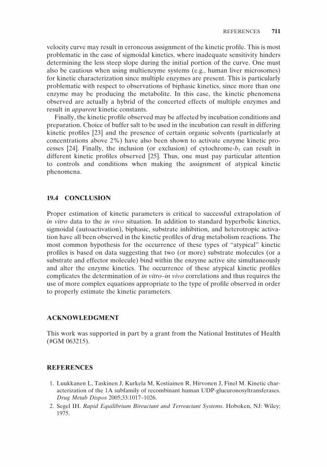
The most straightforward kind of activation in terms of fi tting K m and V m values to velocity versus [S] plots is probably hyperbolic activation. The name refers to the replot of K m / V m against reciprocal activator concentration, which is hyperbolic, not linear. (These plots for sigmoidal and biphasic kinetics are also not expected to be linear.) It is nonessential, because the effector is not necessary for catalysis to occur. While V m and often K m change for a substrate in the presence of an activating effec-tor, the shape of the velocity vesus [S] curve remains a hyperbola. Therefore, the term hyperbolic activation has a more important meaning in drug metabolism kinet-ics because it can also refer to homo - or heterotropic activation that results in hyperbolic curves rather than sigmoidal or biphasic curves. As one might expect, changes in K m and V m can be fi t at each activator concentration using the Michaelis – Menten equation. One of the best documented cases of this heterotropic activation is the activation of fl urbiprofen (S) hydroxylation by the effector dapsone (B) in P450 2C9 and a mechanistic scheme has been proposed (Fig. 19.3 ). A graphi-cal representation of the kinetic profi le for this hyperbolic activation is shown in Fig. 19.4 .
The equation (Eq. 19.11 ) for fi tting multiple curves representing different activa-tor concentrations has a manageable number of parameters including the usual K m (which is assumed to be an estimate of K S in this equilibrium model) and V m for substrate in the absence of activator:
vV
KK
KKK
= ⋅+
++ +
+
m
mB
B
B
B
SB /B /
SB /B /
[ ][ ][ ]
[ ][ ][ ]
11
11β α
αβ α
(19.11)
where [B] is the concentration of activator, α is the factor by which K S for substrate changes, and β can be thought of as the factor by which V m changes (see Fig. 19.3 for further explanation of parameters).
FIGURE 19.3 Kinetic mechanism for nonessential activation [2] demonstrates that the effector, B, is not essential for product formation. Furthermore, it can be seen that this scheme is fl exible as α and β can be greater or less than unity. In the case of fl urbiprofen, metabolism in the presence of effector dapsone results in a decrease in α , indicating an increase in affi nity of P450 2C9 for fl urbiprofen. This was further supported by equilibrium binding experiments. Parameter β was increased, indicating the velocity for oxidation of fl urbiprofen has increased. Activation could also be achieved with α > 1 but this would require higher substrate concen-trations to be observed. Alternatively, if α < 1, but β < 1, activation could result at low sub-strate concentrations and then result in inhibition at higher substrate concentrations.

706 METABOLISM KINETICS
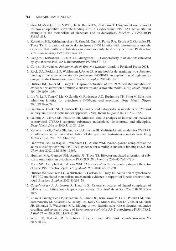
19.3.4 Substrate Inhibition
In the case of substrate inhibition kinetics, as the concentration of substrate is increased, to determine V m an infl ection point in the velocity versus [S] curve occurs so that rather than plateau, the velocity starts to decrease and continues to do so as [S] is raised further (Fig. 19.5 ). Assuming the reactions were carried out appropri-ately (see Section 19.3.6 ), two likely scenarios arise when substrate inhibition is noted:
FIGURE 19.4 Graphical representation of a kinetic profi le exhibiting heterotropic coop-erativity (activation).
FIGURE 19.5 Graphical representation of a kinetic profi le exhibiting substrate inhibition kinetics. Inset demonstrates the Eadie – Hofstee plot of a substrate inhibition kinetic profi le.

ATYPICAL KINETICS DISPLAYED BY DRUG METABOLIZING ENZYMES 707
1. If the experiment is conducted with P450, the most likely cause is analogous to uncompetitive inhibition where two or more ligands (in this case, two sub-strate molecules) binding to the enzyme decrease its ability to catalyze sub-strate oxidation.
2. If the experiment is conducted with UGT, inhibition is likely the result of aglycone substrate binding right after a glucuronide conjugate is released from a previous round of catalysis, thereby trapping UDP and forming an inactive ternary complex. For many UGTs there appears to be a preference to bind UDP - glucuronic acid fi rst, followed by aglycone, suggesting an ordered ternary complex mechanism.
In either case, K m and V m can be estimated via use of the appropriate equation. It should be noted, however, that the derived parameters (e.g., V m and K m ) are not truly equivalent to the usual situation. Because V m is never truly reached (or to simulate it unless the inhibition is very weak), the true K m is also not discernible. In the past, researchers have incorrectly fi t these types of data by either eliminating the points showing decreasing velocity or simply applying the Michaelis – Menten equation to the entire dataset. Lin and colleagues [9] have demonstrated the neces-sity to fi t the entire dataset to the proper equation and the hazards of not doing so. For substrate inhibition occurring through promiscuous binding of a second sub-strate (case 1), the following rate equation (Eq. 19.12 ) is useful. It is derived from the uncompetitive inhibition model using an extra substrate molecule instead of inhibitor, therefore leading to a squared term in the denominator when multiplied out:
vV
KK
=+ +
m
msi
S
SS
[ ]
[ ][ ]2
(9.12)
However, in the course of kinetic analyses one may wish to gain additional infor-mation from the available kinetic data, allowing for estimation of additional kinetic parameters that may provide more insight into the processes taking place. Thus, more complicated equations for kinetic modeling of substrate inhibition kinetics have been derived (e.g., Eq. 19.13 ) that allow for the estimation of interaction factors and additional kinetic parameters.
vV
K K K
K K K K
=+ ⋅( )
+ + +
mS i S
S i S i
S
SS
1
1 1 1
βα
α
[ ]
[ ][ ]
(9.13)
In the case of Eq. 19.13 , K S ≈ K m , K i is the dissociation constant of substrate binding to the inhibitory site, α is the factor by which the dissociation ( K S and K i ) of substrate at both sites changes when a second substrate is bound, and β is the factor by which V m changes when a second substrate is bound. Because of the number of parameters being estimated in Eq. 19.13 , modeling with this

708 METABOLISM KINETICS
equation will generally require substantially more data points than required for Eq. 19.12 .
For substrate inhibition mechanisms involving ordered ternary complex, such as the UGTs (case 2), Eq. 19.14 is applicable:
vV
K K K KK
=+ + + +( )
m
iA mB mB mASiB
A B
A B A BB
[ ][ ]
[ ] [ ] [ ][ ][ ]
1
(19.14)
where K SiB is not a dissocation constant.
19.3.5 Explanations for Atypical Kinetics in P450 Enzymes and Their Detection
Reports on the actual mechanism(s) behind the activation kinetics of drug metabo-lizing enzymes (mainly P450) are few compared to the kinetic models that have been devised to fi t nonhyperbolic velocity data [10 – 12] . The approaches that have been taken to understand the interplay of substrate, effector, and enzyme are, however, diverse and include isotope effects, fl uorescence spectroscopy [13] , and nuclear magnetic resonance [14, 15] in addition to assays for distinct steps in the P450 catalytic cycle [16] . One of the most infl uential hypotheses behind the action of effectors that prompted the use of these varied techniques is that the effector binding site could be located in the active site with the substrate. Over the past few years evidence for simultaneous ligand binding to the same active site has now been provided for at least two microsomal enzymes, human CYP3A4 and CYP2C9 , and three bacterial enzymes, CYP102A1 (BM3) , CYP107A1 (eryF) [17] , and CYP158A2 [18] as discussed below. Of course, allosterism can result from binding at remote sites or through protein – protein interactions.
Presence of Multiple Ligands Temporally Bound in the Same Active Site In the case of 3A4, the substrate pyrene could be used to demonstrate multiple ligand binding to the same enzyme active site during catalysis. Pyrene induces a sigmoidal kinetic response with increasing concentrations, and its planarity, coupled with its multiring fused aromatic structure, gives it a large surface for intermolecular pyrene – pyrene pi - stacking (a.k.a. excimer formation). After showing binding of pyrene to 3A4 by visible difference spectrophotometry and changes in fl uorescence of pyrene and the enzyme, differences in the excitation spectra (absorbance of monomer and excimer in the ground state) gave multiple indications that there are excimers in the presence of enzyme distinct from those that form in solution. Crystal structures of 3A4 do demonstrate active volumes suffi ciently large enough ( > 1000 Å ) for multiple ligands in their closed conformations [19] . Some investigators have even suggested triply occupied active sites [20] , but defi ning the nature of atypical kinetics in terms of different subsites still remains a challenge due to the dependency on the structure of both substrate and effector.
Shortly after the 3A4/pyrene report, a series of isotope effect experiments with P450 BM3 suggested that multiple fatty acids could bind to the same enzyme active site simultaneously. This explained why the F87A mutant produced a different metabolic profi le of hydroxylated products in the presence of laurate. Mutation of

ATYPICAL KINETICS DISPLAYED BY DRUG METABOLIZING ENZYMES 709
F87 to alanine results in an increase in hydroxylation of the ω - 1 position as laurate concentrations are raised. Since the alanine substitution gives a smaller side chain, and therefore more space, it is unclear why the ratio of metabolites would change unless the presence of a second substrate or the collapse of the enzyme constricts the available volume. This is where isotope effects appear to be capable of detecting multiple ligand binding without the need for fl uorescent molecules. The theory is that given two enzyme - bound ligands in rapid equilibrium, mixing protio substrate (all hydrogens) and substrate substituted with deuteriums (the deuterium – carbon bond possesses a lower vibrational energy and requires more energy for abstraction) at the site of metabolism will favor metabolism of the nonlabeled substrate. Hence, a bias in the rate of labeled and un labeled substrate metabolism is produced and it favors the hydrogen - containing substrate (i.e., k H / k D ratio is greater than unity). In this case, the bias for ω - 1 hydroxylation of palmitate was increased even more when deuterated laurate was added. Further validation of this technique is awaited for its use as a powerful tool with possibly untapped potential.
There are now crystal structures for two of the three major families of human microsomal P450s, 2 and 3 [21] , but none have captured multiple ligands in the same pocket. However, crystallography has provided convincing electron density for mul-tiple ligand binding in the same pocket for two bacterial enzymes. While not exactly a high throughput technology, even with the use of robotics, crystallography has generated structures that are invaluable for the predictive modeling of drug – drug interactions. P450 eryF in complex with two molecules of 9 - aminophenanthrene and, more recently, P450 158A2 with two molecules of fl aviolin have been reported. Both structures show one ligand above the heme with the second ligand lying above the fi rst. Interestingly, both ligands are aromatic and planar, possibly owing some of their binding to each other. As of now, there are no examples with two different ligands bound, nor does a single structure indicate the order of their binding or the extent at which they move about inside the enzyme.
With the help of crystallography, nuclear magnetic resonance spectroscopy (NMR) has the capability to provide more details about the sequence and location of binding without the need for assigning every resonance. Yoon et al. [15] have reported on the use of protein NMR spectroscopy to study the binding of 9 - aminophenanthrene to P450 eryF using enzyme expressed with 15 N - phenylalanine so that all phenylalanine residues would have detectable amide nitrogen nuclei. Titration of 9 - aminophenanthrene to levels that are substoichiometric to enzyme altered the signal intensity for Phe residues that are different from those whose intensity changed in the presence of higher levels of 9 - aminophenanthrene ([sub-strate] > [enzyme]). Further arguments regarding the time scale of on and off rates and their interpretation in terms of the mechanism of cooperative binding were also drawn from this study. Surely, the ability to distinguish two binding events at differ-ent locations inside the enzyme will greatly complement other analytical methods, and studies with human P450s will no doubt bring a more detailed understanding to drug – drug interactions induced by atypical kinetics.
An additional use of NMR involves the enhanced relaxation rate of substrate nuclei near the iron of the heme, which is paramagnetic in the ferric state [22] . Using P450 2C9, Hummel et al. [14] studied the proton relaxation times of the substrate fl urbiprofen in the absence and presence of heteroactivator dapsone. Time - averaged distances of each substrate proton can be estimated by carrying out a T 1 relaxation
710 METABOLISM KINETICS
experiment both with the ferric enzyme and the ferrous carbon monoxide complex, which is not paramagnetic. T 1 refers to the longitudinal relaxation and refers to the time it takes for the irradiated substrate protons to recess back to alignment with the magnetic fi eld. The site of hydroxylation (4 - proton) was found to be closer to the iron of the heme group in the presence of heteroactivator, suggesting that its hyperbolic activation may result from increased collision frequency of substrate with the reactive iron - oxene or enhanced spin state conversion via heme dehydration.
Another dimension to the action of dapsone on fl urbiprofen metabolism by P450 2C9 was provided by a study that compared the coupling of NADPH consumption to metabolite formation versus shunting to the side products superoxide, H 2 O 2 , and water. Catalytic coupling can be defi ned as the fraction of metabolite formed per NADPH consumed and is thus a measure of enzyme effi ciency distinct from k cat / K m . In addition to increasing the rate of fl urbiprofen metabolite formation, the effector dapsone was found to stimulate the consumption of NADPH, but decrease the for-mation of H 2 O 2 . Overall, the coupling of fl urbiprofen metabolism was increased. Therefore, the distance of a substrate relative to the heme iron as altered by effec-tors appears to have multiple consequences as shown for P450 2C9. Possible effec-tor - mediated changes in the position of substrate could displace the water that occupies the sixth coordination position of the iron to increase its reduction poten-tial, keep the substrate closer to the active oxidant, and therefore decrease uncou-pling of the catalytic cycle.
19.3.6 Artifactual Sources of Atypical Kinetics
During the conduct of in vitro drug metabolism experiments, it is prudent to be alert for the possible occurrence of atypical kinetics. However, the researcher must also be aware that there are several potential artifactual causes of atypical kinetic pro-fi les that may lead to a misintepretation of the data. Thus, extreme care must be taken to assure the validity of the results. The comments below address several of these potential sources of error and how to minimize their impact on the kinetic profi les observed.
One of the fundamental assumptions when conducting enzyme kinetic experi-ments is that the substrate (and effector concentration if drug – drug interactions are being studied) remains constant throughout the experiment. For substrates and effectors that exhibit substantial turnover, one must worry about substrate/effector depletion. This will result in constantly changing substrate/effector concentrations throughout the experiment and invalidate the assumption of steady state. Related to constantly changing (and decreasing) drug concentrations are potential sources of error when the actual substrate/effector concentration is lower than believed. This can occur if either compound undergoes substantial nonspecifi c binding to the incubation matrix (i.e., protein) or when the drug has very low solubility. In these cases, the free drug concentration (i.e., that available to interact with the enzyme) is substantially lower than believed, resulting in overestimates of kinetic parameters such as K m , and K i .
Although less of an issue in today ’ s environment of widespread LC/MS/MS avail-ability, insuffi cient analytical sensitivity to accurately and reproducibly quantitate metabolite production at the extreme low end of the substrate concentration –
velocity curve may result in erroneous assignment of the kinetic profi le. This is most problematic in the case of sigmoidal kinetics, where inadequate sensitivity hinders determining the less steep slope during the initial portion of the curve. One must also be cautious when using multienzyme systems (e.g., human liver microsomes) for kinetic characterization since multiple enzymes are present. This is particularly problematic with respect to observations of biphasic kinetics, since more than one enzyme may be producing the metabolite. In this case, the kinetic phenomena observed are actually a hybrid of the concerted effects of multiple enzymes and result in apparent kinetic constants.
Finally, the kinetic profi le observed may be affected by incubation conditions and preparation. Choice of buffer salt to be used in the incubation can result in differing kinetic profi les [23] and the presence of certain organic solvents (particularly at concentrations above 2%) have also been shown to activate enzyme kinetic pro-cesses [24] . Finally, the inclusion (or exclusion) of cytochrome - b 5 can result in different kinetic profi les observed [25] . Thus, one must pay particular attention to controls and conditions when making the assignment of atypical kinetic phenomena.
19.4 CONCLUSION
Proper estimation of kinetic parameters is critical to successful extrapolation of in vitro data to the in vivo situation. In addition to standard hyperbolic kinetics, sigmoidal (autoactivation), biphasic, substrate inhibition, and heterotropic activa-tion have all been observed in the kinetic profi les of drug metabolism reactions. The most common hypothesis for the occurrence of these types of “ atypical ” kinetic profi les is based on data suggesting that two (or more) substrate molecules (or a substrate and effector molecule) bind within the enzyme active site simultaneously and alter the enzyme kinetics. The occurrence of these atypical kinetic profi les complicates the determination of in vitro – in vivo correlations and thus requires the use of more complex equations appropriate to the type of profi le observed in order to properly estimate the kinetic parameters.
ACKNOWLEDGMENT
This work was supported in part by a grant from the National Institutes of Health (#GM 063215).
REFERENCES
1. Luukkanen L , Taskinen J , Kurkela M , Kostiainen R , Hirvonen J , Finel M . Kinetic char-acterization of the 1A subfamily of recombinant human UDP - glucuronosyltransferases . Drug Metab Dispos 2005 ; 33 : 1017 – 1026 .
2. Segel IH . Rapid Equilibrium Bireactant and Terreactant Systems . Hoboken, NJ : Wiley ; 1975 .
REFERENCES 711
712 METABOLISM KINETICS
3. Shou M , Mei Q , Ettore MW Jr , Dai R , Baillie TA , Rushmore TH . Sigmoidal kinetic model for two co - operative substrate - binding sites in a cytochrome P450 3A4 active site: an example of the metabolism of diazepam and its derivatives . Biochem J 1999 ; 340 (Pt 3 ): 845 – 853 .
4. Korzekwa KR , Krishnamachary N , Shou M , Ogai A , Parise RA , Rettie AE , Gonzalez FJ , Tracy TS . Evaluation of atypical cytochrome P450 kinetics with two - substrate models: evidence that multiple substrates can simultaneously bind to cytochrome P450 active sites . Biochemistry 1998 ; 37 : 4137 – 4147 .
5. Ueng YF , Kuwabara T , Chun YJ , Guengerich FP . Cooperativity in oxidations catalyzed by cytochrome P450 3A4 . Biochemistry 1997 ; 36 : 370 – 381 .
6. Cornish - Bowden A . Fundamentals of Enzyme Kinetics . London : Portland Press ; 2004 .
7. Rock DA , Perkins BN , Wahlstrom J , Jones JP . A method for determining two substrates binding in the same active site of cytochrome P450BM3: an explanation of high energy omega product formation . Arch Biochem Biophys 2003 ; 416 : 9 – 16 .
8. Hutzler JM , Hauer MJ , Tracy TS . Dapsone activation of CYP2C9 - mediated metabolism: evidence for activation of multiple substrates and a two - site model . Drug Metab Dispos 2001 ; 29 : 1029 – 1034 .
9. Lin Y , Lu P , Tang C , Mei Q , Sandig G , Rodrigues AD , Rushmore TH , Shou M . Substrate inhibition kinetics for cytochrome P450 - catalyzed reactions . Drug Metab Dispos 2001 ; 29 : 368 – 374 .
10. Galetin A , Clarke SE , Houston JB . Quinidine and haloperidol as modifi ers of CYP3A4 activity: multisite kinetic model approach . Drug Metab Dispos 2002 ; 30 : 1512 – 1522 .
11. Galetin A , Clarke SE , Houston JB . Multisite kinetic analysis of interactions between prototypical CYP3A4 subgroup substrates: midazolam, testosterone, and nifedipine . Drug Metab Dispos 2003 ; 31 : 1108 – 1116 .
12. Kenworthy KE , Clarke SE , Andrews J , Houston JB . Multisite kinetic models for CYP3A4: simultaneous activation and inhibition of diazepam and testosterone metabolism . Drug Metab Dispos 2001 ; 29 : 1644 – 1651 .
13. Dabrowski MJ , Schrag ML , Wienkers LC , Atkins WM . Pyrene – pyrene complexes at the active site of cytochrome P450 3A4: evidence for a multiple substrate binding site . J Am Chem Soc 2002 ; 124 : 11866 – 11867 .
14. Hummel MA , Gannett PM , Aguilar JS , Tracy TS . Effector - mediated alteration of sub-strate orientation in cytochrome P450 2C9 . Biochemistry 2004 ; 43 : 7207 – 7214 .
15. Yoon MY , Campbell AP , Atkins WM . “ Allosterism ” in the elementary steps of the cyto-chrome P450 reaction cycle . Drug Metab Rev 2004 ; 36 : 219 – 230 .
16. Hutzler JM , Wienkers LC , Wahlstrom JL , Carlson TJ , Tracy TS . Activation of cytochrome P450 2C9 - mediated metabolism: mechanistic evidence in support of kinetic observations . Arch Biochem Biophys 2003 ; 410 : 16 – 24 .
17. Cupp - Vickery J , Anderson R , Hatziris Z . Crystal structures of ligand complexes of P450eryF exhibiting homotropic cooperativity . Proc Natl Acad Sci USA 2002 ; 97 : 3050 – 3055 .
18. Zhao B , Guengerich FP , Bellamine A , Lamb DC , Izumikawa M , Lei L , Podust LM , Sun-daramoorthy M , Kalaitzis JA , Reddy LM , Kelly SL , Moore BS , Stec D , Voehler M , Falck JR , Shimada T , Waterman MR . Binding of two fl aviolin substrate molecules, oxidative coupling, and crystal structure of Streptomyces coelicolor A3(2) cytochrome P450 158A2 . J Biol Chem 2005 ; 280 : 11599 – 11607 .
19. Scott EE , Halpert JR . Structures of cytochrome P450 3A4 . Trends Biochem Sci 2005 ; 30 : 5 – 7 .

20. He YA , Roussel F , Halpert JR . Analysis of homotropic and heterotropic cooperativity of diazepam oxidation by CYP3A4 using site - directed mutagenesis and kinetic modeling . Arch Biochem Biophys 2003 ; 409 : 92 – 101 .
21. Johnson EF , Stout CD . Structural diversity of human xenobiotic - metabolizing cytochrome P450 monooxygenases . Biochem Biophys Res Commun 2005 ; 338 : 331 – 336 .
22. Yao H , Costache AD , Sem DS . Chemical proteomic tool for ligand mapping of CYP antitargets: an NMR - compatible 3D QSAR descriptor in the Heme - Based Coordinate System . J Chem Inf Comput Sci 2004 ; 44 : 1456 – 1465 .
23. Hutzler JM , Powers FJ , Wynalda MA , Wienkers LC . Effect of carbonate anion on cyto-chrome P450 2D6 - mediated metabolism in vitro : the potential role of multiple oxygenat-ing species . Arch Biochem Biophys 2003 ; 417 : 165 – 175 .
24. Hickman D , Wang JP , Wang Y , Unadkat JD . Evaluation of the selectivity of in vitro probes and suitability of organic solvents for the measurement of human cytochrome P450 monooxygenase activities . Drug Metab Dispos 1998 ; 26 : 207 – 215 .
25. Jushchyshyn MI , Hutzler JM , Schrag ML , Wienkers LC . Catalytic turnover of pyrene by CYP3A4: evidence that cytochrome b5 directly induces positive cooperativity . Arch Biochem Biophys 2005 ; 438 : 21 – 28 .
REFERENCES 713