practitioner's corner - jiaci · investig allergol clin immunol 2 vol 2 - 2 esmon publicidad...
TRANSCRIPT

J Investig Allergol Clin Immunol 2014; Vol. 24(1): 56-71 © 2014 Esmon Publicidad
PRACTITIONER'S CORNER
Cross-reactivity Between Buckwheat and Quinoa in a Patient With Eosinophilic Esophagitis Caused by Wheat
D El-Qutob López,1 B Bartolomé Zavala,2 I Ortiz1
1Clínica Atenea, Aldaia, Valencia, Spain 2R&D Department, Bial-Aristegui, Bilbao, Spain
Key words: Eosinophilic esophagitis. Buckwheat. Quinoa. Allergy. Wheat flour.
Palabras clave: Esofagitis eosinofílica. Alforfón. Quinoa. Alergia. Harina de trigo.
Eosinophilic esophagitis is a chronic, antigen-mediated esophageal disease characterized clinically by symptoms of esophageal dysfunction and histologically by eosinophil-predominant inflammation. The symptoms of eosinophilic esophagitis in adults are (from the least to the most frequent) dysphagia, chest pain, food impaction, and upper abdominal pain [1].
We present the case of a 38-year-old woman with intolerance to nonsteroidal anti-inflammatory drugs (NSAIDs), rhinoconjunctivitis caused by grass pollen, and recently diagnosed eosinophilic esophagitis caused by flour (wheat and other). After following a flour-free diet, the patient was asymptomatic, and biopsy studies revealed no eosinophilic inflammation.
As the patient suggested the possibility of consuming products containing buckwheat flour (Fagopyrum esculentum) and quinoa seed flour (Chenopodium quinoa), neither of which she had eaten before, a sensitization study was performed. The patient was asked to avoid both foods until the study was complete. Extracts from quinoa seed and buckwheat flour were prepared by homogenization in phosphate buffer saline, dialyzation, and lyophilization. Both were diluted in phenol glycerol saline solution at a concentration of 20 mg/mL, and skin prick tests (SPT) were performed with standardized lancets. The results were positive for both extracts. The results of SPT with buckwheat flour and quinoa extracts were negative in 10 controls (5 nonatopic controls and 5 atopic).
Serum specific IgE was measured using the enzyme allergosorbent test with quinoa seed (0.8 kUA/L), buckwheat flour (0.8 kUA/L), wheat seed (1.6 kUA/L), wheat flour (0.6 kUA/L), rice flour (0.9 kUA/L), and soybean (0.7 kUA/L). SDS-PAGE immunoblotting with extracts of quinoa and flours from buckwheat, wheat, and rice was performed following the Laemmli method. After incubation of the polyvinylene difluoride membrane with the patient’s serum, IgE binding bands of 28-30 kDa and 45-50 kDa were detected in all of
Figure. SDS-PAGE immunoblotting results. A, Quinoa extract. B, Buckwheat flour extract. C, Wheat flour extract. D, Rice flour extract. Lane P, patient serum; Lane C, control serum (pool of sera from nonatopic patients); Lane M, molecular mass marker.
the assayed extracts (Figure). Both bands were previously described by Park et al [2]. The first band could correspond to Fag e 1, a legumin of 11S-type globulins, although this allergen is a 24-kDa protein. The second could be Fag e 3, a 7S-vicilin–like globulin. Both are present in soybean. Cross-reactivity has been detected between 2S, 7S, and 11S-globulins of soybean [3]. No allergens have been described for C quinoa flour. Immunoblot-inhibition assays with extracts of quinoa and buckwheat in the solid phase revealed the existence of cross-reactivity between proteins from both extracts and between these proteins and others from wheat and rice flour extracts (results not shown). Consequently, the patient was also instructed to avoid buckwheat flour and quinoa. Three months later, biopsy studies showed no eosinophilic inflammation at all, and the patient continues to avoid dairy products containing flours, quinoa, and buckwheat.
Astier et al [4] reported a case of anaphylaxis to quinoa [4]. Using immunoblotting, the authors detected an IgE-binding band with a molecular mass of 35 kDa, which could belong to the legumin class (11S-type globulins). Hong et al [5] recently reported a case of anaphylaxis to quinoa, although they did not perform an immunoblotting analysis [5]. Several cases of anaphylaxis to buckwheat have been reported [6-8]. Based on the presence of the homologous allergens legumin (Fag e 1)

Practitioner's Corner
J Investig Allergol Clin Immunol 2014; Vol. 24(1): 56-71© 2014 Esmon Publicidad
57
and 2S-albumin (Fag e 2), Heffler et al [9] detected cross-sensitization between buckwheat, peanut, and soy [9]. In 2011, the same authors published a study of patients with a suspected buckwheat allergy [10]. Patients were classified into 3 groups: patients with a 16-kDa band (predominantly gastrointestinal symptoms with grass and wheat flour cosensitizations), patients with a 25-kDa band, and patients with a 40-kDa band. The patient we report does not belong to any of these groups.
The hypersensitivity mechanism involved in eosinophilic esophagitis has not been fully elucidated. We recorded positive results to prick tests. Immunoblotting showed the apparent molecular mass of the IgE-binding proteins that may be responsible for the disease.
In conclusion, we present a case of cross-reactivity between several flours, including quinoa and buckwheat. In the future, patients with hypersensitivity reaction to flours should undergo a cross-sensitization study before starting to eat buckwheat and quinoa as an alternative food.
Funding
The authors confirm that no funding was received for the present study.
Conflicts of Interest
The authors declare that they have no conflicts of interest.
References
1. Liacouras CA, Furuta GT, Hirano I, Atkins D, Attwood SE, Bonis PA, Burks AW, Chehade M, Collins MH, Dellon ES, Dohil R, Falk GW, Gonsalves N, Gupta SK, Katzka DA, Lucendo AJ, Markowitz JE, Noel RJ, Odze RD, Putnam PE, Richter JE, Romero Y, Ruchelli E, Sampson HA, Schoepfer A, Shaheen NJ, Sicherer SH, Spechler S, Spergel JM, Straumann A, Wershil BK, Rothenberg ME, Aceves SS. Eosinophilic esophagitis: updated consensus recommendations for children and adults. J Allergy Clin Immunol. 2011 Jul;128(1):3-20.
2. Park JW, Kang DB, Kim CW, Koh SH, Yum HY, Kim KE, Hong CS, Lee KY. Identification and characterization of the major allergens of buckwheat. Allergy. 2000;55(11):1035-41.
3. Shibasaki M, Suzuki S, Tajima S, Nemoto H, Kuroume T. Allergenicity of major component proteins of soybean. Int Arch Allergy Appl Immunol. 1980;61(4):441-8.
4. Astier C, Moneret-Vautrin DA, Puillandre E, Bihain BE. First case report of anaphylaxis to quinoa, a novel food in France. Allergy. 2009;64(5):819-20.
5. Hong J, Convers K, Reeves N, Temprano J. Anaphylaxis to quinoa. Ann Allergy Asthma Immunol. 2013;110(1):60-1.
6. Davidson AE, Passero MA, Settipane GA. Buckwheat-induced anaphylaxis: a case report. Ann Allergy. 1992;69(5):439-40.
7. Wuthrich B, Trojan A. Wheatburger anaphylaxis due to hidden buckwheat. Clin Exp Allergy. 1995;25(12):1263. Epub 1995/12/01.
8. Schiffner R, Przybilla B, Burgdorff T, Landthaler M, Stolz W. Anaphylaxis to buckwheat. Allergy. 2001;56(10):1020-1.
9. Heffler E, Guida G, Badiu I, Nebiolo F, Rolla G. Anaphylaxis after eating Italian pizza containing buckwheat as the hidden food allergen. J Investig Allergol Clin Immunol. 2007;17(4):261-3.
10. Heffler E, Nebiolo F, Asero R, Guida G, Badiu I, Pizzimenti S, Marchese C, Amato S, Mistrello G, Canaletti F, Rolla G. Clinical manifestations, co-sensitizations, and immunoblotting profiles of buckwheat-allergic patients. Allergy. 2011;66(2):264-70.
Manuscript received January 5, 2013; accepted for publication April 4, 2013.
David El-Qutob LópezClínica Atenea
C./Xest, 16 46960 Aldaia, Valencia
E-mail: [email protected]

Practitioner's Corner
J Investig Allergol Clin Immunol 2014; Vol. 24(1): 56-71 © 2014 Esmon Publicidad
58
Occupational Allergy in a Holm Oak Pruner
P Mur Gimeno,1 A Martín Iglesias,1 M Lombardero Vega,2 Q Malo Casero,3 S Jiménez Alvarez3
1Allergy Department, Santa Bárbara Hospital, Puertollano, Ciudad Real, Spain2R&D Department, ALK-Abelló SA, Madrid, Spain3Clinical Analysis Service, Microbiology, Santa Bárbara Hospital, Puertollano, Ciudad Real, Spain
Key words: Holm oak. Occupational disease. Quercus. Western blotting. Wood dust.
Palabras clave: Encina. Enfermedad ocupacional. Quercus. Inmunoblotting. Polvo de madera.
Wood dust is a cause of occupational respiratory diseases, such as asthma, rhinoconjunctivitis, and hypersensitivity pneumonitis, as well as skin diseases, such as contact urticaria and contact dermatitis.
Sometimes the immunological mechanism is IgE–mediated (eg, obeche wood), although on most occasions this has not been proven [1]. We describe the case of a pruner who was found to have type I sensitization to holm oak wood after other possible allergens were ruled out. Although holm oak is the most prevalent forestry species in Spain, its pollen is only weakly allergenic.
We describe the case of a 55-year-old man who had worked as a farmer and pruner from the age of 18. During his youth, he was diagnosed with joint pain after chronic brucellosis, gout, hepatitis B infection, and anthrax. Six years ago he was diagnosed with rhinitis and urticaria caused by grass, olive, and Artemisia pollens and urticaria-angioedema caused by pyrazolone.
During the last 3 years he has presented episodes of ocular pruritus, tearing, conjunctival hyperemia, and chemosis, all of which are associated with the pruning of holm oak branches from November to March. The episodes ceased in 4-6 hours with antihistamine drops and were not accompanied by nasal or bronchial symptoms. No corneal erosion was detected after fluorescein staining.
The results of prick testing with common inhalant allergens including mites, fungi, animal dander, and pollen were only positive for cypress pollen (8x8 mm) and ryegrass pollen (4x4 mm), with a histamine positive control of 5x5 mm. The results of prick testing with beech, pine, cherry, oak, sapele, and iroko were negative.
Prick testing with holm oak wood dust was positive (4x4 mm), although the result of the rubbing test was negative. Intense ocular conjunctivitis with chemosis was observed 10 minutes after handling holm oak wood.
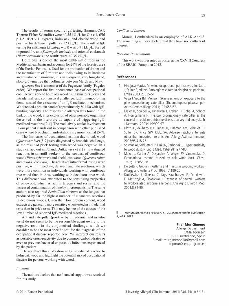
IgE immunoblotting of holm oak wood extract revealed proteins of approximately 50 kDa capable of binding the patient’s IgE (Figure).
In order to determine which part of the trunk the allergen was from, extracts were prepared from the bark of the holm
oak (10% wt/vol with 0.270 mg/mL protein) and from the core (10% wt/vol with 0.164 mg/mL protein). We used the bark extract for prick testing (6x5 mm) and intradermal testing (1/100 dilution, 12x11 mm with histamine 11x11 mm). The conjunctival challenge was positive at a 1/1000 dilution. Curiously, the result of the nasal challenge with bark extract was negative. The results of the tests were all negative for core extract. The results of prick, intradermal, and conjunctival tests performed with bark extract in 3 atopic, 3 nonatopic, and 2 exposed controls were negative.
In order to rule out the possibility that a microorganism nesting on the surface of the trunk might be responsible for the allergic reaction, we prepared and tested a series of extracts.
(a) Oak bark fungi cultivated in specific media (Sabouraud dextrose agar with chloramphenicol and actidione). Colonies of Aspergillus niger, Penicillium species, and Zygomycetes were identified. The results of the prick and intradermal tests and conjunctival challenge with Penicillium were negative.
(b) Lichens. The results of the prick and intradermal tests and conjunctival challenge were all negative.
(c) Oak caterpillars (Tortrix viridana). Prick testing (7x7 mm) with a positive histamine control (6x6 mm) and intradermal testing had similar results to histamine diluted at 1/10. The result of the conjunctival challenge was negative.
(d) Red wood ant (Formica rufa) (2.5% wt/vol, 0.705 mg/mL proteins). Prick testing elicited a wheal (7x8 mm). The result of intradermal testing was similar to histamine at a dilution of 1/1. The result of the conjunctival challenge was negative.
When these tests were carried out in 2 control patients, the results were negative.
Figure. IgE immunoblotting on holm oak wood. Lane 1, patient’s serum; lane 2, negative control. M indicates marker of molecular weight in kDa.
Practitioner's Corner
J Investig Allergol Clin Immunol 2014; Vol. 24(1): 56-71© 2014 Esmon Publicidad
59
The results of serum specific IgE testing (ImmunoCAP, Thermo Fisher Scientific) were <0.35 kUA/L for Ole e 1, rPhl p 1-5, rBet v 1, cypress, holm oak, and obeche wood and positive for Artemisia pollen (2.12 kUA/L). The result of IgE testing for silkworm (Bombyx mori) was 0.91 kUA/L; for red imported fire ant (Solenopsis invicta), and oriental cockroach (Blatta orientalis), the results were <0.35 kUA/L.
Holm oak is one of the most emblematic trees in the Mediterranean basin and accounts for 25% of the forested area of the Iberian Peninsula. Used for the production of timber and the manufacture of furniture and tools owing to its hardness and resistance to moisture, it is an evergreen, very long-lived, slow-growing tree that pollinates between March and May.
Quercus ilex is a member of the Fagaceae family (Fagales order). We report the first documented case of occupational conjunctivitis due to holm oak wood using skin tests (prick and intradermal) and conjunctival challenge. IgE immunoblotting demonstrated the existence of an IgE-mediated mechanism. We detected a protein band of approximately 50 kDa with IgE-binding capacity. The responsible allergen was found in the bark of the wood, after exclusion of other possible organisms described in the literature as capable of triggering IgE-mediated reactions [2-4]. The exclusively ocular involvement in our patient stands out in comparison with other published cases where bronchial manifestations are more normal [5-7].
The first cases of occupational asthma due to oak wood (Quercus robur) [5-7] were diagnosed by bronchial challenge, as the result of prick testing with wood was negative. In a study carried out in Poland, Dutkiewicz et al [8] investigated reactions in sawmill workers to the sawdust of coniferous wood (Pinus sylvestris) and deciduous wood (Quercus robur and Betula verrucosa). The results of intradermal testing were positive, with immediate, delayed, and late reactions, which were more common in individuals working with coniferous tree wood than in those working with deciduous tree wood. This difference was attributed to the sensitizing properties of pinewood, which is rich in terpenes and resins, and the increased contamination of pine by microorganisms. The same authors also reported Penicillium citrinum as the fungus that produced by far the highest number of cutaneous reactions in deciduous woods. Given their low protein content, wood extracts are generally more sensitive when tested in intradermal tests than in prick tests. This may be one of the causes of the low number of reported IgE-mediated reactions.
Ant and caterpillar (positive by intradermal and in vitro tests) do not seem to be the responsible agent owing to the negative result in the conjunctival challenge, which we consider to be the most specific test for the diagnosis of the occupational disease reported here. We interpret our results as possible cross-reactivity due to common carbohydrates or even to previous bacterial or parasitic infections experienced by the patient.
The results of this study show an IgE-mediated reaction to holm oak wood and highlight the potential risk of occupational disease for persons working with wood.
Funding
The authors declare that no financial support was received for this study.
Conflicts of Interest
Manuel Lombardero is an employee of ALK-Abelló. The remaining authors declare that they have no conflicts of interest.
Previous Presentations
This work was presented as poster at the XXVIII Congress of the SEAIC, Pamplona 2012.
References
1. Hinojosa Macías M. Asma ocupacional por maderas. In: Satre J, Quirce S, editors. Patología respiratoria alérgica ocupacional. Emisa 2003. p. 335-51.
2. Vega J, Vega JM, Moneo I. Skin reactions on exposure to the pine processionary caterpillar (Thaumatopoea pityocampa). Actas Dermosifiliogr. 2011;102:658-67.
3. Maier H, Spiegel W, Kinaciyan T, Krehan H, Cabaj A, Schopf A, Hönigsmann H. The oak processionary caterpillar as the cause of an epidemic airborne disease: survey and analysis. Br J Dermatol. 2003;149:990-97.
4. Klotz JH, deShazo RD, Pinnas JL, Fishman AM, Schmidt JO, Suiter DR, Price GW, Klotz SA. Adverse reactions to ants other than imported fire ants. Ann Allergy Asthma Immunol. 2005;95:418-25.
5. Sosman AJ, Schlueter DP, Fink JN, Barboriak JJ. Hypersensitivity to wood dust. N Engl J Med. 1969;281:977-80.
6. Malo JL, Cartier A, Desjardins A, Weyer RV, Vandenplas O. Occupational asthma caused by oak wood dust. Chest. 1995;108:856-58.
7. De Zotti R, Gubian F. Asthma and rhinitis in wooding workers. Allergy and Asthma Proc. 1996;17:199-20.
8. Dutkiewicz J, Skorska C, Krysinska-Traczyk E, Dutkiewicz E, Matuszyk A, Sitkowska J. Response of sawmill workers to work-related airborne allergens. Ann Agric Environ Med. 2001;8:81-90.
Manuscript received February 11, 2013; accepted for publication April 4, 2013.
Pilar Mur GimenoAllergy Department
C/Malagón s/n13500 Puertollano, Spain
E-mail: [email protected] [email protected]

Practitioner's Corner
J Investig Allergol Clin Immunol 2014; Vol. 24(1): 56-71 © 2014 Esmon Publicidad
60
Suspicion of a New Cross-reaction Between Carbamazepine and Olanzapine
D Brajon,1 P Trechot,2 J Waton,1 JF Cuny,1 JL Schmutz,1 A Barbaud1
1Dermatology, Bâtiment Philippe Canton, CHU Nancy – Hôpital Brabois, Vandoeuvre les Nancy, France2Pharmacovigilance, Central Hospital, CHU Nancy – Hôpital Central, Nancy, France
Key words: Carbamazepine. Olanzapine. Cross-reaction. DRESS. Drug hypersensitivity.
Palabras clave: Carbamazepina. Olanzapine. Reacción cruzada. Síndrome de DRESS. Hipersensibilidad a medicamentos.
We report the first case of a cross-reaction between carbamazepine and olanzapine.
In 1990, a 30-year-old man with tuberous sclerosis and recurrent epilepsy developed maculopapular exanthema. Carbamazepine was the only drug he was taking at the time and was therefore suspended. After new seizures in 2010, carbamazepine was reintroduced. Two days later, the patient was hospitalized in the dermatology unit with maculopapular rash, fever, cytolysis (alanine aminotransferase, 5 times normal), and polymorphonuclear eosinophils (PME) (2x10⁶/L). He was diagnosed with severe maculopapular exanthema, and carbamazepine was suspended. Following surgical removal of the frontal tuberosity 16 months later, the patient developed a psychotic episode, which was treated with olanzapine and cyamemazine. He received concomitant treatment with piperacillin, cefotaxime, fosfomycin, and rifampicin for a brain abscess. Six weeks after the initiation of olanzapine, the patient developed generalized maculopapular exanthema associated with fever (39°C), axillary and cervical lymph node enlargement, a PME count of 1540/mm3, liver dysfunction with cytolysis (alanine aminotransferase, 10 times normal), and cholestasis (g-glutamyl transferase, 12 times normal; alkaline phosphatase, 5 times normal). He was diagnosed with drug rash with eosinophilia and systemic symptoms (DRESS) syndrome with a score of 7 according to the criteria of Kardaun et al [1]. The reaction was thought to have been caused by olanzapine, rifampicin, piperacillin, fosfomycin, or iodinated radiocontrast media. The suspect drugs were stopped, and the clinical symptoms and laboratory abnormalities disappeared within 18 days.
An allergy workup 6 months later (patch tests with the European standard series and drugs) yielded negative results except for anticonvulsants (pill containing 200 mg of carbamazepine diluted at 30% in water [+/+], 30% in petrolatum [+/+], and 30% in alcohol [+/+] and a pill containing 10 mg of olanzapine diluted at 30% in petrolatum [+/+]). Patch tests, prick tests, and intradermal tests (IDT) were performed with the other suspect drugs: the results were negative on immediate and delayed readings except for delayed readings in the IDT with piperacillin. Olanzapine
and carbamazepine were discontinued. Two months later, ceftriaxone and iobitridol were reintroduced with caution using gradually increasing doses administered every 10 days. These were the only ß-lactam antibiotic and radiocontrast medium allowed. Given the chronology of the reaction and following criteria established in the literature, olanzapine was considered the main suspect drug. Moreover, the patient had a previous history of DRESS syndrome due to carbamazepine; the results of tests with fosfomycin, rifampicin, and radiocontrast medium were negative. Carbamazepine and olanzapine have similar chemical structures.
We report the first case of cross-reaction between carbamazepine and olanzapine. Similar cross-reactions have been reported between various aromatic antiepileptic drugs (phenytoin, phenobarbital, oxcarbazepine, and primidone) [2]. We did not study these possible cross-reactions in the patient reported here, because his antiepileptic treatment was effective and well tolerated. Carbamazepine is one of the most frequently involved drugs in DRESS syndrome. Lin et al [3] reported 10 cases diagnosed as carbamazepine-induced DRESS syndrome, 7 of which had positive patch test results with carbamazepine and 5 of which had at least 1 positive patch test result with another antiepileptic drug (oxcarbazepine in 50% of the cases, phenytoin in 40%, or lamotrigine in 20%). Aouam et al [4] reported possible cross-reactivity between carbamazepine and lamotrigine in a 14-year-old boy who presented DRESS syndrome after treatment with carbamazepine followed 8 months later by DRESS syndrome due to lamotrigine. Lamotrigine is not an aromatic antiepileptic drug, and carbamazepine and lamotrigine are not structurally related. According to Veyrac et al [5], these 2 occurrences of DRESS syndrome could be explained by an accumulation of toxic metabolites [5], which leads to nonconformational cross-reactivity between an aromatic antiepileptic and a nonaromatic antiepileptic.
The antipsychotic olanzapine is an oxazepine that is prescribed to patients with schizophrenia or recurrent bipolar disorder. Drug-drug interactions have been reported between antiepileptic and antipsychotic drugs [6], although there have been no reports of cross-sensitization. In the case of cutaneous adverse drug reactions due to olanzapine, the literature contains reports on 2 cases of DRESS syndrome (1 with positive patch test results to olanzapine) [7], 2 cases of vasculitis [8], and 1 of acute generalized exanthematous pustulosis [9]. Cross-reactivity between carbamazepine (aromatic antiepileptic drug) and olanzapine (aromatic antipsychotic drug) could be related to the existence of a common conformational component in their chemical structures, namely, benzazepine in the case of carbamazepine and benzodiazepine in the case of olanzapine (Figure).
Another hypothesis would involve pharmacogenetic effects on drug metabolism or immunogenetic predisposition affecting drug presentation and favoring the occurrence of 2 DRESS syndromes in the same patient.
The only antipsychotic agents belonging to the dibenzodiazepines are clozapine and olanzapine, whose chemical structures are largely identical. Therefore, we also discontinued clozapine in our patient as a precaution.
The patient reported here can receive antipsychotic drugs that do not belong to the dibenzodiazepine class. In the case

Practitioner's Corner
J Investig Allergol Clin Immunol 2014; Vol. 24(1): 56-71© 2014 Esmon Publicidad
61
of antiepileptic therapy, carbamazepine is strictly forbidden. If an aromatic antiepileptic was indicated, we would perform patch testing and reintroduce the drug gradually under hospital supervision before the start of treatment.
Relevant positive results in IDTs with piperacillin could indicate a new case of multiple drug reactivity affecting up to 18% of patients with DRESS syndrome [10].
In summary, the occurrence of 2 DRESS syndromes within a 16-month interval—one due to carbamazepine, the other due to olanzapine—would suggest cross-reactivity between the 2 drugs. The interval between the 2 episodes does not indicate multiple drug reactivity. To our knowledge, we report the first cross-reaction between an antiepileptic drug and an antipsychotic drug. Olanzapine and clozapine should not be prescribed in patients with hypersensitivity reactions to carbamazepine.
Funding
The authors declare that no funding was received for the present study.
Conflicts of Interest
The authors declare that they have no conflicts of interest.
References
1. Kardaun SH, Sidoroff A, Valeyrie-Allanore L, Halevy S, Davidovici BB, Mockenhaupt M, Roujeau JC. Variability in the clinical pattern of cutaneous side-effects of drugs with
systemic symptoms: does a DRESS syndrome really exist? Br J Dermatol. 2007;156:609-11.
2. Hirsch LJ, Arif H, Nahm EA, Buchsbaum R, Resor SR Jr, Bazil CW. Cross-sensitivity of skin rashes with antiepileptic drug use. Neurology. 2008;71:1527-34.
3. Lin Y-T, Chang Y-C, Hui RC-Y, Yang C-H, Ho H-C, Hung S-I, Chung WH. A patch testing and cross-sensitivity study of carbamazepine-induced severe cutaneous adverse drug reactions. J Eur Acad Dermatol Venereol. 2012 doi:10.1111/j.1468-3083.2011.04418.x
4. Aouam K, Ben Romdhane F, Loussaief C, Salem R, Toumi A, Belhadjali H, Chaabane A, Boughattas NA, Chakroun M. Hypersensitivity syndrome induced by anticonvulsants: possible cross-reactivity between carbamazepine and lamotrigine. J Clin Pharmacol. 2009;49:1488-91.
5. Veyrac G, Marcade G, Chiffoleau A, Bourin M, Jolliet P. [Characteristics of hypersensitivity syndrome to lamotrigine: review of one case reported in the Regional Center of Pharmacovigilance of Nantes]. Therapie. 2002;57:289-96.
6. De Leon J, Santoro V, D’Arrigo C, Spina E. Interactions between antiepileptics and second-generation antipsychotics. Expert Opin Drug Metab Toxicol. 2012;8:311-34.
7. Prevost P, Bédry R, Lacoste D, Ezzedine K, Haramburu F, Milpied B. Hypersensitivity syndrome with olanzapine confirmed by patch tests. Eur J Dermatol. 2012;22:126-7.
8. Papaioannides D, Sinapidis D, Korantzopoulos P, Charalabopoulos K. A case of cutaneous vasculitis associated with olanzapine. Int J Low Extrem Wounds. 2006;5:116-7.
9. Christen S, Gueissaz F, Anex R, Zullino DF. Acute generalized exanthematous pustulosis induced by olanzapine. Acta Medica (Hradec Kralove). 2006;49:75-6.
10. Barbaud A, Collet E, Milpied B, Assier H, Staumont D, Avenel-Audran M, Grange A, Amarger S, Girardin P, Guinnepain MT, Truchetet F, Lasek A, Waton J. A multicenter study to determine the value and safety of drug patch tests for the three main classes of severe cutaneous adverse drug reactions. British J Dermatol. 2013;168:555-62.
Figure. Common conformational part in the chemical structures of carbamazepine and olanzapine.
Manuscript received January 27, 2013; accepted for publication April 8, 2013.
Delphine BrajonCHU Nancy – Hopital Brabois
Vandoeuvre les Nancy, FranceE-mail: [email protected]

Practitioner's Corner
J Investig Allergol Clin Immunol 2014; Vol. 24(1): 56-71 © 2014 Esmon Publicidad
62
Oral Pancreatic Enzyme Supplements Can Reduce Excretion of Ovalbumin in Breast Milk
A Des Roches,1 M Abbott,2 P Bégin,1 J Paradis,1 L Paradis1
1Immunology-Allergy Service, Sainte-Justine Hospital, Montreal, Canada2Food Directorate, Health Products and Food Branch, Health Canada, Ottawa, Canada
Key words: Breastfeeding. Breast milk. Ovalbumin. Pancreatic enzymes. Food allergy.
Palabras clave: Lactancia. Leche materna. Ovalbumina. Enzimas pancreáticas. Alergia a alimentos.
Over 60% of mothers excrete ingested food allergens in their breast milk [1-5]. In highly sensitized infants, these trace amounts can trigger acute reactions or cause persistent chronic atopic dermatitis. In such cases, the mother should avoid the food. However, restrictive diets may prove difficult, as the 2 most frequent allergens, cow’s milk and hen’s egg, are found in many food products. Vance et al [1] reported that up to 84% of mothers on an egg avoidance diet admit to nonadherence [1]. Switching to a hydrolyzed formula is a viable option, although the benefits of continued breastfeeding mean that it is not favored.
The aim of the present study is to determine whether oral supplemental pancreatic enzymes could reduce expression of ingested allergens in breast milk. The rationale is that additional protease would favor more complete hydrolysis and reduce the amount of intact allergenic protein absorbed and then transferred through lactation. This hypothesis is supported by a case series reporting improvement in allergic milk protein enteropathy following the administration of supplemental pancreatic enzymes to breastfeeding mothers [6] and by our own uncontrolled observation that atopic dermatitis improved in children with food sensitizations treated with these enzymes [7].
To test this hypothesis, 25 healthy breastfeeding mothers were recruited from support groups. The mean age of the infants was 5 months (range, 2-11 months). The protocol included 2 home visits. Briefly, after the first morning feed, the mothers expressed their milk manually or mechanically in order to empty their breasts completely. They then ingested a scrambled egg and produced a 10-mL sample of breast milk every hour for 8 hours. They had to perform the experiment on an empty stomach and were instructed to avoid egg-containing products for the rest of the day by a dietician specialized in food allergy. At 1 of the 2 visits, the subjects received 1 capsule of pancrelipase immediately before eating the egg (PANCREASE MT 16, Janssen-Ortho). Each capsule contained 16 000 USP units of lipase, 48 000 USP units of amylase, and 48 000 USP units of protease. Ovalbumin concentrations in breast milk were then determined using enzyme-linked immunosorbent assay based on a previous publication [1]. Written informed consent was obtained. The protocol was reviewed and approved by the local ethics committee.
As expected, ovalbumin was detected in breast milk samples from 15 mothers (60%). Peak excreted ovalbumin varied between 2.09 ng/mL and 262.12 ng/mL, with a mean of 62.8 ng/mL, and was recorded during the first 2 hours in 12 of the 15 cases (80%). The duration of excretion varied widely between mothers, with a median of 3 hours (range, 1 to 8 hours). Four mothers (27%) still had detectable ovalbumin levels 8 hours after ingestion of the egg.
Figure. Ovalbumin concentration in breast milk following the ingestion of 1 scrambled egg with and without administration of pancreatic enzymes (black and gray lines respectively). A, Peak ovalbumin levels in breast milk for each woman. B, Variation in mean ovalbumin concentration over time (outlier result excluded).
300
250
200
150
100
50
01
1
4
4
7
7
Women
A
BO
valb
umin
, ng/
mL
Ova
lbum
in, n
g/m
L
Time, h
10 132
2
5
5
8
8
11 143
3
6
6
9 12 15
60
50
40
30
20
10
0
Excretion of ovalbumin was reduced after ingestion of pancreatic enzymes in 12 women (80%) previously identified as excreters and was below detection level in 7 women (47%) (Figure, A). Three women (20%) had higher levels of ovalbumin in their breast milk after ingestion of the enzyme, including 1 with a paradoxical increase from 12.22 ng/mL to 195.31 ng/mL. Excluding this outlier, the mean peak level of ovalbumin in breast milk after taking the enzyme was 6.91 ng/mL (range, 0-29.58 ng/mL), which was significantly lower than without taking the enzyme (paired t test, 2.65; P=.02). The mean area under the curve also decreased from 92.26 h x ng/mL to 20.21 h x ng/mL (paired t test, 2.32; P=.04), suggesting that ovalbumin was not simply excreted over a longer period of time (Figure, B). These results did not reach significance when the outlier value was included. After ingestion of the enzyme, the peak generally remained for the first 2 hours after ingestion (66%); 5 women (33%) still had detectable amounts after 8 hours.
Practitioner's Corner
J Investig Allergol Clin Immunol 2014; Vol. 24(1): 56-71© 2014 Esmon Publicidad
63
Taken together, our results suggest that supplemental pancreatic enzymes can modulate excretion of food allergens in human milk. They further add to the literature by showing that normal and complete digestion is important for protection against food allergy [8]. These findings are particularly relevant for mothers of highly sensitized children who wish to continue breastfeeding. The safety profile of pancreatic enzymes is excellent, as they have been used for many years in diseases such as cystic fibrosis, with minimal side effects [9].
We observed high variability in baseline ovalbumin excretion and in the response to the enzymes. The dose used in our study was small and may have been insufficient in some women. In their case series on milk protein enteropathy, Repucci et al [6] reported on a mother who needed up to 3 capsules with every meal for her child’s hematochezia to resolve completely, suggesting that dosing may indeed need to be individualized. The paradoxical increase in ovalbumin excretion in one of the women is perplexing. It does not seem plausible that the use of enzymes would result in such an increase in ovalbumin excretion, and a labeling or laboratory error cannot be completely excluded.
This proof-of-concept study needs to be followed by placebo-controlled trials before it can be applied in clinical practice, as previous literature suggesting efficacy only includes uncontrolled observations. In addition, in the real-life setting, allergen concentration in breast milk varies with meals and amount ingested [2], and doses may need to be adjusted accordingly. Finally, these results need to be confirmed for other food allergens to pass through breast milk [3-5].
In summary, we have shown that oral supplemental enzymes can modulate excretion of ovalbumin in human breast milk. Our results could provide a basis for further study of increased ovalbumin excretion in the breastfeeding mothers of children with food allergy.
Acknowledgments
We thank the Food Directorate, Health Canada, for supplying the materials for the analysis. We also thank Claire Emond RN for clinical assistance.
Funding
Funding was provided by the Allergy Section of CHU Sainte-Justine.
Conflicts of Interest
The authors declare that they have no conflicts of interest.
References
1. Vance GH, Lewis SA, Grimshaw KE, Wood PJ, Briggs RA, Thornton CA, Warner JO. Exposure of the fetus and infant to hen’s egg ovalbumin via the placenta and breast milk in relation to maternal intake of dietary egg. Clin Exp Allergy. 2005;35:1318-26.
2. Palmer DJ, Gold MS, Makrides M. Effect of maternal egg consumption on breast milk ovalbumin concentration. Clin Exp Allergy. 2008;38:1186-91.
3. Jarvinen KM, Makinen-Kiljunen S, Suomalainen H. Cow's milk challenge through human milk evokes immune responses in infants with cow's milk allergy. J Pediatr. 1999 Oct;135(4):506-12.
4. Cant A, Marsden RA, Kilshaw PJ. Egg and cow's milk hypersensitivity in exclusively breast fed infants with eczema, and detection of egg protein in breast milk. Br Med J (Clin Res Ed). 1985;291:932-5
5. Chirdo FG, Rumbo M, Añón MC, Fossati CA. Presence of high levels of non-degraded gliadin in breast milk from healthy mothers. Scand J Gastroenterol. 1998;33: 1186-92.
6. Repucci A. Resolution of stool blood in breast-fed infants with maternal ingestion of pancreatic enzymes. J Pediatr Gastroenterol Nutr 1999;29(4):500.
7. Singer S, Meddings J, Powell J, Desroches A, Seidman E. Pancreatic enzymes supplements improve severe eczema in children with multiple food allergies. J Pediatr Gastroenterol Nutr 2002;35(3):430.
8. Pali-Schöll I, Jensen-Jarolim E. Anti-acid medication as a risk factor for food allergy. Allergy. 2011;66(4):469-77.
9. Wooldridge JL, Heubi JE, Amaro-Galvez R, Boas SR, Blake KV, Nasr SZ, Chatfield B, McColley SA, Woo MS, Hardy KA, Kravitz RM, Straforini C, Anelli M, Lee C. EUR-1008 pancreatic enzyme replacement is safe and effective in patients with cystic fibrosis and pancreatic insufficiency. J Cyst Fibros. 2009;8:405-17.
Manuscript received February 6, 2013; accepted for publication April 8, 2013.
Anne Des RochesService d’immunologie-allergie
CHU Sainte-Justine3175 Côte Sainte-Catherine, Montréal, Québec, Canada,
H3T 1C5.E-mail: [email protected]

Practitioner's Corner
J Investig Allergol Clin Immunol 2014; Vol. 24(1): 56-71 © 2014 Esmon Publicidad
64
A Curious Case of Exercise-Induced Wheat Allergy in Adulthood
I Carrapatoso, AF Santos, E Faria, A Segorbe LuísImmunoallergology Department, Coimbra University Hospital, Coimbra, Portugal
Key words: Exercise-induced anaphylaxis. Aspirin. Wheat allergy. Omega-5 gliadin. Clopidogrel.
Palabras clave: Anafilaxia inducida por ejercicio. Aspirina. Alergia a trigo. Omega-5 gliadina. Clopidogrel.
Food-dependent exercise-induced anaphylaxis (FDEIA) is a rare, life-threatening syndrome characterized by anaphylactic symptoms triggered by the combination of exercise and the ingestion of a specific sensitizing food [1,2]. Concomitant medications and alcohol intake can act as additional facilitating factors. As opposed to what happens in exercise-induced anaphylaxis, exercise on its own is well tolerated in FDEIA, as is ingestion of the sensitizing food if not associated with exercise. We report a case of wheat-dependent exercise-induced anaphylaxis in adulthood.
A 59-year-old man was referred to our department for recurrent episodes of anaphylaxis. He had had 2 similar episodes consisting of the sudden development of rhinoconjunctivitis, generalized urticaria, facial and upper airway angioedema, dyspnea, and loss of consciousness that required medical assistance in the emergency room. These episodes occurred while he was out walking following a home-made dinner that consisted of vegetable soup and wheat-based bread. There were no symptoms when he performed exercise after ingestion of food other than cereals. The patient would take his prescription medication, namely furosemide, atorvastatin, and acetylsalicylic acid (ASA), at dinner time. It is noteworthy that he denied the occurrence of symptoms with the ingestion of wheat and ASA in the absence of exercise. There were no other co-factors such as alcohol consumption or stress.
The patient had no previous history of atopy, allergy, or intolerance to food or drugs. The anaphylactic episodes started
when the patient was 55 years old, following coronary heart surgery and the prescription of furosemide, atorvastatin, and ASA. This also coincided with changes in the patient's lifestyle, with light dinners consisting of soup and bread and regular exercise, mainly walks after dinner.
On admission to our department, 2 months after the second episode of anaphylaxis, the physical examination was unremarkable. Laboratory studies including complete blood count, sedimentation rate, serum electrolytes, liver and renal function tests, complement and serum tryptase levels, and skin biopsy were all normal. The patient had a high level of total IgE (443 IU/mL). Skin prick tests using commercial extracts of common aeroallergens and cereals (ALK, Abelló) as previously described [3] were negative. Prick-to-prick tests with wheat, rye, corn, oat, and rice flours provided by the bakery where the patient had bought the bread on the days of the episodes were performed. Prick-to-prick tests carried out in a control group of 5 healthy individuals were all negative. Specific IgE was measured using the ImmunoCAP FEIA system (Phadia) following the manufacturer´s recommendations. Levels above 0.35 kU/L were considered positive. The results of the prick-to-prick tests and specific IgE determinations are shown in the Table. Oral challenges with wheat and exercise were not performed due to the patient’s coronary heart disease.
The patient was advised to avoid eating wheat and rye 3 hours before and 1 hour after exercise. Patient education included detailed information on foods that may contain hidden cereals. A written emergency plan including self-injectable adrenaline, cetirizine, and prednisolone was delivered. ASA (100 mg/d) was replaced by clopidogrel (75 mg/d), after consulting the patient's cardiologist. The patient was advised to avoid other nonsteroidal anti-inflammatory drugs (NSAIDs). He continued to eat corn, barley, oats, and rice. With the avoidance measures taken, no more allergic symptoms or anaphylactic episodes occurred. Total IgE declined to 173 IU/mL and specific IgE to wheat, rye, and recombinant omega-5 gliadin decreased to 0.38, 0.63, and 4.14 kU/L, respectively.
The diagnosis of FDEIA triggered by wheat ingestion was strongly supported by our results. The presence of specific IgE to recombinant ω-5-gliadin (rω-5-gliadin) was demonstrated. This is the major causative allergen in wheat-dependent exercise-induced anaphylaxis (WDEIA), and has
Table. Skin Prick Tests and Specific IgE Results
Allergen Source Prick Test Prick-to-Prick Test Specific IgE, kU/L (Wheal Diameter) (Wheal Diameter)
Histamine Positive (5 mm) Wheat Negative Positive (8 mm) 0.9 (class 2) Rye Negative Positive (10 mm) 4.2 (class 3) Barley Negative Positive (4 mm) 0.06 (class 0) Corn Negative Positive (4 mm) 0.05 (class 0) Oats Negative Negative 0.04 (class 0) Rice Negative Negative 0.04 (class 0)ω-5-gliadin (rTri a 19) 9.7 (class 3)
Flours

Practitioner's Corner
J Investig Allergol Clin Immunol 2014; Vol. 24(1): 56-71© 2014 Esmon Publicidad
65
been reported to cause reactions in approximately 80% of all patients with this condition [4]. In our patient, the level of specific IgE to wheat and rye was particularly low compared with that to rω-5-gliadin. This finding is in accordance with previous reports [4,5], indicating a higher capacity of the recombinant allergen molecule to detect allergen-specific IgE. Matsuo et al [5] demonstrated that the sensitivity of the in vitro test measuring specific IgE to rω-5-gliadin is highest in the identification of patients with WDEIA, with lower sensitivities seen for wheat, gluten, and the high-molecular-weight glutenin subunit. Furthermore, the authors estimated that the cutoff value for specific IgE to rω-5-gliadin giving maximal efficiency in the diagnosis of WDEIA was 0.89 kUA/L [5]. Because of the high level of specific IgE to rω-5-gliadin (9.7 kU/L) in our patient, we did not consider it essential to perform an oral challenge to confirm the clinical diagnosis. Besides, we considered that oral challenge with wheat and exercise was contraindicated because of the patient’s heart condition.
This case is unusual because of the late age of onset and the combination of different triggering factors. The patient had increased the ingestion of wheat, particularly in the evenings when he used to take ASA, just before exercising. The pathophysiology of FDEIA is not fully understood [2]. Possible explanations involve physiological changes that occur during exercise, such as enhanced plasma osmolality, decreased pH, blood flow redistribution, and increased sensitivity of basophils and mast cells to degranulation [1,6]. Moreover, as both food and exercise are independently tolerated, several factors need to occur synergistically to produce symptoms. One critical factor is the increased intestinal permeability to allergens that occurs with exercise and that is dependent of enhanced tissue enzyme activity, which facilitates additional epitope recognition [2]. It is well described that the intake of ASA and other NSAIDs can enhance the induction of type I allergic symptoms when combined with food and exercise in patients with FDEIA [2,6,7]. These drugs might increase gastrointestinal permeability to allergens and allergic histamine release from human leukocytes [6-8]. It has also been recognized that exercise and ASA intake, even at low doses, act synergistically to produce severe symptoms [7,10]. In the particular case of WDEIA patients, Matsuo et al [9] demonstrated that exercise and ASA increase levels of circulating gliadin. Furthermore, they observed that blood gliadin levels were correlated with clinical symptoms, strongly supporting the hypothesis that exercise and ASA facilitate allergen absorption from the gut.
In our opinion the low baseline absorption of ω-5-gliadin was probably significantly enhanced by the additional effects of exercise and ASA intake, and the synergistic effect of these co-factors was sufficient to amplify the symptoms and trigger anaphylaxis.
Funding
The authors declare that no funding was received for the present study.
Conflicts of Interest
The authors declare that they have no conflicts of interest.
References
1. Robson-Ansley P, Du Toit G. Pathophysiology, diagnosis and management of exercise-induced anaphylaxis. Curr Opin Allergy Clin Immunol. 2010; 10: 312-7.
2. Morita E, Kohno K, Matsuo H. Food-dependent exercise-induced anaphylaxis. Journal of Dermatological Science. 2007; 47: 109-17.
3. Dreborg S, Frew A. Position paper: Allergen standardization and skin tests. The European Academy of Allergology and Clinical Immunology. Allergy. 1993; 48 (14 Suppl): 48-82.
4. Morita E, Matsuo H, Chinuki Y, Takahashi H, Dahlström J, Tanaka A. Food-dependent exercise-induced anaphylaxis-importance of omega-5 gliadin and HMW-glutenin as causative antigens for wheat-dependent exercise-induced anaphylaxis. Allergology Int. 2009; 58: 493-8.
5. Matsuo H, Dahlström J, Tanaka A, Kohno K, Takahashi H, Furumura M, Morita E. Sensitivity and specificity of recombinant ω-5 gliadin specific IgE measurement for the diagnosis of wheat-dependent exercise-induced anaphylaxis. Allergy. 2008; 63: 233-6.
6. Barg W, Medrala W, Wolanczyk-Medrala A. Exercise-induced anaphylaxis: an update on diagnosis and treatment. Curr Allergy Asthma Rep. 2011; 11: 45-51.
7. Harada S, Horikawa T, Ashida M, Kamo T, Nishioka E, Ichihashi M. Aspirin enhances the induction of type I allergic symptoms when combined with food and exercise in patients with food-dependent exercise-induced anaphylaxis. Br J Dermatol. 2001; 145: 336-9.
8. Wojnar RJ, Hearn T, Starkweather S. Augmentation of allergic histamine release from human leukocytes by nonsteroidal anti-inflammatory-analgesic agents. JACI. 1980; 66: 37-45.
9. Matsuo H, Morimoto K, Akaki T, Kaneko S, Kusatake K, Kuroda T, Niihara H, Hide M, Morita E. Exercise and aspirin increase levels of circulating gliadin peptides in patients with wheat-dependent exercise-induced anaphylaxis. Clin Exp Allergy. 2005; 35: 461-6.
10. Fujii H, Kambe N, Fujisawa A, Kohno K, Morita E, Miyachi Y. Food dependent exercise-induced anaphylaxis induced by low dose aspirin therapy. Allergol Int. 2008; 57: 97-8.
Manuscript received February 28, 2013; accepted for publication, April 30, 2013.
Maria Isabel Paiva CarrapatosoServiço de Imunoalergologia
Hospitais de Universidade de CoimbraPraceta Mota Pinto
3000-075 Coimbra, PortugalE-mail: [email protected]

Practitioner's Corner
J Investig Allergol Clin Immunol 2014; Vol. 24(1): 56-71 © 2014 Esmon Publicidad
66
First Case Report of Acute Generalized Exanthematous Pustulosis Due to Intravenous Iopromide
S Bavbek,1 ZÇ Sözener,1 Ö Aydın,1 SK Özdemir,1 Ü Gül,2 AO Heper,3 1Department of Chest Diseases, Division of Immunology and Allergy, Ankara University, School of Medicine, Ankara, Turkey2Department of Dermatology, Ankara Numune Training and Resarch Hospital, Ankara, Turkey3Department of Pathology, School of Medicine, Ankara University, Ankara, Turkey
Key words: Radiocontrast media. AGEP. Acute generalized exanthematous pustulosis. Iopromide.
Palabras clave: Medios de radiocontraste. Pustulosis exantemática aguda generalizada. İopromida.
Acue generalized exanthematous pustulosis (AGEP) is a relatively rare dermatosis that is generally attributed to drugs, particularly β-lactam antibiotics, sulfonamide, and macrolides; AGEP induced by contrast media (CM), however, is very rare [1].
A 52-year-old woman with a history of non-ST elevation myocardial infarction had received intravenous iopromide (Ultravist) for coronary angiography (CAG). Two hours after the CAG, her inguinal region became flushed. Her entire body then became red and she developed itching, fever, swelling of the tongue, and pustular lesions covering her body. She was diagnosed with AGEP at a dermatology department. She denied any history of psoriasis but had received CM injections on several occasions, without complications. There was also a family history of hyperlipidemia and cardiac arrest. It was decided to perform another CAG because of her recurrent cardiac complaints and she was referred to our department before the procedure.
Her physical examination was unremarkable on admission. Skin prick tests (SPTs), intradermal tests (IDTs), and patch tests (PTs) were performed with the CM that had caused the previous reaction, iopromide (Ultravist), as well as with alternative CMs that are widely used in Turkey, namely, iodixanol (Visipaque), iomeprol (Iomeron), diatrizoate (Urografin), and iopamidol (Pamiray). SPTs with undiluted CM were followed by IDTs with 10-fold diluted CM and PTs with 10-fold diluted and undiluted CM. The SPT results were read after 20 minutes and on days 1, 2, and 3. The SPT was considered positive if a wheal of over 3 mm in diameter was observed after 20 minutes (immediate reaction) or if an erythematous induration was observed at the skin test site on days 1, 2, or 3 (delayed reaction). For the IDTs, the test solution (0.03 mL) was injected into the skin to produce a bleb of 3 to 4 mm in diameter. Readings were taken after 20 minutes and on days 1, 2, and 3. The IDT was regarded as positive if the size of the initial wheal had increased by at least 3 mm in diameter and was surrounded by erythema after 20 minutes (immediate reaction) or if an
erythematous induration was present at the skin test site on days 1, 2 or 3 (delayed reaction) [2]. For the PTs, readings were taken 15 minutes after removal of the strips and at 24, 48, and 72 hours in accordance with the recommendations of the European Society of Contact Dermatitis [3]. The immediate SPT and IDT readings were negative with all the CM tested. Only iomeprol and iopamidol were positive on the delayed SPT readings. By contrast, the delayed IDT readings and the PTs showed positive results for all the CM except diatrizoate.
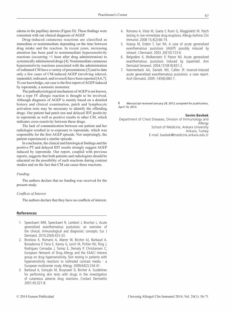
Based on the skin tests, the patient was given written information recommending the use of diatrizoate—with caution—for further CAG procedures. One month later, she was scheduled to undergo another follow-up CAG but was accidentally given intravenous iopromide again. Three to four hours after administration, she developed a generalized erythematous rash that also affected her face, mild pruritus, tongue swelling, dyspnea, and fever, and had to be hospitalized by the dermatology department. On admission she had fever, and her rash and pruritus continued to worsen with a widespread skin rash—with the formation of pustules—affecting the entire body (Figure A-C). Laboratory results revealed an increased white blood cell count of 16 500/μL and an elevated C-reactive protein concentration (129 mg/dL). Blood, urine, throat, and pustule cultures were negative. All other laboratory values were unremarkable. A punch biopsy specimen revealed epidermal acanthosis with multifocal pustule formation in the upper part of the epidermis, with inflammation and mild
Figure. A, Patch test with 1:10 dilution of iopromide. B, Delayed intradermal test reading with 1:10 dilution of iopromide. C, Widespread skin rash, with formation of pustules, affecting the entire body. D, Skin biopsy showing epidermal acanthosis with multifocal pustule formation in the upper part of the epidermis. There was also inflammation in the papillary dermis with mild edema (hematoxylin-eosin, original magnification ×400).
Practitioner's Corner
J Investig Allergol Clin Immunol 2014; Vol. 24(1): 56-71© 2014 Esmon Publicidad
67
edema in the papillary dermis (Figure D). These findings were consistent with our clinical diagnosis of AGEP.
Drug-induced cutaneous reactions are classified as immediate or nonimmediate depending on the time between drug intake and the reaction. In recent years, increasing attention has been paid to nonimmediate hypersensitivity reactions (occurring >1 hour after drug administration) to systemically administered drugs [4]. Nonimmediate cutaneous hypersensitivity reactions associated with the administration of iodinated CM have a variety of presentations [5] and to date only a few cases of CM-induced AGEP (involving iohexol, iopamidol, iodixanol, and ioversol) have been reported [4,6,7]. To our knowledge, our case is the first report of AGEP induced by iopromide, a nonionic monomer.
The pathophysiological mechanism of AGEP is not known, but a type IV allergic reaction is thought to be involved. Although diagnosis of AGEP is mainly based on a detailed history and clinical examination, patch and lymphocyte activation tests may be necessary to identify the offending drugs. Our patient had patch test and delayed IDT positivity to iopromide as well as positive results to other CM, which indicates cross-reactivity between these drugs.
The lack of communication between our patient and her radiologist resulted in re-exposure to iopromide, which was responsible for the first AGEP episode. Not surprisingly, the patient experienced a similar episode.
In conclusion, the clinical and histological findings and the positive PT and delayed IDT results strongly suggest AGEP induced by iopromide. Our report, coupled with previous reports, suggests that both patients and radiologists should be educated on the possibility of such reactions during contrast studies and on the fact that CM can cause these reactions.
Funding
The authors declare that no funding was received for the present study.
Conflicts of Interest
The authors declare that they have no conflicts of interest.
References
1. Speeckaert MM, Speeckaert R, Lambert J, Brochez L. Acute generalized exanthematous pustulosis: an overview of the clinical, immunological and diagnostic concepts. Eur J Dermatol. 2010;20(4):425-33.
2. Brockow K, Romano A, Aberer W, Bircher AJ, Barbaud A, Bonadonna P, Faria E, Kanny G, Lerch M, Pichler WJ, Ring J, Rodrigues Cernadas J, Tomaz E, Demoly P, Christiansen C; European Network of Drug Allergy and the EAACI interest group on drug hypersensitivity. Skin testing in patients with hypersensitivity reactions to iodinated contrast media - a European multicenter study. Allergy. 2009;64(2):234-41.
3. Barbaud A, Gonçalo M, Bruynzeel D, Bircher A. Guidelines for performing skin tests with drugs in the investigation of cutaneous adverse drug reactions. Contact Dermatitis 2001;45:321-8.
Manuscript received January 29, 2013; accepted for publication, April 10, 2013.
Sevim BavbekDepartment of Chest Diseases, Division of Immunology and
AllergySchool of Medicine, Ankara University
Ankara, TurkeyE-mail: [email protected]
4. Romano A, Viola M, Gaeta F, Rumi G, Maggioletti M. Patch testing in non-immediate drug eruptions. Allergy Asthma Clin Immunol. 2008 15;4(2):66-74.
5. Atasoy M, Erdem T, Sari RA. A case of acute generalized exanthematous pustulosis (AGEP) possibly induced by iohexol. J Dermatol. 2003 ;30(10):723-6.
6. Belgodère X, Wolkenstein P, Pastor MJ. Acute generalized exanthematous pustulosis induced by iopamidol. Ann Dermatol Venereol. 2004;131(8-9):831-2.
7. Hammerbeck AA, Daniels NH, Callen JP. Ioversol-induced acute generalized exanthematous pustulosis: a case report. Arch Dermatol. 2009 ;145(6):683-7.

Practitioner's Corner
J Investig Allergol Clin Immunol 2014; Vol. 24(1): 56-71 © 2014 Esmon Publicidad
68
Eosinophilic Bronchitis Caused by Styrene
L Arochena, M Fernández-Nieto, E Aguado, M García del Potro, J Sastre Allergy Department, Fundación Jiménez Díaz, Madrid, Spain
Key words: Eosinophilic Bronchitis. Styrene. Occupational. Induced sputum. Specific bronchial challenge.
Palabras clave: Bronquitis eosinofílica. Ocupacional. Esputo inducido. Provocación bronquial específica.
Eosinophilic bronchitis is a common cause of chronic cough that, like asthma, presents with sputum eosinophilia but, unlike asthma, is not associated with reversible bronchial obstruction or bronchial hyperresponsiveness [1]. Some cases of eosinophilic bronchitis are caused by work-related exposure to low- and high-molecular-weight allergens [2].
Styrene is an organic compound that is a derivative of benzene and a precursor to polystyrene and other copolymers. Its use has become widespread due to its hardening properties, and it is a common component of rubber, plastic, insulation material, fiberglass, pipes, and food containers. It is also present in most glues used nowadays.
We present the case of a nonatopic, nonsmoking 55-year-old man who had been working as a panel beater for 22 years. Over the past 10 years he had developed chronic cough and dyspnea, which became more intense at the end of the work shift and at night; he remained symptom-free during holidays. He handled metal sheets and putty containing styrene as a hardener; the styrene concentration was 10% to 20%, as revealed by product labels and material data safety sheets. He also occasionally handled glues with a high content of acrylates.
He was evaluated at our clinic during sick leave (4 months without working) and the physical examination, blood tests, spirometry, and fraction of exhaled nitric oxide (FeNO) results were normal. Sputum was induced by hypertonic saline inhalation and processed as previously described [3]; the samples were analyzed by flow cytometry, which revealed an eosinophil percentage of 7.4%. Written informed consent was given by the patient, who underwent 3 single-blind inhalation challenges (SICs) in a 7m3-challenge chamber on separate days as previously described [4,5].
At baseline (visit 1) a bronchial challenge with methacholine was negative (20% fall in forced expiratory volume in the first second [FEV1] [PC20] at >16 mg/mL); neither the eosinophil count in sputum nor FeNO rose after the challenge.
At visit 2, a SIC with styrene was performed as previously described [6]. Briefly, the patient was asked to brush paint the putty (10%-20% styrene) for increasing periods of time (30 seconds and 2, 5, 10, and 15 minutes). After a cumulative time of 32 minutes the styrene elicited no reaction, and 24 hours later, no significant changes in FEV1, methacholine PC20, FeNO, or cell counts in induced sputum were observed.
At visit 3 (1 week later), because there had been no positive response to the previous challenges, we decided to perform a
SIC with hexamethylene diisocyanate (HDI) for 30 minutes, but there was no response to this agent either.
Given that there was no evidence of occupational asthma, we asked the patient to go back to work and to come back after 10 working days, when we performed another sputum induction test; the eosinophil percentage was 15%, which was double the baseline count. An increase of over 2% in eosinophil count in sputum after exposure to work is considered to be clinically significant [7,8], and is also a diagnostic criterion of eosinophilic bronchitis. Changes were also observed in FeNO, with a result of 76 ppb.
There are a few reports of styrene being a cause of occupational asthma [6,9,10], but to our knowledge this is the first time that eosinophilic bronchitis due to this compound has been described.
Our belief is that because of the patient’s long period away from work during the initial tests (4 months), the cumulative exposure time of 32 minutes was not enough to elicit an allergic reaction, and it was only after he had been exposed to styrene for longer periods of time at his workplace that we saw a positive reaction. Nonetheless, we have described a case of eosinophilic bronchitis that, in addition to other diagnostic criteria, has the particularity of not being accompanied by bronchial hyperresponsiveness [1].
Styrene is increasingly being used in the manufacture of a wide range of materials. This agent may elicit airway inflammation and asthma-like symptoms, and it is our job to distinguish between real occupational asthma and other conditions that can emulate these symptoms. Occupational eosinophilic bronchitis should be remembered when specific bronchial challenges are successively negative.
Funding
The authors declare that no funding was received for the present study.
Conflicts of Interest
The authors declare that they have no conflicts of interest.
References
1. Gibson PG, Dolovich J, Denburg J, Ramsdale EH, Hargreave FE. Chronic cough: eosinophilic bronchitis without asthma. Lancet. 1989 Jun 17;1(8651):1346-8.
2. Quirce S. Eosinophilic bronchitis in the workplace. Curr Opin Allergy Clin Immunol. 2004 Apr;4(2):87-91.
3. Fernández-Nieto M, Sastre B, Sastre J, LaHoz C, Quirce S, Madero M, Del Pozo V. Changes in sputum eicosanoids and inflammatory markers after inhalation challenges with occupational agents. Chest. 2009; 136:1308-15.
4. Quirce S, Fernández-Nieto M, Bartolomé B, Bombín C, Cuevas M, Sastre J. Glucoamylase: another fungal enzyme associated with baker's asthma. Ann Allergy Asthma Immunol. 2002 Aug;89(2):197-202.
5. Sastre J, Fernández-Nieto M, Novalbos A, De Las Heras M, Cuesta J, Quirce S. Need for monitoring nonspecific bronchial hyperresponsiveness before and after isocyanate inhalation challenge. Chest. 2003 Apr;123(4):1276-9.

Practitioner's Corner
J Investig Allergol Clin Immunol 2014; Vol. 24(1): 56-71© 2014 Esmon Publicidad
69
Rapid Iron Desensitization After Generalized Urticaria and Facial Angioedema
B Rodríguez-Jiménez,1 J Domínguez-Ortega,1 B Nuñez-Acevedo,1 B Cava-Sumner,1 C Kindelan-Recarte,1 C Montojo-Guillén2 1Allergy Unit, Hospital Universitario de Getafe, Madrid, Spain2Pharmacy Service, Hospital Universitario de Getafe, Madrid, Spain
Key words: Iron salts. Desensitization. Drug allergy.
Palabras clave: Sales de hierro. Desensibilización. Alergia a fármacos.
Treatment of iron deficiency anemia is generally based on oral iron supplements in the form of iron salts. Parenteral administration is reserved for patients who have malabsorption or cannot tolerate oral iron. The most common adverse effects are gastrointestinal, and include nausea, epigastric pain, and constipation. Despite the widespread use of iron salts, very few cases of allergic reaction have been reported. The most common reactions are those associated with parenteral iron dextran [1].
Our patient was a 61-year-old woman with a history of intolerance to nonsteroidal anti-inflammatory drugs, arterial hypertension, eroded internal hemorrhoids, benign gastric polyps, and iron deficiency anemia. She also had a metal mitral valve because of rheumatic heart disease. The patient was referred to our department for assessment of a possible allergy to iron salts. Six months previously, 30 minutes after taking a second ferrous sulfate tablet (105 mg), she experienced edema of the eyelids and pinnae and facial wheals that subsequently affected the rest of her body; intramuscular antihistamine and corticosteroids were administered. She subsequently had 2 episodes with similar characteristics after taking the first tablet of ferrous sulfate (80 mg) and ferrimannitol ovalbumin (40 mg). In both cases she required treatment in the emergency department. Treatment with iron salts was stopped, and her anemia worsened. A laboratory workup disclosed the following values (with reference ranges): hemoglobin, 9.9 g/dL (12-16 g/dl); hematocrit, 31.2% (36%-46%); mean corpuscular volume, 75.7 fL (80-98 fL); and iron, 34 µg/dL (60-160 µg/dl). At this point, she was referred to the allergology department.
Since the patient had chronic iron deficiency anemia with accompanying symptoms and had experienced reactions with different iron salts, we considered administering an iron tolerance protocol. We decided on desensitization with intravenous iron (Venofer). This route may be faster and safer than oral desensitization because intravenous drug administration can be stopped abruptly in the event of a reaction, whereas discontinuance of oral administration is followed by continued gastrointestinal absorption for a variable period [2]. In addition, as the patient needed urgent treatment with iron, we could not perform skin tests with iron salts and started the protocol as soon as she gave her signed informed consent. Because we attended the patient on Friday,
6. Fernández-Nieto M, Quirce S, Fraj J, Del Pozo V, Seoane C, Sastre B, Lahoz C, Sastre J. Airway inflammation in occupational asthma caused by styrene. J Allergy Clin Immunol. 2006 Apr;117(4):948-50.
7. Girard F, Chaboillez S, Cartier A, Cote J, Hargreave FE; Labrecque M, Malo JL, Tarlo SM, Lemière C. An effective strategy for diagnosing occupational asthma: use of induced sputum. Am J Respir Crit Care Med. 2004; 170:845-50.
8. Lemière C, Chaboillez S, Malo JL, Cartier A. Changes in sputum cell counts after exposure to occupational agents: what do they mean? J Allergy Clin Immunol. 2001 Jun;107(6):1063-8.
9. Hayes JP, Lambourn L, Hopkirk JA, Durham SR, Taylor AJ. Occupational asthma due to styrene. Thorax. 1991;46:396-7.
10. Moscato G, Bilcaldi G, Cottica D, Pugliese F, Candura F. Occupational asthma due to styrene: two case reports. J Occup Med. 1987;29:957-60.
Manuscript received March 22, 2013; accepted for publication, May 2, 2013.
Lourdes ArochenaFundación Jiménez Díaz.Avda. Reyes Católicos, 2
28040 Madrid, SpainE-mail: [email protected]

Practitioner's Corner
J Investig Allergol Clin Immunol 2014; Vol. 24(1): 56-71 © 2014 Esmon Publicidad
70
we decided to carry out the premedication over the weekend. Premedication was with montelukast (10 mg/12 h), prednisone (20 mg/24 h), and dexchlorpheniramine (6 mg/12 h) 3 days before administration of iron and intravenous dexchlorpheniramine (5 mg) and methylprednisolone (80 mg) 1 hour before the start of the protocol. Dosing was started at 0.1 mg at a concentration of 1 mg/mL (iron is unstable below this concentration) and continued until a cumulative dose of 188.8 mg was reached (Table). The interval between doses was 15 minutes.
Five minutes after the third dose (0.5 mg at 1 mg/mL), the patient experienced malaise and moderate erythema on the face and neck. No other symptoms were present. She received intravenous dexchlorpheniramine (5 mg) and methylprednisolone (40 mg), after which her symptoms began to resolve. Symptoms had resolved completely 30 minutes later. The protocol was continued with the fourth dose until the total required dose was reached.
The patient subsequently received treatment with parenteral iron every 7 days for 6 weeks according to our desensitization protocol. She received the same premedication each time. Tolerance was good, with no new allergic reactions and a marked improvement in her initial anemia (hemoglobin, 13.6 g/dL).
Allergic reactions to iron salts are uncommon. The literature contains reports of maculopapular erythematous rash [3], pustular rash [4], photodermatitis [5], and even anaphylaxis [6]. Desensitization could be a feasible alternative in patients with marked iron deficiency anemia for whom iron is clearly indicated but who have had allergic reactions.
Iron desensitization protocols have been reported, but they have used oral preparations. Ortega et al [3] reached a dose of 30 mg of ferrous sulfate in 4 days, but were unable to reach the therapeutic dose, as the patient developed a skin eruption and generalized pruritus after the 30-mg dose. De Barrio et al [6] carried out a protocol for desensitization to ferrous sulfate in 18 days and reached a therapeutic dose of 525 mg. In that case, premedication was with antihistamines, since the patient had urticaria accompanied by dyspnea.
To date, no rapid desensitization protocol with parenteral iron has been reported. This approach induces temporary tolerance and enables allergic patients to receive appropriate
treatment for their condition. Consequently, patients with IgE-mediated and non-IgE-mediated hypersensitivity reactions, including anaphylaxis, can receive their medication, with minimal or no allergic reactions [7].
Our patient was premedicated with antihistamines, corticosteroids, and montelukast. Both montelukast and acetylsalicylic acid have been used to increase safety and tolerance in rapid drug desensitization protocols. The advantage of using them as premedication has been observed in patients with cutaneous and respiratory symptoms during desensitization to chemotherapy and acetylsalicylic acid. Given that prostaglandins and leukotrienes play an important role in the development of urticaria, flushing, and bronchospasm, the advantages of montelukast and acetylsalicylic acid could be attributed to blockade of these mediators [8,9]. In the present case, the clinical picture and the fact that symptoms reappeared with the third dose could indicate an IgE-mediated reaction, although no clear mechanism was identified.
Our desensitization protocol enabled our patient to tolerate the treatment she needed to reverse the worsening of her anemia. Hemoglobin levels returned to normal after the protocol.
We have presented a case of iron allergy in a patient who needed iron and who eventually tolerated parenteral iron after undergoing a rapid desensitization protocol based on intravenous administration.
Funding
The authors declare that no external funding was received for the present study.
Conflicts of Interest
The authors declare that they have no conflicts of interest.
References
1. Monaghan MS, Glasco G, St John G, Bradsher RW, Olsen KM. Safe administration of iron dextran to a patient who reacted to the test dose. South Med J. 1994; 87: 1010-2.
Table. Protocol for Inducing Tolerance to Parenteral Iron
Dose Parenteral Iron, mg Concentration, mg/ml Quantity, cc Time, min
1 0.1 1 0.1 152 0.2 1 0.2 153 0.5 1 0.5 154 1 1 1 155 2 1 2 156 5 1 5 157 10 1 10 158 20 1 20 159 50 1 50 1510 100 1 100 30
Practitioner's Corner
J Investig Allergol Clin Immunol 2014; Vol. 24(1): 56-71© 2014 Esmon Publicidad
71
Manuscript received March 8, 2013; accepted for publication, May 8, 2013.
Beatriz Rodríguez JiménezUnidad de Alergología
Hospital Universitario de GetafeCarretera de Toledo Km 12,500
28905 Getafe, SpainE-mail: [email protected]
2. Audícana Berasategui MT, Ortega Rodríguez NR, García Ortega P, Uriel Villate O. Prevención y tratamiento de la alergia a los fármacos. Desensibilización. In: Peláez Hernández A, Dávila González IJ, editors. Tratado de Alergología. Ergon 2007. p. 1369-94.
3. Ortega N, Castillo R, Blanco C, Alvarez M, Carrillo T. Oral iron cutaneous adverse reaction and successful desensitization. Ann Allergy Asthma Immunol. 2000; 84: 43-5.
4. Ito A, Nomura K, Hashimoto I. Pustular drug eruption induced by ferrous fumarate. Dermatology. 1996; 192: 294-5.
5. Kawada A, Hiruma M, Noguchi H, Kimura M, Ishibashi A, Banba H, Marshall J. Photosensitivity due to sodium ferrous citrate. Contact Dermatitis. 1996; 34: 77.
6. de Barrio M, Fuentes V, Tornero P, Sánchez I, Zubeldia J, Herrero T. Anaphylaxis to oral iron salts. Desensitization protocol for tolerance induction. J Investig Allergol Clin Immunol. 2008; 18: 305-8.
7. del Carmen Sancho M, Breslow R, Sloane D, Castells M. Desensitization for hypersensitivity reactions to medications. Chem Immunol Allergy. 2012; 97: 217-33.
8. White AA, Stevenson DD, Simon RA. The blocking effect of essential controller medications during aspirin challenges in patients with aspirin-exacerbated respiratory disease. Ann Allergy Asthma Immunol. 2005; 95: 330-5.
9. Breslow RG, Caiado J, Castells MC. Acetylsalicylic acid and montelukast block mast cell mediator-related symptoms during rapid desensitization. Ann Allergy Asthma Immunol. 2009; 102: 155-60.
ERRATUM:
Omalizumab Treatment in 2 Cases of Refractory Heat Urticaria
F Carballada, R Nuñez, J Martín-Lázaro, Y Juárez, I Castiñeira, L Fernández, M Boquete
J Investig Allergol Clin Immunol 2013; Vol. 23(7): 519.
The order of the pictures in the figure does not correspond to the order of the legends. Legend B and C should be reversed.