practice recommendations for preventing heel … recommendations for preventing heel pressure ulcers...
TRANSCRIPT

OstomyWoundManagementOctober 2008, Vol. 54, Issue 10 www.o-wm.com
Practice Recommendations for Preventing HeelPressure UlcersEvonne Fowler, RN,CNS, CWOCN; Suzy Scott-Williams, MSN, RN, CWOCN; andJames B. McGuire, DPM, PT, CPed, CWS, FAPWCA
Contemporary Topics
in Skin, Wound, Ostomy,
and Incontinence Care
The officialjournal of the
LLC,
991_OWM1008_Cover1:Cover1 2/13/09 10:11 AM Page 1

2 OstomyWound Management
FEATURE
Apressure ulcer is a localized injury to the skin
and/or underlying tissue that usually develops
over a bony prominence as a result of pressure
or pressure with shear or friction forces.1 Heel ulcers
are the most common facility-acquired pressure ulcer
in long-term acute care facilities2 and the second most
common pressure ulcer overall.3 Heel ulcers can be
physically debilitating and painful and can lead to seri-
ous complications such as infection, cellulitis,
osteomyelitis, septicemia, limb amputation, or death,
and can increase healthcare and litigation costs.
However, with appropriate evidence-based prevention,
most heel pressure ulcers can be avoided.4 To summa-
rize pertinent information on the clinical, emotional,
and financial significance of heel pressure ulcers,
selected literature and poster presentations are
reviewed and wound care clinicians’ insights on the
challenges of heel pressure ulcers are provided.
Anatomy/Physiology/Pathophysiology ofthe Heel Pressure Ulcer
Pressure ulcers are ultimately a result of tissue damage
due to inadequate tissue perfusion.5 Direct sustained
pressure, repetitive moderate pressure, shear forces, and
Practice Recommendations forPreventing Heel Pressure UlcersEvonne Fowler, RN,CNS, CWOCN; Suzy Scott-Williams, MSN, RN, CWOCN; and James B. McGuire, DPM, PT,CPed, CWS, FAPWCA
Heels are the second most common anatomical location for pressure ulcers. A combination of risk factors, including pressure,may cause ulceration. Heel pressure ulcers are a particular concern for surgical patients. A review of the literature, includingposter presentations, shows that controlled clinical studies to assess the effectiveness and cost-effectiveness of availableinterventions are not available. Case series (with or without historical controls) as well as pressure ulcer guideline recommen-dations suggest the most important aspect of heel ulcer prevention is pressure relief (offloading). It also has been documentedthat the incidence of heel ulcers can be reduced using a total-patient care approach and heel offloading devices. Guidelines,observational studies, and expert opinion intimate that reducing heel ulceration rates can be expected to improve patient out-comes, decrease costs associated with their care, and avoid costs related to hospital-acquired pressure ulcers. The heel pres-sure ulcer prevention strategies reviewed should be implemented until the results of prospective, randomized controlled stud-ies to compare the effectiveness and cost-effectiveness of these strategies are available.
KEYWORDS: heel pressure ulcer, perioperative pressure ulcer, heel protector device, heel offloading device
Ostomy Wound Management 2008;54(10):42–57
Ms. Fowler is a wound/ostomy care specialist, San Gorgonio Memorial Hospital, Banning, Calif. Ms. Scott-Williams is a surgicalquality improvement/research liaison nurse and wound, ostomy, continence nurse, Memphis Veteran’s Affairs Medical Center,Memphis, Tenn. Dr. McGuire is Chairman, Department of Podiatric Medicine and Orthopedics, Temple University School of PodiatricMedicine, Philadelphia, Pa. Please address correspondence to: Evonne Fowler, RN,CNS, CWOCN, San Gorgonio Memorial Hospital,600 N. Highland Springs Avenue, Banning, CA 92220-3090; email: [email protected].
42-57_OWM1008_Fowler:Feature 2/13/09 10:12 AM Page 2

October 2008 Vol. 54 Issue 10 3
reperfusion injury all contribute to tissue ischemia and
thrombotic occlusion of the capillary vasculature.6
Tissue compromised by maceration, friction
injury, or dryness is particularly vulnerable.7
Bacterial colonization of existing ulcers, altered cel-
lular responses, and systemic stress also may con-
tribute to tissue injury.8
According to a model proposed by Mustoe et al,8
reperfusion injury is an exacerbation of tissue injury
that results as blood returns to the tissue and white
blood cells accumulate in and around the damaged
small capillaries. The accumulation of damaged cell
byproducts and white blood cells obstructs the capil-
laries, aggravating ischemia and local inflammatory
response. Free radicals produced in the cells during
ischemia are released when blood flow is restored.
These free radicals damage cellular proteins, DNA,
and cell membranes, contributing to cell death. Thus,
tissues damaged by pressure-induced ischemia are fur-
ther damaged as the tissue is reperfused.
Heels are particularly vulnerable to pressure injury.
The plantar surface of the heel is well adapted to
resisting the forces of standing and ambulation but the
posterior heel is not — it is covered with only a thin
layer of fat and skin. The superficial fascia of the heel
is firmly bound to the deep fascia with fat contained in
the interstices between segments, creating a tough
elastic pad capable of absorbing shock and shear
forces during gait.9 This area is well supplied with
blood from the medial calcaneal branch of the poste-
rior tibial artery and is supported and padded by
attachments of the flexor digitorum bre-
vis and abductor digiti minimi muscles.
When patients are supine, the poste-
rior aspect of the heel is in contact with
the supporting surface. The posterior
aspect of the heel lacks fat-filled fascial
interstices to absorb the compressive
forces of prolonged pressure or shear
forces generated during limb movement
or transfers. The fat layer is thin and the
skin is bound tightly to the underlying
deep fascia and Achilles tendon fibers.
The blood supply to the skin is poor and
there is no underlying muscle to cush-
ion the bone and tendon or distribute
pressure. When the foot is resting with pressure on the
posterior heel, all the pressure from the weight of the
foot and lower leg is concentrated on the small area of
skin overlying the posterior tubercle of the calcaneus.
This skin is supplied by small branches from the cal-
caneal and peroneal arteries and prolonged pressure
on these relatively small vessels can lead to ischemia.9
Pressure Ulcer Prevalence, Mortality, and Morbidity
Prevalence rates vary across patient populations
and are as high as 25.1% in a mixture of acute and
nonacute care settings.10 In a study3 conducted in
intensive care units, prevalence rates reported in a 24-
hour data collection period ranged from 14% to 17%.
Long-term care facilities have reported prevalence
rates as high as 27.3% — of these, 23.6% were heel
ulcers.2 In a single-center, prospective controlled study
of surgical patients (mean age 61.6 years), the inci-
dence of pressure ulcers was 23% — 52% of these were
on the heels.11 Heel pressure ulcers account for
approximately one third of all pressure ulcers in acute
care and mixed acute care/long-term care settings.2,12
Higher proportions of heel pressure ulcers have been
reported in individual institutions; prevalence rates
were as high as 43% for a population of patients
(mean age 74.9 years) in a Midwestern community
acute care hospital.13 Mortality rates are also higher in
patients with pressure ulcers. In an observational
cohort study (n = 3,103), the relative risk for death in
elderly individuals with pressure ulcers was 1.92,
KEY POINTS• Although heel pressure ulcers are very common, the authors of this
review found that currently available literature provides limited evi-dence about optimal heel pressure ulcer prevention and care.
• There is general agreement that offloading is the first step to preventingand healing these ulcers.
• Results of descriptive studies and expert opinion also suggest that theuse of heel offloading reduces the incidence of heel pressure ulcers.
• Until more robust data are available, clinicians should implement theheel ulcer prevention strategies discussed to help prevent heel ulcersand their associated morbidity, mortality, and cost.
Ostomy Wound Management 2008;54(10):42–57
42-57_OWM1008_Fowler:Feature 2/13/09 10:12 AM Page 3

almost double that of persons without pressure
ulcers.14 According to national multiple cause-coded
death records, septicemia is the cause of death in 40%
of pressure ulcer patients.15
Morbidity manifests in most heel ulcer patients as
pain and reduced mobility but also can include loss of
a limb. A prospective study of patients with heel ulcers
who were referred to a specialist multidisciplinary
clinic between January 2000 and November 2003
included 154 heel ulcers. Of the 53 nonhealed ulcers,
11 (71% of 154) resulted in major amputation. A ret-
rospective database and medical record review17 of 788
patients (average age 66.7 years) with 959 consecutive
lower extremity amputations found that amputation
patients are at high risk for surgical complications,
with 30-day mortality rates of 5.7% for below-the-
knee amputation, infection rates of 5.5%, and re-oper-
ation rates of 18.4%.
Clinical Presentation of the Heel Pressure Ulcer
A pressure-related injury may first present clinically
with discoloration, tenderness, and changes in consis-
tency or temperature compared to the surrounding
skin. Nonblanchable erythema is early evidence of
abnormal perfusion due to pressure-related injury or
friction or shearing forces.18 With deep tissue injury (a
category of pressure injury that is not visible as an
ulcer), the skin may be purple or maroon, boggy or
firm, and warmer or cooler than surrounding tissues.
The area may be painful and may develop blood- or
serum-filled blisters. As damage evolves, the blister
roof dries and an eschar develops, which may in time
become an open wound. The progression from local
ischemia to pressure injury and damage to the skin
may be rapid even under circumstances of optimal
treatment.
The National Pressure Ulcer Advisory Panel
(NPUAP)1 recently revised the definition and staging
of pressure ulcers to include deep tissue injury and
unstageable ulcers (see Table 1).
Pressure Ulcer Risk FactorsVarious conditions have been associated with an
increased risk of pressure ulcer development. The
Institute for Healthcare Improvement (IHI)19 lists age,
immobility, incontinence, inadequate nutrition, sen-
sory deficiency, multiple concurrent morbidities (eg,
diabetes), circulatory abnormalities, and dehydration
as key factors contributing to the development of
pressure ulcers. Additional literature includes immo-
bility (eg, spinal cord injury20 or stroke), major sur-
gery, shock/resuscitation, limited or no responsive-
ness, inactivity, skin moisture, inadequate nutrition,
friction and shear forces on the skin21; hypoalbumine-
mia, anemia, diabetes mellitus (DM), peripheral vas-
cular disease (PVD), hip fracture, low Braden Scale
score13; and iatrogenic causes, such as misapplication
of pressure stockings22 among factors that have been
found to be associated with pressure ulcer develop-
ment. Studies specifically designed to assess the risk of
heel pressure ulcers have not been conducted.
Risks in the surgical patient population.Prevalence studies23 have shown that patients who
undergo surgery are at higher risk for developing pres-
sure ulcers than the general patient population.
Overall rates of pressure ulcers in all anatomical loca-
tions ranged from 3.5% in an observational cross-sec-
tional prevalence study24 (n = 281) to 14.3% in a
prospective comparative 6-week incidence study25 of
surgical patients (n = 286). Results from the former
study indicated that long periods of immobility and
decreased sensorium related to the type of anesthesia
used were associated with a higher risk of pressure
ulcer development. In a small study26 of elderly
patients (50 undergoing emergency surgery and 150
undergoing elective surgery), 13% of patients under-
going elective surgery and 14.2% of patients undergo-
ing surgery for hip fracture developed heel pressure
ulcers. Heel pressure ulcers have been reported in a
case study27 of older patients (average 74.3 years)
undergoing nerve blocks for hip or knee replacement.
A retrospective study28 of 34 patients receiving elective
hip or knee replacements conducted from August
2002 to December 2004 concluded if a heel has altered
sensation or unrelieved pressure, patients undergoing
knee replacements can develop heel pressure ulcers.
Further, central and/or peripheral nerve blockade is a
disadvantage due to reduced heel sensation.
A prospective matched control comparison study
of 323 high-risk surgical patients conducted by Scott-
Williams et al11 reported that a combination of age
4 OstomyWound Management
42-57_OWM1008_Fowler:Feature 2/13/09 10:12 AM Page 4

>62, albumin levels <3.5, and American Society of
Anesthesia (ASA) scores ≤3 was predictive of perioper-
ative pressure ulcer development. In this two-group
comparison study, the incidence of perioperative pres-
sure ulcers was 74 (23%) and 61 (52%) of the 118 pres-
sure ulcers that developed were located on the heels.
In addition to the risk of developing pressure ulcers
during surgery, surgical patients may be immobile due to
injury or illness. In observational reports, the presence of
obesity,29 decreased sensation due to central or peripheral
nerve blocks,27,28 and lower body mass index with low
serum albumin have been found to be associated with
October 2008 Vol. 54 Issue 10 5
TABLE 1NATIONAL PRESSURE ULCER ADVISORY PANEL REVISED PRESSURE
ULCER STAGING SYSTEM1
Stage/ClinicalPresentation
Stage I: Red heel
Stage II: Serous-filledblister
Stage II: Partial-thicknessopen wound
Stage III: Full-thicknesswound
Pressure Ulcer DefinitionA pressure ulcer is localized injury to the skin and/or underlying tissue,usually over a bony prominence, as a result of pressure or pressure incombination with shear and/or friction. A number of contributing orconfounding factors also are associated with pressure ulcers; the sig-nificance of these factors is yet to be elucidated.
Stage I:Intact skin with nonblanchable redness of a localized area, usually overa bony prominence. Darkly pigmented skin may not have visibleblanching; its color may differ from the surrounding area
Further description:The area may be painful, firm, soft, warmer, or cooler as compared toadjacent tissue. Stage I may be difficult to detect in individuals withdark skin tones. May indicate "at risk" persons (heralding sign of risk)
Stage II:Partial-thickness loss of dermis presenting as a shallow open ulcer witha red-pink wound bed, without slough. Also may present as an intactor open/ruptured serum-filled blister
Further description:Presents as a shiny or dry shallow ulcer without slough or bruising.*This stage should not be used to describe skin tears, tape burns, per-ineal dermatitis, maceration, or excoriation
*Bruising may be a sign of deep tissue injury
Stage III: Full-thickness tissue loss. Subcutaneous fat may be visible but bone,tendon, or muscle are not exposed. Slough may be present but doesnot obscure the depth of tissue loss. May include undermining andtunneling
Further description:The depth of a Stage III pressure ulcer varies by anatomical location.The bridge of the nose, ear, occiput, and malleolus do not have subcu-taneous tissue and Stage III ulcers can be shallow. In contrast, areasof significant adiposity can develop extremely deep Stage III pressureulcers. Bone/tendon is not visible or directly palpable
Photo
Continued on next page
42-57_OWM1008_Fowler:Feature 2/13/09 10:12 AM Page 5

heel ulceration.25 In a comprehensive literature review30
of studies involving cardiac surgery patients, temperature
manipulation, treatment with vasoactive drugs, periods
of hypotension related to the surgery, and reduced
hemoglobin and hematocrit levels were frequently asso-
ciated with increased risk for pressure ulceration as a
complication of surgery. Hip fracture surgery patients
often are considered to be at high risk of developing
ulcers because many have risk factors such as a limited
mobility, a history of stroke, multisystem failure, DM,
diabetic neuropathy, or abnormal peripheral perfusion.31
In addition, anesthetic drugs, the effects of hypothermia,
and positioning devices that reduce peripheral circula-
tion and oxygen supply to the extremities can contribute
to pressure ulcer formation in surgical patients. Also, spe-
cific surgical positions such as supine, semi-Fowler,
Trendelenburg, and lithotomy can increase pressure,
shear, or friction if the heels are not protected.32
Assessment of the At-Risk Patient The Braden Scale is the most widely used and,
although not perfect, the best tool currently available
6 OstomyWound Management
Stage IV: Full-thicknesswound with muscle andbone exposure (the black mark showsundermining)
Unstageable eschar/slough-covered wound
Deep tissue injury
Deep tissue injury: blood-filled blister
Stage IV:Full-thickness tissue loss with exposed bone, tendon, or muscle. Sloughor eschar may be present on some parts of the wound bed. Ofteninclude undermining and tunneling
Further description: The depth of a Stage IV pressure ulcer varies by anatomical location.The bridge of the nose, ear, occiput, and malleolus do not have subcu-taneous tissue and these ulcers can be shallow. Stage IV ulcers canextend into muscle and/or supporting structures (eg, fascia, tendon,or joint capsule) making osteomyelitis possible. Exposed bone/tendonis visible or directly palpable
Unstageable:Full-thickness tissue loss in which the base of the ulcer is covered byslough (yellow, tan, gray, green, or brown) and/or eschar (tan, brown,or black) in the wound bed
Further description:Until enough slough and/or eschar is removed to expose the base ofthe wound, the true depth, and therefore stage, cannot be determined.Stable (dry, adherent, intact without erythema or fluctuance) escharon the heels serves as the body's natural (biological) cover andshould not be removed
Suspected deep tissue injury:Purple or maroon localized area of discolored intact skin or blood-filledblister due to damage of underlying soft tissue from pressure and/orshear. The area may be preceded by tissue that is painful, firm, mushy,boggy, warmer, or cooler as compared to adjacent tissue
Further description:Deep tissue injury may be difficult to detect in individuals with darkskin tones. Evolution may include a thin blister over a dark wound bed.The wound may further evolve and become covered by thin eschar.Evolution may be rapid, exposing additional layers of tissue even withoptimal treatment
Continued from previous page
42-57_OWM1008_Fowler:Feature 2/13/09 10:12 AM Page 6

to assess patient risk of developing pressure ulcers.33
Use of the Braden Scale increases provider awareness
about patient risk and provides a baseline for a plan of
care. The Braden Scale includes six subcategories of
potential risk: sensory perception (the ability to
respond meaningfully to pressure-related discomfort),
moisture (the degree to which skin is exposed to per-
spiration, wound drainage, urine, and stool), nutrition
(the usual food intake pattern), mobility (the ability to
change and control body position), friction and shear
(the presence of friction and shear forces — eg, the
degree to which the skin slides against a fixed surface
such as the bed or chair), and activity (the degree of
physical activity). The score in each category indicates
the risk potential where 1 indicates the greatest risk
and 4 indicates the least risk. The lowest possible over-
all Braden score (addition of the score in all six cate-
gories) is 6 and the highest score is 23; the lower the
score, the higher the risk. Identified risks using the
Braden Scale can be addressed with category-specific
interventions (see Table 2).
The Braden scale is useful for measuring general
risk factors, as well as risk factors related to activity
and mobility in select patient populations. A cross-
sectional survey34 of 21,574 hospital patients and nurs-
ing home residents found that patients with lower
Braden scores have more severe pressure ulcers. More
recent data from a prospective study11 comparing the
effects of different surgical bed surfaces in various sur-
gical patients suggest that overall Braden scores may
not be predictive of risk in surgical patients because
most patients undergoing general anesthesia will score
low in the immediate postoperative period. In an
acute care prevalence study,35 the Braden subscales
appear to be more useful than the overall Braden score
and provide a valuable way to assess a patient’s risk.
The IHI’s first recommendation for pressure ulcer
prevention is to conduct a skin and pressure ulcer risk
admission assessment for all patients.19 The initial gen-
eral assessment of a patient on admission must
include a complete medical history, head-to-toe phys-
ical examination, and Braden scale assessment to iden-
tify risk factors. Comorbidities such as stroke, cogni-
tive impairment, cardiopulmonary disease, hemody-
namic status, DM, PVD, malnutrition, and hydration
can influence the response to injury and repair and
should be documented. Prompt identification of
patients at risk is essential for timely implementation
of intervention and prevention measures.19
Documentation of skin assessments must include
notations on even minor skin changes such as bruis-
ing.19 Hospital-wide, documentation of skin condi-
tions and wounds must be consistent. When differ-
ences exist between assessment and documentation, a
rationale for the differences should be noted. Many
facilities require reassessment every shift or daily and
as the patient’s condition changes. The authors sug-
gest that the skin of surgical patients should be
assessed preoperatively, immediately postoperatively,
and at least daily for 5 days or until discharge because
perioperative pressure ulcers may not present until
several days after surgery. The Braden tool can be used
for daily risk assessments and a Braden Score of 18 or
below requires immediate implementation of pressure
ulcer prevention measures. An example of a pressure
ulcer prevention and skin care intervention protocol
based on the Braden Subscales is presented in Table 2.
Skin and wound assessment documentation. The
NPUAP1 advises inclusion of the following variables in
all skin and pressure ulcer documentation: location,
stage, size, color, drainage, odor, inflammation, under-
mining, edema, and signs of infection as well as date of
ulcer onset, treatment history, and previous response
to treatments. If the diagnosis of the skin condition is
not known, the appearance of the skin or the ulcer
should be carefully documented. Photographs of the
ulcer and skin changes are useful for documentation
and should, at a minimum (per the authors), be taken
at the time of admission and discharge with appropri-
ate informed consent obtained from the patient. All
photographs should be taken at the same distance and
a measuring device that includes date, location of the
wound, patient initials, and medical record number
should be placed next to the wound. Because pressure
ulcers are painful, appropriate care of patients who
have or who are at risk for pressure ulceration includes
pain assessment and management. The degree and
effectiveness of pain relief efforts must be document-
ed.36 Wound status should be documented after each
dressing change — weekly photographs and an evalu-
ation of changes in skin/wound status help assess the
effectiveness of the treatment plan.36
October 2008 Vol. 54 Issue 10 7
42-57_OWM1008_Fowler:Feature 2/13/09 10:12 AM Page 7

8 OstomyWound Management
TABLE 2INTERVENTION PROTOCOL FOR BRADEN SUBSCALES*
Sensory perception(Cutaneous and cognitive per-
ception of sensory stimuli)
Able to respond meaningfully to pressure-relateddiscomfort
Moisture
Degree to whichskin is exposed to moisture.
4. No impairmenta. Provide routine skin care
3. Slightly limiteda. Teach patient/family the importance of changing positions for prevention of pressure
ulcers. Encourage small frequent position changesb. Encourage/assist with turning and repositioning at least q 2 hours when in bed.
Consider use of pillows to separate pressure areas, with special attention to offload-ing contracted joints
c. Consider offloading heels d. Consider keeping head of bed (HOB) at or below 30°. HOB may be elevated for
meals then lowered within 1 hour p.c. When elevating HOB, gatch the knee area (ele-vate 10° to 20°)
e. When in wheelchair (W/C) instruct/assist with position changes to alter pressurepoints at least every hour.
f. Consider W/C cushion (especially with existing skin breakdown)
2. Very limiteda. Provide above interventionsb. Instruct/assist to shift weight in W/C q 15 minutesc. Consider limitation of W/C to 1- to 2-hour intervalsd. Use draw sheet to lift up in bed or turn
1. Completely limiteda. Provide all of the above as needed
4. Rarely moista. Instruct resident to request care as neededb. Assess and provide routine skin care as needed to keep skin clean and dry
3. Occasionally moista. Provide above with use of incontinent care products after each incontinent episode.
(no-rinse pH-balanced cleanser, protective ointment, absorbent briefs. Baby powderimpairs absorptive ability of briefs. Ensure treatment of fungal dermatitis)
b. Assess and address cause of moisture (eg, diaphoresis, incontinence)c. Apply semiocclusive dressings over ulcers affected by incontinenced. Consider keeping HOB at or below 30°. HOB may be elevated for meals then lowered
within 1 hour p.c. When elevating HOB, gatch the knee area (elevate 10° to 20°)
2. Very moista. Provide all of above as neededb. Consider fecal/urinary incontinence containment device (especially with existing skin
breakdown)
1. Constantly moista. Provide all of aboveb. Apply fecal/urinary incontinence containment device (especially if ulcer healing is
impaired by repeated effluent contamination)
Continued on next page
42-57_OWM1008_Fowler:Feature 2/13/09 10:12 AM Page 8

October 2008 Vol. 54 Issue 10 9
Activity
Degree of physicalactivity
Mobility
Ability to change and controlbody position
4. Walks frequentlya. Encourage activity as tolerated
3. Walks occasionallya. Provide aboveb. Teach patient/family the importance of changing positions for prevention of pressure
ulcers. Encourage small frequent position changes c. Consider OT/PT consult
2. Chairfasta. Provide all of the aboveb. Obtain W/C cushionc. Instruct/assist to shift weight in W/C q 15 minutes. Consider limiting W/C to 1- to 2-
hour intervals
1. Bedfasta. Provide all of above, as neededb. Consider higher level pressure redistribution surface (especially with existing skin
breakdown)
4. No limitationa. Provide routine skin care
3. Slightly limiteda. Teach patient/family the importance of changing positions for prevention of pressure
ulcers. Encourage small frequent position changesb. Encourage turning and repositioning at least q 2 hours when in bed. Consider use of
pillows to separate pressure areas with special attention to offloading contractedjoints
c. Consider offloading heelsd. Consider use of foam wedges to help maintain positioning. Use draw sheet to lift up
in bed or turne. Consider keeping HOB at or below 30°. HOB may be elevated for meals then lowered
within 1 hour p.c. When elevating HOB, gatch the knee area (elevate 10° to 20°)f. Instruct/assist to shift weight in W/C q 15 minutesg. Consider use of assistive device (eg, trapeze)h. Consider OT/PT consult
2. Very limiteda. Provide above interventions as neededb. Limit W/C to 1- to 2-hour intervals c. Consider pressure redistribution surface for W/C and/or bed (especially with existing
skin breakdown)d. Consider offloading the heels with pillow firm enough to float the heels or use a heel
offloading device
1. Completely immobilea. Provide above
Continued from previous page
Continued on next page
42-57_OWM1008_Fowler:Feature 2/13/09 10:12 AM Page 9

CMS Rules and Regulations The Centers for Medicare and Medicaid Services
(CMS) have started the process of selecting hospital-
acquired conditions that are reasonably preventable
through implementation of evidence-based guide-
lines. These conditions, which include pressure ulcers,
are subject to the new nonreimbursement rules that
went into effect on October 1, 2008 through which the
CMS will deny reimbursement for any pressure ulcers
that develop during hospitalization.37
Healthcare providers must demonstrate use of evi-
dence-based pressure ulcer prevention and care. In nurs-
ing homes, failure to comply with recommendations may
result in F-Tag fines.38 Across the continuum of care, the
entire healthcare team must be involved in the assessment
and continuous reevaluation of the patient. This will
require professional collaboration with better communi-
cation and more detailed summaries of patient status.
Preventative Interventions Immobile and other at-risk patients need a care
plan that incorporates heel pressure relief.39 Table 3
summarizes recent articles and posters describing
interventions for heel pressure ulcer prevention.13,18,39-
43 However, it should be noted that randomized con-
trolled clinical studies are not available and the
results of some noncontrolled studies are inconsis-
tent or conflicting. The general consensus is that total
heel offloading is the only effective method for heel
ulcer prevention.
Pillows. Although the literature includes evidence
that pillows are acceptable offloading devices, types of
pillows available and how pillows are used for offload-
ing vary greatly. Some pillows are soft and compressi-
ble, others are firm and less conforming. The NPUAP36
recommends pillows for short-term use with coopera-
tive patients. Pillow usefulness is a factor of several
10 OstomyWound Management
Nutrition
Usual food intake pattern
Friction and shear
4. Excellenta. Provide tray set up and other routine assistance as needed
3. Adequatea. Encourage intake and assist with meals as neededb. Offer ordered supplementsc. Assess needs for oral care, assist PRN
2. Probably inadequatea. Provide above interventions. Patient may need to be fedb. Consider pressure redistribution surface for W/C and/or bed (especially with existing
skin breakdown)b. Consider dietary consult
1. Very poora. Provide above interventions
3. No apparent problema. Provide routine skin care
2. Potential problema. Use a draw sheet to lift up in bed or turnb. Consider keeping HOB at or below 30°. HOB may be elevated for meals then lowered
within 1 hour p.c. When elevating HOB, gatch the knee area (elevate 10° to 20°)d. Consider heel/elbow pads or socks
1. Problema. Provide above interventionsb. Consider use of assistive device (eg, trapeze)
Continued from previous page
* Adapted with permission from Braden Scale for PredictingPressure Sore Risk (copyright1988 by Barbara Braden andNancy Bergstrom) by JenniferHurlow to include offloading rec-ommendations
42-57_OWM1008_Fowler:Feature 2/13/09 10:12 AM Page 10

October 2008 Vol. 54 Issue 10 11
TABLE 3SUMMARY OF SELECTED HEEL ULCER PREVENTATIVE INTERVENTIONS REPORTS*
Authors
Brainard et al 200739
Burda 200740
Jones 200741
Loehne 200742
Meeker 200732
Meyers et al 200743
Vanderwee 200718
Walsh 200713
Heel Ulcer Intervention(Report/study patient sample size)
Use of the heel offloadingdevice in a VeteranAdministration Hospital(acute care and extend-ed care) (N = 240)
Heel offloading device (N = 550)
Heel offloading device (N = 52)
Heel offloading deviceuse in a long-term carefacility (N = 8)
Heel positioning devices
Use of heel offloadingdevices in high-risksedated ICU patients (N = 53)
Frequent turning (N = 235)
Heel offloading devices (N = 46)
Findings
Pre-intervention average facility-acquiredheel pressure ulcer (FAHPU) = 11.7%;Post-intervention = FAHPU 4%
Heel offloading devices resulted in a 95%reduction in heel ulcers in high-riskpatients (Braden score of 18 or lowerand comorbidities)
Application of a heel offloading devicereduced heel ulceration rates from6.38% to 0%
N = 2 IMCU, 6 nursing home — total of16 heel protectors. Despite low Bradenscores and comorbidities, no new pres-sure ulcers developed with intervention
Positioning devices such as IV bags,rolled towels or sheets, and com-pressed beanbags can increase pres-sure and potential skin damage
100% prevention of heel pressure ulcersand plantar flexion contractures; 9.4%with improvement in heel status;11.3% with pre-existing heel skin con-ditions stayed the same or nochange/worsening
Frequent turning reduced rates of heelulceration. Turning more frequently thanevery 4 hours did not lead to furtherimprovement
Patients with low Braden scores and co-morbidities were given heel offloadingdevices and the heel ulcer rate wasreduced to zero
Comments
When using positioningdevices in the OR, it isimportant to use itemsdesigned to redistributeor eliminate pressure
*There are no controlled studies comparing the use of heel-protector devices/prevention efforts. All reports summarized are obser-vational studies with or without historical controls.
42-57_OWM1008_Fowler:Feature 2/13/09 10:12 AM Page 11

characteristics that include size, amount of fill
weight (minimum 18 oz), covering, and patient
comfort.44 Raising the heel off the bed with pillows
is best accomplished when the pillow is placed lon-
gitudinally underneath the calf with the heel sus-
pended in air. This approach allows complete
offloading of the heel.44 Disadvantages of longitudi-
nal pillows include difficulty in maintaining proper
positioning — patient movement and gravity can
cause the pillow to move, placing the patient’s heel
against the bed surface (see Figure 1). Furthermore,
pillows do not prevent plantar flexion contracture
or lateral foot and leg rotation.
Heel offloading devices. Heel offloading devices
(HOLDs) solve most problems associated with pillow
offloading and are more efficient because the devices
stay in intimate contact with the
foot and lower leg and can
remain in place 24 hours a day.
Padding devices such as sheep
skin, bunny boots, and rigid
splints (see Figure 2) protect the
heels but do not remove all
pressure. These padding devices
are designed to remove friction
and shear but do not remove
pressure. Heel offloading
devices can be pillow-based,
foam-based, and air-based (see
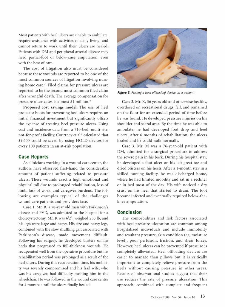
Figure 2). An important advan-
tage of HOLDs is that they not
only reduce friction and shear,
but they also completely offload
the heel while staying in place,
even with patient movement
(see Figure 3).
CostsData are limited
but studies suggest the
cost of care for pres-
sure ulcers is high. In
a tertiary hospital,
total cost of care for
hip fracture patients
with a pressure ulcer
(unspecified type)
averaged $37,288 compared to $13,924 in patients with-
out pressure ulcers.45 Costs of patient care in a New
Mexico Medicaid population hospitalized for the treat-
ment of pressure ulcers (unspecified type) averaged
$15,760.46 Results of a long-term care study showed
that the average cost of treatment per pressure ulcer in
these older adults (mean age 71.4 years) was $2,731 per
ulcer, including hospitalization costs.47 Costs of treat-
ment also were found to increase with ulcer severity,
ranging from an average of $1,119 for Stage II to
$10,185 for Stage III and Stage IV ulcers. Courtney et
al48 reported a per-case additional cost of $3,037 for
patients with a pressure ulcer in a population of
patients in a university hospital.
Additional costs of heel pressure ulcers. The cost
of pressure ulcers extends beyond medical expenses.
12 OstomyWound Management
Figure 1a,b. Inadequate pillow support provides no heel pressure relief. Note that the heels are notoffloaded in the first picture, despite the presence of the pillow.
Figure 2. Heel offloading devices (left) are not the same thing as heel padding devices (right)..
A B
42-57_OWM1008_Fowler:Feature 2/13/09 10:12 AM Page 12
Most patients with heel ulcers are unable to ambulate,
require assistance with activities of daily living, and
cannot return to work until their ulcers are healed.
Patients with DM and peripheral arterial disease may
need partial-foot or below-knee amputation, even
with the best of care.
The cost of litigation also must be considered
because these wounds are reported to be one of the
most common sources of litigation involving nurs-
ing home care.49 Filed claims for pressure ulcers are
reported to be the second most common filed claim
after wrongful death. The average compensation for
pressure ulcer cases is almost $1 million.49
Proposed cost savings model. The use of heel
protector boots for preventing heel ulcers requires an
initial financial investment but significantly offsets
the expense of treating heel pressure ulcers. Using
cost and incidence data from a 710-bed, multi-site,
not-for-profit facility, Courtney et al48 calculated that
$9,600 could be saved by using HOLD devices for
every 100 patients in an at-risk population.
Case ReportsAs clinicians working in a wound care center, the
authors have observed first-hand the considerable
amount of patient suffering related to pressure
ulcers. These wounds exact a high emotional and
physical toll due to prolonged rehabilitation, loss of
limb, loss of work, and caregiver burdens. The fol-
lowing are examples typical of the challenges
wound care patients and providers face.
Case 1. Mr. R, a 78-year old man with Parkinson’s
disease and PVD, was admitted to the hospital for a
cholecystectomy. Mr. R was 6'2", weighed 250 lb, and
his legs were large and heavy. His size and heavy legs,
combined with the slow shuffling gait associated with
Parkinson’s disease, made movement difficult.
Following his surgery, he developed blisters on his
heels that progressed to full-thickness wounds. He
recuperated well from the operative procedure but his
rehabilitation period was prolonged as a result of the
heel ulcers. During this recuperation time, his mobili-
ty was severely compromised and his frail wife, who
was his caregiver, had difficulty pushing him in the
wheelchair. He was followed in the wound care center
for 4 months until the ulcers finally healed.
Case 2. Mr. K, 36 years old and otherwise healthy,
overdosed on recreational drugs, fell, and remained
on the floor for an extended period of time before
he was found. He developed pressure injuries on his
shoulder and sacral area. By the time he was able to
ambulate, he had developed foot drop and heel
ulcers. After 6 months of rehabilitation, the ulcers
healed and he could walk normally.
Case 3. Mr. M was a 76-year-old patient with
DM, admitted for a surgical procedure to address
the severe pain in his back. During his hospital stay,
he developed a foot ulcer on his left great toe and
dried blisters on his heels. After a 1-month stay in a
skilled nursing facility, he was discharged home,
where he had limited mobility and sat in a recliner
or in bed most of the day. His wife noticed a dry
crust on his heel that started to drain. The foot
became infected and eventually required below-the-
knee amputation.
Conclusion The comorbidities and risk factors associated
with heel pressure ulceration are common among
hospitalized individuals and include immobility
and resultant pressure, skin condition (eg, moisture
level), poor perfusion, friction, and shear forces.
However, heel ulcers can be prevented if pressure is
completely alleviated. Heel offloading devices are
easier to manage than pillows but it is critically
important to completely relieve pressure from the
heels without causing pressure in other areas.
Results of observational studies suggest that their
use reduces the rate of pressure ulceration. This
approach, combined with complete and frequent
October 2008 Vol. 54 Issue 10 13
Figure 3. Placing a heel offloading device on a patient.
42-57_OWM1008_Fowler:Feature 2/19/09 10:03 AM Page 13

patient assessment, can improve patient outcomes
while preventing the additional cost of hospital-
acquired pressure ulcers. Although studies to assess
the effect of HOLDs on heel ulcer healing have not
been conducted, clearly pressure reduction is the
first step in healing all pressure ulcers. Prospective,
randomized controlled studies to compare the
effectiveness and cost-effectiveness of heel pressure
reduction strategies are needed to support the
validity of the observations contained herein. - OWM
References1. National Pressure Ulcer Advisory Panel. Pressure defini-
tion and stages revised by NPUAP, February 2007.Available at:www.npuap.org/documents/PU_Definition_Stages.pdf .Accessed December 31, 2007.
2. VanGilder C, Washienko C, Eckstein A, Decker S,MacFarlane G. 2005 international pressure ulcer preva-lence survey results for long-term acute care facilities inthe United States. Available at:www.sawc.net/ses/sawc/abstracts/06323. AccessedDecember 27, 2007.
3. Whittington KT, Briones R. National Prevalence andIncidence Study: 6-year sequential acute care data. AdvSkin Wound Care. 2004;17(9):490–494.
4. Centers for Medicare and Medicaid Services. FederalRegister Part II. 42CFR Parts 411, 412, 413, 489. Changes tothe Hospital Inpatient Prospective Payment systems andfiscal year 2008 rates. Final rule. Federal Register.2007;72(162):47201–46205.
5. Wong VK, Stotts NA. Physiology and prevention of heelulcers: the state of science. J WOCN. 2003;30(4):191-198.
6. Barton AA. The pathogenesis of skin wounds due to pres-sure. J Tissue Viability. 2006;16(3):12–15.
7. Grey JE, Harding KG, Enoch S. Pressure ulcers. BMJ.2006;332(7539):472–475.
8. Mustoe TA, O’Shaughnessy K, Kloeters O. Chronic woundpathogenesis and current treatment strategies: a unifyinghypothesis. Plast Reconstr Surg. 2006;117(7 suppl):35S–41S.
9. Romanes GJ. Cunningham’s Manual of PracticalAnatomy,Vol I, 14th edition. London, UK: OxfordUniversity Press; 1976:156–158.
10. Woodbury MG, Houghton PE. Prevalence of pressureulcers in Canadian healthcare settings. Ostomy WoundManage. 2004;50(10):22–38.
11. Scott-Williams S, Lummus AC. Perioperative pressureulcer assessment and prevention: efficacy study of a multi-layer pressure relief pad in the operating room. Poster pre-sented at the Symposium on Advanced Wound Care.Tampa, Fla. April 28, 2007.
12. Amlung SR, Miller WI, Bosley LM. The 1999 NationalPressure Ulcer Prevalence Survey: a benchmarkingapproach. Adv Skin Wound Care. 2001;14(6):297–301.
13. Walsh JS, Plonczynski DJ. Evaluation of a protocol for pre-vention of facility-acquired heel pressure ulcers. J WOCN.2007;34(2):178–183.
14. Landi F, Onder G, Russo A, Bernabei R. Pressure ulcer andmortality in frail elderly people living in community. ArchGerontol Geriatr. 2007;44(suppl 1):217–223.
15. Redelings MD, Lee NE, Sorvillo F. Pressure ulcers: morelethal than we thought? Adv Skin Wound Care.2005;18(7):367–372.
16. Chipchase SY, Treece KA, Pound N, Game FL, Jeffcoate WJ.Heel ulcers don’t heel in diabetes. Or do they? Diabet Med.2005;22(9):1258–1262.
17. Aulivola B, Hile C, Hamdan AD, et al. Major lower extremityamputation: outcome of a modern series. Arch Surg.2004;139(4):395–399.
18. Vanderwee K, Grypdonck M, Defloor T. Non-blanchableerythema as an indicator for the need for pressure ulcer pre-vention: a randomized-controlled trial. J Clin Nurs.2007;16(2):325–335.
19. Institute for Healthcare Improvement. How to Guide: PreventPressure Ulcers. 2007. Institute for Healthcare Improvement.Available at: www.ihi.org. Accessed May 5, 2008.
20. Haisma JA, van der Woude LH, Stam HJ, et al. Complicationsfollowing spinal cord injury: occurrence and risk factors in alongitudinal study during and after inpatient rehabilitation.J Rehabil Med. 2007;39(5):393–398.
21. De Laat EH, Pickkers P, Schoonhoven L, Verbeek AL, FeuthT, van Achterberg T. Guideline implementation results in adecrease of pressure ulcer incidence in critically ill patients.Crit Care Med. 2007;35(3):815–820.
22. Cock KA. Anti-embolism stockings: are they used effectivelyand correctly? Br J Nurs. 2006;15(6):S4–S12.
23. Uzun O, Tan M. A prospective, descriptive pressure ulcer riskfactor and prevalence study at a university hospital in Turkey.Ostomy Wound Manage. 2007;53(2):44–56.
24. Aronovitch SA. Intraoperatively acquired pressure ulcers: arethere common risk factors? Ostomy Wound Manage.2007;53(2):57–69.
25. Lindgren M, Unosson M, Krantz AM, Ek AC. Pressure ulcerrisk factors in patients undergoing surgery. J Adv Nurs.2005;50(6):605–612.
26. Campbell KE, Woodbury MG, Houghton P. Heel pressureulcers incidence in elderly patients undergoing orthopedicprocedures. Poster presented at the Symposium onAdvanced Wound Care. Tampa, Fla. April 28, 2007.
27. Apsingi S, Dussa CU. Can peripheral nerve blocks con-tribute to heel ulcers following total knee replacement? ActaOrthop Belg. 2004;70(5):502–504.
28. Edwards JL, Pandit H, Popat MT. Perioperative analgesia: afactor in the development of heel pressure ulcers? Br J Nurs.2006;15(6):S20–S25.
29. Baugh N, Zuelzer H, Meador J, Blankenship J. Wound wise:wounds in surgical patients who are obese. Am J Nurs.2007;107(6):40–50.
30. Feuchtinger J, Halfens RJ, Dassen T. Pressure ulcer risk fac-tors in cardiac surgery: a review of the research literature.Heart Lung. 2005;34(6):375–385.
31. Baumgarten M, Margolis D, Berlin JA, et al. Risk factors forpressure ulcers among elderly hip fracture patients. WoundRepair Regen. 2003;11(2):96–103.
32. Meeker MH, Rothrock JC. Alexander’s Care of the Patient inSurgery, 13th edition. St. Louis, Mo: Mosby;2007:130.
33. Tourtual DM, Riesenberg LA, Korutz CJ, et al. Predictors ofhospital acquired pressure ulcers. Ostomy Wound Manage.1997;43(9):24–40.
14 OstomyWound Management
42-57_OWM1008_Fowler:Feature 2/13/09 10:12 AM Page 14

October 2008 Vol. 54 Issue 10 15
34. Lahmann NA, Halfens RJ, Dassen T. Pressure ulcers inGerman nursing homes and acute care hospitals: preva-lence, frequency, and ulcer characteristics. Ostomy WoundManage. 2006;52(2):20–33.
35. Fisher AR, Wells G, Harrison MB. Factors associated withpressure ulcers in adults in acute care hospitals. Adv SkinWound Care. 2004;17(2):80–90.
36. National Pressure Ulcer Advisory Panel. Pressure UlcerTreatment: Competency-based Curriculum. Available at:www.npuap.org/PDF/treatment_curriculum.pdf. AccessedMay 7, 2008.
37. Action plan for (the further improvement of) nursinghome quality. Available at:www.cms.hhs.gov/SurveyCertificationDenInfo/down-loads/2007actionplan.pdf. Accessed October 1, 2008.
38. AHCPR-supported Clinical Practice Guidelines. ClinicalPractice Guideline Number 3: Pressure Ulcers in Adults:Prediction and Prevention. AHCPR Pub. No. 92-0047, May1992.
39. Brainard NR, Ortiz L. Simple low cost intervention savesVA hospital thousands through reduction in heel injury.Poster presented at the Institute for HealthcareImprovement. Orlando, Fla. December 2007.
40. Burda V. A successful heel ulcer prevention programresulting in 95% reduction of heel ulcer incidence.Abstract/poster presented at the Symposium on AdvancedWound Care. Tampa, Fla. April 28, 2007.
41. Jones P, Cadavero A, Payne N. Heel pressure ulcer preven-tion (H-PUP). Available at:www.sawc.net/ses/sawc/abstracts/06171. AccessedDecember 27, 2007.
42. Loehne HB. A trial of a heel pressure relieving deviceproves efficacious in a long term care facility, addressingcomplete heel suspension 24/7 with patient comfort, lead-ing to use throughout the continuum of care. Poster pre-sented at the WOCN Society 39th Annual Conference, SaltLake City, Utah. June 2007.
43. Meyers T, Pezel R, Bennett J, et al. Successful prevention ofheel ulcers and foot drop in the high risk ventilationpatient population. Poster presented at the Institute forHealthcare Improvement. Orlando, Fla. December 2007.
44. Ayello EA, Lyder CH. Protecting patients from harm: pre-venting pressure ulcers in hospital patients. Nursing.2007;37(10):36–40.
45. Allman RM, Goode PS, Burst N, Bartolucci AA, ThomasDR. Pressure ulcers, hospital complications, and diseaseseverity: impact on hospital costs and length of stay. AdvWound Care. 1999;12(1):22–30.
46. Kumar RN, Gupchup GV, Dodd MA, et al. Direct healthcare costs of 4 common skin ulcers in New MexicoMedicaid fee-for-service patients. Adv Skin Wound Care.2004;17(3):143–149.
47. Xakellis GC, Frantz R. The cost of healing pressure ulcersacross multiple health care settings. Adv Wound Care.1996;9(6):18–22.
48. Courtney BA, Ruppman JB, Cooper HM. Save our skin:initiative cuts pressure ulcer incidence in half. NursManage. 2006:37(4):36,38,40 passim.
49. Medical News Today. Clinical trial shows 96% improve-ment in pressure ulcer healing among nursing home resi-dents. March 11, 2006. Available at: www.medicalnewsto-day.com/articles/39327.php. Accessed November 10, 2007.
42-57_OWM1008_Fowler:Feature 2/13/09 10:12 AM Page 15
DMReprint:Layout 1 2/12/09 2:31 PM Page 1