ppt wound healinhg
DESCRIPTION
okTRANSCRIPT

Wound Healing and Platelet Derived Growth Factor in Periodontal RegenerationBy: Martin Leybovich DDSResident, Department ofPeriodontics

Wound Healing
4 Stages1. Hemostasis2. Inflammation
a. Early b. Late
3. Granulation Tissue Formation4. Matrix Formation and Remodeling
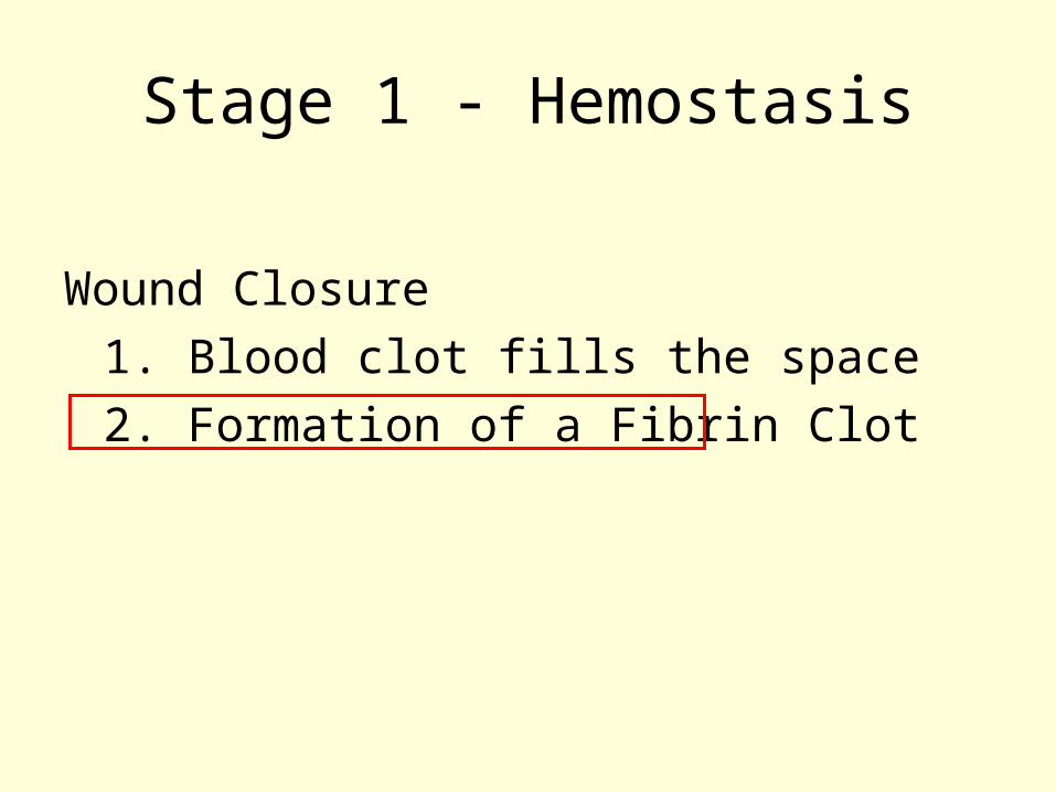
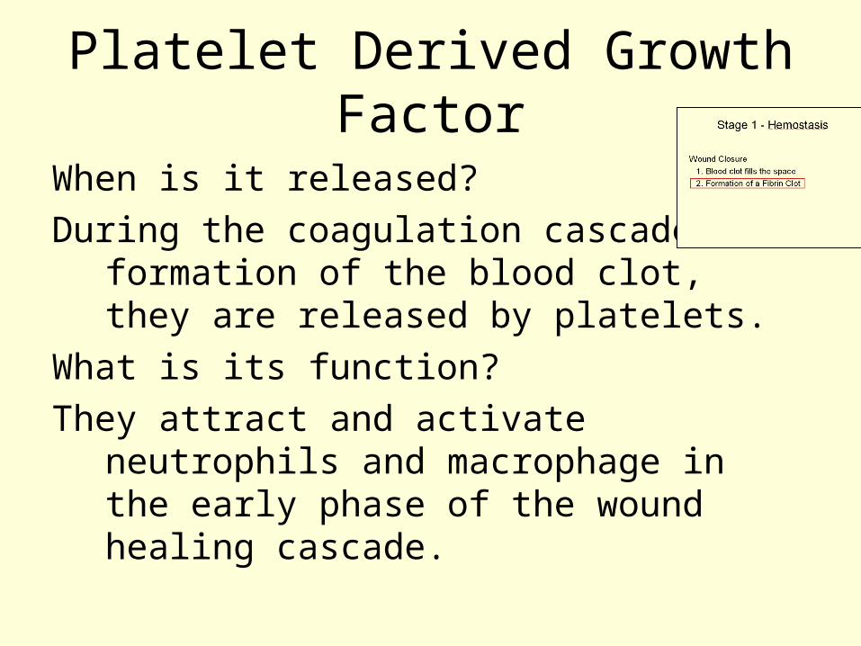
Stage 1 - Hemostasis
Wound Closure 1. Blood clot fills the space2. Formation of a Fibrin Clot
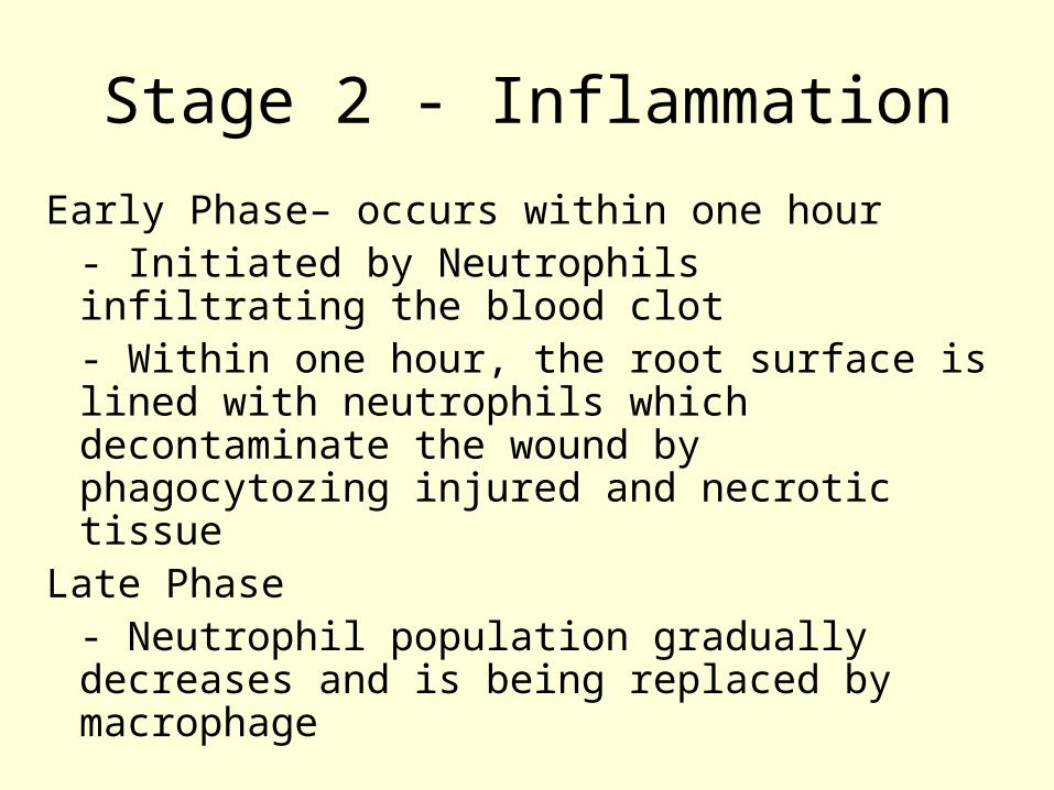
Stage 2 - Inflammation
Early Phase– occurs within one hour- Initiated by Neutrophils infiltrating the blood clot- Within one hour, the root surface is lined with neutrophils which decontaminate the wound by phagocytozing injured and necrotic tissue
Late Phase- Neutrophil population gradually decreases and is being replaced by macrophage
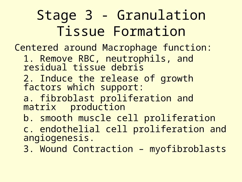
Stage 3 - Granulation Tissue Formation
Centered around Macrophage function:1. Remove RBC, neutrophils, and residual tissue debris2. Induce the release of growth factors which support:
a. fibroblast proliferation and matrix production
b. smooth muscle cell proliferationc. endothelial cell proliferation and
angiogenesis. 3. Wound Contraction – myofibroblasts

Stage 4 - Matrix Formation and Remodeling
As the cells mature from the granulation stage, they undergo the formation of an orderly matrix and start to remodel until they become a functional unit again.
Begins ~ day 7 of wound healing.

Phases of Wound Repair

Wound Healing
4 Stages1. Hemostasis2. Inflammation
a. Early b. Late
3. Granulation Tissue Formation4. Matrix Formation and Remodeling

Stage 3 - Granulation Tissue Formation
Centered around Macrophage function:1. Remove RBC, neutrophils, and residual tissue debris2. Induce the release of growth factors which support:
a. fibroblast proliferation and matrix production
b. smooth muscle cell proliferationc. endothelial cell proliferation and
angiogenesis. 3. Wound Contraction – myofibroblasts

Guided Tissue Regeneration3 Principles based on the concept of
Compartmentalization (Melcher 1976):
1. Clot Stability2. Space3. Selective Cell Repopulation
a. Cementoblastsb. Fibroblasts of the PDLc. Osteoblasts
Players:• Gingival Corium• Periodontal Ligament • Cementum• Alveolar Bone
Players:• Gingival Corium• Cementum • Periodontal Ligament • Alveolar Bone

PDL problem

Stage 3 - Granulation Tissue Formation
Centered around Macrophage function:1. Remove RBC, neutrophils, and residual tissue debris2. Induce the release of growth factors which support:
a. proliferation of fibroblasts and matrix production
b. smooth muscle cell proliferationc. endothelial cell proliferation and
angiogenesis. 3. Wound Contraction – myofibroblasts
Stage 3 - Granulation Tissue Formation
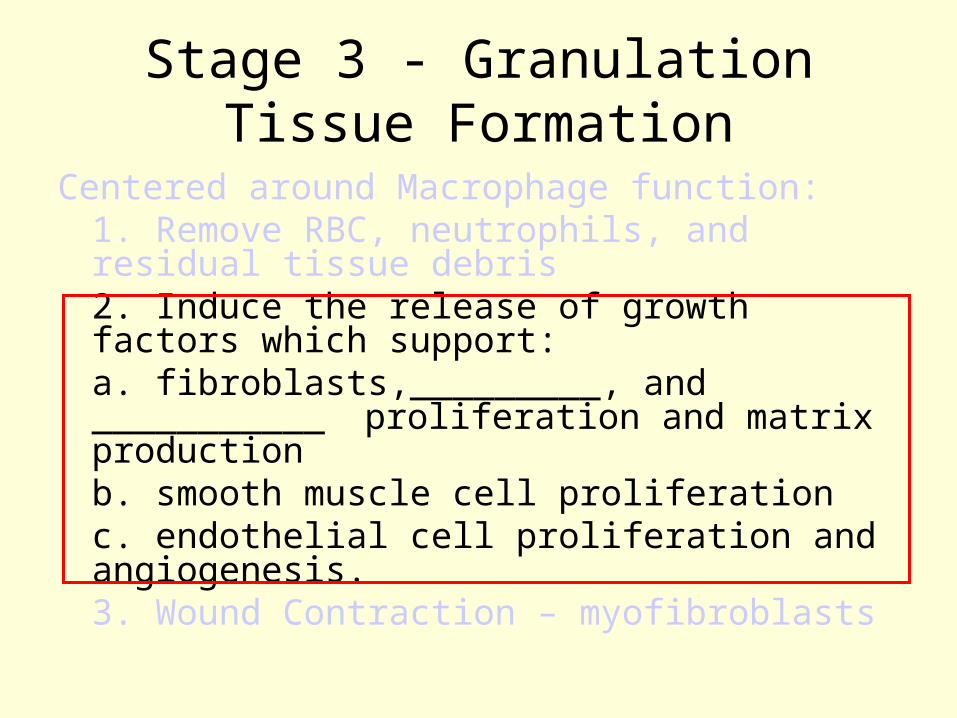
Centered around Macrophage function:1. Remove RBC, neutrophils, and residual tissue debris2. Induce the release of growth factors which support:a. fibroblasts,_________, and ___________ proliferation and matrix productionb. smooth muscle cell proliferationc. endothelial cell proliferation and angiogenesis. 3. Wound Contraction – myofibroblasts

Stage 3 - Granulation Tissue Formation
Centered around Macrophage function:1. Remove RBC, neutrophils, and residual tissue debris2. Induce the release of growth factors which support:
a. fibroblast, osteoblast, and cementoblast proliferation and matrix productionb. smooth muscle cell proliferationc. endothelial cell proliferation and
angiogenesis. 3. Wound Contraction – myofibroblasts

What is a Growth Factor?
1. Proteins that promote cell proliferation and migration.
2. Growth Factors have receptors on various cell types present in the various stages of wound healing that further activate these cells.
3. Growth factors stimulate angiogenesis and mitogenesis.

List of Growth Factors
1. Vascular Endothelial Growth Factor (VEGF)
2. Platelet Derived Growth Factor (PDGF)3. Transforming Growth Factor (TGF)4. Insulin Growth Factor (IGF)5. Fibroblast Growth Factor (FGF)6. Epidermal Growth Factor (EGF)

Platelet Derived Growth Factor1. Family of growth factors representing two
polypeptide chains – A and B – which form pairs:
a. PDGF- AA b. PDGF – BB c. PDGF – AB2. PDGF receptors are transmembrane
structures with extracellular ligand-binding domains and intracellular tyrosine kinase domains – 2 receptors:
1. PDGFR- b. PDGFR-3. The only polypeptide chain to bind to both
receptors is PDGF-BB, and therefore, it made sense to produce this one commercially.
Platelet Derived Growth Factor
When is it released?During the coagulation cascade and
formation of the blood clot, they are released by platelets.
What is its function?They attract and activate neutrophils and
macrophage in the early phase of the wound healing cascade.

PDGF Function
1. Neutrophils and macrophage can secrete and release more PDGF which in turn can stimulate more neutrophils and macrophage, etc.
2. Stimulates macrophage to secrete and release other growth factors important in wound healing
3. Stimulates chemotaxis and mitogenesis of progenitor cells

VEGF
Platelets
FGF
PDGF PMNs/Macrophage
PDGFMacrophage
BMP
Endothelial Cells

Gingival Fibroblasts
VEGF
Platelets
FGF
PDGF PMNs/Macrophage
Macrophage
BMP
Endothelial Cells
PDGF
Osteoprogenitor Cells
TGF
Osteoblasts
PDL Fibroblasts
Cementoblasts

Gingival Fibroblasts
Osteoblasts
PDL Fibroblasts
Cementoblasts
PDGF
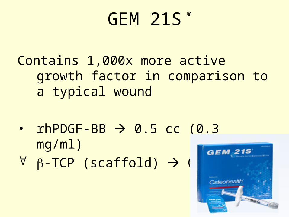
GEM 21S
Contains 1,000x more active growth factor in comparison to a typical wound
• rhPDGF-BB 0.5 cc (0.3 mg/ml) -TCP (scaffold) 0.5 cc
®

Literature: Nevins and Camelo et al “Periodontal Regeneration in Humans Using
Recombinant Human Platelet Derived Growth Factor-BB (rhPDGF-BB) and Allogenic Bone”
Journal of Periodontology 2003:
- Demonstrated in humans that rhPDGF-BB mixed with a synthetic bone graft will yield histologic periodontal regeneration
GEM 21S ®

Literature:Nevins, Giannobile et al“Platelet Derived Growth Factor Stimulated Bone Fill and
Rate of Attachment Level Gain: Results of Large Multicenter Randomized Control Trial”
Journal of Periodontology 2005
1. Demonstrated that purified rhPDGF-BB mixed with a synthetic bone substitute was safe and effective for the treatment of periodontal osseous defects
2. The significant increase in the rate of CAL gain, reduction in gingival recession at 3 months, and improvement in bone fill demonstrated in this study provide substantial evidence for the clinical advantages of this therapy
3. Findings of this study substantiate the hypothesis the addition of rhPDGF-BB improves the effectiveness of the bone substitute
GEM 21S ®

Clinical Case


2 week post op

2 months

The End

References1. Periodontal Wound Healing and Regeneration. Wikesjo UME, Selvig KA. Periodontology
2000 1999;19:31-392. Platelet Derived Growth Factor Expression in Normally Healing Human Fractures.
Andrew JG, Hoyland JA, Freemont AJ, Marsh DR. Bone 1995; 16:455-4603. Hollinger JO, Hart CE, Hirsch SN, Lynch S, Friedlaender GE. Recombinant Human
Platelet Derived Growth Factor: Biology and Clinical Applications. Journal of Bone and Joint Surgery 2008; 90:48-54
4. Li WW, Li VW. The Biology of PDGF and Other Growth Factors in Wound Neovascularization. Contemporary Surgery Supplement 2003, November; 12-17
5. Heldin C-H, Westermark B. Mechanism of Action and In-Vivo Role of Platelet Derived Growth Factor. Physological Reviews 1999; 79: 1283-1316
6. Chen F-M, Jin Y. Periodontal Tissue Engineering and Regeneration: Current Approaches and Expanding Opportunities. Tissue Engineering 2010; 16:219-255
7. Giannobile WV, Periodontal Tissue Engineering by Growth Factors. Bone 1996; 19:23S-37S
8. Nevins M, Camelo M, Nevins ML, Schank RK, Lynch SE. Periodontal regeneration in humans using recombinant human platelet-derived growth factor-BB (rhPDGF-BB) and allogenic bone. J Periodontology 2003; 4:1282-1292
9. Nevins M, Giannobile WV, McGuire MK, et al. Plateletderived growth factor stimulates bone fill and rate of attachment level gain: Results of a large multicenter randomized controlled trial. J Periodontology 2005;76: 2205-2215.