ppt ohinata - review of colorectal cancer - spanish cancer... · review of colorectal cancer aki...
TRANSCRIPT
1
PowerPoint Slides English Text Spanish Translation
Review of Colorectal Cancer
VideoTranscript
Revisión del cáncer colorrectal Transcripción del video
Professional Oncology Education
Review of Colorectal Cancer
Time: 13:01
Educación Oncológica Profesional Revisión del cáncer colorrectal Duración: 13:01
Aki Ohinata, MSPA-C Physician Assistant Gastrointestinal Medical Oncology
The University of Texas, MD Anderson Cancer Center
Aki Ohinata, MSPA-C
Asistente Médica
Oncología Médica Gastrointestinal MD Anderson Cancer Center de la Universidad de Texas
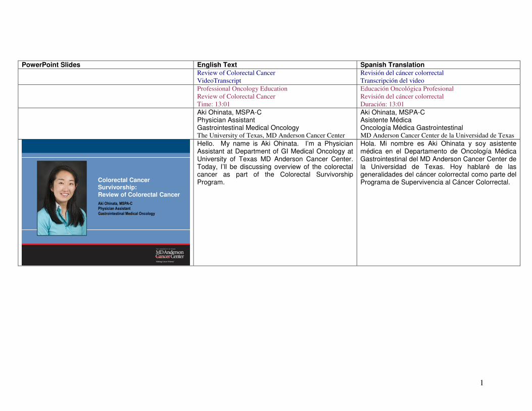
Colorectal Cancer
Survivorship:
Review of Colorectal Cancer
Aki Ohinata, MSPA-C
Physician Assistant
Gastrointestinal Medical Oncology
Hello. My name is Aki Ohinata. I’m a Physician Assistant at Department of GI Medical Oncology at University of Texas MD Anderson Cancer Center. Today, I’ll be discussing overview of the colorectal cancer as part of the Colorectal Survivorship Program.
Hola. Mi nombre es Aki Ohinata y soy asistente médica en el Departamento de Oncología Médica Gastrointestinal del MD Anderson Cancer Center de la Universidad de Texas. Hoy hablaré de las generalidades del cáncer colorrectal como parte del Programa de Supervivencia al Cáncer Colorrectal.

2
Objectives
At the conclusion of this lesson, the participant will be able to:
• Discuss the epidemiology and etiology of colorectal
cancer (CRC)
• Identify those at increased risk of the disease
• List advantages and disadvantages for various screening
tests
• Define the stage of disease using the American Joint
Committee on Cancer (AJCC) staging system
• Order appropriate testing for newly diagnosed patients
At the conclusion of this lesson, the participants will be able to discuss the epidemiology and etiology of colorectal cancer; identify those at increased risk of the disease; list advantages and disadvantages of various screening tests; define the stage of disease using American Joint Committee on Cancer Staging System; and order appropriate testing for newly diagnosed patients.
Al concluir este curso, los participantes podrán analizar la epidemiología y la etiología del cáncer colorrectal; identificar a las personas con mayor riesgo de contraer la enfermedad; enumerar las ventajas y desventajas de los diversos exámenes de diagnóstico; definir el estadio de la enfermedad usando el Sistema de Estadificación del Comité Conjunto Americano sobre el Cáncer; y solicitar las pruebas apropiadas para los pacientes recientemente diagnosticados.
Epidemiology
• Colorectal cancer (CRC) is the second leading cause of
cancer-related deaths in the United States when both sexes
are combined
• Expected death in 2010 is roughly 51,370 (26,580 in men
and 24,790 in women)
• Lifetime risk of men developing CRC is slightly higher than
women (1 in 19 males vs. 1 in 20 females)
http://www.cancer.org/Cancer/ColonandRectumCancer/
DetailedGuide/colorectal-cancer-key-statistics
Colorectal cancer is the second leading cause of cancer-related deaths in the United States when both sexes are combined. Expected death in 2010 is roughly a little over 51,000, with lifetime risk of men developing colon cancer slightly higher than women.
El cáncer colorrectal es la segunda causa principal de muerte relacionada con el cáncer en los Estados Unidos cuando se combinan ambos sexos. La expectativa de muerte en 2010 era de algo más de 51,000 personas, y los hombres tienen un riesgo de por vida ligeramente superior al de las mujeres.

3
Etiology
Carcinogenesis:
Colorectal cancer is thought to
result from sequential accumulation
over years of genetic and molecular
alterations that ultimately lead to
transformation of normal epithelium
into intraepithelial neoplasia/
dysplasia then malignant epithelium
Visual Art: © 2011
The University of Texas
MD Anderson Cancer Center
Colorectal cancer is thought to result from sequential accumulation over the years of genetic and molecular alternations [speaker meant to say alterations] that ultimately lead to transformation of normal epithelium into intraepithelial neoplasia/dysplasia, then malignant epithelium.
Se cree que el cáncer colorrectal es resultado de la acumulación secuencial durante años de alteraciones genéticas y moleculares que transforman el epitelio normal en una neoplasia o displasia intraepitelial, y luego en epitelio maligno.
Etiology
• 75% of all colorectal cancer patients have sporadic disease
• Remaining 25% of the patients have a family history of
colorectal cancer
• Genetic mutations have been identified as the cause of
inherited cancer risk in some colon cancer–prone families;
these mutations are estimated to account for only 5% - 6%
of colorectal cancer cases overall
http://www.cancer.gov/cancertopics/pdq/
genetics/colorectal/HealthProfessional
Seventy-five percent of all colorectal cancer patients have sporadic disease. [The] remaining 25 percent of the patients have a family history of colorectal cancer. Genetic mutations have been identified as the cause of inherited cancer risk in some colon cancer-prone families, but these mutations are estimated to only account for five to six percent of colorectal cancer cases overall.
El 75% de los pacientes con cáncer colorrectal son casos esporádicos, mientras que el 25% restante tienen antecedentes familiares de este cáncer. En algunas familias propensas a desarrollar cáncer de colon se han identificado mutaciones genéticas como causa del riesgo de cáncer hereditario, y se estima que estas mutaciones responden al 5% o 6% del total de los casos de cáncer colorrectal.

4
Estimated Relative and Absolute Risk of
Developing Colorectal Cancer (CRC)
Family History Relative Risk for CRC Absolute Risk of CRC by Age 79a
No family history 1 4%a
One first-degree relative with CRC 2.3 (95% CI, 2.0–2.5) 9%b
More than one first-degree relative
with CRC4.3 (95% CI, 3.0–6.1) 16%b
One affected first-degree relative
diagnosed with CRC before age 45 y3.9 (95% CI, 2.4–6.2) 15%b
One first-degree relative with
colorectal adenoma2.0 (95% CI, 1.6–2.6) 8%b
CI = Confidence interval
a = Data from the surveillance, epidemiology, and end results database
b = The absolute risks of CRC for individuals with affected relatives was calculated
using the relative risks for CRC and the absolute risk of CRC by age 79 ya
Johns LE et al., Am J Gastroenterol 2001 96(10):2992
http://www.cancer.gov
This chart describes the rel --- relative and absolute risk of developing colorectal cancer, based upon the patient’s family history, mainly looking into the involvement of first-degree relatives with colorectal cancer. And, as you could tell, the more patients with first-degree --- more than one first-degree relative with colorectal cancer diagnosis have the higher risk of developing cancer themselves. And, the close second will be one affected first-degree relative diagnosed with colorectal cancer before the age of 45.
Esta tabla describe el riesgo relativo y absoluto de desarrollar cáncer colorrectal, con base en el historial familiar del paciente y buscando principalmente familiares de primer grado con este tipo de cáncer. Como pueden apreciar, los pacientes con más de un familiar de primer grado con diagnóstico de cáncer colorrectal son quienes tienen mayor riesgo de desarrollar cáncer. Los siguientes en riesgo son aquellos con un familiar de primer grado diagnosticado con cáncer colorrectal antes de los 45 años.
Etiology
Inherited gene mutations
• Mutation in APC gene (tumor suppressor gene)
– Familial adenomatous polyposis (FAP)
– Gardner syndrome
• Mutation in DNA repair gene
– Hereditary nonpolyposis colon cancer (HNPCC) aka Lynch syndrome
• Mutation in STK11 gene (thought to be the tumor suppressor gene)
– Peutz-Jeghers syndrome
• Mutation in MADH4 gene
– Juvenile polyposis http://www.cancer.gov/cancertopics/pdq/
genetics/colorectal/HealthProfessional/page4
As mentioned on the previous slide, inherited gene mutations being the cause of colorectal cancer is a small percentage in overall cases. However, these patients do have higher increased risk of developing the cancer compared to the general sporadic colon cancer patients. Mutation in the APC gene, which is a tumor suppressor gene, is shown in familial adenomatous polyposis and Gardner syndrome. Mutation in DNA repair gene is seen in hereditary nonpolyposis colon cancer, or HNPCC, also known as the Lynch syndrome. Peutz-Jeghers syndrome is known to have a mutation in STK11 gene, which is also thought to be a tumor suppressor gene. A mutation in MDAH4 [speaker meant to say MADH4] gene is associated with the juvenile polyposis.
Como ya mencioné, las mutaciones genéticas heredadas que causan cáncer colorrectal corresponden a un pequeño porcentaje de los casos generales. Estos pacientes tienen mayor riesgo de desarrollar cáncer en comparación con los pacientes de cáncer de colon esporádicos. La mutación del gen APC, que es un gen de supresión tumoral, se muestra en la poliposis adenomatosa familiar y el síndrome de Gardner. La mutación del gen de reparación de ADN ocurre en el cáncer de colon no polipósico hereditario o HNPCC, también llamado síndrome de Lynch. El síndrome de Peutz-Jeghers tiene una mutación en el gen STK11, que se considera un gen de supresión tumoral. La poliposis juvenil está asociada a una mutación del gen MADH4.

5
Absolute Risks of Colorectal Cancer for Mutation Carriers in Hereditary Colorectal Cancer Syndromes
SyndromeAbsolute Risk in
Mutation Carriers
FAP 90% by age 45 y
Attenuated FAP 69% by age 80 y
Lynch Syndrome/HNPCC 40% to 80% by age 75 y
Mut Y Homologue-associated neoplasia Not established
Peutz-Jeghers Syndrome 39% by age 70 y
Juvenile polyposis 17% to 68% by age 60 y
Bussey HJ: Familial Polyposis Coli: Family Studies, Histopathology,
Differential Diagnosis, and Results of Treatment, 1975Burt RW, et al., Gastroenterology 2004 127(2):444
Vasen HF et al., Gastroenterology 1996 110(4):1020
Stoffel E et al., Gastroenterology 2009 137(5):1621Hearle N et al., Clin Cancer Res 2006 12(10):3209
Coburn MC et al., Ann Surg Oncol 1995 2(5):386
Desai DC et al., Br J Surg 1995 82(1):14http://www.cancer.gov
This chart describes as --- absolute risk factors for colorectal cancer for mutation care --- carriers in hereditary colorectal cancer syndromes. In this chart, it shows that FAP does have the highest absolute risk in mutation carriers where [there is a] 90 percent risk by age 45 years old. Lynch syndrome also has a higher absolute risk of 40 percent to 80 percent by the age 75.
Esta tabla muestra los factores de riesgo absoluto de cáncer colorrectal para los portadores de mutaciones en los síndromes hereditarios. La poliposis adenomatosa familiar o FAP tiene el mayor riesgo absoluto en los portadores, con un riesgo del 90% a los 45 años. El síndrome de Lynch también tiene un mayor riesgo absoluto, de% 40 a 80% a los 75 años.
Etiology
• Other risk factors for colorectal cancer
– Diet high in total fat and meat (both red and white meat)
– Cigarette smoking
– Sedentary lifestyle
– Inflammatory bowel disease (IBD)
– Older age
– Low fiber diet
– Obesity
Other risk factors for colorectal cancer includes diet high in total fat and meat, including both red and white meat, cigarette smoking, sedentary lifestyle, inflammatory bowel disease, older age, low fiber diet, and obesity.
Otros factores de riesgo de cáncer colorrectal son una dieta con alto contenido de grasa total y carnes rojas o blancas, tabaquismo, estilo de vida sedentario, enfermedad inflamatoria intestinal, edad avanzada, dieta baja en fibra y obesidad.
6
Screening and Diagnosis
• Patients without increased risk of colorectal cancer should
undergo screening at age 50
• Patients with increased risk for colorectal cancer (i.e.
personal/family history of colorectal cancer, adenomatous
polyps, IBD) should undergo screening before age 50
Patients without increased risk of colorectal cancer should undergo screening at age 50. Patients with increased risk of colorectal cancer, for example, personal family history of colorectal cancer, adenomatous polyp, or IBD, should undergo screening before age 50.
Los pacientes sin riesgo incrementado deben hacerse exámenes de detección a partir de los 50 años, mientras que los pacientes con mayor riesgo o historial familiar de cáncer colorrectal, pólipos adenomatosos o enfermedad inflamatoria intestinal deben hacerlos antes de los 50.
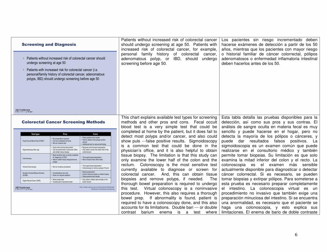
Colorectal Cancer Screening Methods
Test type Pros Cons
Fecal Occult Blood Test (FOBT)
• No preparation required
• Can be completed at home
• Not an invasive test
• False positive test results
• Fails to detect most polyps and/or
cancer
• Additional test for abnormal finding
Sigmoidoscopy (flex sig)
• Quick with minimal discomfort
• Can be performed in physician office
and obtain tissue biopsy
• Requires some bowel preparation
• Only able to exam the lower half of the
colon/rectum
Colonoscopy
• Most sensitive test currently available
for diagnosis of CRC
• Able to obtain tissue biopsy/remove
polyps
• Thorough bowel preparation
• More invasive than other tests
Virtual Colonoscopy • Not an invasive procedure• Thorough bowel preparation
• Colonoscopy to remove polyps if found
Double Contrast Barium Enema
(DCBE)
• Complications are rare
• Does not require sedation
• Bowel preparation
• Cannot remove polyps or obtain biopsy
• False positive test results
Digital Rectal Exam (DRE)• Most simple test
• Usually part of physical exam
• Only able to detect abnormality in the
lower rectum
http://www.cancer.gov/cancertopics/factsheet/Detection/colorectal-screening
This chart explains available test types for screening methods and other pros and cons. Fecal occult blood test is a very simple test that could be completed at home by the patient, but it does fail to detect most polyps and/or cancer, and also could show puls ---false positive results. Sigmoidoscopy is a common test that could be done in the physician’s office, and it is also helpful to obtain tissue biopsy. The limitation is that this study can only examine the lower half of the colon and the rectum. Colonoscopy is the most sensitive test currently available to diagnose or screen for colorectal cancer. And, this can obtain tissue biopsies and remove polyps, if needed. The thorough bowel preparation is required to undergo this test. Virtual colonoscopy is a noninvasive procedure. However, this also requires a thorough bowel prep. If abnormality is found, patient is required to have a colonoscopy done, and this also accounts for its limitations. Double bari --- or double contrast barium enema is a test where
Esta tabla detalla las pruebas disponibles para la detección, así como sus pros y sus contras. El análisis de sangre oculta en materia fecal es muy sencillo y puede hacerse en el hogar, pero no detecta la mayoría de los pólipos o cánceres, y puede dar resultados falsos positivos. La sigmoidoscopia es un examen común que puede realizarse en el consultorio médico y también permite tomar biopsias. Su limitación es que solo examina la mitad inferior del colon y el recto. La colonoscopia es el examen más sensible actualmente disponible para diagnosticar o detectar cáncer colorrectal. Si es necesario, se pueden tomar biopsias y extirpar pólipos. Para someterse a esta prueba es necesario preparar completamente el intestino. La colonoscopia virtual es un procedimiento no invasivo que también exige una preparación minuciosa del intestino. Si se encuentra una anormalidad, es necesario que el paciente se haga una colonoscopia, y esto explica sus limitaciones. El enema de bario de doble contraste

7
complications are very rare. However, this also cannot remove polyps or obtain biopsy, if needed. A digital rectal exam is the most simple test usually done by the --- in the physician’s office. However, it can only detect abnormality in the lower rectum.
tiene complicaciones muy poco frecuentes, pero tampoco permite extirpar pólipos o tomar biopsias. La prueba más simple es un examen rectal digital, generalmente en el consultorio médico, pero solo detecta anomalías en el recto inferior.
Recommendations for Colorectal
Cancer Screening
Patient (Pt) Populations Screening Tests
General population with average riskColonoscopy q 10y (preferred method), or FOBT annually and flex sig q
5 y beginning at age 50 y
First-degree relative with CRC
Two related first-degree relatives with CRC
Colonoscopy every 3-5 yrs beginning at age 40 y or 10 y younger than
youngest affected relative
Two related second-degree relatives with CRC Colonoscopy every 5 yrs starting at age 50 y
Inflammatory bowel diseaseStart screening 8-10 yrs after onset of symptoms. Colonoscopy every 1-
2 yrs.
Family history of Classical FAP, mutation
known
APC +: Flex sig or colonoscopy annually at age 10-15 y
Not tested: Flex sig or colonoscopy annually at age 10-15 y until 24 y.
Then screen every 2 yrs until age 34 y, every 3 yrs until age 44 y, then
every 3-5 yrs thereafter.
APC -: Average risk screening
Family history of HNPCCColonoscopy every 1-2 yrs beginning at age 20-25y or 2-5 yrs prior to
the earliest colon cancer if diagnosed < 25 y
Personal history of CRC
Colonoscopy in 1 yr following resection or within 3-6 mo if no or
incomplete preoperative colonoscopy. Repeat every 1-3 yrs if +
adenoma/SSP. Repeat every 2-3 yrs, then 3-5 yrs if negative/no polyps.
www.NCCN.org; NCCN GuidelinesTM
Colorectal Cancer Screening Version 2.2011
This chart describes --- I’m sorry--- this chart shows the frequency of the testing required for colorectal cancer screening. For general population with average risk, recommendation is for colonoscopy every 10 years or fecal occult blood test annually and flexible sigmoidoscopy every five years beginning at age 50. Depending on [the] patient’s risk factors or family history, the frequency of these screenings are shortened and most of the time the testing are started at a younger age than 50 years old.
Esta tabla muestra la frecuencia de los exámenes para detectar cáncer colorrectal. Para la población general con riesgo promedio, se recomienda una colonoscopia cada 10 años, o una prueba anual de sangre oculta con sigmoidoscopia flexible cada 5 años a partir de los 50. Según los factores de riesgo o el historial familiar, la frecuencia de estos exámenes debe reducirse y las pruebas suelen iniciarse antes de los 50 años de edad.

8
Screening and Diagnosis
• Colonoscopy is currently the most sensitive test available for
detection of colorectal cancer
Colonoscopy currently is the most sensitive test available for detection of colorectal cancer.
La colonoscopia es actualmente el examen más sensible para diagnosticar cáncer colorrectal.
Histology
Normal Colonic Mucosa Hyperplastic Polyp
Adenocarcinoma Adenomatous Polyp
A. B.
C. D.
Images courtesy of Russell Broaddus, M.D., Ph.D.
[Okay]. I’ll be discussing some histologies. Ahora hablaré ahora de histología.

9
Histology
• Immunohistochemical Markers
– Typical colorectal adenocarcinoma staining
• CK7-, CK20+, CEA+, CDX2+ and TTF1-
– Microsatellite instability (MSI) test
• MSI-stable/low: No absent immunostains of mismatch
repair proteins (MLH1, MSH2, MSH6, and PMS2)
• MSI-high: Absent staining in one or more of mismatch
repair proteins
– Consider genetics counseling for MSI-high patients
Immunohistochemical markers: These are the typical colorectal cancer staining when seen under the microscope. These are po --- negative for CK7, positive for CK20, positive for CEA, positive for CDX2, and negative: TTF1. Microsatellite instability test is performed mainly for screening inherited cancer risk factors. MSI-low are stable, shows no absent immunostains of mismatch repair proteins. MSI-high suggests absence of staining in one or more of the mismatch repair proteins and for these MSI-high patients [he or she] should consider genetic testing to rule out or further evaluate possibility of inherited cancer genes, such as the Lynch syndrome.
Marcadores inmunohistoquímicos: Son las típicas tinciones de cáncer colorrectal vistas al microscopio. Resultan negativas para CK7, positivas para CK20, CEA y CDX2, y negativas para TTF1. La prueba de inestabilidad de microsatélites o MSI detecta factores de riesgo de cáncer hereditario. Si es baja o estable, no hay ausencia de inmunotinciones en proteínas reparadoras de desapareamiento. Si es alta, muestra ausencia de tinción en una o más de estas proteínas, en cuyo caso se deben considerar pruebas genéticas para descartar o confirmar la posibilidad de genes de cáncer hereditario, como el síndrome de Lynch.
AJCC (TNM) Staging System
TumorT1: Tumor invades submucosa
T2: Tumor invades muscularis propria
T3: Tumor invades through the muscularis propria
into the subserosa, pericolic or perirectal tissues
T4: Tumor directly invades other organs or structures,
and/or perforates the colon wall
NodeN0: No regional lymph node metastasis
N1: Metastasis in 1 to 3 regional lymph nodes
N2: Metastasis in greater than 3 regional
lymph nodes
MetastasisM0: No distant metastasis
M1: Distant metastasis present
T1 T2 T3 T4
N0 I I IIA IIB
N1 IIIA IIIA IIIB IIIB
N2 IIIC IIIC IIIC IIIC
M1 IV IV IV IV
Greene FL et al. AJCC Cancer Staging Handbook from the AJCC Cancer Staging Manual,
6th Edition; Chap 12, 2002:127
The AJCC staging system is utilized for appropriate staging of colorectal cancer. T stands for the tumor, N for the node, and M for metastatic disease. This chart is actually the Sixth Edition of the AJCC Cancer Staging Guidelines.
El sistema de estadificación del AJCC se utiliza para clasificar el cáncer colorrectal. T significa tumor; N, nódulo; y M, enfermedad metastásica. Esta tabla corresponde a la sexta edición de las Pautas de Estadificación del AJCC.

10
AJCC (TNM) Staging System
Visual Art: © 2011
The University of Texas
MD Anderson Cancer Center
This is an illustration of difference between Stage I through IV of the colon cancer.
Esta es una ilustración de las diferencias entre los estadios I y IV del cáncer de colon.
AJCC (TMN) Staging System
• AJCC 7th edition cancer staging manual is current available
– www.cancerstaging.org
We do have the AJCC Seventh Edition available which can be obtained through this website listed.
Hay una séptima edición de las pautas, que se puede obtener del sitio web indicado en la pantalla.

11
Survivor Rate Based on Staging
Stage 5 Year Survivor Rate (Rectal)
I 74% (74%)
IIA 67% (65%)
IIB 59% (52%)
IIC 37% (32%)
IIIA 73% (74%)*
IIIB 46% (45%)*
IIIC 28% (33%)
IV 6% (6%)
Data based on study of the National Cancer Institute's SEER database, looking
at more than 28,000 people diagnosed with colon cancer between 1998 and 2000
*In this study, survival was better for some stage III cancers than for some stage II cancers.
The reasons for this are not clear.
Edge SB et al. AAJCC Cancer Staging Manual. 7th Edition; Chapter 14, 2010:143
This chart describes the survivor rate based upon the staging. Traditionally, the earlier the staging [the]better the five-year survival rate. And, as higher end staging or increased lymph node involvement decreases the overall five-year survival rate.
Esta tabla muestra la tasa de supervivencia según el estadio del paciente. Cuanto más incipiente es el estadio, tanto mayor es la tasa de supervivencia a cinco años. Si es más avanzado o es mayor el compromiso de los ganglios linfáticos, tanto menor es la tasa de supervivencia general.
Patient Referral
• Once the diagnosis of colorectal cancer has been made…
– Obtain baseline tumor marker, carcinoembryonic antigen
(CEA), along with standard blood work
– Staging workup via imaging studies such as CT, MRI,
or PET/CT
• CT scan is ideal for baseline and restaging work up
• For rectal cancer, flexible sigmoidoscopy with EUS or MRI
can be used to evaluate T and N staging preoperatively
– Refer patient to oncologist for evaluation/treatment
Once the diagnosis of colorectal cancer has been made, patients should have a baseline tumor marker, the CEA, drawn along with standard lab work including CBCs and chemistries. Staging workup via imaging studies such as CT scan, MRI or PET-CT scan should be completed. CT scan is ideal for baseline and restaging workup. For rectal cancer, flexible sigmoidoscopy with EUS or MRI of the pelvis can be used to evaluate the T and the N staging preoperatively. Refer patients to oncologists for evaluation and management.
Si se diagnostica cáncer colorrectal, debe medirse como referencia el antígeno carcinoembrionario o CEA, un marcador tumoral, junto con análisis de laboratorio, un hemograma completo y análisis bioquímicos. También se debe evaluar el estadio con tomografía computarizada, resonancia magnética o tomografía de positrones. La tomografía computarizada es ideal para referencia y reestadificación. En el cáncer rectal, la sigmoidoscopia flexible con ecografía endoscópica y la resonancia magnética de pelvis permiten evaluar el estadio tumoral y nodular antes de la operación. Refiera a los pacientes a oncólogos para su evaluación y tratamiento.

12
Patient Referral – Colon Cancer
Referral to
medical oncology for
systemic therapy*
Low risk
•Observation•Clinical trial
Diagnosis of Colon
Cancer
•CEA•CT C/A/P
Metastasis?No
Yes
Referral to
colorectal surgeon
* if the patient presents with obstructive symptoms, consider sending to surgeon first for diverting colostomy or stent placement
Stage III
Stage II
Stage I Surveillance
High risk
•Observation
• Adjuvant therapy•Clinical trial
• Adjuvant therapy•Clinical trial
The University of Texas MD Anderson Cancer Center Rectal Cancer Guidelines:
http://utm-ext01a.mdacc.tmc.edu/mda/cm/cwtguide.nsf/LuHTML/SideBar1
This chart shows an algorithm on referral process for patients newly diagnosed with colon cancer. Once the diagnosis of colon cancer has been made, it is recommended to obtain baseline CEA and CT including the chest, abdomen and, pelvis. If the patient is found not to have metastatic disease, then they should be referred to a colorectal surgeon for resection. Based upon the staging, the patient will then receive appropriate treatment, if necessary. For patients with metastatic disease at the time of presentation, it is recommended for them to be referred to a medical oncologist to initiate systemic therapy. If the patient presents with obstructive symptoms, they could be considered to be referred to a surgeon for possible diverting surgery or stent placement.
Este gráfico muestra el algoritmo para referir pacientes recientemente diagnosticados con cáncer de colon. Luego del diagnóstico, se recomienda medir el CEA de referencia y hacer tomografías de tórax, abdomen y pelvis. Si el paciente no tiene enfermedad metastásica, debe ser referido a un cirujano colorrectal para una resección. Luego, el paciente recibirá el tratamiento apropiado para su estadificación. Se recomienda referir los pacientes con enfermedad metastásica a un oncólogo médico a fin de iniciar una terapia sistémica. Si el paciente presenta síntomas obstructivos, se puede referir a un cirujano para una cirugía de desviación o colocación de un stent.
Patient Referral – Rectal Cancer
Metastasis?
Diagnosis of
Rectal Cancer
•CEA•CT C/A/P
• Flex sig/EUS or MRI
pelvis for T/N staging
No
Yes
Colorectal surgeon
* if the patient presents with obstructive symptoms, bleeding, or severe rectal pain consider sending to surgeon and/or radiation oncology first
Stage II/III
Stage I Surveillance
Adjuvant
therapy
Radiation and
medical oncology
referral for chemo-
radiation therapy
Colorectal surgeon
Referral to
medical oncology for
systemic therapy*
The University of Texas MD Anderson Cancer Center Rectal Cancer Guidelines:
http://utm-ext01a.mdacc.tmc.edu/mda/cm/cwtguide.nsf/LuHTML/SideBar1
For patients with newly diagnosed rectal cancer, the initial workup is similar, including the baseline CEA and CT of the chest, abdomen, and pelvis. But, in addition to this, we rec --- rec --- recommend obtaining a flexible sigmoidoscopy with EUS or MRI of the pelvis for the preoperative T and N staging. Patients without metastatic disease and was found to have Stage I rectal cancer, they should be referred to the colorectal surgeon for resection followed by surveillance. For patients with Stage II or III rectal cancer, [he or she] should be referred to the radiation oncologist and medical oncologist for neoadjuvant chemoradiation therapy followed by resection by the colorectal surgeon. These patients will then go on to receive the adjuvant therapy as needed. A patient who is found to have metastatic disease at time of presentation should be referred to the medical oncologist for initiation of systemic therapy. Again, if the patient presents with obstructive symptoms, bleeding, or severe rectal pain, they sh --- may benefit from evaluation by the
Para los pacientes con cáncer rectal reciente, la evaluación inicial es similar e incluye el CEA de referencia y tomografías de tórax, abdomen y pelvis. También recomendamos una sigmoidoscopia flexible con ecografía endoscópica o una resonancia de pelvis para determinar el estadio tumoral y nodular antes de la operación. Los pacientes sin metástasis y con cáncer de recto de estadio I deben ser referidos a un cirujano colorrectal para una resección seguida de monitoreo. Los pacientes con cáncer rectal de estadio II o III deben ser referidos al radiooncólogo y al oncólogo médico para terapia de quimiorradioterapia neoadyuvante, seguida de una resección por el cirujano colorrectal. Luego recibirán terapia adyuvante, según sea necesario. Un paciente con enfermedad metastásica debe ser referido a un oncólogo médico para iniciar una terapia sistémica. Si presenta síntomas obstructivos, sangrado o dolor rectal grave, puede beneficiarse de la evaluación del cirujano colorrectal

13
colorectal surgeon for diverting surgery or stent placement as well.
para una cirugía de desviación o colocación de un stent.
Summary
• Colorectal cancer is the second leading cause of
cancer-related deaths in the United States when both
sexes are combined
• It is a preventable disease (in many cases) with
routine screening
• Importance of identifying patient population at increased
risk of CRC early on to start screening
• Efficient referral process to an oncologist once the
diagnosis has been made
In summary, colorectal cancer is the second leading cause of cancer-related death in the United States when both sexes are combined. It is a preventable disease in many cases with routine screening. It is important to identify a patient population at increased risk of colorectal cancer early on to start the screening, and once the col --- diagnosis of cancer has been made, to efficiently refer these patients to an oncologist. Thank you very much for your time. We appreciate any feedback. Thank you.
El cáncer colorrectal es la segunda causa de muerte relacionada con el cáncer en los Estados Unidos cuando se combinan ambos sexos. En muchos casos, es una enfermedad prevenible con exámenes de rutina. Es importante identificar precozmente la población de pacientes con mayor riesgo de cáncer colorrectal para iniciar los exámenes. Si se diagnostica cáncer, deben ser referidos a un oncólogo. Muchas gracias por su tiempo. Agradeceremos cualquier comentario. Gracias.